12 minute read
Adenoid Ameloblastoma with Dentinoid

Adenoid Ameloblastoma with Dentinoid
Case Report of a Rare Entity and Literature Review
Eric Silver, D.M.D., M.D.; Stephen Roth, D.D.S.; Lydia Lam, D.D.S.; John Fantasia, D.D.S.; Steve Yusupov, D.D.S., M.D.
ABSTRACT
Adenoid ameloblastoma with dentinoid (AAD) is a rare, benign neoplasm of odontogenic epithelium, with under 30 cases having been reported in the literature. AAD shares histopathological characteristics with both ameloblastomas and adenomatoid odontogenic tumors. They are unique because of dentinoid deposition without enamel. We present a case of AAD in the maxilla of a 13-year-old. The patient was treated with left infrastructure maxillectomy and left partial infratemporal fossa dissection. This case reports on an extremely rare disease process in an uncommon location in a pediatric patient.
Adenoid ameloblastoma (AAME) is a rare, benign neoplasm of odontogenic epithelium. Even less common is adenoid ameloblastoma with deposits of dentinoid (AAD), with less than 30 cases being reported in the literature.[1] Both share histopathological characteristics with ameloblastomas and adenomatoid odontogenic tumors. It has been suggested by some that AAD is a more aggressive variant of the well-described ameloblastoma. Recurrence rates were previously reported to be between 46% and 71%,[2,3] though recent literature has estimated the recurrence rate to be over 75%,[1] while conventional types of ameloblastoma have documented recurrence rates of between 8.3% and 21%.[4] However, definitive data establishing AAD as more aggressive than conventional ameloblastoma have yet to be established.
Given the aggressive nature of this tumor, it is important for clinicians to recognize this entity and its treatment modalities.[1] While AAD is not yet recognized as a distinct entity by the World Health Organization, there are several features that occur in predictable histopathologic patterns, including an adenomatoid-like proliferation with features of ameloblastic differentiation, in addition to deposition of dentinoid.[2] The deposition of dentin differentiates AAD from the adenoid ameloblastoma (AAME).[3]
In the case reported here, a 13-year-old male presented with a chief complaint of left maxillary facial swelling. Initial incisional biopsy revealed ameloblastoma. The patient was treated with a left infrastructure maxillectomy, and final pathology was consistent with adenoid ameloblastoma with dentinoid production.
Case Report
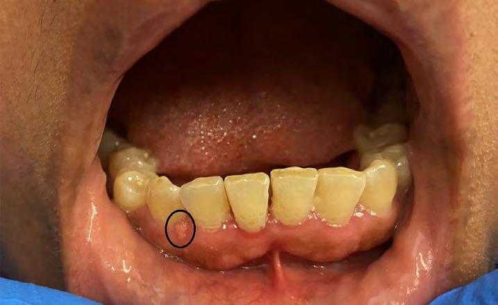
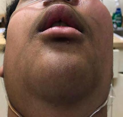
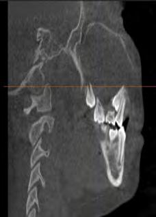
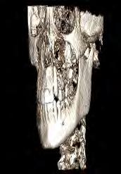
A 13-year-old male with no significant past medical history presented with a chief complaint of left maxillary facial swelling that had rapidly progressed over a six-month course. Social history was noncontributory. He denied any fevers, chills, recent weight loss or any other alarming symptoms. Exam was notable for pronounced upper left facial expansion extending along the malar process to the left eye, with left orbital proptosis and vertical dystopia. Doughy expansion was appreciated along the left malar process extraorally (Figure 1). Intraoral exam revealed extensive expansile doughy swelling at the upper left vestibule and hard palate with intact overlying gingiva. There were retained deciduous teeth in the posterior left maxilla, which were displaced with slight mobility (Figure 2). The patient had difficulty occluding due to pain. Vision was intact, with preserved extraocular movements. Cone beam CT scan showed an expansile cystic radiolucent lesion in the posterior left maxilla extending to the midline through the palatal and buccal bone, and encompassing the maxillary and ethmoid sinuses. The left orbital floor was intact (Figures 3-6).


An incisional biopsy was performed in addition to extraction of tooth #J. Approximately 40 cc of dark cystic fluid was aspirated from the lesion. Cystic appearance was confirmed intraoperatively, and the cyst lining was submerged in formalin. In addition, drain decompression was performed at the time of the biopsy with rapid resolution of the facial asymmetry and ocular dystopia.
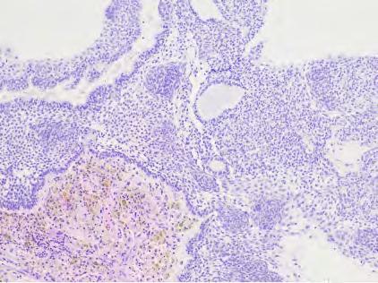
Histologic examination of the initial biopsy (Figure 7) revealed odontogenic epithelium appearing similar to ameloblasts with the associated characteristic reverse polarity at the periphery and areas of stellate-reticulum-like spindle cells centrally. Within the stellate reticulum-like areas were structures mimicking ducts. These histologic features were consistent with an adenomatoid ameloblastoma. Given this diagnosis, treatment options included composite resection with or without definitive free-flap reconstruction.

After lengthy discussion, a surgical plan for delayed osseous reconstruction and infrastructure maxillectomy was decided upon. The patient was then taken to the operating room, where he underwent a left infrastructure maxillectomy and partial left infratemporal fossa dissection, as the lesion abutted the pterygoid plates. Immediate reconstruction was performed with a buccal fat pad and preserved mucosal flaps.
The final surgical pathology report showed an adenoid ameloblastoma with solid, cystic and plexiform growth patterns; focal areas of pseudoglandular/ductal foci; and dentinoid production (Figure 8), making the lesion consistent with AAD. All margins were negative for the tumor by at least 3 mm in all directions. The patient recovered well and is currently under surveillance. Definitive osseous reconstruction and dental implant rehabilitation are planned at growth completion.
Discussion
Adenoid ameloblastoma with dentinoid (AAD) is a rare tumor of epithelial odontogenic origin. Adenoid ameloblastoma (AAME) may occur in isolation without dentinoid deposits, or it may present with dentinoid deposits, resulting in AAD.[3] The histopathological profile of these lesions includes aspects of both ameloblastoma (such as follicular and/or plexiform arrangements of odontogenic epithelium) and adenomatoid odontogenic tumors (such as pseudoducts surrounded by palisaded columnar cells and whorled epithelial structures). They may also demonstrate ghost cells.[5]
The first case was reported by Slabbert et al. in 1992. They described this tumor as having features of ameloblastoma but being distinct due to the deposition of dentin without enamel.[6] Diagnosis of AAD can be challenging and controversial, especially since areas resembling either ameloblastoma or AOT may prevail in the tissue sample.[2,3,5,7-9] As stated above, adenoid ameloblastomas may occur in isolation, but often dentinoid deposits are found, resulting in the diagnosis of AAD. Due to histopathologic similarities, these lesions may be diagnosed on biopsy as another entity, such as ameloblastoma or AOT. Additionally, this lesion may be confused with several hybrid subtypes of lesion, such as AOT originating in a unicystic ameloblastoma or focal adenomatoid changes resembling AOT in ameloblastoma.[2] Despite similarities to other odontogenic tumors, AAD occurs in a predictable pattern and is likely a distinct entity. Further classification and examination of demographic distribution, genetic alteration and long-term follow-up studies are needed to determine if adenoid ameloblastoma and adenoid ameloblastoma with dentinoid are separate entities or variants of the same pathologic process.
As with conventional ameloblastoma, recurrence has been a feature of AAD, and it has been posited that AAD is a more aggressive subtype of ameloblastoma.[2] However, too few cases have been reported to draw specific conclusions in this regard and as of now, it is unclear whether AAME/ AAD behaves more aggressively than conventional ameloblastomas. In a review by DeArruda et al., 45.8% of cases of AAME were found to have recurred; and in a review by Loyola, 71.4% of cases were found to have recurred.[2,3] Importantly, several of these cases showed multiple recurrences, with one case recurring nine times. The hypothesized higher recurrence rate may be due to the more aggressive nature of AAME and the AAD subtype compared to ameloblastoma, or it may be due to previous misdiagnosis. For example, Evans et al. reported on a case of AAD that had been consistently misdiagnosed as AOT and was treated with multiple enucleations before final resection.[10]
The largest and most recent case series of AAME by DeArruda et al. investigated 38 cases that had been reported in the literature. Male-to-female ratio was 1.3:1; average age was reported as 38.0 years, with a range of 4-82; average size was found to be 3.5 centimeters; and location was found to be in the mandible in 25 cases and maxilla in 11 cases.[3] The chief complaint was swelling, though several cases also presented with pain, paresthesia and/or numbness, and 21% of patients were asymptomatic. It was noted in general that several of these cases involved deposition of dentinoid material.
A comprehensive review of AAD was performed by Sachdev et al. in 2021, which found 29 cases of AAD in the literature. The average age was 39 years, while male-to-female ratio was 1.2:1. The ratio of AAD in the mandible to maxilla was 2:1, and seven of the lesions crossed the midline. Additionally, it was noted that 20 of the 29 lesions presented as a well-defined unilocular radiolucency.[1] Given the limited data, while it is likely that the demographic and clinical characteristics are the same for AAME and AAD, there is insufficient data in the literature to draw conclusions at this time. In addition, more cases need to be reported in the literature to elucidate the true characteristics of this tumor and establish whether AAME/AAD behaves differently from conventional ameloblastoma.
With select exceptions, the recommended treatment for ameloblastoma is surgical resection with 10 mm to 15 mm bony margins,[4] and reconstruction, as necessary.[11-13] Given the similarities to ameloblastoma and potential for recurrence, current management of AAD should involve local resection with wide margins and reconstruction, as necessary. In the case of our patient, the treatment plan with regards to reconstruction had to be altered due to the patient’s growth potential and the family’s preferences. Careful, long-term follow-up with both clinical and radiographic examinations is mandatory for this disease process.
Conclusion
Adenoid ameloblastoma with dentinoid is an aggressive, benign tumor that shares histopathological characteristics with both ameloblastomas and adenomatoid odontogenic tumors. It is separated from adenoid ameloblastoma by deposition of dentin without enamel. It is possible that AAD has as high or higher level of recurrence than the conventional ameloblastoma, though further research needs to be performed to confirm this. Treatment of AAD should involve local resection with wide margins, and close patient follow-up is essential.
Queries about this article can be sent to Dr. Silver at esilver0901@gmail.com.
REFERENCES
1. Sachdev SS, et al. Adenoid ameloblastoma with dentinoid. Sultan Qaboos University Medical Journal [SQUMJ] 2021. https://doi.org/10.18295/squmj.9.2021.127.
2. Loyola AM, et al. Adenoid ameloblastoma: clinicopathologic description of five cases and systematic review of the current knowledge. Oral Surgery, Oral Medicine, Oral Pathology, Oral Radiology 2015;120(3):368–377. doi:10.1016/j.oooo.2015.05.011.
3. De Arruda JA, et al. Adenoid ameloblastoma in the posterior maxilla: a case report and review of the literature. Oral and Maxillofacial Surgery 2020;24(2):243–249. doi:10.1007/ s10006-020-00830-1.
4. Carlson ER, Marx RE. The ameloblastoma: PRIMARY, curative surgical management. Journal Oral and Maxillofacial Surgery 2006;64(3):484–494. doi:10.1016/j.joms.2005.11.032.
5. Khalele Bacem AEO, Al-Shiaty RA. Adenoid ameloblastoma with dentinoid and cellular atypia: a rare case report. Italian Journal Medicine 2016;10. doi:10.4081/itjm.2016.639.
6. Slabbert H, et al. Ameloblastoma with dentinoid induction: dentinoameloblastoma. Journal Oral Pathology and Medicine 1992;21(1):46–48. doi:10.1111/j.1600-0714.1992.tb00969.x.
7. Adorno-Farias D, et al. Ameloblastoma with adenoid features: a series of eight cases. Acta Histochemica 2018;120(5):468–476. doi:10.1016/j.acthis.2018.05.006.
8. Allen CM, et al. Adenomatoid dentinoma. Oral Surgery, Oral Medicine, Oral Pathology, Oral Radiology, and Endodontology 1998;86(3):313–317. doi:10.1016/s1079-2104(98)90178-0.
9. Matsumoto Y, et al. Atypical plexiform ameloblastoma with dentinoid: adenoid ameloblastoma with dentinoid. Journal Oral Pathology & Medicine 2001;30(4): 251–254. doi:10.1034/ j.1600-0714.2001.300410.x.
10. Evans BL, et al. Adenoid ameloblastoma with dentinoid: a case report. Oral Surgery, Oral Medicine, Oral Pathology, Oral Radiology, and Endodontology 2004;98(5):583–588. doi:10.1016/j.tripleo.2004.02.077.
11. Dandriyal R, et al. Surgical management of ameloblastoma: conservative or radical approach. National Journal Maxillofacial Surgery 2011;2(1):22. doi:10.4103/0975-5950.85849.
12. Singh M, et al. Treatment algorithm for ameloblastoma. Case Reports in Dentistry 2014;2014:1–6. doi:10.1155/2014/121032.
13. Adeel M, et al. Ameloblastoma: management and outcome. Cureus 2018. doi:10.7759/cureus.3437.
Eric Silver, D.M.D., M.D., is attending oral and maxillofacial surgeon, Kings County Hospital, New York, NY, and in private practice in Philadelphia, PA.
Stephen Roth, D.D.S., is attending oral and maxillofacial pathologist, Donald and Barbara Zucker School of Medicine at Hofstra/Northwell, New Hyde Park, NY.
Lydia Lam, D.D.S., is attending oral and maxillofacial surgeon, Staten Island University Hospital, Staten Island, NY, and in private practice.
John Fantasia, D.D.S., is professor, Donald and Barbara Zucker School of Medicine, Hofstra/Northwell, New Hyde Park, NY.
Steve Yusupov, D.D.S., M.D., is attending oral and maxillofacial surgeon, Nassau University Medical Center, East Meadow, NY, and in private practice.