Intraoperative Assessment of Tumor Margins Tina W.F. Yen, MD, MS Professor, Section of Endocrine Surgery, Division of Surgical Oncology
Bing Yu, PhD Assistant Professor, Marquette University and MCW Department of Biomedical Engineering
The majority of women with newly diagnosed breast cancer in the United States undergo breast-conserving surgery (BCS, lumpectomy). The goal of BCS is to completely remove the tumor with a surrounding rim/margin of normal, unaffected breast tissue while preserving as much normal tissue as possible. Women who have positive margins (cancer cells at the surface/edge of the lumpectomy specimen) have at least a two-fold increased risk of cancer recurrence.1 Since definitive pathologic evaluation of margin status typically is not known until several days after surgery, patients who have positive margins must return to the operating room on another day to undergo additional surgery/surgeries until negative margins (no cancer cells at the edge of the specimen) are achieved. Although re-excision rates have decreased over time in the U.S. to approximately 15-20%,2 additional surgery is associated with more discomfort, increased complications, worse cosmesis, and added emotional stress, time and financial burdens to patients and their caregivers.3,4 Therefore, achieving complete tumor excision with negative margins ideally at the first operation is essential. However, no intraoperative technique currently exists that can accurately and quickly assess margin status. Currently, the lumpectomy specimen typically undergoes X-ray examination to evaluate radiographically how close the tumor is to the margin of the specimen. Although this technique allows for rapid assessment in a few minutes, it has low sensitivity (53%). Other intraoperative techniques to assess margin status, such as frozen section and imprint cytology/touch prep, have much higher sensitivity and specificity (85%-95%) but are labor and time-intensive and require pathology expertise, so are rarely used.5,6 To address this gap, there are many new innovations that have emerged, each hoping to optimize margin assessment and reduce operative re-interventions. These novel techniques can be categorized into four broad groups. Diagnostic imaging uses scanners that provide high-resolution images. Bioimpedance measures cellular/ molecular response to an external electric field at the tissue level. Mass spectrometry involves chemical analysis of 6 | Medical College of Wisconsin Department of Surgery
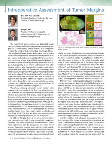
Figure 1. Fluorescence and H&E images of normal benign breast tissue.
cellular contents. Optical spectroscopy evaluates changes in the optical absorption or emission spectrum of cells at the tissue level while optical imaging focuses on the structural information of tissues at the cellular/molecular level. Many of these technologies are in the early stages of development and few have intraoperative trial data. MarginProbe® and ClearEdgeTM are hand-held devices that utilize radiofrequency and bioimpedance spectroscopy, respectively, and are able to assess margins in about five minutes. MarginProbe® is the only FDA-approved device but has limited specificify (<60%) and a high false positive rate (>36%). ClearEdgeTM is approved for sale in Europe. 3,7,8 There are many optical spectroscopy and imaging devices under investigation. With the same field of view among different devices, microscopy with ultraviolet surface excitation (MUSE) has the best surface resolution so is highly desirable for detecting tumor cells on the surface of specimens.9 We decided to focus on this technology given its high resolution, sharpness, contrast, medium field of view and simplicity. We investigated the translational potential of MUSE as an intraoperative tool for margin assessment during BCS by developing a deep ultraviolet scanning fluorescence microsope that can rapidly image fresh breast tissue (1 minute/cm2) with excellent contrast at a resolution sufficient to resolve cells of different tissue types.10 The microscope is portable and housed inside a dark enclosure to prevent personnel exposure to deep ultraviolet light and to eliminate background from room light. In order to obtain both adequate accuracy and time efficiency required for margin assessment introperatively, we chose a combination of deep ultraviolet (DUV; 285 nm) excitation and low maginfication (4x) with slightly reduced spatial resolution (2-3 µm) to achieve a faster imaging speed. We examined 47 fresh human breast tissue specimens acquired from the MCW Tissue Bank. Samples were stained with propidium iodide for nuclear staining and eosin Y for
