
6 minute read
Intraoperative Assessment of Tumor Margins During Breast-Conserving Surgery


Intraoperative Assessment of Tumor Margins
Tina W.F. Yen, MD, MS
Professor, Section of Endocrine Surgery, Division of Surgical Oncology
Bing Yu, PhD
Assistant Professor, Marquette University and MCW Department of Biomedical Engineering
The majority of women with newly diagnosed breast cancer in the United States undergo breast-conserving surgery (BCS, lumpectomy). The goal of BCS is to completely remove the tumor with a surrounding rim/margin of normal, unaffected breast tissue while preserving as much normal tissue as possible. Women who have positive margins (cancer cells at the surface/edge of the lumpectomy specimen) have at least a two-fold increased risk of cancer recurrence.1 Since definitive pathologic evaluation of margin status typically is not known until several days after surgery, patients who have positive margins must return to the operating room on another day to undergo additional surgery/surgeries until negative margins (no cancer cells at the edge of the specimen) are achieved. Although re-excision rates have decreased over time in the U.S. to approximately 15-20%,2 additional surgery is associated with more discomfort, increased complications, worse cosmesis, and added emotional stress, time and financial burdens to patients and their caregivers.3,4
Therefore, achieving complete tumor excision with negative margins ideally at the first operation is essential. However, no intraoperative technique currently exists that can accurately and quickly assess margin status. Currently, the lumpectomy specimen typically undergoes X-ray examination to evaluate radiographically how close the tumor is to the margin of the specimen. Although this technique allows for rapid assessment in a few minutes, it has low sensitivity (53%). Other intraoperative techniques to assess margin status, such as frozen section and imprint cytology/touch prep, have much higher sensitivity and specificity (85%-95%) but are labor and time-intensive and require pathology expertise, so are rarely used.5,6
To address this gap, there are many new innovations that have emerged, each hoping to optimize margin assessment and reduce operative re-interventions. These novel techniques can be categorized into four broad groups. Diagnostic imaging uses scanners that provide high-resolution images. Bioimpedance measures cellular/ molecular response to an external electric field at the tissue level. Mass spectrometry involves chemical analysis of
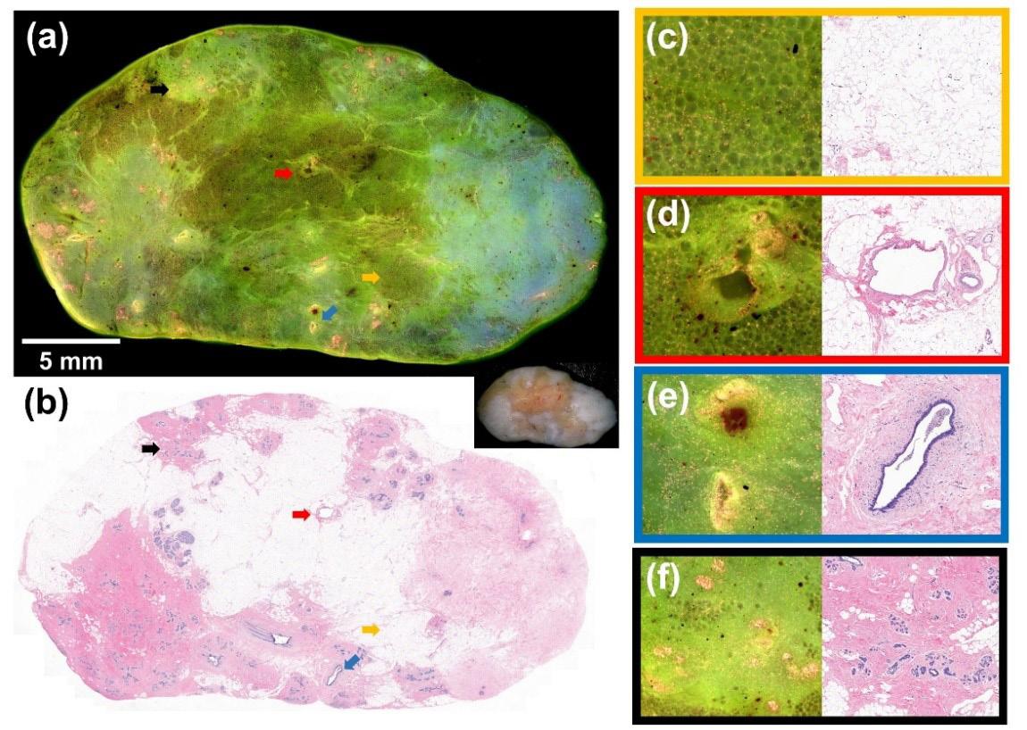
Figure 1. Fluorescence and H&E images of normal benign breast tissue.
cellular contents. Optical spectroscopy evaluates changes in the optical absorption or emission spectrum of cells at the tissue level while optical imaging focuses on the structural information of tissues at the cellular/molecular level. Many of these technologies are in the early stages of development and few have intraoperative trial data. MarginProbe® and ClearEdgeTM are hand-held devices that utilize radiofrequency and bioimpedance spectroscopy, respectively, and are able to assess margins in about five minutes. MarginProbe® is the only FDA-approved device but has limited specificify (<60%) and a high false positive rate (>36%). ClearEdgeTM is approved for sale in Europe. 3,7,8
There are many optical spectroscopy and imaging devices under investigation. With the same field of view among different devices, microscopy with ultraviolet surface excitation (MUSE) has the best surface resolution so is highly desirable for detecting tumor cells on the surface of specimens.9 We decided to focus on this technology given its high resolution, sharpness, contrast, medium field of view and simplicity. We investigated the translational potential of MUSE as an intraoperative tool for margin assessment during BCS by developing a deep ultraviolet scanning fluorescence microsope that can rapidly image fresh breast tissue (1 minute/cm2) with excellent contrast at a resolution sufficient to resolve cells of different tissue types.10 The microscope is portable and housed inside a dark enclosure to prevent personnel exposure to deep ultraviolet light and to eliminate background from room light.
In order to obtain both adequate accuracy and time efficiency required for margin assessment introperatively, we chose a combination of deep ultraviolet (DUV; 285 nm) excitation and low maginfication (4x) with slightly reduced spatial resolution (2-3 µm) to achieve a faster imaging speed. We examined 47 fresh human breast tissue specimens acquired from the MCW Tissue Bank. Samples were stained with propidium iodide for nuclear staining and eosin Y for
During Breast-Conserving Surgery
staining of the cytoplasm and connective tissues. The specimen was immobilized on a motorized stage and images were collected, transformed and stitched together. Figure 1a demonstrates the fluorescence image of benign breast Figure 2. (a) Fluorescence and (b) H&E tissue, which apimages of an invasive ductal carcinoma. pears green. A photograph of the gross specimen is shown in the lower right corner. Figure 1b displays the corresponding H&E image. The four arrows mark structures that are displayed in zoomed fluorescence and H&E images on the right panel. The orange arrow highlights fibroadipose tissue, which appears darker green (c). Since blood vessels (red arrow; d), ducts (blue arrow; e) and lobules (black arrow; f) have higher cell density, these structures appear pink and yellow.
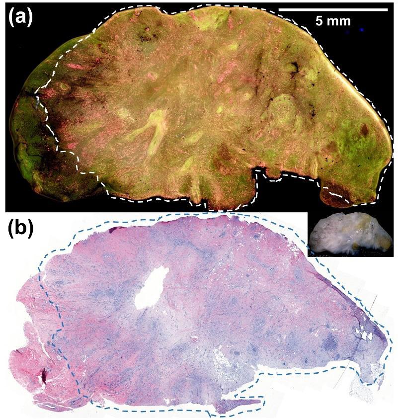
Figure 2 shows fluorescence (a) and H&E (b) images of an invasive ductal carcinoma. The cancer is demarcated by the dashed lines. Since cancer cells have higher cell density, they appear pink and yellow in the fluorescence image and can be easily distinguished from the surrounding benign fatty breast tissue, which appears dark green.
After demonstrating excellent contrast in color, tissue texture, cell density and shape between invasive carcinomas and normal counterparts, we wanted to determine the accuracy of visual interpretation of the MUSE images by non-medical evaluators to demonstrate general feasibility and applicability. We trained 3 people with no prior experience to differentiate cancer from non-cancer tissue using five specimens. The evaluators then interpreted the MUSE images of the remaining 42 specimens, providing a diagnosis of cancer or normal tissue. Visual interpretation was outstanding with an average sensitivity, specificity and accuracy of 97.6%, 92.9% and 96.0%, respectively.
In summary, rapid and accurate intraoperative lumpectomy margin assessment remains an unmet clinical need. We demonstrate the feasibility of using deep ultraviolet scanning fluorescence microscopy as a potential intraoperative tool to detect positive margins. It achieves a good balance between imaging speed and spatial resolution with excellent contrast and should be practical and generalizable to most operating room settings as this technology does not require specialized resources or training. Although these initial results are promising, we acknowledge that additional work is needed to further optimize this device prior to conducting a clinical study to assess its true potential for intraoperative lumpectomy margin assessment.
This work was supported by a Medical College of Wisconsin Department of Surgery We Care grant, a Marquette University College of Engineering GHR Foundation grant, and Marquette University startup grant.
FOR ADDITIONAL INFORMATION on this topic, visit mcw. edu/surgery or contact Dr. Tina Yen at tyen@mcw.edu.
REFERENCES 1. Houssami N, Macaskill P, Marinovich ML, Morrow
M. The association of surgical margins and local recurrence in women with early-stage invasive breast cancer treated with breast-conserving therapy: a metaanalysis. Ann Surg Oncol 2014;21:717-30. 2. Havel L, Naik H, Ramirez L, Morrow M, Landercasper J.
Impact of the SSO-ASTRO Margin Guideline on Rates of Re-excision After Lumpectomy for Breast Cancer: A
Meta-analysis. Ann Surg Oncol 2019;26:1238-44. 3. Leff DR, St John ER, Takats Z. Reducing the Margins of Error During Breast-Conserving Surgery: Disruptive
Technologies or Traditional Disruptions? JAMA surgery 2017;152:517-8. 4. Marinovich ML, Noguchi N, Morrow M, Houssami N.
Changes in Reoperation After Publication of Consensus
Guidelines on Margins for Breast-Conserving Surgery:
A Systematic Review and Meta-analysis. JAMA Surg 2020:e203025. 5. Gray RJ, Pockaj BA, Garvey E, Blair S. Intraoperative
Margin Management in Breast-Conserving Surgery: A
Systematic Review of the Literature. Ann Surg Oncol 2018;25:18-27. 6. St John ER, Al-Khudairi R, Ashrafian H, et al. Diagnostic
Accuracy of Intraoperative Techniques for Margin
Assessment in Breast Cancer Surgery: A Meta-analysis.
Ann Surg 2017;265:300-10. 7. Maloney BW, McClatchy DM, Pogue BW, Paulsen
KD, Wells WA, Barth RJ. Review of methods for intraoperative margin detection for breast conserving surgery. J Biomed Opt 2018;23:1-19. 8. Schwarz J, Schmidt H. Technology for Intraoperative
Margin Assessment in Breast Cancer. Ann Surg Oncol 2020;27:2278-87. 9. Fereidouni F, Harmany ZT, Tian M, et al. Microscopy with ultraviolet surface excitation for rapid slide-free histology. Nat Biomed Eng 2017;1:957-66. 10.Lu T, Jorns JM, Patton M, et al. Rapid assessment of breast tumor margins using deep ultraviolet fluorescence scanning microscopy. J Biomed Opt 2020;25.
