I am currently caring for a 72-year-old man (he looks much younger, even now given his medical challenges) who represents the COVID-delay syndrome which has affected health care as well as many other aspects of our lives. He presented to his family physician in January of 2020 with dyspepsia, heartburn and mild weight loss. Without going into the details, he had multiple challenges in accessing health care during the height of the pandemic and finally presented 11 months later with gastric outlet obstruction. He was operated on under the preoperative diagnosis of probable ulcer disease. At the time of surgery, he was found to have adenocarcinoma, probably arising from the peripyloric antrum, and underwent an incomplete resection (the surgeons were understandably not prepared to perform a Whipple procedure) and a gastrojejunostomy. He was shortly thereafter referred here and his challenges have not abated – biliary obstruction followed by stent-associated cholecystitis, as well as pain and anorexia, have complicated attempts at delivering systemic therapy and chemoradiation. He has recently been found to have peritoneal disease and we are transitioning to best supportive care. As with most all of my patients with cancer in the right upper quadrant of the abdomen, they are lovely people who certainly do not deserve such a turn of events. Would this man have experienced a different outcome if he received the care he needed 10-11 months earlier – very hard to know, but certainly possible. Could he have been cared for during the height of the pandemic – possibly (and clearly yes at this medical center campus). We have been seeing patients with delayed diagnoses for the past 4-6 months and this may continue for a while. We clearly are experiencing an increase in patient demand at a time when many of our physicians, residents and APPs (as well as other members of the health care team) are trying to recover from 18 months of one-dimensional work – characterized by little time off, social isolation and increased clinical effort. There has been much
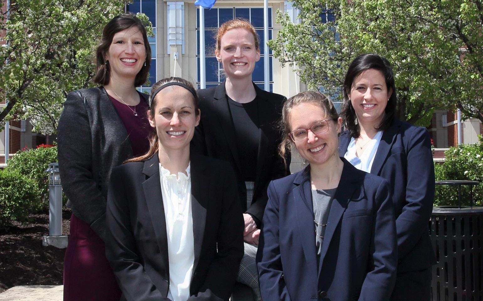
Thank you for supporting our all-star chief residents who will leave us this month for the next chapter in what will be exciting careers – a huge thank you for their many contributions, tireless effort, extraordinary talent and daily smiles!
written about the physical and emotional stress placed on health care workers – no doubt this is correct. How do we manage this reality with the understanding that for patients it may be very hard to navigate the current health care environment and find the doctor they need? Even worse, it might not be that much better now than it was one year ago. For any of you who may be on the receiving end of health care, you may have experienced challenges in obtaining appointments, along with delays. Some of this may be related to the financial implications of COVID on health systems – many have lost employees either due to the need to reduce expenses or because of resignations. When the team gets smaller and the work gets larger, those who remain often look for another job. In addition, there are clearly a number of other reasons
Continued on page 3
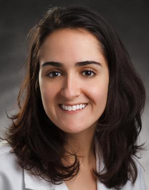
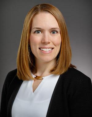
Front row (l to r): Kayla Chapman MD, K. Hope Wilkinson MD MS; Back row (l to r): Kathryn Haberman MD, Elizabeth Traudt MD, Jacqueline Blank MD
Colorectal Cancer Screening in a COVID-19 World
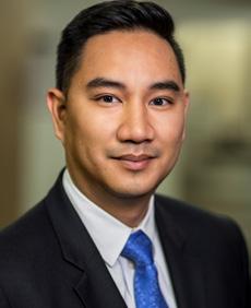
Jed Calata, MD Assistant Professor, Division of Colorectal Surgery
COVID-19 has affected all of us. Back when the pandemic started in late 2019, there was a wave of anxiety and fear that swept throughout the world. There was just so much that we didn’t know. We had to make hard choices to help keep each other safe, while trying our best to continue to care for our patients. Last March, as hospitals began to fill with COVID patients in the US, the Centers of Medicare & Medicaid Services and other professional organizations recommended stopping elective procedures, including colonoscopies and colorectal surgeries, to conserve critical resources and limit COVID exposure to both patients and staff. This emergency action was mirrored in countries around the world as we tried to cope with the number of COVID patients that needed care. We are now over one year into the pandemic and our world is dramatically different from what we once knew; although healthcare centers are now more open, we continue to wrestle with the consequences of the initial strain on our healthcare system. The effects of COVID have extended far beyond those touched by the virus itself and has reverberated into our ability to provide colorectal cancer screening for our patients.
Outcomes from early in the pandemic are now coming to light. In the UK, endoscopic activity dropped to 5% of normal activity by the end of March 2020.1 Ten weeks later, endoscopic activity only increased to 20% of pre-COVID levels.1 During this time, there was an alarming 72% decrease in colorectal cancers detected in the UK.1 In the US, similar trends have been reported. In a study by Whaley et al, they reported a 69.6% reduction in colonoscopy rates among commercially insured patients in the US.2 It has been estimated that 1.7 million fewer colonoscopies were performed in the US over a three-month period in the spring of 2020.3 This suggests that potentially 18,800 patients in the US will have a diagnosis of colorectal cancer delayed as a consequence.3 Alarmingly, conservative estimates predict over 4,000 excess deaths from colorectal cancer in the US related to COVID-19 delays.4
Now, as we learned more about the virus, guidelines on how to safely proceed with elective endoscopy and surgery procedures have been produced and patients and doctors have resumed care. However, as previously noted, colonoscopy rates have been slow to return to normal despite the large backlog of patients that are overdue for screening colonoscopy.1 There are numerous factors that may be affecting this. Patient factors such as anxiety,
loss of insurance, and perception that preventative health should not be prioritized during a pandemic play a role.5 Systemic factors such as time-consuming safety protocols, staff layoffs, furloughs, and COVID-related employee absences within clinics may impact the ability of primary care physicians to see patients to recommend screening as well as for endoscopists to perform screening.5,6 Furthermore, simply reaching pre-COVID colonoscopy rates may not be a sufficient goal. In a study from New York City, one of the areas hardest hit by COVID-19, it was estimated that it could take between one to two years to clear the colonoscopy back log after achieving pre-COVID colonoscopy rates.7
Adding further consternation to this complex picture was the new US Preventive Services Task Force (USPSTF) colorectal cancer screening draft update released on October 27, 2020. In this update, for average risk patients, the USPSTF recommended to begin colorectal cancer screening at age 45 lowering it from the previous age of 50. Although the complexity of this decision is beyond the scope of this article, should the recommendation become finalized, an estimated 20 million additional people would require screening in the United States, further emphasizing the importance of timely colorectal screening and education.8
Nearly overnight, COVID-19 disrupted our lives and changed the way we live. Although these numbers sound daunting, we have the opportunity to save lives together by ensuring colorectal cancer screening is available to all that are interested. Primary care providers are ready to meet with you, in-person or virtually, to discuss colorectal cancer screening options such as a colonoscopy or non-invasive screening options such as fecal immunohistochemistry testing (FIT) or fecal DNA testing (Cologuard) if you are uncomfortable with a procedure at this time. GI labs are open, and gastroenterologists are ready to perform colonoscopies safely. And, should you need help, we, the Colorectal Surgery Division at Froedtert Hospital & MCW, are ready to treat you, utilizing cutting-edge research and the latest in minimally invasive technique.
FOR ADDITIONAL INFORMATION on this topic, visit mcw.edu/surgery or contact Dr. Jed Calata at jcalata@ mcw.edu.
REFERENCES
1. Rutter MD, Brookes M, Lee TJ, et al. Impact of the COVID-19 pandemic on UK endoscopic activity and cancer detection: a national endoscopy database analysis. Gut 2021;70:537-543
2. Whaley CM, Pera MF, Cantor J, et al. Changes in Health Services Use Among Commercially Insured US populations During the COVID-19 Pandemic. JAMA Network Open. 2020; 3(11):e2024984
3. IQVIA. Shifts in healthcare demand, delivery and care during COVID-19 ERA. (2020) www.iqvia.com/ insights/the-iqvia-institute/covid-19/shifts-inhealthcare-demand-delivery-and-care-during-thecovid-19-era
4. Sharpless NE. COVID-19 and cancer. Science. 2020;368(6497):1290
5. Gupta S, Lieberman D. Screening and Surveillance Colonoscopy and COVID-19: Avoiding more Casualties. Gastroenterology. 2020 Oct; 159(4):12051208
6. Quigley, EM. Colon Cancer detection and prevention in the age of COVID-19. Current Opinions in Gastroenterology. 2021, 27:37-38.
7. Aggarwal A, Jain A, et al. SARS-CoV-2 Impact on Screening Colonoscopy: Implications for Postpandemic Recovery
8. Wender, R. Colorectal cancer screening should begin at 45. Journal of Gastroenterology and Hepatology. 2020, 35:1461-1463
From the Chair | Douglas B. Evans, MD, continued
(beyond the scope of this short introduction) for the observation that it is harder to receive timely health care right now.
So how can this be reconciled or even managed –on the supply side, a fatigued physician, resident and APP work force in need of a rebalancing of work-life, and on the demand side, an equally frustrated group of us who need to see a doctor and receive compassionate, timely and hopefully successful treatment. Opinion pieces which I have recently read have largely focused on the struggles and challenges of those on the supply side – and rightfully so as it has been a very hard 18 months in the trenches with little time off and a lack of academic interaction which provides such an effective anecdote for the fatigue of clinical medicine. Perhaps it may be helpful for us all to also consider the demand side – the patient I described above needs us now as much as ever. Although we no longer have the ability to cure him from the cancer that received a competitive advantage due to the confusion of the pandemic, we can provide him the palliative care and emotional support which is making a difference in his life. As we get better at taking care of each other and ourselves, it may be helpful to remember the demand side – after all, we are all future patients – if a health challenge has not affected you or one of your family, it likely will with time.
On a more positive note, please take some time to read the amazing articles in this edition of Leading the Way – they are so well done and full of important information. Additionally, extend a warm welcome to our incoming general surgery PGY 1 residents!
2021-2022 Interns
Fayrouz Abu-Hamdan, MD
Sayeh Bozorghadad, MD - Preliminary
Hannah Holland, MD
Samantha Leonard, MD
Allison Linehan, MD - Plastics
Isaac Melin, MD - Urology
Patrick Moran, MD - Preliminary
Megan Paradzinsky, MD - Urology
Kelley Park, MD - Plastics
Sarah Park, MD
Ujval Pathak, MD, MPH - Urology
Santiago Rolon, MD - Rural
Sarah Suh, MD
Anna Tatakis, MD
Brexton Turner, MD
Brigitte Vanle, MD, PhD - Preliminary
Current Guidelines and Updates in Breast
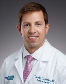
Chandler S. Cortina, MD Assistant Professor, Section of Breast Surgery, Division of Surgical Oncology
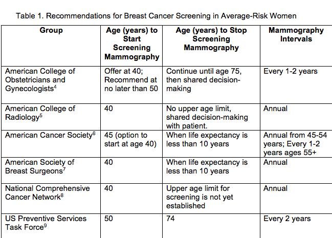
Breast cancer is the most common cancer among American women in the United States, with over 281,000 new cases of invasive breast cancer projected to be diagnosed in 2021.1 The majority will be detected on a screening imaging. Published data has definitively demonstrated that routine use of screening mammography reduces breast cancer mortality and allows for earlier detection and initiation of treatment.2,3 While evidence supports the use of screening mammography, the ages of when to initiate screening, the age to stop screening, and the time intervals between screening exams varies slightly amongst guidelines for average-risk women (Table 1).
A woman is considered to be at average-risk for developing breast cancer if she does not have a personal history of breast cancer, a strong family history of breast cancer, or a genetic mutation known to increase risk of breast cancer, and has not had chest radiation therapy before the age of 30. These factors, when present, may increase a woman’s risk enough to warrant additional imaging methods beyond traditional mammography. The US Preventive Services Task Force (USPSTF) is the only group that recommends an average-risk woman wait until age 50 to start screening and is based on systematic reviews that demonstrated higher rates of false-positive results and smaller numbers of preventable breast cancer deaths in women 40-49.10,11,12 Other organizations support the benefits of annual screening and recommend initiating mammography at age 40. However, most do support a shared decision-making approach, especially when deciding at what age to stop screening. Considering a woman’s individual risk for developing breast cancer, personal comorbidities, and life expectancy, all aid in this decision-making process between patients and healthcare providers.
Breast density has emerged as a hot topic in recent years as it has been established to be a risk factor for breast cancer development, and legislation has been passed requiring reporting of breast density to patients who undergo mammography.13 In an effort to better detect breast abnormalities on screening imaging, technologies such as
automated breast ultrasound (ABUS) and breast magnetic resonance imaging (MRI) may be used for certain patients with extremely dense breasts as an adjunct to routine screening. The decision for adjuvant breast imaging should be a collaborative conversation between patients, healthcare providers, and breast imagers. While these imaging techniques have been demonstrated to be more sensitive at detecting smaller and interval breast cancers, their high-sensitivity rates are also prone to false-positive findings.14 Recent efforts have been made to reduce the time required for MRI by employing abbreviated, or “fast” breast MRI, to allow the process to be more accommodating for patients; however, a consensus on a clear benefit in breast cancer detection has yet to be solidly formed.15 Patients at an elevated risk of breast cancer development, such as those with pathogenic germline genetic variants (ie BRCA or CHEK2 mutations), a strong family history of breast cancer, or those with a personal history of breast atypia or other high-risk lesions, may also benefit from increased breast cancer screening. This is usually by incorporating MRI screening with mammography, with imaging alternating every 6 months in conjunction with physical examinations by a healthcare provider. Additionally, in women who have a first degree relative (such as a sister, or mother) with a previous breast cancer, breast screening is recommended to begin at least 5 years prior to the age that their first degree relative was diagnosed. For example, if a patient has a mother who was diagnosed with breast cancer at age 42, the patient should begin breast cancer screening no later than age 37. Online risk-assessment models such as the Gail and Tyrer-Cuzick (also known as IBIS) models can provide numerical data on a woman’s future risk of breast cancer development compared to their average risk-peer.16,17 While these are readily available online and open to the public, the clinical interpretation of a woman’s risk should be discussed
Cancer Screening
directly with a healthcare provider, particularly one that is well versed in breast cancer risk assessments. Risk assessments allow for a personalized approach to breast cancer screening for those with an elevated risk of breast cancer development.
Breast cancer screening provides for earlier disease detection and continues to evolve as new imaging protocols and data emerge. When to begin screening for breast cancer varies amongst guidelines and should be a collaborative conversation between patients and healthcare providers. Additionally, those women with an increased risk of breast cancer development and those with dense breast tissue may benefit from additional breast cancer imaging. Risk assessment tools are available to assist in determining a women’s relative lifetime breast cancer risk and can assist in personalizing breast cancer screening options.
FOR ADDITIONAL INFORMATION on this topic, visit mcw.edu/surgery or contact Dr. Chandler Cortina at ccortina@mcw.edu
REFERENCES
1. American Cancer Society. How common is breast cancer? Retrieved January 26, 2021 from https:// www.cancer.org/cancer/breast-cancer/about/howcommon-is-breast-cancer.html#:~:text=This%20 means%20there%20is%20a%201%20in%20 8,invasive%20breast%20cancer%20will%20be%20 diagnosed%20in%20women
2. Moss SM, Cuckle H, Evans A, Johns L, Waller M, Bobrow L; Trial Management Group. Effect of mammographic screening from age 40 years on breast cancer mortality at 10 years’ follow-up: A randomised controlled trial. Lancet. 2006 Dec 9;368(9552):205360
3. Tabár L, Vitak B, Chen TH, Yen AM, Cohen A, Tot T, Chiu SY, Chen SL, Fann JC, Rosell J, Fohlin H, Smith RA, Duffy SW. Swedish two county trial: Impact of mammographic screening on breast cancer mortality during 3 decades. Radiology. 2011 Sept 1;260(3):65863
4. American College of Obstetricians and Gynecologists. Breast cancer risk assessment and screening in average risk women. ACOG Practice Bulletin. 2017;179
5. Monticciolo DL, Newell MS, Hendrick RE, Helvie MA, Moy L, Monsees B, Kopans DB, Eby PR, Sickles EA. Breast cancer screening for average-risk women: Recommendations from the ACR commission on breast imaging. J Am Coll Radiol. 2017 Sep;14(9):113743.
6. American Cancer Society. American Cancer Society recommendations for the early detection of breast cancer. Retrieved January 26, 2021 from 8579.00.pdf (cancer.org)
7. American Society of Breast Surgeons. Position statement on screening mammography. 2019. Retrieved January 26, 2021 from Consensus Statement on Screening Mammography (breastsurgeons.org)
8. National Comprehensive Cancer Network. Breast cancer screening and diagnosis. Version 1.2020.
9. US Preventive Services Task Force. Screening for breast cancer: US Preventive Services Task Force recommendation statement. Ann Intern Med 2016; 164: 279-96.
10. Nelson HD, Cantor A, Humphrey L, Fu R, Pappas M, Daeges M, et al. Screening for Breast Cancer: A Systematic Review to Update the 2009 U.S. Preventive Services Task Force Recommendation. Evidence Synthesis No. 124. AHRQ Publication No. 14-05201EF-1. Rockville, MD Agency for Healthcare Research and Quality 2015.
11. Nelson HD, Fu R, Cantor A, Pappas M, Daeges M, and Humphrey L. Effectiveness of breast cancer screening: Systematic review and meta-analysis to update the 2009 U.S. Preventive Services Task Force recommendation. Ann Intern Med. 2016
12. Nelson HD, Pappas M, Cantor A, Griffin J, Daeges M, and Humphrey L. Harms of breast cancer screening: Systematic review to update the 2009 U.S. Preventive Services Task Force recommendation. Ann Intern Med. 2016
13. Kressin NR, Battaglia TA, Wormwood JB, Slanetz PJ, Gunn CM. Dense Breast Notification Laws’ Association With Outcomes in the US Population: A CrossSectional Study [published online ahead of print, 2020 Dec 24]. J Am Coll Radiol. 2020;S1546-1440(20)313478. doi:10.1016/j.jacr.2020.11.01
14. Bakker MF, de Lange SV, Pijnappel RM, et al. Supplemental MRI Screening for Women with Extremely Dense Breast Tissue. N Engl J Med. 2019;381(22):20912102. doi:10.1056/NEJMoa1903986
15. Geach R, Jones LI, Harding SA, et al. The potential utility of abbreviated breast MRI (FAST MRI) as a tool for breast cancer screening: a systematic review and meta-analysis. Clin Radiol. 2021;76(2):154.e11-154. e22. doi:10.1016/j.crad.2020.08.032
16. Costantino JP, Gail MH, Pee D, et al. Validation studies for models projecting the risk of invasive and total breast cancer incidence. J Natl Cancer Inst. 1999;91(18):1541-1548. doi:10.1093/jnci/91.18.1541
17. Tyrer J, Duffy SW, Cuzick J. A breast cancer prediction model incorporating familial and personal risk factors [published correction appears in Stat Med. 2005 Jan 15;24(1):156]. Stat Med. 2004;23(7):1111-1130. doi:10.1002/sim.1668
Intraoperative Assessment of Tumor Margins
Tina W.F. Yen, MD, MS Professor, Section of Endocrine Surgery, Division of Surgical Oncology
Bing Yu, PhD
Assistant Professor, Marquette University and MCW Department of Biomedical Engineering
The majority of women with newly diagnosed breast cancer in the United States undergo breast-conserving surgery (BCS, lumpectomy). The goal of BCS is to completely remove the tumor with a surrounding rim/margin of normal, unaffected breast tissue while preserving as much normal tissue as possible. Women who have positive margins (cancer cells at the surface/edge of the lumpectomy specimen) have at least a two-fold increased risk of cancer recurrence.1 Since definitive pathologic evaluation of margin status typically is not known until several days after surgery, patients who have positive margins must return to the operating room on another day to undergo additional surgery/surgeries until negative margins (no cancer cells at the edge of the specimen) are achieved. Although re-excision rates have decreased over time in the U.S. to approximately 15-20%,2 additional surgery is associated with more discomfort, increased complications, worse cosmesis, and added emotional stress, time and financial burdens to patients and their caregivers.3,4
Therefore, achieving complete tumor excision with negative margins ideally at the first operation is essential. However, no intraoperative technique currently exists that can accurately and quickly assess margin status. Currently, the lumpectomy specimen typically undergoes X-ray examination to evaluate radiographically how close the tumor is to the margin of the specimen. Although this technique allows for rapid assessment in a few minutes, it has low sensitivity (53%). Other intraoperative techniques to assess margin status, such as frozen section and imprint cytology/touch prep, have much higher sensitivity and specificity (85%-95%) but are labor and time-intensive and require pathology expertise, so are rarely used.5,6
To address this gap, there are many new innovations that have emerged, each hoping to optimize margin assessment and reduce operative re-interventions. These novel techniques can be categorized into four broad groups. Diagnostic imaging uses scanners that provide high-resolution images. Bioimpedance measures cellular/ molecular response to an external electric field at the tissue level. Mass spectrometry involves chemical analysis of
cellular contents. Optical spectroscopy evaluates changes in the optical absorption or emission spectrum of cells at the tissue level while optical imaging focuses on the structural information of tissues at the cellular/molecular level. Many of these technologies are in the early stages of development and few have intraoperative trial data. MarginProbe® and ClearEdgeTM are hand-held devices that utilize radiofrequency and bioimpedance spectroscopy, respectively, and are able to assess margins in about five minutes. MarginProbe® is the only FDA-approved device but has limited specificify (<60%) and a high false positive rate (>36%). ClearEdgeTM is approved for sale in Europe. 3,7,8
There are many optical spectroscopy and imaging devices under investigation. With the same field of view among different devices, microscopy with ultraviolet surface excitation (MUSE) has the best surface resolution so is highly desirable for detecting tumor cells on the surface of specimens.9 We decided to focus on this technology given its high resolution, sharpness, contrast, medium field of view and simplicity. We investigated the translational potential of MUSE as an intraoperative tool for margin assessment during BCS by developing a deep ultraviolet scanning fluorescence microsope that can rapidly image fresh breast tissue (1 minute/cm2) with excellent contrast at a resolution sufficient to resolve cells of different tissue types.10 The microscope is portable and housed inside a dark enclosure to prevent personnel exposure to deep ultraviolet light and to eliminate background from room light.
In order to obtain both adequate accuracy and time efficiency required for margin assessment introperatively, we chose a combination of deep ultraviolet (DUV; 285 nm) excitation and low maginfication (4x) with slightly reduced spatial resolution (2-3 µm) to achieve a faster imaging speed. We examined 47 fresh human breast tissue specimens acquired from the MCW Tissue Bank. Samples were stained with propidium iodide for nuclear staining and eosin Y for
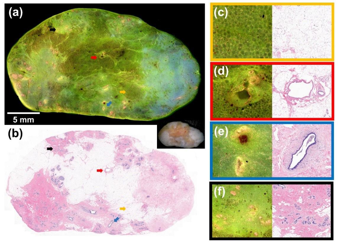
Figure 1. Fluorescence and H&E images of normal benign breast tissue.
During Breast-Conserving Surgery
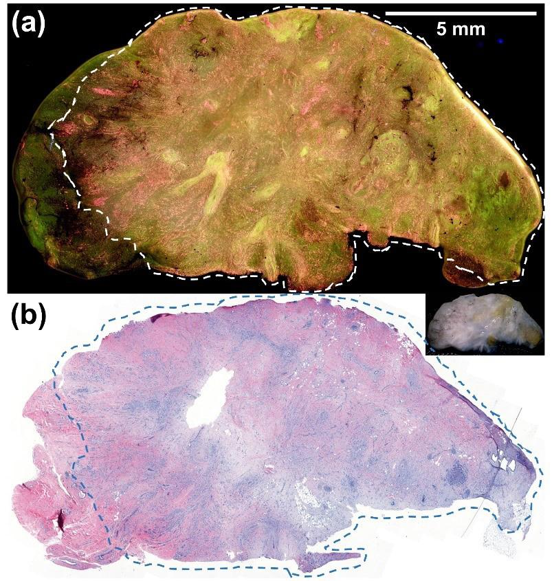
Figure 2. (a) Fluorescence and (b) H&E images of an invasive ductal carcinoma.
staining of the cytoplasm and connective tissues. The specimen was immobilized on a motorized stage and images were collected, transformed and stitched together.
Figure 1a demonstrates the fluorescence image of benign breast tissue, which appears green. A photograph of the gross specimen is shown in the lower right corner. Figure 1b displays the corresponding H&E image. The four arrows mark structures that are displayed in zoomed fluorescence and H&E images on the right panel. The orange arrow highlights fibroadipose tissue, which appears darker green (c). Since blood vessels (red arrow; d), ducts (blue arrow; e) and lobules (black arrow; f) have higher cell density, these structures appear pink and yellow.
Figure 2 shows fluorescence (a) and H&E (b) images of an invasive ductal carcinoma. The cancer is demarcated by the dashed lines. Since cancer cells have higher cell density, they appear pink and yellow in the fluorescence image and can be easily distinguished from the surrounding benign fatty breast tissue, which appears dark green.
After demonstrating excellent contrast in color, tissue texture, cell density and shape between invasive carcinomas and normal counterparts, we wanted to determine the accuracy of visual interpretation of the MUSE images by non-medical evaluators to demonstrate general feasibility and applicability. We trained 3 people with no prior experience to differentiate cancer from non-cancer tissue using five specimens. The evaluators then interpreted the MUSE images of the remaining 42 specimens, providing a diagnosis of cancer or normal tissue. Visual interpretation was outstanding with an average sensitivity, specificity and accuracy of 97.6%, 92.9% and 96.0%, respectively.
In summary, rapid and accurate intraoperative lumpectomy margin assessment remains an unmet clinical need. We demonstrate the feasibility of using deep ultraviolet scanning fluorescence microscopy as a potential intraoperative tool to detect positive margins. It achieves a good balance between imaging speed and spatial resolution with excellent contrast and should be practical and generalizable to most operating room settings as this technology does not require specialized resources or training. Although these initial results are promising, we acknowledge that ad-
ditional work is needed to further optimize this device prior to conducting a clinical study to assess its true potential for intraoperative lumpectomy margin assessment.
This work was supported by a Medical College of Wisconsin Department of Surgery We Care grant, a Marquette University College of Engineering GHR Foundation grant, and Marquette University startup grant.
FOR ADDITIONAL INFORMATION on this topic, visit mcw. edu/surgery or contact Dr. Tina Yen at tyen@mcw.edu.
REFERENCES
1. Houssami N, Macaskill P, Marinovich ML, Morrow M. The association of surgical margins and local recurrence in women with early-stage invasive breast cancer treated with breast-conserving therapy: a metaanalysis. Ann Surg Oncol 2014;21:717-30.
2. Havel L, Naik H, Ramirez L, Morrow M, Landercasper J. Impact of the SSO-ASTRO Margin Guideline on Rates of Re-excision After Lumpectomy for Breast Cancer: A Meta-analysis. Ann Surg Oncol 2019;26:1238-44.
3. Leff DR, St John ER, Takats Z. Reducing the Margins of Error During Breast-Conserving Surgery: Disruptive Technologies or Traditional Disruptions? JAMA surgery 2017;152:517-8.
4. Marinovich ML, Noguchi N, Morrow M, Houssami N. Changes in Reoperation After Publication of Consensus Guidelines on Margins for Breast-Conserving Surgery: A Systematic Review and Meta-analysis. JAMA Surg 2020:e203025.
5. Gray RJ, Pockaj BA, Garvey E, Blair S. Intraoperative Margin Management in Breast-Conserving Surgery: A Systematic Review of the Literature. Ann Surg Oncol 2018;25:18-27.
6. St John ER, Al-Khudairi R, Ashrafian H, et al. Diagnostic Accuracy of Intraoperative Techniques for Margin Assessment in Breast Cancer Surgery: A Meta-analysis. Ann Surg 2017;265:300-10.
7. Maloney BW, McClatchy DM, Pogue BW, Paulsen KD, Wells WA, Barth RJ. Review of methods for intraoperative margin detection for breast conserving surgery. J Biomed Opt 2018;23:1-19.
8. Schwarz J, Schmidt H. Technology for Intraoperative Margin Assessment in Breast Cancer. Ann Surg Oncol 2020;27:2278-87.
9. Fereidouni F, Harmany ZT, Tian M, et al. Microscopy with ultraviolet surface excitation for rapid slide-free histology. Nat Biomed Eng 2017;1:957-66.
10. Lu T, Jorns JM, Patton M, et al. Rapid assessment of breast tumor margins using deep ultraviolet fluorescence scanning microscopy. J Biomed Opt 2020;25.
The Influence of Pre-operative Carbohydrate Loading on
Sarah Suh, BS M4 Student and soon-to-be General Surgery Resident at MCW
Tammy L. Kindel, MD, PhD
Associate Professor, Division of Minimally Invasive Gastrointestinal Surgery; Director, Adult and Adolescent Bariatric Surgery Program
Pre-operative carbohydrate loading is a component of Enhanced Recovery After Surgery (ERAS) protocols and has been shown to improve patient comfort after elective abdominal surgery by decreasing thirst, hunger, anxiety, malaise, and overall length of stay.1,2,3 Despite the known benefits of pre-operative carbohydrate loading, there is limited research in the bariatric surgery patient population. Therefore, the primary objective of this prospective study was to characterize the impact of pre-operative carbohydrate loading on post-operative quality outcomes in bariatric surgery patients.
We conducted a randomized, controlled trial at Froedtert and the Medical College of Wisconsin. Patients undergoing a primary minimally invasive Roux-en-Y gastric bypass (RYGB) or sleeve gastrectomy (SG) between 2018 and 2020 were randomized to standard management or intervention. Standard management patients were nothing by mouth after midnight prior to surgery. Intervention patients consumed two carbohydrate drinks: one the night before and the second three hours prior to surgery. Primary outcomes analyzed included length of stay (LOS), post-operative nausea and vomiting (PONV), and overall complications.
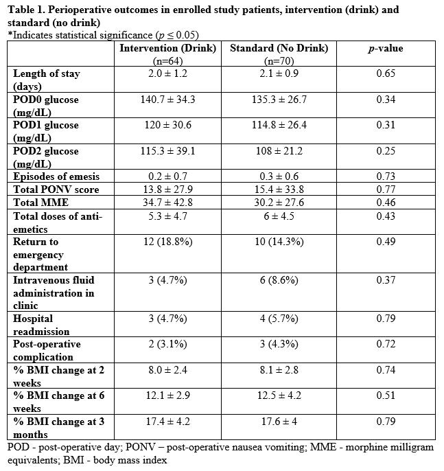
In total, 134 patients were analyzed, 64 (47.8%) intervention (drink) patients and 70 (52.2%) standard (no drink) patients. Among these patients, 49 (36.6%) underwent a RYGB and 85 (53.4%) underwent a SG. Patients in the intervention group had a higher BMI (51.2 kg/m2 ± 8.7 vs 47.1 kg/m2 ± 6.8, p=0.003) and increased proportion of hyperlipidemia (28.1% vs 14.3%, p=0.05). Otherwise, all other demographics were the same between the two groups. There was no statistically significant difference in LOS (2.0 ± 1.2 days vs 2.1 ± 0.9 days; p = 0.65) or overall PONV scores (13.8 ± 27.9 vs 15.4 ± 33.8; p = 0.77) between the intervention and standard management groups (Table 1). There was also no statistically significant difference in any other post-operative outcome between the two groups, including blood glucose, anti-emetic administration, opioid use, weight loss, or overall complications.
Rana M. Higgins, MD Assistant Professor, Division of Minimally Invasive Gastrointestinal Surgery
None of the patients experienced aspiration during anesthesia induction. When divided by procedure type, among patients who underwent a RYGB, those who received the drink had a shorter duration of nausea compared to those who did not receive the drink on post-operative day 1 (POD 1) (0.5 ± 0.7 hours vs 3.2 ± 5.8 hours; p=0.04). A sub-analysis of diabetic versus non-diabetic patients did not reveal significant differences in perioperative glycemic control or post-operative complications.
Pre-operative carbohydrate loading is an integral part of ERAS protocols in various surgical specialties. Although incorporated into some bariatric surgery ERAS protocols, the impact of pre-operative carbohydrate loading alone in an already established ERAS pathway for bariatric surgery patients has yet to be identified. In this study, we identified that pre-operative carbohydrate loading drinks can be administered to bariatric surgery patients without significant post-operative risks. Therefore, carbohydrate drinks can be safely included as a standard of care in bariatric surgery ERAS protocols, including both diabetic and nondiabetic patients.
In our study, there was no significant difference in LOS between standard management and intervention groups. In the literature, several studies demonstrate contrasting results. Many institutions have shortened LOS by at least one day by implementing pre-operative carbohydrate loading.4,5 These findings, however, are difficult to compare to our study because the aforementioned institutions implemented carbohydrate loading and additional ERAS protocols simultaneously. These methods make it challenging to understand the isolated impact of carbohydrate loading on their overall LOS. Given that our institution already has a bariatric surgery ERAS protocol in place, our average LOS of 2 days is lower than the national average of 2.5 days.6 Therefore, no significant change in LOS is not unexpected with one isolated addition to the standard ERAS protocol.
There were no significant differences identified between standard management and intervention groups in the administration of anti-emetics, opioid usage, and overall post-operative complications. These findings are contradictory to other studies which have demonstrated that pre-operative carbohydrate loading can decrease morbidity, major complication rates, opioid usage, and early emergency department visits for bariatric surgery patients.7,8 These studies, however, conducted retrospec-
Post-operative Outcomes in Bariatric Surgery Patients: a Randomized, Controlled Trial
tive reviews after implementing new ERAS programs, which involved multiple components pre, intra, and post-operatively. There are numerous confounding variables amongst those studies that make it challenging to conclude specific outcomes related to the addition of carbohydrate loading alone.
Regarding PONV, patients who underwent a RYGB in the intervention group had a shorter duration of nausea (30 minutes) compared to patients in the standard management group (3.2 hours) on POD 1. We did not see any impact of pre-operative carbohydrate loading on duration of nausea in SG patients. These findings have not been identified elsewhere in the literature. We recently published that SG patients were twice as likely to develop PONV compared to RYGB patients.9 This is likely secondary to the complex mechanisms contributing to PONV that are unique to a SG compared to RYGB, which are not fully understood at this time. Additionally, the mechanism by which carbohydrate loading improves PONV is unclear. Future research investigating the influence of carbohydrate macronutrients on the serotonergic response and changes in gastric emptying will help with better understanding the regulation of nausea among bariatric surgery patients.10,11
drate drinks can be safely included as a standard of care in bariatric surgery ERAS protocols for patients with and without diabetes, although the benefits remain unknown.
Given the overwhelming prevalence of metabolic comorbidities in patients with obesity, it is crucial to determine the effect of pre-operative carbohydrate nutrition on bariatric patients with Type 2 diabetes. Historically, diabetic patients have not been included in randomized controlled trials that involve carbohydrate loading due to concerns for worsening hyperglycemia and risk of aspiration due to delayed gastric emptying.11 In our study, none of the patients suffered from aspiration, and diabetic patients did not have increased rates of post-operative complications or poor glycemic control. Our findings support the notion that carbohydrate loading can be safely utilized and benefit reducing insulin resistance throughout the perioperative period.12
In conclusion, pre-operative carbohydrate drinks can be administered to bariatric surgery patients without significant post-operative risks. Carbohydrate loading prior to surgery can decrease the duration of PONV, specifically in RYGB patients. These results highlight that carbohy-
FOR ADDITIONAL INFORMATION on this topic, visit mcw.edu/surgery or contact Dr. Rana Higgins at rhiggins@mcw.edu.
REFERENCES
1. Hausel J, Nygren J, Thorell A, Lagerkranser M, Ljungqvist O. Randomized clinical trial of the effects of oral preoperative carbohydrates on postoperative nausea and vomiting after laparoscopic cholecystectomy. Clin Nutr. 2003;22(1):76.
2. Nygren J, Thorell A, Jacobsson H, Larsson S, Schnell PO, Hylen L. Preoperative gastric emptying: effects of anxiety and oral carbohydrate administration. Ann Surg. 1995;222:728-734.
Continued on page 11
Changes in Liver Allocation
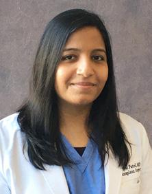
Priyal Patel, MD Assistant Professor, Division of Transplant Surgery
Since the advent of transplantation as a common and successful treatment for organ failure, the transplant community has been attempting to devise an allocation system for donated organs. The unique problems transplantation brings include scarcity, ethics, resource utilization, socioeconomics, and complex medicine. The Final Rule, established in 1998, attempted to establish basic rules for equitable allocation; however, the variables and questions are much more intricate. The use of the Model for Endstage Liver Disease (MELD )score for liver allocation has greatly impacted the reduction of mortality rates in high acuity liver failure patients. Moreover, it has helped prevent transplant-related deaths, due to the inherent risks of transplant, in low MELD patients.1
The continuous distribution policy, specifically for liver allocation in the United States, was adopted in December 2020. The basic premise of this policy removes the previous boundaries of distribution based on region and donation service area (DSA). The geographic boundaries arguably could result in a great disservice to a potential recipient who happens to be on the “wrong” side of DSA3. The current allocation is based on initial MELD score and nautical distance from the donor center. After Status 1A recipients, who are given priority, the MELD score, along with distance, is used for the remainder of the transplant recipient list. Thus, the recipients with the highest MELD (greater than 37) and closest distance to the donor would be given the highest priority. Distance is determined by an initial circle of 150 nautical miles (nm), then 250 nm, and then 500 nm. Within each circle, the matched ABO is first and then unmatched for ABO “O” donors. The list would then progress to the next set of MELD scores from 33-37, and so on. The picture becomes complicated very quickly with the addition of multi visceral, pediatric and “MELD exceptions.” However, the basic principle of the policy tries to account for transport feasibility, equity, and medical severity.
Metrics used to measure “success” in a transplant are extremely complex. The fair allocation has to address pretransplant socioeconomic hurdles. Those in poor economic areas may never make it to transplant evaluation due to challenges with referrals, insurance, distance to transplant center and lack of primary care, to name a few. Even if they do, there are social support requirements and financial burdens to be met prior to being listed for transplant. Even after listing for transplant, half of the waitlist candidates will never make it to transplant due to scarcity of the
resource. Many of the metrics used today are based on the recipients on the waitlist; however, there should be a broader denominator to assess the recipients who never made it to the list or even to evaluation.2
The distances used in the continuous distribution also may seem slightly arbitrary. However, the costs and resources needed to procure organs in a timely manner also affects graft survival. If the graft has an improved rate of survival to a recipient 150 miles away, is it “fair” to use it for a higher MELD patient 250 miles away? The time on the waitlist also raises some questions.
A recipient with hepatocellular carcinoma with intact function will not reach the same MELD as a decompensated patient with cirrhosis and thus requires exception points to “compete” with higher MELDs. However, even with the exceptions, many cancer patients will not receive transplants at a MELD of 33. As these patients wait, they may fall out of criteria. Is it fair to prioritize a patient with a MELD of 40 and decompensated cirrhosis with days of waitlist time over a cancer patient with years of waitlist time?
The liver list caps out at a MELD of 40. The differentiation then falls on waitlist time and geographic proximity. However, as ICU management, intra-operative management and anesthesia capabilities improve, so does the ability to perform transplants on sicker patients. One patient with a MELD of 40 may be on multiple pressors with ongoing massive transfusions while another may be awake and alert on a med/surg floor. In a densely populated area, having a MELD of 40 is the only way to get a transplant due to high demand. So if not all MELD of the same number are equal in resource utilization, management or survival, is it time to extend beyond 40?
With the increase of alcoholic cirrhosis and acute alcoholic hepatitis surpassing Hepatitis C as the primary etiology of transplant, there are many social and ethical considerations that are also brought to the table. Transplant centers have a responsibility to their patients, but also to the generous donors. A high recidivism rate impacts that organ and patient as well as future recipients. Transplants psychologists, social workers, and committees attempt to answer such questions daily. Both alcoholism and Nonalcoholic Steatohepatitis (NASH) are both diseases with high rates of steatosis recurrence without abstinence and weight loss. If we ask alcoholic patients to abstain, should weight loss also be a requirement? Is it fair to defer transplant to these patients until criteria are met?
Due to COVID-19, the impact of the policy since its adoption has been difficult to measure. At the beginning of the pandemic, there was a significant decrease in transplants and donors across the United States for many reasons. Some of the reasons were resource utilization, decrease in donors, decrease in tertiary center evaluations,
and decreases in staff, just to name a few. Some centers, however, did not experience see a decline but rather an increase due to the decrease in utilization at centers in the same organ procurement organization (OPO). In the past few months, while processes have begun to stabilize, they have yet to completely normalize. There will undoubtedly be a need for re-evaluation of the impact of organ allocation in a COVID-free transplant setting. However, many of the questions raised are germane, regardless of COVID. As previously mentioned, there always seem to be more questions than answers. Some questions are directly linked to allocation, but others are an indirect result of a change in the transplant population that affects all aspects of the transplant community. In a setting where annually only half of liver transplant waitlist candidates receive transplants due to scarcity, the socioeconomic, ethical, financial, and medical issues will continue to arise despite allocation policies. Nonetheless, the attempt to move towards improved equity and patient mortality in end organ failure is a moving target that should always be pursued. This month, a new kidney allocation policy is in the process of adoption. Kidney sharing reaches an even broader sharing base due to its longer cold ischemia times and thus will be an interesting study to follow in the coming months.
FOR ADDITIONAL INFORMATION on this topic, visit mcw.edu/surgery or contact Dr. Priyal Patel at priypatel@mcw.edu.
REFERENCES
1. Kasiske, Bertram L.a,b; Pyke, Joshuab; Snyder, Jon J.b,c Continuous distribution as an organ allocation framework, Current Opinion in Organ Transplantation: April 2020 - Volume 25 - Issue 2 - p 115-121 doi: 10.1097/MOT.0000000000000733
2. Schold JD. Assessing the outcome of organ allocation: choosing metrics. Curr Opin Organ Transplant. 2019 Jun;24(3):332-336. doi: 10.1097/ MOT.0000000000000637. PMID: 31090645.
OTHER RESOURCES
3. Snyder JJ, Salkowski N, Wey A, Pyke J, Israni AK, Kasiske BL. Organ distribution without geographic boundaries: A possible framework for organ allocation. Am J Transplant. 2018 Nov;18(11):26352640. doi: 10.1111/ajt.15115. Epub 2018 Sep 29. PMID: 30203912.
4. Leventhal TM, Florek E, Chinnakotla S. Changes in liver allocation in United States. Curr Opin Organ Transplant. 2020 Feb;25(1):52-58. doi: 10.1097/ MOT.0000000000000726. PMID: 31834007.
REFERENCES CONTINUED FROM PAGE 9
The Influence of Pre-operative Carbohydrate Loading...
3. Hausel J, Nygren J, Lagerkranser M, Hellstrom PM, Hammarqvist F, Almstrom C. A carbohydrate-rich drink reduces preoperative discomfort in elective surgery patients. Anesth Analg. 2001;93:1344-1350.
4. Mannaerts GH, van Mil SR, Sepaniak PS, Dunkelgrun M, de Quelerij M, Verbrugge SJ, Zengerink HF, Biter LU. Results of implementing an enhanced recovery after bariatric surgery (ERABS) protocol. Obes Surg. 2016;26:303-312.
5. Lemanu DP, Singh PP, Berridge K. Randomized clinical trial of enhanced recovery versus standard care after laparoscopic sleeve gastrectomy. Br J Surg. 2013;100(4):482-9.
6. Coleman KJ, Huang YC, Hendee F. Three-year weight outcomes from a bariatric surgery registry in a large integrated healthcare system. Surg Obes Relat Dis. 2014;10(3):396-403.
7. Hahl T, Peromaa-Haavisto P, Tarkiainen P, Knutar O, Victorzon M. Outcome of laparoscopic gastric bypass (LRYGB) with a program for enhanced recovery after surgery (ERAS). Obes Surg. 2016;26:505-511.
8. King AB, Spann MD, Jablonski P, Wanderer JP, Phil MD, Sandberg WS, McEvoy MD. An enhanced recovery program for bariatric surgical patients significantly reduces perioperative opioid consumption and postoperative nausea. Surg Obes Relat Dis. 2018;14:849-856.
9. Suh S, Helm M, Kindel TL, Goldblatt MI, Gould JC, Higgins RM. The impact of nausea on post-operative outcomes in bariatric surgery patients. Surg Endosc. 2019;34:3085-3091.
10. Orosco M, Gerozissis K. Macronutrient-induced cascade of events leading to parallel changes in hypothalamic serotonin and insulin. Neurosci Biobehav Rev. 2001;25:167-174.
11. Bilku DK, Dennison AR, Hall TC, Metcalfe MS, Garcea G. Role of preoperative carbohydrate loading: a systemic review. Ann R Coll Surg Engl. 2014;96(1):15-22.
12. Nygren J, Soop M, Thorell A, Efendic S, Nair KS, Ljungqvist O. Preoperative oral carbohydrate administration reduces postoperative insulin resistance. Clin Nutr. 1998;17:65-71.
2020-2021 MCW DOS ANNUAL REPORT!
Read the 2020-2021 Annual Report by scanning the QR code:
MCW Curriculum Changes: Preparing Students
Travis P. Webb, MD, MHPE
Professor, Division of Trauma & Acute Care Surgery; Associate Dean of Medical School Curriculum
As the Associate Dean for Curriculum for the Medical College of Wisconsin School of Medicine, I am excited to provide the MCW surgical community with an update on current efforts to design and implement a new MCW medical student curriculum. Since October 2019, with the formal input, guidance, and dedication of over 150 faculty, students, and staff we have been exploring opportunities to update the curriculum and assure that our students are receiving the best training to prepare them for the future medical practice. Though the pandemic required us to slow the pace for innovation, we have made great strides during the past months to develop a curricular framework for staged implementation beginning academic year 2022. After two successful retreats and countless hours of analysis of published education research, we have defined the goals, role, and principles for the new curriculum.
The goal of our curriculum is to produce competent well-rounded physicians who will be excellent clinicians in any specialty and are prepared to practice in the future health care environment. The curriculum will provide students with opportunities to develop skills to pursue specific career goals.
The following statement reflects the role of the new curriculum in the acquisition and application of knowledge, skills and behaviors:
The acquisition of knowledge is a developmental process focused on sequentially building upon skills in students so that over time they can integrate information and apply that knowledge to clinical care. The curriculum will challenge students to apply what they learn in ways that students may not have encountered in their previous learning settings and which may make them uncomfortable. But through this sequential process of building skills, students will become competent, adaptable, and accomplished life-long learners.
The outstanding work of a Curriculum Exploration Steering Committee and five workgroups populated by a diverse group of faculty, staff and student stakeholders has provided further direction and guidance for us to meet our goals while adhering to defined principles. These workgroups have provided an evidence-based approach to curricular design, including instructional methodology, assessment, and evaluation. The following recommendations have been made to allow us to meet our principled goals:
Principle 1
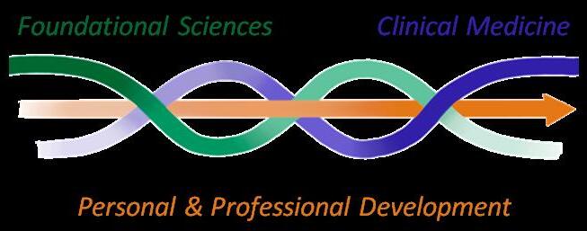
Integration of foundational and clinical science learning throughout all years of the curriculum
Curricular threads will be woven through three curricular phases (pre-core clinical, core clinical, and individualized post-core clinical); each phase will include meaningful foundational science, patient care, and personal and professional development activities. Clinical cases are the recommended functional units of integration in required courses. Phase 1 and 2 must be shortened from the traditional length to allow all students, including those aiming to graduate in three years, to enter phase three prior to starting residency applications and interviews. This will empower students to pursue experiences to realize career goals, such as advanced degrees, research and community service projects, and personal or family health and well-being.
Principle 2
A systematic approach and focus on assessment that drives learning and assures students achieve desired competencies
Medical students need to have an active role in the assessment process to facilitate the development of their professional identities. As part of the Master Adaptive Learner (MAL) cycle, students will plan their learning to identify gaps in understanding, will purposefully participate in learning, will engage in assessment through selfmonitoring and external feedback, and will adjust based on this input. Students in our current system often hide knowledge gaps and avoid feedback, whereas with MAL, students see feedback as growth opportunities. To move through the MAL cycle, students need multimodal, progressive assessments that encourage student curiosity, motivation, mindset, and resilience.
Standardized tools with associated behavioral anchors to support longitudinal assessment of all student competencies and certify that students are safe to take on additional responsibility have been identified and will be implemented. Dashboards to display student progress combined with student-curated portfolios demonstrating mastery and facilitating feedforward of information to support student development will be utilized.
Principle 3
Individualized approaches to learning that are ultimately tailored to student interest and career goals
Curriculum redesign that allows more opportunities for “individualization” after core competencies in foundational and clinical science have been met will require streamlining, integrating, and customizing of the foundational sciences.
for the Future of Healthcare
Streamlining - Essential fundamentals will be taught in a shorter “pre-clinical” or “medical language acquisition” phase, avoiding extensive depth and eliminating unintentional redundancies.
Integrating – Foundational sciences will be taught alongside clinical content during all phases of the curriculum.
Customizing – Advanced integrated science content will be delivered following required clinical phase 2. Students will choose courses in advanced science that aligns with career goals, instead of delivering uniform depth to all students.
It is essential that students have three distinct types of faculty support to create and meet individualized goals. Support of student’s career planning and goal achievement will be through robust use of learning communities, scholarly project guidance, and clinical specialty advising.
Principle 4
A student-centered, inclusive culture with a focus on wellness
Developing a student-centered, inclusive culture with a focus on well-being will be challenging but it is foundational to our new curriculum’s success. Maximizing inclusion and well-being while embracing and valuing diversity and lifelong learning requires MCW and its affiliates to develop and maintain a supportive culture that fully values the diversity, well-being, and ongoing growth and development of all its people. There also needs to be an important component of education regarding well-being, diversity, inclusion, lifelong learning, and personal development threaded throughout the curriculum. The supportive and inclusive culture noted above is paramount. A less than supportive culture at any one of our major affiliates and/ or at MCW will erode and undermine all the other efforts to teach this to our students and will negatively affect the well-being of faculty, staff, and trainees, and our ability to recruit and retain diverse faculty, staff, and trainees.
For MCW to be fully successful at ensuring Principle 4 is achieved in its new curriculum, it will require an investment in culture, people, and education to make sure the resources and personnel necessary are available.
Principle 5
An evidence-based instructional approach that is inquiry driven and utilizes active learning
To promote inquiry and curiosity in medicine and health systems, we will implement an inquiry-based curriculum that features active learning educational methods and appropriate time for work and learning. Student assessment should foster intrinsic motivation and be less dependent on medical knowledge-based grades. Students’ inquiry should be rewarded by assessment of active participation and applied learning outcomes. Ideally, students drive their own learning which is facilitated by multidisciplinary and interprofessional educators and peers. Active learning sessions will take the form of case-based, problem-based, or team-based learning as appropriate to content, stimulating learner investigation and application of concepts, facilitated by faculty, and affording time for student questions. Preparatory material must be engaging and should consist of a diverse array of resources, including high quality lectures. Such small group work will also foster psychosocial skill development, such as teamwork and leadership. A culture endorsing student inquiry and intellectual curiosity will develop students into master adaptive learners where they learn to plan (inquiry), learn (exploration), assess (self-reflect), and adapt (iterative process, comfort with failure as a learning and development opportunity).
We recognize the importance of coordinated, timely, and relevant faculty development in advance of, during, and following implementation of the new curriculum. The focus of faculty development must be on providing faculty with the skills to design and structure educational sessions in the new curriculum that foster student-centered learning, rather than traditional faculty development that focuses on teaching skills to become better within existing systems, e.g., a focus on giving a better lecture. This will require a culture change that can be facilitated through creation of faculty development programs and communities of practice, promoting peer partnerships and skills development, and an emphasis on competency and outcomes-driven instruction and assessment.
On behalf of the many dedicated individuals who have been involved thus far in the design of a new cutting edge curriculum, I would like to welcome any interested individuals to contribute to the ongoing design and implementation of our new curriculum. Much work will be required to accomplish our goals, but with the input and support of the MCW community, I am confident that we will meet our goals in the coming years.
FOR ADDITIONAL INFORMATION on this topic, visit mcw.edu/surgery or contact Dr. Travis Webb at trwebb@mcw.edu.
Right Ventricular Assist Device and Extracorporeal Membrane
Nathan J. Smith, MD General Surgery Research Resident
Michael T. Cain, MD
Cardiothoracic Fellow, University of Colorado at Denver, MCW General Surgery Resident Alum ‘20
“Necessity is the mother of invention” as the old saying goes. The current SARS-CoV-2 pandemic (i.e., COVID-19) has strained our public health system and pressed the medical community to adapt quickly in light of limited knowledge and experience. As we learn more about this disease, its unique pathophysiology and clinical manifestations have challenged clinicians to develop new treatment strategies at a feverish pace, and to look beyond conventional treatment pathways. Among hospitalized patients who develop COVID-19 pneumonia, acute respiratory distress syndrome (ARDS) occurs in approximately 31-67% with a mortality rate as high as 52%.1-3 ARDS is not a new phenomenon, and although clinicians have recognized and treated ARDS long before COVID-19, the high incidence among COVID-19 patients has placed a heavy strain on our intensive care units and consumed resources. Effective treatments such as lung protective ventilation and prone positioning have found their place in the COVID-19 pandemic and are still mainstays of treatment, however there are those who fail these conventional forms of support and require advanced therapies. These patients are candidates for veno-venous extracorporeal membrane oxygenation (VV-ECMO).4 5
VV-ECMO is a form of advanced oxygenation support where blood is removed from the patient using a centripetal pump and passed through a specialized membrane lung that allows for both oxygenation and gas exchange, supplementing, and in some instances replacing, the role of the lungs in those with recalcitrant respiratory failure. Experience in other viral respiratory illnesses has leant evidence to the benefit of VV-ECMO in such situations, but early experience with COVID-19 has been less promising.6 7 What makes COVID-19 so unique compared to other coronavirus illnesses is the multi-organ system involvement, including both cardiac and pulmonary vascular dysfunction. As a result, conventional VV-ECMO may be ineffective due to unsupported cardiac dysfunction in the setting of severe respiratory failure.8-11 This knowledge has forced physicians to rethink the approach to VV-ECMO in patients with severe COVID-19 ARDS.
Lucian A. Durham, III, MD, PhD
Associate Professor, Division of Cardiothoracic Surgery
David L. Joyce, MD, MBA
Associate Professor, Division of Cardiothoracic Surgery
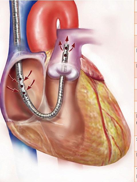
Innovations in VV-ECMO cannula design have provided a unique solution to the treatment of combine cardiopulmonary failure. Use of these advanced devices has become a mainstay of ECMO care here at the Medical College of Wisconsin. The TandemLife Protek Duo™ cannula (LivaNova, London, UK) is a dual-lumen percutaneous right ventricular assist device (RVAD) approved for the treatment of acute right ventricular failure (Figure 1). The cannula is inserted via the right internal jugular vein and advanced into the main pulmonary artery under fluoroscopic and echocardiographic guidance. When connected to an ECMO circuit and oxygenator, deoxygenated blood is removed at the right atrium, oxygenated, and returned to the patient in the main pulmonary artery. In this fashion, the cannula supports the function of the right ventricular by bypassing and unloading the right heart while also assuming the oxygenation role of the lung. This combines the mechanical support function of an RVAD with the gas exchange function of conventional VV-ECMO, providing a combined RVAD/ECMO system. We have previously reported our experience with RVAD/ECMO with favorable results.12
Based on the knowledge of the combined cardiopulmonary pathophysiology at play in COVID-19 pneumonia, RVAD/ECMO has quickly become the mainstay of mechanical circulatory support for COVID-19 at our institution. Led by Dr. Lucian A. Durham, III, MD, PhD, director of ECMO and mechanical circulatory support at the Medical College of Wisconsin, our early experience in this pandemic with RVAD/ECMO for the treatment of severe COV-
Oxygenation for the Treatment of Severe COVID-19 Acute Respiratory Distress Syndrome: Evolving Care in a Pandemic
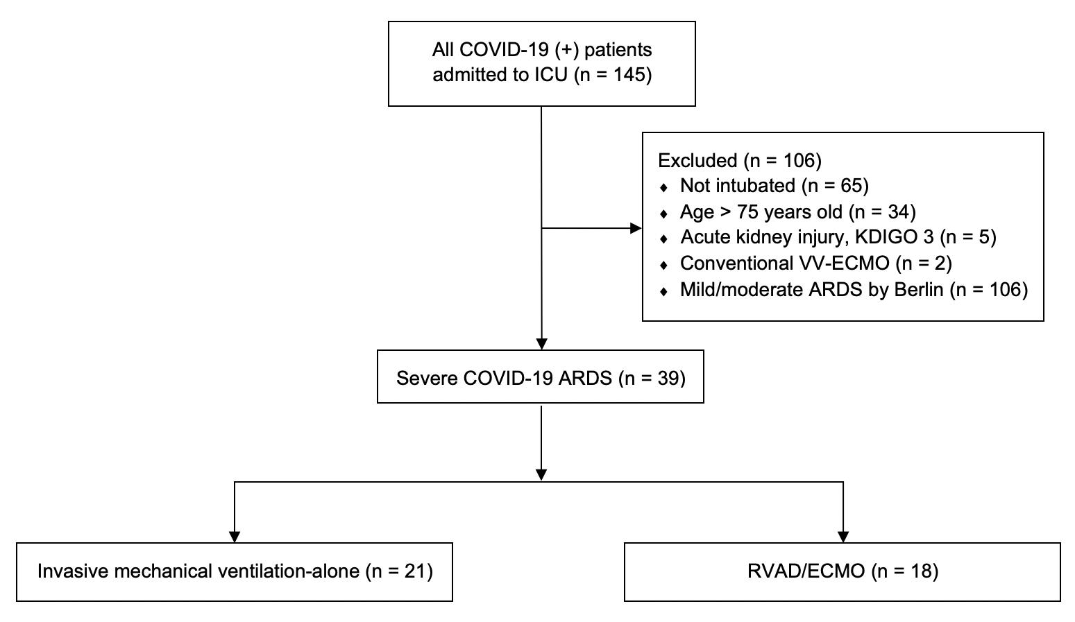
ID-19 ARDS has been remarkably favorable. Through close collaboration with the Division of Critical Care Medicine at the Medical College of Wisconsin, as well as established partnerships with referring institutions throughout Wisconsin, early RVAD/ECMO support in select patients has resulted in significantly lower mortality when compared to similar patients who were treated with conventional mechanical ventilation alone. From March 1 to July 6, 2020, 145 patients were admitted to the intensive care unit at Froedtert Memorial Lutheran Hospital for COVID-19 pneumonia (Figure 2). Patients were not considered ECMO candidates if they were older than 70, not intubated, or had concomitant renal failure. In total, 39 patients met criteria for severe COVID-19 ARDS, of which 18 patients received RVAD/ECMO. The remaining 21 patients received invasive mechanical ventilation (IMV) alone. When comparing
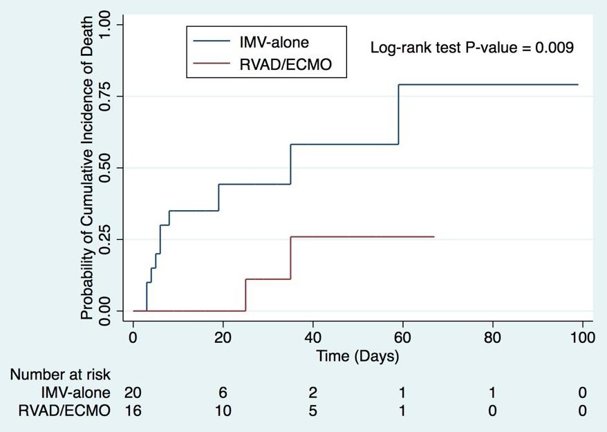
these two groups, RVAD/ECMO patients demonstrated a 11.1% mortality versus 52.4% in those treated with IMV-alone (p=0.008). Thirty-day mortality was also significantly lower for RVAD/ECMO patients (5.6%) versus those treated with IMV alone (42.9%, p=0.011). Length of stay, duration of mechanical ventilation, and the incidence of tracheostomy were similar between the two groups. No patients treated with RVAD/ECMO experienced acute kidney injury after cannulation, compared to 71.4% of mechanical ventilation patients (p<0.001). The average duration of RVAD/ECMO support was 13 days (95% CI 7.5-22 days) with 84.6% of patients surviving to device removal. Kaplan-Meier adjusted mortality was significantly lower for RVAD/ECMO patients (p=0.009) (Figure 3). The most common complication in RVAD/ECMO patients was bleeding. These early results have been echoed by other institutions thus providing support to the novel utility of combined right ventricular support and ECMO in the treatment of severe COVID-19 ARDS.13
Since the beginning of this pandemic, our experience with ECMO for COVID-19 has expanded to include 40 patients. We have learned a tremendous amount about the role of ECMO in COVID-19 and the ideal patients who might benefit from such an aggressive intervention. With the number of reported cases rising in the United States, the strain being felt by our healthcare system is certainly being felt in the field of ECMO, and we are being forced to carefully select which patients offered RVAD/ECMO support. It is fast becoming clear that early referral through close internal and extra-institutional partnerships, early identification of progressive ARDS, and ECMO consideration prior to the onset of multisystem organ failure are critical in achieving optimal outcomes and saving these sick patients. The end of this pandemic is far from near, but one thing is certain; by the end, ECMO care will never be the same and we will have learned much to improve care for the next one. Necessity is, indeed, the mother of invention and, in this case, innovation.
FOR ADDITIONAL INFORMATION on this topic, visit mcw.edu/surgery or contact Dr. Lucian Durham at ldurham@mcw.edu.
References on page 17
Figure 2. Initial COVID-19 experience cohort
Figure 3. Kaplan-Meier mortality for RVAD/ECMO and IMValone patients.
What Learners Tell Us About How They Know When They Matter
Caitlin Patten, MD
Assistant Professor, Section of Breast Surgery, Division of Surgical Oncology
Rana Higgins, MD
Assistant Professor, Division of Minimally Invasive Gastrointestinal
Surgery
We have all been students at some point in our life. Do you remember a day when you were in a classroom, meeting, or clinical learning environment and wondered why you were there? Perhaps, no one acknowledged your presence or asked your name, or they dismissed what you said, or when you returned after a prolonged absence, no one realized you had been gone. These behaviors make you feel like you don’t matter. Knowing that you matter means that others’ behaviors, actions, and words make you feel valued and useful. They are aware of you, make you feel important by paying attention to and going out of their way for you, and rely on you. It turns out that mattering (the perception that you matter) is an enabling concept – when you feel you matter you have more selfesteem and self-efficacy which leads to less anxiety and depression, allowing you to learn more! Helping others matter is one way to improve the learning environment.
How do our learners perceive that they matter? A group of faculty participating in the 2019-2020 KINETIC3 faculty development program asked that question. While participating in this structured program, Dr. Rana Higgins and Dr. Caitlin Patten used qualitative methodology and semi-structured interviews to find out what M3 and M4 students on their surgery clerkship perceived as evidence that they matter.
Mattering focuses on three critical components: awareness, importance and reliance. All medical students interviewed needed to know that others were aware of them. Students felt they mattered when residents and faculty called them by name (yes, it is that simple!). They also felt they mattered by making eye contact and not having the resident or faculty use a phone while talking to them. Part of awareness is also finding out what makes each learner a unique individual – where they are from, their past experiences, and what their current interests and/or career goals are. Identifying learners’ interests and strengths were not only a way to be aware of them, but also made them feel important.
Importance was highlighted when medical students
Karen Marcdante, MD Professor, Department of Pediatrics and Critical Care
Erin Strong, MD, MPH General Surgery Research Resident
were assigned meaningful tasks - making them a valued member of the team. When students perceived their educators relied solely on them to complete an assigned task that was essential to the success of the team, they had a strong sense of mattering. Additionally, learners felt important when those supervising them (residents/faculty) “went out of their way” to see how they were doing, asked about one of their projects, or performed an impromptu mini education session. The third component of mattering is reliance. For medical students this occurred when they were trusted to collect information that was needed and not duplicated by another team member.
This is the first work of its kind with learners in medical education. It is critical for us all to recognize which words, actions and behaviors make learners feel they matter, allowing them to be more curious, comfortable, and creative. It is essential to understand that mattering requires building a relationship. Educators and students alike all want to matter. If a learner has a sense of mattering, they will be more engaged with learning and with their educators. In turn, educators will feel their time spent with students was worthwhile and they will be more motivated to teach. Moving forward, remember how it felt when you did not matter as a learner and how easily you can prevent others from feeling that same way through awareness, importance, and reliance.
FOR ADDITIONAL INFORMATION on this topic, visit mcw.edu/surgery or contact Dr. Rana Higgins at rhiggins@mcw.edu.
REFERENCES FROM PAGE 14
Right Ventricular Assist Device and Extracorporeal...
1. Wu C, Chen X, Cai Y, et al. Risk Factors Associated With Acute Respiratory Distress Syndrome and Death in Patients With Coronavirus Disease 2019 Pneumonia in Wuhan, China. JAMA Internal Medicine 2020;180(7):934-43 doi: 10.1001/jamainternmed.202 0.0994published Online First: Epub Date]|.
2. Yang X, Yu Y, Xu J, et al. Clinical course and outcomes of critically ill patients with SARS-CoV-2 pneumonia in Wuhan, China: a single-centered, retrospective, observational study. Lancet Respir Med 2020;8(5):47581 doi: 10.1016/s2213-2600(20)30079-5published Online First: Epub Date]|.
3. Zhou F, Yu T, Du R, et al. Clinical course and risk factors for mortality of adult inpatients with COVID-19 in Wuhan, China: a retrospective cohort study. Lancet 2020;395(10229):1054-62 doi: 10.1016/s01406736(20)30566-3published Online First: Epub Date]|.
4. Ventilation with Lower Tidal Volumes as Compared with Traditional Tidal Volumes for Acute Lung Injury and the Acute Respiratory Distress Syndrome. New England Journal of Medicine 2000;342(18):1301-08 doi: 10.1056/nejm200005043421801published Online First: Epub Date]|.
5. Thompson AE, Ranard BL, Wei Y, Jelic S. Prone Positioning in Awake, Nonintubated Patients With COVID-19 Hypoxemic Respiratory Failure. JAMA Internal Medicine 2020 doi: 10.1001/jamainternmed. 2020.3030published Online First: Epub Date]|.
6. Davies A, Jones D, Bailey M, et al. Extracorporeal Membrane Oxygenation for 2009 Influenza A(H1N1) Acute Respiratory Distress Syndrome. Jama 2009;302(17):1888-95 doi: 10.1001/ jama.2009.1535published Online First: Epub Date]|.
7. Alshahrani MS, Sindi A, Alshamsi F, et al. Extracorporeal membrane oxygenation for severe Middle East respiratory syndrome coronavirus. Ann Intensive Care 2018;8(1):3 doi: 10.1186/s13613-017-0350-xpublished Online First: Epub Date]|.
8. Argulian E, Sud K, Vogel B, et al. Right Ventricular Dilation in Hospitalized Patients with COVID-19 Infection. JACC Cardiovasc Imaging 2020 doi: 10.1016/j. jcmg.2020.05.010published Online First: Epub Date]|.
9. Zeng JH, Liu YX, Yuan J, et al. First case of COVID-19 complicated with fulminant myocarditis: a case report and insights. Infection 2020:1-5 doi: 10.1007/s15010020-01424-5published Online First: Epub Date]|.
10. Tavazzi G, Pellegrini C, Maurelli M, et al. Myocardial localization of coronavirus in COVID-19 cardiogenic shock. Eur J Heart Fail 2020;22(5):911-15 doi: 10.1002/ ejhf.1828published Online First: Epub Date]|.
11. Inciardi RM, Lupi L, Zaccone G, et al. Cardiac Involvement in a Patient With Coronavirus Disease 2019 (COVID-19). JAMA Cardiol 2020;5(7):1-6 doi: 10.1001/jamacardio.2020.1096published Online First: Epub Date]|.
12. Badu B, Cain MT, Durham LA, 3rd, et al. A Dual-Lumen Percutaneous Cannula for Managing Refractory Right Ventricular Failure. Asaio j 2019 doi: 10.1097/ mat.0000000000001099published Online First: Epub Date]|.
13. Mustafa AK, Alexander PJ, Joshi DJ, et al. Extracorporeal Membrane Oxygenation for Patients With COVID-19 in Severe Respiratory Failure. JAMA Surgery 2020 doi: 10.1001/jamasurg.2020.3950published Online First: Epub Date]|.
To refer a patient or request a transfer/consultation, please use the references below:
MaskUpMKE: The Medical College of Wisconsin’s Collaborative
Jonathan C. Horng M4 Student
Christopher S. Davis, MD, MPH Assistant Professor Division of Trauma & Acute Care Surgery
Zeno Franco, PhD Associate Professor Department of Family and Community Medicine
In March 2020, the outbreak of the novel strain of coronavirus (SARS-CoV-2) was declared a pandemic by the World Health Organization and a national emergency by the United States. In a local response to the crisis and the “100 Million Mask Challenge,” the Milwaukee-based company Rebel Converting donated enough material to make 1 million face masks from electrically charged melt-blown polypropylene. Spearheaded by the early collaboration of a trauma surgeon and students at the Medical College of Wisconsin (MCW), as well as support from faculty and staff at both the Robert D. and Patricia E. Kern Institute for the Transformation of Medical Education and the MCW Office of Community Engagement, the volunteer-fueled project would quickly be known as “MaskUpMKE.” After the immediate shortage of masks for healthcare workers was addressed, MaskUpMKE ultimately produced and delivered 3.5 million face masks to primarily underserved communities and at-risk groups in Milwaukee and throughout Southeast Wisconsin.
Early in the outbreak, when there was a severe shortage of hospital-grade face masks in the United States (U.S.), there was public health messaging which encouraged the lay public not to procure or wear face coverings. While the goal was to preserve the limited supply of face masks for those healthcare workers who were essential to the care of severely ill patients presenting to hospitals around the country, in retrospect this message was detrimental because it caused confusion among the lay public around the benefit of face coverings (even though they were not as protective as N95s, they did serve to reduce aerosolization of the virus and inhalation). It was at this moment that there was great interest and desire from all
Michelle Horng
Physician Assistant Milwaukee Health Services, Inc.
Adina Luba Kalet, MD, MPH
Stephen and Shelagh Roell Endowed Chair
Director, MCW Kern Institute for the Transformation of Medical Education
Mack G. Jablonski M4 Student
sectors to act to protect the public and “flatten the curve”. It became urgent to clarify public health messaging to minimize local spread and empower communities at most risk with information about how to protect themselves and each other. In concert with physical distancing, handwashing, and surface disinfection, face masks/coverings are a mainstay of good practice during an outbreak of a respiratory virus. However, the supply of manufactured hospital-grade masks was inadequate.
Rapid research strategies for active crisis events are well established and are typically driven from Action Research (AR) strategies in which the researchers are, to a greater or lesser extent, embedded in the response while simultaneously documenting these efforts.1-2 Community engagement in disaster response holds as a central tenet the importance of researcher’s participation and partnership, and a variety of community-engaged approaches have been applied in international humanitarian relief efforts and domestic disasters in the US.3-4 Similarly, medical research in the context of epidemics and other epidemiological crises often requires medical practitioners and scholars to serve multiple roles, providing unique insights into the heart of public health crisis response.
In this article, we describe in detail the components of MaskUpMKE as an example of an impactful rapidly coordinated response to a public health threat.
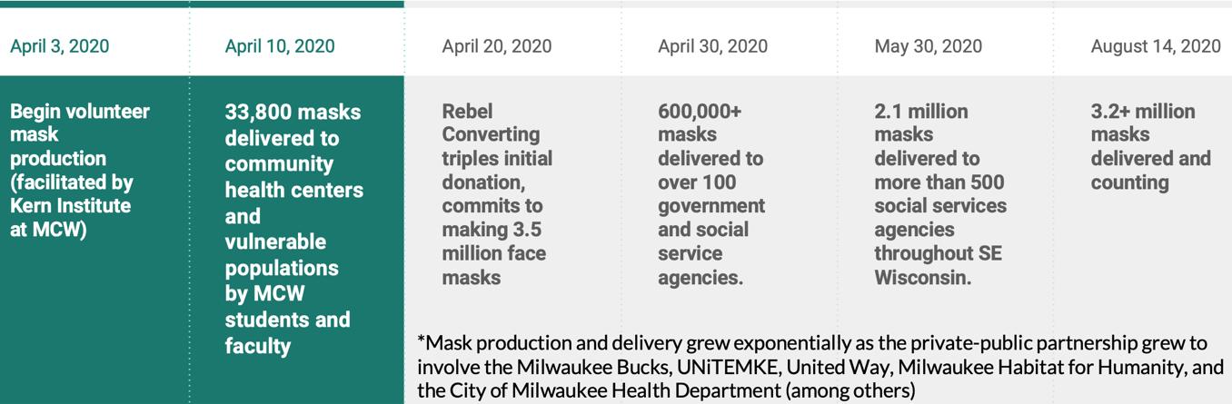
Volunteer mask-production began in the first week of April, 2020 and by April 10th 33,800 masks were delivered to community health centers, homeless shelters, rescue missions, religious shelters, the public school feeding locations, poll workers, and voters. By the end of April 2020, more than 600,000 masks had been delivered to over 100
Approach to the COVID-19 Pandemic in Greater Milwaukee
government and social service agencies throughout greater Milwaukee. As the private-public partnership grew to involve the Milwaukee Bucks, UNiTEMKE, United Way, Milwaukee Habitat for Humanity, and the City of Milwaukee Health Department (among others), mask production and delivery grew exponentially. During May 2020 alone, the formalized project called MaskUpMKE engaged nearly 1,800 volunteers who, through more than 33,000 volunteer hours, had delivered more than 1.5 million additional masks to more than 500 social services agencies throughout Southeast Wisconsin. By August 14, 2020 the total distribution of masks by MaskUpMKE exceeded 3.2 million, and by the end of 2020 would surpass 3.5 million disposable masks as MaskUpMKE turned its attention to reusable masks, broader public health messaging, legislation and advocacy, and other efforts such as TestUpMKE, MaskUp2Vote, and VaccinateMKE.
MaskUpMKE demonstrates a successful example of a grassroots crisis intervention initiative utilizing a public health approach in an effort to curb the spread of COVID-19 in Milwaukee. The project involved many integral components including strategic partnerships, community engagement, intentional social messaging, volunteer efforts, and first-hand educational experiences for medical students. Additionally, it illuminates the unique ways in which medical students, community researchers, and physicians and surgeons can use their leadership skills and approaches to influence their community by responding swiftly and methodically in the face of a crisis. Lastly, MaskUpMKE is a testament to the importance of educat-
ing our future health professionals about the basic principles of public health, community engagement, legislation, and advocacy which are often lacking in their curricula.
FOR ADDITIONAL INFORMATION on this topic, visit mcw.edu/surgery or contact Dr. Chris Davis at cdavis@ mcw.edu.
REFERENCES
1. McCall MK, Peters-Guarin G. Participatory action research and disaster risk. The Routledge Handbook of Hazards and Disaster Risk Reduction Oxford UK: Routledge. 2012.
2. Quarantelli EL, Dynes RR. Response to social crisis and disaster. Annual review of sociology. 1977;3(1):23-49.
3. Mays RE, Braxton M, Berry A, et al. Considering Practitioner-Driven Innovations: Accommodating Information Systems Within Successful Humanitarian Work. 2016 International Conference on Engineering, Technology and Innovation/IEEE lnternational Technology Management Conference (ICE/ITMC): IEEE 2016:1-9.
4. Coletti PGS, Mays RE, Widera A. Bringing technology and humanitarian values together: A framework to design and assess humanitarian information systems. 2017 4th International Conference on Information and Communication Technologies for Disaster Management (ICT-DM): IEEE 2017:1-9.
Figure 1. Timeline of key events in the development of MaskUpMKE
Leading the Way
DIVISION OF CARDIOTHORACIC SURGERY
Nilto C. De Oliveira, MD, joined the Department of Surgery faculty in April as Professor of Surgery and Surgical Director of the Lung Transplant Program at F&MCW. Dr. De Oliveira received his medical degree from Federal University of Parana in Curitiba, Brazil. He completed general surgery residency at the University of Connecticut, followed by a cardiothoracic surgery residency at Barnes Jewish Hospital, Washington University. Dr. De Oliveira completed a Cardiovascular Surgery Research Fellowship at Harvard Medical School/ Massachusetts General Hospital and an Adult Aortic Surgery Fellowship at the University of Toronto.
H. Adam Ubert, MD
H. Adam Ubert, MD, will be joining the Department of Surgery faculty in August as an Assistant Professor of Surgery following completion of a Critical Care Fellowship at the Cleveland Clinic. Dr. Ubert received his medical degree from West Virginia University and completed general surgery residency
DIVISION OF PEDIATRIC SURGERY
Jose Salazar Osuna, MD, PhD, will be joining the Department of Surgery faculty as Assistant Professor of Surgery in September upon completion of the Pediatric Surgery Fellowship program here at MCW. He received his medical degree from Escuela de Medicina del Tec de Monterrey in Mexico, followed by a post-doctoral research fellowship at Johns Hopkins School of Medicine in Baltimore. Dr. Salazar Osuna completed general surgery residency training at Johns Hopkins and the University of Maryland Medical Center and then a second post-doctoral research fellowship at Johns Hopkins. He came to MCW for our
Katie Iverson, MD, MPH
New Faculty
Most recently, he was Surgical Director of Lung Transplantation at the University of Wisconsin-Madison where he also served as Director of Aortic Surgery and the Structural Heart Disease Program. Dr. De Oliveira will also be instrumental in planning the Aortic Surgical Program at F&MCW and will be Co-Director of the program when formally established.
at Charleston Area Medical Center in Charleston, West Virginia. He then completed a Cardiothoracic Surgery Fellowship at the University of Louisville. Prior to his current fellowship training, Dr. Ubert was on faculty at the Charleston Area Medical Center where he was Chair of the Thoracic Surgery Section and Surgical Director of the ECMO program. He will add to our already worldclass team leading the way in mechanical circulatory support.
Pediatric Surgical Critical Care fellowship program and remained here to complete a post-doctoral research fellowship. Dr. Salazar Osuna earned his PhD in Clinical Investigation from the Johns Hopkins Bloomberg School of Public Health. He will provide clinical care for general and thoracic pediatric surgery patients at Children’s Wisconsin and continue his research in developing a model of extracorporeal fetal sheep support with preservation of the placenta.
DIVISION OF TRAUMA & ACUTE CARE SURGERY
Katie Iverson, MD, MPH, will be joining the Department of Surgery faculty as Assistant Professor of Surgery in October upon completion of the Surgical Critical Care Fellowship program at the University of Washington/Harborview Medical Center in Seattle. She received her medical degree from Oregon Health and Science University, followed by general surgery residency training at the University of California, Davis Medical Center. Dr. Iverson completed the
Paul Farmer Research Fellowship Program in Global Surgery and Social Change at Harvard Medical School, during which time she led the Ethiopia Saving Lives Through Safe Surgery project with the Ministry of Health of Ethiopia to develop their national monitoring and evaluation strategy for surgical care access and quality. Continuing her interest in global health, Dr. Iverson earned a Master of Public Health in Global Health from the T. H. Chan School of Public Health at Harvard University. Dr. Iverson will provide clinical care to patients of the Trauma, Acute Care Surgery and Critical Care services at all sites covered by the Division.
Nilto De Oliveira
Jose H. Salazar Osuna, MD, PhD
DIVISION OF SURGICAL ONCOLOGY
Anai N. Kothari, MD, MS, will join the Department of Surgery faculty in August as Assistant Professor of Surgery following completion of a Complex General Surgery Oncology Fellowship at the University of Texas M. D. Anderson Cancer Center. He received his medical degree f.rom the University of Wisconsin School of Medicine and Public Health and completed general surgery residency at Loyola University Medical Center in Maywood, Illinois. Dr. Kothari completed a Research Fellowship at the Cardinal Bernardin Cancer Center Oncology Institute at
Ugwuji N. Maduekwe, MD, MMSc, will join the Department of Surgery faculty in August as Associate Professor of Surgery from the University of North Carolina, Chapel Hill where she is currently leading the HIPEC program. Dr. Maduekwe received her medical degree from Harvard Medical School and completed general surgery residency at Massachusetts General Hospital. Prior to joining the faculty
Jennifer Rabaglia, MD, MSc, will join the Department of Surgery faculty in August concurrent with her appointment as the Chief Medical Officer, Ambulatory Specialty Practice for the Medical College Physicians and Froedtert Health. Dr. Rabaglia is currently the Chief Quality and Safety Officer and Associate Chief Medical Officer/Senior Vice President at Parkland Health and Hospital System in Dallas, Texas. Previously, she served as senior medical director for population health at Parkland Health and Hospital System, and in numerous other hospital and academic appointments at Parkland and UT-Southwestern. Dr. Rabaglia is a graduate of the University of Colorado School of Medicine and completed internship, residency, and endocrine surgery
Anai N. Kothari, MD, MS
Loyola and attained a Master of Science degree in clinical research methods and epidemiology also from Loyola University in Chicago. His clinical practice will be focused on HIPEC and mixed GI tumor. Dr. Kothari will serve as a core faculty in the Collaborative for Healthcare Delivery Science group and will direct a state-wide cancer outcomes registry.
at UNC, she completed a Complex General Surgery Oncology fellowship at the University of Pittsburgh. She earned a Master of Medical Science from the Scholars in Clinical Science Program at Harvard and a Masters in Public Health from the UNC Gillings School of Public Health. Dr. Maduekwe joins the Division of Surgical Oncology as the Director of Regional Therapies and will also serve as Deputy Director for Advancing a Healthier Wisconsin. Her clinical practice will focus on HIPEC and mixed GI tumor.
Jennifer L. Rabaglia, MD, MSc
fellowship at Brigham and Women’s Hospital and Harvard Medical School in Boston. She also completed a postdoctoral fellowship in the Trauma Center Laboratory at Brigham and Women’s and a Medical Device Fellowship Program at the U.S. FDA Center for Devices and Radiologic Health. Dr. Rabaglia received a Master’s in Clinical Science focused on surgical quality improvement/care delivery from the UT Southwestern Clinical Research Scholars Program in Dallas. She will provide care for patients on the Endocrine Surgery service in the Division of Surgical Oncology.
Ugwuji N. Maduekwe, MD, MMSc
THE MEDICAL COLLEGE OF WISCONSIN DEPARTMENT OF SURGERY
Bariatric & Minimally Invasive
Gastrointestinal Surgery Matthew I. Goldblatt, MD
Jon C. Gould, MD, MBA
Rana M. Higgins, MD
Andrew S. Kastenmeier, MD
Tammy L. Kindel, MD, PhD
Kathleen Lak, MD
Cardiac Surgery
G. Hossein Almassi, MD
Nilto C. De Oliveira, MD
Lucian A. Durham III, MD, PhD
Viktor Hraska, MD, PhD
David L. Joyce, MD, MBA
Lyle D. Joyce, MD, PhD
Takushi Kohmoto, MD, PhD, MBA*
R. Eric Lilly, MD*
James E. Mace MD
Michael E. Mitchell, MD*
Paul J. Pearson, MD, PhD
Elyan Ruiz Solano MD
H. Adam Ubert, MD (8/21)
Ronald K. Woods, MD, PhD*
Colorectal Surgery
Jed F. Calata, MD
Kirk A. Ludwig, MD
Mary F. Otterson, MD, MS
Carrie Y. Peterson, MD, MS*
Timothy J. Ridolfi, MD, MS
Community Surgery
Robert J. Brodish, MD
T. Clark Gamblin, MD, MS, MBA
Dean E. Klinger, MD
Kaizad Machhi, MD
Kevin V. Moss, MD
Eric A. Soneson, MD
Mark A. Timm, MD
Pediatric General & Thoracic Surgery
John J. Aiken, MD*
Casey M. Calkins, MD*
Brian T. Craig, MD
John C. Densmore, MD*
Katherine T. Flynn-O’Brien, MD, MPH
David M. Gourlay, MD*
Tammy L. Kindel, MD, PhD
Dave R. Lal, MD, MPH*
Keith T. Oldham, MD*
FACULTY BY SPECIALTY
Pediatric General & Thoracic Surgery, continued
Jose H. Salazar Osuna, MD, PhD (9/21)
Thomas T. Sato, MD*
Jack G. Schneider, MD*
Kyle Van Arendonk, MD, PhD
Amy J. Wagner, MD*
Research Faculty
Mohammed Aldakkak, MD
John E. Baker, PhD
Young-In Chi, PhD
Mats Hidestrand, PhD
Gwen Lomberk, PhD
Angela J. Mathison, PhD
Aoy T. Mitchell, PhD
Kirkwood Pritchard, Jr., PhD
Raul A. Urrutia, MD
Surgical Oncology–Breast Surgery
Chandler S. Cortina, MD*
Amanda L. Kong, MD, MS*
Caitlin R. Patten, MD*
Tina W.F. Yen, MD, MS
Surgical Oncology–Endocrine Surgery
Sophie Dream, MD*
Douglas B. Evans, MD*
Jennifer L. Rabaglia, MD, MSc (8/21)
Tracy S. Wang, MD, MPH*
Tina W.F. Yen, MD, MS
Surgical Oncology–Hepatobiliary and Pancreas Surgery
Leading the Way is written for physicians for medical education purposes only. It does not provide a complete overview of the topics covered and should not replace the independent judgment of a physician about the appropriateness or risks of a procedure for a given patient.