Cancer Cell Lines
In vitro models are crucial tools in cancer research because they assist researchers to identify carcinogens, developing cancer therapies, drug discovery, and study about the molecular pathways of tumour growth and spread. Cancer cells are an essential part of any in vitro tumour model. Cancer cell lines are simple to culture, allow for quick comparisons of experiments, and are commonly employed to research tumour cell biology molecular pathways. Tumour cell biology, 3D cell culture, tissue engineering, biomaterials, microfabrication, and microfluidics advancements have facilitated the dynamic development of in vitro tumour models.
1. Transwell-Based Models The migration of cells from one region to another is fundamental to the metastatic process. Transwell assays are commonly used to evaluate cancer cell migration and invasion. The three most prevalent transwell-based assays are:
MIGRATION ASSAYS: The migration assay includes planting cancer cells on top of a porous membrane and counting how many cells can migrate throughout the membrane toward a chemoattractant. This is a simple assay, the rate of migration via pores towards serum delivers a high throughput in vitro tumour model.
INVASION ASSAYS: Invasion assays bring a new layer of complication to this paradigm by adding an Extracellular Matrix (ECM) layer on the porous membrane. Despite migration and invasion assays targeting relatively comparable cell attributes, it is important to note that some drugs and gene alterations have a greater effect on invasion reduction than migration reduction.
TRANSENDOTHELIAL MIGRATION ASSAYS: Placing a confluent layer of endothelial cells onto a porous layer is used in transendothelial migration assays. The cell to cell connections between endothelial cells and the ECM that they form add intricacy to this concept. These transendothelial assays are most typically employed to research brain capillary endothelium, although other endothelial cell types, such as HUVECs, can be studied as well.
Applications of Transwell-Based Assays Studying the influence of chemoattractants on migration and invasion Studying the influence of other cell types such as fibroblast, macrophages on the invasion of cancer cells Isolation of invasive/non-invasive cell types for molecular analysis Studying relative rates of invasion and migration of various cell types Testing the influence of antibodies o invasion and migration of the cells
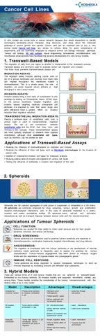
2. Spheroids
Spheroids in Media
Spheroids in Matrix
Coculture Spheroids
Spheroids are 3D cultured aggregates of cells grown in suspension or embedded in a 3D matrix. 3D spheroids are commonly employed for drug screening, tumour growth and proliferation research, immunological interactions, and, in the case of spheroids implanted in a matrix, invasion and matrix remodelling studies. 3D spheroids mimic cell-cell and cell-matrix interactions as well as transport features between tumour cells and the microenvironment.
Applications of Tumour Spheroids CELL FUNCTION: Spheroids are studied for their ability to mimic solid tumours and for their growth dynamics, structure, and tumour cell biology.
DRUG SCREENING: Cancer spheroids are commonly used to examine tumour sensitivity and response to chemotherapeutics, combination treatments, targeted chemotherapy, and drug delivery.
ANGIOGENESIS: The migration of endothelial cells into tumour spheroids or the development of vascular networks inside spheroids is frequently used to assess the potential for tumour vascularization. Tumour-induced angiogenesis has been shown to enhance oxygen levels and the expression of hypoxia-related and proangiogenic genes.
IMMUNE CELL RESPONSE: Tumor spheroids are being exploited to explore therapeutic techniques to elicit an immune response by increasing immune cell infiltration and cytotoxicity.
3. Hybrid Models There are various forms of in vitro tumour models that are not spheroid or transwell-based. Embedded ex vivo tumour sections, 3D invasion models, and avascular microfluidic models are among them. These models combine the complexities of the tumour microenvironment with the relative ease of an in vitro model.
Model Embeded ex vivotumor section
Description Primary tumor sections or biopsies embedded in gel
Advantages Maintains tumor heterogeneity
Disadvantages Lacks flow through vasculature
Patient-specific assay Mimics outgrowth into surrounding tissues
3D invasion models
Tumor cells or clusters embedded in a gel
3D microenvironment
Lacks
vasculature
Allows real-time tracking of cells
Lacks tumor complexity
Balance of complexity and experimental control Avascular microfluidic
Tumor cells grown in a 2D microfluidic device, typically for the study of migration
Simple migration assay
Lacks vasculature
Easy to isolate effect of variables
Typically lacks 3D environment
Allows real time-tracking of cells