International Research Journal of Engineering and Technology (IRJET)
e-ISSN: 2395-0056
Volume: 07 Issue: 02 | Feb 2020
p-ISSN: 2395-0072
www.irjet.net
AN AUTOMATED LEARNING APPROACH FOR DETECTION OF DIABETIC RETINOPATHY USING DEEP LEARNING S. Balaji1, Dr. R. Ramachandiran2, P. Karthikayan3, P. Udhayakumar4, V. Prathap5, 1,2Assistant
Professor, Department of Information Technology, Sri Manakula Vinayagar Engineering College, Puducherry 3Student, Department of Information Technology, Sri Manakula Vinayagar Engineering College, Puducherry 4Student, Department of Information Technology, Sri Manakula Vinayagar Engineering College, Puducherry 5Student, Department of Information Technology, Sri Manakula Vinayagar Engineering College, Puducherry
---------------------------------------------------------------------***---------------------------------------------------------------------
Abstract - Diabetic retinopathy (DR) is an across the board
issue for diabetic patient and it has been a primary explanation behind visual deficiency in the dynamic populace. A few troubles looked by diabetic patients in view of DR can be disposed of by appropriately keeping up the blood glucose and by auspicious treatment. As the DR accompanies various stages and differing challenges, it is difficult to DR and furthermore it is tedious. Right now, build up a computerized division based order model for DR. At first, the Contrast restricted versatile histogram evening out (CLAHE) is utilized for portioning the pictures. Later, deep belief network (DBN) is employed for classifying the images into different grades of DR. For exploratory investigation, the dataset is gotten from Kaggle site which is open source stage that endeavors to construct DR recognition model. The highest classifier performance is attained by the presented model with the maximum accuracy of 84.35 over compared models.
Key Words: Classification, DR, Segmentation, Deep 1. INTRODUCTION Diabetic retinopathy (DR) generally occur to patients who acquires diabetes for long time and because of retinal damage, it causes blindness[1]. By utilizing the strategy of fundus imaging, the DR influenced retinal structure of eyes may be recognized. By centering the eye, the fundus pictures will be commonly caught through fundus camera. The interior surface of eye is exhibited through fundus pictures which involve fovea, retina, veins, optic circle and macula[9]. A typical retina includes veins which conveys supplements and blood required for eye. Ordinarily, the veins are sensitive and in light of extra circulatory strain, they may barge in diabetic patients. Through additional small blood vessels count, the diabetic retinopathy progress because of additional pressure might be found from retinal surface
|
Impact Factor value: 7.34
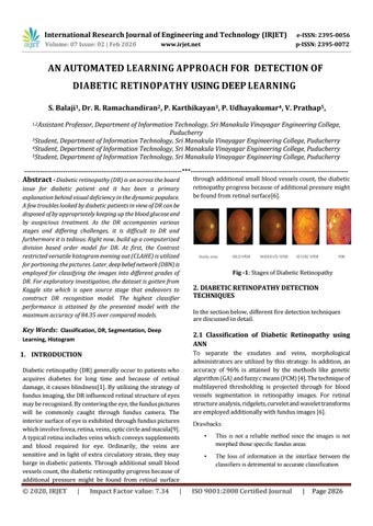
Fig -1: Stages of Diabetic Retinopathy
2. DIABETIC RETINOPATHY DETECTION TECHNIQUES In the section below, different fire detection techniques are discussed in detail.
2.1 Classification of Diabetic Retinopathy using ANN
Learning, Histogram
© 2020, IRJET
through additional small blood vessels count, the diabetic retinopathy progress because of additional pressure might be found from retinal surface[6].
|
To separate the exudates and veins, morphological administrators are utilized by this strategy. In addition, an accuracy of 96% is attained by the methods like genetic algorithm (GA) and fuzzy c means (FCM) [4]. The technique of multilayered thresholding is projected through for blood vessels segmentation in retinopathy images. For retinal structure analysis, ridgelets, curvelet and wavelet transforms are employed additionally with fundus images [6]. Drawbacks: •
This is not a reliable method since the images is not morphed those specific fundus areas.
•
The loss of information in the interface between the classifiers is detrimental to accurate classification.
ISO 9001:2008 Certified Journal
|
Page 2826