International Research Journal of Engineering and Technology (IRJET)
e-ISSN: 2395-0056
Volume: 07 Issue: 02 | Feb 2020
p-ISSN: 2395-0072
www.irjet.net
Breast Cancer Detection from Histopathology Images: A Review Shehras V U 1, Sindhu R 2 1PG
Scholar, ECE Department, NSS College of Engineering, Palakkad, Kerala, India Professor, ECE Department, NSS College of Engineering, Palakkad, Kerala, India ---------------------------------------------------------------------***--------------------------------------------------------------------2
Abstract - Breast cancer is the most prevalent form of
cancer among women. Early detection of breast cancer will increase survival rate. Automatic image analysis methods are necessary to decrease the workload among pathologists and to improve the quality of interpretation. Nuclei detection is the first stage of identifying the cells. Division of single cell and the spreading of cancer from one part to others in the human body can also detected from histopathological images. This paper provides a review on breast cancer detection from histopathology images. Key Words: Histopathology images, Cell nuclei, Metastasis, Mitosis, Convolutional Neural Network (CNN)
biological cells and tissues are known as histopathology. Histopathology is a Greek word in which Histo means “tissues”, Patho means “disease” and logo refers to “study”. Therefore, histopathology means the study of tissues for the identification of diseases. A combination of Hematoxylin and Eosin are the most commonly used stains in histopathology. Hematoxylin gets bound to Deoxyribo Nucleic Acid (DNA) and it dyes purple or blue color to the nuclei. Eosin gets bound to proteins, so it dyes pink color to other structures. These different stains will help to identify the nuclei of the cells.
1. INTRODUCTION Cancer is a cluster of diseases involving abnormal cell growth with the potential to invade or spread to other parts of the body known as malignant tumor. A benign tumor is a tumor that does not spread around the body. Breast cancer is a cancer that forms within the cells of the breasts. Breast cancer can occur in both men and women, but it is more commonly occur in women. Breast cancer is the most common type of cancer diagnosed in women after skin cancer. Almost all cancers can spread. The original tumor is called the primary tumor while the dispersed tumors are called metastatic tumors. Mitosis is a method where single cell divides in to two daughter cells. During mitosis, a cancerous cell makes an exact copy of it and splits into two new cells which are also cancerous. Metastasis is caused by the spread of cancer to other locations in the body. Most cancer deaths are due to metastasis. Early detection plays a key role in cancer detection and can improve long-term survival rates. Medical imaging is a very important technique for early cancer detection and diagnosis. Manual interpretation of enormous number of medical images can be tedious and time consuming and easily causes human bias and mistakes. Therefore, Computer Assisted diagnosis (CAD) systems were introduced to assist doctors in interpreting medical images to improve their efficiency. A biopsy is the physical examination under which a piece of sample tissue is taken out for microscopic examination. The sample is then referred to the laboratory where pathologist examines and analyses tissues under the microscope. This microscopic examination and study of © 2020, IRJET
|
Impact Factor value: 7.34
|
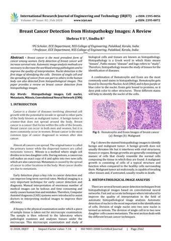
Fig-1: Hematoxylin and Eosin Images of breast cancer (a): Benign (b): Malignant Fig-1 shows the stained histopathology images to identify benign and malignant tumor. A benign growth does not usually threaten life but it interferes with vital structures, tissues or organs. Benign growths are generally consisting of masses of cells that closely resemble the normal cells composing the tissue in which they are found. A malignant growth is consisting of cells of a typical structure and function when compared to the healthy cells surrounding them. Malignant tumor act maliciously, is capable of invading other tissues and, if untreated, usually results in death.
2. HISTOPATHOLOGICAL IMAGE ANALYSIS There are several breast cancer detection techniques from histopathological images based on convolutional neural networks. Fast and accurate techniques where introduced to improve the quality of interpretation in the field of automatic histopathological image analysis. Automatic detection of nuclei is the most important in the identification of cells. Division of single nuclei into two new nuclei is termed as mitosis. The division of single cell in to two new daughter cells causes metastasis. The next section deals with the different breast cancer techniques.
ISO 9001:2008 Certified Journal
|
Page 2562