International Research Journal of Engineering and Technology (IRJET)
e-ISSN: 2395-0056
Volume: 07 Issue: 02 | Feb 2020
p-ISSN: 2395-0072
www.irjet.net
Analysis of Ophthalmic System Applications using Signal Processing Bharati. S. Sakore1, Khemutai Tighare2, Amita Suke2 1M.Tech,
2Asst.
Dept. of Computer Science & Enginnering, WCEM, Nagpur, Maharashtra, India Professor, Dept. of Computer Science & Enginnering, WCEM, Nagpur, Maharashtra, India
---------------------------------------------------------------***----------------------------------------------------------------
Abstract – The aim of this paper is to explore the potential for modern computing technology to advance clinical ophthalmology. In particular we will be investigating the potential for computer programming in the development of novel and objective measures of ophthalmic disease. Ophthalmic diseases like diabetic retinopathy, vision blur etc. have been causing issues with the society at large. Measures for evaluation are discussed in this paper. These measures could ultimately be used for clinical diagnosis or severity measures for clinical decision making. They could also be used as research tools, providing objective outcome measures to power more robust clinical trials.
irreversible blindness if not managed appropriately. Hyperglycaemia initiates a cascade of pathologic complications which eventually brings about devastating damages in the retina such as basement membrane thickening, loss of pericytes, blood retina barrier breakdown, etc. Clinically, DR is diagnosed by the presence of the following features (Table-1) [3]:
Key Words: Retinopathy, Clinical, Membrane, Hyperglycaemia, Macula.
Intraretinal haemorrhage
Table-1: Clinical features observed in DR Clinical feature Microaneurysms
Ophthalmology,
1. INTRODUCTION Soft exudates
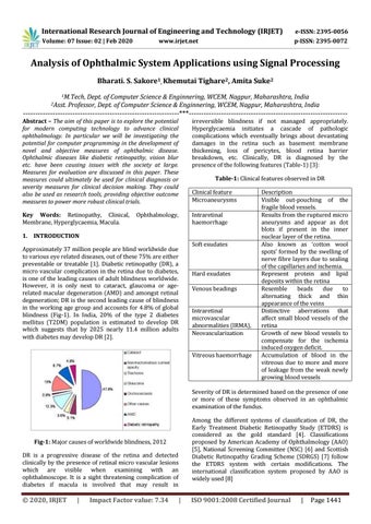
Approximately 37 million people are blind worldwide due to various eye related diseases, out of these 75% are either preventable or treatable [1]. Diabetic retinopathy (DR), a micro vascular complication in the retina due to diabetes, is one of the leading causes of adult blindness worldwide. However, it is only next to cataract, glaucoma or agerelated macular degeneration (AMD) and amongst retinal degeneration; DR is the second leading cause of blindness in the working age group and accounts for 4.8% of global blindness (Fig-1). In India, 20% of the type 2 diabetes mellitus (T2DM) population is estimated to develop DR which suggests that by 2025 nearly 11.4 million adults with diabetes may develop DR [2].
Hard exudates Venous beadings Intraretinal microvascular abnormalities (IRMA), Neovascularization Vitreous haemorrhage
Description Visible out-pouching of the fragile blood vessels. Results from the ruptured micro aneurysms and appear as dot blots if present in the inner nuclear layer of the retina. Also known as ‘cotton wool spots’ formed by the swelling of nerve fibre layers due to sealing of the capillaries and ischemia. Represent protein and lipid deposits within the retina Resemble beads due to alternating thick and thin appearance of the veins Distinctive aberrations that affect small blood vessels of the retina Growth of new blood vessels to compensate for the ischemia induced oxygen deficit. Accumulation of blood in the vitreous due to more and more of leakage from the weak newly growing blood vessels
Severity of DR is determined based on the presence of one or more of these symptoms observed in an ophthalmic examination of the fundus. Among the different systems of classification of DR, the Early Treatment Diabetic Retinopathy Study (ETDRS) is considered as the gold standard [4]. Classifications proposed by American Academy of Ophthalmology (AAO) [5], National Screening Committee (NSC) [6] and Scottish Diabetic Retinopathy Grading Scheme (SDRGS) [7] follow the ETDRS system with certain modifications. The international classification system proposed by AAO is widely used [8]
Fig-1: Major causes of worldwide blindness, 2012 DR is a progressive disease of the retina and detected clinically by the presence of retinal micro vascular lesions which are visible when examining with an ophthalmoscope. It is a sight threatening complication of diabetes if macula is involved that may result in
© 2020, IRJET
|
Impact Factor value: 7.34
|
ISO 9001:2008 Certified Journal
|
Page 1441