Enhancing Lung Cancer Detection with Deep Learning: A CT Image Classification Approach
JEEVIKA K S1, DR. SAVITHA S K21PG Student, Dept. of Computer Science Engineering, Bangalore Institute of Technology, Bengaluru, India
2Professor, Dept. of Computer Science Engineering, Bangalore Institute of Technology, Bengaluru, India

Abstract - Lung cancer is a highly perilous illness ranking as one of the primary causes of disease and death, particularly when diagnosed in its initial stages. It presents significant challenges, as it is often only discernible after it has already diffused. This study proposes a lung cancer prognostication framework that uses deep learning to enhancetheaccuracyof cancer forecasting and disease determination, thereby enabling personalized treatmentapproachesbasedon disease severity. It consists of various steps, including image preprocessing and segmentation of lung CT image features extracted from the segmented images. Three differentmodels, namely a DCNN model, a DCDNN model, and an ANN model, were employed for image classification, and a deep convolutional neural network (DCNN)wasemployedtodetect lung diagnosis based on the extracted feature evaluation results showing the best accuracy of 99.41% in accurately discerning the presence or absence of lung cancer. The GAN model generates realistic lung CT scan images by training a generator to produce authenticimages, andadiscriminatorto distinguish between real and fake images. The outcome of the system depends on the quality of the data, and a well-trained DCNN through training, validation, and testing on diverse datasets is crucial to ensure thereliabilityand generalizability of the model.
Key Words: Lungcancerdetection,Deeplearning,Deep convolutionalneuralnetwork.
1. INTRODUCTION
Lung cancer is a complex and heterogeneous ailment characterizedbyariseinthenumberofcellsinlungtissues. This stands as the main reason for most cancer-related deaths worldwide, accounting for a sizable proportion of cancer-associated morbidity and mortality. Detection assumes a crucial role in identifying the existence of lung canceratanearlystageandfacilitatingtimelytreatment.This study focuses on the development and improvement of detectionmethodsusingdifferenttechniques.
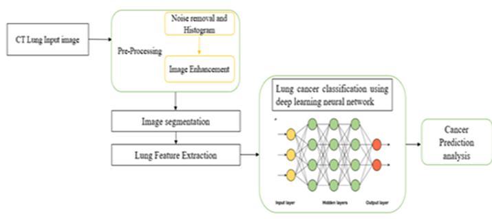
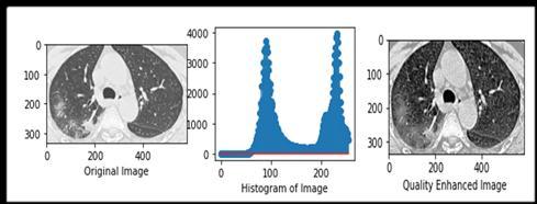
This study provides an input CT image for image preprocessing,whichincludesgrayscaleconversionfornoise removal, histogram calculation, and image quality enhancement for more clearly visible images after image enhancement.
The second step is image segmentation, which detects the edge using Canny edge detection, and lung segmentation techniques using K-means clustering to separate the background and foreground. Morphological operations to refine the lung mask. The original image binary threshold image eroded, dilated color-labeled image-segmented lung mask, and segmented lung area in the original image are displayed. After the segmentation lung feature extraction process, which is not displayed in the system, the model directlyunderwentanalysisassessment.
In the third step, the classification model was trained and evaluated.Collectadatasetoflungimageswithlabelsnormal or cancer and split the dataset into three sets. In the evaluationprocess,itprovidesaccesstothetraininghistory toretrievethetrainingandvalidationaccuracy,andtheloss valuesareplottedusingMatplotlib.Themodelsusedinthis study were the DCNN, DCDNN, and ANN. Every model exhibited good accuracy and loss percentage and plotted graphs.Furtherdetailsareprovidedinthefollowingsections. Inthefourthstep,theGANmodelpredicts,thestructureof the image batch, class names, labels, and filenames in this understood.
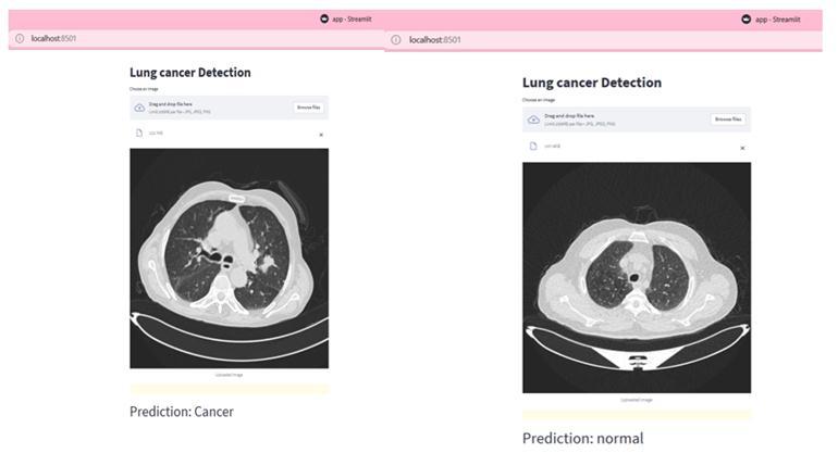
Duringtheultimatestep,theStreamlitappforlungcancer detectionallowsuserstouploadimagesandpredictcancer. User-friendlyinterfacethatinteractswithanapplicationona webbrowser.
1.1 Aims and Objectives
1) Theaimofthisprojectistoenhancetheprecision andeffectivenessoflungcancerdiagnosisbyadvanced deeplearningmethodologies.
2) Todevelopanapplicationthatdetectsandproperly classifiesLungcancerinCTscanimagesusingaDCNN.
Image preprocessing plays a fundamental role in the accurateanalysisofmedicalimages,particularlyindetecting malignancyusingCTscans.Thissectionfocusesonthekey preprocessingsteps:greyconversion,histogramanalysis,and imagequalityenhancement.Grayconversionsimplifiesthe imagebyconvertingittograyscale,therebyfacilitatingthe subsequentprocessing.Histogramanalysisprovidesinsights intothepixelintensitydistribution,aidinginthethreshold determination for image segmentation. Image quality enhancementtechniquesenhancevisibilityandreducenoise and artifacts. The cumulative distribution function is normalizedandscaledtotherange[0,25].Byimplementing these preprocessing steps, CT images were optimized for accuratelungcancerdetection,ensuringimprovedanalysis andmorereliableresults.
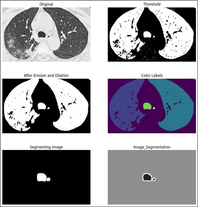
multiplesubregions,allowingthepredictionandlocalization oftheaffectedarea.Thisisaccomplishedbyanalyzingthe similarity of pixel values and grouping superpixels that exhibitsimilarcharacteristics.Thesegmentationalgorithm effectivelyidentifiestheoutlineofthemalignantregionby detectingabruptchangesinpixelvalues.Pixelsimilarityisa fundamental attribute in image segmentation and critical analysis of visual data in accurately shaping clusters. Through quantitative evaluation, the algorithm determines thelevelofsimilaritybetweenpixels,facilitatingthecorrect divisionofthecancerousregion.Thisevaluationconsidered theenhancedqualityoftheimagepixels,ensuringthatthe segmentationprocesswasperformedeffectively.

An undirected graph was constructed to represent the abnormalregionsinthelungimage.Thisgraphprovidesa visual representation of the segmented areas, aiding the identificationandanalysisofcancerousregions.
Image segmentation is an important step in this study. It involves precise identification and separation of the cancerousregionwithintheenhancedtomographyimages. Toachievethis,thek-meansalgorithmistypicallyemployed.
The segmentation process begins by examining the pixel similarityinthelungimages.Thepicturewaspartitionedinto

C. Image Classification
The final stage involves the recognition of malignant. DetectionwasperformedutilizingalgorithmswithCTscan image input. Dataset of lung images with labels indicating cancer or not. After splitting the dataset into training, validation,andtestsetsasafolder.Thealgorithmsusedare deep convolutional neural network, double convolutional deepneuralnetwork,andartificialneuralnetwork.
At this stage, The Generative Adversarial Network (GAN) modelactsasadiscriminator,taskedtodistinguishbetween real and synthetic images, with the ultimate goal of producing life-like images capable of misleading discriminators Throughtraining theGAN model enhances thefidelityofchestCTscanimageswhichwerethensaved and displayed. The saved model is readily deployable for furtheranalysis,evaluation,orapplication-specifictasks.
Aftertheuseruploadsanimageofthelungtissue,theimage is preprocessed and passed through a pre-trained model. Themodelpredictstheprobabilityofanimagebelongingto each class: "CANCER" or "NORMAL.” The class with the highestprobabilitywasconsideredthepredictedclassfor theinputimage.Thepredictedclasswasthendisplayedon the web interface to indicate whether the image was classifiedascancerousornormallungtissue.
Itshouldbeemphasizedthattheaccuracyandreliabilityof cancer identification depend on the excellence of the pretrainedmodel,thedatasetonwhichitwastrained,andthe diversity and quality of the images used for testing and evaluation.Theperformancecanvarybasedonthesefactors, andawell-validatedandreliablemodelisrecommendedfor theaccurateidentificationoflungcancer.
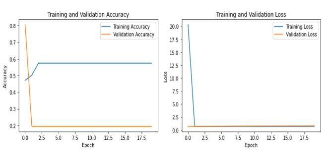
meansclustering,Figure(5)showsthedetectedcancer or not, Figure (6) DCNN (7) DCDNN (8) ANN shows the Accuracyandlossgraphfortrainingandvalidation.
A. Table 1: Accuracy analysis
Figure 5: Detectionofcancerornot
3. RESULTS
Thispaperworkfindsthatimagepreprocessingshowsthe resultsinFigure(2)showstheimagequalityenhancement, Figure (3) shows the edge detection using the canny edge detection,Figure(4)showsthelungsegmentationusingk-
B. Graph for Accuracy and Loss (training and validation)
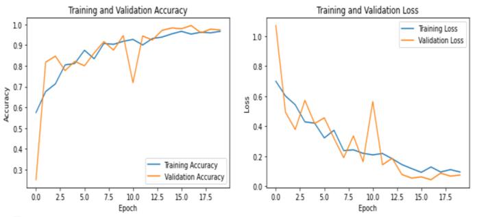
A Deep convolutional neural network model consists of multiple convolutional, pooling, and dense layers. After training, validation, and testing, the data were obtained. Trainingthemodelsavesit,evaluatesitsperformanceusing accuracymetrics,andplotsgraphs.
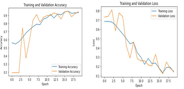

A Double convolutional deep neural network it consists of convolutionalandpoolinglayers,followedbyfullyconnected layers. After training, validation, and testing, the data were analyzed.Trainingthe modelsavesthemodel,evaluatesits performanceusingaccuracymetrics,andplotsthetraining andvalidationaccuracyandlosscurves.
An Artificial neural network in this model consists of a flattenedinputlayer,adensehiddenlayer,andanoutputlayer. Aftertraining,validation,andtestingdata.Trainingthemodel
savedevaluatesitsperformanceusingtheaccuracymetrics, andplotsitisshowninbelowfigure8.
Convolutional Neural Network (3D-CNN)”, in Proceedings of (IJACSA) International Journal of AdvancedComputerScienceandApplications,Volume. 8,2017,pp409-417https://www.ijacsa.thesai.org/
[4]. Prathyusha Chalasani, S Rajesh.: “Lung CT Image RecognitionusingDeepLearningTechniquestoDetect Lung Cancer” .in International Journal of Emerging TrendsinEngineeringResearch.,Volume8.No.7,July 2020, ISSN 2347–3983, https://doi.org/10.30534/ijeter/2020/113872020
[5]. Rasika N. Kachore, Kivita Singh: “Lung Nodule Detection”, in the International Journal of Scientific Research in Science, Engineering and Technology, IJSRSET, Volume 3, Issue 2, ISSN: 2395-1990, Online ISSN:2394-4099.

In conclusion, this paper developed using deep learning techniquesshowedpromisingresults.Themainprediction ofthepresenceorabsenceoflungcancerisachievinghigh accuracyandenablingpersonalizedtreatmentapproaches based on disease severity. The ability of the system to classifyCTimagesprovidesavaluabletoolfordetection.The system development begins with image preprocessing, segmentation, and image classification using different algorithmsormodels,includingDCNN,DCDNN,andANN. In this model, the evaluation results show that the best accuracy is achieved by the DCNN, which yields 99.41% accuracyanddetectslungcancer.
Future research should prioritize the early detection and explorationofthestageofcancer.Integrationwithadvanced medicalimagingtechnologiesandclinicaldatacouldalsobe exploredtoaugmentthepredictivecapabilitiesofthesystem andenablemorecomprehensivelungcanceranalysis.
REFERENCES
[1]. Heng Yu, Zhiqing Zhou, And Qiming Wang: “Deep LearningAssistedPredictofLungCanceronComputed Tomography Images Using the Adaptive Hierarchical Heuristic Mathematical Model” In the IEEE Access, Volume 08 2020, pp 86401-86411, Digital Object Identifier10.1109/ACCESS.2020.2992645
[2]. Mohammad A. Alzubaidi, Mwaffaq Otoom ,(Senior Member,Ieee),AndHamzaJaradat,“Comprehensiveand Comparative Global and Local Feature Extraction Framework for Lung Cancer Detection Using CT Scan Images”, In the IEEE Access, Volume 9, 2021, pp 158140- 158154, Digital Object Identifier 10.1109/ACCESS.2021.3129597
[3]. Wafaa Alakwaa, Mohammad Nassef, Amr Badr, “Lung Cancer Detection and Classification with 3D
[6]. Yaozhi Lu, Shahab Aslani, Mark Emberton, Daniel C. Alexander, And Joseph Jacob: “Deep Learning-Based Long Term Mortality Prediction in the National Lung ScreeningTrial”,intheproceedingsofIEEEAccess,Year – 2022, VOLUME 10, 2022, pp: 34369-34378, Digital ObjectIdentifier10.1109/ACCESS.2022.3161954
[7]. B. Keerthi Samhitha, Suja Cherukullapurath Mana, Jithina Jose, R.Vignesh, D. Deepa: “Prediction of Lung CancerUsingConvolutionalNeuralNetwork(CNN)”,in Proceedings of the International Journal of Advanced TrendsinComputerScienceandEngineering,Volume9, No.3, 2020,ISSN 2278-3091, https://doi.org/10.30534/ijatcse/2020/135932020
[8]. Surbhi Vijh, Prashant Gaurav, Hari Mohan Pandey: “Hybridbio-inspiredalgorithmandconvolutionalneural network for automatic lung tumor detection”, in the springer, Year of publication 19 September 2020, https://doi.org/10.1007/s00521-020-05362-z
[9]. Dr. M. Sangeetha, P Sangeetha, P. Pavithra: “Classification of Lung Cancer using Deep Learning Algorithm”,intheConferenceproceedingsInternational JournalofEngineeringResearch&Technology(IJERT) ISSN:2278-0181Publishedby,www.ijert.orgRTICCT2020.
[10]. S.Perumal,T.Velmurugan,“Lungcancerdetection and classification on CT scan images using enhanced artificialbeecolonyoptimization”,intheInternational Journal of Engineering & Technology Website: www.sciencepubco.com/index.php/IJET, Volume 7, Year-2018,pp74-79.
[11]. OnurOzdemir,Member,IEEE,RebeccaL.Russell, andAndrewA.Berlin,Member,IEEE“A3DProbabilistic Deep Learning System for Detection and Diagnosis of Lung Cancer Using Low-Dose CT Scans”, in the IEEE
TRANSACTIONS ON MEDICAL IMAGING, Volume. 39, NO.5,MAY2020,pp:1419-1429
[12]. Khevna Vasani, Ayushi Shah: “Lung Cancer Detection Using CT Scan Images”, in the International Research Journal of Engineering and Technology (IRJET), e-ISSN: 2395-0056 Volume: 08 Issue: 04 Apr 2021www.irjet.netp-ISSN:2395-0072

[13]. LongxiZhou,ZhongxiaoLi,JuexiaoZhou,Haoyang Li,YupengChen,YuxinHuang,DexuanXie,LintaoZhao, Ming Fan , Shahrukh Hashmi, Faisal Abdelkareem , Riham Eiada , Xigang Xiao, Lihua Li, “Adaptive Local TernaryPatternonParameterOptimized-FasterRegion Convolutional Neural Network for Pulmonary Emphysema Diagnosis”, in the IEEE Access, Volume 9,2021, pp:114135-114152, DOI: 10.1109/ACCESS.2021.3105114
[14]. ChanunyaLoraksa,SirimaMongkolsomlit,Nitikarn Nimsuk, Meenut Uscharapong, and Piya Kiatisevi: “Development of the Osteosarcoma Lung Nodules DetectionModelBasedonSSD-VGG16andCompetency ComparingwithTraditionalMethod”,intheIEEEAccess, VOLUME 10, 2022, pp: 65496-65506, Digital Object Identifier10.1109/ACCESS.2022.3183604
[15]. Aishwarya Kalra, Brijmohan Singh, Himanshu Chauhan,“AnApproachforLungCancerDetectionusing DeepLearning”,intheInternationalResearchJournalof EngineeringandTechnology(IRJET),e-ISSN:2395-0056
Volume:07,Issue: 09Sep 2020www.irjet.net p-ISSN: 2395-0072
[16]. P. Mohamed Shakeel, M. A. Burhanuddin, Mohammad Ishak Desa, “Automatic lung cancer detection from CT image using improved deep neural network and ensemble classifier”, published in the springer pp:349579–9592 https://doi.org/10.1007/s00521-020-04842-6
[17]. Zhang Li, Jiehua Zhang, Tao Tan, Xichao Teng, XiaoliangSun,HongZhao,LihongLiu,YangXiao,“Deep Learning Methods for Lung Cancer Segmentation in Whole-SlideHistopathologyImages-TheACDC@LungHP Challenge2019”,intheIEEEJournalOfBiomedicalAnd HealthInformatics,Volume.25,No.2,February2021
[18]. LuluWang:“DeepLearningTechniquestoDiagnose LungCancer”,intheMDPI,Volume14,Issue22,DOI: 10.3390/cancers14225569
[19]. ImdadAli,MuhammadMuzammil,(Member,IEEE), IHSAN UL HAQ, (Member, IEEE), AMIR A. Khaliq, (Member, IEEE), “Efficient Lung Nodule Classification Using Transferable Texture Convolutional Neural Network”, In the IEEE Access, Volume 8, 2020, pp:
175859- 175870, Digital Object Identifier 10.1109/ACCESS.2020.3026080,Yearofpublication–2020
[20]. LongxiZhou,ZhongxiaoLi,JuexiaoZhou,Haoyang Li, Yupeng Chen, Yuxin Huang, Dexuan Xie,Lihua Li , Member,IEEE,ZhaowenQiu,Member,IEEE,andXinGao, Member,IEEE,“ARapid,AccurateandMachine-Agnostic SegmentationandQuantificationMethodforCT-Based COVID-19 Diagnosis”, in the IEEE Transactions On MedicalImaging,Volume.39,NO.8,AUGUST2020,pp: 2638-2652
