PLANT DISEASE IDENTIFICATION FROM INDIVIDUAL LESIONS AND SPOTS
Sumaya Basheer1 , Er Satnam Singh21Pg Scholar, Department of Electronics and communication Engineering, Sri Sai College of Engineering and Technology
2HOD& Assistant Professor, Department of Electronics and communication Engineering, Sri Sai College of Engineering and Technology ***
Abstract - The current framework the ranchers are utilizing for the location of illnesses in the plants is that they could be distinguished through the unaided eye and there information about plant sickness. For doing as such, on enormous number of plants is tedious, troublesome and exactness isn’t acceptable. Counseling specialists is of extraordinary expense. In such sort of conditions to improve the exactness rate and make it more helpful proposed methods are executed where gadgets are utilized for the programmed location of the illnesses that makes the interaction less expensive and simpler. Serious level of the intricacy is joined by noticing the manifestations on the plant leave optically where the plant infection could be effectively analyzed. Presently days the vast majority of the agro help focuses and numerous ranchers utilize various sorts of innovation to improve creation in agribusiness. The main wellspring of energy is plants. Plants are frequently inclined to infections which may cause social and monetary misfortunes. Numerous infections are at first spotted on the leaves of the plants. It could prompt more damage if the sickness isn't recognized in the primary stage. By recognizing the shading highlights of the Leaves picture handling helps in the identification of the sicknesses and furthermore gives anticipation to the specific diseases. At the absolute first stage the picture is divided by snapping the photo of the plant in the RGB structure and later the green pixel is withdrawn. Surface insights is a course of action of powers in a locale that is utilized for the extraction by the fragments is at last finished and the illness counteraction is given by the investigation.
Key Words: plant,diseases,leafspots,apple.,

1.INTRODUCTION
The agricultural industry has historically been India's most significant economic driver. Agriculture is crucial because of its impact on the global food supply, capital creation, raw materials for manufacturing, consumer demand for manufactured goods, and international trade. Although agriculture's share of the economy is shrinking, it's still the major source of employment in most places, but with some variation as the population grows. It is imperative that we quicken the pace toward agriculture
thatiscompetitive,productive,varied,andsustainable[1, 2].
TherearethreekeyissuesfacingIndianagriculturetoday: increasingagriculturaloutputperacreofland,eliminating ruralpovertythroughacomprehensivesocialprogramme, and ensuring that agricultural expansion responds to the country's growing food security demands. India enjoys a diverse array of climatic and geographical environments thankstoitsfortunategeneticinheritance[3].
1.1 Apple: Diseases and Symptoms
Apple scab, marsonina coronaria, black rot canker, powdery mildew, apple mosaic, and other viral diseases, and alternaria leaf spot are only a few of the many diseasesthatprimarilyattackappleleaves.Herearesome of the illnesses that might strike an apple orchard and whatsignstolookoutfor.
1.2 APPLE SCAB
Apple scab disease, caused by ventura inaequalis virus, is commonly found on the plant's leaves and fruit. The leavesofinfectedplantstwistanddevelopblack,globular patchesontheir top surfaces.The dotsare hairyandmay combinetocoverthewholeundersideoftheleaf.Asevere illnessmaycausetheleavestobecomeyellowandfalloffa plant. Flowers can wilt and die if scab infects their stems. Intime,thesoreswill recedeandturnbrownwithspores dottingtheiredges.Applesafflictedwithadiseasedeform and may crack, allowing secondary viruses to enter the crop. Ventura inaequalis ascospore production is stimulated by conditions of adequate temperature and humidity.
Thesymptomsoftheillnessappearasdarkgreen,circular spotsontheuppersurfaceoftheleaf,ranginginsizefrom 5 to 12 millimetres in diameter, and eventually becoming much darker. In its advanced stage, it spreads on the underside of apple leaves. On the leaf's surface, you can see a few tiny, black acervuli. When there are many differentkindsofspots,theyblendtogethertoformlarge, dark, shadowy patches, and the areas around them turn yellow. Figure 1.5 shows a marsonina coronaria-infected leaf.Highprecipitationandtemperaturesbetween20and 22degreesCelsiusaidthespreadofthisillness
degradedsectiongrows.Cankersformduetobarklesions causedbyeithervirusesorbacteria.Cysts(ascospores)in decomposing organic matter in the soil provide the pathogenwithameansofsurvivalandserveasasourceof secondaryinfection
Indicators of leaf diseases typically appear early in the spring, when leaves begin to unfurl. Apple leaves develop little purple spots on the upper surface that eventually turn into globular lesions between 3 and 6 mm in size. Spotshaveapurpleborderthatfadestotanandbrownin the middle. These leaf patches will develop a secondary growth phase in a matter of weeks. When a plant'sleaves get severely infected, they turn chlorotic and eventually fall off. A series of uniformly sized concentric rings that progress in colour from black to brown form as the
2. METHODOLOGY
Thisstudyisexperimental.Theliteraturesurveyexamines agricultural image processing methods and applications. Experts discuss and choose crops and illnesses. Apple leaves were chosen because apple scab and marsonina, coronaria illnesses are reducing agricultural yields in Jammu and Kashmir, India. After the conversation, apple farmsinbothstatesaresampled.

Experts from agricultural and horticultural institutions discuss the samples. Image processing technologies identify and describe collected pictures. Algorithms segment and classify damaged apple leaves. Finally, severalparametersevaluatethealgorithm'sperformance.
2.1 FIELD SURVEY FOR APPLE FIELDS
Jammu & Kashmir intensively study apple leaves. sopore, shopain,andpulwamafieldsinweresurveyed.Applescab and marsonina coronaria are the main apple plant diseases, according to the report. Apple scab disease is prevalent & marsonina coronaria. June and July saw the field survey. In daylight, an 8-megapixel Smartphone cameracapturessickappleleaves.

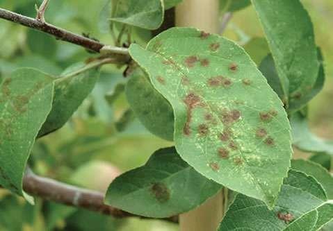
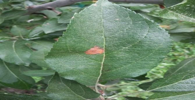
Three different types of leaves, healthy leaves, apple scab diseased leaves and marsonina coronaria diseased leaves have been surveyed and captured with live backgrounds andblackandwhitebackground
2.2 DATA COLLECTION
When a farmer may inspect the field, apple leaf photographsarecapturedinnaturallight.Live,white,and black apple leaf photos are taken. 8-megapixel smartphone cameras produce RGB. color photographs of healthy and sick apple leaves. Digitized photos average 225 KB. June and July collect. 70% of the data base trains the system, 30% tests it. The computer stores all data in JPEGformat.
3. PROPOSED WORK
Researchhaspresentedauniquemethodforimprovement and automatic segmentation of apple leaves illnesses to increase disease detection accuracy. Recommended research technique. Pre-processing, processing, and postprocessingcompriseresearchtechnique.

3.1 PREPROCESSING
Gaussianfilterisusedtocoloredappleleafpictureinpreprocessing. Section 3.6.1 introduces binary preserving dynamic fuzzy histogram equalization (BPDFHE), which enhances the filtered picture. Lastly, performance measures are used to compare the suggested improvementapproachtocurrentstrategies.
3.2 PROCESSING
A unique algorithm segments sick parts of the improved appleleafduringprocessing.Segmentedregionofinterest (ROI) extracts diseased component characteristics to effectivelyclassifyappleillnessesinpost-processing.Then several performance indicators are used to compare the recommendedsegmentationapproachtothecurrentones. Classifying ROI characteristics follows segmentation. Textural characteristics (first- and second-order) are extracted here.1st-order features include mean, variance, skewness, and kurtosis, whereas 2nd-order texture characteristicsincludeGLCMfeatures.
3.3 POST PROCESSING
Four apple scab and marsonina coronaria classification methods are used in post-processing. The most effective method was k closest neighbour, followed by support vector machine, naïve bayes, and decision tree. Several performancemeasurescomparefourclassifiers.
4. IMAGE FILTERING AND ENHANCEMENT
Farmers and anyone interested in apple production may use the proposed algorithm to segment and classify marsonina coronaria and apple scab disease in apple leaves without waiting for specialists. Preprocessing detects apple pathogens. It increases localization and segmentation of damaged apple picture characteristics,
making it crucial for automated apple leaf disease identification.Gaussianfilterisusedtocolouredsickapple leafpictureinpre-processing.Binarymaintaineddynamic fuzzyhistogramequalisationenhancesthefilteredpicture
4.1 IMAGE ACQUISITION AND IMAGE RESIZING
Theinitialstageintheprocessofdevelopingthesystemto capture a picture of a damaged apple leaf is image acquisition. The RGB colour pictures of healthy and sick apple leaves were taken using the digital camera of a cell phone,whichhadaresolutionof8megapixels.Eachofthe digitisedpictureshasasizeofaround225KB.Atotalof20 data samples are gathered, all of which comprise either healthy leaves or leaves that have been afflicted by either applesscabormarsoninacoronaria.Theimagesthathave been utilised for the diagnosis of illnesses all include one of three distinct kinds of backgrounds: a live backdrop, a black background, or a white background,. JPEG is the format used to store the images. The image processing libraryfromMATLABisusedintheprototype.
4.2 IMAGE EHANCEMENT
Imageenhancementisperformedaftertheimagehasbeen filtered, and it is this procedure that increases the reflectivityofinformationortheinterpretabilityofimages for the human eye, in addition to offering additional improvements to the image processing operations. In the workthathasbeenpresented,animprovementtechnique known as binary preserved dynamic fuzzy histogram equalization (BPDFHE) has been introduced for the purpose of enhancing the contrast of the filtered image. Following this, an evaluation of performance indicators is carried out in order to compare the newly proposed enhancement methodology with previously developed enhancement methods. The following paragraph will provide an in-depth analysis of the technique that will be offered, followed by a concise analysis of the techniques thatarecurrentlyinuse.
4.3 SVM CLASSIFICATION
The supervised machine learning approach known as Support Vector Machine (SVM) can be used for both classification and regression. Although we also refer to regression problems, classification is where it really shines. The goal of the Support Vector Machine (SVM) technique is to locate, in an N-dimensional space, a hyper plane that may be used to classify the data points with highaccuracy.Thehyperplane’ssizeisproportionaltothe total number of features. If there are just two input characteristics, the hyper plane is a straight line. When therearejustthreeinputfeatures,thehyperplaneflattens outintotwodimensions.Whentherearemorethanthree distinguishingtraits,visualizationgetsproblematic.
Here we have a blue circle and a red circle serving as our dependent variables, and x1 and x2 as our independent variables.

genetic algorithm for disease diagnosis in plant leaf imagesissegmentation.
5.1 SIMULATION RESULTS
MATLABisusedforeverysingleexperiment.Diseasedleaf samples from plants including roses, beans, lemon trees, bananatrees,andevenbeanswithearlyscorchandfungal disease are taken into account as input data.we see the inputphotos,followedbythesegmentedresults.Different plant diseases can be identified from segmented images. Theappleleafwithearlyscorchdiseaseservesasboththe inputandtheoutputimage,whilethecategorizationofthe disease using the feature extraction approach serves as theoutput.
Diseases on other input plants can be categorized in the similarway.

From the figure above it’s very clear that there are multiple lines (our hyperplane here is a line because we are considering only two input features x1, x2) that segregate our data points or do a classification between redandbluecircles.
5. RESULT AND DISCUSSION
The agricultural sector's output is crucial to the health of the economy. Since plant diseases are a fact of life, it stands to reason that the ability to detect them would be usefulintheagriculturalsector.Thequality,quantity,and productivity of plants are all negatively impacted if this areaisnotproperlycaredfor.Onedangerousdiseasethat affects apple trees in Kashmir is called small leaf disease. Plant disease detection by some automatic technology is helpful because it eliminates the need for constant monitoringinlargecropfarmsandcatchessignsofillness as soon as they manifest on the leaves of plants. An algorithm for an image segmentation technique is presented in this study for the purpose of automating the process of identifying and categorizing leaf diseases in plants. Also included is a review of the many disease classificationmethodsthatcanbeappliedtothedetection of plant leaf diseases. An essential part of applying a
After mapping the R, G, and B components of the input imagetothethresholdimages,theco-occurrencefeatures can be computed. The extracted leaf co-occurrence featuresarethencomparedtotheircorrespondingfeature values in the feature library. First, we use K-Means Clustering to classify the data, and the Minimum Distance Criterion seems to be an effective method with an accuracyof86.54%.
1. Place the folder 'Leaf_Disease_Detection_code' in the Matlab path, and add all the subfolders into that path.
2.Run Detect Disease GUI.M
3.IntheGUIclickonLoadImageandloadtheimagefrom Manu's Disease Dataset, click Enhance Contrast.
4. Next click on Segment Image, then enter the cluster no containingtheROI,i.eonlythediseaseaffectedpartorthe healthy part.
5. Click on classification results. Then measure accuracy (InthiscaseHealthyvs.Alldiseases).
5.2 DATA SET RESULTS
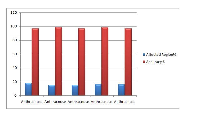
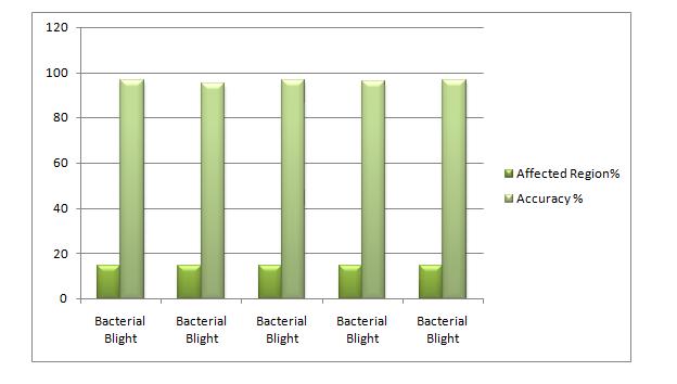
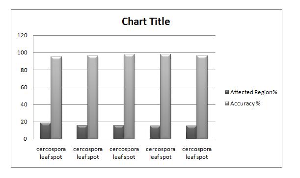
Theistdatasetistestingofleafsusingmatlabarehealthy leaf and alternaria alternate. From the above the results aretabulatedasfollows



6. CONCLUSION
Apple is a crucial product for India's economy, which has played a significant role in our economy in the past, but arenotasplentifultodaybecausetodifferentdiseasesthat affect the crops.Thankfully, more people are becoming awareoftheimportanceofapplefarming,andwhatbetter way to raise awareness of it than by providing a convenience that any farmer can utilise on their smartphone. Since we live in a digital age, we should utilise technology as much as possible across all industries. Machine learning was used as a result. The primary focus of the proposed work is on the apple illnessesmarsoninacoronariaandapplescab,whichyield varied outcomes for disease prediction using artificial intelligence.
Analgorithmforimagesegmentationapproachthatcanbe utilised for automatic detection and classification of plant illnesses, as well as a survey on several diseases classification techniques for plant leaf disease detection, are presented in this study later leaf diseases. On ten different species, including the apple, banana, bean,

jackfruit,lemon,mango,potato,andtomato,theproposed algorithm is tested. As a result, samples of associated diseasesfortheseplantsweretakenforanalysis.Thebest results were attained with a minimum amount of computing work, demonstrating the effectiveness of the suggested algorithm in the identification and categorization of leaf diseases. Utilizing this technology also has the benefit of allowing for the early or first detection of plant diseases. Artificial Neural Networks are usedtoclassifydataandincreaserecognitionrates.
REFRENCES

3. M. Schikora and A. Schikora, ―Image-based Analysis to Study Plant Infection with Human Pathogens,‖ Computational and Structural Biotechnology Journal. 2014.
4. H.M.Asraf,M.T.Nooritawati,andM.S.B.S.Rizam,―A comparative study in kernel-based Support Vector Machine of oil palm leaves nutrient disease,‖ in Procedia Engineering,2012.
5. J.D.Pujari,R.Yakkundimath,andA.S.Byadgi,―Image processing Based Detection of Fungal Diseases in Plants,‖ Procedia Comput. Sci., vol. 46, no. Icict 2014, pp.1802–1808,2015.
6. Y. Atoum, M. J. Afridi, X. Liu, J. M. McGrath, and L. E. Hanson, ―On developing and enhancing plant-level disease rating systems in real fields,‖ Pattern Recognit.,vol.53,pp.287–299,2016.
7. E. Hamuda, M. Glavin, and E. Jones, ―A survey of imageprocessing techniquesforplant extractionand segmentation in the field,‖ Comput. Electron. Agric., vol.125,pp.184–199,2016.
8. B. Tiger and T. Verma, ―Identification and Classification of Normal and Infected Apples using NeuralNetwork,‖vol.2,no.6,pp.4–7,2013.
9. S. D. Khirade and A. B. Patil, ―Plant disease detection usingimage processing,‖ Proc. - 1st Int. Conf. Comput. Commun. Control Autom. ICCUBEA 2015, pp. 768–771, 2015.
10. Y. Lu and R. Lu, ―Histogram-based automatic thresholding for bruise detection of apples by structured-illumination reflectance imaging,‖ Biosyst. Eng.,vol.160,pp.30–41,2017.
11. Z. Wang, K. Wang, Z. Liu, X. Wang, and S. Pan, ―A Cognitive Vision Method for Insect Pest Image Segmentation,‖ IFAC-PapersOnLine,vol.51,no.17,pp. 85–89,2018.
12. N. Krithika and A. Grace Selvarani, ―An individual grape leaf disease identification using leaf skeletons and KNN classification,‖ Proc. 2017 Int. Conf. Innov. Information, Embed. Commun. Syst. ICIIECS 2017, vol. 2018–Janua,pp.1–5,2018.
13. J.G.ArnalBarbedo,―Plantdiseaseidentificationfrom individual lesions and spots using deep learning,‖ Biosyst. Eng.,vol.180,no.2016,pp.96–107,2019.
14. X. E. Pantazi, D. Moshou, and A. A. Tamouridou, ―Automated leaf disease detection in different crop speciesthroughimagefeaturesanalysisandOneClass
Classifiers,‖ Comput. Electron. Agric., vol. 156, no. November2018,pp.96–104,2019.
15. M. Sardogan, A. Tuncer, and Y. Ozen, ―Plant Leaf Disease Detection and Classification Based on CNN with LVQ Algorithm,‖ UBMK 2018 - 3rd Int. Conf. Comput. Sci. Eng.,pp.382–385,2018.
16. J. K. Patil, R. Kumar, B. Vidyapeeth, C. O. E. Kolhapur, andB.Vidyapeeth,―AdvancesinImageProcessingfor Detection of Plant Diseases,‖ J. Adv. Bioinforma. Appl. Res.,vol.2,no.2,pp.135–141,2011.
17. A. B. Payne, K. B. Walsh, P. P. Subedi, and D. Jarvis, ―Estimationofmangocropyieldusingimageanalysis - Segmentation method,‖ Comput. Electron. Agric.,vol. 91,pp.57–64,2013.
18. Y. K. Dubey, M. M. Mushrif, and S. Tiple, ―Superpixel based roughness measure for cotton leaf diseases detection and classification,‖ Proc. 4th IEEE Int. Conf. Recent Adv. Inf. Technol. RAIT 2018,pp.1–5,2018.
19. S. K. Tichkule and D. H. Gawali, ―Plant diseases detection using image processing techniques,‖ Proc. 2016 Online Int. Conf. Green Eng. Technol. IC-GET 2016, pp.1–6,2017.

