• Molecular Characterization Of LEPR Q223R Single Nucleotide Polymorphism (rs1137101) in Different Facial Skeletal Patterns
• Evaluation of Facial Attractiveness using profile silhouettes and selfies in orthodontically treated patients in different skeletal relations: A Comparative Study
• Two-Stage Treatment : The Best of Both Worlds, (Functional and Fixed Orthodontics)
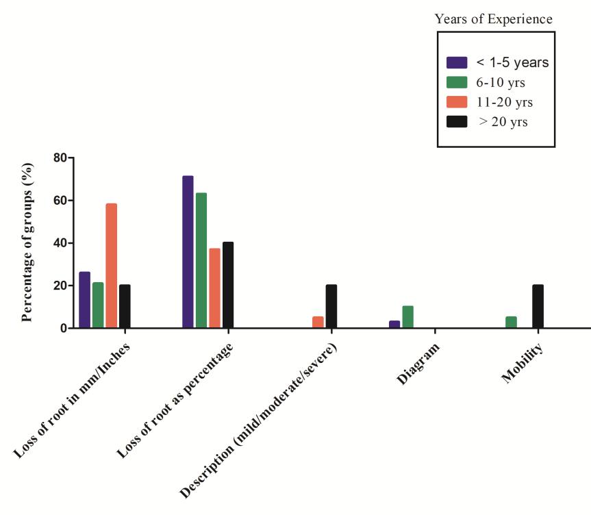
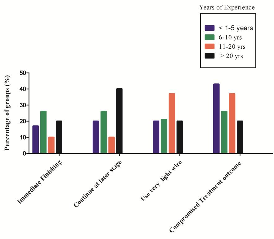
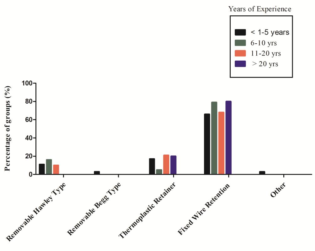
• Orthodontically-Induced External Root Resorption and Its Clinical Management: A Perception of Orthodontists in Pakistan
• Orthodontic Management of Impacted Maxillary Central Incisor: Case Report
Table of Contents
Editor
Rob Pasch, DDS, MSc, IBO
Mississauga, Ontario, Canada
E-mail: paschrob@rogers.com
Managing Editor
Allison Hester
8305 Pennwood Dr Sherwood, AR 72120
E-mail: allisonhijo@gmail.com
Consultants
Adrian Palencar, ON, Canada
Michel Champagne, QC, Canada
Dany Robert, QC, Canada
Scott J. Manning, USA
Mike Lowry, AB, Canada
Edmund Liem, BC, Canada
Yosh Jefferson, NJ, USA
G Dave Singh, CO, USA
Monika Tyszkowski, IL, USA
William Buckley, OH, USA
International Journal of Orthodontics
FEATURES
Molecular Characterization Of LEPR Q223R Single Nucleotide Polymorphism (rs1137101) in Different Facial Skeletal Patterns, by Dr. Danusha Siva Dharma, Dr Noraini Abu Bakar, and Dr Khairani Idah Mokhtar
Evaluation of Facial Attractiveness using profile silhouettes and selfies in orthodontically treated patients in different skeletal relations: A Comparative Study, by Dr. Akshataa Joshi, Dr. Kamal Bajaj, and Dr. Siddharth Mehta
Two-Stage Treatment : The Best of Both Worlds, (Functional and Fixed Orthodontics), by Dr. Gulnaz Husain, Dr. Gyan P Singh, Dr. G K Singh, Dr. Zainab Bint Shams, Dr. Syed Armaan Hussain, Dr. Syed Armaan Hussain
Orthodontically-Induced External Root Resorption and Its Clinical Management: A Perception of Orthodontists in Pakistan, by Dr Rozina Nazir, Dr Ashfaq Alam, Dr Usman Ahmed, and Dr Tania Arshad Siddiqui
Orthodontic Management of Impacted Maxillary Central Incisor: Case Report,, by Dr. Sharon K Sabu, Dr. Gyan P Singh, Dr. Gulshan K Singh, and Dr. Maheshwar Singh
Departments
International Journal of Orthodontics, copyright 2020 (ISSN #1539-1450). Published quarterly (March, June, September, December) by International Association for Orthodontics, 750 North Lincoln Memorial Drive, #422, Milwaukee, WI 53202 as a membership benefit. All statements of opinion and of supposed fact are published on the authority of the writer under whose name they appear and are not to be regarded as views of the IAO. Printed in the USA. Periodical postage paid at Milwaukee, WI and additional mailing offices. Subscription for member $15 (dues allocation) annually; $40 U.S. non-member; $60 foreign. Postmaster: Send address changes and all correspondence to:
International Journal of Orthodontics 750 North Lincoln Memorial Drive, #422 Milwaukee, WI, USA 53202
Phone 414-272-2757 Fax 414-272-2754
E-mail: worldheadquarters@iaortho.org
Writer’s Guidelines
Editorial, by Dr. Rob Pasch, DDS MSc IBO, Editor
Tips from the Experienced: The Utility Arch, Part 2 , by Dr. Adrian J. Palencar, MUDr, MAGD, IBO, FADI, FPFA, FICD
Growing Beautiful Teeth Chapter 8: A Pain-Free Life, by Estie Bav
`
Opinion: The Importance of Myofunctional Therapy in Orthodontics, Part 1, Brock Rondeau, D.D.S. I.B.O., D.A.B.C.P., D-A.C.S.D.D., D.A.B.D.S.M., D.A.B.C.D.S.M.
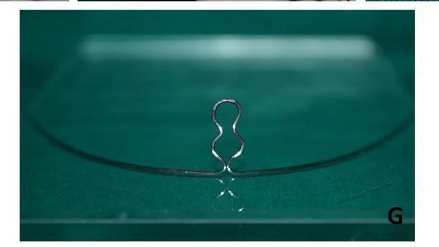
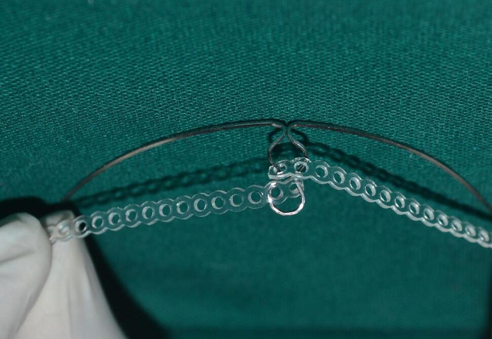
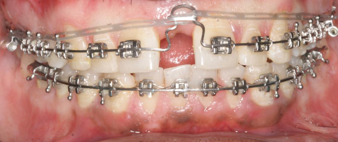
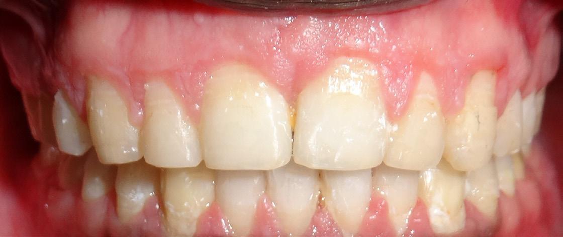
Opinion: Modified Loop for Closure of Midline Diastema, by Dr. Anadha N Gujar, Dr. Janis Shajan, and Dr. Sumit Kalsi
Practice Management Tips: Determining the Best Practice Structure for You, by Scott J Manning, MBA; Founder, Dental Success Today
Author Guidelines
MANUSCRIPT SUBMISSION
Manuscripts are to be submitted electronically at www. editorialmanager.com/iaortho. If the manuscript is written in a language other than English, the author(s) must submit an English translation. The author may also submit a copy in his or her native language that will published in the online version only with a mention in the printed issue that the article is available online in his or her own language. The manuscript must be original and submitted exclusively to IJO.
The Journal invites authors to submit:
•Clinical reports
•Technique articles
•Review articles
•Case reports
MANUSCRIPT FORMAT
Abstract. Must include a short abstract no more than 50 words that describe the significance of the article.
Keywords. Must include keywords to help categorize the article.
Length. Manuscript should be no longer than 15 doublespaced pages, excluding figures and illustrations.
Tooth Numbering. The numbering of teeth should be international numbering. (US numbering can be added and put in parentheses.)
Non-English Manuscripts. Authors are encouraged to submit the manuscript in languages other than English for posting on the IAO website. A mention will be added to the English version published in the International Journal of Orthodontics, directing readers online for other translations.
Illustrations. Images must be available electronically as separate files. High quality digital images must be presented in one of the following formats: .tiff, .eps,.jpg, or .pdf with resolution of a minimum 300 dpi. Images must not be embedded in software programs such as Word or Power Point. The names on the digital files for photo/illustration files should match the manuscript reference. For example, if manuscript copy references Figure 1, electronic file should be titled Figure 1.jpg. No more than 16 photographs, figures, & illustrations are recommended; if greater than 16, IJO has the right to select and limit the number if necessary. Figures must be clearly referenced as to their placement in the manuscript. Brief captions for the figures, identified by number, must be provided. All images must be titled. Radiographs must be of superior quality.
References. References must be included and authors are responsible for the accuracy of references. Manuscripts without them will be returned. Cite references in the text as endnotes and number them consecutively. Citations must be referenced in the following style:
Periodical:
1.Sim JM, Jefferson Y, Dillingham SE, & Keller DC. Diagnosing an orthodontic patient using three different analyses. IJO 1990; 1(4):101-106.
Book:
2.Fonder AC. The Dental Physician. 2nd ed. Rock Falls, IL; Medical Dental Arts; 1985:25-82.
World Wide Web site:
3.Health Care Financing Administration. 1996 statistics at a glance. Available at: http://www.hcfa.gov/stats/stathili.htm”. Accessed Dec. 2, 1996.
Products: Any products mentioned in the manuscript should be footnoted disclosing the company name and address.*
*XYZ Orthodontic Co., 123 Main St., Los Angeles, CA 90000.
REVIEW AND EDITING PROCESS
Editor. Articles will initially be reviewed by the editor. If author fails to adhere to the guidelines set forth, manuscript will be returned to the author for revision and correction.
Peer review. Articles in IJO are subject to an anonymous peer review process. Reviews may take up to eight weeks to complete.
Decision. Once the reviewing consultants have completed their critiques, the editor examines their comments and makes a decision to accept, accept with minor revisions, revise and resubmit, or reject.
Editing. IJO reserves the right to edit manuscript for conciseness, clarity, and stylistic consistency. The author has final approval before publication.
Questons? Contact Managing Editor, Allison Hester at allisonhijo@gmail.com, 501-517-1620.
AUTHOR RESPONSIBILITIES
Copyright transfer. IAO holds the copyright for all editorial content published in the journal. All accepted manuscripts become the permanent property of the IAO, and may not be published elsewhere in full or in part, in print or electronically, without written permission from the IAO.
Reprint permission. The author is responsible for obtaining written permission from the publisher, or the person or agency holding the copyright for any material that is reproduced from a published source.
Consent forms. Any patient clearly identified in the article must sign a form indicating his or her consent to be depicted in the article. It is the author’s responsibility to confirm consent.
Author’s photo and bio. The author(s) must submit a headshot (preferably professional) and current biographical sketch. If author holds a teaching position, the title, department, and school should be included. Any position or relationship with a dental manufacturer must be identified. The sketch should include rank or title and station of authors who are in federal service, and should be limited to 60 words or less.
Conflict of interest. The author will identify any conflicts of interest upon submission of any articles.
REPRINTS
The International Journal of Orthodontics provides the corresponding author a final electronic copy of the Journal in which the article appears as well as an electronic copy (.pdf) of the pages where the article appears. Requests for individual reprints of the article should be directed to Chris McKay, IAO, 414-272-2757 or at chris@iaortho.org.
Patients have a right to privacy that should not be infringed without informed consent. Identifying information, including patients’ names, initials, or hospital numbers, should not be published in written descriptions, photographs, and pedigrees unless the information is essential for scientific purposes and the patient (or parent/guardian) gives written informed consent for publication. Informed consent for this purpose requires that a patient who is identifiable be shown the manuscript to be published. Authors should identify Individuals who provide writing assistance and disclose the funding source for this assistance. Identifying details should be omitted if they are not essential. Complete anonymity is difficult to achieve, however, and informed consent should be obtained if there is any doubt. For example, masking the eye region in photographs of patients is inadequate protection of anonymity. If identifying characteristics are altered to protect anonymity, such as in genetic pedigrees, authors should provide assurance that alterations do not distort scientific meaning and editors should so note. (Source: International Committee of Medical Journal Editors (“Uniform Requirements for Manuscripts Submitted to Biomedical Journals”), February 2006).
Editorial
Dear fellow orthodontic practitioners. I am writing this editorial after having gone over the articles in this edition of the journal. These articles will provide insight on the day to day activity we perform for our patients with a view to understanding diagnosis, mechanical physics, and underlying biological expressions while treating malocclusions.
An interesting article by Noraini Abu Bakar sheds light on the genotype/phenotype expression and causes of malocclusion either from gene expression or environmental influences, and its conclusion, it is well worth a read. The other articles will provide valuable information regarding growth and development discussing mandibular advancement and airway influences on the growing individual.
In showcasing these articles, the IAO has attempted to increase the knowledge base of our members so treatment meted out to our patients is based on the most recent information available. As such, I wish everyone an enjoyable read of the journal this summer, I look forward to communicating with you should you have comments, complaints or suggestions. Thank you to Ms Allison Hester, our Managing Editor, for her invaluable role in completing this good looking journal.
I have a request to all who read this, and the ask is to please help each other to write and submit articles or case reports to the journal, for the journal is YOUR journal and you can make it reflect what is important to you today and in doing so making the journal better for everyone, now and in the future, also send the journal to your friends and share the knowledge.
It is a very good feeling to see your name in print, besides your patients will appreciate it as well.
Yours for accredited GP orthodontic education and better patient care
I remain Respectfully
Dr. Rob Pasch DDS MSc IBO General Practitioner. Summer 2024.
Dr. Rob Pasch Editor
Molecular Characterization Of LEPR Q223R Single Nucleotide Polymorphism (rs1137101) in Different Facial Skeletal Patterns
by Dr. Danusha Siva Dharma, Dr Noraini Abu Bakar, and Dr Khairani Idah Mokhtar
Abstract:
AUTHORS
Dr. Danusha Siva Dharma
Master of Science in Orthodontics
International Islamic University Malaysia Kulliyyah of Dentistry
Kuantan, Pahang MALAYSIA
Dr. Noraini Abu Bakar
Master of Science in Orthodontics
International Islamic University Malaysia Kulliyyah of Dentistry
Kuantan, Pahang MALAYSIA
Dr Khairani Idah Mokhtar
Associate Professor. International Islamic University Malaysia Kulliyyah of Dentistry
Kuantan, Pahang MALAYSIA
Background of the research: Studies on genetic associations have been used to link a person’s genotype to a physical trait or disease. In this investigation, we will examine the association between genetic variants and phenotype for the development of the facial skeletal pattern using the Leptin Receptor gene (LEPR) Q223 Single Nucleotide polymorphism (SNP) and whether it is viable as a diagnostic tool for identifying a certain phenotype. There is a gap in the literature on this subject that we intend to address.
Objective: The aim of this study was to determine the molecular characterization of the leptin receptor (LEPR) Q223R single nucleotide polymorphism (rs1137101) levels between different classes of facial skeletal pattern (Class I, II and III).
Methods: A sample of 82 patients was selected from the International Islamic University Malaysia (IIUM) Specialist Orthodontic Clinic prior to orthodontic treatment. Subjects were categorized into Class I, Class II, and Class III facial skeletal patterns using Eastman and Wits analysis. Unstimulated saliva samples were collected for DNA extraction and amplification, followed by Polymerase Chain Reaction (PCR) analysis under ultraviolet (UV) light. Subsequently, the MspI restriction enzyme was used for Restriction Fragment Length Polymorphism (RFLP) analysis under UV light. Statistical analysis included the Chisquare (χ2) test for genotype and allele frequencies comparison among facial skeletal patterns. The Hardy-Weinberg Equilibrium (HWE) assessed genotype frequency distribution among the facial skeletal patterns.
Results: There was no significant difference in genotype frequency between the Class I facial skeletal pattern in the control group and the Class II and Class III facial skeletal patterns (p=0.48, p=0.16, respectively). Additionally, there was no correlation between allele frequency in the control group (Class I facial skeletal pattern) and Class II and Class III facial
skeletal patterns (p=0.82 and p=0.32, respectively).
Conclusion: There was no significant association between the Q223R (rs1137101) SNP of the LEPR gene in different classes of facial skeletal pattern.
Introduction: Malocclusion is thought to be caused by a combination of genetic and environmental factors, according to a review of its aetiology. With regards to the facial skeletal pattern, discrepancies in the antero-posterior, vertical and transverse dimensions contribute to the development of malocclusions. 1,2 Understanding the growth and development of the facial skeletal pattern is therefore crucial to manage skeletal discrepancies.3
The genetics linked with neurological, muscular and neuromuscular domains also have an indirect effect on the facial skeletal pattern. 4 Therefore, it is advantageous to research genetic variants that may affect the growth of the aforementioned structures as they may provide useful information to be utilised as a diagnostic aid for recognising facial skeletal pattern growth, which in turn would affect orthodontic treatment.5
The association between genetics and different malocclusions has been the subject of numerous studies. Nazirah Yahya et al., 20176 revealed that nucleotide changes in the rs10850110 within the MYO1H gene were found in the Asian Malay population with a mandibular prognathism phenotype. The MYO1H single nucleotide polymorphism (SNP) (rs3825393) on the other hand, did not appear to be significantly associated with mandibular prognathism.7
In addition, it has be demonstrated that individuals with a Class II skeletal pattern have an overrepresentation of the ACTN3 577XX gene, which raises the possibility of
it having a biological influence on growth of the facial skeleton. The same gene is underrepresented in patients with reduced vertical skeletal dimensions suggesting that variances in muscle function play a role in contributing to this facial skeletal pattern.8
Balkhande et al., 2018,9 concluded that a connection between mandibular retrognathism and the matrilin-1 gene (MATN1) gene SNP. When association studies were conducted, a positive correlation between the EPB41, MATN1, SSX2IP, and PLXNA, located within the 1p22-p36 locus and genes COL2A1, MYO1H, TGFB3, and LTBP2 within the 12q13- q24 locus were found.10
In addition to that, SNP in the BMP2, BMP4, SMAD6, RUNX2, WNT3A and WNT11 genes have been linked to both sagittal and vertical discrepancies according to a recent study.11
To date, there have been minimal studies have been done to examine the relationship between the molecular characterization of (LEPR Q223R) single nucleotide polymorphism (RS1137101) in different classes of facial skeletal pattern. As such, this study aims to produce new genetic knowledge which could potentially identify and manage discrepancies in the facial skeletal pattern at its development stage.
Objectives
1. To determine the molecular characterization of the leptin receptor (LEPR) Q223R single nucleotide polymorphism (rs1137101) in patients with Class I, II and III facial skeletal patterns.
2. To investigate the relationship between the molecular characterization of the leptin receptor (LEPR) Q223R single nucleotide polymorphism (rs1137101) in Class I, II and III facial skeletal patterns.
Methods
Study Design: This study is of a quantitative, cross-sectional design with convenience sampling using the active orthodontic patients from the Orthodontic Department, Kulliyyah of Dentistry International Islamic University Malaysia (IIUM) as the target population.
Ethics: Ethical approval was obtained from IIUM Research Ethics Committee (IREC) (REF NUMBER: IREC 2020-028) with investigations being done in accordance with the principles encompassed in the revised Declaration of Helsinki (2008). Prior to the experiment, consent was acquired, and the patients’ privacy was protected at all times.
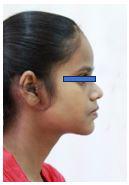
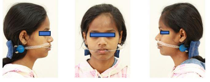
Patient selection: The subjects were patients undergoing active orthodontic treatment from the Orthodontic Specialist Clinic, Kulliyyah of Dentistry, IIUM. Clinical examination and reviews of the patients’ radiological and clinical records were part of the evaluation of the eligible subjects. A mix of study models, cephalometric tracings with the Eastman and Wits Cephalometric analyses performed, and photographs were used to interpret the clinical records.
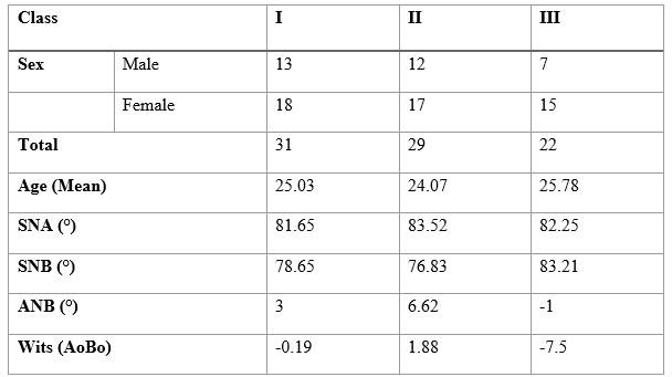
A total of 82 patients participated in this study, including 31 patients from the control group of Class I facial skeletal pattern (13 male and 18 female); 29 patients with a Class II facial skeletal pattern (12 male and 17 female) and 22 patients with a Class III facial skeletal pattern (7 male and 15 female). The following criteria were used to select the patients:
Inclusion Criteria:
• Good health
• Normal weight, according to the WHO body mass index (BMI) categories (BMI of 18.5-24.9)
• No history of anti-inflammatory medication usage in the month prior to the sample collection
• No history of antibiotic medication usage in the six months preceding sample collection
• Good periodontal health with generalized probing depths of no more than 2 mm, minimal bleeding on probing and no evidence of attachment loss
• No periodontal bone loss visible on radiographs
Exclusion criteria:
• Craniofacial anomalies such as cleft lip and palate
• Diseases of the endocrine system
• Dental anomalies including discrepancies in number of teeth, its morphology, and eruption
Cephalometric analysis
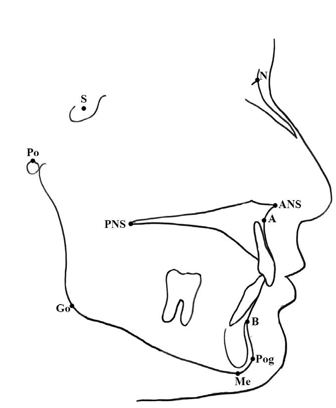
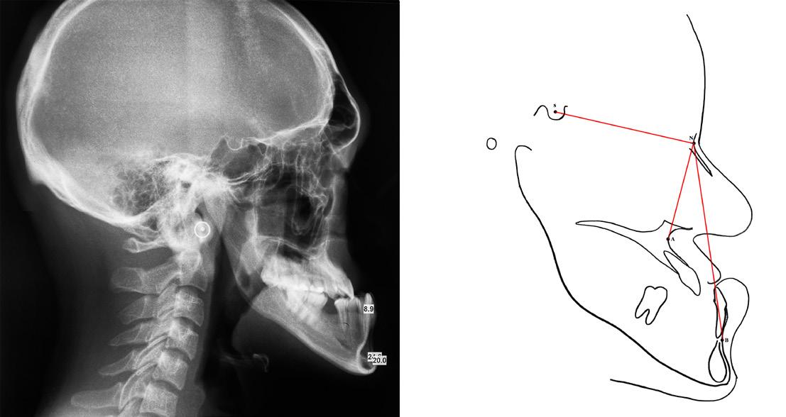
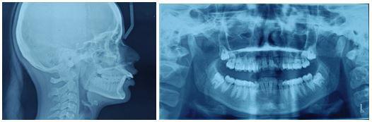
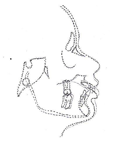
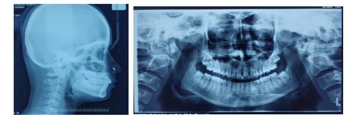
Identified subjects underwent comprehensive clinical examination by one of the researchers at IIUM Specialist Clinic to ensure no anomalies in tooth number, morphology and eruption. A lateral cephalometric radiograph was taken as a record for cephalometric analysis. Eastman analysis,12 measuring sellanasion-A-point angle (SNA), sella-nasion-B point angle (SNB), A point-nasion-B point (ANB) and the Wits appraisal (AoBo) were executed.13 The points as illustrates in Figure 1.
SNA angle was used to assess the position of maxilla to cranial base whilst SNB angle was used to determine the position of mandible to the cranial base. The ANB angle, which has been recognized as the most commonly used antero-posterior skeletal
Figure 1: Reference points on the cephalometric radiograph: Sella (S)midpoint of the sella turcica, Nasion (N), A point (A)- deepest point of concavity on the anterior profile of the maxilla, B point (B)- deepest point of concavity on the anterior surface of the mandibular symphysis, Gonion (Go)- the most posterior, inferior point on the angle of the mandible, Menton (Me)- the most inferior point on the mandibular symphysis, Pogonion (Pog)- the most anterior point of the mandibular symphysis, Porion (Po)- the upper midpoint of the external auditory meatus, Anterior nasal spine (ANS), Posterior Nasal Spine (PNS). (Figure reprinted with permission from Siva Dharma D, Abu Bakar N, Mustafa BE. Evaluation of Salivary Leptin Levels and Its Correlation with Class I, Class II, and Class III Facial Skeletal Pattern: A Prefatory Study. European Journal of Dentistry. Published online August 24, 2021. doi:https://doi. org/10.1055/s-0041-1727552)
discrepancy indicator14 was then calculated. It compares the relationship of the maxilla and the mandible with regards to the cranial base. The classification used denotes that a 2-4 degree value indicates a class I malocclusion, above 4 degrees indicates a Class II malocclusion and below 2 degrees is indication for a Class III malocclusion.15
The Wits appraisal, which compares the relationship of the maxilla and the mandible with regards to the functional occlusal plane was used to further confirm the antero-posterior occlusal disharmony. A line is drawn between the cusp tips of the molars and premolars and this is known as the functional occlusal plane (FOP). A perpendicular line is drawn from point A and point B to the FOP to give points AO and BO. The distance between AO and BO is measured. The average (Class I malocclusion) values are −1 mm (± 1.9 mm) for males and 0 mm (± 1.77 mm) for females.15
Values below the average values denote a Class II malocclusion and values above the average denote a Class III malocclusion.
Subjects were grouped into facial classes based on the analysis criteria below:
Cephalometric analysis criteria for Class I facial skeletal pattern:
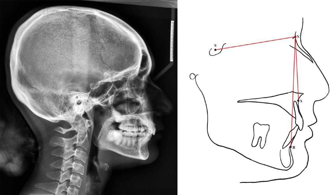
1. Cephalometric value indicative of Class I based on Eastman (ANB within 2°to 4°, SNA within range of 81°±3° and SNB within range of 78°±3°) as shown in Figure 2.
2. Wits appraisal (AoBo) within Class I (- 2mm to +2mm)
3. Straight facial profile
tracing (right) of a Class I lateral cephalometric radiograph (left). To calculate angles SNA and SNB, lines are drawn between the points S, N, and A. The ANB angle is obtained by deducting angle of SNB from SNA. (Figure reprinted with permission from Siva Dharma D, Abu Bakar N, Mustafa BE. Evaluation of Salivary Leptin Levels and Its Correlation with Class I, Class II, and Class III Facial Skeletal Pattern: A Prefatory Study. European Journal of Dentistry. Published online August 24, 2021. doi:https://doi.org/10.1055/s-0041-1727552)
Cephalometric analysis criteria for Class II facial skeletal pattern:
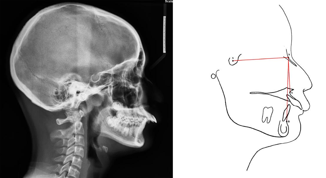
• Cephalometric analysis with value indicative of Class II based on Eastman analysis (ANB should be >4° and SNB should be <78°) as shown in Figure 3.
• SNA within normal range indicative of average maxilla (81°±3°)
• Positive Wits appraisal (AoBo > 2 mm)
• Convex facial profile
Figure 3: Cephalometric tracing (right) of a Class II lateral cephalometric radiograph (left). To calculate angles SNA and SNB, lines are drawn between the points S, N, and A. The ANB angle is obtained by deducting angle of SNB from SNA. (Figure reprinted with permission from Siva Dharma D, Abu Bakar N, Mustafa BE. Evaluation of Salivary Leptin Levels and Its Correlation with Class I, Class II, and Class III Facial Skeletal Pattern: A Prefatory Study. European Journal of Dentistry. Published online August 24, 2021. doi:https://doi.org/10.1055/s-0041-1727552)
Cephalometric analysis criteria for Class III facial skeletal pattern:
• Cephalometric analysis with value indicative of Class III based on Eastman analysis (ANB should be <2° and SNB should be >81°) as shown in Figure 4.
• SNA within normal range indicative of average maxilla (81°±3°)
• Negative Wits appraisal (AoBo) of < - 2mm
• Concave facial profile
Figure 4: Cephalometric tracing (right) of a Class III lateral cephalometric radiograph (left). To calculate angles SNA and SNB, lines are drawn between the points S, N, and A. The ANB angle is obtained by deducting angle of SNB from SNA. (Figure reprinted with permission from Siva Dharma D, Abu Bakar N, Mustafa BE. Evaluation of Salivary Leptin Levels and Its Correlation with Class I, Class II, and Class III Facial Skeletal Pattern: A Prefatory Study. European Journal of Dentistry. Published online August 24, 2021. doi:https://doi.org/10.1055/s-0041-1727552)
Saliva Collection: Saliva samples were taken from patients who were required to fast from 12 am until the samples were collected at 8 am the next day. The patients were informed to not brush their teeth the morning of the appointment to minimise the risk of contamination from gingival trauma/bleeding during collection of the sample. The passive drooling and draining method was used16 during collection whereby the patients were asked to expectorate every thirty seconds over five minutes into dtab;e isposable tubes.
Saliva was collected at the same time of the day (8 am) for all
Figure 2: Cephalometric
patients to reduce differences in circadian rhythmicity.17–19 After collection, the saliva samples were centrifuged at 4000x g;10min and stored at -25°C.
DNA Isolation: The DNA extraction and isolation was done using the GeneAll® ExgeneTM Kit (Korea). 1 ml of saliva was transferred into a 15 ml falcon tube and 5 ml of 1X Phosphate Buffered Saline (PBS) was added. The solution was vortexed and centrifuged at 3000 revolutions per minute (rpm) for five minutes and supernatant was discarded.
The cell pellet was resuspended with 200 μL of 1X PBS and subsequently vortexed and incubated at room temperature for two minutes. 20 μL of Proteinase K (20 mg/ml) solution was pipetted into the tube and 200 μL of buffer BL was added.
The mixture was the transferred to a 1.5 ml centrifuge tube and incubated at 56°C for ten minutes. 200 μL of absolute (pure / 100%) ethanol was added and the solution vortexed. The mixture was then transferred to a spin/vacuum (SV) column carefully and centrifuged for one minute at 6000 xg above (>8000rpm). The pass-through was discarded and the collection tube was replaced with a new one.
Subsequently, 600 μL of buffer BW was added and the mixture was centrifuged for one minute at 6000 xg above (>8000rpm). The pass-through was discarded and the collection tube was replaced with a new Results for Genetic Analysis one.
Next, 700 μL of buffer TW was added and the mixture was centrifuged for one minute at 6000 xg above (>8000rpm). The pass-through was discarded and collection tube was replaced. The mixture was then centrifuged at full speed (12 000 rpm) for one minute to remove residual wash buffer.
The collection tube was replaced with a 1.5 ml centrifuge tube and 200 μL of buffer AE was added. The mixture was incubated for one minute at room temperature and subsequently centrifuged at full speed for one minute. The SV column was discarded and the 1.5 ml centrifuge tube with the DNA sample was and stored at -20°C until further use.
Polymerase Chain Reaction (PCR) Amplification: Genotyping the LEPR gene for the Q223R (rs1137101) polymorphism was conducted by Polymerase Chain Reaction-Restriction Fragment Length Polymorphism (PCR-RFLP). For this purpose, PCR amplification was done with the following primers: forward 5’-AACTCAACGACACTCTCCTT-3’ and reverse 5’-TGAACTGACATTAGAGGTGAC-3’.
PCR amplification of DNA fragments was carried out by using the Bio-Rad T100™ thermal cycler (United States of America). A PCR mixture was made using the Promega GoTaq® Flexi DNA Polymerase (United States of America) consisting of 10 μL of 1X GoTaq® Colourless Buffer, 0.5 μL of forward primer, 0.5 μL of reverse primer, 2 μL of Magnesium Chloride, 25mM, 0.125 μL of GoTaq® polymerase enzyme (5u/ μL), 0.5uL of the deoxynucleoside triphosphate (dNTP) mix, 10mM, 14.375 μL nuclease-free water (dH2O), and 2.0 μL of DNA.
The initial denaturation at 94⁰C for 5 min was followed by 35 cycles of denaturation phase at 94⁰C for 30 seconds, annealing at 58.5ºC for 45 seconds, elongation at 72ºC for 60 seconds, and a final elongation at 72ºC for 5 minutes. The PCR results (80bp) were confirmed by 3% agarose gel electrophoresis at 85V/400mA for 45 min using the CLEAVER nanoPAC-300, MINi 300V 400mA 60W – 110/230V (United Kingdom) electrophoresis power supply. These results were visualized using the Bio-Rad ChemiDoc™ XRS+
System with Image Lab software (United States of America) gel imaging system.
Restriction
Fragment Length (RFLP) Genotyping
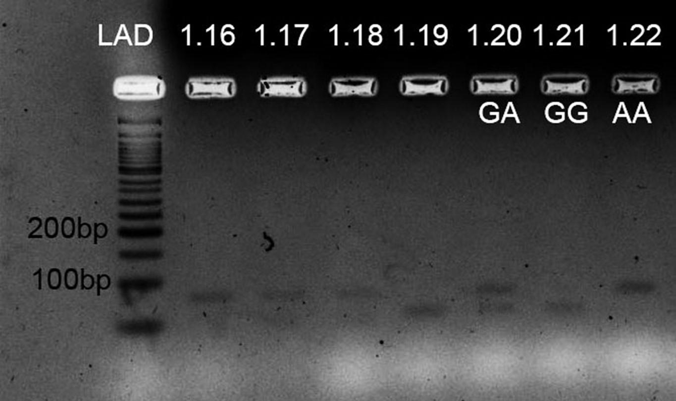
RFLP was conducted using the restriction enzyme MspI from New England Biolabs (United Kingdom); with incubation at 37ºC for 2 hours. The resulting DNA fragments were subjected to 4% agarose gel electrophoresis at 80V/400mA for 60 minutes with the same equipment mentioned above and visualized using gel documentation as before; showed genotyped AA (80bp), GA (80, 59, 21 bp), and GG (59, 21 bp).
Results
Cephalometric analysis
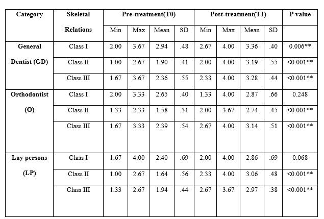
The results of the cephalometric analysis are as shown in Table 1 below.
PCR-RFLP Analysis
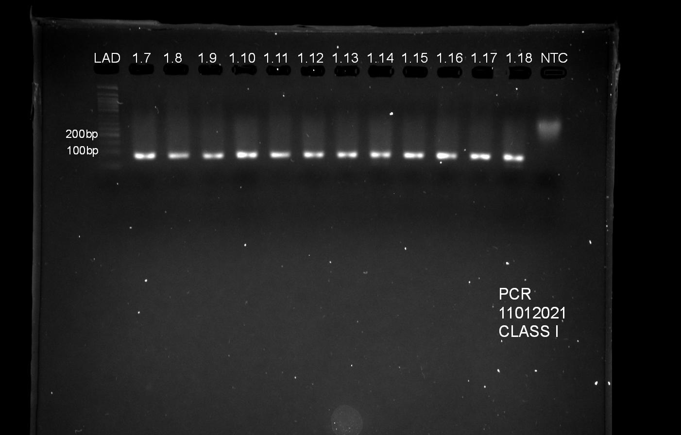
PCR Results: The PCR result obtained was at the value of 80 base pairs (bp). Figure 5 shows the example of the PCR reaction results done for Class I and Class II.
Results for Restriction Enzyme MspI cutting: Figure 6 displays the cutting site indicating that a single black band at 80bp indicates the genotype AA as the enzyme did not cut at the cutting site. Two black bands at 80bp and 59bp indicate GA and a single black band at 59bp indicates the result GG. No band was visible at 21bp as the size of the base pair was too small to be seen.
Results for Restriction Enzyme MspI cutting: Figure 6 displays the cutting site indicating that a single black band at 80bp indicates the genotype AA as the enzyme did not cut at the cutting site. Two black bands at 80bp and 59bp indicate GA and a single
Table 1: The mean values of the demographic data and cephalometric data
Figure 5: The PCR reaction results done for Class I skeletal pattern.
6: The cutting site indicating a single black band at 80bp indicates the genotype AA, Two black bands at 80bp and 59bp to indicate genotype GA and a single black band at 59bp to indicate the result GG. No black band visible at 21bp.
black band at 59bp indicates the result GG. No band was visible at 21bp as the size of the base pair was too small to be seen.
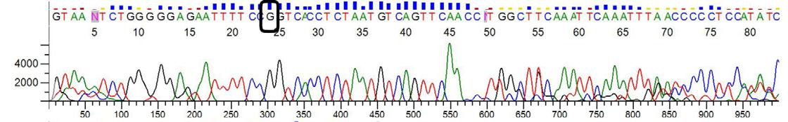
Sanger Sequencing Results: The PCR products were sent for Sanger sequencing to confirm the results of the study.
Figure 7 shows the sequencing done for sample 1.23 whereby a single peak shows the homogenous genotype GG.
Results for Genetic Analysis
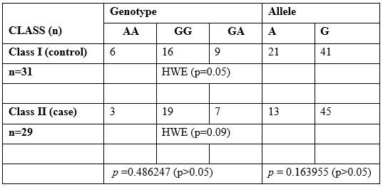
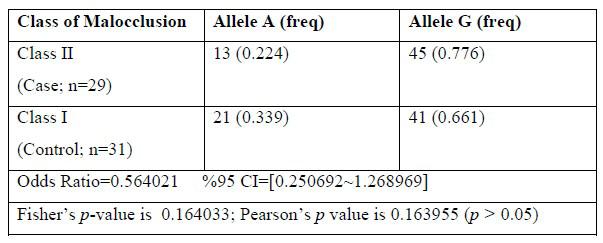
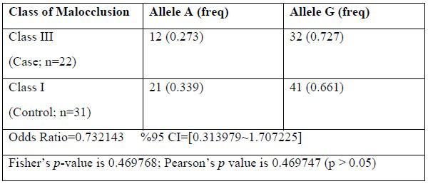
The results of genetic analysis indicate no significant association between genotype frequency [p =0.486247 (p>0.05)] and allele frequency [p = 0.163955 (p>0.05)] with the class II facial skeletal pattern in reference to Table 2 and 3.
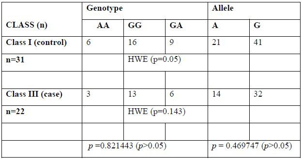
In reference to Tables 4 and 5, our clinical report finds no significant correlation between genotype frequency (p = 0.821443, p > 0.05) and allele frequency (p = 0.326930, p > 0.05) with a class III facial skeletal pattern.
The genotype distribution across all three classes of facial skeletal patterns reveals consistent allele and genotype frequencies in both the case and control groups. Consequently, this suggests the absence of evolutionary influences, aligning with the principles of the Hardy-Weinberg equilibrium (HWE), where the value of p > 0.05.
Discussion
Cephalometric analysis to represent the facial pattern
To evaluate the facial skeletal pattern prior to orthodontic treatment, the lateral cephalometric analysis is frequently utilised as a diagnostic tool. For identifying and classification of anteroposterior discrepancies of the skeletal base, it has been suggested that the use of the ANB angle with Eastman correction and Wits analysis be performed20 and therefore was used to classify the facial skeletal pattern in this study.
Polymorphism of the LEPR Q223 (rs1137101 gene)
The most prevalent type of genetic variation, known as single nucleotide polymorphism, occurs when two alternative bases appear in the human population of more than 1%.21 As a result
Figure
Table 2: Genotype and allele distribution of LEPR Q223 (SNP rs1137101) with Class I and II facial skeletal pattern
Table 3: Single association analysis of rs1137101 with allele frequency of Class I & II facial skeletal pattern
Table 4 :Genotype and allele distribution of LEPR Q223 (SNP rs1137101) with Class I and III facial skeletal pattern
Table 5 :Single association analysis of rs1137101 with allele frequency of Class I & III facial skeletal pattern
of mutation, the amino acid Glutamine is changed to Arginine at codon 223’s second nucleotide (CAG to CGG).22 As a result, it can be said that the mutant allele is G and the normal allele is A. Association of the LEPR Q223 (rs1137101 gene) with the facial skeletal pattern
Chen, 2011 concluded that the Class II facial skeletal pattern in the Chinese population was significantly associated with the LEPR Q223 (rs1137101) polymorphism. According to 24, the genotype AA and allele A of the LEPR Q223 (rs1137101) occurred significantly more frequently in the Class II malocclusion in the Indonesian population.
In our study of the Malaysian population of mixed ethnicity however, there was no significant difference in genotype and allele frequency between the Class II and Class III facial skeletal pattern compared to the Class I control group. The difference in findings compared to previous studies is most likely attributed to the differences in population.
In contrast to the normal allele A, whose frequency was reported for Class I at 0.339, Class II at 0.224, and Class III at 0.273, our investigation demonstrated that the mutant allele G was more common in Class I (0.661), Class II (0.776), and Class III (0.727) skeletal patterns. Our results are consistent with those of the Other Asian population (Asian people who do not identify as East Asian or South Asian), where the frequency of the mutant allele G (0.8786) is higher than the normal allele A (0.1214).25
All of the expected and observed values were in equilibrium according to the Hardy-Weinberg equation (p> 0.05), proving that the genetic variation in this population remains constant.
A small population size is one of our study’s limitations. To define polymorphism more precisely within a population, it is advised that future studies be conducted with a larger population size and an ethnically homogenous population as have been done in the selection criteria of some previous studies.22,26,27
Conclusion
Based on the findings of this study, we can conclude that there was no significant difference in LEPR Q223 polymorphism genotype and allele frequency between the Class II and Class III facial skeletal patterns and the control group, Class I facial skeletal pattern.
References
1. Dehesa-Santos A, Iber-Diaz P, Iglesias-Linares A. Genetic factors contributing to skeletal class III malocclusion: a systematic review and meta-analysis. Clin Oral Investig. 2021;25(4):1587-1612. doi:10.1007/ S00784-020-03731-5
2. Georg e A, Felicita As, Milling Tania S, Priyadharsini Jv. Systematic review on the genetic factors associated with skeletal Class II malocclusion. Indian J Dent Res. 2021;32(3):399. doi:10.4103/IJDR. IJDR_59_20
3. Mokhtar KI, Abu Bakar N, Md Ali Tahir AH. Genetics of malocclusion: A review. IIUM J Orofac Heal Sci. 2020;1(1). doi:10.31436/ijohs.v1i1.2
4. Mossey PA. The heritability of malocclusion: Part 1--Genetics, principles and terminology. Br J Orthod. 1999;26(2). doi:10.1093/ ortho/26.2.103
5. Mossey PA. The heritability of malocclusion: part 2. The influence of genetics in malocclusion. Br J Orthod. 1999;26(3):195-203. doi:10.1093/ ortho/26.3.195
6. Nazirah Yahya S, Syafiqah N, Razak A, et al. Analysis Of MYO1H Single Nucleotide Polymorphism In Class III Malocclusion With Mandibular Prognathism: A Preliminary Study. Vol 16.; 2017. doi:10.31436/IMJM.V16I2.1048
7. Mohd Yusoff N, Khajar N, Mokhtar KI, Abu Bakar N, Wan Taib WR. Application of Polymerase Chain Reaction-Restriction Fragment Length Polymorphism (PCRRFLP) Technique in the Analysis of MYO1H Single Nucleotide Polymorphism in Malay Mandibular Prognathism Patients. Arch Orofac Sci. 2020;15(2):139-147. doi:10.21315/ aos2020.15.2.446
8. Zebrick B, Teeramongkolgul T, Nicot R, et al. ACTN3 R577X genotypes associate with Class II and deepbite malocclusions. Am J Orthod Dentofac Orthop. 20 2014;146(5):603-611. doi:10.1016/j. ajodo.2014.07.021
9. Balkhande PB, Lakkakula BVKS, Chitharanjan AB. Relationship between matrilin-1 gene polymorphisms and mandibular retrognathism. Am J Orthod Dentofac Orthop. 2018;153(2):255-261.e1. doi:10.1016/j. ajodo.2017.06.023
10. Moreno Uribe LM, Miller SF. Genetics of the dentofacial variation in human malocclusion. Orthod Craniofacial Res. 2015;18(S1):91-99. doi:10.1111/ocr.12083
11. Küchler EC, Barreiros D, Da Silva RO, et al. Genetic polymorphismin MMP9 may be associated with anterior open bite in children. Braz Dent J.2017;28(3):277-280. doi:10.1590/0103-6440201600992
12. Shaw K, McIntyre G, Mossey P, Menhinick A, Thomson D. Validation of conventional 2D lateral cephalometry using 3D cone beam CT. J Orthod. 2013;40(1):22-28. doi:10.1179/1465313312Y.0000000009
13. Jacobson A. The “Wits” appraisal of jaw disharmony. Am J Orthod Dentofac Orthop. 2003;124(5):470-479. doi:10.1016/S08895406(03)00540-7
14. Oktay H. A comparison of ANB, WITS, AF-BF, and APDI measurements. Am J Orthod Dentofac Orthop. 1991;99(2):122-128. doi:10.1016/0889-5406(91)70114-C
15. Barber SK, Littlewood SJ, Mitchell L. Cephalometrics. In: An Introduction to Orthodontics (5th Edition). Oxford University Press (OUP); 2019:72-84.
16. Bellag ambi FG, Lomonaco T, Salvo P, et al. Saliva sampling: Methods and devices. An overview. TrAC - Trends Anal Chem. 2020;124. doi:10.1016/j.trac.2019.115781
17. Kubala E, Strzelecka P, Grzegocka M, et al. A Review of Selected Studies That Determine the Physical and Chemical Properties of Saliva in the Field of Dental Treatment. Biomed Res Int. 2018;2018. doi:10.1155/2018/6572381 21
18. Bakar NA, Kamil W, Al Bayati L, Mustafa BE. Saliva leptin levels in tooth movement during initial stage of orthodontic alignment: A pilot study. Brazilian J Oral Sci. 2017;16. doi:10.20396/bjos.v16i0.8651060
19. Thanakun S, Watanabe H, Thaweboon S, Izumi Y. Comparison of salivary and plasma adiponectin and leptin in patients with metabolic syndrome. Diabetol Metab Syndr. 2014;6(1):19. doi:10.1186/1758-59966-19
20. Zamora N, Cibrián R, Gandia JL, Paredes V. Study between ANB angle and Wits appraisal in cone beam computed tomography (CBCT). Med Oral Patol Oral Cir Bucal. 2013;18(4):e725. doi:10.4317/medoral.18919
21. Wang DG, Fan JB, Siao CJ, et al. Large-scale identification, mapping, and genotyping of single- nucleotide polymorphisms in the human genome. Science (80- ). 1998;280(5366):1077-1082. doi:10.1126/ science.280.5366.1077
22. Farzam F, Mahmazi S, Nasseryan J. Association of Leptin Receptor Gene Gln223Arg and lys109Arg Polymorphisms with Obesity and Overweight in an Iranian Young Population. Gene, Cell Tissue. 2017;In Press(In Press). doi:10.5812/gct.57937
23. Chen K. Identification of Genetic Predisposing Factors for Skeletal Class II Malocclusions. The University of Hong Kong; 2011. doi:10.5353/th_b4589155
24. Jazaldi F, Handayani ED, Damayanti YNU, et al. The LEPR Q223R polymorphism as a potential bioindicator of class II malocclusion. J Int Dent Med Res. 2016;9(SpecialIssue).
25. L. Phan, Y. Jin, H. Zhang, et al. ALFA: Allele Frequency Aggregator. National Center for Biotechnology Information, U.S. National Library of Medicine,.
26. Bains V, Kaur H, Badaruddoza B. Association analysis of polymorphisms in LEP 22 (rs7799039 and rs2167270) and LEPR (rs1137101) gene towards the development of type 2 diabetes in North Indian Punjabi population. Gene. 2020;754. doi:10.1016/j. gene.2020.144846
27. Yang J, Du H, Lv J, Zhang L. Association of rs1137101 polymorphism in LEPR and susceptibility to knee osteoarthritis in a Northwest Chinese Han population. BMC Musculoskelet Disord. 2016;17(1). doi:10.1186/s12891-016-1162-0
Acknowledgement
We would like to thank the Ministry of Education and the International Islamic University Malaysia joint grant, FRGS RACER-19-030-0030 for funding this study. We would also like to acknowledge Sponsored Research Collaborative Grant SRGC 20016-0016 for the publication payment. Appreciation also goes to the staff of the Department of Orthodontics, Kulliyyah of Dentistry (IIUM), our research assistants and staff of Central Research & Animal Facility (CREAM) for their assistance.
TTIPS FROM THE EXPERIENCED
The Utility Arch, Part 2
By Dr. Adrian J. Palencar, MUDr, MAGD, IBO, FADI, FPFA, FICD
here are a variety of Utility Arches, and they are classified by their function:
A. Utility Arches without activation in sagittal plane:
1. Passive (neutral)
2. Protrusive
3. Retrusive
B. Utility Arches activated in sagittal plane (tip back, tip forward):
1. Intrusive – protrusive
2. Intrusive – retrusive
3. Extrusive – retrusive
4. Extrusive – protrusive
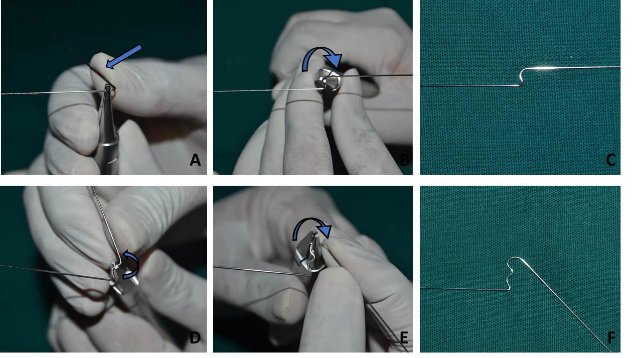
The Utility Arch comprises of anterior step, posterior step, labial segment, buccal segment. The author prefers 3.0 mm high steps. It is also paramount to fabricate the labial segment parallel to the incisal arc (smile arc), providing that it is in correct position.
Utility Arches without activation in the sagittal plane:
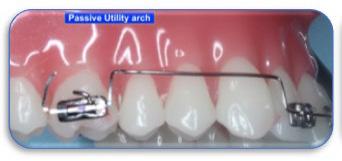
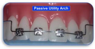
1.Passive (neutral) serves as a space maintainer in the late mixed dentition. It is important to anneal and bend back the arch wire distally to the molar tubes.
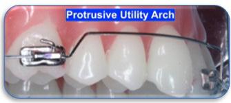
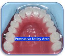
2.Protrusive creates labial moment (proclines, flares} the incisors (i.e., Class II, division 2). The easiest way to fabricate it, is by converting the passive Utility Arch - bending the posterior steps (or anterior steps too if required), to a more obtuse angle (45) thus, gaining the length.
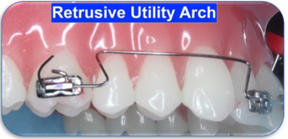
3.Retrusive creates lingual moment (retroclines, de-torques) the incisors (i.e., Class II, ivision 1). The easiest way to fabricate it, is by converting the passive Utility Arch - bending the posterior and anterior steps to letter “Z”, thus reducing the length. This configuration resembles Ricketts “Z” sectional arch. This Retrusive Utility Arch is activated only 1.0 mm per month, bilaterally, by pulling the arch wire distally with a crisp bend-back (cinch). It is beneficial to have the ends of the arch wire annealed for the ease of titration and bending.
It is paramount to have spaces to de-torque to; either diastema, spaced dentition or creation of spaces with IPR (slenderizing).
As the readers may notice, the simplest way to fabricate the intrusive (or any other) arch is Mulligan mechanics. The applied force can be titrated by the amount of Tip back (less acute angle than the Utility arch). The force should be measured by the Gram gage. The following pliers may help you bending the Utility arch.
References
1. Cerum Ortho Organizers catalogue
2. Rondeau Seminars, Level I, Session 3
3. Palencar A. J. Case Finishing and Mechanics manual
Evaluation of facial attractiveness using profile silhouettes and selfies in orthodontically treated patients in different skeletal relations: A Comparative Study
by Dr. Akshataa Joshi, Dr. Kamal Bajaj, and Dr. Siddharth Mehta
AUTHORS
Dr. Akshataa Joshi Post-graduate student, Mahatma Gandhi Dental College and Hospital, Jaipur, Rajasthan
Dr. Kamal BajaJ Head of the Department and Professor, Department of Orthodontics and Dentofacial Orthopedics, Mahatma Gandhi Dental College and Hospital, Jaipur, Rajasthan
Dr. Siddharth Mehta Professor. Department of Orthodontics and Dentofacial Orthopedics, Mahatma Gandhi Dental College and Hospital, Jaipur, Rajasthan.
Abstract:
Introduction: As orthodontist it should be our goal to enhance facial attractiveness along with providing an ideal occlusion. Profile silhouettes provide an unbiased means of assessing facial attractiveness. As the trend of taking selfies is increasing, it is imperative that we assess facial attractiveness in selfies. Our aim is to evaluate if orthodontic treatment can enhance facial attractiveness using profile silhouettes (PS) and selfies(S) in Skeletal Class I (SCI), Skeletal Class II (SCII), Skeletal Class III (SCIII) among layperson (LP), general dentist (GD) and orthodontist(O).
Methods: Pre-treatment(T0) and posttreatment (T1) selfies(S) and PS of patients who underwent orthodontic treatment were collected from various dental colleges in India. The data was divided into SCI, SCII, SCIII with 24 subjects (12 males,12 females) in each category. The selfies(S) were randomly shuffled and the PS at T0 and T1 were placed side by side and shown to a panel of GD, O, LP with 3 examiners in each panel. Their responses were recorded on a 5-point Likert scale.
Results : In SCI of PS, GD gave a statistically highly significant (p<0.001) difference between T0 and T1, SCII & SCIIIGD, LP and O, gave statistically highly significant (p<0.001) difference between T0 and T1. In SCI & SCII of selfies group, GD and O gave a statistically highly significant (p<0.001) difference between T0 and T1 and in SCIII group of selfies GD, O and LP found no statistically significant (p>0.05) difference between T0 and T1.
Conclusions- Orthodontic treatment can increase facial attractiveness which can be assessed in both PS and selfies.
Margaret Wolfe Hungerford once wrote “Beauty lies in the eye of the beholder.” 1 Facial attractiveness is an integral part of overall attractiveness or persona of an individual. People generally perceived as attractive are more self-confident, have high self-esteem and are regarded socially as more accomplished and desirable. 2 There are various factors contributing to an individual’s facial attractiveness, like their smile, eyes, nose, hair, lips, chin, etc.3
With the dawn of the 21st century, soft tissue paradigm replaced the Angle’s paradigm that focused on ideal dental occlusion. The focus is greatly shifted towards providing the occlusion and soft tissue facial form that would benefit the patient the most.4 An orthodontist’s perception of facial attractiveness is greatly based on the profile of the patient.5 Due to this facial attractiveness in the profile view should be taken into consideration. Profile silhouettes are basically profile photographs in which a dark shape or outline of the subject’s face is obtained against a white background. The reason behind converting these photographs into silhouettes is to eliminate distractions like eyes, hair, skin etc and encourage the examiner to only focus on the soft tissue outline of the subject’s face and give an unbiased opinion.6
However, facial attractiveness for laypersons, general population is not necessarily dependent on the profile view of the subject. In a social situation, we see or interact with others, face to face or eye to eye. Hence, an orthodontist should also take other photographic views into consideration. Apart from the photographs taken by the orthodontists, photographs or selfies taken by the patient should also be analyzed to gain a fresh perspective into the soft tissue orientation of the patient. Taking photos of themselves, the socalled “Selfie” is a common trend among
everyone.7 This sudden increase in the craze of selfies lead to “selfies” being crowned as the word of the year by Oxford English Dictionary in 2013.8
Selfies provide people the chance to constantly criticize and judge themselves based on their flaws. People tend to over examine their features and find flaws which sometimes aren’t even that obvious.7,9 People try to mask their imperfections in their selfies by using filters, different facial angles, fancy backgrounds, different facial expressions, etc. People with different skeletal or dental malocclusion may try to hide it in their selfies by smiling with their lips closed, holding the phone at a different angle, covering the problematic parts of their face by accessories, hands, make-up, funny filters etc. Nowadays there are various free applications or software which can instantly beautify your selfies by various means. Every aspect of a selfie can be edited or manipulated by these applications.9
Due to this, it is crucial that we also look at patient’s selfies with their due consent as it will not only help us in identifying the negative attributes of the patient’s face, but also highlight the positive aspects of their soft tissue which must be preserved during orthodontic treatment.
So, in this study our aim is to see if orthodontic treatment can bring about changes in facial attractiveness in profile silhouettes and selfies taken after completion of orthodontic treatment as compared to the one’s taken before. We compared if orthodontist, general dentist and laypersons have different views on facial attractiveness in profile silhouettes and selfies.
Materials and Methods
This study is a retrospective study conducted at Department of Orthodontics and Dentofacial Orthopaedics of Mahatma Gandhi Dental College and Hospital, Jaipur. Ethical approval was granted by the Ethical Committee of Mahatma Gandhi University of Medical Sciences and Technology, Jaipur (Letter number- MGDCH/ IEC/2020-21/T-17)
Pre-treatment (T0) and post-treatment (T1) profile photographs were collected from the treating orthodontist and pre-treatment (T0) and post-treatment (T1) selfies were collected after verbal and written informed consent from the patient. Data was collected from departments of orthodontics from various dental colleges across India.
Keeping type I error=5 % and power of the study at 80%, sample size was estimated to be 24 subjects in each group. Subjects were classified according to their ANB angle values provided by their orthodontists into Skeletal Class I (SCI), Skeletal Class II (SCII), and Skeletal Class III (SCIII). Each skeletal class group had 12 male and 12 female subjects. Three panels were constituted consisting of 3 orthodontists (O), 3 general dentists (GD), 3 lay persons (LP).
All patients who underwent orthodontic treatment of any kind were included in this study. Exclusion criteria consisted of patients with cleft lip and palate, other head and neck syndromes, patients with history of maxillofacial trauma and patients who underwent maxillofacial surgery due to non-orthodontic reasons.
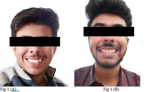
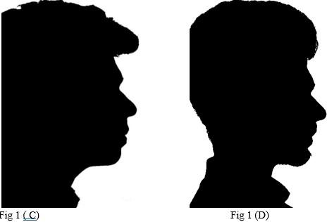
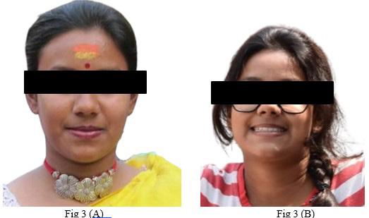
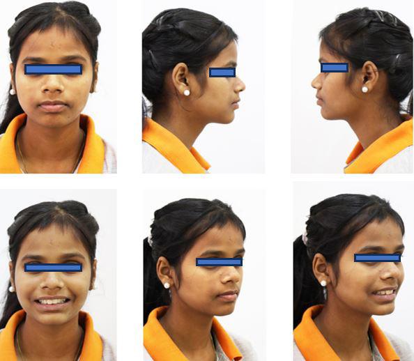
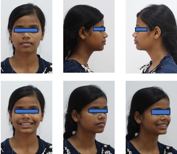
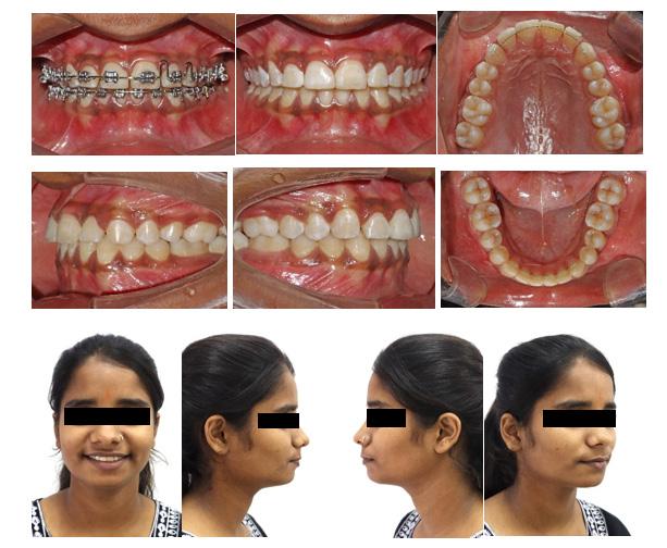
All selfies at T0 and T1 were cropped up to the clavicle of the subject if visible. The backgrounds of the selfies were removed and turned white with the help of Adobe Photoshop CC 2020 version 21.1 software package (Adobe Systems, San Jose, CA). One example of pre-treatment and post-treatment selfies of a subject in skeletal class I is illustrated in Figure 1A and 1B, in skeletal
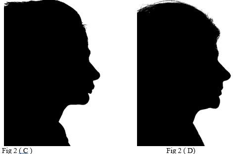
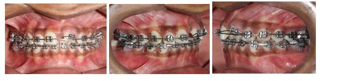
Figure 1: Skeletal Class I Group (SCI) Male.
A-Pre-treatment (T0) Selfie; B- Post-treatment (T1) Selfie; C. Pretreatment Profile Silhouette (T0). D. Post-treatment Profile Silhouette
Figure 2: Skeletal Class II Group (SCII) Female.
A-Pre-treatment Selfie (T0), B- Post-treatment Selfie (T1). C. Pretreatment Profile Silhouette (T0), D. Post-treatment Profile Silhouette (T1).
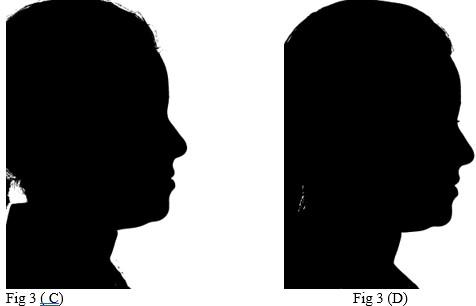
Figure 3: Skeletal Class II Group (SCII) Female.
A-Pre-treatment Selfie (T0), B- Post-treatment Selfie (T1). C. Pretreatment Profile Silhouette (T0), D. Post-treatment Profile Silhouette (T1).
class II is illustrated in Figure 2A and 2B, and in skeletal class III is illustrated in Figures 3A and 3B.
All profile photographs at T0 and T1 were cropped up to the clavicle of the subject. Profile photographs were converted into black silhouettes against a white background using Adobe Photoshop CC 2020 version 21.1 software package (Adobe Systems, San Jose, CA) One example of pre-treatment and posttreatment profile silhouettes of a subject in skeletal class I is illustrated in Figure 1C and 1D, in skeletal class II is illustrated in Figure 2C and 2D, and in skeletal class III is illustrated in Figure 3C and 3D.
In the first evaluation all selfies (T0 and T1) of all the three groups (SCI, SCII, SCIII) were randomly shuffled and shown to the examiners in each panel. Examiners were blinded as they were not informed if the selfies were taken before treatment or after treatment.
In the second evaluation profile silhouettes (T0 and T1) of each patient was placed side by side. In this way, all profile silhouettes (T0 and T1) of all the three groups (SCI, SCII, SCIII) were shuffled and shown to the examiners. Examiners were blinded as they did not know which profile silhouette was patient’s pre- treatment profile and which one was post treatment profile.
In both the evaluations examiners (GD, O, LP) were asked to rate the selfies and profile silhouettes on a 5-point Likert scale, which was as follows-1=Very unattractive, 2=Unattractive, 3=Acceptable, 4=Attractive, 5= Very attractive.
Data Analysis
The statistical analysis was done using statistical package of social sciences (SPSS) software v.22 (IBM, Chicago, USA). Mean values plus standard deviation of the scores given by GD, O and LP was derived for all the T0 and T1 samples in all the skeletal classes. The comparison of mean scores between T0 and T1 samples were calculated by using Paired “t” test. The difference was considered as significant, when the p value was below 0.05 and highly significant when p value was below 0.001.
Results
Profile Silhouettes
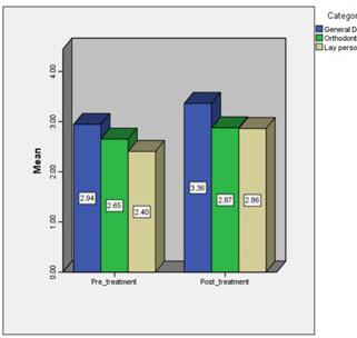
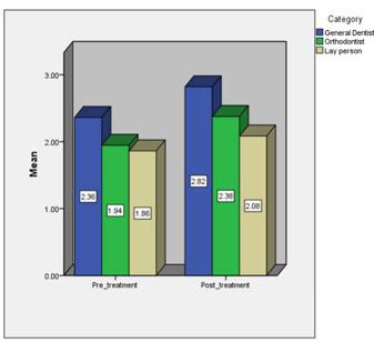
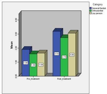
The results are divided into Skeletal Class I, Skeletal Class II, Skeletal Class III. Descriptive statistics of the scores among various skeletal relations of Pre-treatment (T0) and Post- treatment (T1) samples of profile silhouettes (PS) are listed in Table 1. In Skeletal Class I, there was a statistically highly significant difference(p<0.001) seen between the scores for T0 and T1 with higher values for (T1) score among general dentist group. There was statistically no significant difference (p>0.05) seen between the scores for T0 and T1 among Orthodontist and Laypersons. Comparison of T0 and T1 scores of GD, O, LP for SCI of PS is illustrated in Figure 4.
In Skeletal Class II, there was a statistically highly significant difference(p<0.001) seen between the scores for T0 and T1 with higher values for T1 score among General Dentists, Orthodontists and Laypersons. Comparison of T0 and T1 scores of GD, O, LP for SCI of PS is illustrated in Figure 5.
In Skeletal Class III- There was a statistically highly significant difference(p<0.001) seen between the scores for T0 and T1 with higher values for T1 score among General Dentists, Orthodontists
and Laypersons. Comparison of T0 and T1 scores of GD, O, LP for SCI of PS is illustrated in Figure 6.
Selfies
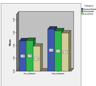
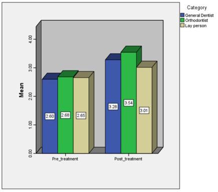
The results are divided into Skeletal Class I, Skeletal Class II, Skeletal Class III. Descriptive statistics of the scores among various skeletal relations of Pre-treatment (T0) and Post- treatment (T1) samples of Selfies (S) are listed in Table 2. In skeletal Class I, there was a statistically highly significant difference(p<0.001) seen between the scores for T0 and T1 with higher values for (T1) score among general dentist and orthodontist group. There was statistically no significant difference (p>0.05) seen between the scores for T0 and T1 among Laypersons. Comparison of T0 and T1 scores of GD, O, LP for SCI of Selfies is illustrated in Figure 7.
In skeletal Class II, there was a statistically highly significant difference(p<0.001) seen between the scores for T0 and T1 with higher values for (T1) score among general dentist and orthodontist group. There was statistically no significant difference (p>0.05) seen between the scores for T0 and T1 among
Table I: Descriptive statistics of the scores among various skeletal relations of Pre-treatment (T0) and Post-treatment (T1) samples of profile silhouettes (PS)
Table 2 Descriptive statistics of the scores among various skeletal relations of Pre-treatment (T0) and Post-treatment (T1) samples of profile silhouettes (PS)
of
post
Figure 4: Comparison of the scores of pre-treatment (T0) and posttreatment (T1) of skeletal class I (SCI) samples of Profile silhouettes (PS)
Figure 5: Comparison of the scores
Pre-treatment (T0) and
treatment (T1) of skeletal class II (SCII) samples of Profile silhouettes (PS)
Figure 6: Comparison of the scores of Pre-treatment (T0) and post treatment (T1) for skeletal class III (SCIII) samples of Profile silhouettes (PS)
Figure 7: Comparison of the scores of Pre-treatment (T0) and post treatment (T1) for skeletal class III (SCIII) samples of Profile silhouettes (PS)
Figure 8: Comparison of the scores of Pre-treatment (T0) and post treatment (T1) of skeletal class II (SCII) samples of selfies (S)(PS)
Figure 9: Comparison of the scores of Pre-treatment (T0) and post treatment (T1) for skeletal class III (SCIII) samples of selfies (S)
Laypersons. Comparison of T0 and T1 scores of GD, O, LP for SCII of Selfies is illustrated in Figure 8.
In skeletal Class III, there was a statistically no-significant difference (p>0.05) seen between the scores for T0 and T1 among General Dentists, Orthodontists and Laypersons. Comparison of T0 and T1 scores of GD, O, LP for SCIII of Selfies is illustrated in Figure 9.
Discussion
Facial attractiveness is an inherent desire of every patient who walks into an orthodontist’s office. In day to day lives we interact with others, eye to eye or face to face. Hence it is important that as orthodontists we make a conscious effort to enhance facial attractiveness along with providing an ideal occlusion. Soft tissue profile greatly influences the treatment plan and outcome for every patient. Hence an orthodontist is greatly dependent on soft tissue profile for his perception of facial attractiveness.5 However, a general dentist or layperson may not necessarily look at the profile of the patient to form an opinion about facial attractiveness.
In our study we focused on profile silhouettes as for an orthodontist profile view is most important during treatment planning. Profile silhouettes were chosen to obtain unbiased ratings on facial attractiveness.
However, facial attractiveness is very subjective. We hardly ever interact or see each other in profile views; hence it is imperative that we also take other photographic views into consideration. An orthodontist should also look at candid photographs, selfies, etc to form a broader picture about the soft tissue layout of the patient.
With the advent of smartphones, every person now owns a high-definition camera with which they record each and every moment of their lives by taking photographs, selfies and videos. Every person is now living dual lives. One in the real world and another in the virtual world of social media. There is an unhealthy tendency to attach one’s self-worth to the number of likes and comments on their pictures and selfies on the internet. Therefore, this constant influence of selfies on the self-esteem of an individual along with comments from peers on social media lead to these individuals seeking orthodontic treatment or other cosmetic procedures. Therefore, it is imperative that we take a look at patient’s selfies as it will provide us a thorough insight on what soft tissue aspect we need to enhance, and which aspect needs to be preserved. All of this will eventually contribute to increased facial attractiveness which should be our ultimate goal.
Speaking of profile silhouettes first, in Skeletal Class I relation, general dentists could appreciate the increase in facial attractiveness after orthodontic treatment whereas laypersons and orthodontists did not find any appreciable increase in facial attractiveness.
In Skeletal Class II relation, General Dentists, Laypersons and Orthodontists, could appreciate the increase in facial attractiveness between pre- treatment and post treatment profile silhouettes. These findings indicate that in skeletal class II group, the changes in profile were so drastic that all three panels were able to easily appreciate and identify it. These findings correlate with the study done by Paduano10 who concluded that the silhouettes of Class II post treatment individuals were more attractive than those of the class II
Pre-treatment and class I groups.
In Skeletal Class III relation, General Dentists, Laypersons and Orthodontists, all reported an increase in facial attractiveness between pre- treatment and post treatment. These findings indicate that in skeletal class III relation group, the profiles are so significantly improved that the changes were appreciable to all three groups. These findings correlate to the study conducted by Watanabe and Fitarelli 11 who concluded that the post treatment silhouettes of class III subjects treated by surgical or compensatory methods were significantly more attractive than Pre-treatment groups.
In our study, regardless of statistical significance it was observed that post treatment groups of all three skeletal relations showed higher scores than pre-treatment groups. Hence it can be concluded that orthodontic treatment can enhance facial attractiveness which can be perceived in profile silhouettes.
To the best of our knowledge, the current study is the first of its kind in the Indian population in which pre-treatment and post treatment selfies of orthodontically treated patients are compared to each other in different skeletal classes. In our study we used selfies to assess overall facial attractiveness in different skeletal relations. Our aim is to find out which skeletal relation showed the most appreciable enhancement in facial attractiveness after orthodontic treatment. We further tried to assess which group of panellists (General Dentists, Laypersons and Orthodontists) were more sensitive towards the changes in facial attractiveness in selfies.
In Skeletal Class I relation, General Dentists and Orthodontists reported an increase in facial attractiveness between pretreatment and post treatment selfies.
In Skeletal Class II relation,orthodontists and general dentists both could appreciate the increase in facial attractiveness in selfies.
In Skeletal Class III relation, General Dentists, Laypersons and Orthodontists found no increase in facial attractiveness between pre-treatment and post treatment selfies. Hence it can be inferred that in skeletal class III relation there was no appreciable changes in facial attractiveness in post treatment selfies as compared to pre- treatment selfies.
Selfies have the potential to play a major role for orthodontists. Regardless of statistical significance, the mean scores of all post treatment groups in skeletal class I, II and III was higher than the mean of pre-treatment groups for all the three panels of General Dentists, Orthodontists and Lay Persons. Hence, it can be concluded that orthodontic treatment contributes to enhancement of facial attractiveness, a change which can be perceived in selfies specifically in skeletal class I and skeletal class II relations.
We faced some limitations while carrying out this research. Although we tried to reduce the noise in selfies as much as possible by removing the background and cropping the selfies up to clavicular level, still standardization of selfies is not possible. Usage of various filters, altering of selfies etc, influence the attractiveness of the selfies, hence their final ratings can also be affected. As the selfies were taken by the patients with their smartphones, camera quality, lighting, distance from which the selfies could have also influenced the final rating of the selfies. Patients with better smartphone cameras had selfies of better quality.
Future studies on selfies can be carried out by finding out a means to standardize selfies. Raw, unaltered selfies should be used to analyse actual facial attractiveness. Old, poor-quality selfies should be discarded, and selfies taken from good cameras or high-definition selfies should be used. To find correlation between selfies and profile silhouettes, more elaborate studies consisting of more panellists and larger sample size are required to explore this aspect. More studies could be conducted on pre-treatment and post treatment selfies in different treatment modalities, for example- extraction versus no-extraction, surgery versus camouflage etc.
References
1. Duchess D. (1878). Molly bawn (Copyright). Bernhard Tauchnitz.
2. Samsonyanová L, Broukal Z. A systematic review of individual motivational factors in orthodontic treatment: facial attractiveness as the main motivational factor in orthodontic treatment. International journal of dentistry. 2014 May 20;2014.
3. Ren H, Chen X, Zhang Y. Correlation between facial attractiveness and facial components assessed by laypersons and orthodontists. Journal of dental sciences. 2021 Jan 1;16(1):431-6.
4. Ackerman JL, Proffit WR, Sarver DM. The emerging soft tissue paradigm in orthodontic diagnosis and treatment planning. Clinical orthodontics and research. 1999 May;2(2):49-52.
5. Kerr WJ, O’donnell JM. Panel perception of facial attractiveness. british Journal of Orthodontics. 1990 Nov 1;17(4):299-304.
6. Bar rer JG, Ghafari J. Silhouette profiles in the assessment of facial esthetics: a comparison of cases treated with various orthodontic appliances. American journal of orthodontics. 1985 May 1;87(5):385-91.
7. Behl DP. Selfies and Orthodontics-A Narrative Review. MAR Dental Scinces.;5.
8. Oxford Dictionaries, The Oxford Dictionaries word of the year 2013, available from: http://blog.oxforddictionaries.com/press-releases/oxforddictionariesword-of-the-year-2013/.
9. Shome D, Vadera S, Male SR, Kapoor R. Does taking selfies lead to increased desire to undergo cosmetic surgery. Journal of cosmetic dermatology. 2020 Aug;19(8):2025- 32.
10. Paduano S, Rongo R, Bucci R, Carvelli G, Cioffi I. Impact of functional orthodontic treatment on facial attractiveness of children with Class II division 1 malocclusion. European Journal of Orthodontics. 2020 Apr 1;42(2):144-50.
11. Julie-Heide-Miyazaki Watanabe FF, de Freitas DS, Cançado RH. Comparison of the facial profile attractiveness in Class III borderline
Growing Beautiful Teeth Chapter 8: A Pain-Free Life
Estie Bav is an active member and senior instructor of IAO. She graduated BDSc from the University of Western Australia, and practises in her own private family dental surgery in Melbourne Australia. In November 2018 she published her first book titled “Growing Beautiful Teeth,” primarily targeting parents, grandparents, teachers or any child health carer to look out for early signs of dental growth issues. It informs the unaware the importance and impact of teeth and jaw on other areas of health such as breathing, sleep, posture, and even behaviour.
Currently the dental profession tends to “supervise and wait” for growth issues to become complex and expensive to correct….”
“My concern is that most parents miss out on basic and important dento-facial growth information until too late.”
The book was designed to be a helpful resource for your patient to read, and for introducing the subject to younger dentists and allied health professionals who may not be familiar with the teeth-occlusion-airway-TMJ-sleep paradigm.
Her message is to get involved with a child’s dento-facialairway development early.
Growing Beautiful Teeth is available from any major online booksellers, or at
• www.drestiebav.com
• www.growingbeautifulteeth.com
She can be reached at estie@drestiebav.com
How my book can be helpful….
It takes time to educate parents on the benefits of treating dental growth issues early and explaining what signs we look for. In writing this book in simple language I hope to bring an awareness to the larger parent community, which will in turn save my dental colleagues chairside time. This book would be a helpful resource for the waiting room, and for introducing the concept to younger colleagues joining your practice.
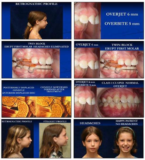
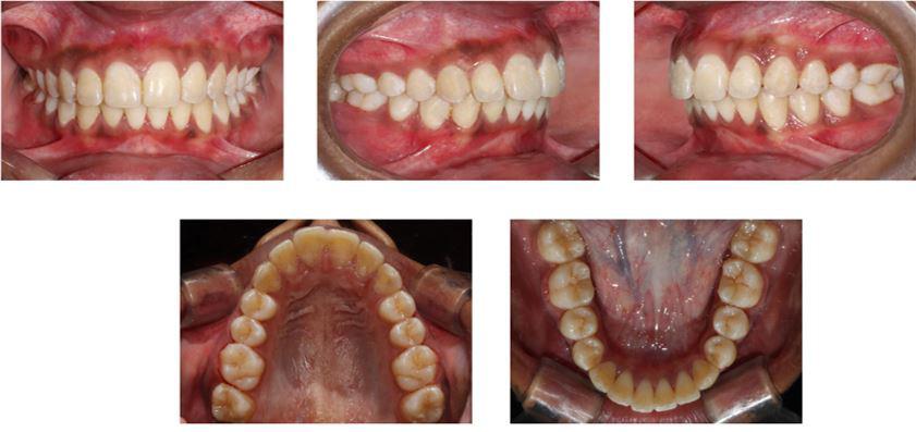
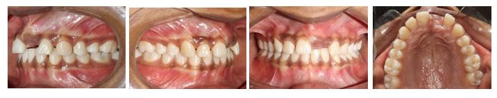
In this chapter, I want to show the reader some typical adult patient cases that I have treated for jaw and tooth problems and pain, who also have small maxilla, poor jaw alignment and a compromised pharyngeal airway. Name initials are used here to protect the identity of the persons.
FO’s Narrow Maxilla and TMD Pain
FO is a 30-year-old female with chronic and debilitating jaw and facial pain. Her range of jaw opening/closing is limited. She had done a circuit of seeing medical doctors and had taken pain medications. She had been treated by several physical therapists for a period of 8 months but the symptoms continued to affect her daily life and her work as a social worker.
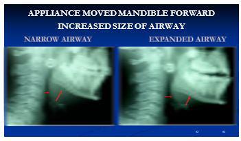
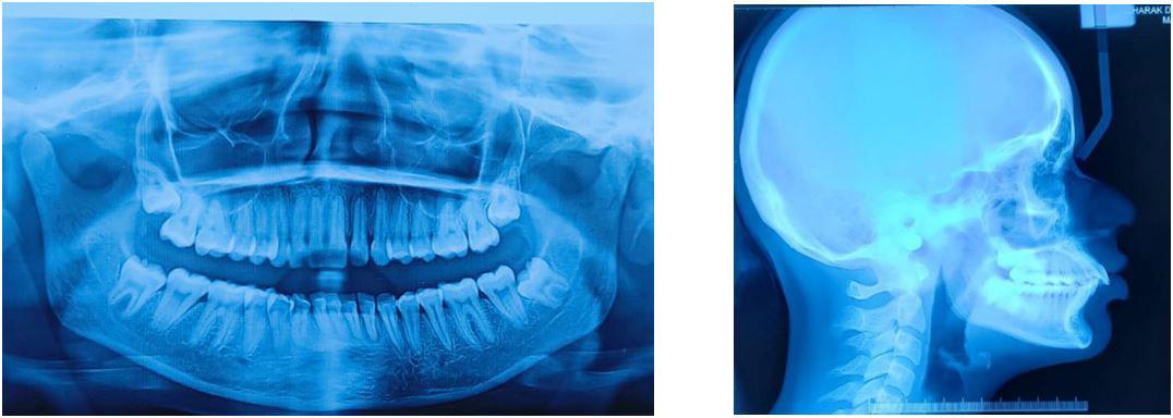
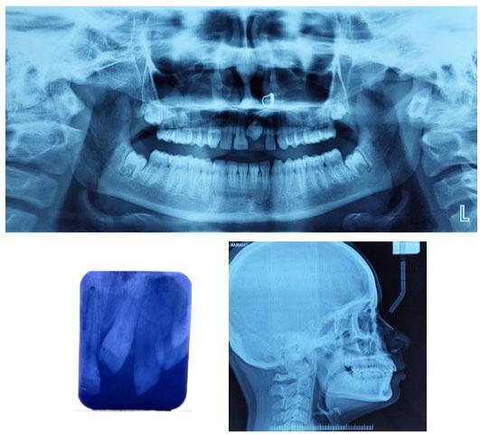
A comprehensive study of her teeth, jaw, head and facial muscles which included CBVT imaging of her jaw joints and a sleep test found that she had set back maxilla and mandible, compressed TMJs and oxygen desaturation during sleep with a sleep disturbance index higher than is considered acceptable. She was observed to have a mouth-breathing habit and she was not aware of the importance of breathing through her nose.
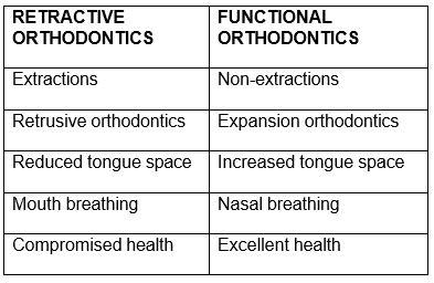
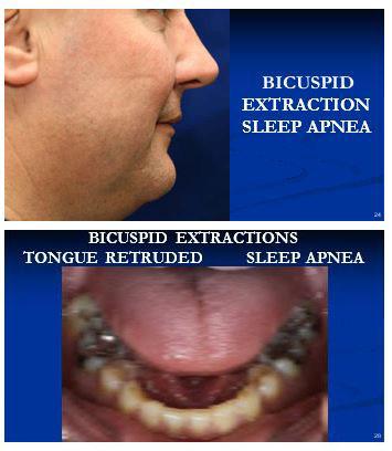
FO in fact had orthodontic treatment in her teens whereby eight permanent teeth had been removed, leaving her with a small maxilla and a lower jaw that was held back.
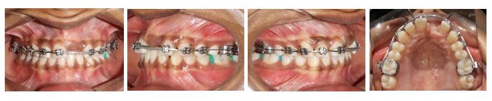
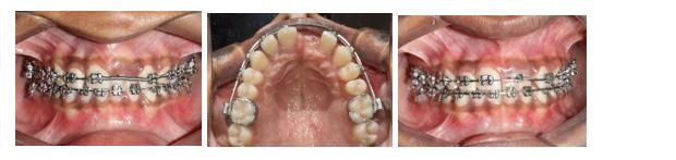
After her jaw pain symptoms were treated with dental splints, she had chosen to have a second course of orthodontic treatment but this time with the goal of widening her maxilla and bringing her jaws more forward to correct the problems and to provide a longer-term resolution.
Some of the teeth that were extracted in her teens have now been replaced with artificial teeth (and implants) that can fit in the treated and wider dental arch.
She has learned the importance of the Big 3 (see Chapter 3, p.22) and she now breathes and sleeps better. The whole series of treatment took several years to complete and presented a significant financial challenge for FO. It was also difficult to take time off work for the treatment.
MK’s Narrow Maxilla and TMD Pain
MK is a 56-year-old female with jaw and facial pain, tooth sensitivity, sleeping problems and depression. She had been seeing medical doctors and specialists, counsellors, physical therapists and taken significant amounts of different medication to control the above symptoms.
MK had orthodontic treatment during her teenage years when four premolar teeth were removed, and the remaining teeth aligned. A few years later, her wisdom teeth were removed as well. Having lost eight of her adult teeth meant that the remaining 24 now appeared well lined up, albeit in smaller dental arches. Though her teeth were reasonably straight, the upper and lower jaws were not relating well and her TMJs were compressed. Comprehensive study and analysis of her teeth, occlusion, TMJs, facial profile, breathing habits and sleep concluded that MK still has narrow dental arches, inadequate tongue space, set back jaws, compressed TMJs and nightly sleep disturbance that may be attributed to choking. MK was habitually breathing through her mouth and had incorrect tongue posture away from her palate.
Initial treatment with dental splints were successful in improving most of her pain symptoms. She is now considering yet another course of orthodontic treatment, possibly in conjunction with surgery, to maximise her maxilla and to allow the mandible to track more forward and enable her tongue to posture correctly. The aim is to improve her airway so that she can breathe and sleep better.
The splint therapy and a planned second round of orthodontic treatment to attempt to max her maxilla will be setting her back several
tens of thousands of dollars. She will also have to have the extracted teeth replaced in the finally treated dental arches. Without this course of treatment to provide more oral space she is likely to continue with TMJ pain, sleep deprivation and debilitation for the rest of her life.
CW’s TMD Pain and Headaches, Class III with Short Maxilla
CW is a 35-year-old male floor manager for a busy industrial firm and was referred to me by his physical therapist who had been treating him for chronic and debilitating jaw and facial pain. He reported that he had been suffering with pain in the jaw area for over 10 years. Physical therapy and medication had provided only temporary relief.
CW’s teeth were sound with no cavity. His dental history included four years of orthodontic treatment in his late teens to correct a prominent and crooked appearance of his upper incisors. He recalled having to use some head-gear with elastic force to pull the upper-front teeth back. At the end of the orthodontic therapy, he had four wisdom teeth removed under general anesthesia due to lack of space in his dental arches
A comprehensive study and analysis of CW’s teeth and jaw showed that he (still) has small dental arches that can accommodate only 28 out of the full complement of 32 teeth. Although his teeth were reasonably straight, the maxilla was short lengthwise and the mandible was trapped. His left-side TMJ was compressed. He also snored, and clenched and grinded his teeth in his sleep. A sleep study conducted showed reduced oxygen saturation and a high index of sleep disturbance. CW habitually breathed through his mouth.
Splint therapy that repositioned his mandible has been able to provide significant symptom relief for CW but is a great inconvenience for him due to the interference with his speech that is important in his work. For a longer-term resolution, he is considering treatment to max his maxilla to free the mandible and TMJs, and to provide more oral space for his tongue and to improve pharyngeal airway.
This planned course of treatment will present a financial and time challenge for CW at this stage of his life as he must take into consideration his situation at work and his new family.
OT’s Class III, Chronic TMD, Back Pain, Bruxing and Broken Teeth
OT is a 57-year-old male. This patient has 12 missing teeth from his mouth. He has lost these teeth one by one over the years due to the stress and breakage from perpetual and severe clenching and grinding. Whilst there is insignificant crowding of his remaining 20 teeth, his upper dental arch is small and his upper and lower jaws are mismatched.
OT has a Class III occlusion with the lower arch constrained by a smaller upper dental arch. Though this discrepancy is not obvious in the first instance, it is enough to have put pressure on the condyles, and to leave insufficient space for his tongue. He reported symptoms of chronic jaw pain, headaches, neck and back pain, and poor-quality sleep all his adult life.
Although his teeth can be repaired, he would ideally need to have upper dental arch made wider and brought more forward, his occlusion repositioned to provide improved support for his TMJs and increased oral space for his tongue so that his airway during sleep is optimal which will give him sound quality sleep.
However, he is now at a stage in his life that to consider the comprehensive but optimal treatment plan to rid his jaw pains is out
of reach. Therefore, it is expected that as time goes by he will continue to suffer gradual breakdown and loss of the remaining teeth, one by one, despite the best restorative repair he can afford. It is also likely that he will continue to suffer the various chronic pains.
NI’s
Deep Overbite, Retruded jaw and OSA
NI is an 80-year-old female who has lost half her teeth. Her remaining teeth are slightly crooked but presented as a deep overbite, collapsed support for her TMJs, a constrained tongue and she uses a Continuous Positive Air Pressure (CPAP) every night to treat her OSA.
Despite her age, I still offered her a comprehensive and doable treatment plan because I wanted to help her rehabilitate her teeth and mouth so that she can eat and enjoy her food, and obtain the necessary nutrients right up to her last meal in her life. Apart from restoring her dentition, I also initiated a conversation about correct orofacial muscle patterns and the all-essential function of nasal breathing during waking hours and sleep.
I knew that once she can eat well, breathe well and sleep better, she would be in a much better position to tackle other degenerative conditions that she has. Perhaps the extensive treatment plan that I carried out for her might not have been necessary had she received good dental growth guidance from a young age.
In fact, the many thousands of dollars that she was spending on dental restorations and dentures at her age might have been put to more advantageous use much earlier, as an investment for a lifelong enjoyment of a full complement of sound and healthy teeth, good occlusion, forward jaws, good airway and sleep. Perhaps the CPAP unit that she is using now may not have been necessary, nor the medication for reflux that she has been taking for the last couple of years.
Conclusion
The above examples show how most of my adult patients with symptoms of tooth breakdown from bruxing share the common characteristics of an undersized maxilla, inadequate room for the tongue, poor breathing habit, sleep disordered breathing and poor sleep. About 60% even had a history of past orthodontic treatment that simply treated their crooked teeth by removing sound teeth but had not addressed these other related factors in a bigger, totalhealth paradigm.
It is challenging to help these patients as their dental, oral, and jaw symptoms are advanced.
The more effective and right approach would have been to begin from the beginning and initiate proper jaw growth in the newborns and pay early attention to the child’s breathing. This approach will mitigate many chronic pains relating to the jaws throughout life. The bonus is that with a properly developed maxilla, and with correct habits such as the Big 3 in place, the teeth will naturally align well, giving the child a broad and beautiful smile for life.
Of course, there could be other causes of TMD such as trauma and injury from accidents and other chronic systemic illness such as arthritis which, of course, must be taken into consideration in treatment planning.
Dentist must join the dots and see beyond the tooth problems that their patient routinely present. They must take the initiative to educate parents of young patients and guide the child’s dentofacial growth early, to maximize their genetic potential to have broad and beautiful jaws, plenty of room for their teeth to line up naturally, nice airway to breathe well and sleep well, and space for their TMJ’s to grow and function without limitation or pain.
The Importance of Myofunctional Therapy in Orthodontics, Part 1
Dr. Rondeau is one of North America’s most sought after clinicians who lectures over 50 days per year for 35 years. He is the past president and master senior certified instructor for the International Association for Orthodontics. Over 24,000 dentists have attended his courses and study clubs. He has an extremely busy practice, which is limited to the treatment of patients with orthodontic, orthopedic, TMD, and snoring and sleep apnea problems. His new textbook on Early Orthodontic Treatment for Children is now available and all his courses are also available live or online, on demand. Go to www.rondeauseminars.com for all the details or email lee@rondeauseminars.com.
The key to optimum health is that all patients must have a patent airway. Orthodontic treatment and myofunctional therapy will help achieve these goals. Mouth breathing is one of the main problems. Mouth breathing can cause poor tongue function. Proper tongue function requires the tongue to fit completely on the roof of the mouth during swallowing. This helps keep the maxilla expanded to prevent malocclusions (teeth crowding), TM dysfunction and sleep disorders (snoring and sleep apnea). The first step would be to orthopedically expand the maxillary arch to normal with an expansion appliance, either removable or fixed. When the maxilla is expanded it frequently converts a mouth breather to a nasal breather. Nasal breathers get 20% more oxygen then mouth
breathers which is important from a total health standpoint.1 The second step would be to have the myofunctional therapist retrain the tongue to achieve the correct swallow so the expanded maxilla will not relapse. An abnormal swallow could also cause dysfunction of the tongue which could cause an anterior open bite. The myofunctional therapist can help solve this open bite also by prescribing tongue exercises to encourage a normal swallow.
Constricted Upper Arch
The width of the maxilla in permanent dentition measuring from the lingual of the first molars is 37-39 mm. The width of a cotton roll is 37 mm. The intermolar measurement should be done routinely on all children and adults. As the photo below demonstrates, if the arch is constricted this causes crowding of the anterior and frequently the lateral incisors erupt on the palate. Mouthbreathing and extractions causes collapse of upper and lower arches. Treatment of choice would be to use expansion appliances to widen the arches to make room for all the permanent teeth and the nasal airway to encourage nasal breathing. Nasal breathers will have a normal swallow pattern where the tongue goes to the roof of the mouth during swallowing and will increase the size of the maxilla approximately ½ mm per year. Therefore, measure the distance between the upper first molars in millimeters. As an example, if the patient is age 10, female, she will grow another 5 years and therefore the maxilla will increase approximately 2 ½ mm. This will help you decide whether or not you need to increase the width of the upper arch to
make room for all the permanent teeth and also create adequate space for the tongue to function normally. You will have to think much differently if the child is a mouth breather as the tongue will not expand the maxilla during swallowing as the tongue drops down to the lower arch. Mouth breathers will almost routinely require maxillary arch expansion to make room for the permanent teeth. Another clinical tip is that you should expand the upper and lower arches with removable or fixed appliances until there is adequate space for the permanent upper and lower lateral and central incisors.
The expansion of the maxilla is one of the most important treatments that a child can receive in preventing future sleepdisordered breathing problems such as mouth breathing, snoring and obstructive sleep apnea. When the maxilla is expanded the nasal airway is increased transversely.2 When the maxilla is expanded the palate drops which increases the nasal airway vertically. The roof of the mouth (palate) is the floor of the nose so arch expansion is the key to success. The objective is to try and convert the child from being a mouth breather to being a nasal breather.
Many authorities believe that the cause of the Class II skeletal malocclusion is an airway obstruction which causes a constriction of the maxillary arch causing the mandible to retrude in order to achieve a satisfactory occlusion.3 A large percentage of these Class II skeletal patients with retrognathic mandibles develop TM dysfunction. When they swallow, their condyles are posteriorly displaced and impinge on the nerves and blood vessels in front of the ear which causes considerable discomfort. Symptoms
include headaches, ringing in the ears, stuffiness in the ears, neck pain, fainting, dizziness, difficulty swallowing, pain behind the eyes, clicking noises when opening and closing, limited opening, numbness in the hands. Obviously, early treatment to expand the maxilla, retrain the tongue and prevent the Class II skeletal malocclusion would be the ideal scenario. The medical profession is trained to treat the symptoms of TM dysfunction with muscle relaxants, pain medication, anti-inflammatories and even anti-depressants. Only an orthodontic clinician working with a myofunctional therapist can achieve the best treatment to successfully treat the symptoms by eliminating the cause of the TM dysfunction. The treatment for the Class II skeletal malocclusions would be to expand the maxilla to normal with an expansion appliance, retrain the tongue to swallow normally and also utilize a functional jaw repositioning appliance to move the lower jaw forward to its correct Class I skeletal position.
This Class II skeletal malocclusion with a normally positioned maxilla and an underdeveloped mandible often causes the tongue to retrude which further obstructs the airway. This can cause sleep disorders, including snoring, obstructive sleep apnea or upper airway resistance syndrome, all of which can negatively affect the health of the child or adult.
Receding Lower Jaw Case #2
• Female, Age 9
• Class II Skeletal malocclusion Normally positioned maxilla • Retruded lower jaw
• Prevents snoring and sleep apnea by opening the airway.\ Moving lower jaw forward 6 mm. Increasing the vertical dimension by correcting the 5 mm overbite by extruding the lower posterior teeth.
• Both increased the size of the airway.
• Posteriorly displaced condyle compressing nerves distal to condyle. Condyle down and forward; increased space distal to condyle for nerves and blood vessels. Headaches eliminated.
Treatment Class II Skeletal Malocclusion
I have outlined two different treatment plans for the treatment of Class II skeletal malocclusion:
1. Functional Philosophy
Treat early in mixed dentition using functional appliance to expand the arches to normal and to use functional appliances to move the retrognathic mandible forward to its proper position. Patients are treated without the extraction of permanent teeth or surgery. When the mandible is advanced this corrects the overjet.
Methods to open the airway: Functional Appliances.
1. Expand the maxilla opens the nasal airway.
2. Erupt the lower posterior teeth to correct deep overbite, open the airway vertically.
3. Advance the mandible open the pharyngeal airway.
2. Retractive Philosophy
Treat in the permanent dentition when all the permanent teeth erupt and 90% of the face is developed. Missed opportunity to guide the growth of the child.
Extract the upper bicuspids to retract the six anterior teeth in order to correct the overjet.
Results of bicuspid extraction: closes the airway.
1. Failure to expand constricted maxilla; does not open the nasal airway.
2. Intrusion of upper or lower incisors in order to correct deep overbite does not open the airway.
3. Retraction of the upper anterior teeth following bicuspid extractions closes the pharyngeal airway.
Since a frequent cause of the Class II skeletal malocclusion is mouth breathing resulting in a constriction of the maxillary arch which causes the mandible to go retrognathic the treatment to me seems very obvious. Just remove the problem that caused the malocclusion. Find out the cause for the mouth breathing: enlarged tonsils or adenoids, allergies, nasal obstruction. Then expand the maxilla to its proper size and width. Then move the mandible forward to an ideal size maxillary arch with a functional repositioning appliance.
The vast majority of Class II skeletal malocclusions have a normal maxilla and a retrognathic (under developed) mandible. Another way to treat Class II skeletal malocclusions with
retrognathic mandibles is to extract the upper first bicuspids and retract the upper six anterior teeth back to close the extraction sites. To retract the maxilla when clearly the problem is an undeveloped mandible is completely illogical. As mentioned earlier in many cases this also predisposes patients to TM dysfunction and sleep apnea. Frequently when the upper
bicuspids are extracted the lower bicuspids are also extracted which can decrease the size of the lower arch by up to 16 mm. This much smaller lower arch creates a serious problem for the tongue. Due to lack of space the tongue retrudes to the back of the throat. This is particularly a serious problem when the patient sleeps supine and the tongue falls back and blocks the airway. Bicuspid extractions flatten the face, significantly reduces the width of the smile, closes the airway, moves the tongue back and increase the future risk of snoring and sleep apnea.
Symptoms include high blood pressure, heart attack, stroke,
Type 2 diabetes, atrial fibrillation, acid reflux, kidney problems, 5 times greater chance of cancer, dementia, Alzheimer’s.4,5,6,7,8,9,10
The extraction of the upper bicuspids causes the maxillary arch to become narrower and the tongue to go to the floor of the mouth. This creates an abnormal swallow. The solution would be to expand the upper and lower arches with expansion appliances and then once space has been created for the tongue, have myofunctional therapy to retrain the tongue to allow for a normal swallow to help expand the maxilla and prevent relapse. First establish a proper form (orthodontically) and then achieve proper function (myofunctional therapy).
In order to achieve a patent airway it is important to evaluate the size of the tonsils and adenoids. If enlarged, you must take photos and send letters to the family medical doctors to request referrals to an ENT Specialist for removal.
Functional Philosophy
As I mentioned at the beginning the key to optimum health is to create and maintain a patent airway. We must keep these objectives in mind whatever technique is utilized to treat the malocclusion. The orthodontic clinician must be responsible for making room for the tongue by creating space by expanding the upper and lower arches.
In the treatment of the Class II skeletal malocclusion it is much preferable to treat by moving the lower jaw forward with functional repositioning appliances that open the airway. The myofunctional therapist is extremely important to help diagnose and treat tongue ties and to retrain the tongue to create the normal swallow to prevent relapse of the orthodontic case.
The treatment of choice would be to use a fixed or removable functional appliance to expand the lower arch to make more room for the tongue. This provides the patient with a wider lower arch, more room for the tongue which encourages patient to have their speech improved.
Orthodontic clinicians are encouraged to utilize the expertise of a myofunctional therapist in their offices. We have been utilizing the services of Alicia Lewis, myofunctional therapist, in our office for the past 2 years and that has been a great asset to the practice and to the patients. I wanted to co-write and article with her.
REFERENCES
1. Recinto c. Efthemeou T. Boffelli PT, Navalta JW. Effects of Nasal or Oral Breathing on Anaerobic Power Output and Metabolic Responses. Int. J Exerc Sci, 2017 Jul 1:10(4):506-514. PMID: 28674596: PMCID: PMC5466403.
2. Mew, J. (2015). The influence of the tongue on dentofacial growth. Angle Orthodontist. 85(4), 715-716.
3. Har vold EP, Torner BS, Vargervik K, Chierici G. Primate experiments on oral respiration. Am J Orthod. 1981 Apr;79(4):359-72. doi:10.1016/0029416(81)90379-1. PMID: 6939331.
4. Shahar E, Whitney CW, Redline S. et al. Sleep-disordered breathing and cardiovascular disease: cross-sectional results of the Sleep Heart Health Study. Am J Respir Crit Care Med. 2001;163:19-25.
5. Dyken ME, Somers VK, Yamada T. Re ZY, Zimmerman MB. Investigating the relationship between stroke and obstructive sleep apnea. Stroke. 1996;27:401407.
6. Peppard PE, Young T, Palta M, Skatrud J. Prospective styd of the association between sleep-disordered breathing and hypertenstion. N Engl J Med.2000;342:1378-1384.
7. Hoffstein V, Mateika S. Cardiac arrhythmias, snoring, and sleep apnea. Chest. 1994:106:466-471.
8. Somers VK, White DP, Amin R, Abraham WT, Costa F, Culebras A, Daniels S, Floras JS, Hunt CE, Olson LJ, Pickering TG, Russell R, Woo M, Young T. Sleep apnea and cardiovascular disease: an American Heart Association/American College of Cardiology Foundation scientific statement from the American Heart Association Council for High Blood Pressure Research Professional Education Committee, Council on Clinical Cardiology, Stroke Council and Council on Cardiovascular Nursing Council. J Am Coll Cardiol 2008:52:686-717.
9. Babu AR, Herdegen J. Fogelfeld L, et al., “Type 2 diabetes, glycemic control and continuous positive airway pressure in obstructive sleep apnea”, Arch Intern Med, 165, 447-452, 2005.
10. Alber t, AM, & Wright CL (2016). The progression of Craniofacial growth and Development: An anthropological study applicable to the forensic and identity sciences. Global Journal of Anthropology Research, 3(1), 16-21.
Two-Stage Treatment : The Best of Both Worlds (Functional and Fixed Orthodontics)
AUTHORS
Dr. Gulnaz Husain
Senior Resident, King Georges Medical University, India
Dr. Gyan P Singh
Professor at Department of Orthodontics and Dentofacial Orthodontics, KGMU Lucknow
Dr. G K Singh
Professor and Head of Department of Orthodontics and Dentofacial Orthodontics, KGMU Lucknow
Dr. Zainab Bint Shams
junior Resident, Department of Orthodontics and Dentofacial Orthodontics, KGMU Lucknow
Dr. Syed Armaan Hussain
junior resident, Department of orthodontics and Dentofacial Orthodontics, Career Postgraduate institute of Dental and Medical Sciences, Lucknow.
*This article has been peer reviewed
ABSTRACT:
The Twin block appliance is an effective functional appliance for attaining great skeletal corrections in growing patients. Two-stage treatment involves usage of a functional appliance during the active phase to move the mandible in its correct orthopaedically stable position until the correction of overjet and overbite are attained, followed by fixed appliance therapy for further refinement and finishing of the occlusion.
Keywords: functional appliance, twin block, class II div 1 ,orthopaedics, two phase therapy
INTRODUCTION:
In a two-stage treatment, the removable twin block appliance is used during the active phase to move the mandible forward until the correction of overjet and overbite are attained.1 When that occurs, first molars will be in touch, and the maxillary and mandibular incisors will be in a harmonious relationship at that point.2 The appliance must be worn for at least 7-9 months in order to prevent the patient from developing a dual bite.
The support phase of treatment begins after the active phase is over. Up until the posterior occlusion is entirely integrated, the corrected incisor relationship is maintained using an upper removable appliance with a steep anterior inclined plane. This typically takes 4-6 months and is continued for an additional 3-6 months to allow for the muscle complex’s functional reorientation. Once this functional appliance phase is completed, fixed orthodontic treatment is essential for the maintenance of the skeletal correction obtained, the settling of the occlusion, and the refinement of any uncorrected dental discrepancy.
CASE REPORT
A 16-year-old female patient presented with a chief complaint of forwardly placed and spacing between the upper front teeth
and gaps between teeth.
Extraoral:
She was mesocephalic, mesoprosopic with convex facial profile and incompetent lips. The incisal show at rest was 4–5 mm with everted lips (Figure 1).
Intraoral:
She had an Angle’s class II molar relation on both sides with overjet of 14 mm and overbite of 5 mm, with mild crowding and rotation in lower arch with coinciding midlines (Figure 2).
2: Intraoral
Radiographic findings:
The radiographic findings are shown in Figure 3.
CVMI Stage 4-5
• SNA 88, SNB 78 (skeletal class II)
• Xecreased mandibular plane angle 23°
• AO ahead of BO by 4 mm
Dr. Gulnaz Husain, Dr. Gyan P Singh, Dr. G K Singh, Dr. Zainab Bint Shams, and Dr. Syed Armaan Hussain
Figure 1: Extraoral facial profile
Figure
• UI to NA 8 mm, 47°
• LI to NB 4 mm, 22°
• Interincisal angle 102° (proclined incisors)
• IMPA 91°
• Base plane angle 30°
• Inclination angle 92°
• Upper lip strain 6 mm
Orthopantomogram (OPG) revealed bunching of roots with respect to lower anteriors.
Diagnosis
• Class II skeletal base with prognathc maxilla and retrognathic mandible
• Class II molar and canine relation
• Maxillary dentoalveolar proclination with incompetent lips
• Convex facial profile
• Hypodivergent growth pattern
Visual Treatment Objective was positive, so a treatment plan involving cervical pull headgear to restrict the maxillary growth along with mandibular advancement with a fixed twin block was considered. (Figure 4)
Treatment objectives
• Correction of proclination of upper anteriors
• Reduction of overjet and overbite
• Decrowding and arch alignment
• Achieve Class I molar and canine relationship
• Enhance facial esthetics
Treatment plan
• Myofunctional appliance therapy to restrict the maxillary growth and advance the mandible
• Using Pre-adjusted edgewise appliance 0.022 slot Roth prescription -Leveling and alignment
• Retention plan – Upper and lower removable retainers.
Cervical pull headgear was given to patient for 12- 14 hour wear per day using molar band. (Figure 5)
Then, a maxillary anterior bite plate with groovings in the anterior palatal region was given for another 6 months to maintain and retain the skeletal corrections.
Post-retention occlusion before the start of fixed orthodontic appliance
During the retention period, the posterior open bite decreased and the occlusion got almost settled (Figs. 7 and 8).
Figure 3: Radiographic Findings
Figure 4: Visual treatment objective was positive
Figure 5: Pahase 1: Cervical pull headgear
Twin Block Twin block appliance was placed for 8 months. (Figure 6)
• Alignment was carried out using round Niti wire. Wire size was progressively increased to 017 -025 inch in both maxillary and mandibular arches and then using 019 - 025
• Lip pads placed for control of mentalis hyperactivity
Figure 10: Bracket repositioning done for root paralleling and settling the occlusion
Post treatment
Post treatment radiographic findings (Figure 13)
• SNA 85, SNB 81
• Increased mandibular plane angle 27°
• AO ahead of BO by 1 mm
• UI to NA 4 mm, 25°
• LI to NB 3 mm, 28°
• Interincisal angle 120°
• IMPA 97°
• Base plane angle 27°
• Inclination angle 90°
Superimposition
Superimposition (Figure 14) of the cephalometric findings shows that the molar and canine relationship had corrected and the upper incisor proclination reduced. The chin lip contour improved with decreased protrusion of the lips. Lower facial height increased.
Discussion
Treatment of Class II patients with a single removable functional device prior to fixed appliance therapy has clear benefits. Improvement in orofacial function can result through
Figure 9: Fixed Appliance
Figure 11: Post treatment extraoral
Figure 12: Post treatment intraoral
Figure 13: Post Treatment RadioGraphic
muscular adaptation, dental modifications, and functional appliances used to treat distal occlusion.3 The pubertal growth spurt is the best time to begin orthopaedic treatment for mandibular deficit,4 but it has worked wonders in our case in later stage of growth as well.
Studies have shown that early treatment comprising two distinct periods of therapy has no benefits, other than a positive effect on self esteem thus the orthopaedic phase and orthodontic treatment phase should be merged into a single treatment.5-8 The buccal segments (molars and canines) must be slightly overcorrected to a super Class I in order for this therapy to be successful. This creates anchoring in the system before the fixed appliances are placed and permits a small amount of relapse.
During the transition to fixed appliances, Class II correction is maintained with an anterior reverse inclined plane. In the twin block phase, lateral open bite reduction is initiated by removing the lower Adams clasps and carefully subsequent trimming the upper blocks. Any occlusal incompetencies that remain after the functional phase, which are typically buccally, can be fixed during the leveling and aligning phase. As in this case, there was too much of upper incisor proclination and crowing as well as rotations in lower anteriors at the end of fixed appliance phase it was corrected using conventional fixed orthodontic appliance therapy.
Conclusion
Form and function walk hand in hand, and a beautiful blend of both can be attained by the combination of functional and fixed therapy. Hence satisfying and achieving all the three goals of -functional stability, esthetics and structural balance.
REFERENCES:
1. F. M. V. Dyer, H. F. McKeown and P. J. SandlerThe Modified Twin Block Appliance in the Treatment of Class II Division 2 Malocclusions Journal of Orthodontics, Vol. 28, No. 4, 271-280, December 2001
2. Christine m. mills, Kara J. Ma culloch. Treatment effects of Twin block appliance a Cephalometric study. Am J Orthod and Dentofacial orthop 1998;114:15-24
3. Harzer et al. Molecular diagnosis in Or thodontics, Facial orthopedics and orthognathic surgery: implications for treatment progress and relapse. Semin Ortho 2010;16:118-127.
4. McNamara et al.Treatment timing for Twin-block therapy. Am J Orthod Dentofacial Orthop 2000;118:159-70.
5. CamillaTulloch, William R.Profitt, and Ceib Phillips. Outcomes in a 2-phase randomized clinical trial of early ClassII treatment. Am J Orthod Dentofacial Orthop 2004;125:657-67
6. McGor ray, Brazeau, King, and Wheeler Timing of Class II treatment :Skeletal changes comparing 1-phase and 2-phase treatment. Am J Orthod Dentofacial Orthop 2007;132:481-9.
7. Kevin O’Brien et al Early treatment for Class II Division 1 malocclusion with the Twin-block appliance:A multi-center, randomized, controlled trial.. Am J Orthod Dentofacial Orthop 2009;135:573-9.
8. Kevin O’Brien et al Effectiveness of early orthodontic treatment with the Twinblock appliance: A multicenter, randomized, controlled trial. Part 2:Psychosocial effect. Am J Orthod Dentofacial Orthop Am J Orthod Dentofacial Orthop 2003;124:488-9
Figure 14: Superimposition
Modified Loop for Closure of Midline Diastema
Dr. Anadha N Gujar, Dr. Janis Shajan, and Dr. Sumit Kalsi
AUTHORS
Dr. Anadha N Gujar
BDS, MDS, Associate Professor Department of Orthodontics
Sri Rajiv Gandhi College of Dental Sciences, Bangalore
Dr. Janis Shajan
BDS, MDS
Assistant Professor Department of Orthodontics
Sri Rajiv Gandhi College of Dental Sciences, Bangalore
Dr. Sumit Kalsi
BDS, MDS
Associate Professor Department of Orthodontics
Sri Rajiv Gandhi College of Dental Sciences, Bangalore
Introduction:
Diastema in Greek, refers to space or gap in between two consecutive teeth. This occurs most frequently in between the maxillary central incisors and is of esthetic concern for most of the patients.1,2 Although its difficult to pinpoint the etiology of midline diastema due to its multifactorial reasons, it shows a prevalence ranging from 1.6% to 25.4% in adults with a greater range in young people.1-3
The use of various loops, removable appliances, composite build-ups and elastics are some of the suggested methods of closing the midline diastemas. 3-6 This article shows a low-cost, faster and easier technique of closing the midline diastema.
Steps in fabrication of the looped archwire
After marking the midpoint on a straight 0.017X0.025” SS arch wire, make an acute angled bend as shown in Fig 1A. Continue bending the wire (Fig 1B-4G) to form a loop (8mm in height, 3-4mm in width) and complete the arch form (Fig 1G). The final position of the loop should be such that it lies at the centre of the midline diastema when placed in the bonded maxillary arch.
Before placing the archwire intra orally, insert sufficient lengths of elastic chains into both the arms of the loop (Fig 2). Place the archwire intra-orally into the bracket slots, stretch it so that the loop opens wider and cinch it. After the arch wire is placed, stretch the elastic chains and engage them into the buccal tube hooks of the opposite sides (Fig 3). This would cause the arms of the loop to come back closer to each other thus helping in closure to each other thus helping in closure of the midline diastema (Fig 4)
Advantages
1. Easy to fabricate with no complex wire bendings or attachments
2. Cost effective
3. Fast closure of midline diastema
4. Does not disturb the arch stability as we have used stainless steel wire
References:
1. Azzaldeen A, Muhamad AH. Diastema Closure with Direct Composite: Architectural Gingival Contouring. J Adv Med Dent Scie Res 2015;3(1):134-139
2. Abu-Hussein M ,Watted N ,Abdulgani A ;An Interdisciplinary Approach for Improved Esthetic Results in the Anterior Maxilla Diastema Journal of Dental and Medical Sciences 2015;14(12), 96-101
3. Tanaka O. Management of a relapsed midline diastema in one visit. J Clin Orthod 2016;46(9):570571
4. Bapat SM, Singh C, Bandejiya P. Closing a Large Maxillary Median Diastema using Bapat Power Arm. Int J Clin Pediatr Dent 2017;10(2):201-204
5. Gandhi M, Hattarki R, Keluskar KM. Closure of a maxillary midline diastema using a modified omega loop. J Indian Orthod Soc 2015;49:161-2
Fig. 2: Placement of elastic chain
Fig. 1:
Fig. 3: Loop placement
Fig. 4: Closure of midline diastema
MANAGEMENT
Determining the Best Practice Structure for You PRACTICE
by Scott J Manning, MBA
Most doctors just keep dreaming about some-day and what-if’s as they wait around for the perfect time when they can make the decisions they really want to, make the changes they know they should, and do the things that will lead to a better life and practice.
If you ask me, I want you to be at a state of ideal now. It won’t ever be perfect, but it will be exceptionally better.
Whatever an ideal state looks like to you, you sure shouldn’t be settling for less. Which, I know often happens more by accident rather than a conscious decision.
The best part about being an independent dentist also happens to be the worst part… no one tells you what to do. Sounds good, doesn’t it? Except when you are trying to figure out what to do next. Or first. Or change. Or improve.
Fear of making the wrong decision results in self-doubt, procrastination, sabotage, and ultimately prevents you from being your best self, pursuing your actual dreams, and doing the things that you really want. However, if you can get over that hesitation, you can achieve your grandest ambitions as anything is possible.
Bluntly, whether you like what you’ve got or you don’t, it’s your fault because you are in charge. You are not just the doctor of the practice but also the leader of the team, the owner of the business, and the decision maker for every possible decision.
Besides, who doesn’t love to be in charge? I’ll tell you who – a person without any direction or plan or goal, that’s who. As long as you have those things, then you are going to be in great shape.
You probably have plans, directions, and goals, though they might or might not be motivating you. Instead what may be more difficult for you is having to constantly go it alone. Decide on your own. Have everything resting on
your shoulders. Carrying the burden of the business instead of it liberating you.
You become your own self-limiting factor.
There are two realities about being a practice owner that you get to choose from as a private practice independent doctor.
First – Allow your practice to become a limitation for you in all ways… money, time, experiences, relationships, autonomy, dreams.
The other option, however. is why I’m here – my purpose in your life and that is…
Second – For you to choose to design and engineer a practice structure that becomes your greatest leverage and liberation for your life.
The question is at what cost do you operate your practice. This cost is not from rent and electric bills, not from equipment or supplies, or even people.
It’s the cost… to you…
To your health.
To your family.
To your peace of mind.
To your other passions and interests.
You hear me say time and time again that dentistry is a vehicle (at least it is supposed to be), to take you wherever you want to go.
No matter how much money you make – and I want you to make as much as possible because you deserve it and because it means you are changing lives of the people you serve (if you are here then I know you are doing dentistry the right way, so I can confidently say that).
No matter how much money you make, we have to look at three things:
1. Do you enjoy it? Are you having fun, feeling fulfilled, and invigorated clinically?
2. Is it on your own terms with a balanced lifestyle and schedule?
3. Do you have anything to show for it when it’s all said and done?
You know those airplane rides that get you where you were headed but were so darn bumpy or turbulent you couldn’t sleep, or maybe you’ve owned a clunker of a car in your life that wasn’t so reliable but it got you where you wanted to go...most of the time.
There are lots of vehicles to take you somewhere, some more preferable than others. In business, as the esteemed owner of your dental practice, you get to choose the vehicle, the speed, the path, the passengers, heck even the color. But most of all… you get to choose the destination!
And that my friend is the key to a career of great significance. It’s about having somewhere this journey is taking you that you actually want to end up, doing what you love along the way, and being at your very best so you have complete fulfillment.
In dentistry, compared to other professions, this isn’t so hard because you can create leverage through a variety of options. You get to choose your own team members, patients, type of dentistry, how and when you want to work. You even choose how much you get paid.
“I can promise you, when you start with a blank page and you outline your state of ideal, you will be more excited and invigorated in your daily execution than if you are just grinding it out and living through it.
Only real professional business owners get to wake up every single day and decide how much they want their lives to be worth, their days to be valued at, and (as long as you work backwards to build the demand to support your objectives) can make it actually happen.
This is the key to take total control over the success in your life and practice. Now is the time for you to get what you want out of all that you have built, sacrificed, worked, studied, and invested. Finally, you can tap the unlimited potential that practice ownership (that is you as a business not just as a dentist), was designed for you to achieve.
The entire point of reverse engineering (the secret to business success and acceleration), is that it prevents waste, frustration, and detours along your journey.
How can you possibly know how to make decisions or adjustments to your business if you do not have an exact place where you want to end up or a specific goal you are trying to achieve.
Like building a house without a blueprint or creating a treatment plan without a diagnosis. If you first know what you want, then you can create it.
There are some things I’m asking you to deeply consider this week before we go another step forward.
What do you want…
Your schedule to be?
Type of dentistry to do?
Daily responsibilities you have?
Level of income to be?
Lifestyle structure balance?
Now, you can’t disconnect these items away from each other. The two most important are reassessing lifestyle and responsibilities because they both give you or take away from you time, freedom, and autonomy. Then you have to determine your income and then build the schedule and get the type of dentistry you want to support all of this.
Most people show up, see the patients in the schedule, and hope there is money left at the end of the day. Without reserve engineering, it’s impossible to be in control of the outcomes.
You might have a sense of what you want this to be one year from now or a few years from now. You might want to change it based on how it is right now in this moment.
The key is that you know what you want – first. Determine this and then we can work backwards to align all components and parts of this for your success.
I can promise you, when you start with a blank page and you outline your state of ideal, you will be more excited and invigorated in your daily execution than if you are just grinding it out and living through it.
Your homework is simple: layout the practice structure you wish to have for the items I have listed above. Rethink this at least once a year and probably more often.
The key is to get ahead of it, not behind it. Be thinking about the future now so you can be headed in the right direction and progressing towards where you want to go next.
It is all about practicing with purpose, for purpose, on purpose by setting everything up to serve your greater vision. There’s some food for far more than just thought.
Orthodontically-Induced External Root Resorption and Its Clinical Management: A Perception of Orthodontists in Pakistan
by Dr Rozina Nazir, Dr Ashfaq Alam, Dr Usman Ahmed, and Dr Tania Arshad Siddiqui
AUTHORS
Dr Rozina Nazir
BDS, FCPS, MHPE, Professor, HOD Orthodontics, Foundation University College of Dentistry
Dr Ashfaq Alam, BDS, Postgraduate Resident, Foundation University College of Dentistry
Dr Usman Ahmed BDS, MOrth RCSEd, FCPS, FFD RCSI, Assistant Professor, Foundation University College of Dentistry
Dr Tania Arshad Siddiqui
BDS, FCPS, ACMED Assistant Professor, Foundation University College of Dentistry
ABSTRACT:
Introduction: Orthodontically induced external root resorption (OIERR) is a common iatrogenic effect of orthodontic treatment and needs to be identified and managed well in time in a systematic way.
Objective: The aim of the current study was to explore the orthodontist’s clinical approaches which are being used to identify, classify and manage OIERR in patients undergoing orthodontic treatment as well as who are at risk of it.
Materials & Methods: In this cross-sectional study a pre-validated questionnaire was made on Google Forms and its web link was shared through emails with 102 Orthodontists practicing in Pakistan.
Results: Out of total 102 orthodontists 78 responded. According to the responses, previous history of root resorption (84%), history of trauma (83%), root shape, root form (73%) and family history of root resorption (70%) were considered as the risk factors for OIERR. Periapical radiographs (74%) and OPG (73%) were advised by most of the orthodontists as the screening method during active treatment of patients at risk of root resorption. When clinical factors were stratified with years of experience, hypermobility was statistically significant with a p-value of 0.002 as a risk factor for OIERR. Treatment approaches of orthodontists in patients with severe root resorption were to compromise on the treatment plan and finish the treatment promptly (36%) and use light forces to finish the treatment (21%).
Conclusion: It is shown that no specific approach is generalized for prevention and management in cases at risk of root resorption during treatment and specialists still rely on their individual perception and experience in all at-risk cases.
The movement of teeth in a prescribed direction is our primary goal in orthodontic treatment. The outcome of force applied using orthodontic appliances is either periodontal ligament’s tension or compression, which later on causes dental trauma followed by the inflammatory mediators’ release. 1 This release of inflammatory mediators causes desired movement of teeth, however on the other hand it also has the potential damaging effect on roots due to the load-induced sterile necrosis in the periodontal ligament resulting in Orthodontically-Induced External Root Resorption (OIERR). 2,3 In severe cases this could even result in permanent resorption of cementum of superficial root denuding the underlying dentin and its irreversible resorption.4-8 Among patients undergoing orthodontic treatment, almost 80% experience OIERR to some extent,8-10 and though it can resorb any type of tooth, it is predominantly observed in the maxillary incisors. 11 Moderate to severe OIERR is seen in only a small number of orthodontic patients, but can be serious enough to cause patient dissatisfaction, mistrust and ultimately a lawsuit.12-14 Due to the lack of a standard clinical approach to prevent OIERR, experience and perception of the clinician as well as the observation of the case is the only way to lessen the incidence of OIERR.15
The evidence in the available literature related to OIERR is based on the outcomes of short term studies which are being implemented on long term treatments. This restricts the evidence-based practice’s strength when targeting to lessen the occurrence as well as the severity of root resorption during active orthodontic treatment. Thus, the clinical approach of orthodontists has to be relied upon for any type of decision-making and consequently the management in this regard.12,16
*This article has been peer reviewed
The aim of the current study was to explore the orthodontists’ clinical approaches which are being used to identify, classify and manage OIERR in patients undergoing orthodontic treatment as well as who are at risk of it. This might help us reduce the irreversible root structure loss and improve the outcomes of orthodontic treatment.
MATERIALS AND METHODS:
A questionnaire-based cross sectional study was conducted to assess the screening, management protocol and postorthodontic retention in patients with or at risk of OIERR among the specialist orthodontists working in Pakistan. The study was performed at Foundation University College of Dentistry and Hospital, Islamabad. Ethical Approval was obtained from the Ethical Review Committee of the institute. Inclusion criterion was specialist orthodontists of both genders currently working in private or university based hospitals in Pakistan. A pre-validated questionnaire was used from a previously published study in Australia.15 The questionnaire consists of 22 questions; the first five questions recorded the bio data of the participant i.e., gender, university of qualification and years of practice as specialist orthodontists. The rest of the questions assessed the preorthodontic diagnosis of OIERR, management during orthodontic treatment, including the treatment option and post orthodontic retention. Participants were enabled to choose more than one option to best describe their answer. A total of 102 specialist orthodontists from all over Pakistan were contacted through email to invite for participation in this study. Questionnaire was made on Google Forms and its web link was shared through emails with all of them. Two reminder emails were sent to the participants with an interval of 2 weeks and the required sample size of 78 was achieved.
Statistical analysis:
All data was analyzed using Statistical Package for Social Sciences (version 21.0; IBM SPSS statistics). Descriptive statistics were applied to analyze demographic data of the respondents in terms of numbers and percentages. Chi-square test and Fisher’s exact test were used to find out the association between the years of experience and the contents of the questionnaire; and p-value ≤ 0.05 was considered to be statistically significant.
Results:
A total of 102 Orthodontists were contacted out of which 78 responded. Table I shows the demographic information of the responders in which the females (57.7%) were slightly higher than males (42.3%). Majority of the responders (44.9%) had clinical experience of less than five years and most were working in hospital based practices as well as doing their private practice (48.7%).
Most commonly used pre-screening methods for patients with no risk of OIERR were Orthopantograms (OPG) (82%) and Periapical Radiographs (74%). The risk factors which were considered important for OIERR by the responders before the start of orthodontic treatment were previous root resorption (84%), history of dental trauma (83%), root shape and position (73%) and family history of root resorption (70%). Regarding the treatment approach in extraction cases with high risk of OIERR, 37% of the orthodontists would change their treatment option to offer
non extraction camouflage treatment, 35% do not recommend treatment for now and planned for recall, 12% offer extraction but allow for migration and 11% advise against treatment for indefinitely. Table II shows the Preoperative methods of screening for OIERR
Patients undergoing orthodontic treatment that are at low risk of OIERR, most of the orthodontists (21%) would opt for screening either after 10-12 months or 3-6 months (20%) into treatment. Patients under treatment with a high risk of OIERR most of the
Table 1: Descriptive Statistics of participating
Table 2: Preoperative methods of screening for OIERR
orthodontics (40%) would do a screening test after a 3 months interval.
Screening methods used were Periapical radiographs (74%) and OPG (73%) in patients undergoing orthodontic treatment with risk of OIERR. Patient-related factors that would lead orthodontists to investigate further for OIERR during the active treatment were hypermobility (83%), force magnitude and direction required during treatment (63%) and treatment duration extended beyond estimated treatment time (49%). Treatment approach in patients having root resorption and remaining extraction site to be closed most of the orthodontist (37%) opt to interrupt the treatment for a period of time and (32%) would stop the treatment immediately and remove all appliances Table 3.
Regarding the patient /or parent information about the mild root resorption most of the orthodontist (48%) would inform only if the condition becomes worse. In case of moderate and severe root resorption (77%) and (96%) of the orthodontist would inform the patient /or parent immediately.
In term of Classification method to describe the OIERR, 59% of the orthodontists would use “loss of root as percentage” as classification method while 32% use “loss of root in mm/inches” as shown in Fig I. In patient undergoing active treatment with more than 4mm of root resorption, most of the orthodontist (36%) would compromise on the treatment outcome and finish the treatment promptly while (21%) recommend the use of light force to finish the treatment as shown in Fig 2.
Treatment duration extended beyond estimated treatment time
Force magnitude and direction required during treatment ( e g large torque )
*statistically significant
19(54) 8(42) 9(47) 2(40) 38(49) 0 818
24(68) 11(58) 11(58) 3(60) 49(63) 0 824
Stop the treatment immediately and remove all appliance 15(43) 5(26) 4(21) 1(20) 25(32) 0 314
Interrupt treatment for a period of time 12(34) 8(42) 6(31) 3(60) 29(37) 0 639
Continue, adapting to treatment mechanics to only involve ‘light force 7(20) 6(31) 4(21) 0(0) 17(21) 0 469
Continue, only to space closure and cease treatment thereafter 1(3) 0(0) 4(21) 1(20) 6(8) 0 035
Other 0(0) 0(0) 1(5) 0(0) 1(1) 0 370
II Management protocol in cases with greater than 4mm Root Resorption
Figure 2: Management protocol in cases with greater than 4mm Root Resorption
Table III Follow-up of patients presented with orthodontic root resorption during active
Table 3: Follow-up of patients presented with orthodontic root resorption during active treatment
Table IV Treatment approach in cases with severe root resorption and remaining extraction sites to be closed
Table 4: Treatment approach in cases with severe root resorption and remaining extraction sites to be closed
Figure
Figure I Methods used to classify OIERR
Figure 1: Methods used to classify OIERR
Regarding post-orthodontic retention protocol in patients with severe root resorption most of the orthodontics (71%) use fixed retention while some of them (15%) use Thermoplastic retainer as shown in Fig III. When asked, 72% of the orthodontists would use the same retention regime as their routine/standard retention protocols.
When compared experience of the orthodontist by clinical method, hypermobility was the only factor that was statistically significant with X2 = 15.05; degree of freedom (df) = 3 and pvalue=0.002 that emphasize further investigation for OIERR.
Discussion:
OIERR is a known iatrogenic consequence of the orthodontic treatment. Although OIERR is a multifactorial phenomenon, evidence indicates that it is likely to exacerbate with orthodontic treatment.17 Up till now, orthodontists from all over the world primarily rely on their clinical experience in identifying and managing OIERR.15,17 In the present study most of the orthodontists considered previous root resorption, history of trauma, root shape and form and family history as risk factors for OIERR. This could be based on their previous knowledge in the literature. Several studies have reported that trauma to the teeth and root shape and form are important risk factors associated with OIERR.18, 19 Mohandesan et al suggests that among all teeth maxillary incisors are most frequently malformed hence more prone to resorption.20
Similarly, familial tendency and genetics have also been found to be associated with OIERR.17 Previous surveys conducted in Greece and Sweden to evaluate the perception of orthodontists regarding screening, diagnosis and treatment of root resorption also came up with nearly the same results in terms of risk factors for OIERR.21
In this study the majority of orthodontists considered both periapical radiographs and OPG as a screening tool for patients with risk of root resorption although literature suggests that OPG estimates the root resorption with an exaggeration of about 20% hence making it less reliable for true estimation of OIERR.21
However, several studies preferred OPG for diagnosis of OIERR over periapical, this is because the comparison of root lengths of adjacent teeth is better accessed on OPG. 15,23,24 Regarding screening intervals for patients at risk of OIERR, almost half of the participants suggest a 3 month interval, however slightly fewer participants support a 6 month protocol which is in agreement with previous studies. In cases with no risk, 10-12 months of follow up is recommended which is in agreement with results from other studies.15,21,22
In patients undergoing active orthodontic treatment with no initial risk of root resorption, most of the orthodontic specialists considered hypermobility as a clinical factor to determine whether OIERR was occurring. Literature shows that tooth mobility is a late sign of root resorption rather than an early indication.15,23 Therefore, this risk factor should be addressed with caution and only be considered as an indicator of advance root loss. In cases with more than 4mm root resorption during active treatment, orthodontists’ decision to compromise the treatment outcome and finish treatment promptly with use of light wire is in agreement with a recent systematic review showing positive association in risk and severity of OIERR with force magnitude, direction and treatment duration.15,23,24 Similarly in cases with risk of root resorption and remaining extraction spaces to be closed, most orthodontists in current study decided to interrupt the treatment and continue at later stages emphasizing the effect of treatment duration on the risk and severity of OIERR as well as the fact that OIERR reduces rapidly as treatment ceases. Regarding classification method to describe the severity of OIERR, orthodontists opt for “loss of root in percentage’’ similar to the approach used by Australian orthodontists. 15 Although different methods are available which differ I their use, literature supports all these methods and its more of an individual preference.21,22 OIERR is a multifactorial phenomenon, orthodontists were asked to make generalized decisions of an otherwise complex issue. This study was lacking regarding the effect of different mechanics used during orthodontic treatment on root resorption. 23 Regarding, post orthodontic retention protocol in patients with root resorption, orthodontists’ decision was fixed retention which is in accordance with previous studies.15,24
Conclusion:
It is shown that no specific approach is generalized in the literature for prevention and management in cases at risk of root resorption during treatment and specialists still rely on their individual perception and experience in all at-risk cases.
Figure III Retention Protocol
Figure 3: Retention Protocol
REFERENCES:
1. Jiang, C., et al., Osteoimmunology in orthodontic tooth movement. Oral Diseases, 2015. 21(6): p. 694-704.
2. Alfuriji, S., et al., The effect of orthodontic therapy on periodontal health: a review of the literature. International Journal of Dentistry, 2014. 2014.
3. KVAM, E., Cellular dynamics on the pressure side of the rat periodontium following experimental tooth movement. European Journal of Oral Sciences, 1972. 80(5): p. 369-383.
4. Krishnan, V., Root resorption with orthodontic mechanics: pertinent areas revisited. Australian dental journal, 2017. 62: p. 71-77.
5. Weltman, B., et al., Root resorption associated with orthodontic tooth movement: a systematic review. American journal of orthodontics and dentofacial orthopedics, 2010. 137(4): p. 462-476.
6. Brudvik, P. and P. Rygh, Root resorption beneath the main hyalinized zone. The European Journal of Orthodontics, 1994. 16(4): p. 249-263.
7. Brezniak, N. and A. Wasserstein, Orthodontically induced inflammatory root resorption. Part I: the basic science aspects. The Angle Orthodontist, 2002. 72(2): p. 175-179.
8. Reitan, K., Initial tissue behavior during apical root resorption. The Angle Orthodontist, 1974. 44(1): p. 68-82.
9. Motokawa, M., et al., Association between root resorption incident to orthodontic treatment and treatment factors. The European Journal of Orthodontics, 2012. 34(3): p. 350-356.
10. Abass, S.K. and J.K. Hartsfield Jr. Orthodontics and external apical root resorption. in Seminars in orthodontics. 2007. Elsevier.
11. Roscoe, M.G., J.B. Meira, and P.M. Cattaneo, Association of orthodontic force system and root resorption: a systematic review. American journal of orthodontics and dentofacial orthopedics, 2015. 147(5): p. 610-626.
12. Abdelkarim, A. and L. Jerrold, Clinical considerations and potential liability associated with the use of ionizing radiation in orthodontics. American Journal of Orthodontics and Dentofacial Orthopedics, 2018. 154(1): p. 15-25.
13. Mizrahi, E., Risk management in clinical practice. Part 7. Dento-legal aspects of orthodontic practice. British Dental Journal, 2010. 209(8): p. 381-390.
14. Li, Y., et al., Intermittent parathyroid hormone (PTH) promotes cementogenesis and alleviates the catabolic effects of mechanical strain in cementoblasts. BMC Cell Biology, 2017. 18(1): p. 1-12.
15. Currell, S.D., et al., The clinical management of orthodontically-induced external root resorption: A questionnaire survey. American Journal of Orthodontics and Dentofacial Orthopedics, 2021. 160(3): p. 385-391.
16. Lim, E., et al., Comparison of Australian and American orthodontic clinical approaches towards root resorption. Australian orthodontic journal, 2012. 28(2): p. 181-189.
17. Brin, I., et al., External apical root resorption in Class II malocclusion: a retrospective review of 1-versus 2-phase treatment. American Journal of Orthodontics and Dentofacial Orthopedics, 2003. 124(2): p. 151156.
18. Sameshima, G.T. and P.M. Sinclair, Predicting and preventing root resorption: Part I. Diagnostic factors. American Journal of Orthodontics and Dentofacial Orthopedics, 2001. 119(5): p. 505-510.
19. Årtun, J., et al., Apical root resorption six and 12 months after initiation of fixed orthodontic appliance therapy. The Angle Orthodontist, 2005. 75(6): p. 919-926.
20. Makedonas, D. and K. Hansen, Diagnosis, screening and treatment of root resorption in orthodontic practices in Greece and Sweden. The Angle Orthodontist, 2008. 78(2): p. 248-253.
21. Sameshima, G.T. and K.O. Asgarifar, Assessment of root resorption and root shape: periapical vs panoramic films. The Angle Orthodontist, 2001. 71(3): p. 185-189.
Orthodontic Management of Impacted Maxillary Central Incisor: Case Report
Dr. Sharon K Sabu, Dr. Gyan P Singh, Dr. Gulshan K Singh, and Dr. Maheshwar Singh
AUTHORS
Dr. Sharon K Sabu
MDS, Department of Orthodontics and Dentofacial Orthopaedics, King George’s Medical University, Lucknow, India
Dr. Gyan P Singh
MDS Fellow W.F.O (U.S.A), F.I.C.D,
M.A.M.S, Professor, Department of Orthodontics and Dentofacial Orthopaedics, King George’s Medical University, India
Dr. Gulshan K Singh
MDS, Professor and Head, Department of Orthodontics and Dentofacial Orthopaedics, King George’s Medical University, India.
Dr. Maheshwar Singh, MDS, Faculty of Dental Sciences Department of Orthodontics and Dentofacial Orthopaedics, King George’s Medical University, India
Abstract:
Impaction of maxillary permanent incisors is not common in dental practice, but its treatment is challenging due to the importance of these teeth in facial esthetics. In order to avoid complications, it is imperative that these teeth be detected as soon as possible. We report a case of a 15-year-old female with an impacted Maxillary left central incisor tooth. Radiographic evidence of impaction is present. The impacted left maxillary central incisor was moved into its correct position in the dental arch by using an orthodontic force.
The objectives of orthodontic therapy are to establish a good occlusion, enhance the health of the periodontium, and most importantly to improve dental and facial esthetics. The non eruption of a permanent tooth may be caused by a variety of clinical abnormalities such as dense overlying bone, excessive soft tissue which prevents
their eruption. Other causes include various local causes such as odontoma, arch space loss, or presence of supernumerary teeth.1 Normally, a tooth erupts into the oral cavity when two-thirds of root development is completed. A tooth that does not erupt into the dental arch within the expected time is said to be impacted. Some teeth which fail to erupt past their normal eruption time need to be surgically exposed and orthodontically aligned into their normal physiologic position in the dental arch. The eruption of impacted maxillary central incisor without surgical exposure of is presented in this case report.
Case Report:
A 15-year-old female patient has been referred to the department of orthodontics with a chief complaint of missing upper central incisor. There was no significant medical history. Past dental history revealed that the patient had no history of trauma. Clinical examination revealed orthognathic facial profile and presence of good facial balance in all proportions. An intraoral examination revealed the presence of all permanent teeth except for the left upper central incisor (Fig 1).
Fig. 1: Pretreatment extraoral and intraoral photographs of 15-year-old female patient
Panoramic (orthopantomogram or OPG), periapical radiographs (Fig 2) were taken to establish a good idea about the position and morphology of unerupted right permanent central incisor in maxilla. Its position was close to the alveolar crest with a thick layer of soft tissue covering the crown. The largest width of the crown of erupted permanent right central incisor was 8 mm. The space available for unerupted left permanent central incisor in maxilla was 5mm. It was decided to do welcome preparation for the tooth and bring down to its normal position.
Fixed orthodontic treatment was started by placement of preadjusted edgewise appliance (MBT 0.022 inch slot). The treatment plan consisted of 2 stages. The first phase involved creation of enough space for the traction of the impacted teeth and their alignment to obtain a good final occlusal relationship. Second stage involved bonding of bracket on the labial surface of the tooth and bring down to its normal position.
We used 0.017*0.025-in CuNiTi archwire for leveling the maxillary left lateral incisor (Fig 3). Once we regained enough space with the help of open coil spring ( Fig 4), the tooth started showing spontaneous eruption. Labial attachment was bonded for tracting impacted central incisor to main wire with occlusal direction (Fig 4). After two months activation, panoramic film was taken for re-evaluation. Then bracket was bonded, for more accurate force direction to tract the impacted central incisor (Fig 5).
After 8 months of active treatment, we achieved a better overbite and overjet. Photographs, dental casts, and panoramic and cephalometric radiographs were taken at the end of the treatment (Fig 6 and Fig 7).The results after completion of the treatment were satisfying, with the tooth in correct esthetic and functional position with normal gingival contour and healthy periodontium.
Fig. 2: Pre-treatment radiographs showing impacted upper left permanent central incisor.
Fig. 3: 0.017*0.025 cu Ni-Ti auxiliary wire was litigated
Fig. 4: Space opening with NiTi open coil spring and lingual button was bonded
Fig. 5: After the emption of impacted central incisor, lingual button was replaced by bracket to bring the tooth into the proper alignment
Fig. 6: Posttreatment intraoral and extraoral photographs of the patient
Fig. 7: Posttreatment radiographs of patient
DISCUSSION
In addition to affecting facial aesthetics, an anterior tooth eruption failure may also result in psychological issues too. A number of methods have been developed to treat teeth that are impacted. Alveolar bone loss is expected if the affected tooth is extracted. Following the healing period, the alveolar ridge becomes thinner and deficient, with these disadvantages in mind, facilitating eruption of the natural tooth and maintaining natural appearance become the goals of orthodontic treatment. Several reports have indicated an impacted tooth can be brought into proper alignment in the dental arch. For the successful treatment of impacted tooth following factors need to be determined : 1. The position and direction of the impacted tooth, 2. The degree of root completion, 3. The degree of dilacerations, and 4. The presence of space for the impacted tooth 2-7.
Delaying orthodontic and surgical intervention can lead to unnecessary challenges in correctly aligning the teeth within the arch.
In this case, the direction of the impacted tooth was inappropriate and the available space for tooth alignment was insufficient therefore orthodontic treatment was carried out to bring the tooth into right anatomical position in the dental arch.
Conclusion
Impaction of maxillary anterior teeth can be a challenging orthodontic problem. The treatment of an unerupted tooth will depend on its state, position, and presence of enough space in the dental arch to accommodate. Creating sufficient space and optimal force during an eruption are important to achieve favorable clinical crown height and consonant gingival margin of the impacted tooth. If eruption is delayed, the permanent tooth should be exposed because it is important to allow the tooth to erupt into correct position as soon as possible. In this patient, long-term observationrevealed an esthetically functional result.
References
1. Becker A, editor. The orthodontic treatment of impacted teeth. 1st ed. Mosby: Company; 1998. p. 53-85.
2. Brin I, Zilber man Y, Azaz B. The unerupted maxillary central incisor: Review of its etiology and treatment. ASDC J Dent Child 1982;49:352-7.
3. Lin YT. Treatment of an impacted dilacerated maxillary central incisor. Am J Orthod Dentofacial Orthop 1999;115:406-9.
4. Wasserstein A, Tzur B, Brezniak N. Incomplete canine transposition and maxillary central incisor impaction a case report. Am J Orthod Dentofacial Orthop 1997;111:635-9.
5. Kolokithas G, Karakasis D. Orthodontic movement of dilacerated maxillary central incisor. Am J Orthod 1979;76:310.
6. Tanaka E, Watanabe M, Nagaoka K, Yamaguchi K, Tanne K. Orthodontic traction of an impacted maxillary central incisor. J Clin
7. Or thod 2001;35:375.
8. Uematsu S, Uematsu T, Furusawa K, Deguchi T, Kurihara S. Orthodontic treatment of an impacted dilacerated maxillary central incisor combined with surgical exposure and apicoectomy. Angle Orthod 2004;74:132.