Manuscripts are to be submitted electronically at www. editorialmanager.com/iaortho. If the manuscript is written in a language other than English, the author(s) must submit an English translation. The author may also submit a copy in his or her native language that will published in the online version only with a mention in the printed issue that the article is available online in his or her own language. The manuscript must be original and submitted exclusively to IJO.
The Journal invites authors to submit:
•Clinical reports
•Technique articles
•Review articles
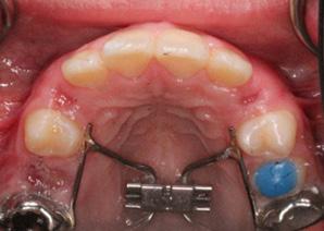
•Case reports
MANUSCRIPT FORMAT
Abstract. Must include a short abstract no more than 50 words that describe the significance of the article.
Keywords. Must include keywords to help categorize the article.
Length. Manuscript should be no longer than 15 doublespaced pages, excluding figures and illustrations.
Tooth Numbering. The numbering of teeth should be international numbering. (US numbering can be added and put in parentheses.)
Non-English Manuscripts. Authors are encouraged to submit the manuscript in languages other than English for posting on the IAO website. A mention will be added to the English version published in the International Journal of Orthodontics, directing readers online for other translations.
Illustrations. Images must be available electronically as separate files. High quality digital images must be presented in one of the following formats: .tiff, .eps,.jpg, or .pdf with resolution of a minimum 300 dpi. Images must not be embedded in software programs such as Word or Power Point. The names on the digital files for photo/illustration files should match the manuscript reference. For example, if manuscript copy references Figure 1, electronic file should be titled Figure 1.jpg. No more than 16 photographs, figures, & illustrations are recommended; if greater than 16, IJO has the right to select and limit the number if necessary. Figures must be clearly referenced as to their placement in the manuscript. Brief captions for the figures, identified by number, must be provided. All images must be titled. Radiographs must be of superior quality.
References. References must be included and authors are responsible for the accuracy of references. Manuscripts without them will be returned. Cite references in the text as endnotes and number them consecutively. Citations must be referenced in the following style:
Periodical:
1.Sim JM, Jefferson Y, Dillingham SE, & Keller DC. Diagnosing an orthodontic patient using three different analyses. IJO 1990; 1(4):101-106.
Book:
2.Fonder AC. The Dental Physician. 2nd ed. Rock Falls, IL; Medical Dental Arts; 1985:25-82.
World Wide Web site:
3.Health Care Financing Administration. 1996 statistics at a glance. Available at: http://www.hcfa.gov/stats/stathili.htm”. Accessed Dec. 2, 1996.
Products: Any products mentioned in the manuscript should be footnoted disclosing the company name and address.*
*XYZ Orthodontic Co., 123 Main St., Los Angeles, CA 90000.
REVIEW AND EDITING PROCESS
Editor. Articles will initially be reviewed by the editor. If author fails to adhere to the guidelines set forth, manuscript will be returned to the author for revision and correction.
Peer review. Articles in IJO are subject to an anonymous peer review process. Reviews may take up to eight weeks to complete.
Decision. Once the reviewing consultants have completed their critiques, the editor examines their comments and makes a decision to accept, accept with minor revisions, revise and resubmit, or reject.
Editing. IJO reserves the right to edit manuscript for conciseness, clarity, and stylistic consistency. The author has final approval before publication.
Questons? Contact Managing Editor, Allison Hester at allisonhijo@gmail.com, 501-517-1620.
AUTHOR RESPONSIBILITIES
Copyright transfer. IAO holds the copyright for all editorial content published in the journal. All accepted manuscripts become the permanent property of the IAO, and may not be published elsewhere in full or in part, in print or electronically, without written permission from the IAO.
Reprint permission. The author is responsible for obtaining written permission from the publisher, or the person or agency holding the copyright for any material that is reproduced from a published source.
Consent forms. Any patient clearly identified in the article must sign a form indicating his or her consent to be depicted in the article. It is the author’s responsibility to confirm consent.
Author’s photo and bio. The author(s) must submit a headshot (preferably professional) and current biographical sketch. If author holds a teaching position, the title, department, and school should be included. Any position or relationship with a dental manufacturer must be identified. The sketch should include rank or title and station of authors who are in federal service, and should be limited to 60 words or less.
Conflict of interest. The author will identify any conflicts of interest upon submission of any articles.
REPRINTS
The International Journal of Orthodontics provides the corresponding author a final electronic copy of the Journal in which the article appears as well as an electronic copy (.pdf) of the pages where the article appears. Requests for individual reprints of the article should be directed to Chris McKay, IAO, 414-272-2757 or at chris@iaortho.org.
Patients have a right to privacy that should not be infringed without informed consent. Identifying information, including patients’ names, initials, or hospital numbers, should not be published in written descriptions, photographs, and pedigrees unless the information is essential for scientific purposes and the patient (or parent/guardian) gives written informed consent for publication. Informed consent for this purpose requires that a patient who is identifiable be shown the manuscript to be published. Authors should identify Individuals who provide writing assistance and disclose the funding source for this assistance. Identifying details should be omitted if they are not essential. Complete anonymity is difficult to achieve, however, and informed consent should be obtained if there is any doubt. For example, masking the eye region in photographs of patients is inadequate protection of anonymity. If identifying characteristics are altered to protect anonymity, such as in genetic pedigrees, authors should provide assurance that alterations do not distort scientific meaning and editors should so note. (Source: International Committee of Medical Journal Editors (“Uniform Requirements for Manuscripts Submitted to Biomedical Journals”), February 2006).
Successful Treatment of Class II Malocclusion in a Young Patient with Headache and Cervical Dystonia Using the Herbst Appliance: A Case Report
by Maryam Bakhtiyari, DDS, IBO, Shahrzad Sadeghi, BS, and Mehrnaz Bakhshzad
Abstract:
AUTHORS
Dr. Maryam Bakhtiyari
BS in Biochemistry, UCLA, California, United States; DDS, University of the Pacific, San Francisco, California, United States. She is a Diplomate of the International Board of Orthodontics. Residency in sleep apnea, Tufts University of Medicine; Diplomate and Master of Excellence, American Board of Craniofacial Pain; Diplomate of the American Board of Dental Sleep Medicine
Shahrzad Sadeghi
BS in Biology, University of the Pacific, California, United States; Third year of dental school at UCSF School of Dentistry, United States
Mehrnaz Bakhshzad
Pre-Dental student, University of Southern California, United States
*This article
This study demonstrates the successful use of the Herbst appliance to manage class II malocclusion and related craniofacial issues. After a comprehensive assessment and appliance treatment, the patient experienced significant improvements in malocclusion, cervical dystonia, and headaches. This multi-faceted approach highlights the importance of individualized treatment planning and specialized orthodontic appliances.
Objective: This study aims to demonstrate the efficacy of the Herbst appliance in managing patients with class II malocclusion and associated craniofacial discomfort manifesting as occasional headaches and cervical dystonia. The primary objectives of this treatment include addressing malocclusion and associated symptoms while simultaneously achieving comprehensive aesthetic and functional dental rehabilitation.
Methods: A comprehensive patient assessment was conducted, including records for the patient’s bite by guiding the mandible more forward to a better physiological position as well as increasing the vertical dimension of occlusion. This record was then sent to the orthodontic lab to construct a Herbst appliance with bilateral molar bands and an occlusal rest. The appliance was subsequently cemented in the patient’s mouth. The appliance was worn by the patient for 12 months, followed by orthodontic braces and removal of the appliance once the desired bite was established.
Results: Following treatment, a night retainer was used to maintain the achieved tooth alignment. The patient became asymptomatic for cervical dystonia within a week after the appliance was delivered, with a resolution of headaches, and these improvements persisted throughout the treatment. The patient remained asymptomatic after two years of followup, and the class II malocclusion was effectively corrected.
Conclusion: Following a thorough clinical evaluation, a custom Herbst appliance with bonded crowns on permanent molars was created. This oneyear treatment successfully corrected class II malocclusion. Subsequently, orthodontic braces aligned the dentition, emphasizing the importance of individualized treatment and specialized appliances.
Keywords: Herbst appliance, Class II malocclusion, Mandibular Advancement Repositioning Appliance, cervical dystonia Conflict of interest: None
Introduction:
Malocclusion is characterized by tooth misalignment or an abnormal relationship between the dental arches that deviates from what is considered within the normal range.1 At the mixed dentition stage, the global prevalence rates of Class I, Class II, and Class III malocclusions are 72.74%, 23.11%, and 3.98%, respectively.2 Class II malocclusion is a frequently encountered clinical issue affecting approximately one-third of the population in the United States. 3 Symptoms of malocclusion affect various aspects of oral and facial health. These include irregular tooth alignment, resulting in an abnormal facial appearance, discomfort or difficulty while biting or chewing, and, in rare cases, speech difficulties, such as lisping. Mouth breathing, characterized by the habitual inhalation and exhalation of air through the mouth without lip closure during respiration, is another symptom. Additionally, malocclusion can lead to an open bite, making it challenging to bite food correctly.4 Recent research has explored the influence of dental occlusion on body balance. Furthermore, dental occlusion can influence muscle tension in both the jaw-related and postural muscles, which are essential for maintaining balance. A thorough examination of the impact of malocclusal characteristics on muscle properties demonstrated that factors such as Angle’s classes of
5B: Upper Occlusal
6A: Upper Occlusal
6B: Lower Occlusal
Fig. 6C: Center
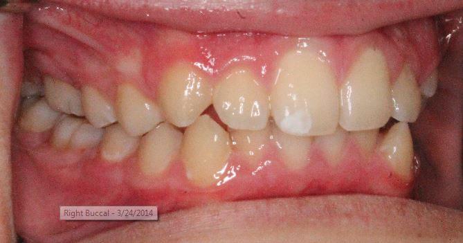
Figure 6: Intraoral images taken of the patient 6 months after placing the Herbst appliance
5C: Center
5D: Lower Occlusal
Fig. 5: Intraoral images are taken of the patient after placing the Herbst appliance
7A: Profile
7B: Frontal Smile
Fig. 7: Facial photographs taken 6 months after placing the Herbst
8A: Right Buccal
8B: Center
8C: Upper Occlusal
8C: Lower Occlusal
8C: Cephlametric Analysis
Fig. 8: Intraoral images taken upon the removal of the appliance and moving onto the next phase and braces
A Comparative Evaluation of Rate of En-Masse Retraction with and without Low-Intensity Laser Therapy –A Randomized Clinical Trial
by Dr. Rishika Arya, Dr. Wasundhara A. Bhad, Dr. Jyoti Manchanda, Dr. Santosh J. Chavan, Dr. Mohammed Niaz Ali A., and Dr. Ahmed Talha
AUTHOR
Dr. Rishika Arya
BDS, Kalinga Institute of Dental Sciences, Bhubaneswar;MDS, Government Dental College and Hospital, Nagpur; Currently Working as Assistant Professor, Government Dental College and Hospital, Nagpur
Dr. Wasundhara A. Bhad (Patil)
BDS, MDS (Orthodontics), Dean, Government Dental College and Hospital, Mumbai
Dr. Jyoti Manchanda,
BDS, MDS (Orthodontics), Associate Professor, Government Dental College and Hospital, Nagpur
Dr. Santosh J. Chavan
BDS, MDS (Orthodontics), Head of Department and Professor, Government Dental College and Hospital, Nagpur
Dr. Mohammed Niaz Ali A.
BDS, MDS (Orthodontics), Assistant Professor, Government Dental College and Hospital, Mumbai
Dr. Ahmed Talha
BDS, MDS (Orthodontics), Senior Resident, Shri. Bhausaheb Hire GMC and Hospital, Dhule
ABSTRACT
Introduction: Prolonged orthodontic treatment can harm tooth-supporting structures and reduce patient compliance. To expedite tooth movement, both surgical and non-surgical methods have been explored. Low-intensity laser therapy (LILT) is a promising non-surgical technique due to its safety and minimal invasiveness. This Randomized Controlled Trial (RCT) was designed to study LILT’s effect on the rate of orthodontic tooth movement during enmasse retraction.
Materials and Methods: This RCT included 32 patients needing first premolar extractions for moderate crowding and protrusion. They were randomly assigned to either an experimental or control group. TADassisted en-masse retraction was performed, with the experimental group receiving laser application every 21 days. Data collection occurred at T0 (start of retraction), T1 (2 months), and T2 (end of retraction).
Results: In the control group, orthodontic tooth movement was 0.81 mm/month for the maxillary arch and 0.69 mm/month for the mandibular arch. In the experimental group, it was 0.99 mm/ month and 0.93 mm/month, respectively. En-masse retraction took 155.7 days (5.12 months) for the maxillary arch and 152.3 days (5.01 months) for the mandibular arch in the experimental group, compared to 180.6 days (5.94 months) and 183.1 days (6.02 months) in the control group.
Conclusion: LILT increased the rate of orthodontic tooth movement by 22.2% in the maxillary arch and 34.7% in the mandibular arch, leading to reduction in total duration of treatment by 16% in the maxillary arch and 20.1% in the mandibular arch.
Orthodontic treatment is widely known for its extended duration, with an average treatment duration of 19.9 months. 1 Prolonged treatment periods can harm
*This article has been peer reviewed
tooth-supporting structures and may lead to a decline in patient compliance.2 Various adjunctive methods can be used to expedite orthodontic tooth movement, broadly categorized as either surgical or non-surgical methods.3
Surgical methods carry risk of potential post-surgical complications such as pain, swelling, 4 loss of crestal bone, bone necrosis, edema, and gingival recession.5,6
On the other hand, non-surgical methods have gained popularity for their effectiveness in biologically accelerating tooth movement. These methods encompass various mechanical and physical approaches, such as lowintensity laser therapy (LILT), direct electric current, pulsed electromagnetic fields, and ultrasonic vibrations.7–10
Among these, LILT has emerged as a focal point in recent studies. LILT is characterized by its low energy output, ensuring that the treated area’s temperature remains within the body’s normal range.11 With its safety and minimally invasive nature, LILT stands out as a promising technique for expediting orthodontic tooth movement.8 The effect of LILT on the rate of OTM has been evaluated during canine retraction7,8,12 and during leveling and alignment,13 however, very limited literature is available on the effect of LILT on orthodontic tooth movement during en-masse retraction.
Hence, the aim of this Randomized Controlled Trial (RCT) was to assess the effectiveness of LILT in accelerating the rate of orthodontic tooth movement in patients undergoing treatment by first bicuspid extraction and en-masse retraction.
MATERIALS AND METHODS
Trial Design, Ethical Approval and Registry
Study design was a randomized controlled trial. The study design was approved by the Institutional Ethics Committee (Ref. no. IEC/05/54 dated on 25.04.2022) and consent from the participating subjects was obtained in advance. The trial was registered in the Clinical Trial Registry – India (Ref. no. CTRI/2022/06/055231).
Participants, Setting, and Eligibility Criteria
The study included patients aged 18-30 years with periodontally sound permanent dentition, presenting with dentoalveolar protrusion and moderate crowding, requiring first premolar extractions as a treatment plan. Since en-masse retraction was being evaluated, the study design involved two separate groups, i.e. experimental and control group. However, to avoid biological variation, patients were selected from the same ethnicity.
Patients with history of systemic illness and patients who had undergone previous orthodontic treatment were excluded.
Sample Size Calculation
The sample size of this study was calculated based on the study conducted by Lalnunpuii H et al, 14 considering a firsttype error (α) level of 5% and second-type error (β) level of 20%. Determination of sample size was done by using OpenEpi Version 3 software and it yielded an approximate sample size of 28 samples. But, considering 10% dropouts, the final sample determined was 32 patients.
These patients were randomly allocated to two groups, experimental group (16 patients) i.e. patients undergoing LILT assisted en-retraction and control group (16 patients) i.e. patients undergoing en-masse retraction without LILT. Randomization was done using computer generated sequence. Allocation concealment to the patient was achieved by asking each patient to draw a sealed envelope containing an allocation.
Orthodontic Treatment Protocol
After thorough case analysis and treatment planning, first premolar extractions were done. Molar bands (0.180” x 0.006”) were customized and cemented with Glass Ionomer Cement (GC Gold Label). Pre-adjusted edgewise MBT brackets (ORTHO R Organizers, USA) of 0.022” slot were bonded with Transbond XT (3M, Unitech). Initial phase of alignment and leveling was initiated using 0.016-in, 0.016 x 0.022-in, 0.019 x 0.025-in heatactivated nickel-titanium archwires (G&H, Orthoforce, USA). At the end of alignment and leveling, a final working wire (0.019 x 0.025 in stainless steel) was inserted. After 21 days of 19x25-in SS wire placement, en-masse retraction was initiated. Incisors were consolidated by using 0.009-in steel ligature wires. Second premolars and first molars were also consolidated to make a single anchorage unit. Under local anesthesia, self-drilling miniimplants (S.K. Surgicals) measuring 1.5 x 8.0 mm were inserted in between the maxillary second premolar and 1st molar, and mandibular second premolar and 1st molar. A Nickel-titanium closed coil spring (G&H, Orthoforce, USA) was placed from the head of the micro implant to crimpable hook (Garmy) of the
working wire. Length of Nickel-titanium closed coil spring chosen depending on the amount of extraction space to be closed, ensuring standardization of retraction force to 200 g using Dontrix gauge.
The low-intensity laser was applied in the experimental group using a semiconductor (GaAlAs) diode laser.
Low-Intensity Laser Therapy (Lilt) Protocol
The laser type used was a semiconductor (GaAlAs) diode laser (Model: DenLase Version: DenLase-SY-A. 1c, China Daheng Group, Inc) emitting infrared radiation with 980+/-10 nm wavelength operated according to the manufacturer’s recommendations. (Figure 1-3)
Laser parameters used in the study are specified in Table 1.
Fig. 1: Laser Kit
Fig. 2: Laser Unit
Table 1: Laser parameters used in the study
Fig. 3: Patient and operator safety goggles
Table 4:Comparison of displacement from T0-T1, T0-T2, and T1-T2 between experimental and control groups
18: Line graph showing descriptive details for the distance between cusp tip of canine and cusp tip of the mesiobuccal cusp of the first molar for the control group
Table 5:Comparison of rate of retraction from T0-T1, T0-T2, and T1-T2 between experimental and control group
20:
showing the comparison of the rate of retraction from T0-T1, T0-T2, and T1-T2 between experimental and control group
Table 2 and Figure 16 depicts descriptive statistics for the distance between the cusp tip of the canine and the cusp tip of the mesiobuccal cusp of the first molar in the experimental group.
Table 3 and Figure 17 depict descriptive statistics for the distance between the cusp tip of the canine and the cusp tip of the mesiobuccal cusp of the first molar in the control group. Figure 17 depicts comparison of mean distance for control and experimental groups at T0, T1, and T2 time.
The comparison of displacement between experimental and control groups (Table 4 and Figure 18) revealed that from T0 to T2 interval, the displacement was greater in the experimental group as compared to the control group; however, there was a non-significant difference.
The comparison of rate of retraction between experimental and control groups (Table 5 and Figure 19) revealed that from T0 to T2 interval, the rate of retraction was significantly greater in the experimental group as compared to the control group. As there was a difference in the rate of orthodontic tooth movement in patients undergoing en-masse retraction with LILT when compared with patients who did not receive any LILT, the Null Hypothesis of the study was rejected.
The comparison of duration of retraction (in months) from T0-T1, T1-T2 and T0-T2 between experimental and control group (Table VI and Graph VI) revealed that from T0 to T2 interval, the total duration of retraction in maxillary arch as well as mandibular arch was significantly lower in the experimental group arch as compared to the control group.
DISCUSSION
The duration of comprehensive fixed orthodontic treatment can vary widely, but according to the recent systematic review, the average duration of fixed orthodontic treatment was 19.9 months.15
There’s a growing demand from patients for shorter treatment times. Possible interventions to accelerate orthodontic tooth movement can be categorized as surgical or non-surgical.3
The surgical methods encompass alveolar decortication, corticotomy, periodontal ligament distraction, and dentoalveolar distraction.16 However, surgical approaches have the disadvantage of being invasive and carry the risk of injuries to the surrounding vital structures, infection, postoperative pain, and edema.8
Non-surgical techniques include low-intensity laser
Fig.
Fig. 19: Bar graph showing descriptive details for the distance between cusp tip of canine and cusp tip of the mesiobuccal cusp of the first molar for the control group
Fig.
Bar graph
irradiation,14 vibration,17 pulsed electromagnetic fields,10 electrical currents9, and pharmacological approaches.18
Over the past decade, numerous research endeavors have focused on exploring all these different modes to expedite orthodontic tooth movement. One such approach is low-intensity laser therapy (LILT). LILT has the advantage of being not only noninvasive but, clinically easily available as well,2 thereby, attracting the attention of several researchers interested in exploring modalities of accelerated orthodontics.7,8,12 14,19,20
Thus, this study aimed to determine the clinical effectiveness of LILT in accelerating the rate of orthodontic tooth movement during en-masse retraction.
The study design was a randomized clinical trial wherein 32 patients (22 females and 10 males) in the age group of 18 to 30 years, presenting with dentoalveolar protrusion and moderate crowding, requiring first premolar extractions as their treatment plan, were included. Patient was sent for premolar extraction and strap was done and leveling and alignment was completed.
TADs (Temporary Anchorage Devices) were placed in between the second premolar and first molar of each quadrant. In the previous study by Lalnunpuii et al,14 second molar banding and cross-arch stabilization was used to prevent anchorage loss during the retraction phase. However, in our study, placement of TADs ensured that absolutely no anchorage loss took place and only en-masse retraction was studied and not the forward movement of the first molar.
Progress models on which Orthodontic tooth movement was measured, were taken at 3 time points: before the commencement of en-masse retraction (T0), at 2 months (T1), and at the end of en-masse retraction (T2). A previous study by Doshi et al.12 on the effect of LILT during canine retraction noted a decrease in the rate of orthodontic tooth movement in later time periods. Therefore, the current study evaluated the effect of LILT on orthodontic tooth movement over the entire duration of en-masse retraction.
Similar to a previous study by Arumughan et al.,19 in all patients belonging to the experimental group, this laser regimen was applied every 21st day till en-masse retraction was complete as it coincides with normal recall visits.
Rate of orthodontic tooth movement at 2 months (T1) (Table 5 and Graph 20).
A mid-treatment progress model was made for each patient at 2 months (T1) to study LILT’s effect on orthodontic tooth movement rate.
In the control group, after 2 months (60.8 days), the mean rate of orthodontic tooth movement was 1.01 mm/month for both the maxillary and mandibular arches. Conversely, in the experimental group, during the same period, the mean rate of orthodontic tooth movement was 1.36 mm/month for the maxillary arch and 1.35 mm/month for the mandibular arch. This indicates that the rate of tooth movement in the experimental group was approximately 1.34 times faster for the maxillary arch and 1.33 times faster for the mandibular arch compared to the control group i.e., there was a 34.6% increase in the rate of tooth movement for the maxillary arch and a 33.6% increase for the mandibular arch in the experimental group compared to the control group in the first 2 months after the commencement of en-masse retraction (T0-T1).
In a similar study by Arumughan et al.,19 the rate of orthodontic tooth movement was evaluated only in the maxillary arch during en-masse retraction. Similar to our study, all six anterior teeth
were irradiated in the experimental group after every 21 days. However, their laser parameters were different from our study, they used 810 nm GaAlAs diode laser with a power output of 0.1 W in a continuous wave mode. The progress model was made on the 84th day (2.7 months) after the commencement of en-masse retraction. Unlike our study, here, the distance between the contact points of the maxillary canine and the second premolar was measured to determine orthodontic tooth movement. They reported a 12.5% increase in the rate of orthodontic tooth movement in the experimental group compared to the control group.
Rate of orthodontic tooth movement at the end of en-masse retraction (T2) (Table 5 and Figure 20).
In the present study, progress records were also taken at the end of en-masse retraction to evaluate the effect of LILT on the rate of orthodontic tooth movement over the entire duration of en-masse retraction.
The present study reported that the mean rate of orthodontic tooth movement from T1 to T2, i.e. from 2 months after the commencement of en-masse retraction till the end of en-masse retraction in the experimental group was 0.77 mm/month and 0.84 mm/month for maxillary and mandibular arch respectively. However, in the control group, it was 0.71 mm/month and 0.53 mm/month for maxillary and mandibular arch respectively. This indicates that the rate of tooth movement in the experimental group was approximately 1.08 times faster for the maxillary arch and 1.58 times faster for the mandibular arch compared to the control group.
As discussed earlier, the highlight of the present study was that the effect of LILT on the rate of orthodontic tooth movement was evaluated over the entire duration of en-masse retraction (T0-T2).
So, the mean rate of orthodontic tooth movement over the entire duration of en-masse retraction in the control group was 0.81 mm/month and 0.69 mm/month for maxillary and mandibular arch respectively. However, in the experimental group, the mean rate of orthodontic tooth movement over the entire duration of en-masse retraction was 0.99 mm/month and 0.93 mm/month for maxillary and mandibular arch respectively. This indicates that the rate of tooth movement in the experimental group was approximately 1.22 times faster for the maxillary arch and 1.34 times faster for the mandibular arch compared to the control group. Therefore, for the T0-T2 interval, there was a 22.2% increase in the rate of tooth movement for the maxillary arch and a 34.7% increase for the mandibular arch in the experimental group compared to the control group.
A previous study by Lalnunpuii et al14 reported similar findings with a 36.7% and 35.4% increase in the rate of orthodontic tooth movement was observed in the experimental group as compared to the control group for maxillary and mandibular arch respectively.
The effect of LILT on treatment duration (Table 6 and Figure 21)
On average, the en-masse retraction was completed in 155.7 days (5.12 months) and 152.3 days (5.01 months) in the maxillary and mandibular arch respectively in the experimental group. However, in the control group, it took 180.6 days (5.94 months) and 183.1 days (6.02 months) for en-masse retraction to be completed in the maxillary and mandibular arch respectively.
Relationship Between Vertical Facial Pattern and Dental Arch Form in Class II Division I Malocclusion
by Dr. Kurapati Krishna Teja, Dr. Mayuri Thomas, Dr. V. Deepti, Dr. Akshay Goje, Dr. Sonali Rathore, Dr. Vennela Gande
AUTHORS
Dr. Kurapati Krishna Teja
Assistant Professor, Department of Orthodontics and Dentofacial Orthopedics, Tirumala Institute of Dental Sciences and Research Center (TIDS), Nizamabad.
Dr. Mayuri Thomas
Professor, Department of Orthodontics and Dentofacial Orthopedics, Sri Sai College of Dental Sciences (SSCDS), Vikarabad.
Dr. V. Deepti
Professor and Head of the Department at Sri Sai College of Dental Surgery (SSCDS) BDS, Madras Dental College; MDS from K.L.E. Dental College
Dr. Akshay Goje
Reader, Department of Orthodontics and Dentofacial Orthopedics, Sri Sai College of Dental Sciences (SSCDS), Vikarabad.
Dr. Sonali Rathore Reader, Department of Orthodontics and Dentofacial Orthopedics, Sri Sai College of Dental Sciences (SSCDS), Vikarabad.
Dr. Vennela Gande
Assistant Professor, Department of Orthodontics and Dentofacial Orthopedics, Sri Sai College of Dental Sciences (SSCDS), Vikarabad.
Abstract
Background: This study investigates dental arch widths relative to vertical facial patterns in Class II Division I malocclusion, examining variations across horizontal, average, and vertical growth patterns.
Methods: A cohort of 120 subjects aged 8-30 years was categorized based on vertical growth patterns. Measurements from cephalograms and dental casts were subjected to statistical analyses including Kruskal-Wallis and Chi-Square tests (p ≤ 0.05).
Results: Significant differences in dental arch widths were observed among vertical growth patterns, with horizontal growth presenting wider arches compared to average and vertical growth patterns. Negative correlations were noted between SN-MP angle and arch widths, indicating narrower arches with increased SN-MP angles.
Conclusion: Vertical facial morphology significantly influences dental arch dimensions in Class II Division I malocclusion. Orthodontic treatment planning should consider these variations to achieve optimal and stable outcomes tailored to individual growth patterns.
Keywords: Dental arch form, Class II Division I malocclusion, Inter-molar and premolar width, Cephalometric analysis, Arch dimensions, SN-MP angle.
Conflict of Interest: None
Introduction
In orthodontic practice, achieving optimal dental arch form is crucial for treatment success and long-term stability. This goal is influenced by a complex interplay of genetic, functional, and environmental factors, particularly in relation to vertical growth patterns. Maintaining appropriate arch forms not only prevents relapse but also enhances overall dental health.
Recent advancements in orthodontic materials and techniques have facilitated faster alignment of dental arches. However, the challenge persists in matching available arch wires with patient-specific arch dimensions effectively. Understanding the impact of vertical facial morphology on dental arch dimensions is pivotal for accurate diagnosis and treatment planning. For instance, individuals with longer facial structures often exhibit narrower maxillary intermolar widths and reduced transverse dimensions, whereas those with shorter facial profiles tend to display larger crosssectional measurements.1
Gender-specific variations in dental arch dimensions further underscore the complexity of orthodontic treatment. Studies by Wei (1970) 2 and Eroz et al. (2000)3 have documented significant differences in maxillary and mandibular inter-canine and intermolar widths across different populations and age groups, highlighting the need for personalized treatment approaches.
The stability of orthodontic outcomes, characterized by achieving and maintaining optimal occlusion, remains a primary concern. This study focuses on exploring the relationship between dental arch widths and vertical growth patterns in Class II Division I malocclusion. By elucidating these connections, the research aims to contribute valuable insights for refining
orthodontic interventions and improving treatment longevity.4
Materials and Methods
The study sample consisted of 120 subjects, both male and female, aged 8-30 years. Vertical facial patterns were categorized into three groups: horizontal growth pattern, average growth pattern, and vertical growth pattern (Figure 1). Pretreatment lateral cephalograms (Figure 2) and dental casts (Figure 3) were obtained for each subject. Measurements of inter-canine widths, intermolar widths, arch lengths of maxillary and mandibular casts, and arch perimeter were taken using digital calipers (Figure 4). Statistical analyses were performed using the Kruskal-Walli’s test and Chi-Square test, with a significance level set at p ≤ 0.05.
Results:
The study revealed notable differences in dental arch widths across various vertical facial growth patterns. Subjects with a horizontal growth pattern consistently displayed the largest inter-premolar and inter-molar distances, with average maxillary inter-premolar and inter-molar distances of 37.51 mm and 47.72 mm, respectively (Table 1,1a,2,2a). Conversely, those with a vertical growth pattern exhibited the smallest measurements, averaging 33.86 mm for maxillary inter-premolar and 44.12 mm for maxillary inter-molar distances. These differences were statistically significant (p < 0.001). Significant negative correlations were found between the SN-MP angle and dental
Fig. 1: Cephalometric tracing for hard tissue analysis
Fig. 2: Lateral cephalogram
Fig. 3: Dental cast
Fig. 4: Digital vernier calipar
Gender Differences and Age Factors
While this study did not specifically analyze gender differences or age-related changes in dental arch dimensions, previous research has indicated that these factors can also influence arch morphology. For instance, males typically exhibit larger dental arch dimensions compared to females, and dental arch dimensions can change with age due to growth and development10.
Future research could further explore these aspects to provide a more comprehensive understanding of the factors influencing dental arch dimensions. Including a larger and more diverse sample could help to generalize the findings and enhance their applicability in clinical practice.
Conclusion:
This study underscores the importance of considering vertical facial morphology in orthodontic diagnosis and treatment planning. Significant differences in dental arch dimensions were observed across different vertical facial growth patterns, with horizontal growth patterns showing the largest arch widths and vertical patterns showing the smallest. Negative correlations between the SN-MP angle and dental arch widths further support that increased vertical facial dimensions are associated with
6:
Table 4: Correlation of Sn-Mp with inter-premolar and inter-molar distance
narrower dental arches. These findings provide valuable insights for orthodontists in devising effective and stable treatment plans, tailored to individual growth patterns.
References
1. McNamara, J. A., Jr. (2000). Maxillary transverse deficiency. American Journal of Orthodontics and Dentofacial Orthopedics, 117(5), 567-570. doi:10.1016/S0889-5406(00)70205-9
2. Wei, S. H. Y. (1970). Maxillary and mandibular arch width in normal occlusion and malocclusion. Angle Orthodontist, 40(1), 36-41. doi:10.1043/0003-3219(1970)040<0036>2.0.CO;2
3. Eroz, U., Ceylan, I., & Aydemir, S. (2000). Intercanine and inter-molar width in a Turkish population. American Journal of Orthodontics and Dentofacial Orthopedics, 117(5), 499-504. doi:10.1016/ S0889-5406(00)70201-1
4. Proffit, W. R., Fields, H. W., & Sarver, D. M. (2012). Contemporary Orthodontics (5th ed.). St. Louis, MO: Elsevier.
5. An evaluation of the errors in cephalometric measurements on scanned cephalometric images and conventional tracings KorkmazSayinsu, FulyaIsik, GöksuTrakyaliand TülinArun Department of Orthodontics, Faculty of Dentistry, Yeditepe University, Istanbul, Turkey
6. Björk, A., & Skieller, V. (1983). Growth of the maxilla in three dimensions as revealed radiographically by the implant method. British Journal of Orthodontics, 10(1), 53-70. doi:10.1179/bjo.10.1.53
7. Schudy, F. F. (1964). Vertical Growth versus Anteroposterior Growth as Related to Function and Treatment. The Angle Orthodontist, 34(2), 75-93. doi:10.1043/0003-3219(1964)034<0075>2.0.CO;2
8. Wagner, D. M., & Chung, C. H. (2005). Transverse growth of the maxilla and mandible in untreated girls with low, average, and high MP-SN angles: a longitudinal study. American Journal of Orthodontics and Dentofacial Orthopedics, 128(6), 716-723. doi: 10.1016/j.ajodo.2004.09.014
9. Kim, Y. H., & Kim, J. H. (2011). Relationship between vertical growth patterns and dental arch dimensions in Korean adults. Journal of Korean Association of Oral and Maxillofacial Surgeons, 37(4), 209-215. doi:10.5125/jkaoms.2011.37.4.209.
10. Bhowmik, S. G., Hazare, P. V., & Bhowmik, H. (2012). Correlation of the arch forms of male and female subjects with those of preformed rectangular nickel-titanium archwires. American Journal of Orthodontics and Dentofacial Orthopedics, 142(3), 364-373. doi: 10.1016/j.ajodo.2012.02.020
Fig.
Mean comparison of Sn-Mp according to the growth pattern
second scan (T2) was obtained at the end of the distalization phase when the distalizer was removed.
The imaging acquisition parameters utilized included five mA, 120 kV, a field of view (FOV) measuring 13 cm in height by 16 cm in diameter, and exposure times of either 20 seconds or 40 seconds. Consistent scanning protocols were applied for each patient at T0 and T1, and the approved protocol did not require a supplementary CBCT scan after treatment.
The 3D analysis was performed on superimposition T1 and T2 using the two open-source 3D Slicer software version 4.10.2, and Romexis software version 5.3.4.39 used a growing tool in the software for manual segmentation tool layer by layer for layer tracing as shown in Fig. 5.
Several landmarks were used in CBCT analysis to evaluate the treatment outcome for class II patients with distalizers using the CMA protocol (Table 1). The same assessor and another observer analyzed pre- and post-CBCT images again to assess the intra- and interobserver reliability statistically.
Outcomes
The primary outcome was anchorage loss in the lower arch, while the secondary outcomes were the amount and type of distalization and the treatment duration.
Sample Size Calculation: This study would be an experimental, interventional, and randomized clinical trial, a convenience sampling technique used for patient selection.
Sample size calculation was performed using G* power 3.1.9.7. By selecting an alpha (α) level of 0.05 (5%), power=80%, and standard deviations (SD) of (2.2) and (4.00) calculated based on the results of the previous study (16) that recorded mandibular central incisor’ torque variable (0.68 ± 2.22) and (5.30 ± 4.00) for Miniscrew and Essix appliances group respectively. The predicted sample size (n) was found to be 10 patients per group.
Randomization: Randomized selections were made for the total number of patients treated with Carriere Motion appliance and class II elastics. Randomization was done using an online Research Randomizer (Version 4.0) computer software. After sample randomization, these patients generated the random allocation sequence by the supervisors of this study. Also,
A: Points placement on the Lateral view
B: Points placement on the Frontal view
C: Fused 3D view of Primary and Secondary datasets
H: Bilateral mandibular central incisors, and 1st molarsAnteroposterior distance measurement (L1 AP, L6 AP).
Fig. 3: Miniscrew anchorage group; A. Pre-treatment; B. Intervention; C. Post-distaization
Fig. 5: DICOM 3d analysis view
Fig. 4: Passive lingual anchorage group; A. Pre-treatment; B. Intervention; C. Post-distaization
Maxillary canine movements: The amount of distal movement of the maxillary canine was found to be statistically significant in both groups. It was almost equal to the amount of distal movement of the molar.16,17,20-22 However, the amount of distalization was much less than that of other skeletal anchorage distalizing appliances or conventional anchorage appliances.29
The distalization of the entire maxillary buccal segment by CMA means that there was no anchorage loss in the premolar area, unlike other distalizers that required retraction of the premolars and canines after molar distalization.30, 31
Maxillary molar movements: On average, with the CMA, distal movement of the maxillary first molars was 1.95 mm, approximately the same as previously reported by Sandifer et al.,20 while only Class II elastics did not show any significant maxillary molar movements.3
It had been claimed that adding a ball-and-socket joint in the molar pad would lead to pure bodily distalization of the maxillary molar without distal tipping. However, in the current RCT, the maxillary molar distal tipping amount was statistically significant and similar in both groups. In the miniscrew group, the molar tipped (3.49), while the lingual appliance group tipped (3.82).15 On the other hand, this degree of tipping was less than that produced by skeletal anchorage distalizing appliances (8.44) and conventional anchorage appliances (8.31). 29 This data did not correlate directly with the manufacturer’s claim of ‘‘distal movement of the canine along the alveolar ridge without tipping.’’15 The data showed tipping of the maxillary canine in both groups. Therefore, the ball-and-socket joint helped minimize molar tipping but did not completely prevent it.16,17
Mandibular molar movements: The mesial tipping, rotation, and extrusion amounts of lower molars significantly differed between the groups. In other studies, the passive lingual group tended to have more significant mesial movement and tipping of the mandibular first molar.20, 23, 28
Fouda et al.16 used indirect anchorage through an SS wire to connect the miniscrew to the tooth, but there was little mesial movement and tipping to the second mandibular molar. There was no mesial movement or tipping as the elastics were loaded directly on the miniscrew, which matched with the infrazygomatic miniscrew study.17
Transferring the anchorage control from indirect to direct miniscrew anchorage eliminates horizontal and vertical components of the forces exerted by class II elastics on lower molars.
Mandibular incisors. The increase in mandibular central incisor movements significantly differed between the two groups. Data showed that the mandibular incisor moved more mesially in the passive lingual group, as noted in several studies.20-23, 25,26 than the miniscrew group that showed no proclination of the lower incisor. This may suggest that, as an anchorage unit, a miniscrew is superior to the passive lingual arch to avoid anchorage loss, as reported in the Ghozy study.17
Overjet: It is typically observed in Class II malocclusions. From the data, although fixed appliances were not used on the anterior teeth, The overjet decreased significantly in both groups. This is due to the spontaneous distal movement of incisors into the space created after the distalization buccal segment in the direct miniscrew group16,17 and the mandibular incisors proclination in the passive lingual group.20-23, 25
Limitations: The trial’s patients and operator could not be blinded to the treatment modality with a small sample size. No treatment was finished during data collection, and the success of the CMA heavily depends on patient compliance with wearing elastics, which can vary significantly and impact the effectiveness of the treatment. Also, this study was limited to the first phase of treatment in which the CMA was used and did not involve observation of the patients during the second phase of treatment, which involved anterior segment retraction.
Generalizability: This study’s generalizability might be constrained as it only involved one dental facility and one PhD candidate performing the treatments on only one ethnic group was investigated.
Conclusion:
• Direct Miniscrew anchored CMA resulted in a more significant distalization of the maxillary buccal segment than the lingual arch anchored one with no significant difference between them regarding the duration of distalization.
• Maxillary first molar and canine rotation with tipping were similar in both groups.
• Using miniscrews in the lower jaw stops class II elastics from negatively affecting the lower teeth and molars. It doesn’t change the lower face height, suggesting that miniscrews can correct class II malocclusion by moving the upper teeth back without affecting the lower teeth.
References
1. Mariani L, Maino G, Caprioglio A. Skeletal versus conventional intraoral anchorage for the treatment of class II malocclusion: dentoalveolar and skeletal effects. Prog Orthod. 2014;15:1-10.
2. Baker. RAHA. Am J Orthod Dentofacial Orthop. 1958;44:940– 2.
3. Janson G, Sathler R, Fernandes TMF, Branco NCC, de Freitas MR. Correction of Class II malocclusion with Class II elastics: a systematic review. Am J Orthod Dentofacial Orthop. 2013;143(3):383-92.
4. Baumrind S, Molthen R, West EE, Miller DM. Distal displacement of the maxilla and the upper first molar. Am J Orthod Dentofacial Orthop. 1979;75(6):630-40.
5. Firouz M, Zernik J, Nanda RJ. Dental and orthopedic effects of high-pull headgear in treatment of Class II, division 1 malocclusion. Am J Orthod Dentofacial Orthop. 1992;102(3):197-205.
6. Keeling SD, Wheeler TT, King GJ, Garvan CW, Cohen DA, Cabassa S, et al. Anteroposterior skeletal and dental changes after early Class II treatment with bionators and headgear. Am J Orthod Dentofacial Orthop. 1998;113(1):40-50.
7. Antonarakis GS, Kiliaridis SJ. Maxillary molar distalization with noncompliance intramaxillary appliances in Class II malocclusion: a systematic review. Angle Orthod. 2008;78(6):1133-40.
8. Papadopoulos MA. Orthodontic treatment of the Class II noncompliant patient: current principles and techniques: Elsevier Health Sciences. 2006. 2006:341-4.
9. Papadopoulos MA. Orthodontic treatment of Class II malocclusion with miniscrew implants. Am J Orthod Dentofacial Orthop. 2008;134(5):604.
10. Franchi L, Alvetro L, Giuntini V, Masucci C, Defraia E, Baccetti TJ. Effectiveness of comprehensive fixed appliance treatment used with the Forsus Fatigue Resistant Device in Class II patients. Angle Orthod 2011;81(4):678-83.
11. Jones G, Buschang PH, Kim KB, Oliver DR. Class II non-extraction patients treated with the Forsus Fatigue Resistant Device versus intermaxillary elastics. Angle Orthod. 2008;78(2):332-8.
12. Pancherz HJ. Treatment of Class II malocclusions by jumping the bite with the Herbst appliance: a cephalometric investigation. Am J Orthod Dentofacial Orthop. 1979;76(4):423-42.
13. Ludwig B, Glasl B, Kinzinger G, Walde KC, Lisson JA. The skeletal frog appliance for maxillary molar distalization. J Clin Orthod. 2011;45(2):77-84; quiz 91.
14. Paulose J, Antony PJ, Sureshkumar B, George SM, Mathew MM, Sebastian J. PowerScope a Class II corrector–A case report. J Cont clinic dent. 2016;7(2):221-5.
15. Carriere L. A new Class II distalizer. J Clin Orthod. 2004;38(4):224–31.
16. Fouda AS, Attia KH, Abouelezz AM, El-Ghafour MA, Aboulfotouh MH. Anchorage control using miniscrews in comparison to Essix appliance in treatment