The commonest disease associated with lymphocytic gastritis is:
a) Crohn’s disease.
b) Common variable immunodeficiency.
c) Collagenous colitis.
d) Coeliac disease.
e) CMV infection
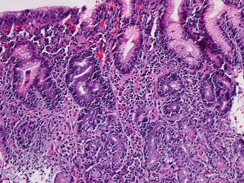
• May be endoscopically normal or present with mucosal nodules or erosions
• Presence of increased mucosal lymphocytes in both the lamina propria and surface and foveolar epithelium (>25/100 epithelial cells)
• Common associations: Coeliac disease, H. pylori gastritis
• Acute erosive
• Helicobacter pylori
• Autoimmune
• Granulomatous
• Eosinophilic
• Reactive (reflux/chemical)
• Radiation
• Drugs
• Ischaemic
• Infectious
Topic 2
MCQ – Question 2
A common complication of chronic gastro-oesophageal reflux is:
a) Eosinophilic oesophagitis.
b) Barrett’s oesophagus.
c) Candida oesophagitis.
d) MALT lymphoma.
e) Squamous cell carcinoma
Answer •.B
General comments
• Types
• Histopathological diagnosis (terminology in the UK)
• Complications – Dysplasia categories
Topic 3
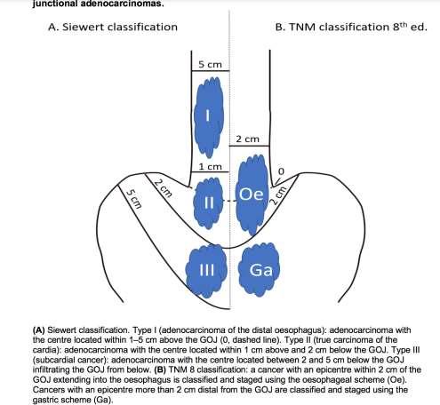
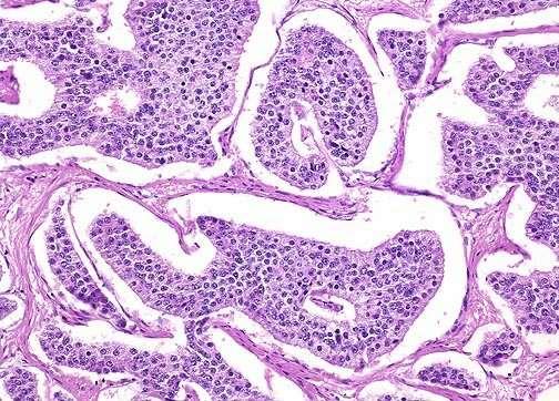
• An oesophagogastrectomy specimen reveals an adenocarcinoma at the gastro-oesophageal junction, about 80% of which lies in the stomach with the background oesophageal mucosa showing no evidence of Barrett’s change. The tumour invades just beyond the muscularis propria into the subserosa and involves two out of fifteen perigastric lymph nodes.
• The pathological stage is:
a) pT1, pN1, pMx.
b) pT2, pN1, pMx.
c) pT3, pN1, pMx.
d) pT1, pN0. pMx.
e) pT2, pN0. pMx.
•.c Answer
General comments
• Minimum datasets
• Difference in staging between oesophageal and gastric carcinomas
• Tumours at the G-O junction
Topic 4
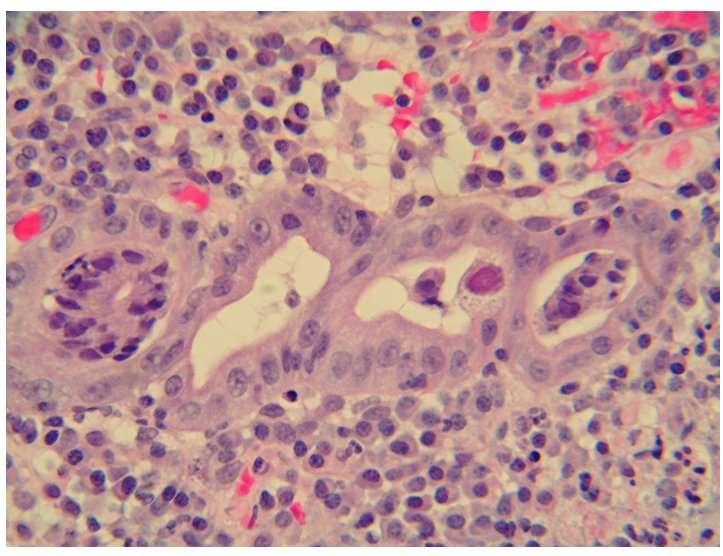
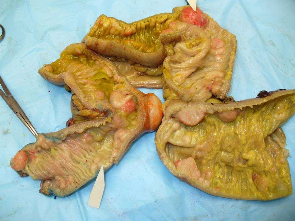
• A common association of gastric endocrine neoplasms is:
a) Helicobacter gastritis.
b) Familial adenomatous polyposis
c) Pernicious anaemia
d) Peptic ulcer disease.
e) NSAID use.
•. C Answer
General comments
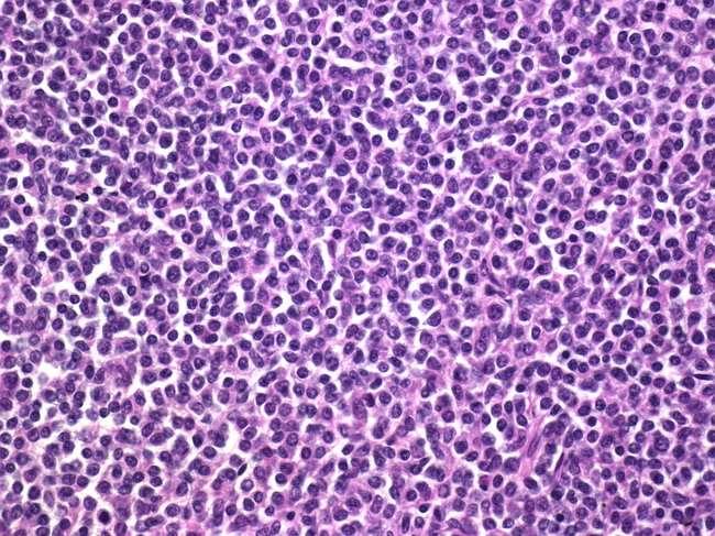
• 3 major types of gastric endocrine neoplasms (“carcinoids”):
1. Type 1 – Atrophic gastritis/pernicious anaemia (multiple, arise against a background of achlorhydria, hypergastrinaemia and endocrine cell hyperplasia)
2. Type 2 – Zollinger Ellison syndrome
3. Type 3 – Sporadic (usually solitary)
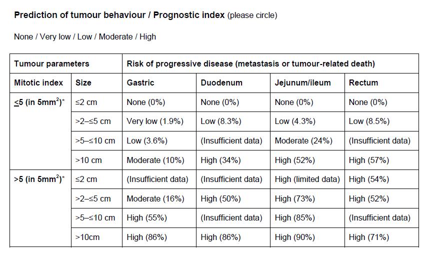
• WHO classification into 4 groups based upon morphology, size, depth of penetration, angioinvasion
Grading – based on Ki-67 staining and/or mitotic rate – low, immediate and high grade
Treatment depends on size and type
Topic 5
• Imatinib (Glivec) is a drug used for the treatment of:
a) Intestinal carcinoids.
b) Gastric MALT lymphoma.
c) H. Pylori gastritis
d) Metastatic GIST.
e) Gastric adenocarcinomas.
•.D Answer
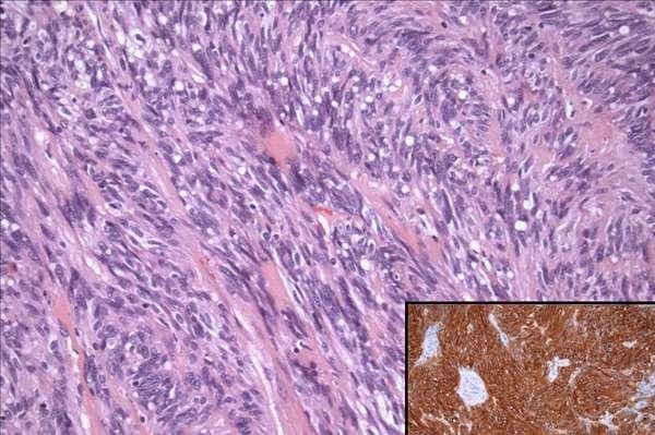
General comments
• Morphology (spindle cell/ epithelioid GISTs)
• Differential diagnosis
• Immunohistochemistry
• Prognostic factors
Topic 6
• A well-known association/complication of Helicobacter pylori gastritis is:
a) Gastric MALT lymphoma.
b) Inflammatory fibroid polyp.
c) Gastric GIST.
d) Fundic gland polyp.
e) Mantle cell lymphoma.
.A
• A: Eosinophilic oesophagitis.
• B: Herpes simplex infection
• C: Barrett’s oesophagus
• D: Reflux oesophagitis.
• E: Chronic radiation damage
• F: Cytomegalovirus infection
G: Adenocarcinoma
H: Malignant melanoma
I: Chronic graft versus Host disease.
J: Crohn’s disease.
K: Granular cell tumour
• Select one of the above oesophageal abnormalities which fits best for the following histological features seen in an oesophageal biopsy:
1. Ulceration, large intranuclear eosinophilic inclusions surrounded by a clear halo with cellular enlargement in the stromal and endothelial cells of the ulcer base.
•.F Answer
• A: Eosinophilic oesophagitis.
• B: Herpes simplex infection
• C: Barrett’s oesophagus
• D: Reflux oesophagitis.
• E: Chronic radiation damage
• F: Cytomegalovirus infection
G: Adenocarcinoma
H: Malignant melanoma
I: Chronic graft versus Host disease.
J: Crohn’s disease.
K: Granular cell tumour
• Select one of the above oesophageal abnormalities which fits best for the following histological features seen in an oesophageal biopsy:
• 2. Basal zone hyperplasia, papillary elongation, vascular dilatation, intraepithelial infiltration by a few neutrophils and occasional eosinophils
•.D Answer
• A: Eosinophilic oesophagitis.
• B: Herpes simplex infection
• C: Barrett’s oesophagus
• D: Reflux oesophagitis.
• E: Chronic radiation damage
• F: Cytomegalovirus infection
G: Adenocarcinoma
H: Malignant melanoma
I: Chronic graft versus Host disease.
J: Crohn’s disease.
K: Granular cell tumour
• Select one of the above oesophageal abnormalities which fits best for the following histological features seen in an oesophageal biopsy:
• 3. Apoptosis of individual squamous cells, necrosis and focal lymphocytic infiltration in a patient with history of bone marrow transplant.
•.I Answer
• A: Eosinophilic oesophagitis.
• B: Herpes simplex infection
• C: Barrett’s oesophagus
• D: Reflux oesophagitis.
• E: Chronic radiation damage
• F: Cytomegalovirus infection
G: Adenocarcinoma
H: Malignant melanoma
I: Chronic graft versus Host disease.
J: Crohn’s disease.
K: Granular cell tumour
• Select one of the above oesophageal abnormalities which fits best for the following histological features seen in an oesophageal biopsy:
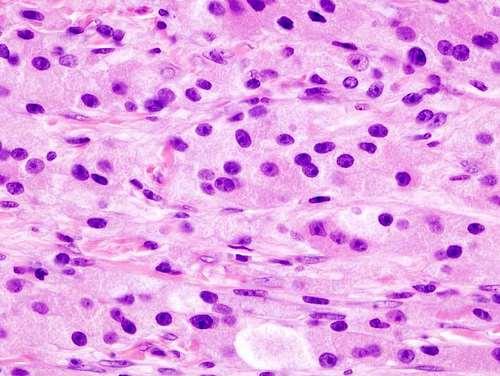
• 4. Pseudoepitheliomatous hyperplasia of the squamous epithelium, well circumscribed lesion in the lamina propria composed of sheets of polygonal cells with bland nuclei and abundant eosinophilic cytoplasm showing PAS positivity .
•.K Answer
• A: Eosinophilic oesophagitis.
• B: Herpes simplex infection
• C: Barrett’s oesophagus
• D: Reflux oesophagitis.
• E: Chronic radiation damage
• F: Cytomegalovirus infection
G: Adenocarcinoma
H: Malignant melanoma
I: Chronic graft versus Host disease.
J: Crohn’s disease.
K: Granular cell tumour
• Select one of the above oesophageal abnormalities which fits best for the following histological features seen in an oesophageal biopsy:
• 5. Ulceration, dyscohesive and multinucleated squamous cells at the margin of the ulcer, ground glass inclusions filling the nucleus.
• E: Bile reflux/chemical gastritis. K: Diffuse signet ring cell carcinoma.
• F: Xanthelasma. L: Lymphocytic gastritis.
• Each of the following subjects has a gastric biopsy. For each one select the most likely condition from the option list above.
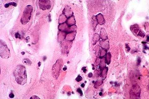
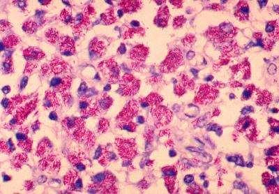
• 1. A 52 year old male with multiple cream coloured plaques in the antrum, ranging in size from 1mm to 4mm: The biopsy shows loosely organised aggregates of foamy histiocytes in the upper lamina propria with bland inconspicuous nuclei. The cytoplasm is PAS and ZN negative.
•.F Answer
General comments
• Xanthelasma are formed of aggregates of foamy histiocytes in the upper lamina propria
• Common in the antrum; single or multiple
• Inconspicuous nuclei; cytoplasm contains lipid
• D/D – Mucin-secreting adenocarcinoma Whipple’s disease MAI infection
• Each of the following subjects has a gastric biopsy. For each one select the most likely condition from the option list above.
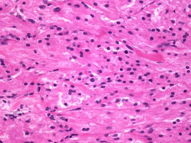
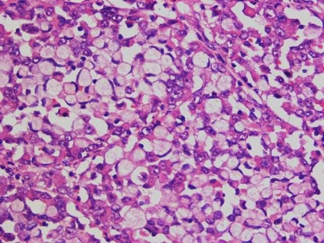
• 2. A 64 year old female with antral ulceration and thickening: The biopsy shows individual and small clusters of atypical cells in the lamina propria. The cells have abundant vacuolated cytoplasm with peripheral enlarged nuclei. The cytoplasm is Alcian blue - PAS positive.
• E: Bile reflux/chemical gastritis. K: Diffuse signet ring cell carcinoma.
• F: Xanthelasma. L: Lymphocytic gastritis.
• Each of the following subjects has a gastric biopsy. For each one select the most likely condition from the option list above.
• 3. A 40 year old male with dyspepsia: The biopsy shows a chronic inflammatory infiltrate and occasional lymphoid follicles with germinal centres in the lamina propria, and several neutrophils infiltrating the foveolar epithelium.
• E: Bile reflux/chemical gastritis. K: Diffuse signet ring cell carcinoma.
• F: Xanthelasma.
L: Lymphocytic gastritis.
• Each of the following subjects has a gastric biopsy. For each one select the most likely condition from the option list above.
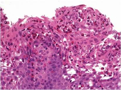
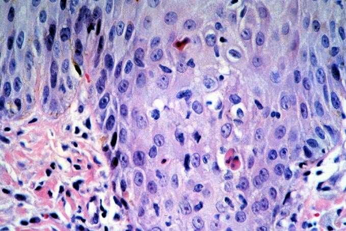
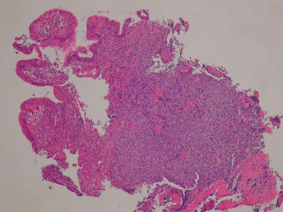
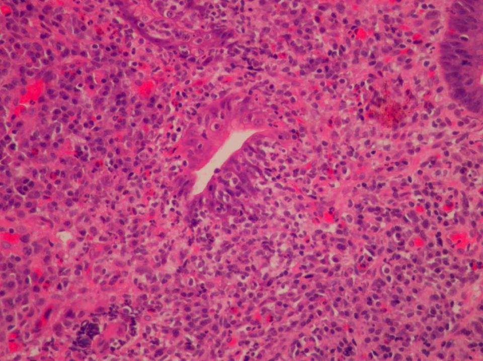
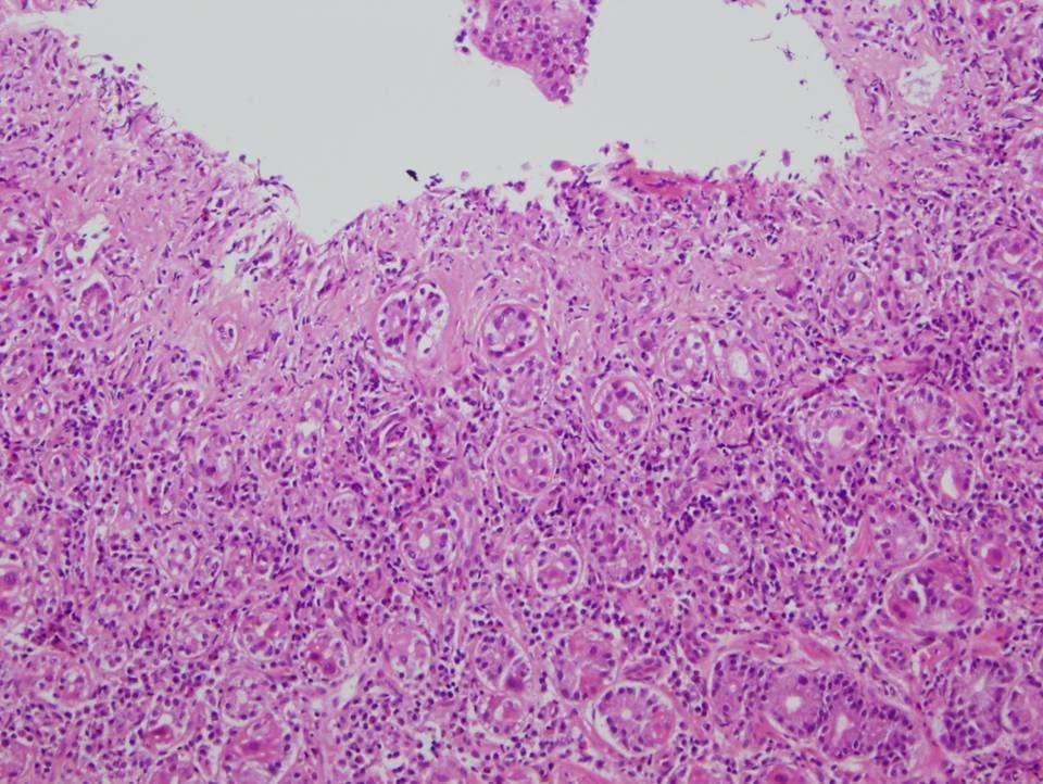
• 4. A 54 year old woman with multiple antral erosions: The biopsy shows a dense and diffuse lymphoid infiltrate in the lamina propria with infiltration of the glands by several aggregates of lymphocytes (lymphoepithelial lesions).
• E: Bile reflux/chemical gastritis. K: Diffuse signet ring cell carcinoma.
• F: Xanthelasma. L: Lymphocytic gastritis.
• Each of the following subjects has a gastric biopsy. For each one select the most likely condition from the option list above.
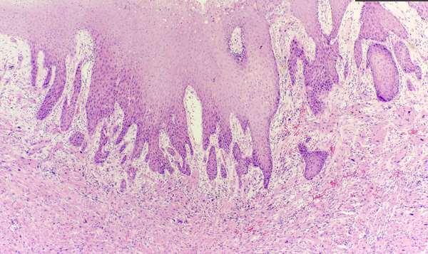
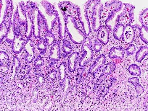
• 5. A 61 year old male with dyspepsia: The antral biopsy shows foveolar hyperplasia and congestion of the lamina propria associated with smooth muscle proliferation.
• One of the above special stains is most helpful in the diagnosis of each of the following upper gastrointestinal conditions. Select the most appropriate option for each.
• One of the above special stains is most helpful in the diagnosis of each of the following upper gastrointestinal conditions. Select the most appropriate option for each.
• One of the above special stains is most helpful in the diagnosis of each of the following upper gastrointestinal conditions. Select the most appropriate option for each.
• One of the above special stains is most helpful in the diagnosis of each of the following upper gastrointestinal conditions. Select the most appropriate option for each.
• One of the above special stains is most helpful in the diagnosis of each of the following upper gastrointestinal conditions. Select the most appropriate option for each.
• 5 -Tablet induced gastric erosion (? Iron tablet).
•.J Answer
• A: CD3 G: CD117.
• B: Chromogranin. H: TTF-1.
• C: CD34 I: S100.
• D: CK20. J: Desmin.
• E: HMB45. K: Cyclin D1.
• F: Alk-1
• One of the above immunohistochemical stains is the most useful in the diagnosis of the following upper gastrointestinal conditions:
• 1-Gastric Schwannoma.
•.I Answer
• A: CD3 G: CD117.
• B: Chromogranin.
H: TTF-1.
• C: CD34 I: S100.
• D: CK20. J: Desmin.
• E: HMB45. K: Cyclin D1.
• F: Alk-1
• One of the above immunohistochemical stains is the most useful in the diagnosis of the following upper gastrointestinal conditions:
• 2-Carcinoid (neuroendocrine tumour) of the duodenum.
•.B Answer
• A: CD3 G: CD117.
• B: Chromogranin. H: TTF-1.
• C: CD34 I: S100.
• D: CK20. J: Desmin.
• E: HMB45. K: Cyclin D1.
• F: Alk-1
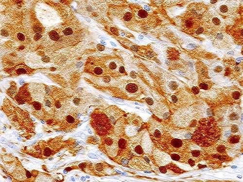
• One of the above immunohistochemical stains is the most useful in the diagnosis of the following upper gastrointestinal conditions:
• 3 -Granular cell tumour of the oesophagus.
•.I Answer
• A: CD3 G: CD117.
• B: Chromogranin. H: TTF-1.
• C: CD34 I: S100.
• D: CK20. J: Desmin.
• E: HMB45. K: Cyclin D1.
• F: Alk-1
• One of the above immunohistochemical stains is the most useful in the diagnosis of the following upper gastrointestinal conditions:
• 4 -Gastrointestinal stromal tumour of the stomach.
•.G Answer
• A: CD3 G: CD117.
• B: Chromogranin. H: TTF-1.
• C: CD34 I: S100.
• D: CK20. J: Desmin.
• E: HMB45. K: Cyclin D1.
• F: Alk-1
• One of the above immunohistochemical stains is the most useful in the diagnosis of the following upper gastrointestinal conditions: