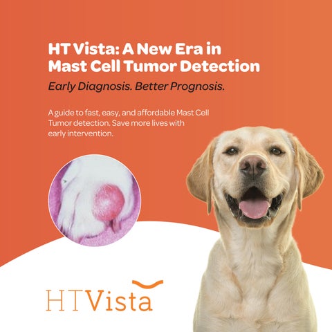
HT Vista: A New Era in Mast Cell Tumor Detection
Early Diagnosis. Better Prognosis.
A guide to fast, easy, and affordable Mast Cell Tumor detection. Save more lives with early intervention.


A guide to fast, easy, and affordable Mast Cell Tumor detection. Save more lives with early intervention.
1 in 4 dogs develop cancer
47% of dogs over 10 die from cancer
1 in 3 deaths are from skin cancer
21% are MCTs
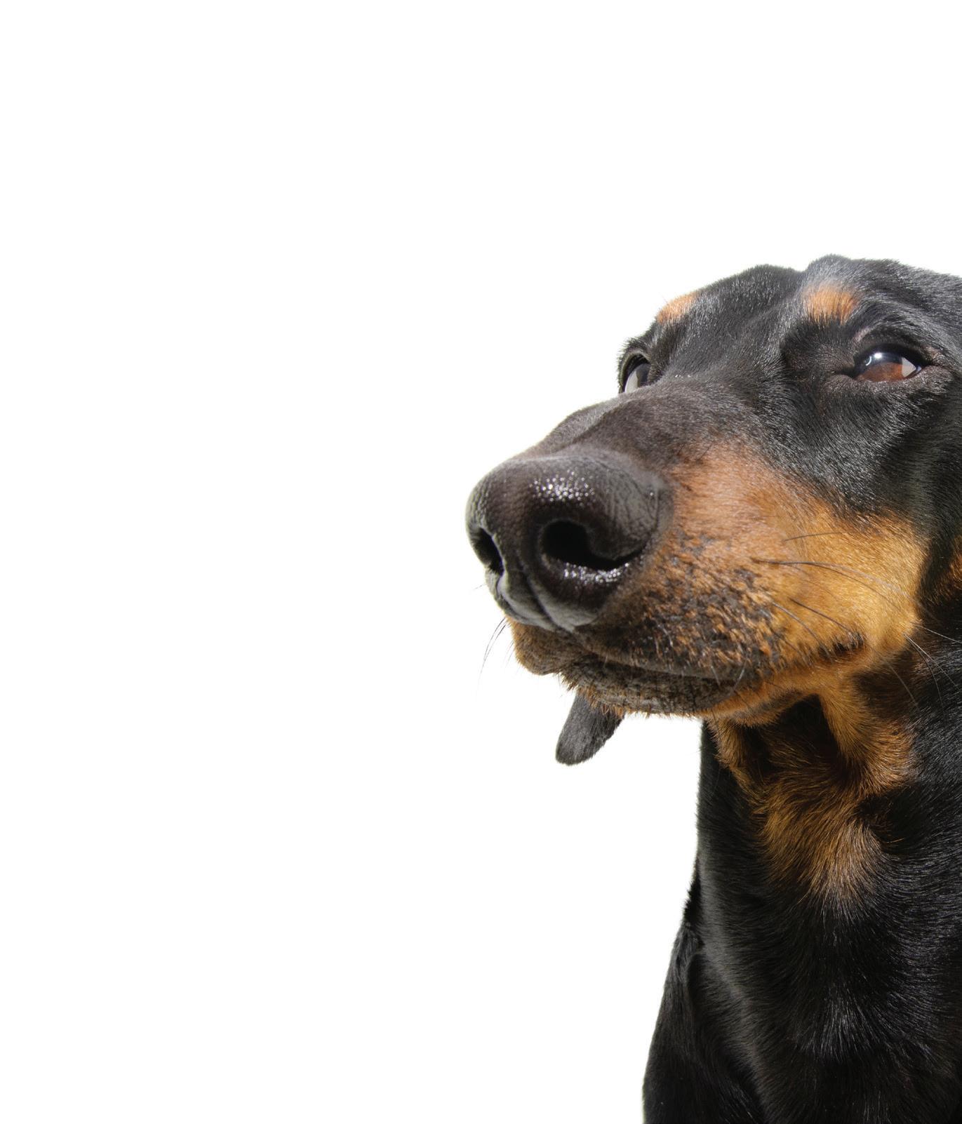
Breeds like Boxers, Boston Terriers, Labrador Retrievers, and Bulldogs are at higher risk.
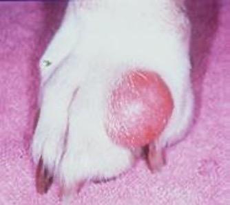
MCTs commonly appear on the trunk, limbs, or perineal area but can develop anywhere on the skin.
MCTs can appear as small, benign-looking lumps or as aggressive, invasive growths. This variability often leads to uncertainty and testing delays.
Each mass must be evaluated independently. A dog may present with a mix of benign and malignant tumors simultaneously.
MCTs can release histamine and other substances that cause local inflammation, itching, swelling, and even systemic effects like gastric ulcers.
The decision to “wait and see” if a mass changes can introduce unnecessary risk, including delayed treatment and worsening outcomes. 50% of dermal/subcutaneous masses go undiagnosed
Worsened Prognosis Risks Lives Delays Needed Services
Needles or surgical sampling can be intimidating and discourage immediate testing—especially for small or seemingly harmless masses.
Diagnostic procedures like cytology and biopsy can be beyond the budgets of pet parents, leading some owners to delay or decline testing.
Cost and invasiveness highlight the need for a non-invasive, affordable, and fast screening tool to evaluate more lumps and bumps early.

Diagnosis (via HT Vista, plus FNA or biopsy)
Staging (assessing disease spread)
Treatment planning (based on diagnosis and staging)

Each tumor type behaves differently. Some metastasize through lymphatics, others via the bloodstream, and some may remain locally invasive. Thus, staging strategies must align with tumor biology.
Most cases begin with an FNA for cytology; however, due to tumor variability, biopsy may be required.
This includes lymph node assessment, splenic and hepatic aspirates, abdominal ultrasound, and bloodwork to evaluate disease spread and overall health.
Surgery is first-line for localized tumors. For high-grade or systemic cases, chemotherapy, radiation, or targeted therapies may be recommended.
A methodical diagnostic approach improves patient outcomes and fosters collaboration between veterinarians and pet owners.
Early identification and classification lead to better outcomes through prompt surgical removal and appropriate staging.


Use analogies to human medicine—Would a person ignore a lump without knowing what it is?—to explain the need for individual mass evaluation.
Educating clients about Mast Cell Tumors and early detection builds trust and encourages them to consent to diagnostics.

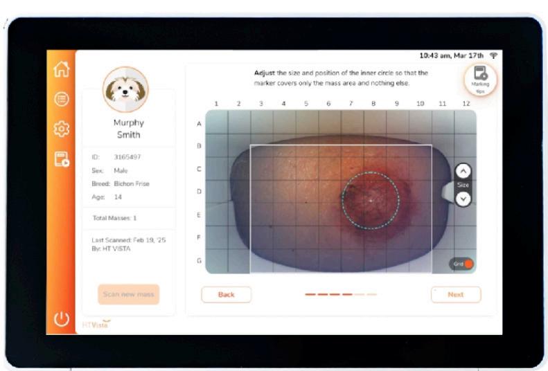
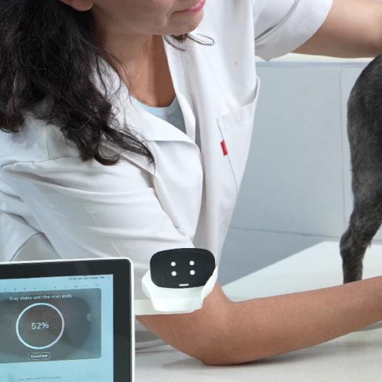
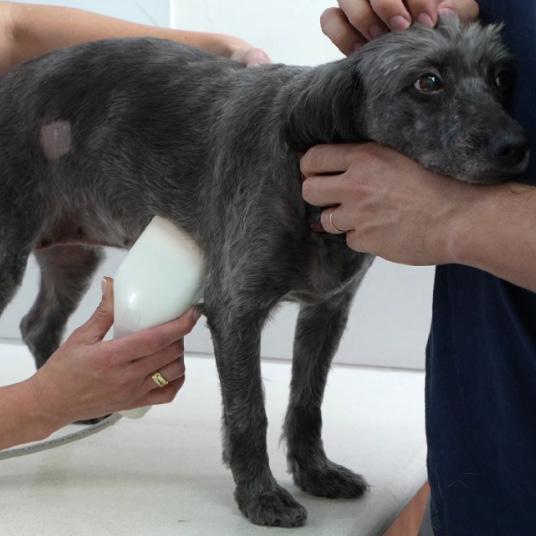
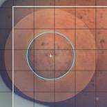
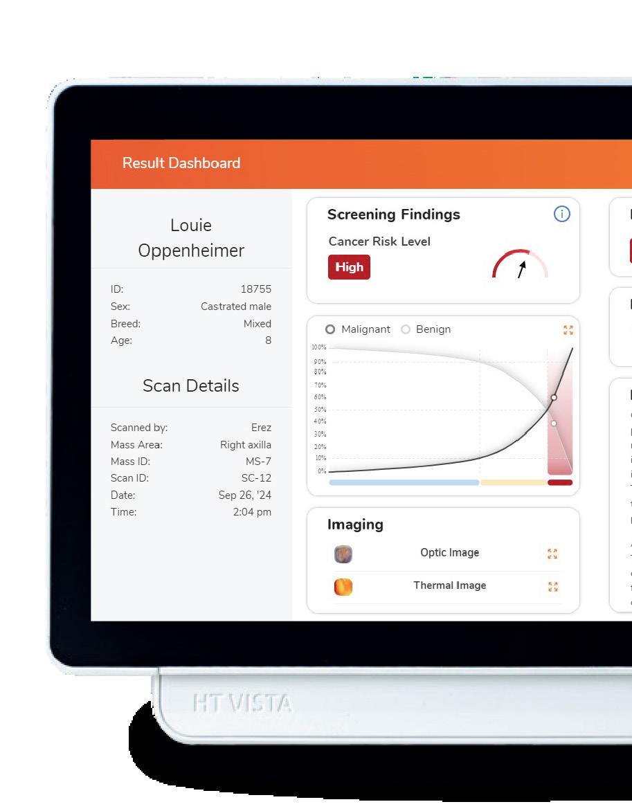
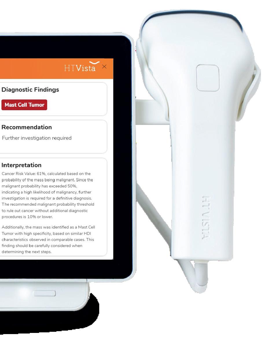
HT Vista measures heat diffusion patterns to identify differences in vascularity, metabolism, composition, and cooling rates indicative of mass malignancy compared to healthy tissue.
Proprietary AI analyzes these patterns to assess cancer risk, and detect specific cancer types, like MCTs.

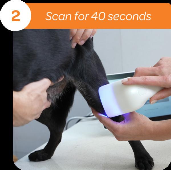
HT Vista screens dermal and subcutaneous masses quickly, delivering a cancer risk assessment and in some cases, a specific diagnosis in under a minute.

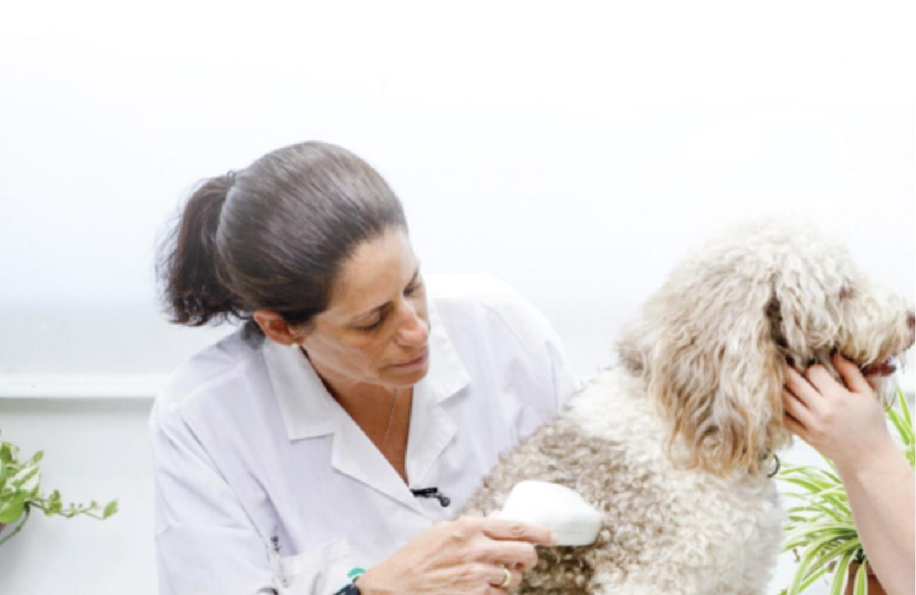
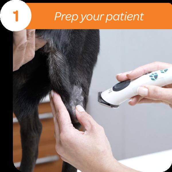
The scan is painless and integrates seamlessly into
HT Vista shifts care from reactive to proactive, ensuring every mass gets the attention

Veterinary nurses/technicians and nurses can operate
HT Vista, integrating mass scans into wellness checks and improving early cancer detection.

Minimal vs. Complete Staging
Minimal staging includes FNA of the regional lymph node and a basic health profile (CBC, chemistry, urinalysis).
Complete staging incorporates abdominal ultrasound with liver/spleen aspirates, thoracic imaging, and potentially bone marrow evaluation.
The decision depends on...
Tumor location
Known prognostic indicators Owner goals and finances


Tumors used to be graded 1, 2, or 3 (Patnaik system), but most were graded 2, creating ambiguity. The Kiupel system now uses a 2-tier grading: low or high grade, which correlates better with outcomes. Lowgrade tumors tend to have prolonged survival (>2 years), while high-grade tumors may yield a median survival of only four months—or even less.
High mitotic activity indicates more aggressive tumors and worse prognosis.
Shar-Peis, for example, often present with aggressive tumors. Tumors in the inguinal, muzzle, or mucocutaneous areas have higher metastatic potential.
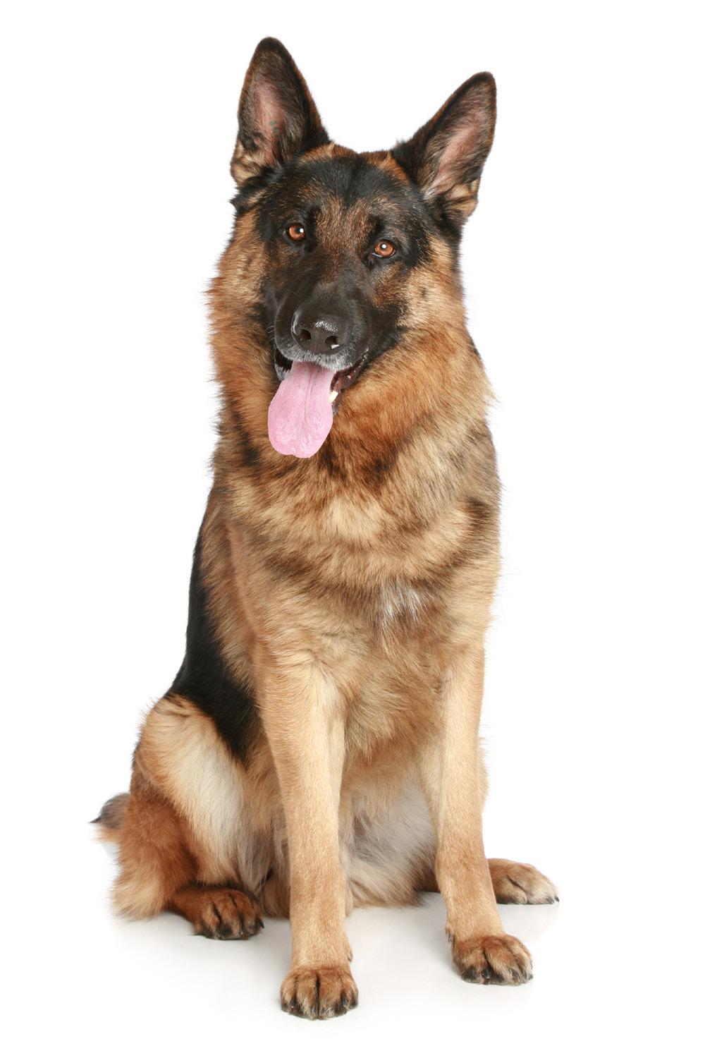
Jemma, a ten-year-old German Shepherd, had a lowgrade MCT removed from her foot.
Months later, she developed a swollen leg and enlarged lymph node. Cytology confirmed metastasis.
Although her liver appeared abnormal on ultrasound, aspirates showed no mast cell infiltration—confirming stage II disease. Mass at the distal part of the suture line scanned with HT Vista to discover a Mast Cell Tumor.
Always aspirate normal-appearing organs; imaging alone can be misleading. HT Vista Heat Diffusion technology identified the presence of a MCT
With surgery and chemotherapy, dogs with stage II MCTs can live several years—median survival over 40 months.
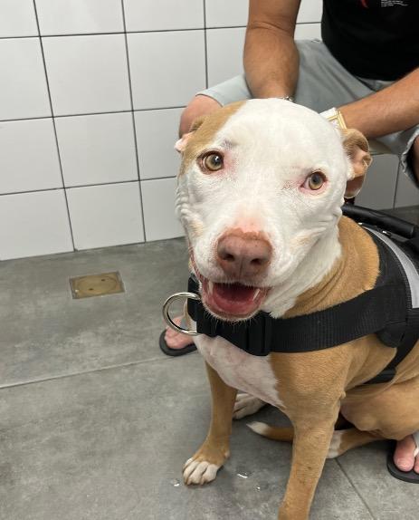
Sigi, a FS 5-year-old Pitbull mix with no issues in her history presented with five small masses her owners noticed in the past month. HT Vista scan of multiple mast cell tumors, all low grade.
Three cutaneous masses are all 0.8-1 cm and are all easily movable.
Prognosis
Excellent Treatment
Surgery alone
Just because a dog has multiple mast cell tumors does not mean that they have a poor prognosis. Multiple cutaneous MCTs, detected by HT Visa, are associated with a low rate of metastasis and a good prognosis for long-term survival.
MCT treatment depends on grade, stage, location, and patient factors, and may include surgery, chemotherapy, radiation, and supportive care.
Research shows that 2 cm margins with one fascial plane deep are often sufficient for lowto mid-grade tumors— reducing unnecessary wide excisions.
A 2004 study found low recurrence rates for grade I and II tumors, even with narrow margins—changing surgical planning.
Used for high-grade tumors, poor prognostic factors, or metastasis. Common drugs: prednisone, vinblastine, CCNU, and tyrosine kinase inhibitors.
All dogs with gross disease should receive prednisone, diphenhydramine, and a gastroprotectant to mitigate effects like vomiting and ulcers.

Eventually, the focus shifts to comfort. Honest communication with pet owners is essential, combining medical guidance and emotional support.
Rules out malignancy with high confidence
Sensitivity
90% — correctly identifies malignant tumors.
98% — correctly classifies that a mass is truly benign. NPV
Low Risk (≤10%) Mass is likely benign.
Moderate Risk (>10%–50%)
Further investigation recommended.
High Risk (≥50%) Suspicious—requires follow-up.
Targeted detection: HT Vista’s AI subclassifier flags MCTs—essential for variable presentations.

Prioritizes high-risk cases
Reduces diagnostic uncertainty Informs timely treatment planning
Performance metrics
Specificity
90% — distinguishes MCTs from other tumors.
77% — flagged cases are likely MCTs. PVV
HT Vista delivers rapid cancer risk assessments— 40 seconds, no needles or sedation.
Rule our or rule in
Confidently clears benign lumps while flagging suspicious masses for follow-up —streamlining decision-making.
Improved care and
Builds trust with pet owners and supports higher-quality care.


MCTs range from local to aggressive cancers—each requiring a unique approach.
Early diagnosis, grading staging, and tailored treatment plans offer the best outcomes.
Combining advanced tools like HT Vista with compassionate, science-backed decision making offers every patient the best chance at a longer, healthier life.


1 Source: Dank Gillian , Buber Tali , Rice Anna , Kraicer Noa , Hanael Erez , Shasha Tamir Aviram Gal , Yehudayoff Amir , Kent Michael S.; Training and validation of a novel non-invasive imaging system for ruling out malignancy in canine subcutaneous and cutaneous masses using machine learning in 664 masses; Frontiers in Veterinary Science (10, 2023) URL=https://www.frontiersin.org/journals/veterinary-science/articles/10.3389/ fvets.2023.1164438; DOI=10.3389/fvets.2023.1164438; ISSN=2297-1769