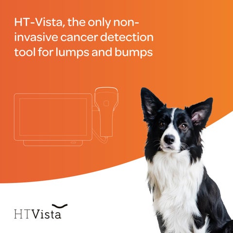
HT-Vista, the only noninvasive cancer detection tool for lumps and bumps
Most dermal and subcutaneous masses are benign, but when they are not, early detection saves lives
dogs over the age of 10 succumb to cancer, making it the leading cause of death 47%
Here are the statistics: BY dogs develop cancer at some stage of their lives 1 in 4 of all tumors in dogs are skin tumors 1 / 3 of dermal and subcutaneous masses are undiagnosed 50%
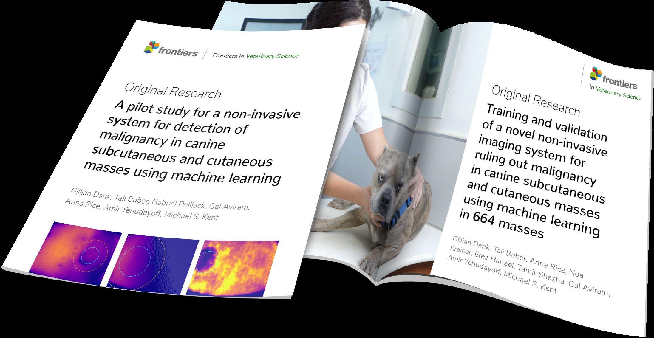
Source: Dank Gillian , Buber Tali , Rice Anna , Kraicer Noa , Hanael Erez , Shasha Tamir Aviram Gal , Yehudayoff Amir , Kent Michael S.; Training and validation of a novel non-invasive imaging system for ruling out malignancy in canine subcutaneous and cutaneous masses using machine learning in 664 masses; Frontiers in Veterinary Science (10, 2023)
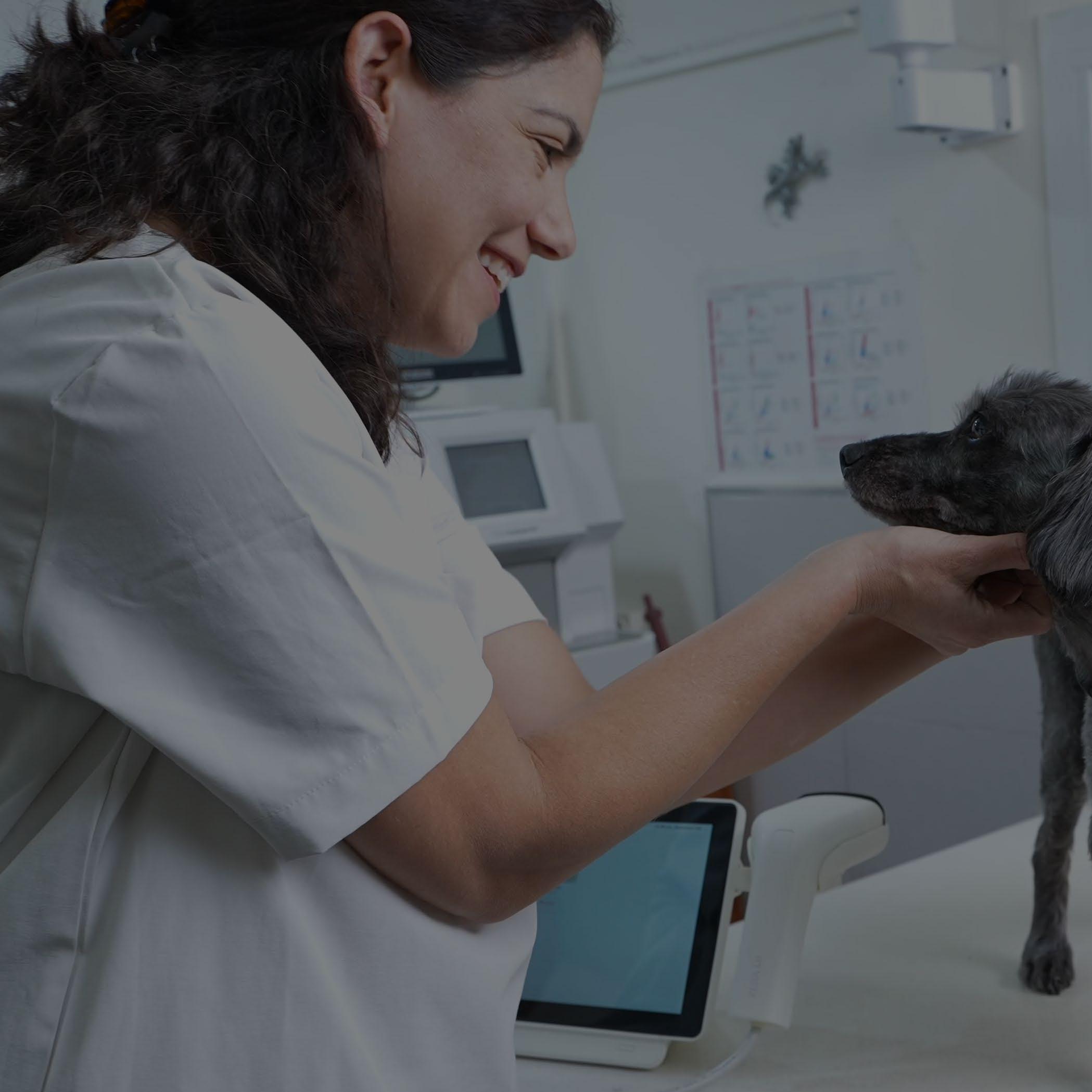
The benefits of HT Vista to your patients and your practice
Early Detection
Supports decision-making that reduces the time between diagnosis and treatment.
Build Stronger Teams
Ease of use means the veterinary support staff can perform the test and reduce the veterinarian's workload.
Less Stress on Patient
Non-invasive, pain-free approach means intrusive diagnostics are necessary only when evidence indicates.
Fosters Growth
Increase diagnostic and treatment opportunities when you scan regularly, and uncover overlooked malignancies in need of biopsy.
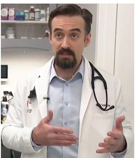
The confidence level of a negative result being a true negative is 98%… in medicine, that’s almost as good as it gets.
Dr. Peter Converse, DVM Owner/Medical Director Advanced Veterinary Care, CO
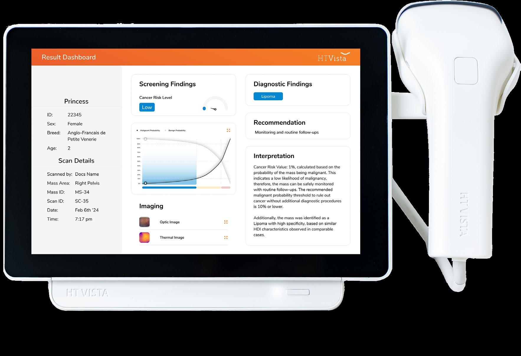
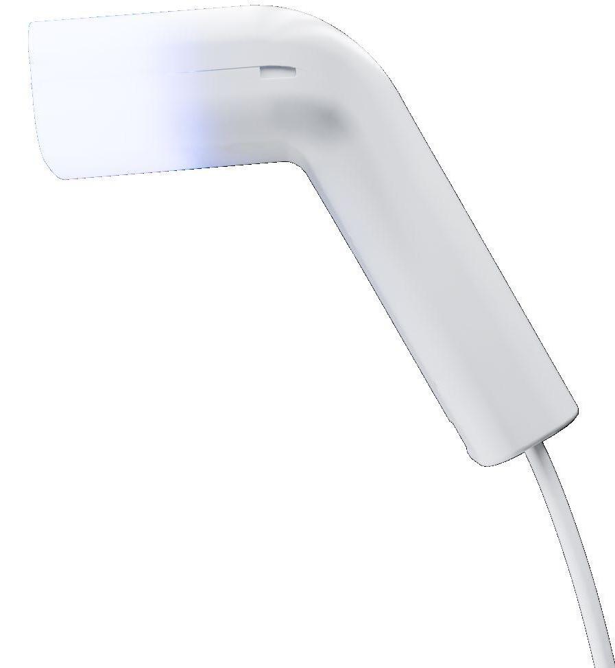
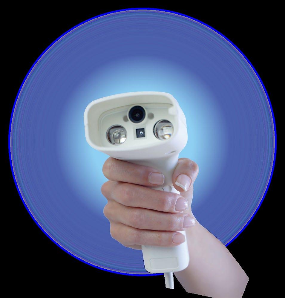
How HT Vista Works
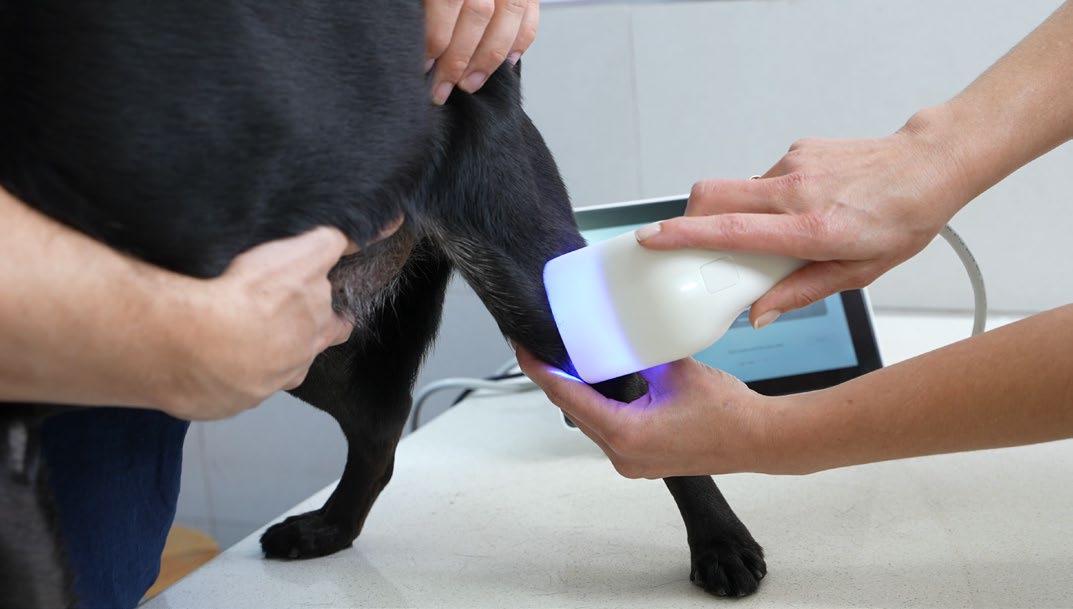
40 sec overall scan time
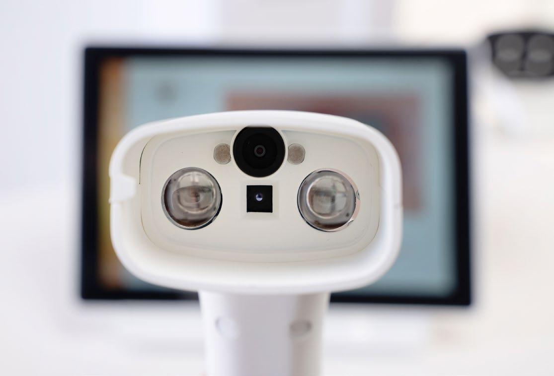
Heat Diffusion Imaging
A patented non-invasive technology
The underlying principle of the HT Vista Heat Diffusion Imaging (HDI) technology, is that benign and malignant tissues display different heat transfer rates. This is due to differences in composition, metabolism, tissue morphology, and vascular network, which affect their thermophysical properties.
This innovative screening modality relies on the unique thermal signals recorded by the device, as the tissue is heated for 10 seconds and left to cool down for 30 seconds. The trained algorithm then processes those signals and delivers a predictive score.
Optical Camera Thermal Sensor
Heat Source
“The HT Vista has proved a really useful bit of kit for our clinic. We are able to assess masses quickly and simply and give owners either immediate peace-of-mind or move forward with investigations knowing there is a good reason for them. Our protocols are set so this is an almost entirely nurse-led process, allowing them to utilise their skillset and creating real efficiency for our clinic.”
Dr Catherine Henstridge MRCVS (AKA 'Cat the Vet')
"The HT Vista is an excellent addition to the primary care vet's tools. The more of us that start using AI tools like this, the more they will learn and become better and better.'
Simon Hayes, MRCVS, Veterinarian, Chief Veterinary Officer, Creature Comforts Vets, UK
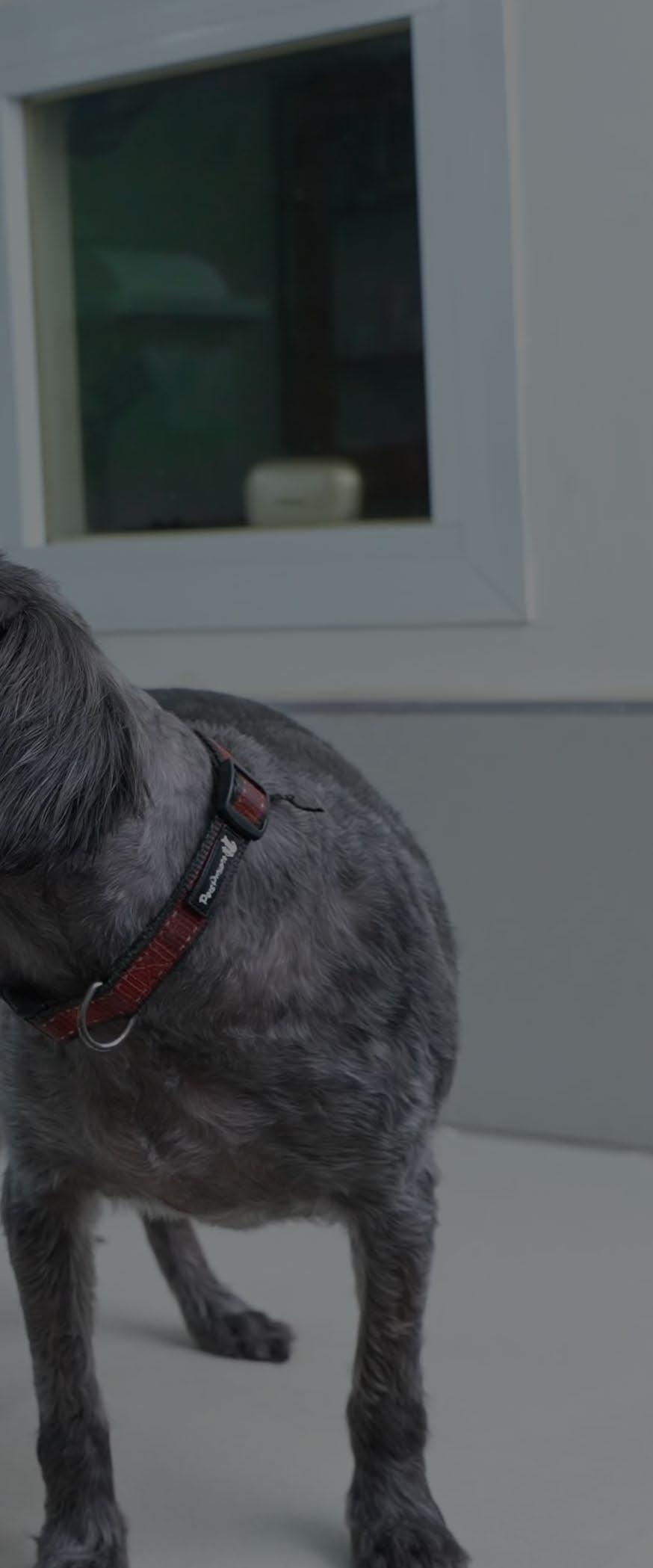
Case Study
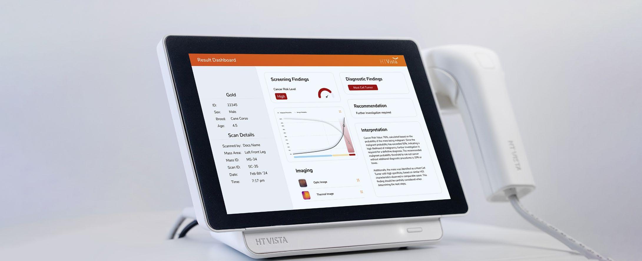
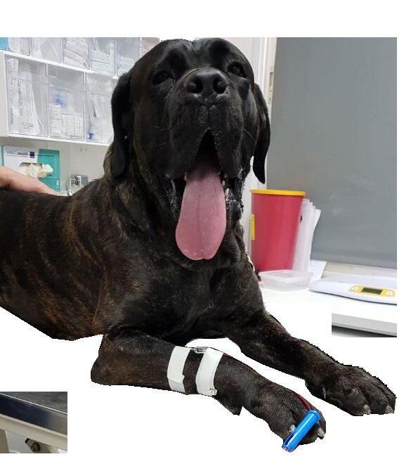
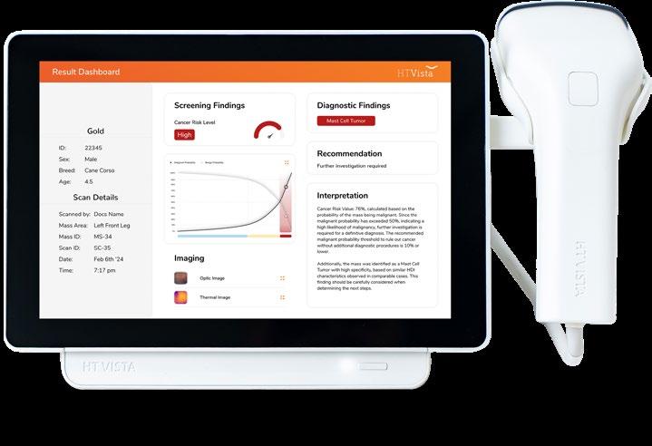
4.5 yo Cane Corso Gold
Gold's owner wanted to take a wait-and-see approach, but was convinced by her veterinarian to do a scan with HT Vista.
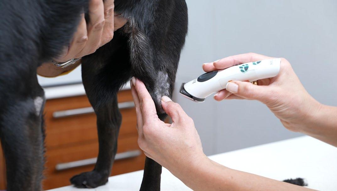
Incidental finding of a SC mass during a routine physical examination
Subcutaneous mass on the left front leg, firm, mobile, 5x5 cm, circumscribed
HEAT DIFFUSION
IMAGING (HDI)
SCAN
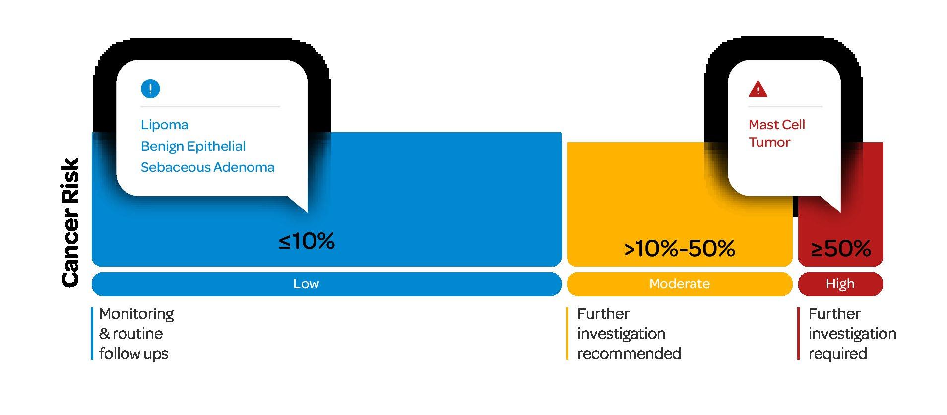
BASED ON HT VISTA RESULTS
A 40-second scan returned a "High Cancer Risk" value with diagnostic alert of a "Mast Cell Tumor." Further Investigation was recommended.
OUTCOME
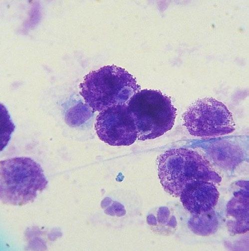
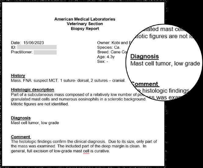
HISTOPATHOLOGY
A fine needle aspirate was performed by the veterinarian.
Results indicated a low-grade Mast Cell Tumor.
Gold underwent a lumpectomy, and returned home healthy after detecting the mast cell tumor early. The pet parent confirmed she would not have opted for surgery without HT Vista!
Guss had several tumors… we immediately took the HT Vista and scanned. And we unfortunately found something that needed further investigation. Unbelievably, it was the original lipoma but underneath it was an invading mast-cell tumor.
Dr. Monica Wlodarchak, DVM
Touch, WA
Tender