Medical Imaging International
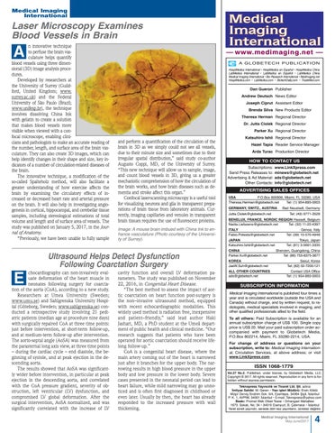
Laser Microscopy Examines Blood Vessels in Brain n innovative technique to perfuse the brain vasculature helps quantify blood vessels using three dimensional (3D) image analysis procedures. Developed by researchers at the University of Surrey (Guildford, United Kingdom; www. surrey.ac.uk) and the Federal University of São Paulo (Brazil; www.unifesp.br), the technique involves dissolving China Ink with gelatin to create a solution that makes blood vessels more visible when viewed with a confocal microscope, enabling clinicians and pathologists to make an accurate reading of the number, length, and surface area of the brain vasculature. They can also create 3D images, which can help identify changes in their shape and size, key indicators of a number of circulation-related diseases of the brain. The innovative technique, a modification of the so-called Spalteholz method, will also facilitate a greater understanding of how exercise affects the brain by examining the circulatory effects of increased or decreased heart rate and arterial pressure on the brain. It will also help in investigating angiogenesis in cortical, hippocampal, and cerebellar tissue samples, including stereological estimations of total volume and length and of surface area of vessels. The study was published on January 5, 2017, in the Journal of Anatomy. “Previously, we have been unable to fully sample
A
–– www.medimaging.net –– A G L O B E T E C H P U B L I C AT I O N HospiMedica International • HospiMedica en Español • HospiMedica China LabMedica International • LabMedica en Español • LabMedica China Medical Imaging International • Bio Research International • Medimaging.net HospiMedica.com • LabMedica.com • BiotechDaily.com • TradeMed.com
Dan Gueron Publisher Andrew Deutsch News Editor Joseph Ciprut Assistant Editor Brenda Silva New Products Editor Theresa Herman Regional Director Dr. Jutta Ciolek Regional Director Parker Xu Regional Director
and perform a quantification of the circulation of the brain in 3D as we simply could not see all vessels, due to their minute size and sometimes due to their irregular spatial distribution,” said study co-author Augusto Coppi, MD, of the University of Surrey. “This new technique will allow us to sample, image, and count blood vessels in 3D, giving us a greater mechanistic comprehension of how the circulation of the brain works, and how brain diseases such as dementia and stroke affect this organ.” Confocal laser-scanning microscopy is a useful tool for visualizing neurons and glia in transparent preparations of brain tissue from laboratory animals. Currently, imaging capillaries and venules in transparent brain tissues requires the use of fluorescent proteins. Image: A mouse brain imbued with China Ink to enhance vasculature (Photo courtesy of the University of Surrey).
Ultrasound Helps Detect Dysfunction Following Coarctation Surgery chocardiography can non-invasively evaluate deformation of the heart muscle in neonates following surgery for coarctation of the aorta (CoA), according to a new study. Researchers at Umea University (Sweden; www.umu.se) and Sahlgrenska University Hospital (Göteborg, Sweden; www.sahlgrenska.se) conducted a retrospective study involving 21 pediatric patients (median age at procedure nine days) with surgically repaired CoA at three time points: just before intervention, at short-term follow-up, and at medium-term follow-up after intervention. The aorto-septal angle (AoSA) was measured from the parasternal long axis view, at three time points – during the cardiac cycle – end diastole, the beginning of systole, and at peak ejection in the descending aorta. The results showed that AoSA was significantly wider before intervention, in particular at peak ejection in the descending aorta, and correlated with the CoA pressure gradient, severity of obstruction, left ventricular (LV) dysfunction, and compromised LV global deformation. After the surgical intervention, AoSA normalized, and was significantly correlated with the increase of LV
E
cavity function and overall LV deformation parameters. The study was published on November 22, 2016, in Congenital Heart Disease. “The best method to assess the impact of aortic coarctation on heart function post-surgery is the non–invasive ultrasound method, equipped with recent echocardiographic modalities. This widely used method is radiation free, inexpensive and patient–friendly,” said lead author Haki Jashari, MD, a PhD student at the Umeå department of public health and clinical medicine. “Our research suggests that patients who have been operated for aortic coarctation should receive lifelong follow-up.” CoA is a congenital heart disease, where the main artery coming out of the heart is narrowed just after it branches for the upper body. The narrowing results in high blood pressure in the upper body and low pressure in the lower body. Severe cases presented in the neonatal period can lead to heart failure, while mild narrowing may go unnoticed and is often first diagnosed in childhood or even later. Usually by then, the heart has already responded to the increased pressure with wall thickening.
Katsuhiro Ishii Regional Director Hazel Tapia Reader Service Manager Arda Turac Production Director
HOW TO CONTACT US Subscriptions: Send Press Releases to: Advertising & Ad Material: Other Contacts:
www.LinkXpress.com minews@globetech.net ads@globetech.net info@globetech.net
ADVERTISING SALES OFFICES USA P.O.Box 800806, Miami, FL 33280, USA Theresa.Herman@globetech.net Tel: (1) 954-893-0003 GERMANY, SWITZ., AUSTRIA Jutta.Ciolek@globetech.net
Bad Neustadt, Germany Tel: (49) 9771-3528
BENELUX, FRANCE, NORDIC REGION Hasselt, Belgium Nadia.Liefsoens@globetech.net Tel: (32) 11-22-4397 ITALY Fabio.Potesta@globetech.net
Genoa, Italy Tel: (39) 10-570-4948
JAPAN Katsuhiro.Ishii@globetech.net
Tokyo, Japan Tel: (81) 3-5691-3335
CHINA Parker.Xu@globetech.net
Shenzen, Guangdong, China Tel: (86) 755-8375-3877
KOREA JaeW.Suh@globetech.net
Seoul, Korea Tel: (82) 02-7200-121
ALL OTHER COUNTRIES ads@globetech.net
Contact USA Office Tel: (1) 954-893-0003
SUBSCRIPTION INFORMATION Medical Imaging lnternational is published four times a year and is circuIated worldwide (outside the USA and Canada) without charge, and by written request, to radiologists, medical specialists involved in imaging, and other qualified professionals allied to the field. To all others: Paid Subscription is available for an annual subscription charge of US$ 100. Single copy price is US$ 20. Mail your paid subscription order accompanied with payment to Globetech Media, P.O.Box 802214, Miami, FL 33280-2214, USA. For change of address or questions on your subscription, write to: Medical Imaging lnternational, Circulation Services, at above address; or visit www.LinkXpress.com
ISSN 1068-1779 Vol.27 No.2. Published, under license, by Globetech Media, LLC. Copyright © 2017. All rights reserved. Reproduction in any form is forbidden without express permission.
Teknopress Yayıncılık ve Ticaret Ltd. S¸ti. adına ˙Imtiyaz Sahibi: M. Geren • Yazı is¸leri Müdürü: Ersin Köklü Müs¸ ir Dervis¸ ˙Ibrahim Sok. 5/4, Esentepe, 34394 S¸is¸ li, ˙Istanbul P. K. 1, AVPIM, 34001 ˙Istanbul • E-mail: Teknopress@yahoo.com Baskı: Promat Web Ofset Tesisi • Orhangazi Mahallesi 1673. Sokak, No: 34 • 34510 Esenyurt, B. Çekmece • ˙Istanbul Yerel süreli yayındır, senede dört kez yayınlanır, ücretsiz dag˘ıtılır.
Medical Imaging International May-June/2017
4