Medical Imaging International
Optical System Reconstructs Virtual 3D Spinal Column novel diagnostic system offers stereo-radiographic imaging and three dimensional (3D) modeling tools particularly suited to orthopedic applications. The Biomod 3S system uses noninvasive optical information technology that is combined with classic two-dimensional (2D) radiographic images of the spine to yield a virtually reconstructed 3D model of the vertebral column that can provide clinicians with complete view of the patient’s spine. This allows a thorough and accurate evaluation of spinal deformities in diverse pathologies, such as scoliosis, kyphosis, vertebral compression, posture and balance anomalies, and dorsopathy, among others. The optical acquisition is realized simultaneously with frontal and sagittal full spine X-rays obtained from an existing radiological set-up, and therefore does not require any change in clinical routine or increased radiation exposure. Patented software performs an automatic correction of patient’s sway during acquisition, thus providing a complete evaluation of the spine and the posture in a minimum time input, with an average spinal reconstruction taking about five minutes. The Biomod 3S system was developed by AXS Medical, a division of Diagnostic Medical Systems (DMS; Mauguio, France; www.dms.com). The technology has also been adopted by Toshiba Medical Systems Europe (TMSE; www.toshibamedical.eu) for OrthoMod 3D spinal imaging technology in order to provide information on rotations
A
–– www.medimaging.net –– A G L O B E T E C H P U B L I C AT I O N HospiMedica International • HospiMedica en Español • HospiMedica China LabMedica International • LabMedica en Español • LabMedica China Medical Imaging International • Bio Research International • Medimaging.net HospiMedica.com • LabMedica.com • BiotechDaily.com • TradeMed.com
Cover Image: Courtesy of Tera Recon Dan Gueron Publisher Andrew Deutsch News Editor Joseph Ciprut Assistant Editor Brenda Silva New Products Editor Theresa Herman Regional Director Dr. Jutta Ciolek Regional Director Parker Xu Regional Director Katsuhiro Ishii Regional Director Arda Turac Production Director
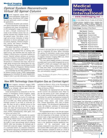
and twists in the spine that are not possible to evaluate in a traditional 2D setup. Before OrthoMod 3D, getting 3D information usually meant relying on a supplemental computerized tomography (CT) or magnetic resonance imaging (MRI) scan, which implied further radiation exposure. “The new spinal imaging solution is extremely accessible and it can be easily integrated into an existing R/F or RAD suite equipped with digital full spine,” said René Degros, business unit manager for X-Ray with TMSE. “It is particularly suited for practices focusing on radiology, pediatrics, orthopedics, and ambulatory surgery.” Image: The Biomod 3S system (Phoro courtesy of AXS Medical/DMS).
New MRI Technology Uses Krypton Gas as Contrast Agent new magnetic resonance imaging (MRI) scanning technology that uses Krypton gas as an inhalable contrast agent could provide insights on lung disease. Under development by researchers at the University of Nottingham (United Kingdom; www. nottingham.ac.uk), the novel technique uses lasers to hyperpolarize the nuclei of a noble gas, aligning them so that the inert gas becomes visible on an MRI scan. While traditional MRI uses hydrogen protons in the body as molecular targets, this does not give a detailed picture of the lungs, since they are full of air. The new technique will make three-dimensional (3D) imaging of the lungs possible, and could also be used with other inhaled, hyperpolarized, noble gases, such as helium and xenon-129. The new method is made possible via catalytic hydrogen combustion, a process associated with clean energy related sciences. In the process, krypton-83 gas is mixed with molecular hydrogen as the buffer in laser spin-exchange optical pumping (SEOP). After laser pumping, the hydrogen gas is mixed with molecular oxygen, “exploding” it away in a controlled fashion via catalyzed combustion, leaving behind high-purity, hyperpolarized krypton gas. The study describing the catalytic process was
A
published on March 9, 2016, in the Proceedings of the National Academy of Sciences of the United States of America (PNAS). “Remarkably, the hyperpolarized state of krypton-83 ‘survives’ the combustion event. Water vapor, the sole product of the ‘clean’ hydrogen reaction, is easily removed through condensation, leaving behind the purified laser-polarized krypton-83 gas diluted only by small remaining quantities of harmless water vapor,” said senior author Prof. Thomas Meersmann, PhD, chair of translational imaging. “This development significantly improves the potential usefulness of laser-pumped krypton-83 as MRI contrast agent for clinical applications.” The hyperpolarized state is very low spin temperature condition that is not in a thermal equilibrium with the temperature of the sample. Low spin temperature leads to high magnetization of the spin ensemble, which results in a very high nuclear magnetic resonance signal intensity. This eventually returns to the depolarized thermal equilibrium temperature. Of the quadrupolar noble gas isotopes, krypton-83 is most likely to serve as a surface sensitive MRI contrast agent, since it displays a slower relaxation compared to xenon as a result of its smaller electron cloud and its larger nuclear spin.
Elif Erkan Reader Service Manager
HOW TO CONTACT US Subscriptions: Send Press Releases to: Advertising & Ad Material: Other Contacts:
www.LinkXpress.com minews@globetech.net ads@globetech.net info@globetech.net
ADVERTISING SALES OFFICES USA P.O.Box 800806, Miami, FL 33280, USA Theresa.Herman@globetech.net Tel: (1) 954-893-0003 GERMANY, SWITZ., AUSTRIA Jutta.Ciolek@globetech.net
Bad Neustadt, Germany Tel: (49) 9771-3528
BENELUX, FRANCE, NORDIC REGION Hasselt, Belgium Nadia.Liefsoens@globetech.net Tel: (32) 11-22-4397 ITALY Fabio.Potesta@globetech.net
Genoa, Italy Tel: (39) 10-570-4948
JAPAN Katsuhiro.Ishii@globetech.net
Tokyo, Japan Tel: (81) 3-5691-3335
CHINA Parker.Xu@globetech.net
Shenzen, Guangdong, China Tel: (86) 755-8375-3877
KOREA JaeW.Suh@globetech.net
Seoul, Korea Tel: (82) 02-7200-121
ALL OTHER COUNTRIES ads@globetech.net
Contact USA Office Tel: (1) 954-893-0003
SUBSCRIPTION INFORMATION Medical Imaging lnternational is published six times a year and is circuIated worldwide (outside the USA and Canada) without charge, and by written request, to radiologists, medical specialists involved in imaging, and other qualified professionals allied to the field. To all others: Paid Subscription is available for an annual subscription charge of US$ 100. Single copy price is US$ 20. Mail your paid subscription order accompanied with payment to Globetech Media, P.O.Box 802214, Miami, FL 33280-2214, USA. For change of address or questions on your subscription, write to: Medical Imaging lnternational, Circulation Services, at above address; or visit www.LinkXpress.com
ISSN 1068-1779 Vol.26 No.3. Published, under license, by Globetech Media, LLC. Copyright © 2016. All rights reserved. Reproduction in any form is forbidden without express permission.
Teknopress Yayıncılık ve Ticaret Ltd. S¸ti. adına ˙Imtiyaz Sahibi: M. Geren • Yazı is¸leri Müdürü: Ersin Köklü Müs¸ ir Dervis¸ ˙Ibrahim Sok. 5/4, Esentepe, 34394 S¸is¸ li, ˙Istanbul P. K. 1, AVPIM, 34001 ˙Istanbul • E-mail: Teknopress@yahoo.com Baskı: Promat Web Ofset Tesisi • Orhangazi Mahallesi 1673. Sokak, No: 34 • 34510 Esenyurt, B. Çekmece • ˙Istanbul Yerel süreli yayındır, iki ayda bir yayınlanır, ücretsiz dag˘ıtılır.
Medical Imaging International May-June/2016
4
