

-Independent and Sponsor freeApril 2025 – Edition - 5th Year-


-Independent and Sponsor freeApril 2025 – Edition - 5th Year-

The journey of Earth's formation holds the key to understanding life's beginnings. Recent research from Rutgers University has transformed our understanding of when water - one of life's essential ingredients - arrived on our young planet.
This arrival timing, occurring during what scientists call late accretion, reshapes our entire perspective on life's origins.
Think of Earth's formation as a cosmic cooking show. The process of accretion worked like making a gigantic snowball in space - but instead of snow, it gathered cosmic dust, gases, and rocky debris. As Earth's mass grew, its gravitational pull increased, allowing it to attract more material.
This process wasn't gentle; violent collisions released enormous energy, keeping our forming planet molten.
During early accretion, heavier elements like iron sank to form Earth's core, while lighter materials floated up to create the mantle. The final phase, late accretion, occurred after this core formation. This stage proved crucial because it delivered the last 10-20% of Earth's building materials - including, as we now know, much of its water. Life required specific ingredients to emerge. Scientists summarize the essential chemical elements such as CHNOPS - Carbon, Hydrogen, Nitrogen, Oxygen, Phosphorus, and Sulfur.
These elements, combined with water and energy, provided the basic recipe for life. But timing was everything - these ingredients needed to arrive in the right order and under the right conditions.
The Rutgers research team used clever chemical detective work to determine when water arrived. They studied variations in the element molybdenum, comparing signatures in meteorites with Earth rocks. Their findings showed that Earth's rocks matched meteorites from the drier inner solar system rather than the wetter outer regions, suggesting water arrived later than previously thought.
This timing revelation carries profound implications. The Moonforming impact, once thought to have delivered significant water
to Earth, likely contributed less than scientists previously believed. Instead, water arrived gradually during late accretion, after the Moon's formation. This new timeline helps explain why Earth became habitable and potentially when life could have first emerged.
After Earth's basic "recipe" was complete, with its precise mixture of elements, water, and energy, our planet became a laboratory for increasingly complex chemical experiments. The molten planet, once a cosmic mixing bowl of CHNOPS, cooled and organized into structured environments where life's chemistry could unfold.

Like a master chef progressing from basic ingredients to sophisticated dishes, Earth's chemical processes grew more intricate. Simple
molecules combined into complex ones, eventually achieving the remarkable feat of self-replication. This molecular choreography set the stage for evolution's grand experiment, leading ultimately to the precisely orchestrated process of human development.
The elements that once swirled in Earth's primitive atmosphere now form the building blocks of our DNA, guiding the intricate dance of embryonic development. Each human cell contains atoms that were present in that original planetary recipe, reorganized through billions of years of evolution into the instructions for building human life.
From the violent mixing of Earth's formation to the delicate precision of embryonic development, we see an unbroken chain of increasing complexity and organization.
Our story begins in the cosmic kitchen of planetary formation and continues through to the sophisticated biological processes that shape human life – an evidence of the remarkable transformation from cosmic dust to conscious beings.
Understanding this progression - from basic elements to complex life - not only illuminates our origins but guides our ongoing search for life elsewhere in the cosmos. We are, in essence, the universe's most complex recipe, billions of years in the making.
Reference: “The non-carbonaceous nature of Earth’s late-stage accretion” by K.R. Bermingham, H.A. Tornabene, R.J. Walker, L.V. Godfrey, B.S. Meyer, P. Piccoli and S.J. Mojzsis, 5 November 2024, Geochimica et Cosmochimica Acta
DOI:10.1016/j.gca.2024.11.005The study was funded by the U.S. National Science Foundation and the NASA Astrobiology Institute.


Our planet formed about 4.5 billion years ago as a hot, molten sphere. Recent discoveries from Curtin University have revealed that freshwater emerged just 500 million years after Earth's formation - much earlier than previously thought.
Analysis of ancient zircon crystals from Western Australia shows that by 4 billion years ago, Earth already had a functioning water cycle, challenging the old theory that our planet was completely covered by oceans.
This earlier emergence of freshwater and landmasses created diverse environments for chemical evolution. While oceans provided vast spaces for chemical reactions, freshwater environments like lakes, rivers, and underground water systems offered different conditions and concentrations of chemicals. Lightning storms, volcanic activity, and cosmic radiation interacted with a mixture of simple molecules in both fresh and saltwater environments, creating nature's first chemical laboratory.
In these varied aqueous environments, organic molecules began to form. The presence of both freshwater and saltwater created different chemical gradients and concentrations, potentially providing more opportunities for complex molecules to form and interact. Simple compounds combined into more complex ones - amino acids, nucleotides, and lipids - concentrating in promising locations like tide pools, clay surfaces, deep-sea vents, and freshwater pools.
A crucial breakthrough came with RNA molecules. Unlike simpler molecules, RNA could both carry information (like modern DNA) and catalyze chemical reactions (like proteins do today). These molecules began to make copies of themselves, competing for resources. For the first time, natural selection - the engine of evolution - began to operate at a molecular level. The most efficient self-replicating RNA molecules survived and produced more copies.
A major leap forward occurred when lipid molecules spontaneously formed microscopic bubbles, or vesicles. These primitive membranes could enclose RNA and other important molecules, creating the first protocells. These weren't quite living cells as we know them, but they
provided a crucial boundary between internal chemistry and the outside world. The most successful protocells were those that could maintain their internal environment and reproduce.
Around 3.5 billion years ago, a critically important organism emergedLUCA, the Last Universal Common Ancestor. This wasn't the first life form, but it was the one from which all current life descended. LUCA had already developed many features we see in modern cells: DNA for storing information, proteins for performing cellular work, and a cell membrane enclosing these components. It used ATP for energy and had a genetic code remarkably similar to what all life uses today.
A revolutionary change occurred when some cells engulfed others, leading to a symbiotic relationship. The engulfed cells became specialized energy-producing structures (mitochondria) within their hosts. These more complex cells, called eukaryotes, developed internal compartments, including a nucleus to protect their DNA. This marked the beginning of cells as we know them in animals and plants today.
Perhaps one of the most remarkable transitions was when single cells began to cooperate, staying together after division and specializing in different tasks. This led to the first multicellular organisms. Some cells specialized in obtaining nutrients, others in reproduction, and others in protection. This division of labor allowed for the evolution of complex bodies with specialized tissues and organs.
From these multicellular beginnings, life diversified explosively. The first animals appeared, followed by an arms race of innovation: hard shells, appendages, sensory organs, and eventually internal skeletons. Plants
colonized land, creating new opportunities for animals to follow. Each innovation built upon previous ones, leading to the rich tapestry of life we see today.
The discovery that freshwater appeared much earlier than previously thought - around 4 billion years ago - suggests that conditions suitable for life developed significantly earlier than once believed.
This new timeline indicates that Earth became habitable within just 600 million years after its formation, providing more time for life to emerge and evolve.
This journey from molecules to modern life spans billions of years, but it's important to understand that evolution continues today. Every organism on Earth is both a product of this ancient history and a participant in ongoing evolution.
While we can't observe every step in this ancient process directly, we can see its evidence in the chemistry of our cells, the structure of our genes, and the patterns of life around us.
The presence of early freshwater and diverse environmental conditions suggests that life's story began in a more complex and dynamic setting than previously imagined, enriching our understanding of how life emerged and evolved on our planet.

A groundbreaking theory by Professors Tlusty and Libchaber presents life as a remarkable hierarchical system of machineproducing machines. Let me break down the key aspects of their framework:
The theory presents life as two intersecting cascades - one descending to the molecular level and another ascending to the biosphere. At the molecular level, we see fundamental machines like ion pumps and enzymes working at atomic scales. These combine to form increasingly complex systems, eventually reaching the critical point
The Critical Point:
Perhaps the most fascinating aspect is the identification of a critical point where these cascades meet - at the scale of about 1 micron and 1000 seconds. This corresponds to the basic unit of microbial life, where self-replicating machines first achieve stable interaction with their aqueous environment. This point represents the minimum conditions necessary for life as we know it.
The framework spans an impressive range - 18 orders of magnitude in space and 30 in time. This means it describes everything from the tiniest molecular machines to entire planetary biospheres, unified under a single conceptual framework.
This work builds on Leibniz's historical observation about the infinite machine-like nature of living bodies, but takes it further by providing a mathematical foundation for understanding life itself. The framework suggests that life's complexity can be understood through this cascade of machine production and interaction.
Reference:
Tlusty, T., & Libchaber, A. (2024). A Machine-Based Theory of Life: Conceptualizing Living Systems as Cascades of Machine-Producing Machines. From the Department of Physics, Ulsan National Institute of Science and Technology (UNIST) and Center for Physics and Biology, Rockefeller University.

Before the first human embryo, before even the first mammals, a revolutionary change happened roughly 2 billion years ago. This was the emergence of eukaryotes - complex cells that would eventually give rise to all animals, plants, and fungi. Recent discoveries have revealed a "lost world" of early eukaryotic organisms, filling an 800-million-year gap in our evolutionary story.
These ancient single-celled organisms, particularly the recently cultivated Asgard archaea, provide crucial clues about our deepest origins.
The story of human origins stretches back millions of years. Our lineage split from chimpanzees around 6 million years ago, beginning a unique evolutionary journey. Through countless generations, our ancestors developed upright walking, larger brains, and complex social behaviors, ultimately leading to Homo sapiens around 300,000 years ago in Africa.
Life begins when a single sperm cell, among millions released, successfully penetrates an egg cell. This moment of fertilization triggers a cascade of precise events. The genetic material from both parents combines, creating a unique genome that contains all the instructions needed to build a human being. This newly fertilized egg, called a zygote, marks the beginning of a new life.
Within hours of fertilization, the zygote begins a remarkable process of division. Like a perfectly orchestrated dance, the single cell divides into two, then four, then eight, and so on.
These early divisions, called cleavage, happen without increasing the total size - imagine cutting a ball of clay into smaller and smaller pieces. By day 3, this cluster of 16-32 cells resembles a tiny mulberry, giving it the name "morula."
Around day 5, the developing embryo undergoes its first major transformation. The cells reorganize to form a hollow sphere called a blastocyst. This is a crucial moment - for the first time, cells begin to
specialize. The outer layer will eventually form the placenta, while the inner cell mass will develop into the fetus. Like a ship finding its harbor, the blastocyst begins to implant in the uterine wall.
Between days 14-16, another remarkable transformation occurs called gastrulation - often considered the most important event in early development. The embryo forms three distinct cell layers, each with a specific destiny. The outer layer (ectoderm) will become the nervous system and skin.
The middle layer (mesoderm) forms muscles, bones, and blood. The inner layer (endoderm) develops into internal organs. This is when the basic human body plan first emerges.
With the fundamental blueprint established, development accelerates. The nervous system begins to form, starting as a simple tube that will become the brain and spinal cord. The heart starts as a simple tube and begins beating by week 4. Limb buds appear, gradually developing into arms and legs. Each organ system develops in a carefully choreographed sequence.
What truly sets human development apart is our brain development. Our brains grow larger relative to body size than any other primate, and this growth continues well after birth. This extended period of development, though making human infants more dependent, allows for remarkable learning and adaptation capabilities.
This complex journey from single cell to human being showcases both our ancient origins and our unique features. When a human embryo
develops, it carries forward an astonishing 2-billion-year legacy of eukaryotic evolution. The specialized organelles in our cells, like mitochondria, trace back to that ancient revolution when complex cells first emerged.
The early stages of our development mirror those of other mammals, reflecting our deep evolutionary connections. Yet subtle differences in timing and growth patterns, particularly in brain development, make us distinctly human.
Each human embryo not only recapitulates the 3.5-billion-year journey of life on Earth but also tells the specific story of how eukaryotes evolved into complex multicellular organisms and eventually into humans.
Throughout this entire process, the precision is astounding. Genes switch on and off in exact sequences. Cells communicate through complex chemical signals. Tissues fold and reshape with origami-like precision.
When we consider that this process has been refined over millions of years of evolution, successfully creating billions of human beings, we glimpse the remarkable achievement that each human life represents.

A groundbreaking discovery in Romania has pushed back the timeline of human migration out of Africa by 200,000 years, challenging previous assumptions about early hominin movements.
At the Grăunceanu site in Romania's Olteț River Valley, researchers have identified compelling evidence of human presence dating back approximately 1.95 million years ago.
The key evidence comes from meticulous examination of over 5,000 ancient bones, among which researchers identified twenty specimens bearing distinct cut marks from stone tools. These marks, analyzed using advanced dating techniques, provide strong evidence of early hominin butchering activities.
This discovery predates the previously accepted earliest evidence of humans outside Africa at Georgia's Dmanisi site by about 200,000 years.
What makes this finding particularly significant is the environmental context. Analysis reveals these early hominins encountered challenging conditions quite different from their African origins. The region experienced seasonal temperature fluctuations and higher rainfall levels, requiring significant adaptation.
The fauna they encountered included novel species such as woolly rhinos, saber-tooth cats, pangolins, and mammoths - a dramatic departure from African wildlife.
The research team, led by Sabrina Curran, Claire Terhune, and Alexandru Petculescu, approached their findings with appropriate scientific rigor.
Their discovery emerged from careful reexamination of collections that had been housed in museums since the 1960s, demonstrating the value of revisiting archived materials with modern analytical techniques.
While no hominin remains have been found at the site, the evidence of tool use is compelling.
"The field of paleoanthropology can be contentious," notes Terhune, explaining their methodical approach to documenting and verifying the cut marks. Their careful analysis has helped establish the authenticity of these significant findings.
This discovery provides valuable insights into early hominin adaptability and migration patterns. It demonstrates that our ancient relatives were capable of adapting to diverse environments much earlier than previously thought, contributing to our understanding of human evolution's complex history.
The findings, published in Nature Communications, represent a significant advancement in our knowledge of early human migration and adaptation capabilities, suggesting a more complex pattern of early human movement than previously understood.

Every living thing on Earth has its own internal clock - from the tiniest bacteria to the tallest trees and us humans. These biological timekeepers evolved long before we invented watches or calendars, helping organisms sync their activities with nature's rhythms.
Think of your body like a symphony orchestra, with each section playing its part at just the right moment. The conductor of this biological orchestra is our internal timing system, which developed over millions of years to match Earth's daily spin. The most important signal? The simple rising and setting of the sun.
Our bodies run on a 24-hour cycle, controlling everything from when we feel sleepy to how our metabolism works. Dr. Partch's team at UC Santa Cruz made a fascinating discovery about the proteins that manage these cycles - they're surprisingly flexible in structure, which turns out to be key to their function.
Beyond daily rhythms, our bodies have a different kind of clock that controls how we grow and develop. Scientists have found that the tiny powerhouses in our cells (mitochondria) play a double role - they not only provide energy but also help keep time for development.
The intricate workings of our biological clocks involve several interconnected layers. At the foundation are basic chemical reactions that set the tempo of life. These reactions work alongside genes that turn on and off in precise patterns throughout the day. Meanwhile, the cellular powerhouses coordinate both energy production and timing, creating a sophisticated timekeeping network.
While all mammals share similar biological clockwork, the speed varies dramatically. A mouse develops from embryo to birth in days, while humans take months - yet they use many of the same genetic
instructions. This variation in timing produces the diverse forms of life we see around us, even though the underlying mechanisms remain remarkably similar.
Understanding these biological clocks has revolutionized medical treatment timing and opened new pathways for treating disorders where timing goes wrong. Scientists can now better understand how metabolism works and its connection to our internal clocks. Dr. Partch and other researchers continue to uncover new details about nature's timekeepers, opening doors to better healthcare and biotechnology innovations.
Just as our ancestors looked to the sun and stars to tell time, our cells carry ancient timekeeping systems that keep life running in rhythm with the world around us. This deep connection between life and time remains one of nature's most elegant solutions to the challenge of survival in an ever-changing world.


Advancements in embryology are marked by significant discoveries, new techniques, and evolving theoretical framework. To metaphorically map the progress of embryology to a versioning system based on major milestones, we might consider a few key eras:
Classical Embryology (1.0):
This era includes the earliest studies of embryology, from ancient civilizations understanding basic aspects of reproduction to the more structured studies in the 18th and 19th centuries that began to detail embryonic development stages in various species.
Starting in the late 19th (1827) and early 20th centuries, this phase introduced experimental approaches to study developmental processes, including manipulation and observation of embryonic tissues, which helped establish many fundamental concepts in developmental biology.
Considered the real birth of Embryology with the research of his founder: Karl von Baer -Next page
Molecular Embryology (3.0): From the late 20th century onward, the focus shifted towards understanding the molecular mechanisms controlling development. Techniques such as gene editing (e.g., CRISPR-Cas9), in vitro fertilization, and stem cell research represent this era's cutting-edge methodologies.
Systems Embryology (4.0): Currently, we are moving towards an integration of molecular data with computational modeling to understand the complex systems governing embryonic development. This approach considers genetics, epigenetics, cellular interactions, and environmental factors in a holistic manner.
Karl von Baer: The Father of Embryology

Born into a noble Baltic German family in 1792, in the Piep Manor of what is now Estonia, young Karl's life straddled multiple cultures and empires. Though ethnically German, he was born in the Russian Empire, creating a unique intellectual perspective that would later influence his groundbreaking work.
His journey into science was almost derailed before it began - his father wanted him to become a military officer. However, a fortuitous illness prevented Karl from entering military service, redirecting him to medical studies at the University of Dorpat (now Tartu). As he would later quit, this "fortunate illness" changed the course of scientific history.
Despite struggling with poor eyesight - an ironic challenge for a man destined to make microscopic discoveries - von Baer developed extraordinary observational skills that would define his career.
His most famous discovery came in 1827: the mammalian egg cell. When colleagues doubted this finding, he famously remarked, "Gentlemen,
I show you not what you want to see, but what is there."
Known as "The Wizard of Königsberg" by his students, von Baer would often become so absorbed in his work that his wife had to drag him away from his microscope for meals. This dedication led to his most profound contribution: Baer's Laws of Embryology.
Working in his dimly lit laboratory, examining countless embryos across different species, he noticed universal patterns in early development. His Laws established that:
- General features of a large group of animals develop earlier in embryos than specialized features
- Development progresses from general to specific characteristics
- Early-stage embryos of different species look remarkably similar before diverging
- A young embryo of a higher animal never resembles an adult of a lower animal, only its embryo
These principles revolutionized our understanding of development. He would often challenge skeptical colleagues to distinguish between
early-stage embryos of different species - a test they frequently failed. Published in his 1828 masterwork "On the Development of Animals," these laws remain fundamental to embryology today.
His work went beyond pure science. While at the St. Petersburg Academy of Sciences, he applied his methodical mind to practical problems, even establishing sustainable fishing practices in Russia's lakes. Yet he maintained his humility, writing that "Science is eternal in its source, unlimited in its scope, endless in its task, unattainable in its goal."
Von Baer's work decisively disproved the then-popular theory of preformation - which suggested organisms existed in miniature form inside sperm or eggs - and established epigenesis, showing how organisms develop progressively through cell differentiation. His comparative embryology studies laid groundwork that would later support Charles Darwin's evolutionary theories.
Karl von Baer's story reminds us that behind every scientific breakthrough is a very human tale of curiosity, perseverance, and sometimes, a fortunate illness that points life in an unexpected direction. His work not only revealed how life begins but set the foundation for our modern understanding of developmental biology.

The story of the human brain weaves together two remarkable narratives: our evolutionary past and our individual development. Recent discoveries from Yale University illuminate how special genetic switches, called Human Accelerated Regions (HARs), bridge these stories, revealing the delicate dance between evolution and embryonic development.
Our brain's journey begins as a simple neural tube in the embryo, yet it develops into the most complex structure known in the universe. This transformation follows precise genetic instructions, including these newly understood HARs, which act like sophisticated volume controls for genes. Instead of creating entirely new genes, these regions modified existing ones we share with chimpanzees, subtly adjusting their activity to create uniquely human brain features.
Using advanced 3D genome mapping, researchers discovered that HARs orchestrate crucial aspects of brain development: they guide neuron formation, enhance brain size, and fine-tune the intricate networks that make our cognitive abilities possible. The same genetic switches that shaped human brain evolution millions of years ago continue to direct brain development in every human embryo today.
What makes this discovery particularly fascinating is its implications for human health. Some HAR-controlled genes are linked to conditions like autism and schizophrenia, suggesting that the very changes that made our brains uniquely human might also make them vulnerable to certain disorders. This understanding opens new possibilities for treating neurodevelopmental conditions by revealing their evolutionary origins. Each human embryo today recapitulates this evolutionary journey, guided by ancient genetic innovations that continue to shape our species' most distinctive feature – our extraordinary capacity for complex thought and culture. Understanding these mechanisms isn't just about appreciating our past; it's crucial for comprehending how our minds develop and function today.
As we decode the intricate relationship between brain evolution and development, we're gaining deeper insights into what makes us uniquely human. This knowledge bridges the gap between our distant past and our present development, showing how ancient changes continue to shape each new human life, one brain at a time.

What if consciousness isn't like flipping a switch, but more like a sunrise? In the final weeks before birth, as billions of neurons complete their intricate dance of connections, something remarkable may be stirring in the developing brain. The fetus, floating in its aquatic world, might be experiencing the first glimmers of awareness - not yet the full daylight of consciousness we know, but perhaps a kind of dawn.
Think of how a fetus responds to music, to voices, to touch through the womb wall. We've long attributed these reactions to pure reflex, but what if they represent something more - the first threads of experience being woven into the tapestry of consciousness? The brain, nearly complete in its architecture during the final month, might be capable of creating not just responses, but experiences.
This isn't to suggest that fetal consciousness mirrors our adult experience. Rather, imagine it as a form of proto-consciousness - a primordial awareness that's neither fully "on" nor entirely "off." It might be closer to the consciousness we experience in deep meditation or just before sleep, where awareness exists but hasn't yet crystallized into the sharp focus of full wakefulness.
The philosophical implications are profound. If consciousness indeed emerges before birth, then our very first experiences occur in a world utterly different from the one we'll soon enter - a liquid environment where sound travels differently, where light is filtered to soft shadows, where movement follows different rules. These pre-birth experiences might form the deepest substrate of our consciousness, laying down patterns that influence how we'll later perceive and interact with the world.
What makes this hypothesis particularly intriguing is how it challenges our traditional understanding of consciousness as something that begins with birth. Instead, birth might be better understood as a transition point - not the beginning of consciousness, but rather its dramatic transformation as it encounters a new world of sensory experiences.
This perspective invites us to reconsider what we mean by consciousness itself. Rather than viewing it as an all-or-nothing phenomenon, we might better understand it as a gradient - one that
begins to emerge in the womb and continues to develop and evolve throughout our lives.
The implications reach beyond philosophy into ethics and medicine. If consciousness indeed begins before birth, how might this influence our understanding of fetal development and our approach to prenatal care? Could early consciousness formation help explain why prenatal experiences seem to have such lasting effects on development?
In the end, perhaps the most beautiful aspect of this hypothesis is how it suggests that consciousness, like life itself, doesn't begin with a bang but with a whisper - a gentle awakening in the quiet depths before we draw our first breath.

A revolution is transforming our understanding of how life begins. Systems embryology, a cutting-edge field merging traditional embryology with advanced technology, is unveiling the intricate dance of early human development like never before.
Think of embryo development as an orchestra where every instrument must play perfectly in time. Scientists now know this symphony isn't conducted by genes alone – it's a complex interplay of genetic code, environmental signals, and cellular communication.
Using powerful tools like artificial intelligence and high-resolution imaging, researchers can now watch this biological concert unfold in real-time. One of the field's most exciting breakthroughs comes fromsingle-cell sequencing and CRISPR technology, which allow
scientists to track individual genes as they switch on and off during development. This precision has revealed how cells decide their fate –whether to become heart, brain, or bone tissue. Alongside this, researchers have discovered that chemical modifications to DNA, like molecular sticky notes, help fine-tune these developmental decisions. The cellular neighborhood matters tremendously in embryo development. Cells constantly chat with their neighbors through chemical signals, physical pushes and pulls, and electrical impulses.
Scientists are decoding this cellular conversation using lab-grown miniorgans called organoids, which help reveal how tissues organize themselves and what goes wrong in developmental disorders.
Artificial intelligence is proving to be a game-changer. Virtual embryo models, built from vast biological datasets, can now predict how development might go awry and suggest ways to prevent birth defects. These computer simulations are particularly valuable because they reduce the need for traditional embryo research while expanding our understanding of human development.
The environment plays a crucial role too. Maternal nutrition, stress levels, and even gut bacteria can influence how genes are expressed in the developing embryo, potentially affecting health long into adulthood. This knowledge is opening new avenues for preventing developmental disorders before birth.
Systems embryology promises to revolutionize reproductive medicine. AI-enhanced IVF selection, personalized treatments for developmental disorders, and even the possibility of correcting genetic abnormalities before birth are all on the horizon. By viewing embryo development through this comprehensive, technology-driven lens, scientists are not just understanding life's beginnings – they're gaining the tools to ensure it starts in the healthiest way possible.

Perinatal technology has transformed the landscape of reproductive medicine, offering new possibilities for individuals facing uterine-related infertility while carefully managing associated risks. This comprehensive overview examines current technological advances in reproductive medicine and their impact on maternal and neonatal outcomes.
Advanced artificial intelligence systems are revolutionizing embryo selection, analyzing multiple parameters to optimize implantation success rates and improve pregnancy outcomes. Time-lapse imaging provides continuous embryo development monitoring, enabling unprecedented precision in selecting the healthiest candidates for transfer. Single Embryo Transfer has evolved significantly, reducing multiple pregnancy risks while maintaining high success rates through improved selection techniques. Vitrification advances have transformed
freezing procedures, dramatically improving embryo and egg survival rates, which enables more flexible treatment timing and safer delayed implantation options.
Uterine transplantation represents a groundbreaking solution for absolute uterine factor infertility, with approximately 73% of procedures successfully restoring uterine function and 42% achieving pregnancy. The integration of personalized medicine through genetic profiling enables precisely tailored treatment protocols, significantly reducing required IVF cycles and improving success rates.
Artificial ovary development continues to advance through innovative research in tissue engineering and ovarian organoids, showing considerable promise for future fertility preservation options. These experimental approaches undergo rigorous testing while offering hope for previously untreatable conditions.
Modern fertility protocols incorporate sophisticated monitoring systems for both maternal and fetal health. Maternal health tracking includes comprehensive assessment of gestational hypertension, preeclampsia risk factors, and gestational diabetes markers. Advanced fetal monitoring systems track developmental milestones, evaluate potential congenital anomalies, and assess growth patterns throughout pregnancy.
Multiple pregnancy management has evolved to include specialized care pathways with enhanced monitoring protocols, though modern techniques increasingly focus on prevention through single embryo transfer when appropriate.
Growing evidence supports the advantages of frozen embryo transfer protocols, which demonstrate reduced perinatal risks and improved endometrial preparation compared to fresh transfers.
Contemporary fertility care integrates multiple treatment modalities, combining advanced reproductive technologies with genetic counseling and personalized treatment planning. Risk-specific monitoring protocols are tailored to individual patient profiles, ensuring optimal care throughout the fertility journey. The field continues to refine these approaches based on emerging research and technological capabilities.
The rapidly evolving landscape of fertility technology continues to push boundaries while maintaining unwavering focus on safety and success rates. Each advancement brings new hope to those facing reproductive challenges, with increasingly sophisticated solutions becoming available through ongoing research and development.
This integration of cutting-edge technology with careful risk management represents the current state of modern fertility care, offering unprecedented opportunities for family building while prioritizing optimal outcomes for both parents and children.

Recent breakthroughs in human germline research have revealed unprecedented insights into epigenetic reprogramming mechanisms, fundamentally advancing our understanding of reproductive biology. This process proves essential for developing functional gametes and ensuring proper embryonic development.
Epigenetic regulation operates through various molecular mechanisms, with DNA methylation serving as a primary control switch. These modifications act as cellular memory, determining which genes are expressed or silenced without changing the DNA sequence itself. During germ cell development, the genome undergoes comprehensive epigenetic restructuring, establishing a clean slate for the next generation.
Development and ReprogrammingThe formation of primordial germ cells represents a critical juncture in human development. During this phase, cells undergo extensive epigenetic erasure, removing accumulated modifications to achieve totipotency. This reset ensures that developing germ cells can properly differentiate into functional gametes carrying appropriate epigenetic instructions for the next generation.
Recent research has successfully replicated this complex reprogramming process in laboratory conditions. Scientists have cultivated human PGC-like cells that demonstrate natural epigenetic reset patterns. This achievement provides direct observation of human germline development, previously accessible only through limited animal models.
These advances offer promising applications for reproductive medicine. Understanding epigenetic reprogramming mechanisms could lead to improved fertility treatments and novel approaches to addressing reproductive disorders. The ability to generate functional gametes in vitro could revolutionize assisted reproductive technologies.

In a remarkable scientific leap forward, researchers have achieved what was once thought impossible: creating complete human embryo models that mirror development up to 14 days after fertilization. Using only naive embryonic stem cells, scientists have developed structures that recapitulate the intricate organization and development of early human life, opening new windows into previously inaccessible stages of human development.
These sophisticated models, developed from stem cells without using eggs or sperm, demonstrate an unprecedented level of complexity. They form structures that mirror real embryonic development, including the formation of the embryonic disc, yolk sac, and even early placental tissues. What makes this achievement particularly noteworthy is the models' ability to demonstrate key developmental milestones, such as the formation of distinct cell layers and the beginning of organ system organization.
The implications of this breakthrough are profound, offering potential insights into miscarriage prevention and developmental disorders. However, this advancement also raises important ethical considerations. While these models are far from being actual human embryos - more like sophisticated 3D sketches of early developmentthey push us to carefully consider the boundaries between model and reality.
Professor Jacob Hanna from the Weizmann Institute of Science, one of the pioneers in this field, acknowledges both the scientific potential and ethical complexity of this work. These models, while remarkably detailed, remain fundamentally different from natural embryos and have no potential to develop into a fetus. Nevertheless, they can provide crucial insights into the mysterious early stages of human development that have long been impossible to study.
The research operates within established ethical guidelines, particularly the 14-day limit for embryo research - a timepoint when the embryo is barely visible to the naked eye. This boundary reflects both technical and ethical considerations that have long guided embryological research.
As we advance in our ability to create more sophisticated models, the scientific community continues to engage in thoughtful dialogue about the ethical implications of this work. The goal remains clear: to advance our understanding of human development while maintaining rigorous ethical standards and respect for human life at all stages. These developments represent not just technical achievements, but careful steps forward in our quest to understand human development, potentially leading to breakthroughs in treating developmental disorders and preventing pregnancy complications, all while navigating the complex ethical landscape of embryological research.

Dr. Sarah Chen adjusted her safety goggles, watching the CRISPRengineered iPSCs (induced pluripotent stem cells) on her screen.
These weren't the controversial embryonic stem cells of the early 2000s – these were adult cells reprogrammed using Yamanaka's Nobel Prizewinning technique, which had revolutionized the field by transforming ordinary skin cells into pluripotent stem cells.
Their lab was attempting something that would have seemed impossible just a decade ago: growing miniature heart organoids using their advanced bioprinting system. The lab hummed with cutting-edge equipment. In one corner, their single-cell RNA sequencing machine analyzed gene expression patterns, while nearby, a specialized bioreactor maintained their "organs-on-chips" – microfluidic devices that mimicked human organ systems for drug testing.
"Dr. Chen!" Maya called excitedly. "The cardiomyocytes are showing synchronized beating patterns!" Sarah hurried over, noting the rhythmic contractions on the screen.
They were using a refined version of direct cardiac reprogramming, which could transform cardiac fibroblasts directly into beating heart cells, combined with a new extracellular matrix scaffold.
The implications stretched beyond cardiac tissue. In the adjacent lab, her colleagues were working on converting brain cells called astrocytes into neurons, while another team developed protocols for creating dopamine neurons for Parkinson's treatment.
As she documented the results, Sarah reflected on recent successes in the field: human-monkey chimeric embryos, mitochondrial replacement therapy, and breakthrough intestinal organoids. Each advance had opened new possibilities while raising important ethical questions.
The lab's AI system finished analyzing their cardiac tissue's electrical patterns. The results showed something extraordinary: their bioprinter heart tissue was displaying conduction patterns remarkably similar to natural heart tissue.
Later that evening, reviewing their latest data on neural differentiation, Sarah passed their cryogenic storage unit containing specialized stem cell lines. Each frozen vial represented years of collective scientific knowledge, building on breakthrough moments in stem cell research.
As she stepped into the cool evening air, Sarah couldn't help but feel optimistic. Tomorrow, they would begin testing a new protocol combining CRISPR techniques with epigenetic reprogramming methods.
In her pocket was a letter from a patient named Michael, thanking them for their work on the clinical trial using epithelial stem cell therapy for genetic skin disorders.
The future of regenerative medicine was growing, one breakthrough building on another, in labs around the world. As she headed home, the lab's incubators continued their vital work, maintaining careful conditions for the next generation of stem cell discoveries.


In a remarkable advancement for infant cardiac care, researchers at Texas Children's Hospital and Rice University have achieved a breakthrough that offers hope to thousands of families. They've successfully transformed stem cells found in amniotic fluid – the protective liquid surrounding a developing baby – into cells that can form blood vessels, paving the way for revolutionary heart defect treatments.
Led by bioengineer Jeffrey Jacot, the team envisions a future where babies diagnosed with heart defects in the womb could receive custommade tissue patches grown from their own cells. The elegance of this approach lies in its timing: since most severe heart defects are detected during pregnancy via ultrasound, doctors could collect amniotic cells early enough to grow the necessary tissue by the time the baby needs surgery.
What makes this research particularly compelling is its practical approach to an urgent need. Every year, about 32,000 babies in the United States are born with congenital heart defects, with 10,000 requiring surgical intervention in their first year of life. Current solutions often involve synthetic materials like Dacron or Teflon, which can't grow with the child and frequently require multiple surgeries.
The team's innovation centers on a special type of stem cell found in amniotic fluid – roughly one in every 10,000 cells – that carries markers similar to both embryonic and mesenchymal stem cells. These cells, when properly cultured, can develop into the endothelial cells that form blood vessels, a crucial first step in creating viable heart tissue.
Perhaps most promising is the cells' impressive growth rate. According to Jacot's calculations, the amount of cells obtained from a standard amniocentesis could theoretically grow enough tissue for an entire heart by the time of birth. While a complete heart replacement isn't the immediate goal, this rapid growth potential makes the creation of smaller repair patches very feasible.
This research represents a significant step forward in regenerative medicine, offering a practical solution that elegantly sidesteps both the ethical concerns of embryonic stem cells and the rejection risks of donor tissue. By using the baby's own cells, the approach ensures perfect genetic compatibility while providing a renewable source of healing tissue

In a scientific leap forward, researchers have achieved what was once thought impossible: creating complete human embryo models that mirror development up to 14 days after fertilization. Using only native embryonic stem cells, scientists have developed structures that recapitulate the intricate organization and development of early human life, opening new windows into previously inaccessible stages of human development.
These sophisticated models, developed from stem cells without using eggs or sperm, demonstrate an unprecedented level of complexity. They form structures that mirror real embryonic development, including the formation of the embryonic disc, yolk sac, and even early placental tissues. What makes this achievement particularly noteworthy is the models' ability to demonstrate key developmental milestones, such as the formation of distinct cell layers and the beginning of organ system organization.
The implications of this breakthrough are profound, offering potential insights into miscarriage prevention and developmental disorders. However, this advancement also raises important ethical considerations. While these models are far from being actual human embryos - more like sophisticated 3D sketches of early developmentthey push us to carefully consider the boundaries between model and reality.
Professor Jacob Hanna from the Weizmann Institute of Science, one of the pioneers in this field, acknowledges both the scientific potential and ethical complexity of this work. These models, while remarkably detailed, remain fundamentally different from natural embryos and have no potential to develop into a fetus. Nevertheless, they can provide crucial insights into the mysterious early stages of human development that have long been impossible to study.
The research operates within established ethical guidelines, particularly the 14-day limit for embryo research - a timepoint when the embryo is barely visible to the naked eye. This boundary reflects both technical and ethical considerations that have long guided embryological researc
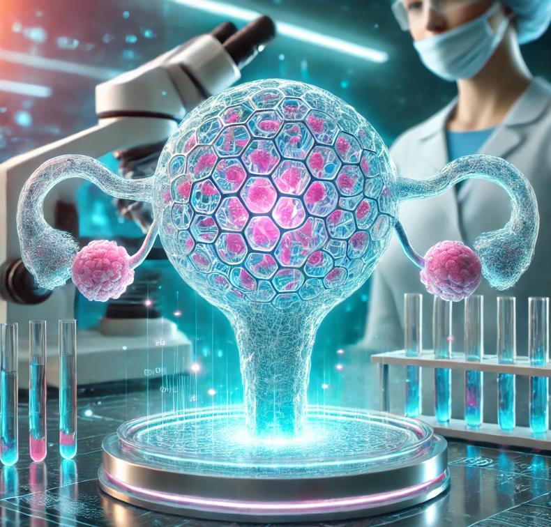
In a fasicnating convergence of bioengineering and reproductive science, researchers have made remarkable strides in creating artificial ovaries and ovarian organoids, offering new hope for individuals with compromised ovarian function. This innovative field combines the precision of 3D bioprinting with the complexity of human tissue engineering to recreate one of the body's most intricate organs.
At the forefront of this revolution are ovarian organoids, nicknamed "ovaroids," which represent miniature versions of human ovaries grown in laboratory conditions. These sophisticated structures have achieved what was once thought impossible: they form follicle-like structures, respond to hormonal signals, and even support the development of cells similar to human egg precursors. Most remarkably, these organoids can be developed in less than a week through careful manipulation of stem cells and specific transcription factors.
The engineering approach to artificial ovaries has taken an equally fascinating turn with the advent of 3D bioprinting technology. Scientists are now able to create customized scaffolds using biocompatible materials that mimic the natural ovarian environment. These scaffolds serve as an architectural framework where human ovarian cells can grow and function. A significant breakthrough came when researchers successfully supported human follicles on these bioengineered scaffolds, marking a crucial step toward functional artificial ovaries.
The innovation doesn't stop at 3D printing. Researchers are exploring multiple avenues, including the use of decellularized ovarian tissue –essentially natural ovaries stripped of their cellular content but retaining their structural framework. This biological scaffold preserves the complex architecture needed for proper ovarian function while providing a natural environment for new cell growth. Additionally, synthetic hydrogels are being developed to encapsulate ovarian follicles, offering a controlled environment for their development and function.
These developments represent more than just technical achievements; they offer potential solutions for individuals facing fertility challenges due to medical treatments, genetic conditions, or age-related factors. While still in developmental stages, this research paves the way for personalized reproductive medicine, where artificial ovaries could be tailored to individual patients' needs.
The success of these efforts hinges on the delicate balance between technological innovation and biological complexity. Scientists must ensure these artificial structures not only look like ovaries but also function properly, producing hormones and supporting egg cell development. As this field continues to evolve, it promises to transform our understanding of reproductive biology while offering new therapeutic possibilities for ovarian function restoration.

In a remarkable display of surgical precision, Japanese scientists have accomplished what once seemed impossible: transplanting a kidney from one rat fetus to another while still in the womb. This achievement, smaller than any transplant previously attempted, opens new possibilities for treating organ failures before birth.
The delicate procedure involves extracting kidney tissue no larger than two millimeters from donor rat fetuses, carefully chosen at just the right stage of development. Using specialized tools, surgeons then navigate through the uterine wall to place this tiny organ into a recipient fetus, all while maintaining the delicate balance of life within the womb.
What makes this achievement particularly extraordinary is not just the technical feat, but the remarkable success that followed. The transplanted kidneys didn't just survive – they thrived. These miniature organs integrated with their new hosts, developing blood vessels and performing their essential function of producing urine. Perhaps most surprisingly, they continued functioning for months without requiring the usual immune-suppressing drugs that transplant patients typically need.
This success carries profound implications for human medicine, particularly for conditions like Potter syndrome, where babies develop without functioning kidneys. Currently, these infants face grim prospects, but this research hints at a future where intervention could begin before birth. The technique could provide a crucial bridge, supporting these tiny patients until they're strong enough for more conventional treatments.
The research team didn't stop at same-species transplants. They've also successfully transferred mouse kidney tissue into rat fetuses, suggesting possibilities that extend beyond single-species solutions. While the path to human application remains long and complex, this tiny kidney transplant represents a giant leap forward in fetal surgery,
offering hope for treating conditions that currently have few options for intervention.
This achievement, though still in its early stages, paints a picture of a future where some of life's earliest medical challenges might be addressed before a baby takes their first breath.

In a remarkable advancement for reproductive medicine, uterine transplantation (UTx) has emerged as a beacon of hope for women with absolute uterine factor infertility. This pioneering procedure represents one of the most significant breakthroughs in transplant medicine, offering a chance for women born without a uterus or those who have lost uterine function to experience pregnancy.
The journey of uterine transplantation combines surgical precision with the intricate dance of reproductive medicine. The procedure involves carefully transferring a uterus from a donor – either living or deceased – to a recipient, creating the possibility for pregnancy where none existed before. Once the transplanted uterus heals and demonstrates normal function, physicians can proceed with in vitro fertilization, allowing the recipient to experience the profound journey of pregnancy. The success rates of this groundbreaking procedure have been encouraging. Research indicates that nearly three-quarters of transplanted uteruses successfully restore function, with over 40% of recipients achieving pregnancy. These statistics represent more than just numbers – they represent dreams realized and boundaries pushed in medical science.
However, like all pioneering medical procedures, uterine transplantation carries its own set of challenges. Recipients must undergo carefully managed immunosuppressive therapy to prevent rejection of the transplanted organ. The procedure requires extensive monitoring and is currently available only at select medical centers with specialized expertise in this field.
What makes this advancement particularly remarkable is its temporary nature – unlike other organ transplants, the uterus is typically removed after one or two successful pregnancies, limiting the time a recipient needs to remain on immunosuppressive medications. This approach balances the desire for childbearing with long-term health considerations.

In a fascinating fusion of cutting-edge technologies, artificial intelligence and gene editing are transforming reproductive medicine, offering unprecedented precision in the journey toward successful pregnancies. This convergence of AI and CRISPR technology represents a quantum leap in how we approach fertility treatments and genetic health.
At the heart of this revolution is AI's remarkable ability to analyze embryo development. Advanced algorithms can now process thousands of microscopic images of developing embryos, identifying subtle patterns and characteristics that even experienced embryologists might miss. This technology works like a tireless observer, monitoring embryos through time-lapse imaging, tracking their development moment by moment, and using this wealth of data to predict which embryos have the highest potential for successful implantation.
Meanwhile, CRISPR technology is opening new frontiers in genetic medicine. This precise gene-editing tool, often described as molecular scissors, offers the potential to address genetic disorders before they can impact a developing embryo. While still in experimental stages, CRISPR's capability to target and modify specific genes with remarkable accuracy has generated excitement in the medical community for its potential to prevent inherited diseases.
The synergy between these technologies is particularly powerful. AI can help identify genetic patterns and potential issues, while CRISPR provides the tools to potentially address them. This combination offers a level of precision and possibility that was unimaginable just a decade ago. Time-lapse imaging systems, enhanced by AI analysis, can now track embryo development continuously, creating detailed profiles of embryo health and viability.
However, scientists approach this technology with appropriate caution and ethical consideration. The ability to edit genes and select embryos based on AI analysis raises important questions about the boundaries
of human intervention in reproduction. Research continues within carefully controlled parameters, focusing on preventing serious genetic disorders while maintaining rigorous ethical standards.
As these technologies mature, they promise to revolutionize not just IVF success rates but our entire approach to reproductive medicine. The future points toward more personalized, precise, and successful fertility treatments, offering new hope to countless individuals and families while carefully balancing scientific progress with ethical considerations

In a profound shift in our understanding of cancer, scientists at the Van Andel Institute have discovered that our cancer destiny may be partially written in our epigenetic code even before birth. This groundbreaking research challenges the traditional view that cancer is primarily a disease of aging and genetic mutations, revealing instead that crucial
risk factors are established during early development through epigenetic patterns inherited from our parents.
The study, published in Nature Cancer, has identified two distinct epigenetic states that emerge during development, each carrying different cancer risk profiles. What makes this discovery particularly remarkable is that these states appear to be inherited and established naturally, rather than being solely the result of environmental factors or random mutations. When cancer develops in individuals with the lowerrisk epigenetic state, it tends to manifest as blood cancers like leukemia or lymphoma. In contrast, those with the higher-risk state are more prone to developing solid tumors, such as lung or prostate cancer.
The research team, led by J. Andrew Pospisilik and Ilaria Panzeri, made a crucial observation while studying mice with reduced levels of the Trim28 gene. They found that even genetically identical mice could develop different patterns of epigenetic marks on cancer-related genes. These patterns, established during early development and potentially inherited from parents, effectively set the stage for future cancer risk.
This revelation challenges the notion that cancer development is merely "bad luck." As Dr. Panzeri explains, while everyone carries some level of risk, the presence of these inherited epigenetic states suggests a more deterministic component to cancer development than previously thought. More importantly, unlike genetic mutations, epigenetic patterns can potentially be modified, opening new avenues for cancer prevention and treatment.
The implications of this research are far-reaching. Evidence of these two epigenetic states has been found throughout body tissues, suggesting that this developmental epigenetic risk framework might be common across various cancer types. This understanding could revolutionize our approach to cancer prevention, potentially allowing for intervention before cancer even begins to develop.
As we continue to unravel the complex relationship between inherited epigenetic patterns and cancer risk, this research opens up exciting possibilities for future therapeutic approaches. By understanding how these patterns are established and maintained across generations, we might one day be able to modify cancer risk factors before they can influence disease development, fundamentally changing our approach to cancer prevention and treatment.

Recent advances in synthetic genomics have enabled significant progress toward the development of synthetic embryos. This convergence of genomic engineering and developmental biology presents both opportunities and challenges for future applications.
Synthetic genomics has achieved notable milestones, including the synthesis of functional genomes for various organisms. These developments, combined with tools like **CRISPR** and advanced sequencing technologies, enable precise genetic programming of cells. This capability has proved crucial for directing cellular self-organization into embryo-like structures.
Synthetic embryos are stem cell-derived structures that replicate specific stages of natural embryonic development. Unlike conventional embryos, they do not require gametes (eggs or sperm) and currently cannot develop into viable organisms. Instead, they serve as research models for studying development and disease. Current technical limitations reflect both scientific challenges and established regulatory frameworks, with most jurisdictions restricting research to early developmental stages (typically 14 days).
Regenerative medicine, novel fertility treatments, and solutions for demographic challenges represent key applications of this technology. These become particularly relevant in the context of global denatalisation trends, where synthetic embryos could provide alternative solutions for population sustainability and reproductive healthcare.
The integration of artificial wombs for controlled reproduction, population management applications, and enhanced genomic modifications represent likely developments by 2300.

In a critical examination of space exploration challenges, scientists are investigating one of the most complex aspects of long-term space missions: the significant risks and potential barriers to human reproduction beyond Earth. As discussions of Mars missions and space colonies continue, understanding these challenges has become increasingly important for realistic space planning. The challenges begin with the fundamental force we take for granted on Earth: gravity. In the microgravity environment of space, researchers have discovered concerning effects on basic biological processes. Through animal studies, scientists have observed that the absence of gravity impacts embryonic stem cell development and even interferes
with the crucial process of implantation in the womb. The implications for human pregnancy are serious – the very first steps of life formation could be compromised in space.

Radiation presents a particularly serious obstacle. Unlike on Earth, where our atmosphere shields us from cosmic radiation, space travelers face constant exposure to high-energy particles. Research suggests this exposure could dramatically reduce a woman's ovarian reserve by up to 50% during a typical Mars mission. This radiation not only affects fertility but poses significant risks to a developing fetus, raising serious concerns about potential birth defects.
The physiological changes that astronauts experience in space add substantial medical concerns. Bone loss and muscle atrophy, common challenges in space travel, could significantly impact a mother's health during pregnancy. The human body's adaptation to space environments also affects the immune system, potentially leaving both mother and developing child more vulnerable to complications.
Looking at potential births on Mars or other planetary colonies, scientists must confront how different gravitational environments might affect fetal development. Studies in rats have shown that gravity plays a crucial role in developing the vestibular system, which controls balance and orientation. Children born on Mars, with its reduced gravity, would likely face significant developmental challenges.
While no human pregnancies have occurred in space, researchers continue to study these substantial risks. Current research focuses on understanding the full scope of these challenges and whether protective measures could ever make space pregnancy safely possible.


The world faces unprecedented demographic changes characterized by varying patterns of denatalisation across regions. These shifts create distinct economic challenges and opportunities, fundamentally reshaping the global economic landscape.
Japan represents the most advanced case of denatalisation without migration compensation. Its sustained sub-replacement fertility rates, combined with minimal immigration, have created a laboratory for understanding the economic implications of population decline. The Japanese economy has experienced persistent deflationary pressures, reduced domestic consumption, and increased automation adoption. Labor shortages in key sectors have driven technological innovation but also constrained growth. The strain on pension and healthcare systems continues to challenge fiscal sustainability, despite high national savings rates and substantial automation investments.
China faces demographic challenges similar to Japan but at an earlier stage. The legacy of the one-child policy, combined with rapid urbanization and changing social norms, has accelerated denatalisation. While China's population decline appears less severe than Japan's, its economic implications may be more significant due to China's development stage. The country must manage the transition to a higher-income economy while simultaneously addressing demographic decline. Unlike Japan, China faces these challenges before achieving high-income status, potentially creating a "demographic trap" that could complicate economic development.
Europe presents a unique case where denatalisation intersects with migration politics. Traditional demographic through immigration faces increasing political constraints as domestic policies shift toward restricting non-EU migration. This changing political landscape limits the historical balance between low fertility rates and positive migration flows. Individual European nations show varying capacities to attract and integrate migrants, creating divergent demographic trajectories
within the region. These differences strain EU cohesion and complicate economic policy coordination.Some statistician are predicting that today Europe total population of 600 Million (EU only) will scale down to 480/500 million in

Africa and India maintain higher fertility rates due to cultural factors and domestic policies supporting population growth. These regions represent demographic counterpoints to aging societies, potentially offering economic advantages through demographic dividends. However, realizing these benefits requires substantial investments in education, healthcare, and job creation. The challenge lies in converting population growth into sustainable economic development while avoiding the low-income trap.
Demographic divergence between regions reshapes global markets. Aging societies experience reduced domestic consumption and increased savings rates, while younger populations drive consumption growth in developing regions. This disparity creates global imbalances in savings and investment patterns, affecting capital flows and interest rates worldwide.
Global labor markets reflect demographic tensions. Aging societies compete for skilled migrants, while younger populations struggle with job creation. International labor mobility becomes increasingly critical for economic balance, yet faces growing political constraints in destination countries.
Demographic pressures drive technological adoption differently across regions. Labor-scarce economies accelerate automation and productivity-enhancing innovations, while labor-abundant regions focus on employment-generating growth strategies. This divergence affects global competitive advantages and economic specialization patterns.

Demographic shifts influence global capital markets through changing savings patterns and investment needs. Aging societies typically show higher savings rates but lower domestic investment opportunities, while younger populations offer investment potential but often lack domestic capital resources.
The global nature of demographic shifts requires coordinated policy responses while acknowledging regional differences. Aging societies must focus on productivity enhancement and social system sustainability. Younger populations need to emphasize human capital development and job creation. International cooperation becomes crucial for managing migration flows and capital movements.
The future global economy will likely feature increased regional specialization based on demographic advantages. Success will depend on adapting economic systems to demographic realities while maintaining social stability and international cooperation. Countries that effectively manage their demographic transitions while maintaining economic dynamism will define the next phase of global economic development.

As we contemplate the gradual emergence of humanity, we find ourselves grappling with the fundamental question of boundariesnot just physical or genetic, but the very essence of what defines us as human beings.
Evolution presents us with a fascinating paradox - there was never a "first human couple" in the way we might imagine. Like a sunrise gradually illuminating the world, humanity emerged slowly across populations over thousands of generations. No single mother suddenly gave birth to a dramatically different child; instead, subtle changes accumulated over vast periods of time.
This evolutionary understanding raises deeper questions about human identity. What truly makes us human? Is it our genetic makeup, our consciousness, our ability to question our own existence? When did our ancestors first look at the stars and wonder about their place in the universe?
Perhaps the beauty of our origin story lies in its complexity. We are both the product of an ancient, gradual process and something uniquely new. Each human birth continues a story billions of years in the making while beginning a new chapter. Our genes carry the wisdom of countless generations, yet each person is uniquely themselves.
Instead of seeking a single point of origin, we might better understand ourselves as part of an unbroken chain of consciousness. Each generation has added to the human story, contributing to our collective understanding, culture, and wisdom. We are not just descendants of two first parents, but inheritors of humanity's entire journey.
This uncertainty about our precise origins doesn't diminish our story; it enriches it. Our quest to understand our origins reflects a uniquely human trait - the ability to contemplate our own existence and seek meaning in our story.
interesting engineering popular mechanics
Inverse mckinsey
technology review azoai scientific american the conversation
neuroscience news medium live science eetimes
investorplace spectrum scmp robb report science post serverobotics future sciences futurism spectra we forum gates notes azorobotics
Business insider wired technology review arstechnica new atlas trust my science
robotics and automation phonandroid
silicon india freethin


