13 Decoding the Digital Heart: Practical AI for Precision Diagnostics
Jenna Lorge
17 Diabetic Neuropathies in the Era of Precision Medicine: Unravelling Complex Mechanisms Through Multimodal Approaches
Bertie Pearcey
23 Big Changes on a Small Scale: Microbiome-Targeted Therapies for Lung Disease
Ada Enesco
27 Reshaping Prostate Cancer Therapies with Biomarker-Driven Strategies
Katrina Thornber
32 Emerging Therapeutic Frontiers in Cardiology: What Does the Future Hold?
Niamh Holmes Congress Sessions Review
36 Real-World Data and Treatment Guidelines Support Better Bronchiectasis Management: A Year in Review Symposium Review
45 Breakthroughs in Becker: Unveiling New Natural History Insights and a Novel Agent’s Clinical Progress Interviews
53 Michael Snyder
57 Rishindra Reddy
Infographics
60 Beyond the Disease: Understanding the Impact of Chronic Hand Eczema on Patients
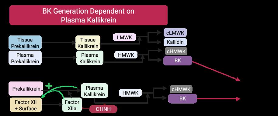
62 Bradykinin-Mediated Angioedema: Pathways, Physiology, and Disease Mechanism Articles
65 Evaluating the Effectiveness of Hypertension Treatment in Delaying/Slowing the Progression of Chronic Kidney Disease in Adults Aged 18 Years and Above with Impaired Glucose Regulation: A Systematic Review Protocol
Jadhakhan F et al.
76 Cohort Profile: The COVID-19 Ticino Biobank
Barda B et al.
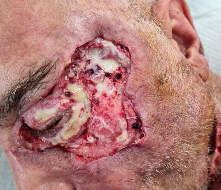
85 Surviving the Uncommon: A Case of Unilateral Periorbital Necrotising Fasciitis
Kassem M et al.
93 Cystic Fibrosis: A Review Study
Roy R et al.
"Less than 10% of people who should be getting lung cancer screening are getting screened"
Editorial Board
Editor-in-Chief
Prof Markus Peck-Radosavljevic Klinikum Klagenfurt am Wörthersee, Austria
Current Chairman and Head of the Department of Gastroenterology and Hepatology, Endocrinology, Rheumatology and Nephrology at Klinikum Klagenfurt am Wörthersee, with expertise in portal hypertension, hepatocellular carcinoma, and HIV–HCV coinfection.
Prof Ahmad Awada
Jules Bordet Institute, Belgium
Prof Sorin T. Barbu
“Iuliu Hațieganu” University of Medicine and Pharmacy, Romania
Prof Abdullah Erdem Canda
Yildirim Beyazit University, Türkiye
Prof Ian Chikanza
Harley Street Clinic, UK
Prof Lászlo Vécsei
University of Szeged, Hungary
Dr Pierfrancesco Agostoni
St. Antonius Hospital, the Netherlands
Dr Fernando Alfonso
Hospital Universitario de La Princesa, Spain
Dr Emanuele Angelucci
IRCCS Ospedale Policlinico San Martino, Italy
Dr George Anifandis University of Thessaly, Greece
Dr Riccardo Autorino
Virginia Commonwealth University, USA
Dr Mátyás Benyó University of Debrecen, Hungary
Prof Andrew Bush Imperial College London, UK
Dr Hassan Galadari
United Arab Emirates University, United Arab Emirates
Dr Amir Hamzah Abdul Latiff
Pantai Hospital, Malaysia
Dr Lorenz Räber
Bern University Hospital, Switzerland
Aims and Scope
EMJ, the flagship journal of the EMJ portfolio, is an openaccess, peer-reviewed eJournal, committed to elevating the quality of healthcare globally by publishing high-quality medical content across the 18 clinical areas covered in our portfolio. The journal is published quarterly and showcases the latest developments across these clinical areas.
EMJ publishes peer-reviewed research papers, review articles, and case reports across all therapy areas of the EMJ portfolio. In addition, the journal publishes features and opinion pieces create a discussion around key topics in the field and broaden readers’ professional interests. The journal also features interviews with leading experts in various clinical disciplines.
The journal covers advances within the pharmaceutical arena by publishing sponsored content from congress symposia, which is of high educational value for healthcare professionals. This undergoes rigorous quality control checks by independent experts and the in-house editorial team.
EMJ endeavours to increase knowledge, stimulate discussion, and contribute to the delivery of world-class updates in the clinical realm. We do not publish veterinary science papers or laboratory studies that are not linked to patient outcomes. Further details on coverage can be found here: www.emjreviews.com
Editorial Expertise
EMJ is supported by various levels of expertise:
• Guidance from an Editorial Board consisting of leading authorities from a wide variety of disciplines.
• Invited contributors who are recognised authorities in their respective fields.
• Peer review, which is conducted by expert reviewers who are invited by the Editorial team and appointed based on their knowledge of a specific topic.
• An experienced team of editors and technical editors.
• A team of internal and independent medical writers.
Peer Review
Every review article, case report, feature, and research article published in EMJ undergoes peer review by at least two independent experts.
On submission, all manuscripts are assessed and undergo a technical check by the EMJ Editorial staff to determine their suitability for the journal and appropriateness for peer review. Editorial staff identify appropriate reviewers who are selected based on their specialist knowledge in the relevant area. All peer review is double-blind.
Following review, manuscripts are either accepted without modification, returned to the author(s) to incorporate required changes, or rejected. Editorial staff are responsible for ensuring that necessary amendments to the manuscript have been made, with input from our Editorial Board or the original reviewers where necessary. The Editor of EMJ has final discretion over any proposed amendments. Manuscripts authored by members of the Editorial Board are subjected to the same double-blind process. Short opinion pieces are published following internal review and publication is at the discretion of the Editor. Congress-associated content authored by the EMJ Editorial staff undergoes internal quality control checks. Congress-related content sponsored or funded by our industry partners undergoes quality control checks independently. Industry-supported content that falls into any of
the categories that are eligible for peer review, undergoes the same peer review process.
Submissions
We welcome contributions from professionals, consultants, academics, and industry leaders on relevant and topical subjects. We seek papers with the most current, interesting, and relevant information in each therapeutic area and accept original research, review articles, case reports, and features.
We are always keen to hear from healthcare professionals wishing to discuss potential submissions, please email: editorial.assistant@emjreviews.com
To submit a paper, use our online submission site: www.editorialmanager.com/e-m-j
Submission details can be found through our website: www.emjreviews.com/contributors/authors
Reprints
All articles included in EMJ are available as reprints (minimum order 1,000). Please contact hello@emjreviews.com if you would like to order reprints.
Distribution and Readership
EMJ is distributed through controlled circulation to healthcare professionals in the relevant fields globally.
Indexing and Availability
EMJ is indexed on DOAJ, the Royal Society of Medicine, and Google Scholar®.
EMJ is available through the websites of our leading partners and collaborating societies.
EMJ journals are all available via our website: www.emjreviews.com
Open Access
This is an open-access journal in accordance with the Creative Commons Attribution-Non Commercial 4.0 (CC BY-NC 4.0) license.
Congress Notice
Staff members attend medical congresses as reporters when required.
All information obtained by EMJ and each of the contributions from various sources is as current and accurate as possible. However, due to human or mechanical errors, EMJ and the contributors cannot guarantee the accuracy, adequacy, or completeness of any information, and cannot be held responsible for any errors or omissions. EMJ is completely independent of any event reviews in this issue and the use of the organisations does not constitute endorsement or media partnership in any form whatsoever. The cover photo is of Madrid, the location of work for the primary author of Editor's Pick.
A comprehensive portfolio for uro-gynaecological health
IBSA develops interconnected solutions in the uro-gynaecological field, moving beyond the concept of single products and isolated treatments to provide comprehensive responses tailored to each patient’s needs.
SYNERGISTIC APPROACH
The synergy between solutions that work together enables targeted interventions on multiple aspects of uro-gynaecological conditions.
MORE OPPORTUNITIES
An integrated portfolio allows adaptation to different patient profiles, maximizing clinical outcomes and enhancing treatment adherence.
GAGs: glycosaminoglycans.
SILDENAFIL Orodispersible films
For the treatment of erectile dysfunction (inability to achieve or maintain a penile erection sufficient for satisfactory sexual performance).
PEROVIAL®
For the treatment of the acute phase of Peyronie’s Disease.
HYALUXELLE®
For the treatment of vulvovaginal atrophy. With IBSA NAHYCO® Hybrid Technology.
IALURIL® Prefill
For the restoration of the GAGs layers of the urothelial vesical tissue in cases in which their loss can cause frequent and recurring problems (such as, cystitis, chronic inflammation).
IALURIL® Soft Gels
With PearlTec® Technology. Based on curcumin, quercetin, hyaluronic acid, and chondroitin sulfate.
VITAMINS AND MINERALS
Iron, B12 and D3 orodispersible films With IBSA FilmTec® Technology.
Editorial Director
Andrea Charles
Managing Editor
Darcy Richards
Copy Editors
Noémie Fouarge, Sarah Jahncke
Editorial Leads
Helena Bradbury, Ada Enesco
Editorial Co-ordinators
Bertie Pearcey, Alena Sofieva
Editorial Assistants
Niamh Holmes, Katrina Thornber, Aleksandra Zurowska
Creative Director
Tim Uden
Design Manager
Stacey White
Senior Designers
Tamara Kondolomo, Owen Silcox
Designers
Shanjok Gurung, Fabio van Paris
Creative Artworker
Dillon Benn Grove
Junior Designer
Stephanie Corbett
Head of Marketing
Helena Spicer
Chief Executive Officer
Justin Levett
Chief Commercial Officer
Dan Healy
Founder and Chairman
Spencer Gore
Welcome
Dear Readers,
We are delighted to welcome you to the fourth and final 2025 flagship issue of EMJ, which focuses on targeted therapies. This promising field holds the key to personalised treatment, tailored to individual needs with the aim of minimising side effects, and the potential to improve not only quality, but also quantity of life.
Within this issue, we feature exclusive interviews with Michael Snyder, Stanford University School of Medicine, California, USA; and Rishindra Reddy, University of Michigan, Ann Arbor, USA, who discuss integrative personal omics profiles to predict genetic risk, and genomic markers and circulating tumour DNA to diagnose early-stage lung cancer.
Among our peer reviewed content, you can find a timely and informative study exploring real-world biobanking efforts that took place during the COVID-19 pandemic. The study highlights the utility of biobank-based research and provides a potential framework for future collaborative efforts to further improve our understanding of infectious diseases and patient outcomes through earlier diagnosis and the uncovering of new therapeutic targets. You can also explore an interesting systematic review protocol outlining a roadmap for the evaluation of antihypertensive treatment in delaying progression to chronic kidney disease in adults with impaired glucose regulation.
We would like to take this opportunity to thank our Editorial Board, the authors, peer reviewers, and interviewees for their invaluable contributions to this issue. We hope you enjoy reading and find insightful takeaways for your clinical practice and to help further elevate the quality of healthcare.
Contact us
Editorial enquiries: editor@emjreviews.com
Sales opportunities: salesadmin@emjreviews.com
Permissions and copyright: accountsreceivable@emjreviews.com
Reprints: info@emjreviews.com Media enquiries: marketing@emjreviews.com
Collagen Bank Introducing
Patented* Micro-Peptide Technology
Developed with Dermatologists
Targets 5 Extra-Cellular Matrix Building Markers**
Welcome to our final EMJ Flagship issue of 2025. This edition highlights a central theme shaping clinical practice across disciplines: the expanding role of personalised medicine.
Each of the major congresses covered in this issue illustrates how precision approaches are moving from specialist areas into everyday decision-making. At the European Association for the Study of Diabetes (EASD) Annual Meeting 2025, experts discussed how precision medicine is redefining the assessment and management of diabetic neuropathies. The European Society for Medical Oncology (ESMO) Congress 2025 explored biomarker-based therapies in metastatic prostate cancer, and the European Respiratory Society (ERS) Congress 2025 highlighted how microbiome-targeted strategies are driving new treatments for respiratory disease. Finally, the European Society of Cardiology (ESC) Congress 2025 examined emerging therapeutic frontiers in cardiology and the growing use of AI in precision diagnostics.
This issue’s articles provide a broad and clinically relevant selection of research. The COVID-19 Ticino Biobank shows how highquality, well-curated samples can deepen our understanding of disease progression and support more tailored clinical insights. A systematic review protocol assesses how hypertension treatment may slow
chronic kidney disease progression in adults with impaired glucose regulation, a group where targeted evidence is limited. We also present a rare case of unilateral periorbital necrotising fasciitis, emphasising the importance of early recognition and coordinated care. Finally, a comprehensive cystic fibrosis review summarises current treatment approaches and the need for continued innovation as survival improves.
Each of the major congresses covered in this issue illustrates how precision approaches are moving from specialist areas into everyday decision-making
We are also pleased to feature interviews with Michael Snyder, discussing longterm health monitoring and personalised prevention, and Rishindra Reddy, who shares insights into advances in earlystage lung cancer care.
Thank you to all authors, reviewers, interviewees, and Editorial Board members for your contributions throughout the year.
Prof Markus Peck-Radosavljevic
Professor of Medicine and Chairman, Department of Gastroenterology and Hepatology, Endocrinology, Rheumatology and Nephrology, Klinikum Klagenfurt am Wörthersee, Austria
Decoding the Digital Heart: Practical AI for Precision Diagnostics
THE LANDSCAPE of modern cardiology is undergoing a profound transformation, driven by the emergence of AI as a powerful tool to enhance diagnostic precision and patient management. In a session titled ‘Practical Artificial Intelligence Solutions for Precision Diagnostics in the Clinic’ at the 2025 European Society of Cardiology (ESC) Congress, held in Madrid, Spain, leading experts converged to demonstrate how AI is moving from a futuristic concept to a pragmatic, indispensable part of everyday clinical care.
MINING BIG DATA FOR NEW BIOMARKERS
AI is reshaping cardiovascular medicine by enhancing the way clinicians interpret medical images, diagnose disease, and guide treatment decisions. According to Alexios Antonopoulos, 1st Cardiology Department, National Kapodistrian University of Athens, Greece, AI-driven imaging tools are addressing major unmet needs in cardiovascular care, particularly in early disease detection, personalised risk prediction, and workflow efficiency.
AI has already transformed routine imaging tasks, automating measurements such as ventricular volumes on cardiac magnetic resonance, saving clinicians valuable time and improving diagnostic consistency. More advanced applications, including automated plaque segmentation and quantification of coronary or epicardial fat, are redefining how clinicians perceive and classify disease.1 For instance, AI tools can detect subtle imaging features of conditions like cardiac amyloidosis, often before symptoms appear, helping clinicians identify high-risk patients who would otherwise go undiagnosed.2
Antonopoulos explained that, by integrating imaging biomarkers with
large-scale datasets such as the UK Biobank,3 AI enables large cohort analyses and the discovery of new predictors of cardiovascular risk, such as vascular inflammation or perivascular fat attenuation. These insights support a shift toward individualised risk stratification and more precise management strategies.
To fully integrate AI into clinical practice, Antonopoulos emphasised the need for rigorous validation, regulatory oversight, biological grounding, and evidence from RCTs. When proven effective and costefficient, AI-assisted imaging can deliver on its promise to not only enhance diagnosis and prediction, but also to transform patient care through truly personalised cardiovascular medicine.
USING AI TO READ BEHIND THE ECG TRACES
Following on, Rohan Khera, Yale University, New Haven, Connecticut, USA, explored how AI can extract hidden clinical information from ECGs, transforming one of the world’s most accessible diagnostic tests into a powerful tool for early disease detection and risk prediction. Every year, more than 300 million ECGs are performed globally, far exceeding all cardiac imaging
tests combined, making it an ideal foundation for scalable AI innovation.4
Traditionally, clinicians interpret ECGs visually, identifying rhythm or conduction abnormalities, Khera explained. However, much richer information lies within the underlying voltage data that AI can process through deep learning techniques such as convolutional neural networks.4 Khera’s team at the Yale Cardiovascular Data Science (CarDS) Lab, New Haven, developed models that can detect structural heart diseases (such as left ventricular dysfunction, hypertrophic cardiomyopathy, aortic stenosis, and cardiac amyloidosis) using only ECG images, rather than specialised raw data. Remarkably, these models achieved >90% accuracy in distinguishing patients with reduced ejection fraction and validated consistently across global populations.5
The group also created smartphone-based tools that allow clinicians, or even patients, to capture ECG images with a phone camera or wearable device and instantly receive AI-driven insights without requiring internet connectivity. This opens the door to community-level screening for heart disease using affordable, portable devices.
Khera continued that, beyond diagnosis, AI-enhanced ECGs can also predict future risk of heart failure, outperforming standard clinical risk models and offering prognostic value even in asymptomatic individuals. As he emphasised, AI can “see what the human eye cannot,” revealing disease signatures hidden in plain sight and democratising cardiovascular diagnostics worldwide.
BRIDGING MOLECULAR DATA AND DIGITAL DIAGNOSTICS
Tanja Zeller, The University Medical Center Schleswig-Holstein, Lübeck, Germany, explored how digital and molecular technologies can help decode the complexity of cardiovascular disease by integrating diverse layers of biological data. She likened biomarkers to puzzle pieces: each provides useful but incomplete information. To see the whole picture of
To see the whole picture of disease, clinicians must combine molecular, clinical, and digital data in multidimensional analyses
disease, clinicians must combine molecular, clinical, and digital data in multidimensional analyses.
Her first example focused on improving the diagnosis of acute myocardial infarction. Traditional triage relies on fixed troponin cut-offs and standard algorithms, which often leave many patients in an indeterminate zone.6 Using machine learning models such as those developed in the Acute Coronary Syndrome (ACS) Pathfinder and Collaboration for the Diagnosis and Evaluation of Acute Coronary Syndrome (CoDE-ACS) consortia, Zeller’s team incorporated multiple routine clinical parameters to calculate an individual’s probability of acute myocardial infarction.7,8 These digital tools, validated in >20,000 patients, allowed three times more people to be immediately and safely ruled out compared with current practice.7,8
Moving beyond diagnosis, she described how integrating multi-omics data, including genomics, transcriptomics, proteomics, and immune profiling, can uncover the molecular pathways driving acute and chronic coronary syndromes. Studies using such approaches have identified immune cell shifts and cytokine signalling patterns that may predict outcomes after infarction.9
Her second example addressed atrial fibrillation, where combining genetic, RNA, and protein data with AI tools identified molecular variants linked to disease development and improved risk prediction.10 In a striking proof of concept, her team trained deep learning models to predict blood N-terminal pro-B-type natriuretic peptide (NT-proBNP) levels directly from ECG recordings, demonstrating how digital tools might replicate expensive laboratory tests.11
Zeller concluded that merging omicsbased biomarkers with digital diagnostics could transform cardiovascular care, but success will depend on data quality, crossdisciplinary collaboration, and careful clinical validation.
DIGITAL BIOMARKERS IN ACTION
The final speaker, Florian A. Wenzl, Center for Molecular Cardiology, University of Zurich, Switzerland, explored the current role and future potential of AI in clinical decision-making. He began by emphasising that AI is no longer a future concept but an established reality that already influences many aspects of daily life and medicine. The rapid progress in computing power and data availability has led to exponential growth in AI capabilities, enabling systems to achieve superhuman performance in selected tasks, including diagnostics and prognostics.
Wenzl outlined how AI models in cardiovascular medicine are increasingly applied across four key domains: disease phenotyping, diagnostics, prognostics, and treatment decisions.12 He stressed that a range of data sources, such as clinical
findings, imaging, and laboratory values, can be used to train predictive models, though selecting the most relevant data for each clinical question remains crucial.12 Rigorous evaluation, including external validation and comparison with current standards of care, was highlighted as essential for ensuring reliability and trust.
A central theme of his talk was that AI model assessment should follow the same methodological principles as conventional statistical models, using metrics like sensitivity, specificity, and calibration. Wenzl also addressed concerns about explainability, suggesting that practical utility can sometimes outweigh full mechanistic understanding, as is often the case with effective drugs.
He concluded with examples of AI applications, from predicting coronary artery disease by facial photographs13 to detecting atrial fibrillation by speech analysis,14 and emphasised that transparent reporting, external validation, and integration into clinical guidelines will be vital for safely realising AI’s promise in personalised medicine.
CONCLUSION
AI is transforming cardiology, enhancing early diagnosis, personalised risk prediction, and patient management. From imaging and ECG analysis to multi-omics integration, this session demonstrated how AI uncovers
References
1. Lin A et al. Deep learning-enabled coronary CT angiography for plaque and stenosis quantification and cardiac risk prediction: an international multicentre study. Lancet Digit Health. 2022;4(4):e256-65.
2. Antonopoulos AS et al. Computed tomography-derived myocardial radiomics for detection of transthyretin amyloidosis in patients with severe aortic stenosis. Amyloid. 2025;32(3):226-37.
3. UK Biobank. 2025. Available at: https:// www.ukbiobank.ac.uk/. Last accessed: 14 October 2025.
4. Mason F et al. AI-enhanced reconstruction of the 12-lead electrocardiogram via 3-leads with accurate clinical assessment. NPJ Digit Med. 2024;7(1):201.
hidden disease signatures, automates workflows, and informs clinical decisions. Rigorous validation and interdisciplinary collaboration remain essential to safely translate these innovations into improved cardiovascular care.
5. Sangha V et al. Detection of left ventricular systolic dysfunction from electrocardiographic images. Circulation. 2023;148(9):765-77.
6. Byrne RA et al. 2023 ESC Guidelines for the management of acute coronary syndromes. Eur Heart J. 2023.44(38):3720-826.
7. Neumann JT et al. Personalized diagnosis in suspected myocardial infarction. Clin Res Cardiol. 2023;112(9):1288-301.
8. Doudesis D et al.; CoDE-ACS Investigators. Machine learning for diagnosis of myocardial infarction using cardiac troponin concentrations. Nat Med. 2023;29(5):1201-10.
9. Pekayvaz K et al. Multiomic analyses uncover immunological signatures in acute and chronic coronary syndromes. Nat Med. 2024;30(6):1696-710.
10. Assum I et al. Tissue-specific multiomics analysis of atrial fibrillation. Nat Commun. 2022;13(1):441.
11. Neyazi M et al. Deep learning-based NT-proBNP prediction from the ECG for risk assessment in the community. Clin Chem Lab Med. 2023;62(4): 740-52.
12. Lüscher TF et al. Artificial intelligence in cardiovascular medicine: clinical applications. Eur Heart J. 2024;45(40):4291-304.
13. Lin S et al. Feasibility of using deep learning to detect coronary artery disease based on facial photo. Eur Heart J. 2020;41(46):4400-11.
14. Golovchiner G et al. Automated detection of atrial fibrillation based on vocal features analysis. J Cardiovasc Electrophysiol. 2022;33(8):1647-54.
Diabetic Neuropathies in the Era of Precision Medicine: Unravelling Complex
AT THE European Association for the Study of Diabetes (EASD) Annual Meeting, held in Vienna, Austria, from 15th–19th September 2025, the session ‘Diabetic Neuropathies in the Era of Precision Medicine’ offered a comprehensive and forward-looking examination of sensory, autonomic, and inflammation-driven neuropathic complications in diabetes. Chaired by Leszek Czupryniak, Central University Hospital, Warsaw, Poland; and Anca Pantea-Stoian, INDNBM Paulescu, Bucharest, Romania, the symposium brought together experts who highlighted emerging pathomechanisms, advanced diagnostic technologies, and the promise of tailored therapeutic strategies.
ACKNOWLEDGING PREDIABETIC POLYNEUROPATHY
Julia Szendroedi, University Hospital Heidelberg, Germany, opened the session by emphasising the substantial morbidity and mortality associated with diabetic sensorimotor polyneuropathy (DSPN). Affecting more than half of individuals with Type 2 diabetes (T2D), DSPN markedly increases the risk of foot ulceration, major amputation, and mortality by nearly 3.6fold in Type 1 diabetes (T1D) and 1.6-fold in T2D.1 Yet, as Szendroedi noted, determining the true prevalence of neuropathy in prediabetes remains challenging. Definitions of prediabetes vary, and screening methods for neuropathy differ significantly between studies, resulting in widely divergent prevalence estimates ranging from greater than 10% in most studies,2 to as high as 70–80%. This variability has prevented robust meta-analyses, leaving individual studies to shape the field.
Early Small-Fibre Injury and Phenotype Progression
Szendroedi contrasted studies showing minimal sensory changes in normoglycaemic individuals with others reporting unexpectedly high neuropathy rates, underscoring both the sensitivity and the limitations of quantitative sensory testing (QST). The accumulating evidence suggests that neuropathy in prediabetes begins with early metabolic and vascular injury to small fibres. As individuals progress to T2D, large-fibre dysfunction and central sensitisation become more prominent, with age and nephropathy acting as amplifiers of risk, while HbA1c alone does not fully account for the observed sensory patterns.3
Longitudinal QST phenotyping has revealed dynamic transitions across sensory subtypes. Patients may evolve from profiles dominated by thermal hyperalgesia to mechanical hyperalgesia, and eventually to sensory loss, reflecting distinct and measurable clinical trajectories.4
DSPN markedly increases the risk of foot ulceration, major amputation, and mortality by nearly 3.6-fold in T1D and 1.6-fold in T2D
Szendroedi emphasised that how prediabetes is defined substantially influences neuropathy risk prediction. An HbA1c-based approach, while convenient, is the least predictive for DSPN. In contrast, an oral glucose tolerance testbased hierarchy, where impaired glucose tolerance predicts risk more strongly than impaired fasting glucose, and both of these predict risk more strongly than HbA1c, better reflects true DSPN susceptibility (unpublished data). She also discussed the emerging prediabetes clusters (cluster 1-6), highlighting that individuals in cluster 5 have a notably elevated risk of neuropathy, demonstrating the biological heterogeneity present even before diabetes onset.5
Metabolic Drivers and Patchy Small-Fibre Damage
Applying QST to individuals without overt neuropathy has shown that insulin resistance is a key driver of early preclinical neuropathy in those without diabetes. Among people with T2D, further metabolic syndrome components and accumulation of skin advanced glycation end-products (AGE) contribute to progression.6 Early investigations using corneal confocal microscopy and skin biopsy have revealed small-fibre loss shortly after diabetes diagnosis. Yet, the overlap between corneal and skin findings was present in only 3% of individuals, indicating a 'patchy', organspecific pattern of nerve involvement.7
“This is evidence we need more longitudinal studies,” Szendroedi remarked.
This is evidence we need more longitudinal studies
Ischaemia, Oxidative Stress, and Neuroimmune Alterations
Peripheral nerve ischaemia is emerging as a major contributor to DSPN, with dynamic contrast-enhanced magnetic resonance neurography (MRN) revealing that microvascular permeability correlates strongly with age, BMI, and neuropathy severity.8 Skin biopsy analyses indicate early oxidative stress, reflected in increased dermal superoxide dismutase 2 (SOD2) expression, while the markedly reduced Langerhans cell density in newly diagnosed T2D points to early neuro-immune dysregulation that appears independent of intraepidermal nerve fibre density.9
Individuals in cluster 5 have a notably elevated risk of neuropathy
Proximal Nerve Involvement and Distinct Patterns in Type 1 Versus Type 2 Diabetes
Advanced MRN has shed light on early microstructural remodelling in the proximal sciatic nerve, extending understanding beyond distal small-fibre pathology. Comparative analyses between T1D and T2D indicate distinct remodelling patterns, with glycaemia more influential in T1D and dyslipidaemia playing a stronger role in T2D. These findings support the emerging
'patch hypothesis', suggesting that different mechanisms may drive neuropathy in different nerve regions.10 Neuron-specific biomarkers, including neurofilament light chain protein and circulating myelin-related mRNA, have shown predictive value for both hypoalgesic and hyperalgesic phenotypes, emphasising their utility for mechanistic longitudinal studies.11 Szendroedi concluded her talk by emphasising that DSPN is not a linear disease but a mosaic, in which compensated segments, immune-active regions, and ischaemic lesions coexist within the same nerve. Because different mechanisms dominate in different regions and patients, multimodal assessment, using QST, MRN, biopsy markers such as SOD2, and circulating biomarkers like neurofilament light chain, is essential. She called for early screening, including in prediabetes, and phenotype-guided management strategies in the future.
UPDATE ON CARDIOVASCULAR AUTONOMIC NEUROPATHY
In the second talk, Péter Kempler, Semmelweis University, Budapest, Hungary, delivered an “unorthodox perspective” on cardiovascular autonomic neuropathy (CAN), starting his talk by invoking the words of the late Aaron Vinik: “Know autonomic neuropathy and you will know the whole of medicine.”
Mortality Risk and Long-Term Data
CAN has been recognised for decades, and early data suggested a fivefold increase in mortality among affected individuals.12 More recent 20-year follow-up data in T2D indicated a 1.5-fold increase in mortality risk, bringing it closer to that seen in sensory neuropathy and refining earlier estimates.13 Additional pooled analyses have shown that having more than two CAN abnormalities confers a 3.5-fold increased risk of mortality.14
ACCORD Revisited:
Sex-Specific
Pooled analyses have shown that having more than two CAN abnormalities confers a 3.5-fold increased risk of mortality x 3.5
Signals
Kempler revisited findings from the ACCORD study,15 which reported increased
mortality in the intensive glucose-lowering arm. Notably, a self-reported history of neuropathy, despite the absence of formal neuropathy assessment in the study, emerged as the strongest independent predictor of mortality under intensive treatment. Newly presented 2025 analyses revealed striking sex differences: women with CAN exhibited significantly higher risks of both all-cause and cardiovascular mortality, while men did not show this association.16 These findings echo signals from the original ACCORD dataset that had previously gone unrecognised.
Pathogenic Therapies and Cardiometabolic Protection
He continued by outlining the mechanisms by which hyperglycaemia induces microand macrovascular endothelial injury, including glucose toxicity, oxidative stress, inhibition of glyceraldehyde-3-phosphate dehydrogenase, and activation of harmful alternative pathways such as AGE formation. These pathways underpin the rationale for agents such as benfotiamine, which reduces AGE formation and diverts glucose into non-harmful pathways, and alpha-lipoic acid, mitigating oxidative stress. Although widely used in Germany, Austria, and parts of Central Europe for decades, these agents remain underutilised in the UK, France, and the USA due to a lack of recent large trials.
Finally, Kempler presented compelling real-world evidence from a Hungarian study of nearly 24,000 patients comparing pathogenically oriented alphalipoic acid therapy with symptomatic treatments. Individuals receiving alphalipoic acid showed approximately 30% fewer myocardial infarctions requiring percutaneous coronary intervention, improved stroke outcomes, a 30% reduction in hospitalisations for heart failure, a 17% reduction in cancer events, and a 45% decrease in all-cause mortality, with no significant difference in lowerlimb amputation rates.17 He closed with quotations from writer and philosopher, Aldous Huxley, and Murphy’s Law to highlight the consequences of ignoring complex, multifactorial evidence in favour of overly simplistic explanations, leaving the audience a moment to reflect.
INFLAMMATION, DIABETES HETEROGENEITY AND POLYNEUROPATHY
The final talk, delivered by Christian Herder, German Diabetes Center, Düsseldorf, Germany, explored the interplay between inflammation, diabetes heterogeneity, and DSPN risk.
Inflammatory Biomarkers and Neuropathy Incidence
Using data from the KORA cohort (a subgroup of over 500 participants aged 61–82 years), Herder described the first prospective evidence linking inflammatory biomarkers with incident DSPN. Elevated levels of high sensitivity C-reactive protein, IL-6, and TNF-α were associated with greater neuropathy risk, while adiponectin showed an inverse association.18 To capture a broader view of inflammatory processes, the team analysed 71 highquality serum biomarkers using the OLINK Inflammation Assay (OLINK Proteomics, Uppsala, Sweden). Twenty-six proteins were positively associated with future DSPN, reflecting coordinated signalling across innate and adaptive immune pathways. Upstream regulatory analysis identified TNF-α and IL-1β as key drivers, reinforcing their therapeutic potential.19
Diabetes Subtypes and Differential Risk
Individuals receiving alpha-lipoic acid showed approximately 30% fewer myocardial infarctions requiring percutaneous coronary intervention, improved stroke outcomes, a 30% reduction in hospitalisations for heart failure, a 17% reduction in cancer events, and a 45% decrease in all-cause mortality
Herder examined heterogeneity within diabetes and prediabetes using data from the German Diabetes Study,20 Europe’s largest prospective cohort of individuals with recent-onset diabetes. Significant differences in inflammatory biomarker profiles emerged across diabetes subtypes. The severe insulin-resistant diabetes phenotype exhibited the highest inflammatory load, accompanied by elevated leukocyte counts and increased neutrophil-lymphocyte ratios, while the severe insulin-deficient diabetes subtype displayed the lowest inflammatory burden.19 Parallels were observed in prediabetes. In a call back to Szedroedi’s earlier discussion of clusters, Herder explained that cluster 5, marked by high visceral adiposity, hepatic fat accumulation, and pronounced
insulin resistance, had both the highest inflammatory load and the greatest DSPN risk. In contrast, cluster 2 showed the lowest inflammatory burden.5
Inflammasome Inhibition:
A Targeted Therapeutic Pathway
Herder highlighted the role of NLRP3 inflammasome activation in generating IL-1β, a potent pro-inflammatory cytokine. Within the INTERCEPT-T2D consortium, an ongoing RCT is testing an inflammasome inhibitor in individuals with T2D and elevated inflammatory markers, with neuropathy outcomes designated as a key secondary endpoint.21 Results are anticipated within the next few years and may help pave the way for targeted anti-inflammatory therapies.
Key Takeaways
Herder concluded by emphasising that DSPN risk is strongly linked to multi-marker inflammatory signatures, that diabetes and prediabetes subtypes differ markedly in inflammatory burden, and that identifying high-inflammation subgroups may facilitate precision medicine approaches to neuropathy prevention and management.
CONCLUSION
Across all three talks, a clear narrative emerged: diabetic neuropathies are mechanistically heterogeneous, multifactorial, and ill-suited to one-sizefits-all approaches. Early small-fibre injury, proximal nerve remodelling, vascular dysfunction, inflammation, and metabolic diversity each contribute to a mosaic of pathology rather than a linear progression.
The speakers collectively emphasised the importance of early and multimodal screening, including in prediabetes, along with phenotype-guided assessment and treatment, better recognition of autonomic and inflammatory contributors, and the promise of targeted therapies spanning metabolic, vascular, and inflammatory pathways.
As precision medicine evolves, integrating multimodal diagnostics with mechanistic understanding offers a path towards genuinely individualised care, with the potential to alter the natural history of diabetic neuropathies in the years ahead.
References
1. Hicks CW et al. Peripheral neuropathy and all-cause and cardiovascular mortality in U.S. adults: a prospective cohort study. Ann Intern Med. 2021;174(2):167-74.
2. Kirthi V et al. Prevalence of peripheral neuropathy in pre-diabetes: a systematic review. BMJ Open Diabetes Res Care. 2021;9(1):e002040.
3. Kopf S et al. Deep phenotyping neuropathy: an underestimated complication in patients with prediabetes and type 2 diabetes associated with albuminuria. Diabetes Res Clin Pract. 2018;146:191-201.
4. Tsilingiris D et al. Sensory phenotypes provide insight into the natural course of diabetic polyneuropathy. Diabetes. 2024;73(1):135-46.
5. Wagner R et al. Pathophysiologybased subphenotyping of individuals at elevated risk for type 2 diabetes. Nat Med. 2021;27(1):49-57.
6. Tsillingiris D et al. Dysmetabolismrelated early sensory deficits and their relationship with peripheral neuropathy development. J Clin Endocrinol Metab. 2023;108(10):e979-88.
7. Herder C et al. Novel insights into sensorimotor and cardiovascular autonomic neuropathy from recentonset diabetes and population-based cohorts. Trends Endocrinol Metab. 2019;30(5):286-98.
8. Jende JME et al. Sciatic nerve microvascular permeability in type 2 diabetes decreased in patients with neuropathy. Ann Clin Transl Neurol. 2022;9(6):830-40.
9. Ziegler D et al. High prevalence of diagnosed and undiagnosed polyneuropathy in subjects with and without diabetes participating in a nationwide educational initiative (PROTECT study). J Diabetes Complications. 2015;29(8):998-1002.
10. Jende JME et al. Diabetic neuropathy differs between type 1 and type 2 diabetes: insights from magnetic resonance neurography. Ann Neurol. 2018;83(3):588-98.
11. Morgenstern J et al. Neuron-specific biomarkers predict hypo- and hyperalgesia in individuals with diabetic peripheral neuropathy. Diabetologia. 2021;64(12):2843-55.
12. Ziegler D. Diagnosis and management of diabetic peripheral neuropathy. Diabet Med. 1996;13(Suppl 1):S34-8.
13. Vinik AI et al. Diabetic autonomic neuropathy. Diabetes Care. 2003;26(5): 1553-79.
14. Vági OE et al. Association of cardiovascular autonomic neuropathy and distal symmetric polyneuropathy with all-cause mortality: a retrospective cohort study. J Diabetes Res. 2021;2021:6662159.
15. ACCORD Study Group; Buse JB et al. Action to Control Cardiovascular Risk in Diabetes (ACCORD) trial: design and methods. Am J Cardiol. 2007;99(12A):21i-33i.
16. Zhou Z et al. Sex differences in the association between cardiovascular autonomic neuropathy and mortality in patients with type 2 diabetes: the ACCORD study. J Am Heart Assoc. 2025;14(2):e034626.
17. Jermendy G et al. Morbidity and mortality of patients with diabetic neuropathy treated with pathogenetically oriented alphalipoic acid versus symptomatic pharmacotherapies - a nationwide database analysis from Hungary. Diabetes Res Clin Pract. 2023;201:110734.
18. Herder C et al. Proinflammatory cytokines predict the incidence and progression of distal sensorimotor polyneuropathy: KORA F4/FF4 study. Diabetes Care. 2017;40(4):569-76.
19. Herder C et al.; GDS Group. Differences in biomarkers of inflammation between novel subgroups of recent-onset diabetes. Diabetes. 2021;70(5):1198-208.
20. Szendroedi J et al. Cohort profile: the German Diabetes Study (GDS). Cardiovasc Diabetol. 2016;15:59.
21. Meier DT et al. Targeting the NLRP3 inflammasome-IL-1β pathway in type 2 diabetes and obesity. Diabetologia. 2025;68(1):3-16.
Big Changes on a Small Scale: Microbiome-Targeted Therapies for Lung Disease
TARGETED and personalised therapies are expanding into a new area, focused not just on molecular receptors or immune signatures, but on the complex microbial communities that live inside us. In a ‘hot topic’ session presented at the European Respiratory Society (ERS) Congress 2025, researchers offered a compelling look at how the respiratory microbiome is reshaping our understanding of airway disease pathogenesis and opening novel therapeutic avenues. Together, their talks moved from precision pathogen targeting, to early-life microbial imprinting, to the potential of ‘beneficial bacteria’ in the chronically diseased lung.
PHAGE THERAPY: PATHOGEN TARGETING WITHOUT COLLATERAL DAMAGE?
The session opened with Georgia Mitropoulou, Lausanne University Hospital, Switzerland, who brought the clinical urgency of precision antibacterial therapy into focus. Her talk centred on one of the most challenging scenarios in respiratory medicine: a child with cystic fibrosis (CF) and multidrug-resistant Pseudomonas aeruginosa, for whom no conventional antimicrobial options remained.
Mitropoulou described the rationale and real-world execution of bacteriophage therapy, a highly targeted approach in which viruses that naturally infect bacteria are used therapeutically. Unlike broadspectrum antibiotics, phages are narrow in host range, leaving commensal bacteria largely untouched. This makes phage therapy uniquely compatible with the principles of personalised, microbiomesparing intervention.
In her case example, inhaled phage therapy was delivered under compassionate use, combined with standard antibiotics. Clinical improvement followed, and sputum monitoring showed
Unlike broad-spectrum antibiotics, phages are narrow in host range, leaving commensal bacteria largely untouched
early increases in phage titres. Yet the story quickly became more complex. Over repeated cycles, the patient developed increasing levels of anti-phage antibodies (IgA, IgG, and IgM), which correlated with a steady decline in recoverable phage in sputum. Ultimately, phage neutralisation diminished therapeutic effect.
From this, Mitropoulou explored one of the central challenges in phage therapy: immune recognition. While phages can evolve alongside bacteria, they cannot escape a host immune response that flags them as foreign. Experimental frameworks are now emerging to quantify this risk. One such tool, a ‘phage immunogenicity risk index’, based on specific genetic features, may eventually help clinicians choose phages that are less likely to be neutralised.
She contextualised these clinical observations within the broader evidence base. Across hundreds of case reports, observational success rates appear high
(up to 80% achieving infection control or eradication), but randomised trials report more modest outcomes, reflecting publication bias and the concomitant use of antibiotics in nearly all cases. Notably, the first randomised, inhaled phage therapy trial in humans (BioMIX BX004 for chronic CFassociated Pseudomonas) demonstrated safety, tolerability, and preliminary microbiological signals, though efficacy results remain pending.1
Mitropoulou concluded with a nuanced message: phage therapy holds great promise as a personalised, targeted antibacterial intervention, especially for drug-resistant infections in CF and bronchiectasis, but its implementation required rigorous phage-host matching, immune-aware treatment planning, and much stronger clinical trial data.
EARLY-LIFE IMPRINTING AND RESTORING LOST MICROBIAL SIGNALS
Olaf Perdijk, Utrecht University, the Netherlands, extended the concept of targeted therapy from targeting microbes to replacing the molecular signals they provide. His research shines light on a complex paradox: when early-life antibiotics are given to young mice, the microbiome composition eventually normalises, yet the animals retain a heightened vulnerability to allergic airway disease. Something in those early-life interactions imprints a long-lasting susceptibility, even when the microbial community appears restored.
Perdijk’s mouse model, which uses a short antibiotic course followed by cohousing to allow complete microbial recovery, enabled his team to identify the crucial missing link: epithelial memory. Airway structural cells from mice treated with early-life antibiotics showed exaggerated chemokine release and heightened inflammatory responses long after microbial normalisation. Singlecell sequencing revealed mitochondrial stress signatures and altered metabolic pathways in epithelial subsets, signs of a tissue 'stress imprint' beneath an apparently healthy surface.2
Untargeted metabolomics provided a clue to this imprint: mice treated with earlylife antibiotics exhibited reduced levels of indole-3-propionic acid (IPA), a microbially derived, tryptophan-based antioxidant with strong reactive oxygen species-scavenging properties. IPA supplementation during the early-life antibiotic window prevented mitochondrial dysfunction, normalised epithelial transcriptional signatures, and protected mice from developing exaggerated allergic inflammation. Interestingly, microbial dysbiosis and exacerbated airway inflammation were absent when antibiotics were given in adulthood, pointing to a narrow developmental window during which microbial metabolites shape epithelial resilience.2
Perdijk’s findings position microbial metabolites, not just microbes themselves, as personalised therapeutic targets. Restoring these missing metabolic ‘messages’, whether via metabolite supplementation, engineered probiotics, or defined microbial consortia, may one day allow clinicians to correct early-life perturbations that predispose individuals to asthma and allergic disease.
BENEFICIAL BACTERIA IN CHRONIC LUNG DISEASE: PROTECTING THE ‘GOOD ONES’
The third speaker, Aurélie Crabbé, University of Antwerp, Belgium, extended the discussion into chronic lung conditions such as COPD, CF, and bronchiectasis, asking a central question: while pathogens fuel the well-known vicious circle of chronic airway inflammation, could non-pathogenic members of the lung microbiome play a modulating, or even protective, role?
Chronic airway disease microbiomes (such as in bronchiectasis) typically include a substantial proportion of pathogens like P. aeruginosa, Haemophilus, and Streptococcus pneumoniae 3 But alongside them exist an overlooked second half composed of commensal or opportunistic microbes. Over the past decade, multiple studies have reported associations between certain airway bacteria and lower inflammation, better lung function, or improved prognosis. However, study heterogeneity makes it difficult to evaluate which findings are robust.
To address this, Crabbé and colleagues conducted a systematic review and meta-analysis of chronic lung microbiome studies, synthesising data from 34 studies and over 4,000 participants (submitted, unpublished data). Across diseases spanning CF, COPD, asthma, and bronchiectasis, six genera were consistently associated with lower inflammation or better clinical outcomes. Three stood out: Prevotella, linked with lower cytokine levels, reduced neutrophil elastase activity, and better lung function; Rothia, associated with reduced inflammatory biomarkers and shown experimentally to protect against viral infection; and non-pathogenic Streptococcus, correlated with lower inflammation and capable of suppressing Pseudomonas through metabolic by-products.
Understanding how beneficial bacteria function requires credible in vitro systems that mimic the architecture and behaviour of human airway tissue. Using a variety of sophisticated platforms, including a rotating-wall bioreactor that produces highly differentiated airway tissue, Crabbé and her team found how specific nonpathogenic bacteria exert protective effects. Prevotella dampened epithelial inflammatory responses and inhibited Pseudomonas biofilms;4 Rothia reduced cytokine production and protected mice from influenza-induced mortality by lowering viral loads;5 and certain Streptococcus strains inhibited Pseudomonas growth via acetate production.6 Importantly, these effects are strain-specific: genus-level sequencing cannot distinguish protective strains from neutral or even pro-inflammatory ones. This complicates interpretation of microbiome data and reinforces the need for precision approaches that function at strain resolution.
Crabbé closed with a real-world example illustrating why this nuance matters. In the CFMATTERS trial,7 patients were randomised to either standard Pseudomonas-targeted therapy or an intensified regimen targeting Pseudomonas plus the nextmost-abundant genera, which in many individuals included Prevotella, Rothia, Veillonella, and commensal Streptococcus. The broader regimen showed no benefit in lung function. More worryingly, extended follow-up revealed more exacerbations and lower quality of life in patients who received the broader antimicrobials.
Across diseases spanning CF, COPD, asthma, and bronchiectasis, six genera were consistently associated with lower inflammation or better clinical outcomes
While causality cannot be proven, the pattern is consistent with the possibility that eliminating commensal strains removed ecological buffers that protect against pathogen overgrowth or inflammation. As Crabbé summarised: “Let’s not kill the good ones.”
A NEW ERA OF MICROBIOMEINFORMED PRECISION MEDICINE
Across these talks, a conceptual shift emerged, redefining the targets of respiratory therapeutics. Mitropoulou highlighted the promise of precision pathogen killing that spares the wider microbial ecosystem. Perdijk
References
1. Chan BK et al. Personalized inhaled bacteriophage therapy for treatment of multidrug-resistant Pseudomonas aeruginosa in cystic fibrosis. Nat Med. 2025;31(5):1494-501.
2. Perdijk O et al. Antibiotic-driven dysbiosis in early life disrupts indole-3-propionic acid production and exacerbates allergic airway inflammation in adulthood. Immunity. 2024;57(8):1939-54.e7.
demonstrated that microbial metabolites, not just microbes, are essential signals that shape immune development and disease susceptibility. Crabbé argued for recognising and protecting beneficial commensals that actively modulate inflammation and pathogen behaviour.
Together, they point toward a future where respiratory medicine embraces the lung as a dynamic ecosystem. The next generation of personalised therapies may involve not only eradicating harmful microbes, but cultivating protective ones, restoring missing metabolites, and respecting the ecological balance that underpins respiratory health.
3. Richardson H et al. The microbiome in bronchiectasis. Eur Respir Rev. 2019;28(153):190048.
4. Goeteyn E et al. Commensal bacteria of the lung microbiota synergistically inhibit inflammation in a threedimensional epithelial cell model. Front Immunol. 2023;14:1176044.
5. Maia AR et al. Intranasal exposure to commensal bacterium Rothia mucilaginosa protects against influenza A virus infection. Antiviral Res. 2025;234:106076.
6. Tony-Odigie A et al. Airway commensal bacteria in cystic fibrosis inhibit the growth of P. aeruginosa via a released metabolite. Microbiol Res. 2024;283:127680.
7. Plant BJ et al. Cystic Fibrosis Microbiome-directed Antibiotic Therapy Trial in Exacerbations Results Stratified (CFMATTERS): results of a multicentre randomised controlled trial. Eur Respir J. 2025;66(2):2402443.
Reshaping Prostate Cancer Therapies with Biomarker-Driven Strategies
TARGETED treatments for prostate cancer were a central theme at the European Society for Medical Oncology (ESMO) Congress 2025, where speakers highlighted how advances in biomarker-driven approaches are reshaping patient management across the metastatic disease spectrum. Two presentations in particular, one on overcoming resistance through next-generation androgen receptor (AR) targeting, and another on exploiting vulnerabilities in homologous recombination repair (HRR), revealed both the opportunities and ongoing challenges of tailoring therapy based on tumour biology, from decoding AR alterations to expanding the role of poly (ADP-ribose) polymerase (PARP) inhibition.
TARGETING ANDROGEN RECEPTOR ALTERATIONS
Alice Bernard-Tessier, Institut Gustave Roussy, Villejuif, France, gave an insightful overview of the evolving landscape of targeting AR alterations in metastatic castration-resistant prostate cancer (mCRPC). Her talk highlighted the importance of the AR axis in prostate cancer pathology, and the clinical complexity of overcoming resistance driven by AR pathway reactivation.
Bernard-Tessier explained that AR alterations are the most frequent genomic events that occur in mCRPC, and that they accumulate as the disease progresses under the selective pressure of ARtargeted therapies.1 Describing the role of the androgen receptor as both a problem and a solution, Bernard-Tessier explained that, although AR alterations are well established as prognostic markers, they have not yet consistently demonstrated predictive value for treatment selection. She outlined the three main classes of AR alterations: amplification, splicing variants, and mutations, explaining that each have
different prevalences and prognostic implications. AR amplification is common (present in 40–60% of patients), and is associated with worse prognosis.2,3 Splicing variants, particularly AR-V7, are also prognostic, and emerging evidence suggests that their presence may predict greater efficacy of chemotherapy compared to AR pathway inhibitors.4 AR mutations increase in frequency with cumulative AR-directed treatment exposure, and are likewise associated with poorer outcomes.5 These alterations have been known about for decades, Bernard-Tessier revealed, yet developing targeted agents remains an ongoing challenge.
AR amplification is common (present in 40–60% of patients), and is associated with worse prognosis
Challenges in Targeting Androgen Receptor Alterations
Bernard-Tessier described the challenges in translating the biological understanding of AR alterations into therapeutic success. Several targeted approaches have failed to progress, including galeterone, a CYP17 inhibitor evaluated in patients with AR-V7positive mCRPC, which was terminated early due to a high screening failure and early dropout.6 Additionally, third-generation AR antagonists, including compounds designed to more effectively inhibit the ligand-binding domain, have failed to demonstrate efficacy in patients who have experienced disease progression on an AR pathway inhibitor.7
Emergence of Androgen Receptor Degraders
In contrast, AR degraders have recently emerged as a promising therapeutic avenue. Bernard-Tessier highlighted BMS986365, a novel drug candidate that is a dual AR degrader and antagonist capable of inducing AR degradation while retaining antagonistic activity.8 In an early-phase trial in patients with heavily pre-treated mCRPC, progression-free survival responses in both AR-wild-type and AR-mutant disease were achieved, with particularly notable activity in the mutant subgroup. Among patients with AR mutations, prostatespecific antigen ≥50% was achieved in 55% of patients, with a median progression-free
survival of 8 months. A similar pattern was observed with the AR degrader ARV-766, in which prostate-specific antigen ≥50% was achieved in 43% of patients with AR ligand-binding-domain mutant tumours.9 Why AR degraders demonstrate enhanced activity in mutant disease remains under investigation, Bernard-Tessier explained, but hypotheses include increased binding affinity and a heightened dependency of mutated tumours on AR signalling. Despite this promising research, Bernard-Tessier emphasised that toxicity associated with AR degraders remains an important consideration, with common side effects including gastrointestinal adverse events and prolonged QT on ECGs.8,9
Ongoing Research
Looking ahead, Bernard-Tessier explained that most upcoming trials have not been designed to target AR alterations specifically. Instead, for some of them, AR mutations are a stratification biomarker, and for others, they are “not even part of the pre-plan analysis.” On reflection, she stated that we are still a long way from achieving precision medicine in this space. Advancing precision medicine will require more refined biomarkers and better detection methods. She highlighted circulating tumour DNA as a promising biomarker technique, although optimal timing and reimbursement remain unresolved challenges.
EXPLOITING HOMOLOGOUS RECOMBINATION WITH POLY (ADP-RIBOSE) POLYMERASE THERAPY
In the next session, the spotlight fell on HRR alterations and the rapidly evolving role of PARP inhibitors across the prostate cancer continuum. Neeraj Agarwal, Huntsman Cancer Institute, University of Utah Health, Salt Lake City, USA, began by establishing the prevalence of HRR alterations, noting that around 12% of men with metastatic prostate cancer carry germline pathogenic variants.10 His own large somatic sequencing study, conducted with Foundation Medicine, Boston, Massachusetts, USA, and involving more than 3,400 patients, identified HRR alterations in roughly 25% of tumours,11 emphasising the clinical importance of this population and the urgent need to expand genomic testing.
Evidence from Key Trials
Agarwal then outlined how PARP inhibitors (drugs that target DNA-repair defects) entered the mCRPC landscape. He highlighted a Phase II trial that was the first prospective evidence of their efficacy in this setting, whereby patients demonstrated
radiographic progression-free survival and overall survival.12 He then moved on to the pivotal Phase III PROfound and TRITON3 trials, explaining that both olaparib and rucaparib significantly improved radiographic progression-free survival compared with alternate AR pathway inhibitors, with TRITON3 also showing superiority over docetaxel.13,14 The PROfound trial, he emphasised, demonstrated an overall survival benefit despite a 67% crossover rate from the control arm. These findings led to regulatory approvals for both agents in patients with mCRPC and HRR alterations.
Next, Agarwal devoted much of his session to combination strategies, explaining the rationale behind this approach being that AR inhibition appears to upregulate PARP activity, while PARP inhibition suppresses AR signalling. He explained that the link between these pathways inspired the PROpel, MAGNITUDE, and TALAPRO-2 trials.15-17 Although their designs differed, ranging
The advantages of PARP inhibition can only be realised through broader adoption of genomic testing
from all-comer cohorts to strictly biomarkerselected populations, and they involved different PARP inhibitors, all three trials showed substantial reductions in the risk of progression when a PARP inhibitor was added to an AR pathway inhibitor. The most significant effects were seen in BRCA1/2mutated tumours, where PROpel and TALAPRO-2 demonstrated risk reductions of up to 80% for risk of progression or death. In TALAPRO-2, Agarwal added, for patients with HRR-altered mCRPC, enzalutamide plus talazoparib reduced the risk of death by 40%, extending median survival from around 31 months to 45 months. He described the data as “striking, with an unprecedented survival benefit.”
Sequencing versus Upfront Combination
On the question of sequencing versus upfront combination, Agarwal acknowledged the absence of direct comparative trials to answer this question, but pointed to the Phase II BRCAAway study for insight.18 In this study, sequential therapy yielded combined progression-free survivals of about 16 months, whereas upfront abiraterone plus olaparib extended this to 39 months. Although the trial was small, he described the difference as convincing. He also highlighted high attrition rates as a reason to use upfront combination, as real-world data show that fewer than half of patients proceed to second-line therapy.19
References
1. Watson PA et al. Emerging mechanisms of resistance to androgen receptor inhibitors in prostate cancer. Nat Rev Cancer. 2015;15(12):701-11.
2. Robinson D et al. Integrative clinical genomics of advanced prostate cancer. Cell. 2015;161(5):1215-28.
3. Conteduca V et al. Androgen receptor gene status in plasma DNA associates with worse outcome on enzalutamide or abiraterone for castration-resistant prostate cancer: a multi-institution correlative biomarker study. Ann Oncol. 2017;28(7):1508-16.
4. Antonarakis ES et al. Androgen receptor splice variant 7 and efficacy of taxane chemotherapy in patients with metastatic castration-resistant prostate cancer. JAMA Oncol. 2015;1(5):582-91.
“If we don’t combine upfront, we lose half the patients to prostate cancer,” Agarwal said. Looking ahead, he described emerging data in metastatic hormone-sensitive prostate cancer, which are expected to further define the role of these agents earlier in the disease course.
Agarwal closed with a call to action, stressing that the advantages of PARP inhibition can only be realised through broader adoption of genomic testing. “It is unacceptable that NGS testing happened in fewer than 30% of patients in the USA in 2023,” he said, urging clinicians to improve identification of biomarker-eligible patients and maximise benefits.
IN SUMMARY
Together, the presentations by BernardTessier and Agarwal illustrated the expanding possibilities of biomarker-guided therapy in prostate cancer while highlighting persistent gaps in precision implementation. Both speakers emphasised that therapeutic innovation must go hand-in-hand with better biomarker detection. As the field moves towards next-generation AR degraders, earlier PARP inhibitor use, and more refined genomic stratification, the future of prostate cancer therapy will increasingly depend on integrating biological insights into routine clinical decision-making.
5. Bernard-Tessier A et al. Androgen receptor (AR) mutations in men with metastatic castration-resistant prostate cancer (mCRPC): incidence and natural history. J Clin Oncol. 2023;41:221.
6. Taplin ME et al. Clinical factors associated with AR-V7 detection in ARMOR3-SV, a randomized trial of galeterone (Gal) vs enzalutamide (Enz) in men with AR-V7+ metastatic castration-resistant prostate cancer (mCRPC). J Clin Oncol. 2017;35:5005.
7. De Bono JS et al. PI3K/AKT pathway biomarkers analysis from the phase III IPATential150 trial of ipatasertib plus abiraterone in metastatic castrationresistant prostate cancer. J Clin Oncol. 2021;39:13
8. Rathkopf DE et al. Safety and clinical activity of BMS-986365 (CC94676), a dual androgen receptor ligand-directed degrader and antagonist, in heavily pretreated patients with metastatic castrationresistant prostate cancer. Ann Oncol. 2025;36(1):76-88.
9. Petrylak DP et al. ARV-766, a proteolysis targeting chimera (PROTAC) androgen receptor (AR) degrader, in metastatic castrationresistant prostate cancer (mCRPC): initial results of a phase 1/2 study. J Clin Oncol. 2024;42:5011.
10. Pritchard CC et al. Inherited DNArepair gene mutations in men with metastatic prostate cancer. N Engl J Med. 2016;375(5):443-53.
11. Chung JH et al. Prospective comprehensive genomic profiling of primary and metastatic prostate tumors. JCO Precis Oncol. 2019;3:PO.18.00283.
12. Mateo J et al. DNA-repair defects and olaparib in metastatic prostate cancer. N Engl J Med. 2015;373(18):1697-708.
13. de Bono J et al. Olaparib for metastatic castration-resistant prostate cancer. N Engl J Med. 2020;382(22):2091-102.
14. Fizazi K et al. Targeted inhibition of CYP11A1 in castration-resistant prostate cancer. NEJM Evid. 2024;3(1):EVIDoa2300171.
15. Clarke NW et al. Abiraterone and olaparib for metastatic castrationresistant prostate cancer. NEJM Evid. 2022;1(9):EVIDoa2200043.
16. Chi KN et al. Niraparib and abiraterone acetate for metastatic castrationresistant prostate cancer. J Clin Oncol. 2023;41(18):3339-51.
17. Agarwal N et al. Talazoparib plus enzalutamide in men with first-line metastatic castration-resistant prostate cancer (TALAPRO-2): a randomised, placebocontrolled, phase 3 trial. Lancet. 2023;402(10398):291-303.
18. Hussain M et al. Abiraterone, olaparib, or abiraterone + olaparib in firstline metastatic castration-resistant prostate cancer with DNA repair defects (BRCAAway). Clin Cancer Res. 2024;30(19):4318-28.
19. Swami U et al. Treatment pattern and outcomes with systemic therapy in men with metastatic prostate cancer in the real-world patients in the United States. Cancers (Basel). 2021;13(19):4951.
Emerging Therapeutic Frontiers in Cardiology: What Does the Future Hold?
AN ENLIGHTENING session, entitled ‘Emerging therapeutic frontiers in cardiology: what does the future hold?’, delivered at the European Society of Cardiology (ESC) 2025 Congress, held in Madrid, Spain, brought together leading specialists to explore transformative advances in cardiovascular health and future directions in patient care.
MULTI-OMICS IN CARDIOVASCULAR HEALTH
Konstantinos Stellos, Heidelberg University, Germany, opened the session by highlighting the significant potential of multi-omic approaches in cardiovascular research. He emphasised that largescale, high-resolution data are essential for personalised management strategies, given that: “Cardiovascular diseases, or circulatory diseases, are not diseases that are caused just by one risk factor or by one gene.” Many ageing-related cardiovascular diseases are shaped by primary, antagonistic, and integrative hallmarks of ageing, including loss of proteostasis, cellular senescence, and gut dysbiosis.1 Stellos described the heterogeneity of cardiovascular health, and how it affects diverse cell types and organs as well as cardiac cells.
Multi-omics allows clinicians and researchers to unravel this complexity across transcriptomics, proteomics, radiomics, and microbiomics. Each contribute their own unique insights into diseases at a molecular and cellular level, supporting the discovery of novel cell types, biomarkers, and pathways.
Using atherosclerosis as an example, Stellos illustrated how single-cell multi-omics has allowed precise subtyping of contributing immune and vascular cells. As described,
the transformation of cardiovascular health through multi-omics-based approaches can be categorised into four sections: therapeutic target identification, drug discovery, biomarker discovery, and drug mechanisms and repurposing.
Stellos cited several recently published success stories, including a study in which PCSK9 was shown as a prime example of a molecular target discovered through genetic analysis, leading to the development of PCSK9 inhibitors that substantially reduce cardiovascular risk.2 Similarly, genomic variants of lipoprotein(a) have reinforced its value as a therapeutic target due to its strong association with adverse cardiovascular outcomes.3 A third example highlighted angiopoietin-like 3 inhibition, which reduces triglycerides, low-density lipoprotein cholesterol, and apolipoprotein B.4
Leading on from this, Stellos outlined the role of multi-omics in biomarker discovery. These biomarkers, together with large longitudinal data collection, can subsequently be measured for sophisticated patient stratification and risk assessment. In 2024, a study based on this concept identified multiple genes associated with atherosclerotic ischaemic cardiovascular disease,5,6 and Stellos spotlighted IL-6 as one of the most promising targets from this list.
Beyond this, he explained the role of multiomics in contributing to the understanding of drug mechanisms, efficacy, safety, and subsequently their repurposing for cardiovascular health. He noted that glucagon-like peptide-1 receptor agonists, sodium-glucose co-transporter 2 (SGLT2) inhibitors, and colchicine are now more extensively known and understood from a combination of transcriptomics, metabolomics, and proteomics approaches.
However, Stellos also noted the challenges associated with multi-omics and their assessment. He cautioned that in order to accurately assess data, knowledge of the collection methods, technologies applied, depth of sequencing, and how missing values are approached is required.7
He concluded by emphasising how spatiotemporal multi-omics can transform cardiovascular medicine through enabling earlier diagnosis, sharper risk stratification, and more targeted therapies.8
METABOLIC REPROGRAMMING FOR CARDIOVASCULAR HEALTH
Continuing the session, Yibin Wang, Duke University, Durham, North Carolina, USA, provided insight into the origins and biological significance of metabolism. He related metabolism to ageing, before diving into more detail regarding the role of branched chain amino acids in life span extension. Valine, leucine, and isoleucine are all essential, branched-chain amino acids (BCAA) that play key roles in metabolic homeostasis. Wang explained their contributions to insulin resistance, cardiovascular disease, and obesity, and described how mitochondrial catabolism of BCAAs, mediated by specific enzymes,
SGLT inhibitors have consistent benefits regardless of the ejection fraction, regardless of NT-proBNP, regardless of heart failure duration or characteristics
is closely tied to reactive oxygen species generation and protein nitrosylation. Multiomics approaches have proven BCAA catabolic defects to be a key driver of a failing heart, which, Wang explained, can be exploited as a therapeutic target for cardiometabolic disease.9
SODIUM-GLUCOSE COTRANSPORTER INHIBITORS FOR CARDIOVASCULAR HEALTH
Following on from Wang, Subodh Verma, University of Toronto, Canada, introduced the topic of SGLT inhibitors, whose evidence for use in heart failure has rapidly transformed and multiplied.10 He highlighted that “SGLT inhibitors have consistent benefits regardless of the ejection fraction, regardless of NT-proBNP, regardless of heart failure duration or characteristics.” These benefits include improved quality of life, renal efficacy and safety, and rapid onset of said benefits. Despite differences in hypertrophic responses, SGLT inhibitors are highly versatile and largely agnostic to the aetiology of heart failure.11
Verma then described his predictions for developments in this space, highlighting erythropoietin and iron metabolism as one of the most significant. He noted that the SGLT class of inhibitor has a diuretic sparing benefit, demonstrated by the EMPEROR and EMPA-RESPONSE trials.12,13 Speaking to physiologists in particular, Verma noted that the EMPULSE trial further demonstrated that empagliflozin reduces pulmonary artery pressure.14
Ventricular remodelling was presented as another promising area in the future of cardiovascular health. The EMPA-HEART and EMPA-HEART2 trials have shown SGLT2 inhibitors to reduce diastolic tension and passive myofilament stiffness, as well as increase erythropoietin production, a critical component of oxygen delivery.15,16,17 Erythropoietin as a driver of cardiac outcome has been further supported by the ERPEROR programme. With this in mind, Verma reiterated the value of SGLT inhibitors as a therapy that improves myocardial performance regardless of heart failure aetiology.18
Moving on to discuss cardiac energetics and challenging the assumption of ketone oxidation being a driver of improved myocardial energy supply, Verma noted that the origins of additional ATP following SGLT inhibition were actually due to glucose oxidation, not ketone oxidation.19 Beyond the immediate effects on the heart, he emphasised the profound bidirectional relationship between cardiac failure, haemodynamics, neurohormonal mechanisms, and inflammation.20
INFLAMMATION AND IMMUNITY IN CARDIOVASCULAR DISEASE
The session concluded with Peter Libby, Harvard Medical School, Boston, Massachusetts, USA, who provided an overview of inflammation and immunity in cardiovascular disease, a rapidly expanding area of the field. He explored targeted antiinflammatory therapy for cardiovascular disease, noting the CANTOS study, which demonstrated that blocking IL-1β, a proinflammatory cytokine, significantly reduces mortality.21 Interestingly, a decrease in severity of arthritis, osteoarthritis, and gout was also observed. However, there was an increased susceptibility to infection, given the role of IL-1β in immune defence.22
Colchicine, a well-known anti-inflammatory drug, also showed cardiovascular benefits
in the COLCOT and LoDoCo2 trials, as did anti-IL-6 in the RESCUE study.23 Libby also referenced other potential antiinflammatory therapies, including regulatory cytokine therapy, depleting antibodies, anticytokine neutralising antibodies, antibodies against oxidised low-density lipoprotein, and cytoskeleton targeting therapies.24
He then moved on to discuss the significance of the inflammasome complex, which activates IL-1β and subsequently induces IL-1 and IL-6. This in turn activates the hepatic acute phase and thrombosis formation.25 Libby argued that IL-6, given its downstream position from IL-1β, may be a more favourable target in order to preserve innate defence and mitigate the risks associated with infection. The RESCUE trial supported this approach, displaying a significant reduction in C-reactive protein following inhibition of IL-6.26
Incretin mimetics were noted as a promising approach, having shown impressive cardiovascular benefits. The SELECT trial presented a similar CRP drop following semaglutide treatment.27
Libby concluded his session by describing the current position of inflammation in atherosclerosis research as a crucial inflection point providing a surge in research momentum across the field.
CONCLUSION
The session highlighted a field undergoing rapid evolution on many levels. From the deep molecular insights provided by multiomics approaches and the metabolic and haemodynamic benefits of SGLT inhibitors, to the promise of targeted antiinflammatory therapies.
References
1. Liberale L et al. Roadmap for alleviating the manifestations of ageing in the cardiovascular system. Nat Rev Cardiol. 2025;22(8):577-605.
2. Belkadi A et al. Identification of PCSK9-like human gene knockouts using metabolomics, proteomics, and whole-genome sequencing in a consanguineous population. Cell Genom. 2022;3(1):100218.
3. Mack S et al. A genome-wide association meta-analysis on lipoprotein (a) concentrations adjusted for apolipoprotein (a) isoforms. J Lipid Res. 2017;58(9):1834-44.
4. Gobeil É et al. Genetic inhibition of angiopoietin-like protein-3, lipids, and cardiometabolic risk. Eur Heart J. 2024;45(9):707-21.
5. Pekayvaz K et al. Multiomic analyses uncover immunological signatures in acute and chronic coronary syndromes. Nat Med. 2024;30: 1696-710.
6. Tarazona S et al. Undisclosed, unmet and neglected challenges in multiomics studies. Nat Comput Sci. 2021;1(6):395-402.
7. Chen C et al. Applications of multiomics analysis in human diseases. MedComm. 2023;4(4):e315.
8. Tual-Chalot S, Stellos K. Unravelling heart failure cellular signalling heterogeneity with spatial transcriptomics. Eur Heart J. 2025;46(31):3115-7.
9. Wende AR et al. Metabolic origins of heart failure. JACC Basic Transl Sci. 2017;2(3):297-310.
10. Verma S et al. Pump, pipes, and filter: do SGLT2 inhibitors cover it all? Lancet. 2019;393(10166):3-5.
11. Verma S et al. One size fits all: the story of SGLT2 inhibitors in heart failure. Med. 2022;3(11):735-9.
Each presentation highlighted pathways that are reshaping cardiovascular care. Together, these advances point toward a future in which earlier detection, precision-guided treatment, and integrated cardiometabolic and immunologic strategies become central to improving patient outcomes.
12. Dhingra NK et al. Efficacy and safety of empagliflozin according to background diuretic use in HFrEF: post-hoc analysis of EMPERORReduced. JACC Heart Fail. 2024;12(1):35-46.
13. Dammon K et al. Randomized, double-blind, placebo-controlled, multicentre pilot study on the effects of empagliflozin on clinical outcomes in patients with acute decompensated heart failure (EMPA-RESPONSE-AHF). Eur J Heart Fail. 2020;22(4):713-22.
14. Voors AA et al. The SGLT2 inhibitor empagliflozin in patients hospitalized for acute heart failure: a multinational randomized trial. Nat Med. 2022;28(3):568-74.
15. Verma S et al. Effect of empagliflozin on left ventricular mass in patients with type 2 diabetes mellitus and coronary artery disease: the EMPA-HEART CardioLink-6 randomized clinical trial. Circulation. 2019;140(21):1693-702.
16. Connelly KA et al. Empagliflozin and left ventricular remodeling in people without diabetes: primary results of the EMPA-HEART 2 CardioLink-7 randomized clinical trial. Circulation. 2023;147(4):284-95.
17. Pabel S et al. Empagliflozin directly improves diastolic function in human heart failure. Eur J Heart Fail. 2018;20(12):1690-700.
18. Mazer CD et al. Effect of empagliflozin on erythropoietin levels, iron stores, and red lood cell morphology in patients with type 2 diabetes mellitus and coronary artery disease. Circulation. 2020;141(8):704-7.
19. Verma S et al. Empagliflozin increases cardiac energy production in diabetes: novel translational insights into the heart failure benefits of SGLT2 inhibitors. JACC Basic Transl Sci. 2018;3(5):575-87.
20. Schefold JC et al. Heart failure and kidney dysfunction: epidemiology, mechanisms and management. Nat rev Nephrol. 2016;12(10):610-23.
21. Ridker PM et al. Interleukin-1β inhibition and the prevention of recurrent cardiovascular events: rationale and design of the Canakinumab Anti-inflammatory Thrombosis Outcomes Study (CANTOS). Am Heart J. 2011;162(4):597-605.
22. Ridker PM et al.; CANTOS Trial Group. Antiinflammatory therapy with canakinumab for atherosclerotic disease. N Engl J Med. 2017;377(12):1119-31.
23. Libby P. Inflammation in atherosclerosis-no longer a theory. Clin Chem. 2021;67(1):131-42.
24. Ait-Oufella H, Libby P. Inflammation and atherosclerosis: prospects for clinical trials. Arterioscler Thromb Vasc Biol. 2024;44(9):1899-905.
25. Libby P. Targeting inflammatory pathways in cardiovascular disease: the inflammasome, interleukin-1, interleukin-6 and beyond. Cells. 2021;10(4):951.
26. Ridker PM et al. IL-6 inhibition with ziltivekimab in patients at high atherosclerotic risk (RESCUE): a double-blind, randomised, placebocontrolled, phase 2 trial. Lancet. 2021;397(10289):2060-9.
27. Lincoff AM et al.; SELECT Trial Investigators. Semaglutide and cardiovascular outcomes in obesity without iabetes. N Engl J Med. 2023;389(24):2221-32.
Real-World Data and Treatment Guidelines Support Better Bronchiectasis Management: A Year in Review
Support: This article was initiated, reviewed, and funded by Insmed Incorporated.
Author: Caroline E. Cross1
1. Freelance Medical Writer, Reading, UK
Disclosure:
Cross was commissioned by EMJ to write this article.
Disclaimer: The purpose of this non-promotional article is for awareness of recently published data relating to real-world data on treatments, exacerbations, and healthcare burden of bronchiectasis, and is not meant to imply the efficacy or safety of any Insmed Incorporated products or other medications. The information is intended for healthcare professionals only.
Non-cystic fibrosis bronchiectasis (NCFB) is a heterogeneous disease characterised by a ‘vicious vortex’ of pathophysiological processes, with neutrophilic inflammation serving as one of the key drivers. NCFB can lead to exacerbations that significantly impact patients’ quality of life and place a considerable burden on healthcare systems. This year, 2025, Insmed Incorporated presented real-world evidence (RWE) at the American Thoracic Society (ATS) International Conference in San Francisco, California, USA, and the European Respiratory Society (ERS) Congress in Amsterdam, the Netherlands. The presenters highlighted the unmet needs in global treatment patterns of NCFB, the high hospitalisation burden amongst patients with NCFB on standard of care, and risk factors for exacerbations. The updated ERS Clinical Practice Guideline for the Management of Adult Bronchiectasis was also published in 2025 for improved diagnosis and proactive treatment of patients with bronchiectasis at high risk for exacerbations.
A USA study reported that half of patients with NCFB were managed with medications, including corticosteroids, antibiotics, and β-agonists, within 2 years of their first health insurance claim. Complications, including respiratory failure requiring hospitalisation, were common, suggesting that with current management strategies, patients with bronchiectasis continue to experience exacerbations.
Hospitalisation data from a separate USA study found that, on admission, 85.6% of patients with NCFB had pulmonary exacerbations, 29.5% had respiratory failure, and 39.8% had a respiratory infection. Hospital stays required medications, including antibiotics, bronchodilators, or corticosteroids in a majority of cases. A third of patients required mechanical ventilation and 17% needed intensive care, incurring high healthcare
PHARMA
costs and long-term post-discharge care. Hospital readmissions were also common. Data from the UK and French cohorts showed that patients who had experienced ≥2 baseline exacerbations were at a markedly higher risk of exacerbations (160% in the UK; 309% in France), especially those with comorbid COPD, asthma, gastrooesophageal reflux disease (GORD), or heart failure. Japanese data revealed that older patients (≥75 years) had higher hospitalisation rates, long-term macrolide use, and exacerbations requiring intravenous (IV) antibiotics.
In 2025, the ERS published updated treatment guidelines for adults with bronchiectasis. The guidelines are recommendations for proactive NCFB management, providing diagnostic algorithms and recommending timely intervention if exacerbation risk is high. Collectively, these advances will inform strategies to reduce exacerbations and alleviate the healthcare burden of bronchiectasis.
Introduction
Bronchiectasis is a chronic and progressive inflammatory lung disease, characterised by permanent and abnormal dilatation of the bronchi and accompanied by cough, sputum production, and recurrent bronchial infection and exacerbations.1,2 Bronchiectasis that is not associated with cystic fibrosis is also known as NCFB.3
NCFB is a heterogeneous disease that is often associated with co-morbidities, including asthma, COPD, and nontuberculous mycobacterial infection, which can make it challenging to diagnose.2,4 The most frequent cause of bronchiectasis is post-infectious, but up to a third of cases are idiopathic.5
The pathophysiology of bronchiectasis has been described as a vicious vortex, driven by inflammation, chronic airway infection, impaired mucociliary clearance, and structural lung damage.3,6 Neutrophils are important in the pathogenesis of bronchiectasis, and are considered to be one of the key drivers in disease severity and progression.3,6
NCFB is associated with high disease burden on patients and healthcare systems, often presenting with recurrent exacerbations and progressive lung function decline, increased hospitalisations, and worsening health-related quality of life.7,8 Recent retrospective studies of patient registries and insurance claim databases
are helping to identify risk factors for exacerbations that will inform better bronchiectasis management strategies.9,10
International ERS guidelines for the management of bronchiectasis, informed by the latest research, recommend a multimodal approach to management, focusing on controlling symptoms and infection, reducing exacerbations, and enhancing mucociliary clearance.11 However, despite better diagnostic and treatment algorithms to improve bronchiectasis management, there remains a clear and unmet need for specific targeted medications that could help to reduce the patient and healthcare burden of bronchiectasis.
The Vicious Vortex of Inflammation in Bronchiectasis
Bronchiectasis pathophysiology is a complex mix of self-perpetuating components, referred to as a vicious vortex, that together contribute to progressive lung damage.7,12 There are four key drivers: chronic airway inflammation, recurrent airway infection, impaired mucociliary clearance, and lung damage. Each driver can influence the others, promoting disease progression, permanent bronchial dilation, and further lung function decline.12
At a cellular level, the airways of patients with bronchiectasis are infiltrated by a range of inflammatory cells, primarily neutrophils.
However, other inflammatory patterns, including Type 2 eosinophilic, epithelial, or systemic, can also coexist.3,7,12 In response to infection or inflammatory stimuli, neutrophils infiltrate the tissue to exert effector functions such as phagocytosis, cytokine release, and reactive oxygen species production (Figure 1).13 They also release neutrophil serine proteases, which have proteolytic qualities that, in pulmonary infections, help to protect the lungs against pathogens (Figure 1B). However, in bronchiectasis, neutrophil response becomes unresolved, leading to dysregulated chronic inflammation with elevated levels of neutrophil serine proteases (Figure 1C), contributing to structural airway damage and dysfunction (Figure 1D), and driving disease progression, including increased risk of exacerbations (Figure 1E).8,12,14
Addressing Challenges of Bronchiectasis Diagnosis
The reported prevalence of bronchiectasis varies between countries, but overall, cases are increasing globally, partly due to more effective diagnosis.15-17 Almost twice as many women as men are affected, and the disease occurs mainly in people over 65 years of age.15-17 NCFB is often initially diagnosed as COPD or asthma,18 but can also present as a co-morbidity alongside COPD, asthma, GORD, and some autoimmune conditions, including inflammatory bowel disease and rheumatoid arthritis.19,20 Delayed diagnosis is common, particularly in women,4 and the time between onset of symptoms and diagnosis can be over a decade.4 Accurate diagnosis of clinically significant bronchiectasis requires both clinical symptoms, such as cough or sputum production, and radiological findings (Figure 2),21 with a chest CT scan forming an integral part of the diagnostic algorithm.19,21 Diagnostic features include dilation of the bronchial wall such that the internal lumen diameter exceeds that of the accompanying pulmonary artery, and lack of tapering and visibility of airways in the periphery. Other clinically relevant radiological features include bronchial wall thickening and mucus plugging, as well as centrilobular nodules in a ‘tree-in-bud’ pattern.19
Real-World Data Provide Novel Insights
Currently, the primary management goals of bronchiectasis focus on symptom control and reducing exacerbation frequency to improve health-related quality of life.11 However, the clinical heterogeneity of the condition and limited treatment options available create challenges for clinicians and patients alike. Real-world data on clinical outcomes and treatment patterns for NCFB are providing novel insights that will enhance understanding of bronchiectasis management practices and help to identify more effective approaches. During 2025, several retrospective studies of bronchiectasis patient data were presented at international conferences, including ATS 2025 and ERS 2025,22,23 providing new data on treatment patterns, complications, and hospitalisations.
Exacerbation Risk Factors
An exacerbation in bronchiectasis is defined as a person with bronchiectasis experiencing deterioration in three or more key symptoms for at least 48 hours, combined with a clinician’s determination that a change in bronchiectasis treatment is required. Core symptoms of exacerbation include a change in cough, sputum volume and/or consistency, sputum purulence, dyspnoea and/or exercise intolerance, fatigue or malaise, and haemoptysis.2 Additional clinical features can indicate a severe exacerbation requiring hospitalisation or IV antibiotic treatment.11
Approximately half of patients with bronchiectasis experience at least two exacerbations per year, and these have a significant impact on patient quality of life, clinical outcomes, and healthcare resources.24
Figure 1: The role of neutrophils in bronchiectasis pathogenesis.
Bronchiectasis
Permanent dilation of the bronchi, with chronic cough, sputum production, respiratory infections, and recurrent exacerbations
A
Failure to resolve acute inflammation
Driven by elevated neutrophil levels, excessive neutrophil serine protease release, and impaired phagocytosis and apoptosis
Neutrophils Are essential cells of the innate immune system
D
Mobilise to sites of infection or tissue damage to exert effector functions during an inflammatory response
Chronic neutrophilic inflammation is central to the pathogenesis and disease progression of bronchiectasis
Proteases NSPs (NE, Cat G, PR3) Metalloproteases
Antiproteases α1-AT Elafin SLPI
Lung damage
Elastin degradation and MMP activation
Impaired mucociliary clearance
Imbalance of proteases and antiproteases
E
Increased mucus production and epithelial damage
B
Degranulation
Neutrophils release neutrophil serine proteases, which have proteolytic qualities to protect the lungs against pathogens
C
Resolution of acute inflammation
Resolution of neutrophilic inflammation is critical for restoring normal tissue homeostasis
Clearance of apoptotic neutrophils by macrophages once trigger is eliminated
Vicious vortex
Excessive NSPs play a key role in bronchiectasis pathogenesis via multiple mechanisms
Chronic airway inflammation primarily neutrophil-mediated activation of inflammatory effectors, including cytokines
F
Increased NSPs, specifically neutrophil elastase are associated with markers of disease severity
Frequency of exacerbations
Lung damage
Chronic airway infection Impaired innate immunity and pathogen clearance
2: Diagnosis of clinically significant bronchiectasis requires both clinical and radiological criteria.19,21
Incidental evidence of bronchiectasis on a chest CT scan10
≥1 of the following on HRCT:
Inner airway-artery diameter ratio ≥1.0
Outer airway-artery ratio ≥1.0
Lack of tapering of the airways
Visibility of airways in the periphery
Yes
≥2 of the following:
Cough most days of the week
Sputum production most days of the week
History of exacerbations
Clinical suspicion of bronchiectasis10
≥2 of the following:
Cough most days of the week
Sputum production most days of the week
History of exacerbations
Yes Yes No No Yes
Radiological evidence of bronchiectasis in absence of significant disease
HRCT: high-resolution CT.
≥1 of the following on HRCT:
Inner airway-artery diameter ratio ≥1.0
Outer airway-artery ratio ≥1.0
Lack of tapering of the airways
Visibility of airways in the periphery
Radiological evidence of clinically significant bronchiectasis; consider as a chronic lung disease
Does not have bronchiectasis; other causes for symptoms should be considered
Morbidity and Exacerbations in Patients with Non-cystic Fibrosis
Bronchiectasis in the UK and France
Two retrospective studies that explored patient data from The Health Improvement Network (THIN®, London, UK) UK and France databases23 defined exacerbations as primary care physician visits with codes for exacerbations, haemoptysis, lower respiratory tract infection, or antibiotic prescription with either bronchiectasis or ≥1 lower respiratory tract infection symptom.23 A total of 12,106 patients were included in the UK study and 6,194 in the France study, of whom 15.5% and 12.5%, respectively, experienced ≥2 exacerbations during the baseline period based on this database. Data analysis revealed that patients experiencing ≥2 exacerbations during the baseline period were significantly more likely to have comorbidities such as asthma, COPD, GORD, and heart failure compared to those with <2 exacerbations.23
Approximately 70% of patients in the UK and 75% of patients in France with ≥2 exacerbations during the baseline period experienced a subsequent exacerbation within the first year of follow-up. In contrast, in patients who had <2 exacerbations during baseline, 30% and 25% of patients in the UK and France cohorts had a subsequent exacerbation during the follow-up period. Further analysis of the data determined that having ≥2 exacerbations in the baseline period correlated with a 160% increased risk of further exacerbations during followup in the UK and a 309% increased risk in France.23 In the UK cohort, comorbid COPD was associated with a 48% increased risk of further exacerbations, while asthma increased the risk by 21%. In the French cohort, COPD and heart failure also increased the risk of further exacerbations by 27% and 21%, respectively. 23
Figure
These data reveal that patients with bronchiectasis who had ≥2 exacerbations during the baseline period were at the highest risk of experiencing exacerbations during follow-up. In addition, patients with ≥2 exacerbations during baseline had more comorbidities, including asthma in the UK and COPD in both countries, and a higher overall treatment burden than those with <2 exacerbations.23 Overall, these data illustrate the importance of effective strategies aimed at reducing and managing exacerbations as a means of alleviating the overall burden of bronchiectasis.
Exacerbations Among Incident Cases of Non-cystic Fibrosis
Bronchiectasis
in Japan
In Japan, a retrospective analysis of two insurance claims databases provided by JMDC, Inc. (Tokyo, Japan) included individuals aged <75 years from February 2015–April 2023 and aged ≥75 years from April 2019–March 2023. Patients with bronchiectasis were identified according to claims-based diagnostic criteria and defined as meeting bronchiectasis diagnosis criteria (index date) and without any other bronchiectasis-related claim in the year before the index date.23,25
A total of 6,288 patients aged <75 years and 1,127 patients aged ≥75 years identified as incident cases of bronchiectasis were included in the analysis. Compared with those aged <75 years, a higher proportion of patients aged ≥75 years had all-cause hospitalisations, respiratory-related hospitalisations, and long-term macrolide use during the 1-year period before the diagnosis of NCFB. Overall, 63.6% of patients aged <75 years (mean followup duration of 2.8 years) and 67.2% of patients aged ≥75 years (mean followup of 1.4 years) experienced at least one exacerbation during the follow-up period.23,25
The proportion of patients who required hospitalisation or IV antibiotics for an exacerbation was approximately three-times higher in the ≥75-year group compared to that in the <75-year group (67.6% versus 23.0%, respectively). Similarly, the annualised rate of
exacerbations requiring hospitalisation or IV antibiotics was around six-times higher among patients aged ≥75 years than among those aged <75 years (0.43 versus 0.07 per person-year, respectively). The study also concluded that patients with comorbidities, including COPD, asthma, non-tuberculous mycobacterial infection, or chronic rhinitis, presented in higher proportions with exacerbations.23
The findings demonstrate a substantial burden of exacerbations, evident irrespective of age or the presence of preexisting respiratory diseases. These results underscore the unmet need for effective management strategies aimed at reducing or preventing exacerbations.22,23,25-29
Treatment Patterns
Data from a retrospective cohort study using de-identified claims from the Optum® Market Clarity database (Optum, Eden Prairie, Minnesota, USA)26 provided new evidence on treatment patterns and complications in NCFB. The study included patients diagnosed with NCFB in the USA between 1st January 2017–31st March 2020.26 Eligible patients were those aged ≥12 years who had made more than two outpatient claims more than a month apart, or one inpatient claim with a bronchiectasis diagnosis code. The analysis identified 12,018 people with NCFB (mean age: 68.5±13.2 years; 67.0% female).26 During the 2-year follow-up period, commonly prescribed medications included corticosteroids, short-acting β-agonists, antibiotics, and long-acting β-agonists, with around half of patients receiving one or more medications during the follow-up period. The study also reported on complications that included respiratory failure (22.0%), heart failure (19.6%), having a lung transplant, and experiencing one or more bronchiectasis exacerbations (Table 1).22
Additional real-world data on treatment patterns come from retrospective analyses of patient cohorts from the THIN® UK and France databases.27,28 The studies demonstrated that patients who
Table 1: Complications and all-cause mortality during follow-up.17
*Continuous enrollment or clinical activity were not required during follow-up.
experienced ≥2 exacerbations during baseline were more likely to have received medications such as antibiotics (oral and inhaled), inhaled corticosteroids, oral steroids, mucolytics, and bronchodilators, either at baseline or during follow-up, compared to those with fewer than two exacerbations. In the UK, the use of longterm antibiotics remained consistent between 2018–2022 among patients with ≥2 exacerbations during baseline, with approximately one in four patients receiving these therapies (23.8% and 24.5%, respectively).23 In France, antibiotic prescriptions declined over time in patients with ≥2 exacerbations, from 91.9% of patients in 2018 to 80.4% in 2022. However, the use of long-term antibiotics remained relatively stable across the same period (15.5% and 13.5% in 2018 and 2022, respectively).23 These data demonstrate the complexity of treating NCFB due to heterogeneity. Frequent switching of antibiotics, reliance on multiple symptombased therapies and steroids, along with ongoing exacerbations, suggest
current approaches often fall short of effectively managing bronchiectasis and preventing complications.22
Hospitalisations and Implications for Healthcare Costs
A separate retrospective study investigated patient data recorded in the USA Premier Healthcare database (Premier Inc., Charlotte, North Carolina, USA), with 73,656 patients identified aged ≥12 years (mean [SD] age: 71.8±14.7 years; 58.3% female), hospitalised between 1st January 2018–30th June 2022, with a primary or secondary diagnosis of bronchiectasis. For analysis, patients with a primary diagnosis of COPD, asthma, or cystic fibrosis were excluded.29
At index hospitalisation, 39.8% of patients had an existing respiratory infection, 29.5% had respiratory failure, and 85.6% had a pulmonary exacerbation.29 Length of hospital stay varied (7.1±9.4 days),
and the most common treatments during hospitalisations were antibiotics (83.9%), bronchodilators (76.8%), and corticosteroids (68.6%).29 Additionally, 34.6% of patients required mechanical ventilation.29
Approximately 17% required intensive care, costing 16,375±35,847 USD. Following the index hospitalisation, 5.9% of patients died.29
The study also found that, postdischarge, most patients had significant post-acute care needs, with over half (53.8%) requiring specialised care plans; 23.3% were discharged to specialised healthcare facilities for long-term, around-the-clock care, and 21.9% received home healthcare.29 Patients were also likely to be readmitted to hospital, resulting in higher healthcare costs; 22.6% were readmitted within 90 days of discharge, for on average 7.9±9.6 days, costing 24,576±41,189 USD.29
Updated Bronchiectasis Guidelines Set New Standards for Bronchiectasis Management
Since the first international guidelines for bronchiectasis were published by the ERS in 2017,30 there has been a notable increase in clinical trials and research activity in bronchiectasis, including extensive data from patient registries.9,10 Updated guidelines, published in 2025,11 recommend a more holistic approach to patient care.
As recommended in the ERS Guidelines,11 all newly diagnosed patients should be assessed for known comorbid illnesses, including cardiovascular disease, osteoporosis, depression, rhinosinusitis, and GORD. Importantly, all newly diagnosed patients should have a Bronchiectasis Severity Index (BSI) calculated to assess the risk of future complications and mortality.11
Recommended treatments focus on airway clearance with muco-active agents to reduce symptom burden and improve quality
of life. Long-term antibiotic use requires antimicrobial stewardship, including a treatment algorithm to support best practice. Patients at high risk of exacerbations include patients with a history of two or more exacerbations in the prior year, or one severe exacerbation, or one exacerbation plus severe daily symptoms. Long-term macrolides are recommended for patients at high risk of exacerbations, including patients with chronic Pseudomonas aeruginosa infection, patients with airway infection caused by other pathogens, and those without evidence of airway infection, but should not be prescribed as monotherapy for NTM.11 The ERS guidelines make it clear that a more proactive patient-centred approach to treatment is needed, based on identifying patients with high disease activity (frequency, severity, and impact on quality of life of symptoms and exacerbations), and therefore at high risk of progression, to help prevent further deterioration.11
Conclusion
Current management of bronchiectasis is still suboptimal, based on RWE that shows patients still experience exacerbations. Increased awareness of early diagnosis of bronchiectasis can lead to earlier management strategies and potentially a decrease in exacerbations. Studies have identified risk factors for exacerbations, including comorbidities, recent history of exacerbation, and age.
RWE from retrospective studies highlights that exacerbations and complications as a result of bronchiectasis are contributing to high hospital readmission rates and direct medical costs, and negatively impacting patient quality of life. New treatment guidelines emphasise the need for a more proactive and patient-centred approach to bronchiectasis management and treatment.
3. Dente FL et al. Neutrophilic bronchial inflammation correlates with clinical and functional findings in patients with noncystic fibrosis bronchiectasis. Mediators Inflamm. 2015;2015:642503.
4. Girón RM et al. Sex bias in diagnostic delay in bronchiectasis: an analysis of the Spanish Historical Registry of Bronchiectasis. Chron Respir Dis. 2017;14(4):360-9.
5. Gomez-Olivas JD et al. Etiology of bronchiectasis in the world: data from the published national and international registries. J Clin Med. 2023;12(18):5782.
6. Chalmers JD et al. Neutrophilic inflammation in bronchiectasis. Eur Respir Rev. 2025;34(176):240179.
7. Flume PA et al. Advances in bronchiectasis: endotyping, genetics, microbiome, and disease heterogeneity. Lancet. 2018;392(10150):880-90.
8. Chalmers JD et al. Neutrophil elastase activity is associated with exacerbations and lung function decline in bronchiectasis. Am J Respir Crit Care Med. 2016;195(10):1384-93.
9. Tkacz J et al. Real-world treatment patterns, health care resource utilization, and costs in a US Medicare population with bronchiectasis. J Manag Care Spec Pharm. 2024;30(9):967-77.
10. Shoaib S et al. Real-world disease burden, mortality, and healthcare resource utilization associated with bronchiectasis. Chron Respir Dis. 2025;22:14799731241310897.
11. Chalmers JD et al. European Respiratory Society Clinical Practice Guideline for the management of adult bronchiectasis. Eur Respir J. 2025; DOI:10.1183/13993003.01126-2025.
12. Keir HR, Chalmers JD. Pathophysiology of bronchiectasis. Semin Respir Crit Care Med. 2021;42(4):499-512.
13. Burn GL et al. The neutrophil. Immunity. 2021;54(7):1377-91.
14. Chalmers JD et al. Characterization of the “frequent exacerbator phenotype” in bronchiectasis. Am J Respir Crit Care Med. 2018;197(11):1410-20.
15. Weycker D et al. Prevalence and incidence of noncystic fibrosis bronchiectasis among US adults in 2013. Chron Respir Dis. 2017;14(4):377-84.
16. Aliberti S et al. Prevalence and incidence of bronchiectasis in Italy. BMC Pulm Med. 2020;20(1):15.
17. Snell N et al. Epidemiology of bronchiectasis in the UK: findings from the British Lung Foundation’s ‘Respiratory Health of the Nation’ project. Respir Med. 2019;158:21-3.
18. Dente FL et al., “COPD and asthma overlap with bronchiectasis,” Chalmers JD et al. (eds.) Bronchiectasis (2018), Sheffield: European Respiratory Society, pp.167-85.
19. Hill AT et al. British Thoracic Society guideline for bronchiectasis in adults. Thorax. 2019;74(Supp 1):1-69.
20. Long MB et al. Rethinking bronchiectasis as an inflammatory disease. Lancet Respir Med. 2024;12(11):901-14.
21. Aliberti S et al. Criteria and definitions for the radiological and clinical diagnosis of bronchiectasis in adults for use in clinical trials: international consensus recommendations. Lancet Respir Med. 2022:10(3):298-306.
22. Sadikot RT, Deverajan SR. Exploring the clinical and economic impact of non-cystic fibrosis bronchiectasis. Resp AMJ. 2025;3(1):36-41.
23. Loebinger MR et al. The burden of exacerbations in non-cystic fibrosis bronchiectasis: real-world evidence from the UK, France and Japan. EMJ Respir. 2025;13(1):59-66.
24. Flume PA et al. Pulmonary exacerbations in insured patients with bronchiectasis over 2 years. ERJ Open Res. 2023;9(4):00021-2023.
25. Asakura T et al. Exacerbations among incident cases of bronchiectasis in Japan. PA3953. ERS Congress, 27 September-1 October, 2025.
26. Sadikot RT et al. Treatment patterns and outcomes among patients with non-cystic fibrosis bronchiectasis. Poster presentation, P90. ATS International Conference, 16-21 May, 2025.
27. Loebinger MR et al. Morbidity in patients with non-cystic fibrosis bronchiectasis (NCFB) and exacerbations in the THIN® UK database. PA6002. ERS Congress, 27 September-1 October, 2025.
28. Burgel PR et al. Morbidity in patients with non-cystic fibrosis bronchiectasis (NCFB) and exacerbations in the THIN® France database. PA6001. ERS Congress, 27 September-1 October, 2025.
29. Devarajan SR et al. Hospitalizations and risk of readmissions in patients with non-cystic fibrosis bronchiectasis. Poster presentation P80. ATS International Conference, 16-21 May, 2025.
30. Polverino E et al. European Respiratory Society guidelines for the management of adult bronchiectasis. Eur Respir J. 2017;50(3):1700629.
Breakthroughs in Becker: Unveiling New Natural History Insights and a Novel Agent’s Clinical Progress
This symposium took place on 8th October 2025, as part of the 30th Annual International Congress of the World Muscle Society (WMS) held in Vienna, Austria
Support: The publication of this article was supported by Edgewise Therapeutics.
Chairperson: Roxana Donisa Dreghici1
Speakers: Eric Niks,2 Craig McDonald3
1. Edgewise Therapeutics, Boulder, Colorado, USA
2. Department of Neurology, Leiden University Medical Center (LUMC), the Netherlands
3. University of California, Davis, USA
Disclosure: Dreghici is an employee of Edgewise Therapeutics. Niks has served on advisory boards for Edgewise Therapeutics, BioMarin, Entrada, Pfizer, Roche, Italfarmaco, Sarepta, and Solid Bioscience; is Principal investigator at LUMC for clinical trials from Edgewise Therapeutics, Italfarmaco, Entrada, Sarepta, Solid Bioscience, Fibrogen, NS Pharma, Reveragen, Santhera, ML Bio, Janssen, Alexion, and Argenx; and has received grants from Duchenne Parent Project, PPMD, Spierfonds, Spieren voor Spieren, Pfizer, Edgewise Therapeutics, ReNEW, EU, and Dutch Research Council. McDonald has served on advisory boards, carried out consulting work on Becker and Duchenne muscular dystrophy clinical trials; and received research funding for the conduct of clinical trials from Edgewise Therapeutics.
Acknowledgements: Medical writing assistance was provided by Juliet George, Chester, UK.
Disclaimer: The opinions in this article belong solely to the named speakers. Sevasemten is an investigational agent that is not approved for use by any regulatory authority in any territory.
Keywords: Becker muscular dystrophy (Becker), biomarkers, dystrophin, function, myosin inhibitor, natural history, North Star Ambulatory Assessment (NSAA), prediction model, sevasemten.
This symposium was held at the 30th Annual International Congress of the World Muscle Society (WMS) 2025, in Vienna, Austria. Speakers raised awareness of the importance of natural history studies to support the development of novel treatments in Becker muscular dystrophy (Becker), and presented the latest clinical data for the investigational agent, sevasemten.
Becker is a rare, serious muscular dystrophy that presents predominately in males. It is caused by a mutation in the dystrophin gene, which leads to contraction-induced muscle injury and progressive impairment of mobility and cardiac function, amongst other effects. There are currently no approved treatments for Becker.
Eric Niks, Consultant and Paediatric Neurologist at Leiden University Medical Center (LUMC), the Netherlands, explained how natural history studies provide better understanding of disease severity and progression in Becker. Using the North Star Ambulatory Assessment (NSAA) as a clinically meaningful longitudinal measure of function, Niks described the development and validation of a new Becker NSAA prediction model, and its importance in providing benchmarks for treatment outcomes in Becker clinical studies. Craig McDonald, Director of the Muscular Dystrophy Association (MDA) Neuromuscular Diseases Clinic, University of California, Davis, USA, shared updates from the sevasemten Becker clinical programme, including recently released data from the adolescent population in the CANYON study. Sevasemten treatment was associated with significant decreases in biomarkers of muscle damage. In addition, a Becker prognostic model illustrated long-term stabilisation of disease progression across 18 months (versus predicted natural history control) in sevasemtentreated participants continuing from CANYON into the MESA extension study.
Introduction
Becker is a serious, progressive neuromuscular condition that is X-linked, affecting approximately one in 18,000 male births.1,2 It is caused by mutations in the DMD gene that encodes dystrophin and is driven by contraction-induced muscle damage.1,3,4 Characteristic clinical features include progressive muscle weakness and cardiomyopathy, although multiple body systems may be affected.1 Once disease progression begins, individuals living with Becker are on an irreversible path to loss of mobility, function, and independence.5,6 There is currently no approved treatment for Becker, and disease management mainly consists of symptomatic treatment to preserve cardiac function, and exercise and physiotherapy to help retain mobility.1,3 Harnessing new insights from natural history studies, the speakers described the latest clinical progress in the development of sevasemten, a novel agent for the treatment of Becker.
Becker Natural History and Modelling
Natural History of Becker
Introducing the topic of natural history, Niks began: “Becker muscular dystrophy natural history data have evolved hugely over recent years, helping us to characterise disease progression, and identify subpopulations in decline.” Within these studies, the validated NSAA has been used as a longitudinal measure of ambulatory function in Becker.5-9 The NSAA consists of 17 items to test different aspects of motor function (e.g., rise from chair, walk, stand, jump), scored as 0 (cannot perform), 1 (can perform with compensation due to muscle weakness), or 2 (can perform normally), giving a total maximum score of 34 points.6 As Niks illustrated in several patient videos, the NSAA test items clearly correspond to activities in daily life; for example, the item ‘rise from chair’ may reflect an individual’s level of independence in toileting or getting out of bed, while the items ‘jump/hop/run’ and ‘walk’ link with everyday mobility and participation in recreational activities. Thus, NSAA scores are clinically meaningful in a real-world context.
Combined data from Becker natural history studies show consistent mean annual rates of decline in NSAA between 1.0–1.7 points.5-9 Most recently, two long-term (3and 6-year follow-up) studies demonstrated that this rate of decline is linear, and also most evident, in patients with baseline NSAA values of 10–32.5,9 The studies also revealed that the rate of decline differs across genotypes, with the greatest decline consistently associated with the common del 45–47 genotype.5,9 Thus, overall, natural history data have shown that functional decline in Becker, as measured by NSAA, is predictable, which has led to the development of a prediction model.
Prediction Modelling
Niks explained that prediction models for natural history disease trajectories can provide contextualisation for clinical trial outcomes, which is especially valuable over multi-year periods in Becker and other muscular dystrophies for which placebo-controlled studies are not feasible. A predictive model to examine NSAA trajectories and changes in individuals living with Becker was developed based on longitudinal natural history data from the Netherlands.10 The input data set was the changes from baseline NSAA total score from the Dutch cohort: an ambulatory subpopulation (n=24) with a median follow-up of 37.2 months, and a baseline NSAA total score of 23.9. Several baseline predictors were applied: NSAA, age, 4-stair climb velocity, 10-metre walk/run velocity, and time to rise from floor, and the proportion of variability in the model explained by these predictors was 0.53.10
The model was then validated against two independent published studies of natural history data by Bello et al.6 (2016, Italy; n=69) and De Wel et al.8 (2024, Belgium; n=21), which both showed a significant decline in NSAA score from baseline to follow-up.6,8 The prediction model performed well against these study data, with predicted means closely matching observed values.10 Compared to Bello et al.,6 the predicted mean change in NSAA at 12 months (-0.9; 95% CI: -1.3–-0.5) closely matched the observed value (-0.9).
Compared to De Wel et al.,8 the predicted mean change in NSAA showed smaller declines of -0.9 (95% CI: -1.2–-0.7) versus -1.3 at 9 months, and -1.9 (95% CI: -2.4–1.4) versus -2.5 at 18 months, although the absolute differences were within 1 unit.
Niks concluded: “The importance of studying natural history cannot be emphasised enough, not only in Becker but in many neurological diseases. It’s central to the understanding of both disease progression and severity, but also for work on clinical endpoints. The prediction models for these trajectories can also provide important benchmarks for the outcome of patients receiving novel therapies (as discussed in the following presentation), and we think this is especially valuable for chronic, slow, progressive diseases like Becker muscular dystrophy.”
Advancements with an Investigational Agent: Sevasemten Clinical Programme Update
Contraction-induced muscle damage is the root driver of disease progression in muscular dystrophy. In Becker, fast (Type II) skeletal muscle fibres become disproportionately injured by contraction due to the lack of a fully functional dystrophin protein.11-13 McDonald explained that sevasemten, a first-in-class, oral, fast myofibre (Type II) myosin inhibitor, has been designed to prevent contraction-induced muscle damage while preserving strength:14 “It allows muscles to function normally, but protects them from higher loads and the risk of contraction-induced injury.” This action is independent of the specific dystrophin mutation (‘mutation agnostic’) and, due to being inactive against slow and cardiac myofibre (Type I) myosin, sevasemten does not directly affect cardiac function.14 The sevasemten clinical trial programme in Becker is ongoing, using both biomarkers of muscle damage and functional assessments to evaluate the treatment’s ability to protect susceptible muscle fibres and impact disease progression. McDonald summarised findings from the clinical programme, including recently released adolescent data
from the CANYON study and the latest long-term data from the MESA extension study. Interpretation of the study findings was enhanced by the availability of the Becker NSAA prediction model, as defined by Niks in the preceding presentation.
ARCH and DUNE
ARCH was a Phase I, open-label, dosefinding, single-centre study that assessed the safety and pharmacokinetics of sevasemten (10–20 mg daily) in 12 adult males (18–55 years) with Becker and moderately severe functional impairment (median NSAA: 15.5).15,16 Based on the full 24 months of study data, sevasemten was well tolerated, with sevasemten 10 mg/ day selected as the optimal clinical dose. In terms of efficacy, early and rapid reductions in biomarkers of muscle damage (creatine kinase [CK] and fast skeletal muscle troponin I, Type 2 [TNNI2])17 were observed, sustained to 24 months.15 There was also stabilisation of function, with a mean change in NSAA score of -0.2 points at 24 months, notably diverging from the average natural history change of -2.4 points.
DUNE was a Phase II, placebo-controlled exercise-challenge study that examined the effect of sevasemten on the transient elevation in circulating muscle injury proteins, TNNI2 and CK, following exercise.18 Including nine adults with Becker, the study showed that sevasemten-treated participants had a significant reduction in injury biomarkers during a period of normal activity (compared to placebo), as well as a significant reduction in post-exercise increases in CK. Sevasemten was also well tolerated across 16 weeks in this study.
CANYON
The Phase II CANYON study was a 12-month, multicentre, randomised, double-blind, placebo-controlled trial that evaluated the safety and pharmacokinetics of sevasemten, and also included muscle biomarkers and function (NSAA) as key endpoints.19,20 CANYON enrolled ambulatory males aged 12–50 years with a dystrophin mutation, a Becker phenotype, and, based on the key learnings from natural
history studies (discussed earlier), adults were required to have an NSAA of 5–32 points, enriching the study population for those participants most likely to show linear progression over 12 months. A total of 40 adults and 29 adolescents were enrolled and randomised 2:1 to receive oral sevasemten or placebo: adults 10 mg daily (n=28) or placebo (n=12); adolescents 5 or 12.5 mg daily (n=20) or placebo (n=9). The primary efficacy endpoint was change from baseline in CK averaged across Months 6, 9, and 12; the key secondary endpoint was change from baseline to Month 12 in NSAA score. McDonald highlighted the statistically significant imbalance in baseline levels of functional severity in the adult population, with participants in the sevasemten group being more severely affected than placebo (mean NSAA: 18.4 versus 24.2 points, respectively), while the adolescent participants were relatively mildly affected overall (mean NSAA: 31.3 versus 29.3 points, respectively).
The study met its primary efficacy endpoint in adults, with a statistically significant reduction from baseline in CK of 28% with sevasemten versus placebo (least squares [LS] mean between-group difference; p=0.02) sustained across Months 6–12. There was also a rapid and sustained decrease in plasma TNNI2 of 77% (LS mean between-group difference; p<0.001), which McDonald described as “impressive and encouraging.” In the adolescent population, participants treated with sevasemten 12.5 mg/day showed reductions from baseline in biomarker levels similar to those seen in adults, with a 41% decrease in CK, and an 84% decrease in TNNI2, versus placebo (both LS mean between-group differences; p<0.05; Figure 1). McDonald noted that the 12.5 mg/day dose was “pharmacokinetically similar” to the 10 mg/day dose in adults, and highlighted the efficacy of sevasemten in both adult participants and the more mildly affected adolescent population.
In addition, adults treated with sevasemten showed functional stabilisation (NSAA, key secondary endpoint), while the placebo group declined in line with natural history. Although the study was not powered to detect a difference versus placebo, change
Figure 1: Adolescents treated with sevasemten 12.5 mg had significant decreases in creatine kinase and fast skeletal troponin, biomarkers of muscle damage, similar to changes in adults (CANYON study).19
Adolescents Treated with 12.5 mg Sevasemten had Significant Decreases in CK and TNNI2, Similar to Changes in Adults
Primary Endpoint: CK% Change from Baseline
TNNI2% Change from Baseline
*p-value <0.05.
LS means, LS mean differences, and 95% CI shown for the safety population; CK and TNNI2 values were logtransformed. LS means, LS mean differences, and CIs were back-transformed to percent scale.
Avg: average; CFB: change from baseline; CK: creatine kinase; LS: least squares; mo: months; TNNI2: fast skeletal troponin.
in NSAA over 12 months in sevasemtentreated participants equated to a difference of 1.12 points greater than participants treated with placebo, (p=0.16 versus placebo). In participants treated with sevasemten, 63% were considered NSAA responders (stable or improved score after 12 months; odds ratio: 3.4; p=0.16). There were also trends in favour of sevasemten across other secondary endpoints (4-stair climb; 100 m timed test; 10 m walk/run) at 12 months, adding further support to the functional effects of sevasemten in this double-blind placebo-controlled study.
In terms of safety, sevasemten was well tolerated at all doses in both adults and adolescents across 12 months of treatment in CANYON, with no safety concerns identified. The most common adverse events were headache and dizziness, which were generally transient effects. Due to its
specificity for skeletal muscle over cardiac muscle, sevasemten is not expected to produce adverse cardiac effects, but the absence of any toxicity on the myocardium in the CANYON study (which included participants with cardiomyopathy) was described as ‘reassuring’.
GRAND CANYON
Following the observations of NSAA stabilisation, the 12-month CANYON study was expanded to include an additional cohort of adult participants referred to as GRAND CANYON. The primary endpoint of this pivotal cohort study is NSAA at 18 months, powered at >90% to demonstrate a statistically significant difference between sevasemten and placebo groups, as supported by data from the CANYON study. Currently ongoing, the GRAND CANYON study has enrolled 175 participants (NSAA
Sevasemten Placebo
5–32) to evaluate the effect of sevasemten on biomarkers of muscle damage and functional measures versus placebo over 18 months.20 Topline results are expected in late 2026.
MESA: Long-Term Data
MESA, an open-label extension study, was planned to assess the long-term (3year) safety and durability of sevasemten treatment effects in Becker.21 MESA is enrolling participants from the ARCH, DUNE, CANYON, and GRAND CANYON studies and, to date, 99% of eligible participants have enrolled (n=85). On the high level of enrolment, McDonald commented: “I think it’s indicative of how well these patients are feeling, and their motivation, that 99% of eligible patients have chosen to carry over and enrol in the MESA open-label extension trial. Sevasemten is a well-tolerated small molecule, and we’ve seen very little dropout in the development programme.” Initial output from the MESA study was discussed
in the context of natural history data, as well as versus the Becker prognostic model. On entering MESA, all participants received sevasemten (placebo participants were transitioned) but remained blinded to their original treatment assignment. After 6 months in MESA, former CANYON participants who had transitioned from placebo to sevasemten (n=12) showed stabilisation of disease progression, with a change in NSAA score of 0.2 points (Figure 2). Moreover, participants who remained on sevasemten across both studies (n=28) had an increase in NSAA of 0.8 points versus original baseline after a total of 18 months of treatment (Figure 2). Applying baseline characteristics from the treated patient group in CANYON, the Becker prognostic model developed by Niks predicted an NSAA trajectory of -2.2 points over the 18-month time period, equating to an overall improvement of 3.0 points in the sevasemten group, with 92% of participants improved versus their predicted NSAA score.
After Transitioning To MESA, CANYON Participants Showed an Increase in NSAA After 18 Months
Through Month 12, LS means and 95% CI shown for safety population; post-Month 12 timepoints, observed means and 95% CI are presented.
LS: least squares; NSAA: North Star Ambulatory Assessment; vs: versus.
Figure 2: After transitioning to MESA, CANYON participants showed an increase in North Star Ambulatory Assessment after 18 months.
After 3 years of sevasemten treatment (12 months in MESA), former ARCH participants (n=12) displayed what McDonald described as “long-term stabilisation of disease progression” with a change from original ARCH baseline of 0.2 points versus a predicted decline of -4.4 points in NSAA (difference of 4.6 points; Figure 3). In total, 89% of ARCH participants achieved higher NSAA scores versus those predicted by the Becker prognostic model.
McDonald summarised that sevasemten continues to be investigated in Becker in the ongoing MESA and GRAND CANYON studies,20,21 as well as in Duchenne muscular dystrophy in the Phase II LYNX and FOX studies,22,23 and concluded: “We have a rigorous clinical development programme across both Becker and Duchenne muscular dystrophy, which is providing a lot of exciting data in terms of the long-term implications of this therapy.”
Figure 3: After transitioning to MESA, ARCH participants remained stable versus a predicted −4.4 point NSAA decline after 3 years.
NSAA Score Change from Baseline
*Subject with meniscal tear at Month 15 excluded from subsequent NSAA measures.
Means and 95% CI shown for safety population; natural history comparators not available for timepoints earlier than 12 months.
NSAA: North Star Ambulatory Assessment; yrs: years.
References
1. Magot A et al. French BMD working group. Diagnosis and management of Becker muscular dystrophy: the French guidelines. J Neurol. 2023;270(10):4763-81.
2. Romitti PA et al.; MD STARnet. Prevalence of Duchenne and Becker muscular dystrophies in the United States. Pediatrics. 2015;135(3):513-21.
3. Salari N et al. Global prevalence of Duchenne and Becker muscular dystrophy: a systematic review and
meta-analysis. J Orthop Surg Res. 2022;17(1):96.
4. Petrof BJ et al. Dystrophin protects the sarcolemma from stresses developed during muscle contraction. Proc Natl Acad Sci U S A. 1993;90(8):3710-4.
5. Bello L et al. Longitudinal changes of motor function in Becker muscular dystrophy. Neurol Genet. 2025;11(4):e200285.
6. Bello L et al. Functional changes in Becker muscular dystrophy:
implications for clinical trials in dystrophinopathies. Sci Rep. 2016;6:32439.
7. van de Velde NM et al. Selection approach to identify the optimal biomarker using quantitative muscle MRI and functional assessments in Becker muscular dystrophy. Neurology. 2021;97(5):e513-22.
8. De Wel B et al. Lessons for future clinical trials in adults with Becker muscular dystrophy: disease progression detected by muscle
magnetic resonance imaging, clinical and patient-reported outcome measures. Eur J Neurol. 2024;31(7):e16282.
9. Schrama EJ et al. Sensitivity of different clinical outcome measures in assessing adults with becker muscular dystrophy: a 3-year natural history study. Neurology. 2025;105(7):e214071.
10. Niks E et al. Trajectory of North Star Ambulatory Assessment with sevasemten compares favorably to natural history modeling in Becker muscular dystrophy. Poster 629P. WMS Annual Congress, 7-11 October, 2025.
11. Moens P et al. Increased susceptibility of EDL muscles from mdx mice to damage induced by contractions with stretch. J Muscle Res Cell Motil. 1993;14(4):446-51.
12. Webster C et al. Fast muscle fibers are preferentially affected in Duchenne muscular dystrophy. Cell. 1988;52(4):503-13.
13. Barthel BL et al. Elevation of fast but not slow troponin I in the circulation of patients with Becker and Duchenne muscular dystrophy. Muscle Nerve. 2021;64(1):43-9.
14. Russell AJ et al. Modulating fast skeletal muscle contraction protects skeletal muscle in animal models of Duchenne muscular dystrophy. J Clin Invest. 2023;133(10):e153837.
15. Donovan J et al. Effects of sevasemten (EDG-5506), a fast myosin modulator, on function and biomarkers of muscle damage in adults with Becker muscular dystrophy (Becker). Presentation 002. AAN Annual Meeting, 13-18 April, 2024.
16. Edgewise Therapeutics, Inc. A study of EDG-5506 in adult males with Becker muscular dystrophy (ARCH). NCT05160415. https://clinicaltrials.gov/ study/NCT05160415.
17. Stemmerik MG et al. Biological biomarkers in muscle diseases relevant for follow-up and evaluation of treatment. Brain. 2025;148(2):363-75.
18. Stemmerik MG et al. Post-exercise biomarkers of muscle injury are reduced by sevasemten, a fast myosin inhibitor, in adults with Becker muscular dystrophy. Poster 732LBP. WMS Annual Congress, 8-12 October, 2024.
19. McDonald C et al. CANYON Trial Results: sevasemten, an investigational fast skeletal myosin inhibitor, reduced
muscle damage biomarkers and stabilized function in BMD. Poster 614P. WMS Annual Congress, 7-11 October, 2025.
20. Edgewise Therapeutics, Inc. Phase 2 study of EDG-5506 in Becker muscular dystrophy (GRAND CANYON). NCT05291091. https://clinicaltrials.gov/ study/NCT05291091.
21. Edgewise Therapeutics, Inc. Open-label extension of EDG-5506 in participants with Becker muscular dystrophy (MESA). NCT06066580. https:// clinicaltrials.gov/study/NCT06066580.
22. Edgewise Therapeutics, Inc. A study of EDG-5506 in children with Duchenne muscular dystrophy (LYNX). NCT05540860. https://clinicaltrials. gov/study/NCT05540860.
23. Edgewise Therapeutics, Inc. Phase 2 study of EDG-5506 in Becker muscular dystrophy (FOX). NCT06100887. https://clinicaltrials.gov/study/ NCT06100887.
Interviews
EMJ is honoured to feature two pioneering figures in the field of targeted therapies: Michael Snyder, Director, Center for Genomics and Personalized Medicine, Stanford University School of Medicine, California, USA; and Rishindra Reddy, Section of Thoracic Surgery, Department of Surgery, University of Michigan, Ann Arbor, USA. Snyder discusses how systems-level omics, wearables, and longitudinal monitoring enable early, personalised, and preventative health insights. Meanwhile, Reddy spotlights how early detection shifts lung cancer care towards minimal, biology-guided treatment and personalised interventions.
Featuring: Michael Snyder and Rishindra Reddy
Michael Snyder
Director, Center for Genomics and Personalized Medicine, Stanford University School of Medicine, California, USA
Your lab was the first to perform a largescale functional genomics project. Looking back, what were the biggest challenges and breakthroughs from that effort?
It really was a paradigm shift at the time. People were very focused on studying genes and proteins one at a time, a very reductionist approach. Our claim to fame was that we began studying thousands of genes and proteins simultaneously, working on yeast at the time, to understand how they functioned together in biological processes. The goal was to get a more complete picture.
the medical school gave me the opportunity to dig into medicine at a systems level and also to study health in that same way.
I always thought it was strange that we focused so much on illness rather than health
When I moved to Stanford about 16 years ago, we wanted to apply that same concept to medicine to get a more comprehensive view of what is happening in health and disease. I always thought it was strange that we focused so much on illness rather than health. Joining
It struck me when I first arrived at Stanford, California, USA, and experienced the USA healthcare system. They take your blood, heart rate, and a few other measurements, and then give you maybe 15 numbers back. I thought, this is crazy, we should be measuring so much more to get a full view. That is really how it started: applying systems-level approaches to medicine and health. We were also a bit unusual in that we were comfortable working across disciplines, including DNA, RNA, proteins, and later metabolites, whereas most people specialised in only one. When I came to Stanford, we built new capabilities like metabolomics and expanded our proteomics work. We also developed RNA sequencing, which was our invention. So, stepping back, the
goal was to get a comprehensive view of health and disease. Along the way, we created tools like RNA sequencing and protein chips to make that possible.
Q2
When you began emphasising health rather than illness, did you face resistance or difficulties convincing others of the importance of that shift?
Yes, people were just not thinking that way at the time. We weren’t funded for it. I was able to start because, when you move to a new institution, you get some unrestricted funds, and I used that to begin this work. We’ve never actually had dedicated funding to study health directly.
We were fortunate when the Human Microbiome Project (HMP) came along because it focused on the microbiome in a healthy context, which fit perfectly with what we were doing. More recently, there is the Human Virome Project (HVP), which aims to catalogue all viruses associated with humans, including those in
bacteria and microbes. Again, our cohort aligned well with that. Our study started with about 109 very enthusiastic volunteers who heard what we were doing. Initially, we started with me as the first subject and, during the profiling, we actually predicted and then detected my diabetes. I was a bit of a test case or guinea pig for getting the methods working.
We follow people longitudinally, measuring them deeply while they are healthy and also when they become ill. We collect billions of measurements, about 135,000 parameters per person, and we have seen tremendous results. In the first 3.5 years, we identified 49 major health discoveries, often before symptoms appeared. Some were potentially life-saving, like early lymphoma or serious heart issues. That is the power of deep data. It gives a much deeper dive into a person’s health. And yes, we always test procedures on me first. For example, in an exercise study, we learned the hard way that we should not place a catheter at the elbow while someone is running to their VO₂
max. Doing test runs avoids those issues for participants.
Q3 What inspired you to design the first longitudinal integrative personal omics profile (iPOP)? And can you give a quick explanation of what this is?
What made it possible were the new technologies emerging at the time, some of which we helped develop. We sequence each person’s genome once, which lets us predict genetic risk. Then we collect blood, urine, and microbiome samples, everything from immune cells to plasma to stool, and analyse them for RNA (the transcriptome), proteins, metabolites, and lipids. We even look at autoantibodies in some cases.
About 11 or 12 years ago, fitness trackers became available, and we realised that they could be powerful health monitors. We added them to our cohort. This was not recognised at the time, but it has turned out to be incredibly valuable.
So, in short, iPOP is about combining all these data layers, including genomics, transcriptomics, proteomics, metabolomics, microbiome data, wearable data, and clinical data, to create a comprehensive picture of a person’s health. In the first few years, we had 49 major health discoveries, all pre-symptomatic. The analogy I like is that if your health is a 1,000-piece jigsaw puzzle, traditional medicine gives you maybe five or six pieces. With iPOP, we are getting 700 or 800.
We can see genetic risk, early signs of disease, and biochemical changes before symptoms arise. It is still a research study, much deeper and more expensive than clinical care, but the goal is to learn what is most important and eventually make it accessible to everyone.
As noted before, in the USA, healthcare focuses on treating illnesses, not maintaining health. We want to shift that paradigm. Some of this work is now being scaled through companies, since that is often the only way to bring it to the public here.
Q4
You have pioneered the use of wearable devices for detecting disease signals. How do you envisage their role evolving over the next decade in managing chronic conditions like diabetes or cardiovascular disease?
Wearables tell you when something is off, not necessarily what it is. For example, if your heart rate rises and variability drops, something is wrong. It could be a viral infection or even stress. We think they will also be powerful for monitoring mental health, and we are working on that now.
We are very focused on remote monitoring, combining wearables with micro-sampling, like small drops of blood. From a few drops, we can measure thousands of molecules, including metabolites and proteins, very reproducibly. One of our spin-out companies now profiles 650 metabolites from mailed-in blood samples, covering 20 wellness categories such as inflammation, oxidative stress, and organ health.
With all this data, and with AI, we can integrate lifestyle and health information to make highly specific recommendations. The goal is not just longer lifespan, but longer health span. Right now, the average person spends 11–15 years in poor health at the end of life. We want people to stay healthy until the very end.
Q5
You have followed over 100 individuals with iPOP. What new biological insights have emerged from that cohort?
Some major findings include detecting early lymphoma and precancers that could have developed into aggressive cancers. We also identified people with serious heart issues; one detected through genome sequencing and another through wearables. We found a BRCA mutation in one participant, allowing them to get proactive screening. Nine participants who were thought to have Type 2 diabetes were reclassified, with one having maturity onset diabetes of the young, a rare form requiring different treatment.
These are the kinds of insights that come from deep profiling. Wholebody MRI, for example, has been controversial. Physicians often discourage it for healthy people, but I believe it is valuable when done longitudinally. Everyone has nodules; the key is to know your baseline and see if any are growing. That is the essence of precision health.
It can also be reassuring. I know someone with a family history of ovarian cancer who was relieved to see that her scans were normal. So, it is not just about detecting problems; it is about knowing what is normal for you.
Q6
Since the Human Genome Project, genomics has advanced rapidly. In 2016, you published “Genomics and Personalised Medicine: What Everyone Needs to Know.” Which innovations since then do you wish you could include if you were writing it now?
We managed to include wearables in the book at the last minute, when they were still just fitness trackers. They have turned out to be far more powerful than I expected. For example, my smartwatch detected my Lyme disease, and we can now detect COVID-19 about 3 days before symptoms appear.
If I were updating the book now, I would include things like retinal
scans and smartphone-based measurements, and even voice recognition for mental health tracking. When we first used wearables for respiratory infection detection, we also picked up alerts caused by workplace stress. Mental health is an under-studied area with few biomarkers, and I think that this deep profiling approach can help identify new ones.
The goal is not just longer lifespan, but longer health span
We also now understand ageing much better. Everyone ages differently, what we call ageotypes. Some people age faster metabolically, others immunologically. By identifying your ageing patterns, you can take targeted action through diet, exercise, and lifestyle.
Continuous glucose monitors are another game-changer. Once people use them, they completely change how they eat because they can see what spikes their glucose and what does not. We have even subtyped diabetes using this kind of data, revealing differences like β-cell defects versus insulin resistance, which respond to different treatments. So yes, I
would include all of that, including wearables, AI integration, mental health monitoring, and precision approaches to ageing and chronic disease.
Q7If you could give one piece of advice to individuals navigating the growing world of personalised medicine, what would it be?
We are all going to have AI doctors, systems that integrate all your data and make specific recommendations. I have petabytes of data about myself, and AI tools have already told me things my physicians had not noticed. These systems will not replace doctors but will augment them, providing insights that they might not see otherwise. There is simply too much information for any one person to manage. I think the most important concept is longitudinal monitoring, tracking your own trajectory over time. If something consistently changes or drops, that means something is wrong. Medicine today often misses that.
So, my advice is to embrace data, monitor yourself over time, and use that information to stay healthy. And, as I always say, strength training is essential too.
Rishindra Reddy
Section of Thoracic Surgery, Department of Surgery, University of Michigan, Ann Arbor, USA
With the widespread adoption of lung cancer screening, we are diagnosing more cancers at sub-centimetre, preclinical stages. How has this shift from late to early detection changed the surgical philosophy from radical resection toward minimal, biologically informed intervention?
Q2
Tumour size and stage have long guided early lung cancer management. What emerging molecular or genomic markers, such as specific driver mutations or tumour mutational burden, do you think will be key to defining a true precision classification for Stage I non-small cell lung cancer?
I think we'll see a difference in practice patterns in the future, where all patients, even those with very small tumours, will have biopsies before surgery
One thing that we're seeing is lung cancers being diagnosed earlier and earlier, whereas, historically, 75% of lung cancers were diagnosed at Stage IV, which is when they've already spread. Now, we're seeing the stages shifting. Historically, lobectomy, which is the removal of about 20–25% of someone's lung volume, has been the standard of care, but now if we catch a tumour that is under a centimetre in size, one of the questions is, is that too much to take? There is a risk of those patients developing lung cancer in the remaining lung, so part of the strategy now is lung preservation and how we maintain that for these patients.
Separate from that are also the biologically aggressive cancers. We know that some of these small tumours can be aggressive, while some are not. How can we differentiate between these? Some patients with 3 cm tumours may just need surgery, while some patients may need chemotherapy, immunotherapy, or targeted therapy more than surgery. But that's what the future is about, understanding some of that nuance.
I think we'll see a difference in practice patterns in the future, where all patients, even those with very small tumours, will have biopsies before surgery, even if we think they're amenable to surgery, that will be genetically tested. I think that will change as we go forward. Right now, we don't have molecular targets for every cancer, but if you look back 10–15 years ago, I think less than 50% of lung cancers had a driver mutation that we could identify. That has now changed more and more. If we get to the point where most lung cancers have an identifiable target, I think that personalised therapy will become a much more realistic option.
Q3
Circulating tumour DNA (ctDNA) and other liquid biopsy technologies promise to assess tumour biology noninvasively. How close are we to using ctDNA or similar assays to distinguish aggressive from indolent early lesions, and to guide whether a patient should undergo surveillance, ablation, or surgery?
There are a lot of different ways that we're going about that. One is through biopsy, but when diagnosing someone with lung nodules in advance, there are a lot of different options out there. I
think there's still a lot of validation work being done in those spaces.
The other option is after surgery, but can we identify patients who, after surgery, still have residual ctDNA, and do those patients benefit from adjuvant chemotherapy or targeted therapy? There are a couple of companies in that space right now. We're currently evaluating the use of one of these tests routinely after surgery, though we have not started to test this routinely.
Q4 You have been directly involved in the development of lung ablation techniques. Where do you see ablation fitting into the treatment algorithm for earlystage tumours, particularly as we refine patient selection based on biology rather than just tumour location or operability?
I think that ablation therapy is a great option. Going back to that idea of minimally invasive treatment and how we can treat tumours that are under 1 cm in size, one of the challenges is if we can treat those tumours and ablate them with a single
stage option. We've always had radiation therapy as an option for these, but now we have the ability to do a biopsy and ablate in the same setting and then track these patients, even if they have a very small lesion. Most likely they don't need a lobectomy, and even a segmentectomy or a wedge may not be necessary if we catch these early enough. The combination of ablation plus these circulating liquid biopsies is really going to change how we approach things.
Q5
Your prior work with David G. Beer, University of Michigan Medical School, Michigan, USA, explores novel molecular pathways such as checkpoint kinase 1 (Chk1) in lung cancer. How might pathwayspecific targeting, whether Chk1 or others, translate into actionable therapies for early-stage disease, perhaps in the neoadjuvant or adjuvant setting?
Chk1 is a precursor for PDL1. It was interesting as, when we did that, there were no Chk1 inhibitors on the market, and they were not being supported for research, so we stopped doing
that research. But then a lot of work shifted to PDL1, which is on the same pathway.
That's where a lot of the investments have gone. It's all related in terms of the immunestimulation aspects. If we talk about driver mutations, either a liquid biopsy or doing a percutaneous/needle biopsy of the tumour can help us understand what the driver mutations are. Then we can treat them, and we know that some patients are going to be at high risk for recurrence or spread, while some are going to be low risk. So, maybe we treat those high-risk patients with 1 cm tumours with a high-risk driver mutation and a high level of aggressiveness.
Right now, we would just treat those patients with surgery and surveillance. So, I think there will be game changers in terms of how we approach that. But it depends on the market, and there are different levels of regulatory clearance in the USA, Europe, and Asia. Those will be some differences in terms of challenges. There is also the associated cost. There are different investments in
different countries and regions for prevention or surveillance.
Where I live in Michigan, USA, which is a state of about 10 million people, I think we estimate that less than 10% of people who should be getting lung cancer screening are getting screened. So, that's where I think we need to go first. And I think that, if we can make inroads there, we'll see growth in terms of all these other needs. I think it's going to be a stepwise approach.
I wish I could say that in 2 years we'll be there, but I think it may be a little closer to 10 or 20 years.
Q6
As imaging and AI evolve, do you see a role for computational models in predicting tumour behaviour or treatment response, essentially creating a ‘digital biopsy’ that could complement or even replace tissue-based diagnostics?
I do. The challenge there is how you frame AI versus machine learning. I think we do a lot of those things already, and I believe
it'll have a role. In terms of how that will be defined, I've been a part of some grant proposals looking at AI interpretation of radiology film and CT scans of the chest to try to do that. Those have not been funded, but I know other people are working in that space.
Q7
You have highlighted the importance of quality of life and long-term functional outcomes. How can we better standardise quality of life assessment across trials comparing minimally invasive surgery, stereotactic radiotherapy, and ablation for early-stage disease?
So, there are studies and parameters in terms of how to do that. We measure breathing function and similar things. I think patient reported outcomes and quality of life will be critical in terms of assessing all of these future treatment plans. But we know that patients will prefer minimally invasive surgery. I think the proof is there on patient preference, but now we have to prove similar long-term efficacy
and then balance that against the quality-of-life benefits.
Q8
If you could drive one major change in the global management of early-stage lung cancer, whether in clinical practice, research direction, or policy, what would it be and why?
I think it's really about encouraging lung cancer screening. I think that's the lowest hanging fruit. Lung cancer screening rates are still abysmal for patients who really need it. I think there is a potential role for expanding the criteria as well, but we first have to get to the people who are in the current criteria guidelines. If we can do that, I think we would see a huge shift towards earlier and earlier stage lung cancers, which can be treated with surgery. If we also bring in these personalised therapies, it will make a real impact in terms of making lung cancer more of a chronic disease rather than a death sentence, which it historically has been for most patients.
Beyond the Disease: Understanding the Chronic Hand Eczema on Patients
Infographic 2 of 2 in the ‘CHE disease education’ series
What Is Chronic Hand Eczema?
CHE is a prevalent and multifactorial inflammatory skin disease characterised by persistent or recurrent eczema of the hands and wrists 1,2
Patients with CHE experience long-term inflammatory symptoms that include:
Abbreviations:
AE: adverse event; CHE: chronic hand eczema; TCS: topical corticosteroid.
References:
1. Silverberg JI et al. Dermatitis. 2021;32(5):319-26.
2. Mense SA et al. Dermatol Ther (Heidelb). 2025;15:1953-71.
3. Dalgard FJ et al. J Invest Dermatol. 2025;135:984-91.
4. Grant L et al. Adv Ther. 2020;37:692-706.
5. Ahmed A et al. Clin Expe Dermatol. 2015;40:495-501.
6. Cortezi PA et al. Contact Dermatitis. 2013;70(3):158-68.
7. Thyssen JP et al. Contact Dermatitis. 2022;86(5):357-78.
8. Dubin C et al. Therapeut Clin Risk Management. 2020;16:1319-32.
9. AllergyUK. Available at- https://www.allergyuk.org/about-allergy/ types-of-allergies/eczema/chronic-hand-eczema/. Last accessed: 6 September 2025.
10. Mohandas P et al. Poster PO532. Presented at EADV, 25-28 September 2024.
11. Ghezzi G et al. Dermatol Ther (Heidelb). 2025;15:771-95.
12. Sheu HM et al. Br J Dermatol. 1997;136(6):884-90.
13. Egeberg A et al. JAAD Int. 2023;14:77-83.
14. Hengge UR et al. J Am Acad Dermatol. 2006;54(1):1-15.
15. Maksey AR et al. Front Allergy. 2025;6:1547923.
16. Rönsch H et al. J Eur Acad Dermatol Venereol. 2023;37:1396-405.
17. Balato A et al. J Eur Acad Dermatol Venereol. 2025;DOI:10.1111/ jdv.70068.
CHE is Associated with a Heavy
of CHE patients feel embarrassed or selfconscious 5 Which can cause social distress and prevent patients from interacting with loved ones.
“
When I have on my hands, do not want to touch me.
Clinical depression and anxiety are >2x more common in patients with hand eczema than people without skin disease.3
working days missed per patient per month. 6 There can be a high cost of sick leave, loss in productivity, and loss of employment.
I think I have been to the doctors about my CHE more than I can count.9
Real Patient Quote Allergy UK Website
These impacts for greater psychosocial patients and subsequent improve options
Real Patient Quote Allergy UK Website
Burden2
have a flare-up hands, I really want anyone me.9
CHE can severely impact daily activities and impair quality of life 2
It can affect the ability to work, particularly in jobs involving wet work or frequent exposure to irritants/allergens as these are risk factors for CHE.2,7
CHE can impact the ability to perform domestic tasks since prolonged exposure to skin irritants, such as detergents, increases the risk of the disease.4,7
impacts underscore the need greater awareness of the psychosocial burden on patients with CHE subsequent need to improve the treatment for these patients.2,8
The publication of this infographic was funded by Leo Pharma A/S and is intended for healthcare professionals only.
• TCS have long been the cornerstone for short-term management of CHE.11
• Long-term intermittent use may be considered as maintenance therapy, although evidence of efficacy is limited 7
• The cumulative effect of long-term intermittent use can further impair the epidermal barrier and lead to adverse effects:11,12
of patients report experiencing AEs with TCS use for CHE.13
of patients report worsening of CHE signs or symptoms with TCS use.13
of patients with CHE report skin atrophy with TCS,13 caused by the suppression of cell proliferation and the inhibition of collagen synthesis.14
Patients with eczema can develop symptoms of steroid addiction and withdrawal, with a cycle of increasing dependency on TCS. 15
Most patients with CHE (76.4%) would prefer a non-steroidal topical treatment rather than TCS.13
There is a need for effective, well-tolerated, topical treatments for long-term control of CHE.8
Key Learnings:
> CHE has a high emotional and social impact on patients.16
> There is a need for more targeted and effective long-term therapies. 17
> Patient-centered, timely interventions may optimise outcomes.2
Bradykinin-Mediated Angioedema:
Pathways, Physiology, and Disease Mechanism
Bradykinin Production
Bradykinin B2 Receptor Antagonism
Consequently, B2 receptor antagonism represents a potential therapeutic leakage and tissue swelling.6,7
BK is formed through several biological pathways, which can be either kallikrein-dependent or -independent.1-3 Bradykinin exerts its effects by converging on and activating specifically the bradykinin B2 receptor.4,5
Bradykinin
Based on their pathophysiology, bradykinin B2 receptor antagonism has been investigated for potential therapeutic effects in:
Bradykinin B2 Receptor
Bradykinin B2 Receptor Antagonist
The dysregulation of bradykinin B2 receptor signalling can contribute significantly to a range of inflammatory disorders6,7
Characterising AE-BK
The WAO/EAACI guidelines, and more recently the DANCE consensus, were developed to provide global consensus in defining different subtypes of AE.6,12
HEREDITARY ANGIOEDEMA
Had significant therapeutic effects in the acute treatment setting8
CHRONIC SEVERE ASTHMA
Improvement in measured pulmonary function vs placebo; however, there was no significant clinical benefit9
INTRADIALYTIC HYPOTENSION
Reduced the reduction in blood pressure in patients with IDH, and had no evident effects in patients without IDH10
COLD-INDUCED URTICARIAL SYNDROME
Had effects on cold-induced urticarial symptoms including rash and headache11
The differential diagnosis of AE subtypes remains complex and time-consuming, driving ongoing efforts to identify measurable biomarkers to facilitate the diagnosis and classification of AE-BK and AE subtypes.13
Abbreviations: AE: angioedema; Arg: arginine; BK: bradykinin; cHMWK: cleaved high molecular weight kininogen; cLMWK: cleaved low molecular weight kininogen; CPN: carboxypeptidase HAE: hereditary angioedema; HMWK: high molecular weight kininogen; HS: heparan sulfate; HS3ST6: heparan sulfate 3-O-sulfotransferase 6 gene; IDH: intradialytic hypotension; tPA: tissue plasminogen activator; v: variant; WAO/EAACI: World Allergy Organization/European Academy of Allergy and Clinical Immunology.
References: 1. Gakuba C et al. “Cerebrovascular activity of peptides generated by central nervous system,” Caplan LR et al. (eds.), Primer on Cerebrovascular Diseases (2017) 2nd edition, London: Academic Clinical Immunology. Acta Pharmacol Sin. 2023;44(3):489-98. 5. Leeb-Lundberg LM et al. Pharmacol Rev. 2005;57(1):27-77. 6. Maurer M et al. Allergy. 2022;77(7):1961-90. 7. Sharma JN, Al-Sherif GJ. ScientificWorldJournal. 2023;24(1):134. 11. Scheffel J et al. Nat Commun. 2020;11:179. 12. Reshef A et al. J Aller gy Clin Immunol. 2024;154(2):398-411.e1. 13. Porebski G et al. Clin Rev Allergy Immunol. 2021;60(3):404-15. 14. Marcelino-Rodriguez J Allergy Clin Immunol Pract. 2024;12(6):1614-21. 18. Yong PFK et al. Clin Exp Immunol. 2024;217(1):109-16. 19. Mendivil J et al. Orphanet J Rare Dis. 2021;16(1):94. 20. Betschel S et al. Allergy Asthma Clin
BK
The publication of this infographic was funded by Pharvaris, and is based on a Pharvaris-sponsored symposium which took place on the 29th May 2025 in Budapest, Hungary.
therapeutic target for AE-BK, characterised by vascular
Current targets of therapeutic intervention
Unmet Needs in AE-BK
Unmet needs in AE-BK for ODT and LTP management approaches should address underlying mechanisms from all subgroups of AE-BK, as well as patient needs and expectations.
Efficacy, safety, tolerability, and overall treatment convenience16
Effective on mechanisms across subtypes of HAE16
Moreover, access to testing procedures varies between clinics, causing delays in diagnosis.6
Pathway-specific testing is limited, with current diagnostic indicators unable to distinguish between AE-BK and other AE types.14,15
Goals for ODT include:
Retain patient ability to perform daily activities17 and decrease utilisation of healthcare resources16
Empower patients to achieve disease control and improve QoL6
Decrease use of healthcare resources6,19
Minimised impact on daily living18,20
Treatment efficacy across and for specific types of AE16
Early onset and durable response without multiple administrations17
Minimise tolerability issues related to administration16
Minimise attack frequency to reach attack freedom18
Goals for LTP include:
Fast achievement of reliable protection20 Low tolerability issues6 Convenience around handling, accessibility, portability, and storage (also applicable for ODT)18,20
Academic Press, pp.82-6. 2. Kaplan AP. Blood. 2022;138(18):2732-3. 3. Cap AP. Blood. 2016;128(20):2375-6. 4. Shen JK et al. ;WAO/EAACI: World Allergy Organization/European Academy of Allergy and ScientificWorldJournal. 2006;6:1247-61. 8. Cicardi M et al. N Engl J Med. 2010;363(6):532-41. 9. Akbary AM et al. Immunopharmacology. 1996;33(1-3): 238-42 10. Gamboa JL et al. BMC Nephrol. Marcelino-Rodriguez I et al. Front Genet. 2019;10:900. 15. Grumach AS et al. Front Immunol. 2021;12:78573. 16. Caballero T et al. J Investig Allergol Clin Immunol. 2023;33(4):238-49. 17. Petersen RS et al. Clin Immunol. 2020;16:33.
B2R
Evaluating the Effectiveness of Hypertension Treatment in Delaying/ Slowing the Progression of Chronic Kidney Disease in Adults Aged 18
Years and Above with Impaired Glucose Regulation: A Systematic Review Protocol
Authors: *Ferozkhan Jadhakhan,1 Ayazullah Safi,1,2 Basiru Gai,1 Muhammad Hossain,1 Ahmad Alkhatib1
1. Department of Life and Sports Sciences, School of Life and Health Sciences, Birmingham City University, UK
2. Department of Life and Sports Sciences, School of Life and Health Sciences and Centre for Life and Sport Science (C-LaSS), Birmingham City University, UK
*Correspondence to ferozkhan.jadhakhan@bcu.ac.uk
Disclosure: The authors have declared no conflicts of interest.
Introduction: Antihypertensive drugs effectively reduce chronic kidney disease (CKD) progression, yet research into their effectiveness for individuals with impaired glucose regulation (IGR) is limited. IGR, which refers to intermediate hyperglycaemia, including impaired fasting glucose and impaired glucose tolerance, represents a high-risk metabolic state associated with both hypertension and accelerated CKD progression. This systematic review evaluates the effectiveness of hypertension treatment in delaying CKD progression in individuals with IGR and aims to provide insights into optimal drug-based treatments for this population.
Methods/Design: The electronic databases CINAHL, EMBASE, MEDLINE, Web of Science, PubMed, Zetoc, Scopus, Cochrane Central Register of Clinical Trials, and grey literature will be searched for relevant studies from inception to 30th November 2025. Two independent reviewers will screen results, extract data, select studies for inclusion, and assess quality. Inclusion criteria encompass RCTs and non-randomised studies involving adults with IGR and hypertension, using CKD markers like estimated glomerular filtration rate, albumin creatinine ratio, protein creatinine ratio, serum creatinine, and creatinine clearance levels. The authors will estimate between-group and within-group differences, extracting effect measures such as relative risk, hazard ratio, or pre- and post-intervention means and SD, with 95% CIs. If applicable, study results will be pooled for a meta-analysis; high heterogeneity will prompt a narrative synthesis. Evidence quality and risk of bias will be evaluated using the Grading of Recommendations Assessment, Development, and Evaluation (GRADE) and Risk of Bias in
Non-randomised Studies of Interventions (ROBINS-I), respectively. This systematic review protocol is registered with PROSPERO (CRD42024529193).
Conclusion: Current evidence supports antihypertensive drugs in slowing CKD progression, but research on individuals with IGR is limited. This review explores effective drug-based treatment strategies for adults with CKD and IGR, enhancing clinical practice and patient outcomes.
Key Points
1. People with impaired glucose regulation and hypertension are at higher risk of chronic kidney disease (CKD). Treatment decisions should consider kidney function, stage of CKD, blood pressure control, patient preferences, medication side effects, and likelihood of adherence.
2. Various antihypertensive drugs have been studied, including angiotensin-converting enzyme inhibitors, angiotensin II receptor blockers, beta-blockers, diuretics, and newer agents such as sodium-glucose cotransporter 2 inhibitors or glucagon-like peptide-1 receptor agonists, either alone or in combination, with a focus on protecting kidney function.
3. No single treatment works for everyone. The review aims to clarify which drug strategies are most effective in slowing CKD progression in adults with impaired glucose regulation, helping clinicians make personalised treatment decisions.
INTRODUCTION
Chronic kidney disease (CKD) is a significant public health concern that leads to higher mortality, increased illness, and reduced quality of life 1 It is a common, progressive condition that is often asymptomatic and can occur alongside other health issues.2 CKD is a long-term condition characterised by a gradual decline in kidney function lasting more than 3 months, regardless of the presence of kidney damage.3
CKD is classified into five stages based on estimated glomerular filtration rate (eGFR) and markers of kidney damage. Stage 1 reflects normal or high eGFR (≥90 mL/min/1.73m²) with evidence of kidney damage; Stage 2 indicates mildly decreased eGFR (60–89 mL/min/1.73m²); Stage 3 is divided into moderate reduction (3a: 45–59; 3b: 30-44 mL/min/1.73m²); Stage 4 represents severely reduced eGFR (15–29 mL/min/1.73m²); and Stage 5 corresponds to kidney failure (<15 mL/min/1.73m²) or end-stage renal disease (ESRD), with proteinuria or albuminuria used to further characterise severity.4
The estimated annual cost to the NHS for CKD is 1.4 billion GBP.5 The most common method for diagnosing CKD is by using the eGFR derived from serum creatinine. CKD is diagnosed when there are two or more eGFR values of less than 60 mL/min/1.73m2 at least 3 months apart.6 More recent data from the Global Burden of Disease Study (2023) estimate a global age-standardised CKD prevalence of 14.2%, which shows that CKD is still a common condition worldwide.7 Individuals with Type 1 and Type 2 diabetes (T2D) are reported to have a significantly higher risk of developing CKD compared to the general population, with a 1.75–5.00 times increased risk.8 The United Kingdom Prospective Diabetes Study (UKPDS) found that 29% of patients with newly diagnosed T2D developed renal impairment over a median follow-up period of 15 years.9 Additionally, data from the National Health and Nutrition Examination Survey (NHANES) from 1999–2006 showed that the prevalence of CKD was 39.6% in individuals with diagnosed diabetes, 41.7% in those with previously undiagnosed diabetes (fasting plasma glucose [FPG]: ≥126 mg/ dL), 17.7% in individuals with pre-diabetes (FPG: ≥100 and <126 mg/dL), and 10.6% in those without any glycaemic abnormalities.10
Hypertension and impaired glucose regulation (IGR) share common underlying mechanisms such as insulin resistance, oxidative stress, inflammation, and endothelial dysfunction.11 These factors contribute to the development of hypertension and worsen insulin resistance, leading to IGR. In CKD, the coexistence of hypertension and IGR complicates disease progression. Hypertension can directly damage the kidneys and exacerbate metabolic disturbances associated with IGR, accelerating renal dysfunction.
CKD in individuals with prediabetes is less investigated compared with T2D, despite the similar aetiology, including insulin resistance, impaired glucose tolerance (IGT), and early β-cell dysfunction, and the common progression of prediabetes into T2D. Insulin resistance, a hallmark of IGR, has been independently associated with the development and progression of CKD, even in individuals without diabetes.12-15 This provides a rationale for specifically reviewing the effectiveness of antihypertensive interventions in this population. Pre-diabetes refers to impaired fasting glucose (IFG) and IGT, collectively known as IGR. People with IGR have blood glucose levels that are higher than normal, but not high enough to be diagnosed with diabetes.16 The International Expert Committee (IEC) recommends using the HbA1c assay for diagnosing diabetes, with a threshold of ≥6.5% (≥48 mmol/mol). Prediabetes/IGR is defined as an HbA1c of 6.0–6.4% (42–47 mmol/mol), an FPG of 6.1–6.9 mmol/L (WHO) or 5.6–6.9 mmol/L (American Diabetes Association [ADA]), or a 2-hour oral glucose tolerance test (OGTT) plasma glucose of 7.8–11.0 mmol/L. Individuals meeting any of these criteria are considered at high risk of developing diabetes.17,18
According to the International Diabetes Federation (IDF) in 2021, an estimated 10.6% (541 million) of adults worldwide have IGT, which is projected to increase to 11.4% (730 million) by 2045. Conversely, an estimated 6.2% (319 million) have IFG, which is projected to rise to 6.9% (441 million) by 2045.19 Furthermore, a study conducted in England revealed a significant increase
in pre-diabetes prevalence from 11.6% in 2003 to 35.3% in 2011 among adults aged 16 years and older.20 These data highlight the growing burden of IGR on a global scale, and the need to understand the treatment options for IGR and associated CKD-related risks and treatments.
Diabetes and hypertension are significant risk factors for the development and progression of CKD. CKD caused by these conditions affects nearly 5–7% of the global population.1 The coexistence of diabetes and hypertension, when not well controlled, significantly increases the risk of CKD and cardiovascular morbidity and mortality. While current treatments can slow the progression of diabetic-hypertensive nephropathy, many patients still develop ESRD.9,21
A comprehensive meta-analysis conducted by Casas et al.22 examined 127 clinical trials evaluating the effectiveness of various classes of antihypertensive drugs in patients with high-risk hypertension, including patients who are diabetic and nondiabetic. The study found that angiotensinconverting enzyme (ACE) inhibitors and angiotensin II receptor blockers (ARB) slightly reduced the incidence of ESRD in patients with nondiabetic nephropathy compared to other antihypertensive drugs. However, this effect was less significant in larger studies with ≥500 participants.
In patients with diabetic nephropathy, ACE inhibitors and ARBs were not more effective in slowing renal disease progression compared to other antihypertensive drugs. The researchers cautioned that the results should be interpreted carefully due to potential bias from smaller studies. Since then, additional analyses have been published, including Xie et al.23 and Zhang et al.,24 which further examined the effects of ACE inhibitors and ARBs on CKD progression. Overall, these studies support a modest benefit of renin-angiotensin system blockade in reducing the incidence of ESRD in both diabetic and non-diabetic populations, though effect sizes vary according to study design and patient characteristics.
In a separate study, the ROADMAP trial examined whether the ARB olmesartan could delay the onset of microalbuminuria in patients with T2D and hypertension. Although many ARB studies have demonstrated renoprotective effects, ROADMAP is notable because it evaluated early renal outcomes and included individuals at the stage of dysglycaemia prior to advanced kidney damage. The trial reported that olmesartan delayed the development of microalbuminuria by 25% compared with placebo over a median follow-up of 3.2 years, independent of baseline blood pressure (BP) levels and the degree of BP reduction.25 In contrast, a meta-analysis of 11 RCTs comparing the effectiveness of antihypertensive regimens containing ACE inhibitors in patients who are non-diabetic with renal disease compared to placebo found that regimens containing ACE inhibitors were more effective in slowing the progression of kidney disease compared to regimens without ACE inhibitors, after adjusting for patient and study characteristics.26
However, a Cochrane review concluded that there was insufficient evidence to determine whether ACE inhibitors and ARBs, either alone or in combination, were more effective in preventing the progressive decline of kidney function or reducing urinary protein and creatinine clearance in patients who are non-diabetic. More recent evidence provides additional clarity: a Cochrane review by Cooper et al.27 evaluated ACE inhibitors and ARBs in adults with Stage 1–3 non-diabetic CKD and reported benefits, including slower progression of kidney function decline and reductions in albuminuria.
In a prospective study of 652 non-diabetic individuals aged ≥65 years from Taiwan, the impact of metabolic syndrome and insulin resistance on the progression of CKD and decline in renal function was assessed. The presence of individual components, such as high BP, serum triglycerides, fasting plasma glucose, waist circumference, and low high-density lipoprotein cholesterol levels, was linked to a higher prevalence of CKD. Individuals with elevated BP (≥130/85 mmHg) were found to be twice as likely to
develop CKD, with an odds ratio (OR) of 2.0 (95% CI: 1.4–2.9; p<0.001). After adjusting for factors like body weight, systolic BP, fasting blood glucose, and serum creatinine, the risk of developing CKD was attenuated, but remained statistically significant (OR: 1.8; 95% CI: 1.2–2.5; p=0.004).28
Treatments of hypertension in individuals with IGR are crucial for controlling CKD progression and mitigating associated metabolic risks. Drug-based interventions using antihypertensive medications have a dual role in improving BP as well as glucose metabolism, insulin sensitivity, and renal function outcomes. However, current treatments primarily centre around managing individuals who are diabetic with CKD through hypertensive medications and present inconclusive results, with the majority of studies having small sample sizes.29-31 Comparable interventions in individuals with IGR are lacking. A systematic review is necessary for assessing the effectiveness of antihypertensive drug interventions for CKD stages and IGR. This review aims to fill knowledge gaps and provide recommendations for personalised interventions in this underrepresented population.
RESEARCH QUESTION
To test whether and how antihypertensive drugs are effective in slowing CKD progression at different stages in adults with IGR.
AIMS
To systematically review and evaluate both RCTs and non-RCTs that examine the impact of hypertension treatment in delaying/slowing the progression of CKD in individuals with IGR.
To investigate different drug treatment approaches, such as comparing the effectiveness of various hypertensive medications/treatments in delaying/slowing the progression of CKD in individuals aged 18 years and older with IGR.
METHODS
This review protocol adheres to the reporting guidelines outlined in the Preferred Reporting Items for Systematic Reviews and Meta-Analysis Protocols (PRISMA-P)32 (Appendix 1), and follows the methodological recommendations for conducting systematic reviews as outlined in the Cochrane Handbook for diagnostic test accuracy.33 This systematic review protocol is registered with PROSPERO (CRD42024529193).
Search Strategy
The following citation databases, MEDLINE, EMBASE, CINAHL, ZETOC, Web of Science, Scopus, Cochrane Central Register of Clinical Trials, PubMed, and Google Scholar, along with database-specific filters for RCTs (where available), and key journals/grey literature, will be searched from inception to 30th November 2025. An optimum search strategy (Appendix 2) has been developed to identify relevant articles focusing on key terms such as CKD, prediabetes, IGT, IFG, metabolic syndrome, IGR, hypertension treatment, including interventions such as ACE inhibitors, angiotensin receptor blockers, diuretics, beta-blockers for hypertension, RCTs, and clinical trials. Only articles published in English will be included.
Inclusion criteria
Studies that have been published in peer-reviewed journals as well as grey literature will be considered for inclusion. Both RCT and non-RCT studies focusing on BP-lowering treatment will be included, together with CKD defined by a validated measure at baseline. As Kidney Disease: Improving Global Outcomes (KDIGO) criteria3 for CKD were introduced in 2002, studies conducted before this may use different definitions of CKD. These differences will be considered a potential source of heterogeneity, and pre-2002 studies will be assessed as higher risk of bias in the selection domain of Risk of Bias in Non-randomised Studies of Interventions (ROBINS-I) due to the possible misclassification of CKD. The studies will be followed up in individuals with IGR/ prediabetes and hypertension or on an anti-
hypertensive drug. The selection criteria for including or excluding studies will adhere to the participants, interventions, comparators, outcomes, and study design framework.34
Population
The review population consists of individuals with IGR and hypertension who also have baseline kidney damage. Adults (≥18 years) diagnosed with IGR, also known as ‘pre-diabetes’ or ‘pre-diabetic state’. Pre-diabetes can refer to either IGT or IFG,35 or metabolic syndrome, where IGR is part of the metabolic syndrome. For the purposes of this review, IGR will be defined as an FPG level <7 mmol/L or an OGTT result ≥7.8 mmol/L and <11.1 mmol/L, or an HbA1c level of 6.0–6.4% (42–47 mmol/ mol).36 The authors will also consider studies using alternative definitions for prediabetes according to the ADA, which defines HbA1c as 5.7–6.4% (39–47 mmol/ mol) and FPG as 5.6–6.9 mmol/L. Where studies report differing definitions, sensitivity analyses will be conducted to explore the impact of these variations on outcomes.18 The second requirement is a diagnosis of hypertension, indicated by a systolic BP of 140 mmHg or higher, a diastolic BP of 90 mmHg or higher, or the use of anti-hypertensive medication.37 The third requirement is evidence of kidney damage, indicated by markers such as proteinuria, abnormal findings on imaging, reduced eGFR, or histological abnormalities identified on biopsy.
Study type and intervention
RCTs and non-RCTs investigating interventions for lowering BP, involving a range of pharmacological interventions, including ACE inhibitors, ARBs, diuretics, and beta-blockers. These include comparing BP-lowering drugs with placebos, assessing the effectiveness of different BP-lowering medications, and examining different BP-lowering targets. Studies in which participants are receiving glucagon-like peptide-1 (GLP-1) receptor agonists or sodium-glucose cotransporter 2 (SGLT2) inhibitors will also be included; however, their independent effects on renal function will be extracted and considered in sensitivity analyses as potential confounders. If
studies do not report stratification by these medications, this will be noted as a potential source of bias in the risk of bias assessment. In trials with multiple active groups, including those comparing different drug classes, the group with the greater reduction in BP will be considered the intervention, while the other treatment group(s) will be considered comparators. Trials comparing more intense versus less intense treatments will be categorised as intervention and comparator groups, respectively.
Comparator
In the case of RCTs, individuals may receive no hypertension/BP lowering treatment. Comparator groups can include true controls (i.e., no intervention provided or standard care) or groups receiving alternative hypertension/BP lowering treatments. In non-RCTs, individuals in the comparator groups may receive either standard care (i.e., routine clinical practice without specific intervention) or alternative hypertension/BP-lowering treatments.
Outcome measures
Studies will need to define CKD using various measures, such as eGFR Stages 3A, 3B, 4, and 5; albuminuria; albumin creatinine ratio (≥2.5 mg/mmol or ≥30 mg/g), protein creatinine ratio (≥45 mg/mmol or ≥300 mg/g), serum creatinine (1.0 mg/dL or ≥50 μmol/L), and creatinine clearance (≥60 mL/ min),38 or other relevant markers of CKD progression.
Follow-up
To reduce the potential impact of small study effects, all RCTs investigating BP/ hypertension treatment must include at least 1,000 person-years of follow-up in each study arm.39 If some studies do not reach 1,000 person-years of follow-up, sensitivity analysis will be performed, and a meta-analysis will be considered if feasible. A meta-analysis will only be conducted if there are sufficient studies with comparable interventions, populations, and outcome measures, and if statistical heterogeneity is low (I² <50%). If the included studies are too few, too heterogeneous, or report outcomes are in incompatible formats, a narrative synthesis will be performed instead.
Measures of effect
For RCTs, differences between groups at the end of the intervention will be assessed, while within-group differences will be analysed for non-randomised study interventions. Effect measures such as relative risk (RR) or hazard ratio (HR) and their corresponding 95% CIs will be extracted. HR was included as it considers time-to-event data and accounts for censoring.40 To account for the competing risk of death from cardiovascular disease, particularly in non-ACE inhibitor/ARB comparator groups, studies reporting cause-specific hazards or accounting for competing events will be extracted separately. Where competing risks are not addressed, this will be noted and considered in sensitivity analyses, as it may mask differences in CKD outcomes between intervention and comparator groups. Studies reporting mean difference or standardised mean difference (SMD) for changes in kidney function or biomarker levels over time will be extracted along with 95% CI and p values if available or calculable from raw data. For this review, only guideline-endorsed biomarkers such as eGFR (based on serum creatinine or cystatin C) and albuminuria will be considered as primary outcomes. Other biomarkers will only be extracted if they are reported consistently and are relevant to CKD progression, but they will be analysed separately and considered exploratory. Group effect size will also be extracted and reported.
Exclusion criteria
1. Aged <18 years.
2. Studies that do not focus on individuals with IGR.
3. Studies focusing solely on Type 1 diabetes or T2D.
4. Studies that do not assess the effectiveness of hypertension treatment in preventing CKD or slowing progression in individuals with baseline CKD Stages 3–4 (eGFR: 15–59 mL/ min/1.73 m²) will be excluded.
5. Studies with insufficient data or inadequate reporting of the outcome of interest.
6. Review articles, single case studies, case reports, letters, editorials,
studies with only abstracts, and any other literature with no full-text availability, as well as articles not published in the English language, will be excluded.
7. Severe medical or psychiatric conditions.
8. Drug or alcohol abuse.
9. Specialist CKD care, including dialysis.
ELIGIBILITY SCREENING PREPARATION
Prior to starting the eligibility screening process, the search results from the specified databases will be compiled into a digital library and organised by database using EndNote V.20 software (Clarivate Analytics, London, UK), a reference management tool. Duplicate articles will be identified and removed in this phase.
Study Selection
Two reviewers (Jadhakhan and Safi) will independently review titles and abstracts in the digital library to identify studies that potentially meet the predetermined inclusion criteria. They will then independently screen full-text articles and apply eligibility criteria to select studies for inclusion in the review. Any disagreements over eligibility will be resolved through consensus, with a third reviewer (Alkhatib) available to arbitrate if needed. An inclusion criteria checklist (Table 1) has been developed to ensure that studies are classified and interpreted correctly. A PRISMA-P flow diagram will be included to outline the selection process and reasons for exclusions.
PATIENT AND PUBLIC INVOLVEMENT
No patients or members of the public were directly engaged in the design, writing, or editing of this systematic review protocol.
DATA EXTRACTION
Data will be organised using EndNote V.20 software (Clarivate Analytics) to facilitate reviewers’ access, eliminate duplicates, create groups by database, and store full texts and abstracts efficiently. Data from the studies will be extracted by two reviewers independently. Any disagreements regarding study eligibility will be resolved by engaging in discussions with a third reviewer. Efforts will be made to contact study authors at least twice via email and/or phone to obtain additional information for any missing data. The following information will be extracted from each study: authors and year of publication, study location, study design, participant characteristics, outco mes of interest (markers of CKD), sample size, duration of follow-up, study setting, items related to risk of bias, summary statistics, and statistical analysis methods. Details of the intervention, including BP-lowering/anti-hypertensive treatment, types, duration, frequency, and control/comparison group where applicable, as well as study methodology, outcomes, and measurement/follow-up, will be extracted and reported. Two reviewers will independently carry out data extraction from each study using a predetermined data extraction form. Extracted outcome data will consist of pre-intervention and post-intervention mean and SD, as well as RR and HR where applicable. Between-group differences will be assessed at the end of the intervention, with within-group differences analysed for non-randomised study interventions. Data presented as medians or other measures of spread will be converted to mean and SD. If only figures are provided without numerical data in the text, the data will be extracted and analysed where possible using software tool such as Web Plot Digitizer (Webplot Digital Services LLP, Gurugram, India).41
RISK OF BIAS ASSESSMENT
The Cochrane Risk of Bias tool V.2 (RoB 2; Cochrane Bias Methods Group, London, UK)42 will be used to assess the risk of bias in each of the randomised trials. Potential biases may include selection bias (random sequence generation and
Table 1: Review eligibility criteria checklist.
Study design RCTs and non-randomised studies
• Full-text articles
• Grey literature
Study characteristics
Participants
Comparator
Outcome
• Study identified through medical database search, research archive, including theses/dissertations or reference lists of eligible studies
• Adults aged ≥18 years
• Studies with categorised aged group, some of the participants must be adults (≥18 years)
• With IGR
• With pre-diabetes (can refer to either IGT or IFG)
• With metabolic syndrome (where IGR is part of metabolic syndrome)
• With hypertension:
o SBP of 140 mmHg or higher
o DBP of 90 mmHg or higher
o Anti-hypertensive medication
• Presence of kidney damage:
o Proteinuria
o Abnormal imaging tests
o Reduced eGFR
o Biopsy
o Haematuria
• Placebo/sham group
• No hypertension/BP treatment
• Other BP-lowering treatment
• Standard care
• CKD (eGFR Stages: 3A, 3B, 4, and 5)
• Albuminuria
• ACR ≥30 mg/mmol
• PCR ≥50 mg/mmol
• SCr data
• CrCl data
The table details the study design, characteristics, participant criteria, comparator groups, and outcome measures specified in the systematic review protocol. It includes specific criteria for selecting RCTs and non-RCTs that assess the efficacy of antihypertensive medications in delaying the progression of CKD in adults with IGR.
allocation concealment), performance bias (blinding of patients/research team), detection bias (blinding of outcome assessment), attrition bias (incomplete or missing outcome data), and reporting bias (selective reporting of outcome data). The ROBINS-I tool will be used to assess bias in non-randomised intervention studies.43 The quality assessment of the studies will be done by two
reviewers (Jadhakhan and Safi), with any disagreements resolved by a third reviewer (Alkhatib). This review will use the Grading of Recommendations Assessment, Development and Evaluation (GRADE) methodology to evaluate the quality of the pooled evidence.44
DATA ANALYSIS AND SYNTHESIS
A random-effects meta-analysis will be performed, taking into account the effect measures presented in the studies and the similarities among individual studies regarding interventions and outcomes.45 Statistical heterogeneity will be assessed following guidelines by Higgins et al.46 Meta-analysis will be conducted if there is low heterogeneity between the studies (I² <50%). The variability in study outcomes will be evaluated through the I² statistical analysis. Individual study HRs and RR with 95% CIs will be extracted for each outcome before pooling. For the continuous measurement of CKD markers, SMD and 95% CIs will be extracted and reported as effect estimates. SMD and corresponding Cohen’s D values, where available, will be extracted and reported or calculated using the Cohen’s D formula. Effect size will be categorised as small (0.0–0.2), medium (0.3–0.7), and large (>0.8), with a corresponding 95% CI calculated where possible. If there is significant heterogeneity and bias present in the studies, preventing a pooled analysis, a narrative summary of the outcomes from selected studies will be conducted and included in the final review. The analysis will be carried out using Stata V.17.0 (Stata Corp LLC, College Station, Texas, USA).
Heterogeneity Assessment
Univariate and multivariate metaregression will be conducted to examine sources of variation between studies. Statistical significance will be set at p<0.05. Covariates such as sample size, country, study setting, duration of hypertension/CKD, medication adherence, baseline renal function/IGR, comorbidities, BMI/obesity, age, lipid profiles, smoking status, and diversity of outcome measures will be examined to explore sources of heterogeneity. Significant covariates from univariate models will be included in a multivariate meta-regression model. The meta-regression analysis will be carried out in Stata using the ‘metareg’ command.47
Sensitivity Analysis
Various sensitivity analyses will be performed to assess the methodological rigour and address potential sources of heterogeneity among the included studies. Factors such as the assessment tools for hypertension, CKD, and IGR, duration of follow-up, baseline renal function, type of hypertension treatment, comorbidities, BMI/ obesity, age, lipid profiles, smoking status, sampling strategies, and response rates to treatment will be considered. These factors will be stratified, and separate sensitivity analyses will be carried out to examine their potential influence on the outcomes. Additionally, a sensitivity analysis will be conducted by excluding studies with a high risk of bias to ensure the reliability of the results.
Narrative Synthesis
If there is a high level of heterogeneity between studies that prevents pooling the data, a narrative summary of the outcomes from the selected studies will be provided in the final review. This detailed analysis will explain the reasons for the results reported in each study.
Publication Bias and Overall Quality of the Evidence
Publication bias will be evaluated through visual examination of the inverted funnel plot technique, as well as using the Begg rank test48 and the Egger regression test.49 The extent of publication bias will be assessed using the trim and fill method,50 which estimates the number of missing studies due to publication bias and imputes missing effect sizes until the funnel plot is symmetrical. The effect size will be recalculated using the standard metaanalysis approach. The Stata command metatrim51 will be utilised for the nonparametric trim and fill method. The GRADE framework44 will be applied to assess the quality and consistency of studies, considering factors such as publication bias, imprecision, inconsistency, and indirectness of study results. The quality of the summary evidence will be evaluated as high, moderate, low, or very low in accordance with GRADE. It is recommended to include
a minimum of 10 studies when assessing publication bias.52
DISCUSSION
The proposed systematic review aims to fill a gap in current research by investigating the effectiveness of hypertension treatment in individuals with IGR in preventing CKD. Existing studies have primarily focused on individuals with T2D, leading to inconclusive findings and limitations such as small sample sizes. This review will specifically examine which antihypertensive regimens are beneficial for individuals with IGR. The review will employ a comprehensive search strategy tailored to each database, rigorous quality appraisal, and heterogeneity assessment methods. Potential limitations include variations in diagnostic methods, study settings, and publication bias. A
References
1. Couser WG et al. The contribution of chronic kidney disease to the global burden of major noncommunicable diseases. Kidney Int. 2011;80(12): 1258-70.
2. NICE. Chronic kidney disease in adults. 2011. Available at: https://www.nice. org.uk/guidance/qs5. Last accessed: 20 March 2024.
3. Stevens PE, Levin A; Kidney Disease: Improving Global Outcomes Chronic Kidney Disease Guideline Development Work Group Members. Evaluation and management of chronic kidney disease: synopsis of the kidney disease: improving global outcomes 2012 clinical practice guideline. Ann Intern Med. 2013;158(11):825-30.
4. Arnold R et al. Neurological complications in chronic kidney disease. JRSM Cardiovasc Dis 2016;5:2048004016677687.
5. NHS Digital Health Survey for England. 2016. Available at: https://digital.nhs. uk/data-and-information/publications/ statistical/health-survey-for-england/ health-survey-for-england-2016. Last accessed: 20 March 2024.
6. Matsushita K et al. Association of estimated glomerular filtration rate and albuminuria with allcause and cardiovascular mortality in general population cohorts: a collaborative meta-analysis. Lancet. 2010;375(9731):2073-81.
narrative summary of study outcomes will be provided to address any discrepancies. While no prior reviews have explored this specific research question, relevant existing reviews will be considered, and a meta-analysis may be conducted if feasible, dependent on the number of studies analysed.
CONCLUSION
In summary, this systematic review protocol outlines a comprehensive approach to assess the efficacy of antihypertensive drugs in slowing CKD progression in adults with IGR. By synthesising data from various studies, the goal is to provide insights for evidence-based interventions in hypertension treatment for the management of CKD in this specific population.
7. GBD 2023 Chronic Kidney Disease Collaborators. Global, regional, and national burden of chronic kidney disease in adults, 1990-2023, and its attributable risk factors: a systematic analysis for the Global Burden of Disease Study 2023. Lancet. 2025;406(10518):2461-82.
8. Brück K et al. CKD prevalence varies across the European general population. J Am Soc Nephrol. 2016;27(7):2135-47.
9. Retnakaran R et al.; UKPDS Study Group. Risk factors for renal dysfunction in type 2 diabetes: U.K. Prospective Diabetes Study 74. Diabetes. 2006;55(6):1832-9.
10. Plantinga LC et al.; CDC CKD Surveillance Team. Prevalence of chronic kidney disease in US adults with undiagnosed diabetes or prediabetes. Clin J Am Soc Nephrol. 2010;5(4):673-82.
11. Yesupatham A, Saraswathy R. Role of oxidative stress in prediabetes development. Biochem Biophys Rep. 2025;43:102069.
12. Dengel DR et al. Insulin resistance, elevated glomerular filtration fraction, and renal injury. Hypertension. 1996;28(1):127-32.
13. Chen J et al. Insulin resistance and risk of chronic kidney disease in nondiabetic US adults. J Am Soc Nephrol. 2003;14(2):469-77.
15. Naderpoor N et al. Higher glomerular filtration rate is related to insulin resistance but not to obesity in a predominantly obese non-diabetic cohort. Sci Rep. 2017;7:45522.
16. Evans JMM et al. The risk of total mortality and cardiovascular mortality associated with impaired glucose regulation in Tayside, Scotland, UK: a record-linkage study in 214,094 people. BMJ Open Diabetes Res Care. 2015;3(1):e000102.
17. Roberts S et al. Preventing type 2 diabetes: systematic review of studies of cost-effectiveness of lifestyle programmes and metformin, with and without screening, for pre-diabetes. BMJ Open. 2017;7(11):e017184.
18. American Diabetes Association Professional Practice Committee. Diagnosis and classification of diabetes: standards of care in diabetes-2024. Diabetes Care 2024;47(Suppl 1):S20-42.
14. Melsom T et al. Prediabetes and risk of glomerular hyperfiltration and albuminuria in the general nondiabetic population: a prospective cohort study. Am J Kidney Dis. 2016;67(6):841-50.
21. Arauz-Pacheco C et al.; American Diabetes Association. Hypertension management in adults with diabetes. Diabetes Care. 2004;27 (Suppl 1): S65-7.
22. Casas JP et al. Effect of inhibitors of the renin-angiotensin system and other antihypertensive drugs on renal outcomes: systematic review and meta-analysis. Lancet. 2005;366(9502):2026-33.
23. Xie X et al. Renin-angiotensin system inhibitors and kidney and cardiovascular outcomes in patients with CKD: a Bayesian network metaanalysis of randomized clinical trials. Am J Kidney Dis. 2016;67(5):728-41.
24. Zhang Y et al. ACE inhibitor benefit to kidney and cardiovascular outcomes for patients with non-dialysis chronic kidney disease stages 3-5: a network meta-analysis of randomised clinical trials. Drugs. 2020;80(8):797-811.
25. Menne J et al. Prevention of microalbuminuria in patients with type 2 diabetes and hypertension. J Hypertens. 2012;30(4):811-8.
26. Jafar TH et al. Angiotensin-converting enzyme inhibitors and progression of nondiabetic renal disease: a metaanalysis of patient-level data. Ann Intern Med. 2001;135(2):73-87.
27. Cooper TE et al. Angiotensinconverting enzyme inhibitors and angiotensin receptor blockers for adults with early (stage 1 to 3) nondiabetic chronic kidney disease. Cochrane Database Syst Rev. 2023;7(7):CD007751.
28. Cheng HT et al. Metabolic syndrome and insulin resistance as risk factors for development of chronic kidney disease and rapid decline in renal function in elderly. J Clin Endocrinol Metab. 2012;97(4):1268-76.
29. Bakris GL et al. Effect of finerenone on chronic kidney disease outcomes in type 2 diabetes. N Engl J Med. 2020;383(23):2219-29.
30. Brenner BM et al. Effects of losartan on renal and cardiovascular outcomes in patients with type 2 diabetes and nephropathy. N Engl J Med. 2001;345(12):861-9.
31. Lewis EJ et al. Renoprotective effect of the angiotensin-receptor antagonist irbesartan in patients with nephropathy due to type 2 diabetes. N Engl J Med. 2001;345(12):851-60.
32. Shamseer L et al. Preferred reporting for systematic review and metaanalysis protocols (PRISMA-P) 2015: elaboration and explanation. BMJ. 2015;350;g7647.
33. Deeks JJ et al. (eds.), Handbook for systematic reviews of diagnostic test accuracy (2023) 1st edition, Chichester: John Wiley & Sons.
34. McKenzie JE, Brennan SE. “Synthesizing and presenting findings using other methods”. Higgins JPT et al. (eds.), Cochrane Handbook for Systematic Reviews of Interventions (2019) Version 6.5, Chichester: John Wiley & Sons.
35. Buysschaert M, Bergman M. Definition of prediabetes. Med Clin North Am. 2011;95(2):289-97.
36. Nathan DM et al. International Expert Committee report on the role of the A1C assay in the diagnosis of diabetes. Diabetes Care. 2009;32(7):1327-34.
37. Chobanian V et al.; National High Blood Pressure Education Program Coordinating Committee. The seventh report of the joint national committee on prevention, detection, evaluation, and treatment of high blood pressure. JAMA. 2003;289(19):2560-71.
38. Levey AS et al. Definition and classification of chronic kidney disease: a position statement from Kidney Disease: Improving Global Outcomes (KDIGO). Kidney Int. 2005;67(6):2089-100.
39. Kjaergard LL et al. Reported methodologic quality and discrepancies between large and small randomized trials in meta-analyses. Ann Intern Med. 2001;135(11):982-9.
40. Annesi I et al. Efficiency of the logistic regression and Cox proportional hazards models in longitudinal studies. Stat Med. 1989;8(12):1515-21.
41. Automeris LLC. WebPlotDigitizer. 2020. Available at: https://automeris. io/WebPlotDigitizer/. Last accessed: 20 March 2024.
42. Sterne JAC et al. Rob 2: a revised tool for assessing risk of bias in randomised trials. BMJ. 2019;366:14898.
43. Sterne JAC et al. ROBINS-I: a tool for assessing risk of bias in nonrandomised studies of interventions. BMJ. 2016;355:i4919.
44. Balshem H et al. GRADE guidelines: 3. Rating the quality of evidence. J Clin Epidemiol. 2011;64(4):401-6.
45. Gagnier JJ et al. Investigating clinical heterogeneity in systematic reviews: a methodologic review of guidance in the literature. BMC Med Res Methodol. 2012;12:111.
46. Higgins JPT et al. Measuring inconsistency in meta-analyses. BMJ. 2003;327(7414):557-60.
47. Harbord RM, Higgins JPT. Metaregression in Stata. Stata J. 2008;8(4):493-519.
48. Begg CB, Mazumdar M. Operating characteristics of a RANK correlation test for publication bias. Biometrics. 1994;50(4):1088-101.
49. Egger M et al. Bias in meta-analysis detected by a simple, graphical test. BMJ. 1997;315(7109):629-34.
50. Duval S, Tweedie R. Trim and fill: a simple funnel-plot-based method of testing and adjusting for publication bias in meta-analysis. Biometrics. 2000;56(2):455-63.
51. Sterne JAC, Cox NJ (eds), MetaAnalysis in Stata: an updated collection from the Stata Journal (2016) 2nd edition, College Station: Stata Press.
52. Sterne JAC et al. Recommendations for examining and interpreting funnel plot asymmetry in meta-analyses of randomized controlled trials. BMJ. 2011;343:D4002.
Cohort Profile: The COVID-19 Ticino Biobank
Authors: *Beatrice Barda,1 Rossella Forlenza,1 Bruno Di Mari,1 Adriana Baserga,1 Christian Garzoni,2 Maurizia Bissig,1 Claudia Di Bartolomeo,1 Mario Uhr,2,3 Andreas Cerny1,2
Disclosure: This project was funded by Fondazione Leonardo, Fondazione Metis Mantegazza, and a private supporter. The authors have declared no conflicts of interest.
Acknowledgements: The authors would like to thank the sponsor for supporting their project, and therefore, helping them build and maintain the Biobank COVID-19 Ticino. They would also like to thank Medisyn, Bioggio, Switzerland, the laboratory that stored and is currently still storing their samples, for supporting them with their great technical experience. The authors would also like to thank all their colleagues at Clinica Luganese Moncucco, Switzerland, and Epatocentro Ticino, Switzerland, for their work and dedication during the rough pandemic time. Finally, the authors would like to thank all the patients who adhered to the Biobank and attended the clinical visits.
Introduction: When the COVID-19 pandemic arose, samples from patients infected with SARS-CoV-2 were needed to study the new virus, and a new ‘Biobank COVID-19 Ticino’ of patients was established. The Biobank gathered biological specimens and high-quality data on the first pandemic wave to elucidate clinical presentation, natural history, response to treatment, immune response and cytokine activation, and outcomes of the disease.
Material and methods: The authors collected a full set of clinical and biological data (nasopharyngeal swab, blood, stool and urine samples together with a PAXgene [PreAnalytiX GmbH, Zurich, Switzerland]) from the patients at baseline and at scheduled follow-ups until 1 year after the infection. Patients hospitalised at Moncucco clinic in Lugano, Switzerland, who were older than 18 years and positive for a SARS-CoV-2 swab, were eligible for the Biobank.
Results: A total of 135 patients, hospitalised in Fondazione Epatocentro Ticino, Switzerland, from 25th April 2020–13th December 2021, were included. The mean age of patients was 65 years. Most participants in the COVID-19 Biobank were male (68.9%) and White (98.5%). Two patients were hospitalised in the ICU directly during the enrollment visit, while 133 patients were hospitalised in the internal medicine ward. Of the latter, 16 patients' clinical condition worsened, and 12 required ICU admission, of whom one died. Data from this Biobank have supported four nested research projects.
Discussion and conclusion: The project of Biobank COVID-19 Ticino was created with the primary objective of collecting data and samples of patients who were hospitalised with COVID-19. During the project, the authors collected a total of 10,116 specimens. The authors’ biobank is essential because it provides high-quality, well-annotated biological samples that enable reliable research, foster medical discoveries, and accelerate the development of personalised treatments.
Key Points
1. The Biobank COVID-19 Ticino, established in 2020, collected a full set of data from 135 patients hospitalised with COVID-19.
2. This represents an important dataset captured during the first wave of the pandemic. These samples remain valuable, as they have supported the development of several nested projects.
3. The future of medicine and research lies in the collection of consistent, comprehensive datasets that pair patient samples and clinical records for specific diseases. Here, patients were followed for a full year after hospitalisation, allowing researchers access to important records that extend beyond the narrow window of infection.
INTRODUCTION
When the COVID-19 pandemic reached Europe, little was known. It was mostly reported by the Chinese authorities, and therefore, healthcare professionals did not know what they were dealing with or how to treat this infection.1
In early March 2020, the authors’ region was the first in Switzerland to be confronted with rapidly increasing numbers of cases. It became clear that clinical data and biological samples from patients who were infected with SARS-CoV-2 were a very important tool for future research projects. To this end, the Fondazione Epatocentro Ticino, Swtizerland, decided to establish a new Swiss webbased cohort that would gather biological and clinical data from patients hospitalised because of COVID-19 at Clinica Luganese Moncucco in Lugano, Switzerland.2
The aim of the authors’ project was to collect high-quality data on this first phase of the pandemic and build a ‘Biobank COVID-19 Ticino’, with samples collected from patients with COVID-19 who were hospitalised at the Clinica Luganese Moncucco at different timepoints. The cohort provides a platform for carrying out scientific research projects on COVID-19, and allows collaborations with reference networks.3,4
The stored samples were intended to be used to answer research questions. They are unique in the sense that they were collected during the first pandemic wave of a novel, highly contagious coronavirus interacting with an immunologically naïve population. The authors herein describe the patient population, their clinical characteristics, and the set of biological samples that they collected. They also provide a brief overview of some of the research projects done using their biobank.
METHODS OF SAMPLING
Within the Biobank data collection, after getting the written informed consent from the patients, the authors obtained biological samples suitable for genetic, serological, microbiological, and immunological studies, which have been stored in order to facilitate such studies. All patients who were not conscious at arrival were excluded because of a lack of informed consent. All enrolled patients were assigned a unique COVID-19 Biobank code, and all further information was anonymised. The authors collected clinical data with a case report form (CRF) filled in by the physician, alongside data derived from the electronic patient record. All data were collected at baseline (day of hospitalisation) and at scheduled follow-up
visits, up until 1 year after the infection. Further samples were collected after any clinically relevant change, including worsening or amelioration of clinical conditions.
Worsening was considered as evolution towards pneumonia or acute respiratory distress syndrome, worsening of clinical or laboratory parameter values according to medical judgement, implementation of therapy (such as administration of tocilizumab or remdesivir), admission to the ICU, dyspnoea, and desaturation that led to intubation and mechanical ventilation or extracorporeal membrane oxygenation or death.
Amelioration was considered as extubation, no further need for oxygen therapy, and/or discharge from the ICU.
Collection of samples was performed by the trained nurses at the clinic, while labelling and storage were conducted by a dedicated clinical research unit staff. Missing data were reported in the RedCAP (REDCap Consortium, Nashville, Tennessee, USA) form for each patient and registered in the dedicated log.
In order to minimise data entry bias and errors, double-entry validation was performed by the clinical research unit members. Whenever possible, the authors aimed to obtain a complete data set of samples for each patient, considering the hospitalisation and critical conditions during the pandemic. This approach allowed them to collect the highest-quality data set possible.
Ethical Approval
The COVID-19 Biobank project was approved by the local ethics committee (Comitato Etico del Canton Ticino) and registered with the project ID 2020-00771. All patients signed the informed consent before enrolment and were assigned a unique Biobank code.
Enrolment
At enrolment, a nasal and a nasopharyngeal swab, a blood sample (three tubes of blood
samples included), a sputum sample, a urine sample, and a stool sample were collected. In addition, at ICU admission, one tracheal secretion was collected and stored. A questionnaire (Appendix 1 and 2) was filled out by the physician, which focused on patients’ characteristics, vital signs, oxygen levels, early warning score, medical history, and symptoms. Laboratory analysis results were filled out after the clinical evaluation, and when available.
All samples were collected at Clinica Luganese Moncucco during hospitalisation and ICU stay.
All hospital records were registered in an electronic CRF. The CRF was filled in any time treatment or clinical outcomes changed. Any further record of the enrolled patients, such as results from radiological, cardiological, or pneumological exams, or others performed during the hospitalisation, was collected.
Follow-up
Participants were followed during hospitalisation and for 12 months after discharge. Samples were collected at hospitalisation, on Day 7, Day 14, and Day 21, at discharge, and at 1, 2, 3, 6, and 12 months after discharge. Additional samples were collected in case of worsening or amelioration of the clinical condition.
If the patient was transferred to a rehabilitation ward, the medical staff was contacted in order to collect the samples at the scheduled follow-up points.
Follow-up visits were scheduled at the outpatient Clinic of Epatocentro Ticino. If a patient failed to attend the appointment, he/she were sent at least two reminders via mail. If the patients did not attend an appointment after two further invitations, he/she were withdrawn from the project and listed as lost to follow-up.
LABORATORY PROCEDURES
The study physician was responsible for collecting clinical and laboratory data,
and the study nurses were responsible for taking blood specimens. The collaborating laboratory, Medisyn Ticino, Switzerland, performed regular quality controls to ensure the accuracy of the results. At scheduled time points, filled blood test tubes were stored for the COVID-19 Biobank. During hospitalisation, blood samples were collected at Clinica Luganese Moncucco and, after discharge, at the Epatocentro Ticino. The specimens were handled and stored at Medisyn Ticino, a centralised, authorised laboratory for the handling of biosamples at biosafety level 2. The specimens collected and stored included sodium citrate blood, ethylenediaminetetraacetic acid blood, mRNA (PAXgene [PreAnalytiX GmbH, Zurich, Switzerland]) samples, serum, nasopharyngeal swabs, urine, sputum, stool, and tracheal aspirates.
Results were collected and entered in RedCAP for storage and further exportation and consultation.
Approval Process for Nested Projects
In order to gain access to clinical samples and data, researchers had to submit their research proposal to the scientific committee
of the COVID-19 Biobank, which is composed of a panel of national experts in infectious diseases, virology, and immunology.
RESULTS
Patient Characteristics
Overall, 162 patients were eligible for the COVID-19 Biobank; 27 were excluded from enrolment for different causes, listed in the flowchart (Figure 1). In total, 135 patients with COVID-19 were enrolled, and a total of 10,116 samples were collected. The first patient was enrolled on 25th April 2020, and the last patient was enrolled on 13th December 2021. All patients signed the informed consent at the moment of hospitalisation.
The infection was diagnosed with a nasopharyngeal swab at admission. As reported in Figure 2, 135 patients were hospitalised at the internal medicine ward, and two went directly to the ICU. Among those patients at the internal medicine ward, 22 had a clinical change. Of these, 16 patients underwent worsening of the clinical condition, which led 12 of them to be transferred to the ICU, of which one died.
162 eligible patients
24 patients did not sign the ICF 3 refused the enrolment examination
135 patients included
47 stopped the biobank samples collection
41 have incomplete data sets
7 died
40 complete data sets with 1-year follow-up
ICF: Informed consent form.
Figure 1: Study flowchart.
N=2
*Two patients died after discharge.
Another three patients died at the internal medicine ward (Figure 2). All the patients hospitalised in the ICU survived.
Main characteristics are presented in Table 1. Briefly, most participants in the COVID-19 Biobank were male (68.9%) and White (98.5%). The mean age of patients was 65 years, and of these, 9.6% had previously had a SARS-CoV-2 infection.
The authors registered the BMI value of 111 patients; 67 of them (60.3%) had a BMI >25 kg/m2
Most patients presented with more than one symptom at hospitalisation. At onset, only seven (5.2%) patients displayed no symptoms. The most frequently described symptoms were fever (60.0%), fatigue (62.2%), and dyspnoea (61.5%).
During hospitalisation, 69 (51.1%) patients had a chest radiograph and 75 (55.5%) had a CT chest. The findings on radiography were focal thickening (50.7%) or bilateral interstitial infiltrates (31.9%). On CT scan,
Dead N=1
Alive N=11
Dead N=4*
52.3% of patients had pneumonia and focal thickening, and 22.7% had bilateral pneumonia (Table 2).
All patients provided samples at enrolment. During the follow-up period, the rate of adherence dropped to an overall 40%. The main decrease in adherence was registered during outpatient clinic followup. In total, there were 135 enrolment visits and 249 follow-up visits.
Forty-seven patients were asked to stop the sample collection, and 41 were lost to follow-up. The average age of the lost to follow-up group was 69 years (median: 73 years); 71% of them had more than one comorbidity. The main cause of drop-out or missing samples was a lack of interest in the study once discharged from the acute clinic, and the high number of samples required.
The cause of death in five out of seven patients was respiratory failure associated with SARS-CoV-2 infection; the other two patients died after discharge.
Figure 3 describes the specimen characteristics. The largest amount of retained samples were ethylenediaminetetraacetic acid and serum aliquots (15% of whole samples each), followed by nasal and oral swabs (14%), urine samples (12%), stool samples (8%), saliva samples (4%), and RNA samples (1%). The most requested samples for nested projects were the serum, RNA, and stool samples.
Nested Projects
This Biobank project was created with the primary objective of collecting data and samples from patients hospitalised with COVID-19 during the first wave of the SARS-CoV-2 pandemic. The aim was to obtain well-characterised, professionally collected, and stored samples, deposited in the COVID-19 Biobank alongside detailed clinical information of every single patient with their clinical evolution in order to allow investigators to carry out genetic, serological, microbiological, and immunological studies as nested projects.
Sodium citrate
EDTA
Serum
Nasal swab
Oral swab
Urine
Stool
Sputum
RNA
All projects described had been approved by the scientific committee of the COVID-19 Biobank.
The first approved nested project is by Albrich et al.5 This project comprises a multicentre study aimed at understanding the interaction between gut microbiota and the host immune and metabolic systems that influence COVID-19 outcomes. The researchers performed a multi-omics analysis on the Biobank samples and compared those who had a fatal outcome (n=41) to those with severe but nonfatal outcomes (n=89), or mild/moderate diseases with non-fatal outcomes (n=42).
They identified eight cytokines and 140 metabolites in the sera of patients who had a fatal outcome, together with elevated levels of multiple pathobionts and lower levels of protective or anti-inflammatory microbes. They also found metabolism patterns that were correlated with a fatal outcome, together with the presence of pathobionts such as Enterococcus spp. In patients with less severe outcomes, they identified clusters of anti-inflammatory microbes Bifidobacterium spp or Ruminococcus spp, short fatty chain acids, and IL-17A.
The second approved nested project was by Hensen et al.6 The aim of this project was to characterise the serum metabolomic trajectories of 71 patients hospitalised because of moderate or severe COVID-19. The study included three Swiss hospitals. Researchers found statistically significant differences in the serum metabolite concentrations of 444 out of 901 metabolites studied. The findings included markers of hospitalisation and markers of physiological functioning that might play a role in worsening lung injuries.
The third nested project is by Petkidis et al.7 The primary objective of this study was to develop an AI framework for ready detection of virus-induced cytopathic effect. They used the convolutional neural network EfficientNet-B0 (Google AI, Mountain View, California, USA), and transmitted light microscopy images of infected cell cultures, including SARS-CoV-2. They found out that the cytopathic effect induced by each virus is highly specific and provides unbiased infectivity scores of infectious agents.
Another study conducted with the COVID-19 Biobank samples was published in 2021.8 The authors formed a multinational network of researchers spread across the world to investigate the role of human genetics in
EDTA: ethylenediaminetetraacetic acid.
Figure 3: Specimen characteristics (%, N=3,679).
COVID-19 symptoms and disease severity. They presented the results of a metaanalysis that included more than 49,000 patients from 46 studies across 19 countries. They found 13 genome-wide significant loci that are associated with a severe infection. Some of them correspond to already known associations between the genome and autoimmune and inflammatory diseases.
DISCUSSION
The COVID-19 Biobank is the first collection of clinical and epidemiological data on patients with COVID-19 hospitalised in Southern Switzerland. Its primary aim was to collect high-quality, well-annotated biological samples and clinical data. This objective has been achieved through rigorous standardisation of data collection and centralised storage of samples at Medisyn Ticino under the supervision of Fondazione Epatocentro Ticino. By ensuring consistent sampling protocols across hospitals and the Epatocentro Ticino and implementing double-entry validation of data, the Biobank has generated a dataset that is reliable, comprehensive, and suitable for both immediate and future research applications.
Importantly, this cohort covers a critical period of the pandemic, from the first wave (first patient enrolled 25th April 2020), when over 90% of patients were SARS-CoV-2 naïve, through the emergence of clinically significant viral variants (last patient enrolled 13th December 2021). This has allowed the collection of a unique set of naïve samples, providing an invaluable resource for studying disease outcomes, viral biology, and immune responses without the confounding effects of prior infection or vaccination.
References
1. Huang C et al. Clinical features of patients infected with 2019 novel coronavirus in Wuhan, China. Lancet. 2020;395(10223):497-506.
2. Coppola L et al. Biobanking in health care: evolution and future directions. J Transl Med. 2019;17(1):172.
3. Swiss Biobanking Platform (SBP). SBP Directory. Available at: https:// swissbiobanking.ch/sbp-directory/. Last accessed: 20 January 2025.
In the broader Swiss and European context, the COVID-19 Biobank complements other initiatives such as the Swiss Biobanking Platform, providing a region-specific, longitudinal resource with intensive sampling during acute hospitalisation. By integrating clinical, epidemiological, and microbiological data, the Biobank strengthens collaborative opportunities and contributes to a more comprehensive understanding of COVID-19 at both national and international levels.
The study faced several limitations inherent to pandemic conditions. Frequent sampling of hospitalised patients was challenging due to high patient volumes, rapid clinical changes, and the strain on hospital personnel, which occasionally led to missed samples. Followup adherence declined over time, with only 40 of 135 patients completing the 1-year visit. Patient comorbidities and the milder disease course during later pandemic waves further reduced follow-up compliance. Despite these challenges, the Biobank retains a robust dataset and substantial sample repository suitable for ongoing and future studies.
FUTURE DIRECTIONS
Beyond COVID-19, the Biobank provides a platform for investigating other respiratory infections, immune responses in naïve versus previously exposed individuals, and long-term sequelae such as long COVID-19. Its standardised collection and centralised storage facilitate multicentre collaborations and nested studies, making it a valuable resource for both translational and epidemiological research.
4. Biobanking and Biomolecular Resources Research Infrastructure - European Research Infrastructure Consortium (BBMRI-ERIC). Available at: https://www.bbmri-eric.eu/. Last accessed: 27 January 2025.
5. Albrich WC et al. A high-risk gut microbiota configuration associates with fatal hyperinflammatory immune and metabolic responses to SARS-CoV-2. Gut Microbes. 2022;14(1):2073131.
6. Hensen T et al. The effects of hospitalisation on the serum metabolome in COVID-19 patients. Metabolites. 2023;13(8):951.
7. Petkidis A et al. A versatile automated pipeline for quantifying virus infectivity by label-free light microscopy and artificial intelligence. Nat Commun. 2024;15(1):5112.
8. Niemi MEK et al. Mapping the human genetic architecture of COVID-19. Nature. 2021;600(7889):472-7.
Surviving the Uncommon: A Case of Unilateral Periorbital Necrotising Fasciitis
Disclosure: The authors have declared no conflicts of interest.
Acknowledgements: The authors would like to acknowledge the medical and nursing staff who helped in taking care of the patient, G. Khurtsudze, General Surgeon, G. Topchishvili, N. Akhobadze, and Anesthaesiologist M. Ziraqadze from the Caucasus medical center, Tbilisi, Georgia. We would like to extend our gratitude to the Georgian International Medical Student Society (GIMSOC) for their assistance in the writing process of this case report. Their efforts and collaborative efforts greatly enhanced the clarity and coherence of the manuscript.
Disclaimer: The team has received informed consent from the patient to write this case report.
Received: 14.02.25
Accepted: 26.11.25
Keywords: Case report, fasciitis, periorbital necrotising fasciitis (PNF), Streptococcus.
Periorbital necrotising fasciitis (PNF) is a devastating and rare infection of the subcutaneous soft tissues with necrosis of the superficial fascia. PNF can cause vision loss, disfigurement and, if left untreated, can lead to multi-organ failure, shock, and eventually death. There is no definite protocol to manage PNF, but it is universally accepted that the treatment should consist of broad-spectrum antibiotics and surgical debridement of the affected tissue. The authors present the case of a 68-year-old man who arrived at the emergency department with severe pain, swelling, and eschar in the left eye. He also reported neck and lower-jaw pain with restricted movement, difficulty swallowing, and complete loss of vision in the left eye. These symptoms developed 2 days after he sustained a facial injury from a fall on his bathroom floor. Necrotising fasciitis started to develop after a few days, and he was diagnosed with streptococcal PNF. The patient thus required multiple surgical debridements, along with a course of broad-spectrum antibiotics. This case highlights the importance of early detection of PNF. Rapid detection and a coordinated multidisciplinary approach can be crucial in saving patients' lives. Patients with redness, swelling, and pain in the periorbital area that developed after a trauma, even if minor, must seek medical attention to avoid serious complications.
Key Points
1. Periorbital necrotising fasciitis (PNF) is a rare but rapidly expanding infection with significant morbidity, and mortality reaching 10–15% of cases. This case highlights unusually extensive PNF without prior risk factors, stressing the need for immediate recognition and intervention.
2. In this case report, the authors detail a unilateral streptococcal PNF extending from the orbit to the chest; repeated surgical debridement and targeted antibiotics were required due to rapid necrosis and recurrent tissue involvement.
3. Early diagnosis, aggressive antimicrobial therapy, and prompt multidisciplinary surgery are critical to survival in periorbital necrotising fasciitis. Timely management can prevent systemic complications and unnecessary enucleation, even in advanced presentations.
INTRODUCTION
Periorbital necrotising fasciitis (PNF) or streptococcal gangrene is a rare infection that affects the periorbital region, the surrounding fascia, and the underlying subcutaneous tissue.1,2 This soft-tissue infection can cause extensive damage and necrosis of the underlying tissue, resulting in gangrenous effects and secondarily affecting the overlying skin. β-haemolytic Streptococcus is responsible for a large number of PNF cases, averaging about 50%, with 18% of these cases being superimposed with a Staphylococcus aureus infection.3
Periocular involvement in necrotising fasciitis is quite uncommon and can result in facial disfigurement, ocular problems and, in some cases, death. Elner et al.4 stated that the mortality rate resulting from PNF averaged around 10–15%, and was linked to shock and septicaemia.4,5 This mortality rate is lower in comparison to necrotising fasciitis occurring in other locations in the body. Early diagnosis and management of this condition, both medical and/or surgical, is essential for the survival of the patient and the reduction of systemic complications.6
This case is unique due to the unilateral involvement of the periorbital region rapidly extending to the neck and chest without any risk factors such as diabetes or immunosuppression. Despite the severity of the disease and the multiple surgeries performed, the patient survived without enucleation, which is rare in such advanced cases.
CASE PRESENTATION
Patient Information and Clinical Findings
A 68-year-old man was brought to the Emergency Medicine Department of the Caucasus Medical Center, Tbilisi, Georgia, by an emergency brigade. According to the patient, he sustained a facial injury 2 days prior and did not seek medical attention. Within hours of the injury, he developed swelling on the left side of his face, tearing from the left eye, and swelling of the left periorbital and cheek area, along with a temperature of 38 °C and severe pain. The patient had no history of chronic diseases, allergies, or current medications. Upon admission, he complained of severe pain and swelling in the left eye, cheek, and lower jaw. He also experienced restricted movement of the neck and lower jaw, difficulty swallowing, and loss of vision in the left eye.
Clinically, the patient presented with asymmetry in his face because of the swelling in the periorbital region, the left chewing jaw area, the left half of the upper lip, and the left lower jaw. Swelling extended to the left temporal forehead and the left side of the neck. The skin appeared reddened and stretched, and palpation revealed fluctuation, particularly beneath the cheek and jaw. A 5 mm diameter eschar was observed on the lower eyelid of the left eye (Figure 1). Notably, the patient had completely lost vision in his left eye.
Subcutaneous lymph nodes under the jaw were enlarged, and the patient also exhibited right cheek swelling, along with transient wrinkles in both upper jaws. The mouth appeared swollen, stiffened, and hyperaemic.
Diagnostic Assessment and Initial Management
The preliminary diagnosis for the patient was a facial, neck, forehead, and parietaloccipital phlegmon. Empiric intravenous (IV) antibiotics were initiated immediately upon admission: ceftriaxone 2 g every 12 hours, clindamycin 600 mg every 8 hours, and metronidazole 500 mg every 8 hours.
CT of the head, face, and neck revealed primarily on the left side, extending caudally from the convexity of the forehead, ventrally forming, viscous, non-contoured masses. Swelling of fatty tissue at the same level of trapezoidal muscle and scapula was also seen. Both sinuses were filled with viscous masses.
Orbital CT showed proptosis, obliteration of periorbital fat planes, and opacification in the extraconal space, consistent with orbital cellulitis. Optic nerve sheath enhancement
was noted, suggesting possible ischaemic optic neuropathy, correlating with the clinical vision loss. There was no intracranial extension or cavernous sinus involvement.
Therapeutic Intervention
Performed by maxillofacial surgeons, 2 cm below the lower edge of the lower jaw, an incision was made in the skin and subcutaneous soft tissues, extending the wound towards the left cheek and the area of superficial muscle dissection. Approximately 50 mL of inflamed, yellowish, viscous fluid was collected. Bilateral antrostomy was performed, yielding 30 mL of inflamed exudate. General surgeons drained exudate from incisions along the trapezius and chest. Multiple incisions were connected with drainage.
On Day 2 of admission, the patient was transferred to the critical care unit. The patient’s condition was severe, requiring vasopressor and inotropic support. The following day, necrosis signs appeared on the left eyelid and temporal skin.
The patient underwent daily wound care and antiseptic washing. On Day 12 of admission, necrotic tissues and eye muscles
Figure 1: Pre-operative clinical presentation.
were removed (Figure 2). Haemodynamics improved postoperatively, though necrotic fasciitis reappeared in the left cheek and chewing area by Day 15. Further surgery on Day 20 involved extensive necrotomy and curettage of facial tissues.
Microbiological Findings
Bacteriological analysis confirmed Streptococcus pyogenes, EMM Type 1. Antibiogram showed it was sensitive to penicillin, clindamycin, vancomycin, and linezolid. Blood cultures were negative, most likely due to the empiric antibiotic treatment started before taking the cultures.
Antibiotic Treatment
After microbiology confirmed the presence of Streptococcus pyogenes, treatment with antibiotics was narrowed to penicillin G 4 million IU IV every 4 hours with clindamycin 600mg every 8 hours; metronidazole and ceftriaxone were discontinued. The total IV course lasted 21 days, followed by amoxicillin-clavulanate 1 g orally every 12 hours for 7 days after discharge from the hospital.
Follow-up and Outcomes
Inflammatory markers (Table 1) showed improvement with treatment. Granulation tissue developed. Despite vision loss, enucleation was not needed. Skin recovered, and facial movements improved (Figure 3).
DISCUSSION
This case, in Georgian medical practice, represents a rare case of Streptococcal PNF characterised by the rapid spread of surgical necrotic infection and a high risk of mortality. PNF is mostly caused by Grampositive A β-haemolytic Streptococcus, but other bacteria, such as Pseudomonas, and other organisms like fungal (Cryptococcus, Aspergillus) or viral (chicken pox, herpes zoster) pathogens have also been reported as causative agents.7 The incidence of necrotising fasciitis is estimated to be about 3.5 cases per 100,000 individuals, with a mortality rate ranging from 10–40%, which can rise to up to 80% without medical and surgical intervention.8
Necrotising fasciitis more frequently affects the lower abdomen, the groin, and the lower limbs. The head and neck and periorbital area are rarely affected due to their abundant blood supply.3 In fact, necrotising fasciitis displays unique characteristics
Figure 2: Necrotising fasciitis spread to the left cheek and chewing area.
compared to its manifestation in other areas of the body. The skin in the eyelids is thin, with no subcutaneous tissue, and an abundant blood supply. This abundant blood supply acts as a barrier between the skin and the periorbita, which helps in preventing infection. However, when infections happen, necrosis appears rapidly, thus allowing an early recognition of the infection. In addition, the spread of infection is somewhat prevented by the firm attachment of the dermis to the nasojugal fold medially and the malar fold laterally. But once this barrier is broken, the infection spreads to the neck, as in the authors’ case. The area over the nasal bridge from one eyelid to the other is the path of least resistance, making the bilateral periorbital infections more frequent than unilateral.5
It has been reported that the risk factors for developing necrotising fasciitis include a variety of immunosuppressive conditions like diabetes, chronic renal failure, vascular disorders, active smoking, alcoholism, systemic malignancy, and the use of immunomodulating drugs such as steroids
or chemotherapy. When the patient’s age is higher than 50 years, this has been shown to increase the risk of death from the infection.5 PNF can develop in people of all ages, sexes, or races, and the most reported causative agents can be penetrating injuries, lacerations, abrasions, insect bites, or hypodermic needle injections.9 Sometimes, the injury can be too insignificant for the patient to notice.7
In most cases of PNF, early symptoms will show significant skin changes that indicate severe inflammation, like colour change, hyperaemia, and vesicle formation. When there’s worsening pain, oedema, and crepitus, it signifies a serious subcutaneous necrosis and vascular compromise.10 It is crucial to differentiate PNF from other similar conditions like Wegener’s granulomatosis, erysipelas, cellulitis, and phlegmon. Acute fulminant skin disorders like pyoderma gangrenosum, cavernous sinus thrombosis, and rhino-orbital mucor mycosis must be excluded.11 PNF can have terrible outcomes like necrosis of orbital muscles, severe disfigurement, and eye
Table 1: Longitudinal laboratory markers.
CRP: C-reactive protein; WBC: white blood cells.
loss, and the complications can range from blindness to meningitis and other neurological disorders, to death.3 Death is usually the result of a delayed diagnosis or no intervention that leads to septicaemia, systemic shock, and multi-organ failure.
The authors’ patient received aggressive antibiotic treatment as well as surgical debridement, which are both necessary in the management of necrotising fasciitis. However, despite that treatment, complications still arose, and their patient still needed multiple surgical interventions due to the rapid spread of the necrosis. According to a review by Sarani B et al.,12 multiple surgical interventions are almost always needed because a single surgery can rarely be enough to eradicate the infection. Usually, antibiotics alone are not enough to treat NF, especially if the management is delayed, because antibiotics do not reach the effective concentration in the necrotic tissues.12 So, surgical debridement would be required to get better results. Hyperbaric oxygenation and IV gamma globulins have also been suggested in specifically severe instances.13 However, multiple studies have reported cases where antibiotic treatments alone were enough and surgery was not needed,14,15 emphasising the importance of early diagnosis and intervention.
Though initially misdiagnosed as a phlegmon, the authors’ patient experienced a favourable outcome of his PNF. Rhino-orbital zygomycosis was also considered as a differential diagnosis due to the rapid progression and orbital involvement. Zygomycosis, also known as mucormycosis, is a highly invasive fungal infection. It affects the lungs, sinuses, skin, gastrointestinal tract, and the craniofacial region, including the orbit. Early identification and management are crucial to avoid devastating complications.16 Cutaneous zygomycosis can result from a skin trauma leading to the invasion of the muscles and bones, such as in the authors’ case. However, zygomycetes cause infections almost always in immunocompromised patients, which is not the case with the authors’ patient. In addition, mycological tests were negative, and no black necrotic eschars were seen. Moreover, Streptococcus pyogenes was confirmed via culture, aligning more with bacterial necrotising fasciitis.
The rapid evolution of the infection and necrosis allowed the rule out of other differential diagnoses, and ultimately resulted in a multidisciplinary approach with aggressive antibiotics and repeated surgical debridement, which eventually led to saving the patient’s life.
Figure 3: Complete recovery of the patient's facial skin.
The surgical debridement usually serves to limit the spread of the infection to the neighbouring healthy tissues, and thus enables us to preserve the maximum amount of tissues, and facilitate local healing.17 The authors’ patient presented to the clinic when vision loss had already started, so saving the sight in his left eye was not possible; however, there was no necessity to remove the left eyeball despite the severity of the infection because of the growth of granulation tissue over the necrotic area.
Due to the severity of the disease and the involvement of various anatomical structures, a multidisciplinary approach is always necessary when managing such a condition. The majority of cases require maxillofacial surgeons, plastic surgeons, ophthalmologists, and sometimes it might require the presence of a psychiatrist, because it could be a traumatising injury that requires giving the patient support.8
Finally, this case highlights the critical need for early recognition and aggressive intervention in managing necrotising fasciitis, particularly in cases involving facial injuries. The rapid progression and severity that was observed emphasise the importance of timely surgical procedures, including incision, drainage, and necrotomy. Uncommon complications, such as complete loss of vision, underscore the necessity for vigilant monitoring and thorough microbiological examination for an appropriate antimicrobial therapy. The recurrence of necrotising fasciitis in spite of initial surgical intervention stresses the challenges in completely containing the infection.
References
1. Flavahan PW et al Incidence of periorbital necrotising fasciitis in the UK population: a BOSU study. Br J Ophthalmol. 2014;98(9):1177-80.
2. Balaggan KS, Goolamali SI. Periorbital necrotising fasciitis after minor trauma. Graefes Arch Clin Exp Ophthalmol. 2006;244(2):268-70.
3. Lazzeri D et al. Periorbital necrotising fasciitis. Br J Ophthalmol. 2010;94(12):1577-85.
CONCLUSION
This case highlights the significance of imaging, thorough investigation of the tissue, and standard debridement of necrotic tissue. PNF, although rare, can have devastating outcomes and must be included in the differentials of a rapidly spreading inflammation in the periorbital area, because early detection and management can have satisfying results. Recommendations include advocating for multidisciplinary collaboration, considering potential visual impairment in treatment plans, and promoting patient education on warning signs. These insights aim to enhance clinical practice, emphasising regular follow-up and surveillance to address complications and improve outcomes in facial necrotising fasciitis cases.
Patient Perspective
Reflecting on his journey, the patient recalls the horrific experience that led him to the emergency department at the Caucasus Medical Centre. Initially dismissing his symptoms, he was soon faced with excruciating pain and alarming symptoms that signalled a grave underlying condition.
The patient attributes his condition to the occupational hazards inherent in farming. His loss of vision has significantly impacted his life. However, despite these challenges, he expresses satisfaction with the treatment received and commends the medical team for their exceptional care.
4. Elner VM et al. Periocular necrotising fasciitis with visual loss pathogenesis and treatment. Ophthalmology. 2006;113(12):2338-45.
5. Amrith S et al. Periorbital necrotizing fasciitis - a review. Acta Ophthalmol. 2013;91(7):596-603.
7. Nadal J et al. Periorbital necrotizing fasciitis without initial trauma: a rare case report. J Fr Ophtalmol. 2019;42(5):e209-11.
8. Rothschild MI et al. Predicting severity of periorbital necrotizing fasciitis. Orbit. 2023;42(3):228-32.
9. Tambe K et al. Multidisciplinary management of periocular necrotizing fasciitis: a series of 11 patients. Eye (Lond). 2012;26(3):463-7.
10. Casey K et al. A recent case of periorbital necrotizing fasciitis--
presentation to definitive reconstruction within an in-theater combat hospital setting. J Oral Maxillofac Surg. 2014;72(7):1320-4.
11. Hadizamani Y et al. Pathophysiological considerations in periorbital necrotizing fasciitis: a case report. Ocul Immunol Inflamm. 2023;31(2):468-73.
12. Sarani B et al. Necrotizing fasciitis: current concepts and review of the literature. J Am Coll Surg. 2009;208(2):279-88.
13. Singam NV et al. An eye-popping case of orbital necrotizing fasciitis treated with antibiotics, surgery, and hyperbaric oxygen therapy. Am J Case Rep. 2017;18:329-33.
14. Lee Hooi L et al. Group a streptococcus necrotizing fasciitis of the eyelid: a case report of good outcome with medical management. Ophthalmic Plast Reconstr Surg. 2012;28(1):e13-5.
15. Luksich JA et al. Conservative management of necrotizing fasciitis
of the eyelids. Ophthalmology. 2002;109(11):2118-22.
16. Drago F et al. Rhino-cerebral and cutaneous zygomycosis: an increasingly emerging lifethreating infection. Dermatol Ther. 2020;33(6):e14379.
Cystic fibrosis (CF), which occurs due to abnormal transport of sodium, chloride, and bicarbonate across epithelial cells, is a multisystemic disorder. The CFTR gene has been found to include over 1,000 mutations that cause CF, with ΔF508 being one of the most severe and common variants. CFTR dysfunction causes mucus retention, persistent infection, and ultimately local airway inflammation, which is detrimental to the lungs. CF is characterised by chronic pulmonary infection and inflammation, pancreatic exocrine insufficiency, and gallstones, and might include several comorbidities such as CF-related diabetes or CFassociated hepatobiliary disease. Diagnosis of this autosomal recessive condition can be done as part of newborn screening, or recognition of the documented multiorgan clinical symptoms may also lead to diagnosis. People with CF now have a longer life expectancy thanks to management techniques like increasing mucociliary clearance, bronchodilators, recombinant DNase, and CFTR modulators. With the rise in survival rates and the number of individuals with CF, clinical trial institutes are actively investigating a wide range of alternative strategies, which will become increasingly necessary.
Key Points
1. A recent study indicates that 105,352 individuals across 94 countries have been diagnosed with cystic fibrosis globally, with patient registries accounting for 90% of this population.
2. This study aims to examine the evolving dynamics of long-term disease management, encompassing mental health and multisystemic comorbidities, against the context of increasing survival rates and innovative therapeutics.
3. Subsequent to the endorsement of CFTR modulators in 2019, the median survival age markedly increased from 48.5 to 68.0 years during a 5-year span.
INTRODUCTION
Two mutated alleles of the cystic fibrosis transmembrane conductance regulator (CFTR) gene result in the autosomal recessive hereditary illness known as cystic fibrosis (CF), where the CFTR protein is either absent or deficient. The transepithelial passage of the chloride ion via the channel is prevented when the CFTR protein is not functioning.1 Symptoms include dehydration of secretions, intraductal obstruction, inflammation, fibrosis, and ultimately damage to specific organs due to aberrant CFTR protein, which inhibits the production of water, chloride, and other ions.2 Due to a lack of knowledge and difficulty in accessing diagnostic resources, CF is not recognised at many outlying centres. Recent developments in disease treatment, such as multifaceted symptomatic CF care, systematic neonatal screening, and more recently, highly effective CFTR modulators, have had a notable impact on the prognosis of individuals with CF over the past few years. Following their approval in 2019, the median survival age rose significantly from 48.5 to 68 years over a 5-year period.3
In this age, the significance of a review on CF lies in its ability to bridge critical gaps in global understanding. As a condition, it has a much wider prevalence than historically anticipated, thanks in part to improved national data and diagnostic tools. This necessitates a comprehensive understanding of its pathophysiology and clinical management. Clinically, it educates healthcare providers in recognising CF in populations where it is often misdiagnosed, while socially, it advocates for equitable care and policy reform. This review seeks to address the ever-shifting landscape of long-term management of the disease, including mental health and multisystemic comorbidities, amidst the backdrop of improving survival rates and groundbreaking therapies.
METHODOLOGY
The authors conducted an electronic search using various databases, including Medline
through Ovid, PubMed, Embase, Cochrane, and Google Scholar, for studies related to the authors’ study that were published after 1st January 2016. The goal was to find articles related to keywords such as “Cystic Fibrosis (CF)”, “CFTR,” and “Mutation,” in conjunction with “Prevalence,” “Diagnosis,” “Pathophysiology,” and “Therapeutic Measures.” This search focused solely on studies involving humans, without any language restrictions. Moreover, the authors manually checked reference lists from relevant studies to see how the information applied to their review and considered 43 papers for this research study. To prevent overlap in patient groups, if authors wrote about the same cohorts in multiple publications, the authors only included the most recent or detailed study in their analysis. To minimise bias during data abstraction, four reviewers independently gathered the required data.
EPIDEMIOLOGY
The disease was initially thought to affect only White people, but there is growing awareness that CF is more prevalent than previously thought in other ethnic groups, such as Asian, African, and Hispanic populations.4 The majority of the data that are now available come from national registries, which are present in numerous nations, but vary in their level of comprehensiveness.5 A new study estimates that 105,352 people from 94 countries have received a diagnosis of CF worldwide, with patient registries identifying 90% of this population.6 More than 50,000 individuals with CF from 38 European nations provided data to the European Cystic Fibrosis Patient Registry (ECFSPR) in 2019. In contrast, 33,989 patients with CF were listed in the United States Cystic Fibrosis Foundation Patient Registry (USCFFPR) in 2024.7 For many years, only North America, Western Europe, and Australia-New Zealand have had data on the epidemiologic state of CF. It has become clear during the last 10 years that CF is also common in other parts of the world, such as Africa, the Middle East, Latin America, and Eastern Europe.6
Adapted from UK Respiratory Gene Therapy Consortium.8
MOLECULAR GENETICS
Chromosome 7q31.2 contains the CFTR gene, which codes for the CFTR protein, belonging to the family of membrane transporters known as adenine-binding cassette transporters, or transport ATPases. Two transmembrane domains (each with six α-helices), two cytoplasmic nucleotide binding domains, and a regulatory domain (R domain) that houses protein kinase A and C phosphorylation sites are all present in the 1,480-amino acid polypeptide CFTR. Figure 1 displays the typical CFTR protein.8 The regulatory domain’s cAMP-dependent phosphorylation is controlled by protein kinase A. The chloride channel opens as a result of ATP binding, causing the nucleotide binding domains to dimerise. The dimer is broken, and channel closure is started by ATP hydrolysis.9
CFTR is a bicarbonate channel as well as an ATP-gated chloride channel. Furthermore, it has been proposed that CF-like disease could result from an accumulation of mutations in the CFTR gene and the gene encoding the epithelial sodium channel (ENaC).10 The level of CFTR content and function is influenced by the particular genetic mutation type, which in turn influences the severity of the disease. Based on their effects on the function of the protein, six types of CFTR mutations have been discovered. Figure 2 illustrates the types of CFTR mutations.5 The deletion of phenylalanine at position 508 (ΔF508) in the CFTR protein is one of the most severe, common, and significant mutations in all populations.11
Generally speaking, pancreatic insufficiency and increased rates of pulmonary infections
Figure 1: CFTR structure.
Mature functional CFTR Absent functional CFTR
Nascent CFTR
Type of mutations
Specific mutation examples
Protease
Unstable truncated RNA Absent nascent CFTR
Nonsense; frameshift; canonical splice
G542X, Trp1282X, Arg553X, 621+1G→T
Adapted from Bell SC et al.5
Missense; aminoacid deletion
Phe508del, Asn1303Lys, Arg560Thr
Missense; aminoacid change
Gly551Asp, Gly178Arg, Gly551Ser, Ser549Gly
channel conductance Reduced synthesis of CFTR Decreased CFTR stability
Missense; aminoacid change
Arg117His, Arg347Pro, Arg334Trp, Ala455Glu
Splicing defect; missense
3849+10kb→T, 2789+5G→A, 3120+1G→A, 5T
Missense; aminoacid change
4326delTC, Gln1412X, 4279insA
are linked to mutations of Classes 1–3 (classified as severe mutations) that result in a total or nearly total lack of CFTR function. On the other hand, modest mutations of Classes 4–6 are associated with better nutritional status and less severe lung disease, and they are also likely to have greater residual CFTR function.
PATHOPHYSIOLOGY
In their natural state, the epithelial tissues affected in CF perform distinct roles. Some, like those in the airways and distal intestinal epithelium, are volume-absorbing, where fluid balance is maintained through the absorption of sodium (Na⁺) and chloride (Cl⁻), with water following via the ENaC. Others, like the sweat duct epithelium, are salt-absorbing but not volume-absorbing, where CFTR mediates Cl⁻ absorption, which in turn enables Na⁺ absorption via ENaC,
without accompanying water reabsorption. In CF, CFTR dysfunction disrupts these processes. In volume-absorbing epithelia, this leads to dehydration of the airway surface liquid, resulting in viscous mucus that adheres to the epithelium. This promotes bacterial colonisation and infection, which triggers inflammation. The inflammatory response further increases mucus production and obstruction, establishing a vicious cycle of infection, inflammation, and mucus plugging.
Lung
The primary theory of airway pathology is that the airway surfaces are ‘dehydrated’ due to improper regulation of Na⁺ absorption and an inability to release Cl⁻ via CFTR.12 Mucus adheres to the airway surface because of both thickening and periciliary liquid depletion; hence, both ciliary and airflow-dependent (cough)
processes fail to remove mucus. CF airways are characterised by infections that affect the mucus layer instead of the airway wall or epithelium. Failure to remove mucus is consistent with the airways of people with CF being more susceptible to longterm infections by Staphylococcus aureus and Pseudomonas aeruginosa. 13 Recent research indicates that the mucus of people with CF has extremely low O₂ tension.14 The tendency of Pseudomonas species to form biofilm colonies inside mucus plaques adhering to CF airway surfaces may be influenced by both mucus hypoxia and mucus stasis.15,16
The inflammatory process is also influenced by a protease-antiprotease imbalance created even in the absence of organisms.17 The prolonged presence of inflammation causes the release of proteases from the neutrophil and bacteria, if present. The common serine proteases include elastase, cathepsin G, proteinase III, collagenase, gelatinase, and plasminogen activator. These, along with bacterial proteases, cause structural damage, impair ciliary damage, and reduce immunity. The antiprotease production is normal but is outnumbered by proteases and is also inactivated in the presence of bacterial protease.
Pancreas
The secretion of bicarbonate and Na⁺ ions into the pancreatic ducts via an apical chloride-bicarbonate exchanger is impaired in CF, primarily due to mutations in the CFTR chloride channel. Dysfunctional or absent CFTR leads to the production of thick, dehydrated secretions, causing enzyme retention and progressive destruction of pancreatic tissue.18 This mechanism is responsible for pancreatic insufficiency, which commonly affects individuals with Class I, II, III, and VI CFTR mutations, mutations that result in little to no functional CFTR protein. In contrast, individuals with Class IV and V mutations retain partial CFTR function. This allows for more effective ion and fluid secretion, reducing the risk of ductal obstruction and preserving exocrine pancreatic function. These individuals are considered pancreatic sufficient and typically experience milder
gastrointestinal symptoms. However, even pancreatic sufficient individuals remain at risk for progressive pancreatic deterioration over time, underscoring the need for ongoing monitoring.
Intestine
The intestinal epithelium is unable to clear off produced mucins and other products from intestinal crypts due to defective Cl⁻ and water secretion. Excessive liquid absorption, which reflects anomalies in CFTR-regulated Na⁺ absorption, may worsen the reduced CFTR-mediated secretion of liquid. Dysfunctions lead to the desiccation of intraluminal contents and obstruction of both the small and large intestines.19
Hepatobiliary
Bile duct proliferation, localised biliary cirrhosis, and thicker biliary secretions are all consequences of defective hepatic ductal salt (Cl⁻) and water secretion. Cholelithiasis and chronic cholecystitis can result from the CF gallbladder epithelium’s incapacity to produce water and salt.20
The European Society for Paediatric Gastroenterology Hepatology and Nutrition (ESPGHAN) and the North American Society for Pediatric Gastroenterology, Hepatology and Nutrition (NASPGHAN) recommend the use of the term CF hepatobiliary involvement (CFHBI) to refer to “all liver and biliary tract-related signs, clinical and/or biochemical diagnostic findings” in patients with CF. The proposed CFHBI classification is based on:21
• Elevation in liver enzymes (AST/ALT/ GGT 1.5-times the upper limit of normal)
• Liver imaging
• Histopathology of the liver
• Stiffness of the liver
• Portal hypertension
• Biliary manifestations
• Malignancies of the liver and biliary tract
Patients with evidence of liver disease are categorised as having either advanced CFassociated liver disease (CFLD) or CFHBI. Any patients with CF having evidence of cirrhosis with or without portal hypertension are defined as having advanced CFLD. The
European criteria were outlined by Debray et al.22 in 2011, which suggests that the diagnosis of CFLD should be taken into consideration if at least two of the four categories (liver function tests, ultrasound, liver biopsy, and physical examination) show significant abnormalities.
CLINICAL MANIFESTATION
Though most individuals with CF come to the doctor with respiratory and gastrointestinal complaints along with malnutrition, respiratory symptoms are still observed in most cases of CF.23,24 Patients with respiratory system involvement often present with airway obstruction, bronchiectasis, pneumothorax, or haemoptysis. Other than the previously stated clinical conditions, patients do come with distal intestinal obstruction syndrome (DIOS), insulin-dependent diabetes, gallstones, and acute salt depletion.
DIAGNOSTIC CRITERIA
The following simplifies the diagnosis of CF in suspected individuals.25
• ANY ONE OF
• One or more of distinctive phenotypic traits, OR
• A history of CF in a sibling, OR
• A positive newborn screening test result
• AND ANY ONE OF
• An elevated concentration of Cl⁻ in sweat on ≥2 instances, OR
• Presence of 2 CFTR mutations, OR
• Demonstration of irregular epithelial ion transport
INVESTIGATIONS
Testing for Carrier
Testing for the presence of the abnormal gene associated with CF can be advantageous under specific circumstances. This includes situations where an individual has a family member who is a confirmed carrier of the gene, such
as a parent, sibling, or child. Additionally, if an individual has a close relative diagnosed with CF, testing may provide valuable insights. Furthermore, if an individual’s spouse is known to carry the CF gene, undergoing genetic testing can facilitate informed decision-making regarding family planning and health management. A blood test or, occasionally, a specific mouthwash can be used to obtain a sample of cells for this kind of genetic testing. After that, the sample is sent to a lab to be examined for the defective gene. A person is considered a carrier of CF when they inherit a normal CFTR gene from one parent and a pathogenic variant CFTR gene from the other. Although they are usually healthy, CF carriers might pass on the mutated CFTR gene to their offspring. In order to identify potential carriers of CF, the American College of Medical Genetics and Genomics (ACMG) recommended a panel of 23 core mutations (including ΔF508, G551D, R553X, and 1717-G>A) that will help in diagnosing 49–98% of carriers depending on ethnicity (Ashkenazi Jewish, Caucasian, Hispanic American, African American, and Asian American).26 The ACMG panel includes known CF-causing mutations with an allele frequency of ≥0.1% in patients with CF. Additional testing should be done to confirm the diagnosis following a positive screening test.
Prenatal Screening
Chromosomal abnormalities can be detected via amniocentesis and chorionic villus sampling. Amniocentesis between 15–20 weeks and chorionic villus sampling between 10–13 weeks can be performed to detect CFTR variants.27
Newborn Screening
A medical practitioner will prick the baby’s heel when they are 2–3 days old and gather blood droplets on a Guthrie card or the blood spot card. After that, the blood is transported to a lab to be examined for anomalies that might point to CF. By the time the child is 6–8 weeks old, the results should be available. The immunoreactive trypsinogen test is used for screening. All current newborn screening procedures primarily utilise functional
detection of immunoreactive trypsinogen, a pancreatic exocrine biomarker elevated in the bloodstream of the majority of patients with either partial or nonfunctional CFTR mutations.228 Although it is an early screening test, the diagnosis should be confirmed by additional testing.
Though the above-mentioned procedure can be performed to confirm the diagnosis of CF, the pilocarpine sweat test is still considered the gold standard test for diagnosis. Sweat test is generally considered after a positive newborn screening test and performed as soon as 72 hours after birth.29 These infants, with a presumptive diagnosis, should undergo a repeat sweat test after 2–4 weeks of treatment.29 Possible results obtained from the sweat test can be classified into three groups: diagnostic, borderline, and unlikely.24 CF is diagnosed when sweat Cl⁻ concentration is more than 60 mmol/L, and the test should be repeated when sweat concentration is between 30–59 mmol/L.
Nasal Potential Difference Test
To evaluate the intake or secretion of Na⁺ and Cl⁻, the assay essentially entails dripping tiny amounts of the test solutions into the anterior nostril.
Other tests, including X-ray of the chest, ultrasound of the abdomen, spirometry, sputum cultures, and CT and MRI of the chest, abdomen, and sinuses can all support the diagnosis of CF.
COMPLICATIONS
Pulmonary Complications
Pneumothorax
Though in recent years the number of pneumothorax occurring due to CF has decreased, a study estimated pneumothorax to occur in one in 167 patients suffering from CF every year.30 Infection with P. aeruginosa or Burkholderia cepacia, a projected FEV₁ of less than 30%, a history of severe haemoptysis, and Aspergillus colonisation are risk factors for pneumothorax.
Pulmonary exacerbation
Common signs and symptoms suggesting pulmonary exacerbation in patients with CF are increased cough, voluminous cough, appetite loss, weight loss, and respiratory rate changes.31 Mild pulmonary exacerbation tends to present as a precursor of severe exacerbation, and studies suggest that early aggressive treatment improves disease outcome in the long run.32,33
Haemoptysis
Haemoptysis related to CF is usually associated with chronic inflammation and can also present as a part of pulmonary exacerbation, presenting in 3% of patients with CF each year.34-36 Romàn et al.37 found that 31 out of 132 patients with CF did experience haemoptysis.
Non-Pulmonary Complications
Distal intestinal obstruction syndrome
DIOS is the most common complication of CF and occurs due to stagnation of viscous faecal material in the bowel lumen along with thick mucus, which gets adherent to the distal ileum.38,39 Patients with DIOS often present with abdominal mass, pain in the right lower quadrant, distension, and vomiting.39 Patients with CF with genotypes associated with severe loss of function variants usually present with DIOS.40
Cystic
fibrosis-related diabetes
The pancreas gets damaged by CF because of its thick secretions, which cause fatty infiltration and islet cell remodelling, ultimately leading to diabetes.41 Usually identified in individuals with pancreatic insufficiency, CFRD has been linked to higher morbidity and mortality rates due to poorer pulmonary function and nutritional condition.42 Since CFRD is frequently asymptomatic, latent CFRD may be the cause of unexpected declines in weight, growth, or lung function.43
Gallstones
Gallstones are commonly found in patients with CF.44 For a long time, it was believed that patients with CF had cholesterol stones, but over time, it was discovered that black pigmented stones were more frequently observed than cholesterol
stones.20,45 In fact, it is believed that aberrant bile acidification, a mechanistic flaw caused by the absence of CFTR in the biliary epithelium itself, is the cause of black, distinctly coloured stones.
Psychological symptoms
TIDES, a multinational psychological screening study conducted across nine countries, showed that depression and anxiety are 2–3 times more prevalent in patients with CF, and recommended annual screening for psychological symptoms.46 There is growing apprehension regarding the incidence of anxiety, proceduralrelated anxiety/trauma, depression, and behavioural difficulties among children with CF, and their effects on health and health behaviours.47 The influence of social deprivation, food poverty, and family dynamics on health outcomes is evident early in life with CF, underscoring childhood as a pivotal phase where psychosocial interventions could enhance future health trajectories.48 Therefore, when a caregiver notices clinically heightened symptoms of anxiety among children aged 7–10 years, a clinical evaluation is advised.34 Depressive symptoms are common in adults with CF and correlate with diminished health-related quality of life.49
Osteoporosis
Low bone density and a higher risk of fractures are features of CF-associated bone disease, another consequence of CF. The persistent inflammatory condition of these individuals, along with episodes of infectious exacerbations, stimulates osteoclastogenesis and results in bone resorption.50 In addition to inadequate dietary status, malabsorption of fat-soluble vitamins like vitamin D and K is probably the cause of poor bone health.
MANAGEMENT
CF affects multiple organs, thereby making its treatment difficult. A comprehensive multidisciplinary treatment team is, therefore, beneficial to diagnose the complications of the disease early and provide proper treatment so as to improve quality of life. The team should consist of paediatricians or adult physicians,
nurses, dieticians, physiotherapists, clinical psychologists, and social workers, coordinated by a specialist physician.51 Patients with CF should preferably be treated in a regional ‘cystic fibrosis centre’ equipped with such a multidisciplinary team.52 Current guidelines recommend centre-based treatment, as this has been shown to have improved life expectancy and better health outcomes.53-55 Up until very recently, the treatment was primarily symptomatic, correcting the anomalies as a result of the loss of function of CFTR. But before treating, we also need to prevent the pulmonary infections that cause the exacerbations. Prevention of pulmonary exacerbation is the most practical application of preventive therapy, which prevents decline in lung function, decline in quality of life, hospitalisations, and decreased survival.30 Multiple strategies are employed to attain this objective, including enhancing nutrition and managing diabetes effectively.30 Inhaled colistin combined with oral ciprofloxacin has effectively prevented P. aeruginosa infections, a well-documented infection among patients with CF.56 Routine bronchoscopy may be performed to acquire airway cultures, facilitating early diagnosis and preventing potential infection dissemination, if applicable.
Pulmonary Mucolytics
Both hypertonic saline 3% or 7% and mannitol as a dry powder by metereddose inhaler have been shown to enhance mucociliary clearance.57,58
Bronchodilators
These are used to reduce the narrowing of bronchioles. Despite the widespread and regular use of short-acting inhaled bronchodilators in patients with CF, there is no strong evidence to support such use. Nevertheless, they are often given before airway clearance to open the airways and allow better expectoration of mucus.59
Inhaled corticosteroids
Corticosteroids in CF are used to improve lung function and quality of life, and reduce pulmonary exacerbations. Multiple studies have failed to demonstrate any statistical or clinical benefits of inhaled
corticosteroid (ICS) use in CF.60 The Cystic Fibrosis Foundation (CFF) currently discourages the regular use in patients ≥6 years of age without asthma or allergic bronchopulmonary aspergillosis.61
Use of ICS poses a theoretical risk of growth impairment. A retrospective study conducted by Balfour-Lynn et al.62 revealed that ICS therapy was linked with a significant reduction in the lung function decline but decreased linear growth.62,63 A review by Sheikh et al.,64 however, failed to demonstrate any substantial benefit and concluded that the risks related to use of inhaled CS exceed the benefits.
Antibiotics
Since persistent infection and inflammation are key factors in the course of lung illness, studies have shown that people with CF benefit from the prudent use of antibiotics.65 Common antibiotics used in patients with CF are amoxicillin, dicloxacillin, cephalexin, flucloxacillin, co-trimoxazole, vancomycin, linezolid, and rifampin.65,66
Airway clearance techniques
This includes interventions like:56
• Conventional chest physiotherapy (postural drainage, percussion, and vibration)
• Positive expiratory pressure (10–25 cm of H₂O) or high-pressure positive expiratory pressure therapy (40–140 cm of H₂O)
• Autogenic drainage
• Airway oscillating devices
• Active Cycle Breathing Technique67
Dietary and Nutritional Advice
A systematic review by Thornton et al.68 found dietary intake of adult patients with CF to be less than optimal. The 2021 updated European Society for Clinical Nutrition and Metabolism - European Society for Paediatric Gastroenterology Hepatology and Nutrition - European Cystic Fibrosis Society (ESPEN-ESPGHAN-ECFS) guidelines recognise a shift from high-fat diet to healthy eating, attributed to the new class of highly effective drugs, CFTR modulators:69
• Fat-soluble vitamin supplementation
• Oral nutritional supplements for patients who fail to achieve optimal nutritional status with dietary intake and pancreatic enzyme replacement therapy
• Trace elements
a) Calcium (assessed annually, at least)
b) Zinc, for those with proven or at risk of deficiency
c) Iron, to be given for deficiency persisting even after resolution of underlying inflammation
d) Fractional excretion of sodium, to be used for determining the need for sodium supplementation
• No recommendations are made pertaining to glutathione, essential fatty acid and the use of appetite stimulants.
Molecular Therapy
Recombinant DNase
The purified form of recombinant DNase present natively in humans is produced industrially through bacterial synthesis and administered in humans through inhalation route. Dornase alfa, which is a recombinant DNase, reduces mucous viscosity in the respiratory tract and helps in mucous clearance.70-72 DNase is currently the only mucus-degrading drug that has been shown to be effective in CF, and it is frequently used in combination with conventional medications to treat and control CF.73
CFTR Modulators
Ivacaftor is a potentiator that was the first CFTR modulator to be sold commercially. Research on individuals with Class III gating mutations showed notable improvements in weight, sweat chloride, occurrence of exacerbations, and FEV1. 74 A corrector called lumacaftor was created to help cure CFTR misfolding caused by Class II mutations, such as F508del, the most prevalent CFTR mutation.75 However, in patients homozygous for the F508del mutation, lumacaftor/ ivacaftor combo has been demonstrated to benefit lung function and exacerbations.76 In 2019, a new corrector called tezacaftor was authorised for use in conjunction with ivacaftor in patients who had at least one of 26 additional mutations and F508del homozygotes. Alyftrek (vanzacaftor/
tezacaftor/deutivacaftor) has been approved by the US FDA in 2024 for use in patients aged more than 6 years and having at least one F508del mutation.77
Treatment for Associated Problems
Pancreatic enzyme replacement therapy is administered in capsule form to aid digestion.78 Nonsteroidal anti-inflammatory drugs, such as ibuprofen, are utilised to reduce inflammation. Proton pump inhibitors are prescribed to diminish acid secretion, thereby enhancing the efficacy of pancreatic enzymes. Enemas may be employed to manage gastrointestinal obstructions, while stool softeners are recommended to prevent bowel obstructions. Insulin therapy is essential for managing CFRD, and bisphosphonates are indicated for the treatment of osteoporosis. Routine vaccinations are crucial in preventing superinfections.79 Psychological interventions like cognitive behavioural therapy, motivational interviewing, and counselling are reported to be effective in helping patients cope with the disease, improve adherence to therapy, and improve their quality of life.80
RECENT UPDATES
In 16 different countries, a European study known as HIT-Cystic Fibrosis is currently underway.81 By examining the mini-guts (organoids) of patients with (ultra)-rare mutations in vitro, the initiative aims to predict clinical treatment response and provide modulator medicines to these patients.81
The forskolin-induced swelling assay is a preclinical functional assay on patient derived intestinal organoids used to assess CFTR function.82 It is a repeatable and reproducible in vitro biomarker of CFTR function, thereby being potentially used to better evaluate individual disease expression, progression, and potential benefit of CFTR modulators.83
Currently, Georgia Institute of Technology, Atlanta, USA, is conducting an experiment on mRNA treatment, in which a nebulising
device delivers normal CFTR-encoded mRNA to the lungs.84 MRT5005, a biosynthetic, codon-optimised mRNA, delivered by aerosol in lipid nanoparticles, failed to show any beneficial effects on FEV1 in clinical trials.85 Translate Bio (Lexington, Massachusetts, USA) has since discontinued the development of the drug.86 By attaching itself to the decoding location in the ribosome’s small subunit, Elx-02, an experimental read-through agent undergoing Phase II clinical trials, can cause translational read-through.87
Recently, the RNA editing tool, RESTORE (recruiting endogenous adenosine deaminase acting on RNA to specific transcripts for oligonucleotide-mediated RNA editing) has been able to modify the UGA nonsense codon of CFTR-G542X into a sense codon, allowing for synthesis of a full-length CFTR protein, offering a potential novel treatment strategy.88
CONCLUSION
Worldwide, people of all nations, races, and ethnicities are aware of the multisystem, life-limiting genetic disease known as CF. When making a differential diagnosis for children who have persistent or recurring respiratory infections, chronic malabsorption, and/or failure to thrive, CF should be considered as a differential because it is not as uncommon as previously believed. Research and treatment for CF has advanced significantly since its discovery, and nowadays an individual born with the condition can anticipate a moderately long and reasonably pleasant life. The issues surrounding screening, early identification, timely diagnosis, proper care, and enough follow-up continue to be a challenge that physicians and the national health system as a whole must accept. The development of medical centres and diagnostic facilities for the treatment of people with CF, as well as a management strategy for these patients based on locally accessible resources, is urgently needed.
References
1. Hanssens LS et al. CFTR protein: not just a chloride channel? Cells. 2021;10(11):2844.
2. Morrison CB et al. Mucus, mucins, and cystic fibrosis. Pediatr Pulmonol. 2019;54(Suppl 3):S84-S96.
3. Rubin JL et al. Impact of CFTR modulators on longitudinal cystic fibrosis survival and mortality: review and secondary analysis. Pulm Ther. 2025;DOI:10.1007/s41030-025-002154.
4. Stewart C et al. Cystic fibrosis on the African continent. Genet Med. 2016;18(7):653-62.
5. Bell SC et al. The future of cystic fibrosis care: a global perspective. Lancet Respir Med. 2020;8(1):65-124.
6. Guo J et al. Worldwide rates of diagnosis and effective treatment for cystic fibrosis. J Cyst Fibros. 2022;21(3):456-62.
7. Cystic Fibrosis Foundation. Patient Registry Highlights. Available at: https://www.cff.org/medicalprofessionals/2024-patient-registryhighlights. Last accessed: 18 July 2025.
8. UK Respiratory Gene Therapy Consortium. CFTR Protein Structure. Available at: https://www. respiratorygenetherapy.org.uk/cftrprotein-structure. Last accessed: 17 July 2025.
9. Mihalyi C et al. Obligate coupling of CFTR pore opening to tight nucleotidebinding domain dimerization. eLife. 2016;5:e18164.
10. Farinha CM et al. Molecular mechanisms of cystic fibrosishow mutations lead to misfunction and guide therapy. Biosci Rep. 2022;42(7):BSR20212006.
11. Chung WJ et al. Increasing the endoplasmic reticulum pool of the F508del allele of the cystic fibrosis transmembrane conductance regulator leads to greater folding correction by small molecule therapeutics. PLoS One. 2016;11(10):e0163615.
12. Haq IJ et al. Airway surface liquid homeostasis in cystic fibrosis: pathophysiology and therapeutic targets. Thorax. 2016;71(3):284-7.
13. Bhagirath AY et al. Cystic fibrosis lung environment and Pseudomonas aeruginosa infection. BMC Pulm Med. 2016;16:174.
14. Montgomery ST et al. Hypoxia and sterile inflammation in cystic fibrosis airways: mechanisms and potential therapies. Eur Respir J. 2017;49:1600903.
15. Haidar A et al. Biofilm formation and antibiotic resistance in Pseudomonas aeruginosa. Microbe. 2024;3:100078.
16. Rawat N et al. Different aspects of Pseudomonas aeruginosa biofilm: an in-depth analysis from formation to detection. Naunyn Schmiedebergs Arch Pharmacol. 2025;DOI:10.1007/ s00210-025-03971-w.
17. Rada B et al. Interactions between neutrophils and Pseudomonas aeruginosa in cystic fibrosis. Pathogens. 2017;6(1):10.
18. Thakur S et al. Understanding CFTR functionality: a comprehensive review of tests and modulator therapy in cystic fibrosis. Cell Biochem Biophys. 2024;82(1):15-34.
19. Dorsey J et al. Bacterial overgrowth, dysbiosis, inflammation, and dysmotility in the cystic fibrosis intestine. J Cyst Fibros. 2017;16(Suppl 2):S14-S23.
20. Assis DN et al. Gallbladder and bile duct disease in cystic fibrosis. J Cyst Fibros. 2017;16(Suppl 2):S62-9.
21. Bodewes FAJA et al. Towards a standardized classification of the hepatobiliary manifestations in cystic fibrosis (CFHBI): a joint ESPGHAN/ NASPGHAN position paper. J Pediatr Gastroenterol Nutr. 2024;78(1):153-65.
22. Debray D et al. Best practice guidance for the diagnosis and management of cystic fibrosis-associated liver disease. J Cyst Fibros. 2011;10(Suppl 2):S29-36.
23. Sabharwal S et al. Gastrointestinal manifestations of cystic fibrosis. Gastroenterol Hepatol (N Y). 2016;12(1):43-7.
24. Dickinson KM et al. Cystic fibrosis. Pediatr Rev. 2021;42(2):55-67.
25. Farrell PM et al. Diagnosis of cystic fibrosis: consensus guidelines from the cystic fibrosis foundation. J Pediatr. 2017;181S:S4-15.
26. Deignan JL et al. CFTR variant testing: a technical standard of the American College of Medical Genetics and Genomics (ACMG). Genet Med. 2020;22(8):1288-95.
27. American College of Obstetricians & Gynaecologists (ACOG). Cystic fibrosis: prenatal screening and diagnosis. Available at: https://www. acog.org/womens-health/faqs/ cystic-fibrosis-prenatal-screeningand-diagnosis. Last accessed: 17 July 2025.
28. Fingerhut R et al. Immunoreactive trypsinogen in healthy newborns and infants with cystic fibrosis. Arch Dis Child Fetal Neonatal Ed. 2023;108(2):176-81.
29. Cirilli N et al. Standards of care guidance for sweat testing; phase two of the ECFS quality improvement programme. J Cyst Fibros. 2022;21(3):434-41.
30. Gemma E et al. Pulmonary exacerbations in adults with cystic fibrosis: a grown-up issue in a changing cystic fibrosis landscape. Chest. 2021;159(1):93-102.
31. Ng C et al. Treatment of pulmonary exacerbations in cystic fibrosis. Curr Opin Pulm Med. 2020;26(6):679-84.
32. Locke ER et al. Care-seeking and delay of care during COPD exacerbations. NPJ Prim Care Respir Med. 2022;32:7.
33. West NE et al. Standardized treatment of pulmonary exacerbations (STOP) study: physician treatment practices and outcomes for individuals with cystic fibrosis with pulmonary exacerbations. J Cyst Fibros. 2017;16(5):600-6.
34. Cystic Fibrosis Foundation. Patient Registry Annual Data Report Technical Supplement. 2018. Available at: https:// www.cff.org/media/24916/download. Last accessed: 18 July 2025.
35. Pavaut G et al. Predictors of massive haemoptysis after a first episode of mild-to-moderate haemoptysis in patients with cystic fibrosis. ERJ Open Res. 2020;6(3):00382-2020.
36. Yu C et al. Respiratory infection and inflammation in cystic fibrosis: a dynamic interplay among the host, microbes, and environment for the ages. Int J Mol Sci. 2023;24(4):4052.
37. Romàn CM et al. Hemoptysis from the perspective of people with cystic fibrosis. Clin Respir J. 2020;14(3):299303.
38. Sandy NS et al. Distal intestinal obstruction syndrome: a diagnostic and therapeutic challenge in cystic fibrosis. J Pediatr (Rio J). 2020;96(6):732-40.
39. Green J et al. Interventions for preventing distal intestinal obstruction syndrome (DIOS) in cystic fibrosis. Cochrane Database Syst Rev. 2018;6:CD012619.
40. Tabori H et al. Abdominal symptoms in cystic fibrosis and their relation to genotype, history, clinical and laboratory findings. PLoS One. 2017;12(5):e0174463.
41. Gibson-Corley KN et al. Pancreatic pathophysiology in cystic fibrosis. J Pathol. 2016;238(2):311-20.
42. Norris AW et al. Survival in a bad neighborhood: pancreatic islets in cystic fibrosis. J Endocrinol. 2019;241(1):R35-50.
43. Terliesner N et al. Cystic-fibrosis related-diabetes (CFRD) is preceded by and associated with growth failure and deteriorating lung function. J Pediatr Endocrinol Metab. 2017;30(8):815-21.
44. Tagliati C et al. Gallbladder sludge and microlithiasis disappearance in a cystic fibrosis patient 1 year after triple combination therapy initiation. Clin Case Rep. 2024;12(10):e9481.
45. Dosch AR et al. Bile metabolism and lithogenesis: an update. Surg Clin North Am. 2019;99(2):215-29.
46. Quittner AL et al. Prevalence of depression and anxiety in patients with cystic fibrosis and parent caregivers: results of the international depression epidemiological study across nine countries. Thorax. 2014;69:1090-7.
47. Georgiopoulos AM et al. Promoting emotional wellness in children with CF, part II: mental health assessment and intervention. Pediatr Pulmonol. 2021;56(Suppl 1):S107-22.
48. Harrigan M et al. Psychosocial and mental health in cystic fibrosis in the modern era of care: time to evolve. BMJ Open Respir Res. 2025;12:e002606.
49. Knudsen KB et al. Associations between adherence, depressive symptoms and health-related quality of life in young adults with cystic fibrosis. SpringerPlus. 2016;5:1216.
50. Fonseca Ó et al. Cystic fibrosis bone disease: the interplay between CFTR dysfunction and chronic inflammation. Biomolecules. 2023;13(3):425.
51. National Institute for Health and Care Excellence (NICE). Cystic fibrosis: diagnosis and management. 2017. Available at: https://www.nice. org.uk/guidance/ng78/chapter/ Recommendations. Last accessed: 26 October 2024.
52. Conway S et al. European cystic fibrosis society standards of care: framework for the cystic fibrosis centre. J Cyst Fibros. 2014;13(Suppl 1):S3-22.
53. Southern KW et al. Standards for the care of people with cystic fibrosis; establishing and maintaining health. J Cyst Fibros. 2024;23(1):12-28.
54. Sankararaman S et al. Collaboration between registered dietitians and gastroenterologists in cystic fibrosis care: results of an international crosssectional survey. Nutr Clin Pract. 2025;40(1):195-208.
55. Hevilla F et al. Impact of elexacaftortezacaftor-ivacaftor therapy on body composition, dietary intake, biomarkers, and quality of life in people with cystic fibrosis: a prospective observational study.
Nutrients. 2024;16(19):3293.
56. Wang J et al. Dry powder inhalation containing muco-inert ciprofloxacin and colistin co-loaded liposomes for pulmonary Pseudomonas aeruginosa biofilm eradication. Int J Pharm. 2024;658:124208.
57. Terlizzi V et al. Hypertonic saline in people with cystic fibrosis: review of comparative studies and clinical practice. Ital J Pediatr. 2021;47(1):168.
58. Wark P et al. Nebulised hypertonic saline for cystic fibrosis. Cochrane Database Syst Rev. 2023;6:CD001506.
59. Smith S et al. Short-acting inhaled bronchodilators for cystic fibrosis. Cochrane Database Syst Rev. 2022;6(6):CD013666.
60. Balfour-Lynn IM et al. Inhaled corticosteroids for cystic fibrosis. Cochrane Database Syst Rev. 2019;7:CD001915.
61. Ren CL et al. Cystic fibrosis foundation pulmonary guidelines. Use of cystic fibrosis transmembrane conductance regulator modulator therapy in patients with cystic fibrosis. Ann Am Thorac Soc. 2018;15(3):271-80.
62. Balfour-Lynn IM et al. Inhaled corticosteroids for cystic fibrosis. Cochrane Database Syst Rev. 2016;8:CD001915.
63. Peprah K, McCormack S, Inhaled corticosteroids for cystic fibrosis: a review of clinical effectiveness [Internet] (2019) Ottawa (ON): Canadian Agency for Drugs and Technologies in Health. Available at: https://www.ncbi.nlm.nih.gov/books/ NBK545064/. Last accessed: 20 July 2025.
64. Sheikh Z et al. Is there a role for inhaled anti-inflammatory drugs in cystic fibrosis treatment? Expert Opin Orphan Drugs. 2018;6(1):69-84.
65. Cystic Fibrosis Foundation. Antibiotics. Available at: https://www.cff.org/ managing-cf/antibiotics. Last accessed: 2 August 2025.
66. Royal Brompton & Harefield Hospitals, NHS Foundation Trust. Clinical Guidelines: Care of Children with Cystic Fibrosis. 2023. Available at: https://www.rbht.nhs.uk/sites/ default/files/Cystic%20fibrosis%20 guidelines/CF%20G%202023/CF%20 guideline%202023%20FINAL_081223. pdf. Last accessed: 27 October 2024.
67. International Physiotherapy Group for CF. Physiotherapy for people with cystic fibrosis: from infant to adult. Available at: https://www.ecfs.eu/ sites/default/files/general-contentfiles/working-groups/ipg-cf/blue%20 booklet%202009%20website%20 version%20%2B1.pdf. Last accessed: 26 October 2024.
68. Thornton RR et al. Dietary intake and quality among adults with cystic fibrosis: a systematic review. Nutr Diet. 2024;81(4):384-400.
69. Wilschanski M et al. ESPENESPGHAN-ECFS guideline on nutrition care for cystic fibrosis. Clin Nutr. 2024;43(2):413-45.
70. Terlizzi V et al. Dornase alfa in cystic fibrosis: indications, comparative studies and effects on lung clearance index. Ital J Pediatr. 2022;48(1):141.
71. Dentice R et al. Timing of dornase alfa inhalation for cystic fibrosis. Cochrane Database Syst Rev. 2021;3:CD007923.
72. Yang C et al. Dornase alfa for cystic fibrosis. Cochrane Database Syst Rev. 2018;9:CD001127.
73. Castellani C et al. ECFS best practice guidelines: the 2018 revision. J Cyst Fibros. 2018;17:153-78.
74. Yu H et al. Ivacaftor potentiation of multiple CFTR channels with gating mutations. J Cyst Fibros. 2012;11(3):237-45.
75. Bagdany M et al. Chaperones rescue the energetic landscape of mutant CFTR at single molecule and in cell. Nat Commun. 2017;8:398.
76. Favia M et al. Treatment of cystic fibrosis patients homozygous for F508del with lumacaftor-ivacaftor (Orkambi®) restores defective CFTR channel function in circulating mononuclear cells. Int J Mol Sci. 2020;21(7):2398.
77. U.S. Food and Drug Administration (FDA). ALYFTREK. 2024. Available at: https://www.accessdata. fda.gov/drugsatfda_docs/ label/2024/218730s000lbl.pdf. Last accessed: 18 July 2025.
78. Lee AA et al. PPI and PERT for exocrine pancreatic insufficiency— pertinent or problem? Dig Dis Sci. 2025;70:1288-9.
79. Cystic Fibrosis Foundation. Use the right gastrointestinal medications. Available at: https:// www.cff.org/managing-cf/use-rightgastrointestinal-medications. Last accessed: 8 August 2025.
80. Campagna G et al. Treatment of psychological symptoms in patients with cystic fibrosis. J Clin Med. 2024;13(19):5806.
81. European Cystic Fibrosis Society (ECFS). HIT-CF: Personalised treatment for cystic fibrosis patients with ultra-rare CFTR mutations. Available at: https://www.ecfs.eu/hitcf. Last accessed: 26 October 2024.
82. Spelier S et al. Readthrough compounds for nonsense mutations: bridging the translational gap. Trends Mol Med. 2023;29(4):297-314.
83. Bierlaagh MC et al. Repeatability and reproducibility of the forskolininduced swelling (FIS) assay on intestinal organoids from people with cystic fibrosis. J Cyst Fibros. 2024;23(4):693-702.
84. Sanchez ADS et al. Treating cystic fibrosis with mRNA and CRISPR. Hum Gene Ther. 2020;31(17-18):940-55.
85. Rowe SM et al. Inhaled mRNA therapy for treatment of cystic fibrosis: interim
results of a randomized, double-blind, placebo-controlled phase 1/2 clinical study. J Cyst Fibros. 2023;22(4):656-64.
86. Cystic Fibrosis Foundation. Drug development pipeline. Available at: https://apps.cff.org/trials/pipeline/. Last accessed: 8 August 2025.
87. Leubitz A et al. A randomized, doubleblind, placebo-controlled, multiple dose escalation study to evaluate the safety and pharmacokinetics of ELX-
02 in healthy subjects. Clin Pharmacol Drug Dev. 2021;10(8):859-69.
88. Titoli S et al. RNA editing applied to cystic fibrosis: RESTORE can target G542X CFTR mRNA and revert the nonsense mutation. Gene. 2025;951:149384.