


The year 2022 has seen an unprecedented peak of Vector Borne Diseases mainly Dengue and Malaria. Going by unconfirmed reports in Print Media number of cases in West Bengal crossed 20000 on first October and 924 cases have been diagnosed on that day. Some valuable lives have already been lost due to Dengue. Not only Urban but Semi-urban and rural areas are also reporting patients.
Dengue is an age-old disease with recurrent outbreaks occurring every three to five years. Last two years have witnessed an unprecedented pandemic of COVID-19 when social and economic activities almost came to a halt. All other diseases were also seen less frequently. As we were slowly recovering from the impact of COVID19 the old villain has struck with much vengeance.
Dengue is transmitted by female Aedes mosquitoes which bite classically in the early morning and late afternoon. The mosquitoes breed in very small collections of clean water including left over Plastic Cups, Buckets, Tubs, Old Tyres, Discarded Shoes, Flowerpots, Brick Hole, Roof Guttering etc. After biting an infective Dengue patient (patients remain infectious for mosquito from onset of fever to up to five to six days) the mosquito becomes infectious after 8 to 12 days. This mosquito is now ready to transmit infection. After bite by an infected mosquito a susceptible person develops Dengue illness after an incubation period of 5 to 7 days (range 3 to 10 days). Classically three stages are seen : Febrile, Critical and Convalescent stages. Most of the time the illness is a mild and self-limiting while around five percent of patients develop complications.
There are mainly four serotypes of Dengue virus. Infection by one serotype (known as primary infection) leads to lifelong immunity against the strain and a short-lasting immunity (for about three months) against the other strains. After three months, one can again be infected by other serotype (secondary infection) which can be quite serious.
Two main pathological changes occur in Dengue namely, plasma leakage and Thrombocytopenia. Due to increased capillary permeability fluids come out of intravascular space causing pleural effusion and ascites. In extreme cases, this may lead to hypotension and shock. There is also progressive thrombocytopenia and sometimes it may lead to minor (Purpura, Epistaxis, Nose/gum bleed) or very occasionally major haemorrhages (Gastrointestinal/Vaginal etc). Besides haemorrhages, complications may set in many organs of the body eg, Liver, Kidney, Heart, Brain, Pancreas etc. collectively known as Expanded Dengue syndrome.
Classical Dengue patients complain of Fever, Headache, Retro Orbital Pain, Arthralgia, Nausea, Vomiting etc. There may be an initial blanching rash appearing after two to three days of illness. The fever usually does not last for more than five to six days. There may be a late diffuse erythematous rash with white areas in between
(white island in sea of red) in some patients. Some patients may complain of itchy rash in late stage. When fever remits a small percentage of the patients go on to develop complications. Classical warning signs are severe pain abdomen, repeated vomiting, extreme weakness, difficulty in breathing, passing very low volume of urine, bleeding from some area etc. Very young children, elderly people, pregnant women, people with serious underlying Liver, Kidney, Heart Diseases, Metabolic Diseases, immunosuppressed conditions, malignancies etc are at special risk to develop complications.
Diagnosis of Dengue is made by demonstration of NS1 antigen (non-structural protein) in first five days and IgM dengue antibody after five days of illness, both by ELISA methods. IgG antibody is not routinely tested, when present, it indicates secondary dengue. Rapid tests may give false positive or negative results and are to be discarded. Malaria must be tested for in all cases of acute onset fever. Complete haemogram shows progressive leukopenia followed by thrombocytopenia. Special attention is to be paid to Packed Cell Volume (PCV ) /Haematocrit. Normally PCV is three times of Haemoglobin. In unstable patients a high PCV: platelet ratio indicates ongoing plasma leakage whereas a low ratio may suggest internal haemorrhage. So, in unstable patients, monitoring of Hb, PCV and platelets must be repeated at least twice daily. Liver Function Test shows SGOT(AST) more than SGPT(ALT). Besides there may be evidence of other organ dysfunctions. USG may show features of pericholecystic oedema, ascites, pleural effusion etc.
Most of the patients can be managed at home with advice for taking adequate oral fluids (ORS water, Fruit Juice, Cocoanut water, Plain water etc) and administration of Paracetamol as necessary (usually not more than 3 grams per day for an adult). Those with warning signs or high-risk patients need to be admitted. IV fluids are to be administered for those who are unable to take adequate fluids orally or those with dehydration. Crystalloids (normal saline, Ringer’s lactate etc.) are usually administered. In patients with hypotension IV fluids are initiated in jet followed by reduced rates when patients tend to stabilise haemodynamically. Colloids are infused in refractory hypotension. IV fluids are usually not necessary beyond 24 to 48 hours. Platelet transfusion is given in case of major bleeding along with transfusion of packed red blood cells. Prophylactic platelet transfusion is given in patients with platelet count less than 10,000/ cu mm even in absence of any bleeding. However, recent evidence is accumulating against prophylactic platelet
transfusion. Unnecessary platelet transfusion should be avoided at all costs. Supportive treatment is given for any other organ dysfunction. There is no recommendation for steroids, Carica papaya (pepe) leaf tablets etc. Patients are discharged when they are afebrile for at least 48 hours, not needing any support and platelet count shows a rising trend, at least more than 50000/cu mm.
Usually, Dengue fever does not last beyond five or six days. In patients with persistent fever other causes like Malaria, Typhoid, Scrub Typhus, Leptospirosis, Secondary Bacterial Infection etc, need to be excluded. Rarely in a patient where other causes of fever have been excluded and with persistent cytopenia, jaundice with raised liver enzymes, raised ferritin, triglyceride etc. macrophage activation syndrome has to be considered. They are to be managed with steroids.
Available Dengue vaccines have limitations and have not yet been approved for India. Trials are on way for other vaccines. The most practical way to prevent Dengue till now is to prevent breeding of mosquitoes which needs concerted efforts from civil (Municipal/ Panchayat) and public health departments. But none will succeed unless there is appreciation among public regarding their duty towards not offering breeding places to mosquitoes by throwing things here and there. Those who need to store water must clean water reservoirs in houses at least once a week with thorough scrubbing of walls of the emptied containers.
To conclude, the menace of Dengue is going to be there for few more months this year till the temperature goes down consistently below 20oC when breeding of mosquitoes will automatically decrease. Suspicion regarding Dengue in all patients with fever should be high in the mind of physicians. NS1 antigen in first five days and IgM antibody by ELISA method thereafter gives the diagnosis. Malaria must be excluded in all cases of fever. Possibility of COVID-19 is to be kept at mind. There is progressive leukopenia followed by Thrombocytopenia in Dengue. Correlation between Hb and PCV gives an idea about Plasma leakage and need for fluids. Most patients can be managed at home with adequate oral fluids and paracetamol as necessary. High risk patients or those with warning signs need admission. Fluids are the mainstay for management. Platelet or other blood products are needed less often. Supportive treatment is needed for any other organ dysfunction. The morbidity can be brought down to great extent and mortality to almost zero level if management is done as per standard protocol.
MBBS, DTM&H, MD (Tropical Medicine) Bibhuti Saha Professor and Head Department of Infectious Diseases and Advanced Microbiology, School of Tropical Medicine, Kolkata 700073
Background : Seasonal variations in the incidence of Intracerebral Haemorrhage (ICH) have been extensively evaluated in the studies conducted in various parts of the world. The prevalence per 100,000 person-years of spontaneous cerebral haemorrhage is regularly highest in the winter and lowest in the summer. However, these seasonal variations of ICH in India have not been comprehensively described in any published literature.
Methodology : In this retrospective cross-sectional study, data of 15000 patients were collected from various State Government-owned Hospitals of India of the months April, May and June. The present study examined the association between temperature variations and spontaneous ICH incidence during recent severe Cyclonic Storms ‘Yaas’, and ‘Tauktae’ in India with the brain’s Computed Tomography (CT) scans. A CT brain persists in being the investigation of choice in the initial diagnosis of ICH, as it is readily available, accessible and fast.
Results : During these Cyclones, there was a significant temperature drop associated with an increased incidence of ICH in the specified time.
Conclusion : Sudden temperature drop during a Cyclone can cause spontaneous Hypertension, which causes rupture of arteries in the brain and results in Stroke . The Government, Physicians and the general public need to be made aware of such associations.
[J Indian Med Assoc 2022; 120(10): 15-8]
Key words :Intracerebral haemorrhage, Hypertension, Temperature.
Acute Stroke, due to spontaneous (non-traumatic) ICH, is a major global health issue that creates death and permanent weakness in several million people Worldwide every year1. ICH, also known as intraparenchymal bleed, Cerebral bleed and Hemorrhagic Stroke, is sudden bleeding into the brain’s tissues, into its ventricles or both. It is one of the classifications of bleeding inside the skull and one kind of Stroke. ICH is a lethal type of Stroke due to which the brain is deprived of blood and oxygen supply. ICH is twice as common as Subarachnoid Haemorrhage and has an associated risk of 40% death. Symptoms can include headache, seizures, vomiting, unilateral weakness, decreased level of consciousness and neck stiffness. Often, symptoms get worse with time. Fever is also common among these patients. ICH occurs quite more-ordinarily among men than women and is more frequent among young
Department of Radiodiagnosis, Pacific Institute of Medical Sciences, Udaipur, Rajasthan 313001
1MBBS, Resident Doctor
2MD, DNB, Associate Professor and Corresponding Author
3MD, Assistant Professor
4MD, Senior Consultant Physician, Department of Medicine, Kalpana Nursing Home, Udaipur, Rajasthan 313001
5MD (Medicine), Senior Consultant Physician, Department of Medicine, S N Pareek Memorial Hospital, Kota, Rajasthan 324009
Received on : 25/05/2022
Accepted on : 23/09/2022
Editor's Comment :
During cyclones, hypertensive patients should be alarmed to take proper medications and regularly monitor their Blood Pressure to keep a strict watch with the weather change warning.
The Government should declare a health guideline for such hypertensive patients in the Cyclone-affected area. Investigation of choice in ICH and regular monitoring & follow up is the CT brain.
and middle-aged Indians. Approximately 70% of patients developed long-term deficits after an ICH. Hypertension and advancing age are the most critical risk factors for ICH. Other causes include head trauma, arteriovenous malformations, or amyloidosis etc2
Few small arteries supply blood to brain areas deep inside. These thin-walled arteries are ruptured due to high Blood Pressure, which releases blood into the brain tissue. Clotted blood and fluid build-up within the rigid skull increase the pressure to shift and herniate the brain against the bone. As blood leaks into the brain, the area is now deprived of oxygen-rich blood –leading to a Stroke. As blood cells enclosed by the clot die, toxins are liberated that damage brain cells in the region nearby to the hematoma. Haemorrhages within cerebral parenchyma are frequently categorized into the primary injury - the sudden tissue injury from the haematoma and the secondary injury – the
Recent Cyclones Causing an Increased Incidence of Intracranial Haemorrhage in India — A Cross-sectional StudyVikash Sharma1, Rajaram Sharma2, Tapendra Tiwari3, Saurabh Goyal3, Kritika Sharma1, Sunil Chugh4, Girish Mathur5
subsequent pathological change resulting from the haemorrhage. An ICH can occur in deep areas of the brain or close to the surface. Sometimes deep haemorrhages may extend into the ventricles. Blockage of the normal Cerebrospinal Fluid (CSF) circulation may enlarge the ventricles. Although ICH is usually examined as a single event disease, recently it is being supposed as an operational condition with multiple phases3, these being:
(1)The initial discharge of blood into the parenchyma -An acute ICH causes an abrupt increase in mass effect confined by the brain’s parenchyma, which causes disruption and compression of the surrounding brain tissue, leading to a potential loss or compromise of the nearby cell signalling pathways and causing a focal neurological deficit4.
(2)Due to expansion around the clot, subsequent bleeding occurs - Blood released within white matter causes small foci of intact brain tissue to be surrounded by the haematoma, which is recoverable in theory5.
(3)Oedema or swelling around the haematoma.
Initial manifestations of brain haematoma can be decreased levels of alertness to unconsciousness and suppression of the Cardiopulmonary axis that may cause arrest. Repeat CT scan is a crucial factor to predict prognosis and patient’s functional outcome, measured by expansion of the haematoma as a volume increase of 33 to 55%6
Usually, Hypertension causes spontaneous ICH and is responsible for almost one-fifth of all Stroke cases. It is one of the critical types of Stroke which causes severe morbidity and mortality. Seasonal and climate variations in Blood Pressure have been studied with unappreciated and contradictory results. Detection of patients with high Blood Pressure increases in number across the colder months of Winter & Spring and decrease during Autumn & Summer. Due to seasonal variations and temporal patterns, as already known, ICH occurrence is highest during winters or a sudden drop in temperature7. The aetiology has been not explained fully; however, it may be associated with seasonal and climate variation in some blood components, Serum Lipids, Blood Pressure and a Hypercoagulable state (plasma fibrinogen concentration and viscosity) during the winter & spring season.
Extremely severe Cyclonic Storm Tauktae was a powerful, deadly and damaging tropical Cyclone in the Arabian sea that became the giant tropical, robust Cyclone to make landfall in the Indian state of Rajasthan and Gujarat between 14 to 19 May, 2021. Tauktae prompted heavy rainfall and flash floods to affected areas. Due to the enormous area of convection over the Cyclone, it dumped heavy rainfall over the affected states of the country that caused a sudden
drop in temperature by about 5-6oC due to heavy rains, gusty winds and heat dissipation. The Cyclone also caused widespread agricultural and infrastructure damage to the affected areas of India.
On May 28, 2013, Yaas’s cyclone brought a destructive landfall in Odisha, West Bengal and Bihar with significant and damaging effects. The moderate shear winds, the low-level circulation centre, large masses of rainbands in the storm contributed to the cyclone causing floods, heavy rains, heat dissipation and gusty winds, which caused a wide range of destruction to farmlands and power outages. All of this lead to a sudden fall in temperature by about 6-80C.
Because of Cyclones, the sudden temperature falls caused a significant increase in the number of patients with hypertensive ICH in the affected areas. There were almost ten times surges in the incidence of ICH observed in these states.
In this study, we conclude the association of cold temperature and its risk with haemorrhagic Stroke. Sudden fall in temperature and cold temperature have independent associations with haemorrhagic Stroke. Addressing environmental risk factors concerning such a fall in temperature would increase public awareness, help in prevention and planning a better approach if such situations are encountered. Environmental alerts and public awareness campaigns should be encouraged to inform the public about such medical conditions.
Seasonal variations in the incidence of ICH have been extensively evaluated in studies conducted in Worldwide. However, seasonal variation of ICH in India has not been comprehensively reported in the literature. This is the first large scale study done in India as per our knowledge.
The aim of the present study was to examine the seasonal variation of spontaneous ICH incidence the Indian population.
Here we have discussed spontaneous intracranial haemorrhage with hypertensive bleed at the time of Indian Cyclone Yaas and Cyclone Tauktae.
The primary data source for the present study was hospital discharge statistics from district hospitals of Rajasthan, Bihar & West Bengal of India qualified to treat patients with Stroke. From April, 2021 to June 2021, all patients with ICH were included in the discharge diagnosis was coded as ICH under the World Health Organization’s International Classification of Diseases, 11th revision (ICD-11). In all cases, CT scans of the head confirmed the diagnosis. Demographic details and Neurological Imaging Examinations were recorded for all patients.
Patients who transferred within one or more
hospitals were considered as an only single admission. Some patients who died outside the hospital with a death certificate diagnosis of ICH were also included in the study. Patients whose residence was outside of the affected area were not included in the study.
Approximately 15000 patients who underwent CT scans of the head for various reasons were studied over three months in the year 2021(April to June). Approximately ten times increase in the patients with CT scans confirming ICH during the Cyclone period.
This study is a retrospective based on hospital registration, which does not analyze the components or causes associated with ICH’s temperature variation. To establish the mechanisms causing these seasonal (temperature variation) trends, longitudinal data for the associations between variations in environmental factors, physical activity and Stroke occurrence among Cyclones would be needed.
The variation in brain temperature is highly dependent on the metabolic activity of neural tissue. In medical emergency situations also monitoring the brain temperature is suggested in the case of brain injury as it is extremely sensitive and venerable to small variation in temperature. Mechanism behind the increase incidence of ICH in winter or during sudden fall of temperature is explained however disturbed auto regulation by the sympathetic nervous system is the probable suggested cause8
A non-contrast CT brain persists in being the first choice of imaging modality in the initial diagnosis of
ICH, as it is readily available, accessible and fast9. A non-contrast CT brain can reveal and differentiate between the several intracranial & extracranial pathology, including Ischemic Stroke, Subarachnoid Haemorrhage, and ICH. It can also demonstrate the extension of the haemorrhage regarding surrounding oedema, size, intraventricular clot extension, the mass effect (Fig 1) and raised intracranial pressure.
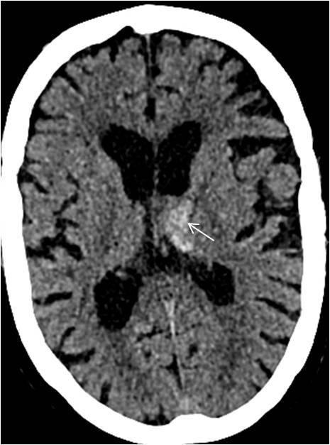
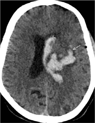
Spontaneous or acute ICH appears on the CT head as an area of hyperdensity within the Parenchyma (Fig 2) or extended into the ventricle (Fig 3), with surrounded hypodense perivascular oedema.
The prompt risk factors affecting 38% of ICH is clot expansion and rebleeding. The investigation of choice for ICH is CT head and CT angiography of the intracranial vessels to exclude the vascular pathology. The vascular abnormalities should always be diagnosed before clot removal, especially for surgeons to deal with vascular malformations. Many signs on imaging help identify clot or active bleeding, eg, the hyperdense signal within the hematoma indicates active bleeding on CECT scans, also known as ‘spot sign’ and other signs like the presence of SAH, hematoma shape and its location to major territorial vessels10.
Such sudden changes in temperature may trigger events of acute Stroke. Temperature variations have been reported as a significant contributor to an increased rate of such Stroke incidences11
Like Blood Pressure, which tends to stay elevated during cold weather and lower in warmer conditions, the chances of Stroke increase in the colder temperature. Additionally, there is an association of
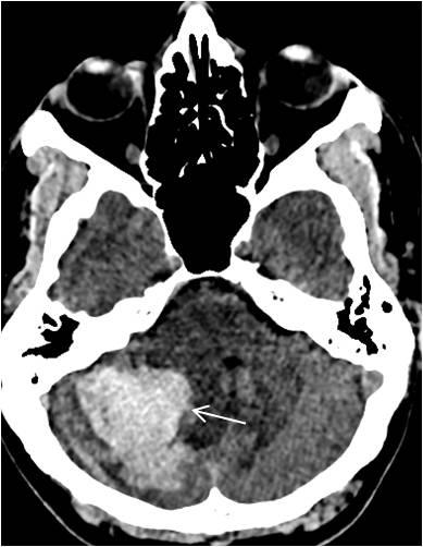
Fig 1 — Axial CT scan of brain at the level of posterior fossa demonstrates an area of high attenuation in the right cerebellum which denotes acute hematoma.(white arrow) Also note the effacement of the adjacent fourth ventricle and compression of the brainstem.
Fig 2 — Computed tomography axial image of head demonstrating an illdefined ovoid, hyperdense focus at left thalamic nucleus level, in keeping with a hematoma. (white arrow)
Fig 3 — Non-contrast axial image of head CT showing acute intracranial haemorrhage involving the left thalamus and basal ganglia(white arrow) with extension into the ventricle. (black arrow)
increased Blood Pressure with increased chances of Stroke, especially during the winters12
Spontaneous, non-traumatic ICH remains a consequential cause of mortality and morbidity worldwide. ICH results from increase Blood Pressure that cause bursting of intracerebral arteries, with majority of mortality or deaths occurring in the first two days of the onset of symptoms. One fifth patients of ICH have neurological deterioration in the pre-hospitalization period and one fourth in hospitalization period. ICH patients present with focal neurological deficits, headache, vomiting, high Blood Pressure and with sudden onset decreased consciousness. But in majority of cases clinical history (hypertension) is needed to reach the diagnosis. CT scan is the gold standard imaging investigation for ICH but in few cases magnetic resonance imaging can be an alternative to differentiate between the chronic and acute stage of haemorrhage13.
So far, the Government focus has been limited to evacuating the affected areas and disaster management. We propose that hypertensive patients be alarmed to take proper medications and regularly monitor their Blood Pressure to keep a strict watch with the weather change warning. Physicians should be advised to take extra care of hypertensive patients. We also feel that more detailed studies should be done worldwide to form a hypothesis to prompt policymakers to form new guidelines.
The environment is changing drastically because of Global Warming that is causing a larger number of natural calamities to happen. Cyclones and heavy rainfalls are becoming more frequent and causing a sudden drop in temperature level, increasing the chances of hypertensive bleeds in chronic untreated cases. Thus, Government should emphasise on making the general public aware about such disasters and possible health problems associated with the same.
On literature review, only a Letter to Editor by Kumar Pradeep, et al in 2015 is found, that described a small group observation was done in All India Institute of Medical Sciences, New Delhi14. In this study, the authors also found highest incidence of ICH in winter seasons.
Seasonal climatic fluctuations cause various changes in vital parameters such as Blood Pressure, leading to increased Stroke chances. Blood Pressure tended to be elevated in colder weather and lowered in warmer conditions. Sudden climate changes cause a sudden spike in Blood Pressure, which reaches beyond the body’s regulatory mechanisms and results in Stroke. These changes become significant in population when predictable climatic changes such as Cyclones or floods occur. As these events are
predictable, measures must be taken quickly to avoid extra health burdens. A health guideline/advisory must be circulated, including avoiding skipping any medication or increasing the doses if required before such predictable natural events. Advisory for the vulnerable population should also include avoiding sudden variation in body temperature by any means.
Prior publication - Nil
Support - Nil
Conflicts of interest - Nil
Permissions - Nil
Acknowledgements - Nil
Funding - Nil
1Krishnamurthi RV, Feigin VL, Forouzanfar MH, Mensah GA, Connor M, Bennett DA, et al — Global and regional burden of first-ever ischaemic and haemorrhagic stroke during 1990–2010: findings from the Global Burden of Disease Study 2010. Lancet Glob Health 2013; 1(5): e259–81. 10.1016/S2214109X(13)70089-5
2Woodhouse P, Khaw KT, Plummer M — Seasonal variation in blood pressure and its relation to ambient temperature in an elderly population. J Hypertens 1993; 11: 1267-74.
3Elliott J, Smith M — The acute management of intracerebral hemorrhage: a clinical review. Anesth Analg 2010; 110(5): 1419-27.
4Aiyagari V — The clinical management of acute intracerebral hemorrhage. Expert Rev Neurother 2015; 15(12): 1421-32.
5Qureshi AI, Tuhrim S, Broderick JP, Batjer HH, Hondo H, Hanley DF — Spontaneous intracerebral hemorrhage. N Engl J Med 2001; 344(19): 1450-60.
6Balami JS, Buchan AM — Complications of intracerebral haemorrhage. Lancet Neurol 2012; 11(1): 101-18.
7Rothwell PM, Wroe SJ, Slattery J, Warlow CP — Is stroke incidence related to season or temperature? The Oxfordshire Community Stroke Project. Lancet 1996; 347(9006): 934-6.
8Zheng, Danni — Low Ambient Temperature and Intracerebral Hemorrhage: The INTERACT2 Study. PloS one vol. 11,2 e0149040. 9 Feb. 2016, doi:10.1371/journal.pone.0149040
9Flower O, Smith M — The acute management of intracerebral hemorrhage. Curr Opin Crit Care 2011; 17(2): 106-14.
10Hemphill JC, Greenberg SM, Anderson CS, Becker K, Bendok BR, Cushman M, et al — American Heart Association Stroke Council. Council on Cardiovascular and Stroke Nursing. Council on Clinical Cardiology. Guidelines for the Management of Spontaneous Intracerebral Hemorrhage: A Guideline for Healthcare Professionals From the American Heart Association/American Stroke Association. Stroke 2015; 46(7): 2032-60
11Khaw KT — Temperature and cardiovascular mortality. Lancet 1995; 345: 337-8.
12MacMahon S, Peto R, Cutler J — Blood pressure, stroke, coronary heart disease. Part I: prolonged differences in blood pressure: prospective observational studies corrected for the regression dilution bias. Lancet 1990; 335: 765-74.
13Kim, Jun Yup, Hee-Joon Bae — Spontaneous Intracerebral Hemorrhage: Management. Journal of Stroke 2017; 19(1): 28-39. doi:10.5853/jos.2016.01935
14Kumar, Pradeep & Kumar, Amit & Pandit, Awadh & Pathak, Abhishek & Prasad, Kameshwar — Seasonal Variations in Stroke: A Study in a Hospital in North India. Journal of Stroke 2015; 17: 219-20. 10.5853/jos.2015.17.2.219.
Introduction : Urinary Tract Infection (UTI) is a common infection and a major health problem. Considering the bacterial resistance developed globally, knowledge regarding sensitivity and resistance pattern of isolated uropathogens in a defined area becomes critically important for choosing appropriate antimicrobial agents for treatment.
Objectives : We conducted this study to detect the common UTI causing microorganisms and to evaluate their culture sensitivity pattern in a Tertiary Care Hospital.
Methods : This retrospective record based observational study was conducted over a period of two months (January and February, 2021). Patients in the General Ward in the Department of General Medicine, Medical College, Kolkata whose urine samples were collected within 48 hours of admission were included. Identification of bacteria was done by standard microbiologic methods and using Kirby disc diffusion test their antimicrobial susceptibility test was performed. The causative organisms for UTI along with its antibiotic sensitivity pattern were retrospectively reviewed and analysed.
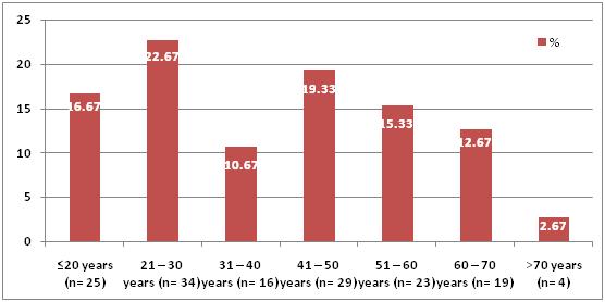
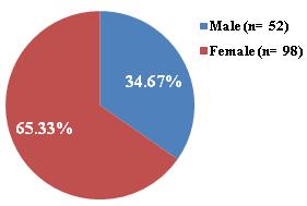
Results : Among 150 culture positive samples 34.67% were from male and 65.33% were from female with highest prevalence in the age group of 21-30 years (22.67%). Most prevalent uropathogens isolated was Escherichia coli (E coli) (60.66%) followed by Enterobactor (21.33%) and Klebsiella (9.33%). E coli showed most sensitivity against ceftazidime, clarithromycin, piperacillin-tazobactam and clindamycin (100% in all cases). Resistance (>70%) of E coli was found against levofloxacin and cefotaxime.
Conclusion : The present study reveals microbiological profile regarding UTI in patients attending our hospital. As resistant to first line antibiotic is increasing, antibiotic stewardship programme should be strengthened. Antibiotic policies agreed among Clinicians, Microbiologists and Pharmacologists will guide good prescribing, provide maximum coverage for treating infections and ensure antibiotic cycling.
[J Indian Med Assoc 2022; 120(10): 19-23]
Key words :Urinary Tract Infection, Antibiotic therapy, Retrospective, Drug-sensitivity pattern.
As treatment failure may occur with commonly used antimicrobials, urinary culture and sensitivity may be considered as a routine investigation in suspected cases of UTI. In this regard timely microbiologic surveillance and assessment of antimicrobial resistance may form an important tool to identify microbial resistance and to limit its spread.
Department of Pharmacology, Medical College and Hospital, Kolkata 700073
1MBBS, MD (Pharmacology), Assistant Professor
2B Tech (Biotechnology), Research Associate, Department of Biotechnology, Tradeshow Operations Organization - Cvent, Haryana 122002
3MBBS, MD (Pharmacology), Professor
4MBBS, MD (Microbiology), Associate Professor, Department of Microbiology, Jalpaiguri Medical College and Hospital, Jalpaiguri 735101
5MBBS, MD (Pharmacology), Assistant Professor, Department of Pharmacology, Calcutta National Medical College & Hospital, Kolkata 700014 and Corresponding Author
Received on : 11/05/2022
Accepted on : 26/07/2022
Urinary Tract Infection (UTI) is a common infection in the community caused by different species of bacteria resulting in very high morbidity. Globally UTI affects about 150 million people per year. This data indicates UTI a major health problem in the community and it may have an adverse impact on World Economy1. UTI may be asymptomatic (subclinical infection) or symptomatic (disease). Thus, the term Urinary Tract Infection encompasses a variety of clinical entities, including Asymptomatic Bacteriuria (ASB), cystitis, prostatitis and pyelonephritis. The distinction between symptomatic UTI and ASB has major clinical implications. In both of the cases bacterial presence in the urinary tract is usually accompanied by urinary white blood cells and inflammatory cytokines2. Lower urinary tract is the usual beginning point of the infection which spreads through the upper urinary tract. Depending upon the selection of therapy UTI may be divided into two classes: uncomplicated and complicated3. Females are more prone to develop UTI compared to males considering the fact that structurally female urethra is not much competent to inhibit the entry of bacteria in the urinary tract4. Factors attributing
to this may be proximity of the urethra and genital tract, urothelial mucosal adherence to the lining by muco-polysaccharide layer, poor and unhygienic practices during menstruation and use of diaphragm for contraceptive purpose.
In most of the cases of uncomplicated UTIs, Escherichia coli (E coli,) the gram-negative bacillus are the causative organisms, other pathogens being Staphylococcusaureus(Saureus),Klebsiellaspp and Proteusmirabilis5 . Presence of 10 5 cfu/mL in midstream urine is considered as significant number of bacteria for UTI6. Effective management of patients suffering from bacterial UTIs commonly relies on the identification of the type of organisms that caused the disease and the selection of an effective antibiotic agent against the organism in question.
However, due to early starting of antibiotic therapy even before the laboratories results are available may result in antibiotic misuse. Global development of antibiotic resistance may be the result of extensive, indiscriminate and inappropriate use of these agents. This has posed a great threat and challenge to the management of UTI. Close monitoring and supervision of uropathogens’ antibiotic susceptibility in a particular area should be done on a regular basis to have the knowledge regarding the antibiotic resistance pattern in UTI.
For the effective selection of empirical antibiotic agents to treat UTI, data supplied by local microbiology laboratories regarding the susceptibility pattern of uropathogens to different antibiotics may be of great help 7 . The patterns of antimicrobial resistance developed in micro-organisms have wide variations. This variation has been found among hospitals as well as among countries. Presently, India lacks any local or national level surveillance program to guide the stakeholders on actual prevalence of resistance8.
In the view of bacterial resistance developed globally with epidemiological significance, physicians should have adequate knowledge regarding microorganisms’ antimicrobial sensitivity and resistance pattern in a certain area for choosing the appropriate antibiotic therapy for treatment of UTI.
However, published literature regarding the susceptibility and resistance pattern of community acquired uropathogens in India is few9. Moreover, to have the adequate knowledge regarding local antibiotic susceptibility pattern of micro-organism, extensive and thorough studies should be conducted in different area. So, we conducted this study to identify the microorganisms commonly cause UTI and to make out the culture sensitivity pattern of those pathogens in a Tertiary Care Hospital in Eastern India.
This retrospective study was conducted in the Department of Pharmacology along with Department of Microbiology, and Department of General Medicine, Medical College & Hospital, Kolkata. Prior to the commencement of the study, approval from Institutional Ethics Committee was taken (Ref No: MC/KOL/IEC/ NON-SPON/796/09/20 dated: 04/09/2020).
Patients admitted in the General Ward in the Department of General Medicine, Medical College & Hospital, Kolkata over a period of two months (January and February 2021), whose urine samples were collected within 48 hours of admission were included in the study. Patients who received antibiotic therapy within 48 hours of admission or patients with known anatomic abnormalities of the genitourinary tract were excluded.
For the purpose of avoiding contamination from urethra, patients were provided adequate instructions regarding collection of urine sample aseptically. Collected samples form the study subjects were clean catch midstream urine. The diagnosis of UTI was based on culture finding of more than 105 organisms (Colony Forming Unit [cfu])/ml. Identification of organisms were done by conventional methods through culturing of samples followed by biochemical tests including their distinct colony characteristics. First culture was observed following inoculation at 37ºC for 16 hours. Using Kirby disc diffusion test the Antimicrobial susceptibility test was performed. ‘Sensitive’ or ‘Resistant’ interpretation was determined depending on the diameters of inhibitory zones of bacterial growth as recommended by the disc manufacturer.
Statistical Methods : For the analysis of the data, Statistical Package for the Social Sciences (SPSS) version 20.0 was used. Qualitative data was presented as frequency and percentage, quantitative data were expressed as percentage.
A total of 395 urine samples from the General Ward in the Department of General Medicine were collected for culture and sensitivity test in the Department of Microbiology. Out of 395 samples, 150 were cultured positive (37.97 %), out of which 52 (34.67%) were from males and 98 (65.33%) were from females (Fig 1). UTI was found to be most prevalent among the age group of 21-30 years (22.67%) (Fig 2).
E coli was the most prevalent uropathogens isolated, the prevalence rate being 60.66%. This was followed by Enterobactor (21.33%), Klebsiella (9.33%), Acinetobacter (3.33%), Pseudomonas (3.33%), Gram positivecocci (0.67%), NonLactoseFermenters(NLF) (0.67%) and S. aureus (0.67%) (Table 1).
Fig 1
Fig
From the antibiotic sensitivity pattern of predominant micro-organisms it was found that Acinetobacter was most sensitive to clarithromycin (100%), followed by amikacin (80%). However, it was resistant to meropenem, ertapenem, amoxyclav, nitrofurantoin, Imipemen and cefotaxime (100% in all cases). E coli showed most sensitivity to clarithromycin, ceftazidime, piperacillin-tazobactam and clindamycin (100% in all cases). Resistance (>50%) of Ecoliwas found against cefotaxime (91.4%), levofloxacin (86.2%), ciprofloxacin (75%), amoxyclav (70%), cefepime (68.1%), amikacin (58.4%) and ertapenem (57.8%).
Enterobactor was most sensitive to vancomycin, linezolid and clarithromycin (100% in all cases). Resistance of Enterobactor was found to be 100% in meropenem, ertapenem, amoxyclav, cefepime, cefotaxime, cefoperazone-sulbactam and piperacillintazobactam. Grampositivecocci were most sensitive to vancomycin, linezolid and nitrofurantoin (100% in all cases). Resistance of Gram positive cocci was found to be 100% in penicillin, amoxycillin, doxycycline and levofloxacin (Table 2A).
Klebsiella was most sensitive to clarithromycin and
Table 1 — Distribution of isolated uropathogens (n=150)
OrganismTotal (n=150)
Acinetobacter 5 (3.33%)
E coli 91 (60.66%)
Enterobactor 32 (21.33%)
Gram positive cocci 1 (0.67%)
Klebsiella 14 (9.33%)
Non lactose fermenters 1 (0.67%)
Pseudomonas 5 (3.33%)
Staphylococcus aureus 1 (0.67%)
clindamycin (100% in both the cases). Klebsiella showed 100% resistance to amoxyclav, cefotaxime and cefoperazone-sulbactam. NLF showed most sensitivity to roxithromycin, levofloxacin, nitrofurantoin, meropenem, ertapenem and ciprofloxacin (100% in all cases). This organism was completely resistant (100%) to amikacin. Pseudomonas was found to be sensitive to cefoperazone-sulbactam and amikacin (100% in both the cases). 100% resistance was shown by this organism to amoxyclav and imipenem. S aureus showed sensitivity to vancomycin, doxycycline, gentamicin, nitrofurantoin, linezolid, cefotaxime and amoxyclav (100% in all cases). This organism was found to be completely resistant (100%) to penicillin (Table 2B)
The present study included the types and antibiotic susceptibility pattern of bacterial organisms isolated from different samples of critically ill patients after 48 hours of admission to identify hospital acquired infections.
In this study, appalling results were obtained about the sensitivity/resistance pattern of microbes to antibiotics. The number of positive isolates was 150 out of 395 samples with an infection rate of 37.97 %. In some other studies conducted in India, prevalence rate of UTI accounted for 34.5%10 and 36.68%11
In our study we found UTI to be highly prevalent in females (65.33%) than in males (34.67%) which is in accordance with the findings of other studies. This may be due to closeness of the anus and urethral meatus as well as females’ shorter urethra4.
We found E coli to be the most predominant isolates (60.66%). This was in accordance with the other studies12
In our study the second most prevalent isolate was Enterobactor (21.33%) followed by Klebsiella (9.33%). However, in several studies Klebsiella was found to be the second most prevalent isolate13. These isolates were tested to find the antimicrobial sensitivity pattern and the pattern was obtained.
Table 2A — Antibiotic sensitivity pattern of predominant micro-organisms isolated from patients Antimicrobial Uropathogens agents Acinetobacter (n=5) E coli (n=91) Enterobactor (n=32) Gram positive cocci (n=1) T*No.(%)S** No.(%)T No.(%)S No.(%)T No.(%)S No.(%)T No.(%)S No.(%)
Vancomycin27(84.4)27(100)1(100)1(100)
Linezolid 28(87.5)28(100)1(100)1(100) Penicillin 27(84.4)2(7.4)1(100)0 (0)
Amoxycillin 23(71.9)1(4.3)1(100)0 (0)
Doxycycline 27(84.4)1(3.7)1(100)0 (0)
Levofloxacin 5(100) 1(20)87 (95.6) 12(13.8)31(96.9)1(3.2)1(100)0 (0)
Amikacin 5(100) 4(80) 89(97.8)37(41.6)5(15.6) 1(20)
Gentamicin 5(100) 3(60)91 (100) 49(53.8)5(15.6) 1(20)
Roxithromycin 4(80)1(25) 48(52.7)32(66.7)5(15.6) 1(20)
Meropenem 4(80)0 (0) 91(100)53(58.2)4(12.5)0 (0)
Ertapenem 5(100) 0(0) 83(91.2)35(42.2)3(9.4)0 (0)
Amoxyclav 1(20)0 (0) 20(21.9) 6(30) 1(3.1)0 (0)
Nitrofurantoin5(100)0 (0)80(87.9)52(65)28(87.5)9(32.1)1(100)1(100)
Cefepime 3(60)0 (0) 22(24.1)7(31.9)1(3.1)0 (0)
Ceftazidime 1(1) 1(100)
Clarithromycin 1(20)1 (100) 15(16.4)15(100)3(9.4)3(100)
Imipemen 1(20)0 (0) 39(42.8)20(51.3)
Cefotaxime1 (20)0 (0) 35(38.4)3(8.6)3(9.4)0 (0)
Cefoperazone-Sulbactam7(7.6)4(57.1) 2(6.2)0 (0)
Piperacillin-Tazobactam 1(1) 1(100)2(6.2)0 (0)
Ciprofloxacin4(4.3) 1(25)
Clindamycin 2(2.1)2(100)
*T= Tested ; **S= Sensitive
Table 2B
Antibiotic
micro-organisms isolated from patients Antimicrobial Uropathogens agents Klebsiella (n=14)
(n=5) S. aureus (n=1)
No.(%)S No.(%)T No.(%)S No.(%)
Vancomycin
1(100)1(100)
1(100)1(100) Linezolid 1(100)1(100) Penicillin 1(100)0 (0) Amoxycillin Doxycycline
Levofloxacin 13(92.8) 3(23)1 (100) 1(100)5(100) 1(20) Amikacin 14(100)4(28.6)1(100)0 (0)5(100)5(100) Gentamicin 14(100)5(35.7)5(100) 4(80) 1(100)1(100)
Roxithromycin 4(28.6) 3(75) 1(100)1(100) 4(80)3(75)
Meropenem 14(100)6(42.9)1(100)1(100)5(100) 3(60)
Ertapenem 13(92.8)2(15.4)1(100)1(100)
Amoxyclav 3(21.4)0 (0)
1(20)0 (0) 1(100)1(100)
Nitrofurantoin12(85.7)2(16.7)1(100)1(100) 2(40)1(50) 1(100)1(100)
Cefepime 5(35.7) 1(20) 5(100) 3(60)
Ceftazidime 5(100) 1(20)
Clarithromycin 4(28.6) 4(100) Imipemen 10(71.4) 4(40)1(20)0 (0)
Cefotaxime 4(28.6)0 (0)1(100)1(100)
Cefoperazone-Sulbactam 1(7.1)0 (0) 1(20) 1(100)
Ciprofloxacin1(100)1(100)
Clindamycin 3(21.4)3(100)
We found E coli to be most sensitive to clarithromycin, ceftazidime, piperacillin-tazobactam and clindamycin (100% in all cases). Resistance (>50%) of Ecoliwas found against cefotaxime (91.4%), levofloxacin (86.2%), ciprofloxacin (75%), amoxyclav (70%), cefepime (68.1%), amikacin (58.4%) and
ertapenem (57.8%).
The fact that micro-organisms show high resistance to fluoroquninolones was suggested by various other works conducted in different parts of the world like Spain14 and India15,16. Indiscriminate and unrestricted use of antibiotics may result this reduced susceptibility.
Another study showed that the driving factor for the development of high resistance of micro-organisms against fluoroquninolones was the physicians’ high prescribing habits of this group of antibiotic17. In the study done by Mostafa, et al18, E coli had a sensitivity rate of 95.2% to cefotaxime in contrast to our study in which cefotaxime was sensitive only in 8.6 % of cases. Extensive use of third generation cephalosporins both as oral and intravenous route may be the reason for increase in resistance in this group of antibiotics.
Compared to the study done by Yolbas, et al19, in which E coli was resistant to amikacin in 3%, nitrofurantoin 9%, in our study E coli showed more resistant pattern to these antibiotics ie, amikacin (58.4%) and nitrofurantoin (35%).
In our study, we found most of the organisms were resistant to a number of antibiotics. Resistance of Enterobacteriaceae, especially E coli and Klebsiella spp, against multiple antibiotics has significantly increased globally considering high use of empiric antimicrobial therapy for treating UTI.
We found Klebsiella to be highly resistant to cephalosporins which was in similarity to a study conducted by Stephanie A, et al20 which showed increased resistant pattern of this micro-organism to third generation cephalosporins in hospital admitted children suffering from UTIs.
Resistance to antibiotics poses a serious and growing problem, because such resistant bacteria are becoming more difficult to treat. The susceptibility data from this study may be worth consideration while implementing empiric treatment strategies for bacterial infections. Avoidance of indiscriminate, unrestricted and empirical use of antibiotics should be followed in order to curtail the emergence and the spread of drug resistance among pathogens.
The authors would like to thank the Department of Medicine and Department of Microbiology, because this study was not possible without their help and cooperation.
Source of Funding : Nil.
Conflict of Interest : Nil.
1Öztürk R, Murt A — Epidemiology of urological infections: a global burden. World J Urol 2020; 38(11): 2669-79
2Powers AC — Diabetes mellitus. In: Kasper DL, Fauci AS, Longo DL, Ameson JL, Loscalzo J, Hauser SL, et al, editors. Harrison’s Principles of Internal Medicine. 20th Edition. New York: The McGraw-Hill Companies, Inc.; 2018.
3Sabra SM, Abdel-Fattah MM — Epidemiological and
Microbiological Profile of Nosocomial Infection in Taif Hospitals, KSA (2010-2011). World J Med Sci 2012; 7(1): 1-9.
4Al-Badr A, Al-Shaikh G — Recurrent Urinary Tract Infections Management in Women: A review. Sultan Qaboos Univ Med J 2013; 13(3): 359-67.
5Blondeau JM — Current issues in the management of urinary tract infections: extended-release ciprofloxacin as a novel treatment option. Drugs 2004; 64(6): 611-28.
6Kass EH — Bacteriuria and the diagnosis of infections of the urinary tract; with observations on the use of methionine as a urinary antiseptic. AMA Arch Intern Med 1957; 100(5): 709-14.
7McNulty CAM, Richards J, Livermore DM, Little P, Charlett A, Freeman E, et al — Clinical relevance of laboratory-reported antibiotic resistance in acute uncomplicated urinary tract infection in primary care. J Antimicrob Chemother 2006; 58(5): 1000-8.
8Wattal C, Goel N, Oberoi JK, Raveendran R, Datta S, Prasad KJ — Surveillance of multidrug resistant organisms in tertiary care hospital in Delhi, India. J Assoc Physicians India 2010; 58 Suppl: 32-6.
9Biswas D, Gupta P, Prasad R, Singh V, Arya M, Kumar A. Choice of antibiotic for empirical therapy of acute cystitis in a setting of high antimicrobial resistance. Indian J Med Sci 2006; 60(2): 53-8
10Dash M, Padhi S, Mohanty I, Panda P, Parida B — Antimicrobial resistance in pathogens causing urinary tract infections in a rural community of Odisha, India. J Family Community Med 2013; 20(1): 20-6.
11Mehta M, Bhardwaj S, Sharma J — Screening of Urinary Isolates for the Prevalence and Antimicrobial Susceptibility of Enterobacteria Other Than Escherichia Coli. Int J Life Sci Pharma Res 2013; 3(1): 100-4.
12Yismaw G, Abay S, Asrat D, Yifru S, Kassu A — Bacteriological profile and resistant patterns of clinical isolates from pediatric patients, Gondar University Teaching Hospital, Gondar, Northwest Ethiopia. Ethiop Med J 2010; 48(4): 293300.
13Haghi-Ashteiani M, Sadeghifard N, Abedini M, Soroush S, Taherikalani M — Etiology and antibacterial resistance of bacterial urinary tract infections in Children’s Medical Center, Tehran, Iran. Acta Medica Iranica 2006; 45(2): 153-7.
14Gobernado M, Valdés L, Alós JI, García-Rey C, Dal-Ré R, García-de-Lomas J — Antimicrobial susceptibility of clinical Escherichia coli isolates from uncomplicated cystitis in women over a 1-year period in Spain. Rev Esp Quimioter 2007; 20(1): 68-76
15Sood S, Gupta R — Antibiotic Resistance Pattern of Community Acquired Uropathogens at a Tertiary Care Hospital in Jaipur, Rajasthan. Indian J Community Med 2012; 37(1): 39-44.
16Sabharwal ER. Antibiotic susceptibility patterns of uropathogens in obstetric patients. N Am J Med Sci 2012; 4(7): 316-9 .
17Kahlmeter G — An International Survey of the Antimicrobial Susceptibility of Pathogens from Uncomplicated Urinary Tract Infections: the ECO.SENS Project. J Antimicrob Chemother 2003; 51(1): 69-76.
18Sharifian M, Karimi A, Tabatabaei SR, Anvaripour N — Microbial sensitivity pattern in urinary tract infections in children: a single centre experience of 1,177 urine cultures. Jpn J Infect Dis 2006; 59(60): 380-2.
19Yolbas I, Tekin R, Kelekci S, Tekin A, Okur MH, Ece A, et al — Community-acquired urinary tract infections in children: pathogens, antibiotic susceptibility and seasonal changes. Eur Rev Med Pharmacol Sci 2013; 17(7): 971-6
20Lutter SA, Currie ML, Mitz LB, Greenbaum LA — Antibiotic resistance patterns in children hospitalized for urinary tract infections. Arch Pediatr Adolesc Med 2005; 159(10): 924-8.
Background : Coronavirus is a highly infectious novel virus we are in urge to know more about their clinical characteristics and laboratory findings for the characterization and selection of treatment protocol.
Methods : Prospective, single centre study. Two months data was collected, clinical characteristics data from patient case sheet and the laboratoryvalues from the Hospital Information System (HIS) for the month of July and August 2020.
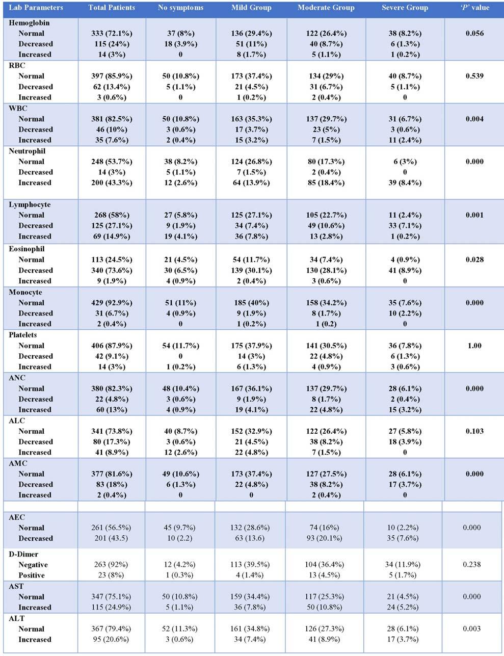
Results : Of 462 patients, 55 (11.9%) are falls under asymptomatic category, 194 (42%) are in mild category, 167 (36.1%) are in moderate category and 46 (10%) in severe category. Fever 230 (49.8%) and cough 211 (45.7%) was most common clinical symptom with p value < 0.01. Non-severe vs severe, 340 (73.6%) and 201 (43.5%) showed decreased in eosinophil count and absolute eosinophil count, 125 (27.1%) and 80 (17.3%) patient showed decrease in lymphocyte count and absolute lymphocyte count, 200 (43.3%) showed increase in neutrophil count with a significance of p value >0.05. 186 (40.3%) patients had one or more co-morbidities. Laboratory findings between Asymptomatic VS symptomatic, showed significance changes in neutrophil, lymphocyte, Aspartate aminotransferase, Alkaline phosphatase, globulin values (p value <0.05).
Conclusion : Clinical severity categorization at the time of admission was very helpful for the treating doctors in proper understanding of disease progression and appropriate treatment of the patient. Presence of co-morbidity, abnormal laboratory values, old age group patients, higher Computed Tomography score, higher mortality rate are seen more in patients who were in clinical severity grade severe category than in non-severe category patients.
[J Indian Med Assoc 2022; 120(10): 24-30]
Key words :COVID-19, Laboratory findings, Clinical severity, Mortality, Computed Tomography.
Currently, the World is in the stage of childhood in understanding of novel coronavirus (COVID-19) in this prevailing pandemic situation. Since the signs and symptoms of this novel virus was non-specific1 and similar to other viral infections, it is important to correlate the laboratory values with the clinical features of the Coronavirus infected patients for better understanding of the disease progression.Although COVID-19 has various clinical manifestations, most patients had no symptoms or mild symptoms, especially in the early disease stage2. The average incubation period of COVID-19, extending from exposure to onset of disease symptoms is estimated at approximately 5.2 days3. Laboratory medicine
Melmaruvathur Adhiparasakthi Institute of Medical Sciences and Research, Melmaruvathur, Tamil Nadu 603319
1MD (General Medicine), Associate Professor, Department of General Medicine
2MD (Immunohematology & Blood Transfusion), Assistant Professor, Department of Immunohaematology and Blood Transfusion and Corresponding Author
3MD (Microbiology), Professor, Department of Microbiology
4MD (General Medicine), Professor, Department of General Medicine
5MD (Anaesthesia), Professor, Department of Anaesthesia
6MD (Biochemistry), Professor, Department of Biochemistry
Received on : 09/03/2022
Accepted on : 19/06/2022
Editor's Comment :
Proper categorisation based on clinical severity at the time admission along with basic laboratory tests plays vital role in the patient management.
plays an essential role in diagnosing and managing this COVID-19 even in the early stage of infection4 Various studies published the reports of Complete Blood Count (CBC) in COVID-19 patients with contradictory results as leukopenia, leukocytosis, and lymphopenia5,6, which can influence the outcomes of COVID-19 patients So on correlating clinical features and basic laboratory investigation of COVID-19 cases in this study will be helpful for doctors to understand the changes happening in different clinical severity and also helps in diagnosis, categorization and appropriate treatment protocol of the patient by adding more novel information with the previous existing data.
It was a prospective descriptive study conducted at Melmaruvathur Adhiparasakthi Institute of Medical Sciences and Research, Melmaruvathur. The data was collected from the patients, for whom COVID-19 infection was confirmed by PCR test and admitted in our institutional COVID-19 wing in the months of July
T Ramesh1, T Ramanathan2, G Hemalatha3, S Appendraj4, C Prasath5, S Sakthidasan6
and August, 2020 and the patient data was collected after obtaining the approval from our institutional Ethical Committee. Patients clinical data was collected from case sheets and the laboratory data are extracted from the HIS.No specific interventions are utilized to conduct the study. Both clinical and laboratory data are entered in an enrolled patient’s data collection sheet and entered in the SPSS software for statistical analysis. Patients were categorized into non-severe (asymptomatic, mild, moderate) and severe group at the time admission based on clinical severity guidelines prepared in our institution based on National Guidelines (ICMR & MoH&FW/DGHS, Clinical Management Protocol: COVID-19, version 4, 27.06.2020)7. Another correlation was done between Asymptomatic and Symptomatic (mild, moderate, severe) groups. Informed consent was obtained from the patient or from their relatives for using their clinical and laboratory data for research purpose.
All statistical analysis was performed using SPSS statistical software version 21.0, IBM. The categorical variables were described as frequency and percentages and the continuous variables were described using Mean, Median and Interquartile Range (IQR). Normally distributed data were analysed by independent sample t test; for non-parametric values the Mann-Whitney U test was used. The P values <0.05 were considered as statistically significant.
Totally 462 patients were enrolled, among which 279 (60.4%) were males and 183 (39.6%) were females. Among study participants, 30 (6.5%), 143 (31%), 198 (42.8%) and 91 (19.7%) patients were aged <18 years, 18 to 40 years, 41 to 60 years, > 60 years, respectively (Median Age 46, IQR 35-58).
The clinical presentation and clinical parameters analyzed in our study were noticed on the day of admission only. The most common clinical symptoms were fever in 230 (49.8%), cough in 211 (45.7%), dyspnea in 121 (26.2%), sore throat in 70 (15.2%) and body pain in 54 (11.7%) which was statistically significant with p value <0.01. Out of 462 patients, 186 (40.3%) had one or more co-morbidities. Most common was diabetes mellitus 141 (30.5%) and hypertension 79 (17.1%). Of 462 patients, 55 (11.9%) are falls under asymptomatic category, 194 (42%) are in mild category, 167 (36.1%) are in moderate category and 46 (10%) in severe category (Table 1).
Laboratory parameters and their significance of nonsevere versus severe groups and Asymptomatic versus symptomatic refer (Tables 2&3). Laboratory parameters and their significance between among 55
asymptomatic individuals, only 4 (7.8%) belongs to >60 years of age and majority 38 (70%) individual in this group are belongs to <40 years. Duration of hospital stay was also less ie, 23 (42%) had discharged within 1 to 7 days of admission and importantly no one in this group had any co-morbidity.
Computed Tomography (CT) imaging studies has been done for 219 (47.4%) patients, among which 113 (51.6%) revealed CT severity score between 1 to 12, 57 (26%) revealed CT severity score between 13 to 19 and 49 (22.4%) revealed CT severity score >20 (scoring was done based on study by Yang R, et al8. Of 49 patients in severe CT score, majority ie, 43 are in severe category shows, greater the CT score greater the clinical severity and lower CT score patients are more seen in mild clinical severity category.
In 398 patients had mild oxygen saturation drop ie, SpO2 <94, 31 (6.7%) had SpO2 between 90-93% and 33 (7.1%) had SpO2 drop <89%.
Considering respiratory rate, it was significantly affected (p value <0.01), 118 (25.5%) patients had mildly increased rate (19 to 23 beats /min), 228 (49.4%) had moderately increased rate (24 to 30 beats /min) and 78 (16.9%) had rate >30 beats/min. Heart rate and Blood Pressure showed no significant changes.
Majority, 346 (74.9%) patients stayed in Hospital for 8 to 14 days, 92 (20%) patient stayed 1 to 7 days, 22 (4.7%) stayed 15 to 22 days and 2 (0.4%) stayed 22 to 28 days. Median duration of stay 10 days, IQR 3. 22 (4.7%) patients required ICU admission in our study population at the time of admission.
In our study, Male patient was 279 (60.4%) and female was 183 (39.6%) though there was slight male predominance there was no significant difference in clinical severity between gender. Similar findings were seen in a study by Wang D, et al gender was not a risk factor for the disease severity9
The most common clinical presentation and clinical parameters noticed in our study was fever [230 (49.8%)], cough [211 (45.7%)] and dyspnea [121 (26.2%)]. Similarly, in a study by Junli Li, et al, found fever [29 (78%)], dry cough [28 (76%)] and dyspnea [9 (57%)] was most common clinical presentation10 but in another study by Mohan A, et al, found cough [31 (34.7%)] was most common followed by fever [25 (17.4%)] and nasal symptoms [31 (21.5%)]11
Out of 462 patients, 55 (11.9%) are falls under asymptomatic category, 194 (42%) are under mild category, 167 (36.1%) are under moderate category and 46 (10%) under severe category. Mohan A, et al in his analyzes reported among 144 patients, 140 (97.2%)
Clinical ParametersAll Patients‘p’ value Total No (%)
Demographic Characteristics : Age (years)
< 1830 (6.5)
18 – 40143 (31)
41 – 60198 (42.3) 0.000 > 6091 (19.7)
Gender : Male279 (60.4)
Female 183 (39.6)
Signs & Symptoms :
Fever 230 (49.8) 0.000
Cough211 (45.7) 0.000
Sore Throat70 (15.2) 0.001
Dyspnea 121 (26.2) 0.000
Body Pain54 (11.7) 0.004
Diarrhea20 (4.3) 0.085
Expectoration 32 (6.9) 0.031
Headache16 (3.5) 0.135
Loss of Taste28 (6.1) 0.045
Loss of Smell12 (2.6) 0.699 Tremor1 (0.2) 0.713
Comorbidities : No of cases = 186 (40.3%)
Diabetes Mellitus (DM)81 (17.5)
Hypertension (HTN)24 (5.2)
DM + HTN43 (9.3)
Cardiac Diseases2 (0.4)
DM + Cardiac Diseases7 (1.5)
DM + HTN + Cardiac Diseases8 (1.7)
Cancer2 (0.4)
HTN + Cardiac Diseases3 (0.6) 0.012
Thyroid Diseases7 (1.5)
Bronchial Asthma4 (0.9)
Seizure Disorders1 (0.2)
Hypercholesterolemia1 (0.2)
DM + HTN + CKD1 (0.2)
DM + Cardiac Diseases + CKD1 (0.2)
Tuberculosis1 (0.2)
falls in mild to moderate disease and remaining 4 (2.8%) falls under severe category11. Similarly, in another research by SakikoTabata, et al reported total of 104 patients, 43 are classified asymptomatic, 41 (39%) had mild COVID-19 and 20 (19%) had severe COVID-1912
SakikoTabata, et al noticed that the patients in the severe group are mostly older than those in the mild group12. In our survey also we had 92 (19.9%) patients with the age >60 years, of which 47 (51.1%) falls in moderate and 16 (17.3%) patients fall in severe group at the time of admission. Remaining, 25 (27%) patients falls in mild group and only 4 (4.3%) of patients falls in asymptomatic group. So it shows that patient who ages > 60 years are mostly had symptoms and they mostly fall in moderate and severe group (Fig 1). Interestingly, in another study by Soysal A, et al conducted in Turkey on children’s found that the rate of symptomatic cases increases with age increases (p=0.049) ie, <11% in children <1 year, 19% in children
Clinical ParametersAll Patients‘p’ value Total No (%)
Clinical Severity :
Asymptomatic55 (11.9)
Mild 194 (42)
Moderate167 (36.1) Severe46 (10)
Vitals
Systolic Blood Pressure : Normal 152 (32.9) Mild 302 (66)0.001 Severe5 (1.1)
Heart Rate : Normal 441 (95.5)0.085 High21 (4.5)
Respiratory Rate (per minute) : Normal38 (8.2)
Mild118 (25.5)0.000
Moderate228 (49.4) Severe78 (16.9)
SpO2 : Mild 398 (86.1)
Moderate31 (6.7)0.002 Severe33 (7.1)
Temperature : Normal 432 (93.5)0.038 High30 (6.5)
Cause of Death: Total = 14 (3%) cases : COVID Pneumonia14 Kidney Diseases12 Diabetes Mellitus11 Systemic Hypertension4 Cardiac Diseases3 Sepsis2 Bronchial Asthma1
<5 years and 36% in children >5 years13
From our investigation non-severe VSsevere,340 (73.6%) and 201 (43.5%) patients showed decrease in eosinophil count and absolute eosinophil count. similarly, in a study by Hu Yun, et al reported, 21 (66%) and 24 (75%) patients had decrease in eosinophil count and their proportions and explained this might be due to the early stage of infection so the decline of eosinophils is faster14. Likewise, our study participants might have got admitted at the early stage of disease since the majority had eosinophils counts at the lower side.
In our study we have found, there was decreased lymphocyte count in 125 (27.1%) and absolute lymphocyte count in 80 (17.3%) and decreased albumin level in 90 (19.5%). Similarly, in a study by Hu Yun, et al, found that among 32 patients with COVID-19, 15 (47%) and 16 (50%) patients showed decreased lymphocyte count and lymphocyte ratio, 21 (66%) and contrastly increased albumin level14
Weiliang Cao, et al also reported that Lymphocytes counts are significantly (P < 0.01) lower in severe group than non-severe groups15.
We also observed that decreased Haemoglobin in 115 (24%) and Red Blood Cells (RBC) in 62 (13.4%) Similarly, Xuemei Liu, et al figured out there was decrease in Haemoglobin in 40% and RBC in 39%16.
Further we noticed, Liver function test values are significantly elevated, AST in 115 (24.9%), ALT in 95 (20.6%), Direct Bilirubin in 258 (55.8%), LDH in 140 (77.3%) and C-reactive Protein (CRP) is increased in 56 (12.1%). Total Bilirubin and Indirect Bilirubin are not affected. Similarly, SakikoTabata, et al, in their study noticed that there was increased AST in 4 (9%), ALTin 5 (12%) and LDH in 9 (21%)12. Weiliang Cao, et alpublished that CRP, ALT and AST levels are increased significantly (P<0.01) in severe group patients15
On comparing Laboratory parameters of asymptomatic versus symptomatic, neutrophils, lymphocytes, AST, ALT, ferritin are increased
significantly (p<0.01) and albumin are decreased significantly (p<0.01)in symptomatic patients. CRP was increased in all symptomatic patients and it was negative in all asymptomatic patients. LDH increased in most of the patients. Supporting our findings, Li Y, et al published the symptomatic patients had a significantly higher Lymphocyte count than asymptomatic patients (P = 0.03)17. In Contrast, studies in children’s showed decreased Lymphocytes and LDH was raised 13 Leucocyte, eosinophil, monocyte, Aspartate Aminotransferase (AST), total bilirubin, total protein, albumin, ferritin counts are affected significantly in symptomatic individuals. But these were significantly affected in severe groups (p value <0.05). Among Asymptomatics, 19 (4.1%) and 28 (5.6%) showed increase in Lymphocyte count and direct bilirubin level, 30 (6.5%) showed decreased count which was minimal number and not statistically significant.
In our survey, out of 462 patients, 186 (40.3%) had one or more co-morbidities. In which the most common
was Diabetes Mellitus 141 (30.5%) and hypertension 79 (17.1%). Similarly, in a studies by Mammen JJ, et alalso reported that diabetes 43.5% was most common co-morbidity18 and Mohan, et al reported 23 (15.9%) out of 144 study participants had co-morbidity, in which 16 (11.1%) are diabetic was the common11 and it was similar to other published studies1,9,19
Junli Li, et al published that, the patients in the death group are mostly older (p=0.002), had higher incidence of hypertension (p=0.045), coronary disease (p=0.002) and dyspnea (p=0.020) at the time of admission11. Mortality rate in our study was 14 (3%), of which 8 falls in severe group and 6 falls in moderate group. No death was reported in Mild and Asymptomatic group in our study. 5 out of 14 death patients had CT score of severe grade and 11 of them
had co-morbidities. In a study by Mohan A, et al, reported death rate of 1.4% ie, 2 of 144 patients and both were belonged to severe group11
Of 14 expired patients, 11 had co-morbidity and all 11 had Diabetes Mellitus has a co-morbidity along with other disease association (Table 1). Likewise, in study by, Acharya, et al published that, higher mortality was seen among the diabetes than non-diabetic patients (20% versus 4.8%) among COVID-19 patients20, but in contrast Mammen JJ, et al noted that presence of diabetes was not significantly different between survivors and non-survivors (42.5% versus 49.2%, p=0.310)18 This shows that death rate was higher in a patient with co-morbidity especially Diabetes Mellitus.
Many studies concluded that older age, comorbidity association, higher CT score, Lymphopenia
are major factors for risk factors for disease progression and morbidity in severe group with p<0.0112,19 because of their poor immune response.
Clinical severity categorization along with laboratory findings guides treating physicians to decide specific treatment protocol for every single patient promptly. Another important finding from our study was patient who falls in severe category are aged >60 years, comorbidity association and higher CT score than in asymptomatic, mild and moderate category (non-severe) patients When comes to asymptomatic and symptomatic individuals, not much derangements seen in asymptomatic, this might be because of more number of younger age group and nil co-morbidity makes them asymptomatic. So from this point of view, patients with older age group and co-morbidity should be given extra care in their management.
The American Journal of the Medical Sciences 2020; 360(3): 229-35.
11Mohan A, Tiwari P, Bhatnagar S, Patel A, Maurya A, Dar L, et al — Clinico-demographic profile & hospital outcomes of COVID-19 patients admitted at a tertiary care centre in north India. The Indian Journal of Medical Research 2020; 152(12): 61.
1Guan WJ, Ni ZY, Hu Y, Liang WH, Ou CQ, He JX, et al — Clinical characteristics of coronavirus disease 2019 in China. New England Journal of Medicine 2020; 382(18): 1708-20.
2Alhazzani W, Møller MH, Arabi YM, Loeb M, Gong MN, Fan E, et al—Surviving Sepsis Campaign: guidelines on the management of critically ill adults with Coronavirus Disease 2019 (COVID19). Intensive Care Medicine 2020; 46(5): 854-87.
3Alamdari DH, Moghaddam AB, Amini S, Alamdari AH, Damsaz M, Yarahmadi A — The application of a reduced dye used in orthopedics as a novel treatment against coronavirus (COVID19): a suggested therapeutic protocol. Archives of Bone and Joint Surgery 2020; 8(suppl1): 291.
4Pourbagheri-Sigaroodi A, Bashash D, Fateh F, Abolghasemi H — Laboratory findings in COVID-19 diagnosis and prognosis. ClinicaChimica Acta; International Journal of Clinical Chemistry 2020; 510: 475.
52020. Centers for Disease Control and Prevention. Coronavirus Disease 2019 (COVID-19)https://www.cdc.gov/ coronavirus/2019-ncov/hcp/clinical-guidance-managementpatients.html.
6Tan L, Wang Q, Zhang D, Ding J, Huang Q, Tang Y — Lymphopenia predicts disease severity of COVID-19: a descriptive and predictive study. Signal Transduct Tar Ther 2020; 5(1).
7Clinical Management Protocol: COVID-19.Government of India Ministry of Health and Family Welfare/Directorate General of Health Services. version 4, 27.06.2020.
8Yang R, Li X, Liu H, Zhen Y, Zhang X, Xiong Q, et al — Chest CT severity score: an imaging tool for assessing severe COVID-19. Radiology: Cardiothoracic Imaging 2020; 2(2): e200047.
9Wang D, Hu B, Hu C, Zhu F, Liu X, Zhang J, et al — Clinical characteristics of 138 hospitalized patients with 2019 novel coronavirus–infected pneumonia in Wuhan, China. JAMA 2020; 323(11): 1061-9.
10Li J, Xu G, Yu H, Peng X, Luo Y — Clinical characteristics and outcomes of 74 patients with severe or critical COVID-19.
12Tabata S, Imai K, Kawano S, Ikeda M, Kodama T, Miyoshi K, et al — Clinical characteristics of COVID-19 in 104 people with SARS-CoV-2 infection on the Diamond Princess cruise ship: a retrospective analysis. The Lancet Infectious Diseases 2020; 20(9): 1043-50.
13Soysal A, Gönüllü E, Arslan H, Kibar BS, Pop S, Yurttaº GN, et al — Comparison of clinical and laboratory features and treatment options of 237 symptomatic and asymptomatic children infected with SARS-CoV-2 in the early phase of the COVID-19 pandemic in Turkey. Japanese Journal of Infectious Diseases 2020: JJID-2020.
14Yun H, Sun Z, Wu J, Tang A, Hu M, Xiang Z — Laboratory data analysis of novel coronavirus (COVID-19) screening in 2510 patients. Clinica Chimica Acta 2020; 507: 94-7.
15Cao W — Clinical features and laboratory inspection of novel coronavirus pneumonia (COVID-19) in Xiangyang, Hubei. MedRxiv. 2020 Jan 1.
16Liu X, Lv J, Gan L, Zhang Y, Sun F, Meng B, et al — Comparative analysis of clinical characteristics, imaging and laboratory findings of different age groups with COVID-19. Indian Journal of Medical Microbiology 2020; 38(1): 87-93.
17Li Y, Shi J, Xia J, Duan J, Chen L, Yu X, et al — Asymptomatic and symptomatic patients with non-severe coronavirus disease (COVID-19) have similar clinical features and virological courses: a retrospective single center study. Frontiers in Microbiology 2020; 11: 1570.
18Mammen JJ, Kumar S, Thomas L, Kumar G, Zachariah A, Jeyaseelan L, et al — Factors associated with mortality among moderate and severe patients with COVID-19 in India: a secondary analysis of a randomised controlled trial. BMJ Open 2021; 11(10): e050571.
19Chen N, Zhou M, Dong X, Qu J, Gong F, Han Y, et al — Epidemiological and clinical characteristics of 99 cases of 2019 novel coronavirus pneumonia in Wuhan, China: a descriptive study. The Lancet 2020; 395(10223): 507-13.
20Acharya D, Lee K, Lee DS, Lee YS, Moon SS — Mortality rate and predictors of mortality in hospitalized COVID-19 patients with diabetes. In Healthcare 2020; 8(3): 338. Multidisciplinary Digital Publishing Institute.
Background : Coronavirus disease 2019, first reported in December 2019 mainly presented with the symptoms of Cough, Fever, Shortness of breath, Myalgia, Weakness and anosmia. C-reactive Protein (CRP) is an acute-phase reactant protein which is synthesized by the liver in response to raised levels of interleukin-6 (IL-6) which is a biomarker of inflammation.
Methods : This was a prospective observational study, done on 110 COVID-19 patients after applying inclusion and exclusion criteria. Detailed history, vaccination status, presence of comorbidities and thorough clinical examination was performed. Serum CRP levels was assessed and Computed Tomographic scan (CT scan) of Thorax was done. CORADS scoring and CT severity grading as per CT scan was done. All the above parameters were recorded in the preformed proforma and data was entered in excel spreadsheet and was analysed using SPSS v26 software.
Results : Majority were males (56.3%) and majority were from 61-80 years of age. Majority (57.3%) patients were non-smokers. Hypertension was the most common associated comorbidity (86.4%)(r=0.743, p=0.000). There is a strong positive correlation between CRP levels and CTSS in COVID 19 patients and a strong negative correlation between the CRP levels and outcome of COVID-19 patients (r= -0.449, p=0.000).
Conclusion : Elevated serum CRP value is associated with disease progression and poorer outcome.
[J Indian Med Assoc 2022; 120(10): 31-3]
Key words :Acute phase reactant, COVID-19, Severe COVID, CORADS.
Coronavirus disease 2019, also known as COVID19 was first reported in Wuhan, China, in December, 2019. Patients of COVID-19 mainly present with the symptoms of cough, fever, shortness of breath, myalgia, weakness and anosmia 1 . Cases are diagnosed based on Nucleic Acid Amplification by RTPCR test from oropharyngeal or nasopharyngeal swab. High Resolution Computed Tomography (HRCT) scan of thorax has high sensitivity in detecting COVID19 among people2.
The hallmark feature of Severe Acute Respiratory Syndrome Coronavirus-2 (SARS-CoV-2) infection is the systemic inflammatory response to COVID-19 and most hospitalized patients with COVID-19 have abnormal inflammatory biomarkers3. C-reactive Protein (CRP) is an acute-phase reactant protein which was first described by Tillet and Francis and is synthesized
Department of Respiratory Medicine, Burdwan Medical College and Hospital, Burdwan 713104
1MBBS, MD (Respiratory Medicine), Associate Professor
2MBBS, MD (Respiratory Medicine), Professor & Head
3MBBS, MD (General Medicine), FICP, Associate Professor, Department of General Medicine, Murshidabad Medical College and Hospital, Berhampore, 742101 and Corresponding Author
4MBBS, Postgraduate Resident
Received on : 25/08/2022
Accepted on : 08/09/2022
Editor's Comment :
There is an association between inflammatory biomarker, serum CRP and the chest CT severity scores among COVID 19 patients. Elevated serum CRP value on admission is associated with severe disease and poorer outcome. In these patients, prompt treatment and optimal monitoring is mandatory to reduce morbidity and mortality.
by the liver in response to raised levels of interleukin6 (IL-6), a biomarker of inflammation3. Elevated CRP concentrations are seen in cardiovascular diseases like Myocardial Infarction, Acute Kidney Injury (AKI), inflammatory rheumatic diseases such as rheumatoid arthritis and gout, and with incident Venous ThromboEmbolism (VTE)3. C-reactive Protein has also been previously associated with severe disease in patients with H1N1 influenza pneumonia4
Previous studies have shown that there is a good correlation between CT Severity Scores (CTSS), severity of clinical disease and blood CRP values among patients diagnosed with COVID-19. Since CRP is an acute phase reactant hence its values increase greatly during inflammation and indicate the severity of disease. With an increase in inflammation there is also activation of the coagulation cascade in the body leading to formation of microthrombi as identified by
Correlation between C-reactive Protein (CRP) Level and Clinicoradiological Profile in COVID-19 Patients Admitted in a Tertiary Care Hospital in Eastern India
Anirban Das1, Santanu Ghosh2, Atanu Roy Chowdhuri3, Pronoy Sen4, Hrishikesh Barui4, Preetam Goswami4
postmortem studies1,5.
In low resource areas like our country, CRP can used as a substitute for CTSS for determining the severity of the disease. Hence, it was decided to carry out this study to find out the utility of serum CRP as a marker of severity of COVID-19 disease as well as the distribution pattern of the disease in this part of Eastern India.
This study was a prospective observational study on 110 COVID 19 patients which was conducted after approval from Institutional Ethics Committee (IEC). Participants who gave consent for the study and were haemodynamically stable were included in the study. Detailed history, vaccination status, presence of comorbidities and thorough clinical examination was performed with relevant biochemical and radiological investigations. Serum CRP levels was assessed for all patients. Computed Tomographic scan (CT scan) of Thorax was done. CORADS scoring and CT severity grading as per CT scan was done. Duration of Hospital stay and clinical outcome of the patient was assessed. All the above parameters were recorded in the preformed proforma after taking written consent from all patients/relatives. Data was entered in excel spreadsheet and was analysed using SPSS v26 software. P-value of <0.05 was considered as statistically significant and Pearson correlation have been used to show correlation between various parameters.
This study was conducted after getting approval from Institutional Ethics Committee (IEC).
Out of 110 patients, 62 (56.3%) were males and 48 (43.6%) were females. 63 (57.2%) were from 61-80 years of age. Most of the male (64.5%) and female patients (68.8%) who were hospitalized were above 60 years of age (Table 1).
In 63 (57.3%) patients were non-smokers while 47 patients (42.7%) were smokers (Table 2).
Hypertension was the most common associated comorbidity seen in 95 (86.4%) patients, followed by Type 2 Diabetes Mellitus in 75 (68.2%) patients (Table 3).
Mean CRP levels were 91.25±32.948 mg/dl while mean CTSS was found to be 16.24±6.486. There is a strong positive correlation between CRP levels and CTSS in COVID-19 patients and is statistically significant (r=0.743, p=0.000) (Table 4).
Table 1— Distribution of COVID patients according to age and gender
AgeGender Male Female No of PatientsPercentage No of PatientsPercentage Below 210 0.0%00.0% 21-4058.1%12.1% 41-601727.4%1429.2% 61-803658.1%2756.2%
814 6.5%612.5%
Table 2 — Smoking status of COVID patients
Smoking StatusNo. of PatientsPercentage
Table 3 — Distribution of co-morbidities of COVID patients
ComorbiditiesNo of PatientsPercentage
Hypertension : Present95 86.4 Absent1513.6
Type 2 Diabetes : Present75 68.2 Absent3531.8
Ischemic Heart Disease : Present38 34.5 Absent7265.5 Chronic Respiratory Disease : Present20 18.2 Absent9081.8
Table 4 — Correlation between CRP levels and CT Severity score
ParametersMean Value ± SDr valuep value CRP levels 91.25±32.9480.7430.000 CT Severity Score16.24±6.486
There is a strong negative correlation between the CRP levels and outcome of COVID-19 patients and is statistically significant. (r= -0.449, p=0.000) (Table 5)
The current study explores the benefit of using serum CRP value as an indicator of severity of COVID19 disease. When there is clinical suspicion of COVID19 with initial negative RT-PCR testing, WHO had advised the use of chest imaging as diagnostic tool. CT scan is an appropriate means to assess individual disease severity6. In the current study a previously validated score based on lobar extent of the disease as reported by Pan, et al has been adopted to quantify the disease severity7. The liver synthesizes various acute phase proteins like CRP. Serum CRP levels
Table 5 — Correlation between CRP levels and Outcome
valuep value
levels -0.4490.000 Outcome
increase during inflammatory responses. It is stated by Liu, et al that severe cases of COVID-19 exhibited higher levels of CRP8
In the present study, age of most of the patients is above 60 years (57.2%). Comparable findings were observed in studies by Wang, et al and Francone, et alin which the study population comprised of a relatively older age9,6. Existing literature has mentioned that presence of comorbidities such as Hypertension, diabetes, Coronary Artery Diseases and Chronic Lung diseases makes the prognosis grim6,10. In our study the most prevalent comorbidity was hypertension similar to the study by Guan, et al in which hypertension followed by diabetes were the two most frequent comorbidity among the patients. In our study, majority of the patients were non-smokers in accordance with the finding of low prevalence of smokers among patients hospitalized with COVID-19 in a multicentred study by Meini, et al11
In the current study there is statistically significant correlation between serum CRP levels and Chest CT severity scores (p<0.0001, r=0.743), which is consistent with the findings of the research by Saeed et al (p<0.0001, r=0.556) and Francone, et al (p<0.0001, r= 0.6204)6,7. Thus in centres which are lacking CT facility, serum CRP value of the patient can be utilized to anticipate severe COVID-19 disease and initiate prompt management such as intravenous steroid at the earliest. In the present study serum CRP value is significantly negatively correlated to outcome of COVID-19 patients, which is equivalent to the finding of significant increase inflammatory markers like CRP in more severe COVID-19 illness in a study by Hachim, et al12,13
The current study takes into account a short sample size. So, the results cannot be universalized. Also, multiple serum CRP value measurements at regular intervals could be done to augment the findings of this study.
To conclude with, the current study establishes an association between inflammatory biomarker namely serum CRP and radiological parameter particularly the Chest CT severity scores among COVID-19 patients. Elevated serum CRP value is associated with disease progression and poorer outcome. Thus, careful monitoring of COVID-19 patient is mandatory to avert mortality. Future research should be directed to consider a large sample size and to obtain serial data for analysis.
1Wiersinga WJ, Rhodes A, Cheng AC, Peacock SJ, Prescott HC. Pathophysiology, Transmission, Diagnosis, and Treatment of Coronavirus Disease 2019 (COVID-19): A Review. JAMA 2020; 324(8): 782-93. doi:10.1001/jama.2020.12839
2Ai T, Yang Z, Hou H, Zhan C, Chen C, Lv W, Tao Q, Sun Z, Xia L — Correlation of Chest CT and RT-PCR Testing for Coronavirus Disease 2019 (COVID-19) in China: A Report of 1014 Cases. Radiology 2020; 296(2): E32-E40. doi: 10.1148/ radiol.2020200642. Epub 2020 Feb 26. PMID: 32101510; PMCID: PMC7233399.
3Smilowitz NR, Kunichoff D, Garshick M, Shah B, Pillinger M, Hochman JS, Berger JS — C-reactive protein and clinical outcomes in patients with COVID-19. Eur Heart J 2021; 42(23): 2270-9. doi: 10.1093/eurheartj/ehaa1103. PMID: 33448289; PMCID: PMC7928982.
4Vasileva D, Badawi A — C-reactive protein as a biomarker of severe H1N1 influenza. Inflamm Res 2019; 68: 39-46.
5Sproston NR, Ashworth JJ — Role of C-Reactive Protein at Sites of Inflammation and Infection. Front Immunol 2018; 9: 754. doi: 10.3389/fimmu.2018.00754. PMID: 29706967; PMCID: PMC5908901.
6Saeed GA, Gaba W, Shah A, Al Helali AA, Raidullah E, Al Ali AB, et al — Correlation between chest CT severity scores and the clinical parameters of adult patients with COVID-19 pneumonia. Radiology Research and Practice 2021; 2021.
7Francone M, Iafrate F, Masci GM, Coco S, Cilia F, Manganaro L, et al — Chest CT score in COVID-19 patients: correlation with disease severity and short-term prognosis. European Radiology 2020; 30(12): 6808-17.
8Pan F, Ye T, Sun P, Gui S, Liang B, Li L, et al — Time course of lung changes on chest CT during recovery from 2019 novel coronavirus (COVID-19) pneumonia. Radiology 2020 Feb 13.
9Liu F, Li L, Xu M, Wu J, Luo D, Zhu Y, et al — Prognostic value of interleukin-6, C-reactive protein, and procalcitonin in patients with COVID-19. Journal of clinical virology. 2020 Jun 1; 127:104370 The Paradox of the Low Prevalence of Current Smokers Among COVID-19 Patients Hospitalized in Nonintensive Care Wards: Results From an Italian Multicenter Case–Control Study. Nicotine and Tobacco Research 2021; 23(8): 1436-40.
10Wang G, Wu C, Zhang Q, Wu F, Yu B, Lv J, et al — C-reactive protein level may predict the risk of COVID-19 aggravation. In Open forum infectious diseases 2020 May (Vol. 7, No. 5, p. ofaa153). US: Oxford University Press.
11Guan WJ, Liang WH, Zhao Y, Liang HR, Chen ZS, Li YM, et al Comorbidity and its impact on 1590 patients with COVID19 in China: a nationwide analysis. European Respiratory Journal 2020; 55(5)
12Meini S, Fortini A, Andreini R, Sechi LA, Tascini C — The Paradox of the Low Prevalence of Current Smokers Among COVID-19 Patients Hospitalized in Nonintensive Care Wards: Results From an Italian Multicenter Case-Control Study. Nicotine Tob Res 2021; 23(8): 1436-40. doi: 10.1093/ntr/ ntaa188. PMID: 32964233; PMCID: PMC7543586.
13Hachim IY, Hachim MY, Hannawi H, Naeem KB, Salah A, Hannawi S — The inflammatory biomarkers profile of hospitalized patients with COVID-19 and its association with patient’s outcome: A single centered study. Plos one 2021; 16(12): e0260537.
Ramachandran Kaliaperumal1, Srikanth Krishnagopal2, Vinod Babu S3, Srinivasan Abu Raghavan4, Kuzhandaivelu Vengatapathy5, Anitha T S6
Background : Lipid Peroxidation (LPO) plays a major initiative factor of cataractogenesis in both age-related or senile cataract and Diabetic cataract. Recently, 8-isoprostaglandin F2α (8-iso-PGF2α) is a reliable biomarker of in-vivo LPO and used as potential indicator of oxidative stress. However, serum 8-iso-PGF2α concentration and its association with glycemic control (HbA1c) in the pathogenesis of diabetic cataract subjects are still unknown.
Objectives : The present study was designed to estimate 8-iso-PGF2α and antioxidant enzymes levels in serum of Type 2 Diabetes Mellitus patients with senile cataract compared to healthy individuals without cataract as control. To assess the magnitude of the association between 8-iso-PGF2α and glycemic status in diabetic cataract.
Materials and Methods : 60 Diabetic Senile Cataracts (DSC) and 60 healthy individuals without cataract in the age group between 45-75 years of both genders. 8-iso-PGF2α, Superoxide Dismutase [Cu-Zn] (SOD3) and Catalase (CAT) concentration were estimated in serum by ELISA method.
Results : The mean concentration of 8-iso-PGF2α was significantly increased (541.6±142.7 pg/ml, p<0.001) and mean concentration of SOD3 (102.1±32.8 ng/ml, p=0.007) and Catalase (1005±274.5 IU/ml, p<0.001) were significantly decreased in serum of diabetic senile cataract when compared to healthy individuals without cataract (control). A negative correlation between serum 8-iso-PGF2α and SOD3 and positive correlation between serum 8iso-PGF2α and fasting blood glucose were observed in Diabetic Senile Cataracts.
Conclusion : The present findings indicate that increased 8-iso-PGF2α is associated with oxidative stress which plays a significant role in the pathogenesis of cataract in diabetic patients.
[J Indian Med Assoc 2022; 120(10): 34-8]
Key words :8-isoprostaglandin F2α α α α, Senile cataract, Diabetic cataract, Superoxide dismutase, Catalase.
Cataract is defined as cloudiness or opacity of the eye lens and the leading cause of blindness Worldwide. Oxidative stress play an important role in the pathogenesis of cataract formation in both senile cataract (Age-related cataract) and hyperglycemia induced cataract (diabetic cataract)1. Oxidative stress represents an imbalance between pro-oxidant and antioxidant status which leads to generation of free radicals resulting in cellular damage 2,3 . Lipid Peroxidation (LPO) levels are used as a vital marker of oxidative stress4. Increasing evidence indicates that free radical induced lipid peroxidation represent one
Department of Biochemistry, Mahatma Gandhi Medical College and Research Institute, Sri Balaji Vidyapeeth, Puducherry 607402
1MSc, PhD, Assistant Professor
2MS, Professor & Head, Department of Ophthalmology
3MD, Associate Professor, Department of Biochemistry, Saveetha Medical College & Hospital, SIMATS, Chennai 602105 and Corresponding Author
4MSc, PhD, Professor, Department of Biochemistry
5MSc, PhD, Assistant Professor
6MSc, PhD, Scientist, Central Inter-Disciplinary Research Facility (CIDRF), Sri Balaji Vidyapeeth, Puducherry 607402
Received on : 08/05/2022
Accepted on : 04/06/2022
The association between oxidative stress and glycemic index is now a matter of concern in patients with diabetic cataract and this would serve the patient community to heal better if they are tested for markers of oxidative stress whenever they check for glycemic index for overall wellness in every individual who have cataract.
of the primary pathogenic factors of ocular changes in senile cataract 4-7 and diabetic cataract patients 8 Though several reports are available on oxidative stress markers in cataracts subjects, the results are conflicting as there is paucity in specificity and sensitivity9. Recent studies have reported that 8isoprostane F2-alpha (8-iso-PGF2α) is the most stable product and reliable biomarker of in vivo lipid peroxidation and oxidative stress10,11. However, there are very few studies with reference to 8-iso-PGF2α concentration in diabetic cataract patients. The relationship between 8-iso-PGF2α and hyperglycemia is still unknown. Hence the present study was designed to investigate the lipid peroxidation, 8-isoPGF2α and antioxidant enzymes levels in serum of Type 2 Diabetes Mellitus patients (T2DM) with senile
Association of Serum 8-isoprostaglandin F2 α Levels with Glycemic Control in Type 2 Diabetes Patients with Senile Cataract
cataract compared to healthy individuals without cataracts.
Study design : This observational comparative study included 60 Healthy individuals without cataract subjects (Group I) and 60 T2DM patients with senile cataract (Group II) aged between 45 to75 years of both genders. The study was conducted in the Department of Biochemistry in collaboration with Department of Ophthalmology in a Tertiary Care Hospital, Puducherry from June 2018 to July 2019. The subjects were selected based on inclusion and exclusion criteria from Ophthalmic OPD. The study was approved by Institutional Human Ethics Committee (IHEC Project No: Faculty Project/2017/05/16) and informed consent was obtained from all the study subjects. Necessary clinical parameters were assessed by a physician and then cataract was defined on the basis of slit lamp examination by an ophthalmologist. Lens opacities classification system (LOC III) was used for grading the cataract. The purely nuclear type of cataractous lens was obtained from the patients during Small Incision Cataract Surgery (SICS) followed by Intraocular Lens (IOL) implantation.
Group I : Healthy individuals without cataract subjects recruited from the Ophthalmology OPD for eye check-up.
Group II : T2DM patients having more than 5 years of duration who are under treatment of oral hypoglycemic drugs with senile cataract were included in this study.
Exclusion criteria : Patients having history of Steroid intake, Ophthalmic disease, Renal disease, Autoimmune disorders, Hypothyroidism, Hyperthyroidism, Hepatic disease, Traumatic or Toxic cataract, Alcohol, Smoking and other systemic disorders were excluded from study.
Sample collections : Blood samples were drawn and placed in EDTA and sodium fluoride-Potassium oxalate vials after 12 hours of fasting. The plasma sample was separated by centrifuging at 3500 rpm for 15 minutes. Plasma sample was used for the estimation blood glucose. Whole blood was used for the estimation of Glycated Haemoglobin (HbA1c).
Biochemical analysis in blood : Fasting plasma glucose was estimated by glucose oxidase –Peroxidase (GOD–POD) method using clinical chemistry Beckman Coulter Olympus AU400 autoanalyzer. Glycated haemoglobin (HbA1c) was estimated in whole blood by HPLC method using Biorad D10 HbA1c analyser.
Estimation of 8-isoprostaglandin F2α α, SOD3 and Catalase in serum: 8-isoprostaglandin F2 (8-iso-PGF2α), Superoxide dismutase [Cu-Zn] (SOD3) and CAT were estimated by sandwich ELISA (Bioassay technology Lab, Shanghhai, China). Samples and standards were added to the microtitre plates pre-coated with anti-8-isoPGF2á, anti-SOD and anti-CAT antibody. After the removal of unbound proteins, anti-8-iso-PGF2α, antiSOD and anti-CAT antibodies conjugated with streptavidin HRP were added to the micro titre plate. After washing, enzyme bound was assayed by the addition of a chromogenic substrate, TMB. The reaction was terminated by adding stop solution. The quantity of 8-iso-PGF2α, SOD and CAT in the lens samples were calculated from the standard curve by measuring absorbance at 450 nm.
Statistical analysis :
The results were expressed as Mean ± Standard Deviation (SD). Data was analysed using JASP 8.4. The statistical significant differences between groups were analysed using the Student’s t-test. Spearman’s correlation coefficient (rho) was carried out for the assessment of association between the variables. Multiple linear regression analysis was performed to assess independent relationship between 8-iso-PGF2α, antioxidant enzymes and glycemic status. A p value of < 0.05 was considered as statistically significant.
The general characteristics of the healthy individuals without cataract as control group (group I) and Type 2 Diabetics with senile cataract patients (group II) were shown in Table 1.We found that cataract formation occurs at an early age group in patients with diabetic cataract ( 59.18±7.51), p=0.016 when compared to healthy individuals without cataract (62.57±7.56).
Fasting Blood Glucose was significantly elevated in group II (157± 62), p<0.001 when compared to group I (87±14.00). Similarly HbA1c was also significantly elevated in group II (7.7±1.88), p<0.001 in comparison
Table 1 — General characteristics of Group I and Group II ParametersGroup I Group IIp Value (Healthy individuals(Type 2 DM without cataract) patients with senile (n=60) cataracts) (n=60) Age (Years) 62.57 ± 7.5659.18 ± 7.510.016* Gender (Female/Male) 36/2432/28Duration of diabetes(years)-8.27 ± 0.37* p<0.05 significant, ** p<0.01, ***p<0.001
with group I (5.1 ± 0.44) as shown in Table 2.
Table 3 shows the mean levels of serum 8-isoPGF2α, SOD3 and catalase in group I and group II. The mean concentration of serum 8-iso-PGF2α was significantly elevated in group II (541.6±142.7), p<0.001 when compared to group I (413.4±168.6).We found that serum SOD3 was significantly lower in group II (102.1±32.8) in comparison to group I (127.7±51.3), p=0.007. Similarly catalase activity in serum of group II (1005±274.5, p<0.001) subjects was significantly reduced when compared to group I (1575 ± 655.3).
In addition, there was significantly positive correlation between 8-iso-PGF2α and fasting blood glucose (r= 0.299*; p=0.02) and negative correlation between 8-iso-PGF2α and SOD (r= -0.263*; p=0.04) in serum of Type 2 diabetic with senile cataract patients (Fig 1 & 2 ).
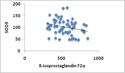
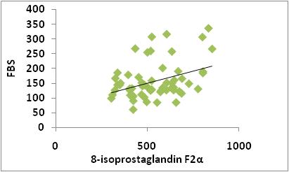
Table 4 shows the results of multiple linear regression analysis for group II (diabetic cataract) with 8-iso-PGF2α as dependent variable. Fasting blood glucose and SOD3 showed positive and negative influence with respect to oxidative stress marker.
Cataract is one of the major causes of visual impairment and blindness in diabetic population12.
Table 2 — Fasting blood glucose and HbA1c levels between Group I and Group II
ParametersGroup I Group IIp Value (Healthy individuals(Type 2 DM without cataract) patients with senile (n=60) cataracts) (n=60)
Fasting plasma glucose (mg/dL)87 ± 14.00157 ± 62<0.001***
HbA1c (%) 5.1 ± 0.447.7 ± 1.88<0.001***
* P<0.05 significant, ** p<0.01, ***p<0.001
Table 3
Several lines of evidence suggest that cataract formation occurs more often at an early age and advances much faster in diabetics when compared with non-diabetic subjects 13,14 . Multiple pathogenic mechanisms have been proposed to explain the cataract formation in Diabetes Mellitus such as increased sorbitol concentration, abnormal glycosylation of proteins and enhanced free radical production in lens, but still not fully understood15-17 Among several risk factors, oxidative stress plays a major role in the development of cataract in diabetic subjects. However, the exact mechanism by which oxidative stress contributes to development of cataract remains unclear.
Lipid Peroxidation (LPO) is caused by an imbalance between the free radical production and the antioxidant defenses and plays a significant role in the cataractogenesis 18 . Lipid peroxidation is the major marker of oxidative stress and the oxidative damage can be measured by estimating the primary or secondary LPO end-products. The Primary LPO end-products are conjugated dienes and lipid hydroperoxides, while
secondary end-products which include Thiobarbituric Reactive Substances (TBARS), gaseous alkanes and F2- isoprostanes (F2-IsoPs). Among the LPO markers, measurement of 8-iso-PGF2α has advantage over the other markers. Since they are biologically active, stable, specific and easily identified in biological fluids and tissue and has been widely used as a valid marker of oxidative stress19–21
We found that 8-iso-PGF 2α levels in serum of Diabetic cataract patients were significantly elevated when compared to healthy individuals without cataract. In line with our findings, Amena Rahim et al have shown that mean concentration of 8-isoprostaglandin F2α level in aqueous humor was significantly higher in diabetic patients with cataract than age matched senile cataract patients22. Another study also reported that the mean concentration of 8-iso-PGF2α in aqueous humor was approximately 5 times higher in patients with exfoliation syndrome and cataracts23. Also few studies have shown the elevated lipid peroxides levels in diabetic cataractous patients when compared with senile cataract8,24. Although very limited studies are available with reference to 8-iso-PGF2 α levels in cataract subjects, our findings were supported with these reports. It was also shown that hyperglycemia generate excess free radicals due to auto-oxidation of glucose and glycosylation of proteins. Thus hyperglycemia induced free radical attacks the membrane lipids especially polyunsaturated fatty acid resulting in increased lipid peroxidation product, 8-isoPGF2á which plays a major role in the development of microvascular complications in diabetic subjects. Hence the accumulation of 8-iso-PGF2α in cataract lens is a key factor in the early development of cataractogenesis in diabetic subjects when compared to healthy controls.
In the present study, fasting blood glucose level and HbA1c were significantly elevated in diabetic cataract subjects. Also there was significant positive correlation between 8-iso-PGF2α and fasting blood glucose levels in diabetic cataract subjects. Our results suggest that hyperglycemia in diabetic subjects may contribute to oxidative damage in lens via increased 8-iso-PGF2α levels which could be a causative factor in early development of cataract formation in Type 2 Diabetic patients.
Human lens has several defense mechanisms against the oxidative damage caused by ROS. The major protective enzymes in the lens are Superoxide Dismutase (SOD), Catalase (CAT)25 and Glutathione Peroxidase (GSH-Px)26 SOD is a chain breaking most predominant antioxidant enzyme in lens, which acts by removing the toxic superoxide radical, O 2 " by
converting it into H 2 O 2 which in turn, can be decomposed by CAT and or GSH-Px27. In vitro and in vivo studies have shown that SOD has protective properties against cataract development in Diabetes Mellitus8,28-30. But some studies have reported the contradictory findings on SOD levels and other antioxidant enzyme levels. We found reduced anti-oxidant enzyme activities in diabetic patients with senile cataract as illustrated by decreased SOD3 and CAT activity. These findings were consistent with previous report in which lens copper, zinc, SOD and catalase levels were significantly lower in the diabetic patients when compared to senile cataract subjects8. Similar studies have shown that serum and erythrocyte SOD levels were decreased in diabetic subjects with cataract when compared with senile cataract subjects6,30. We also observed negative correlation between 8-iso-PGF2α and SOD in serum of Type 2 diabetic with senile cataract patients. Thus it is evident that Hyperglycemia and Lipid peroxidation as shown by elevated levels of 8-iso-PGF2α may contributed to the reduced anti-oxidant enzyme levels of SOD3 and CAT in the current study. The reduced anti-oxidant enzyme activity of SOD3 and CAT in serum leads to elevation of H2O2 and superoxide radicals which may contribute to oxidative damage of lens and associated with early development of cataract formation in Type 2 Diabetic patients.
The cardinal concept in the present study indicates that oxidative stress was significantly higher in serum of diabetic cataracts which is evidenced by elevated levels of 8-iso prostaglandin F2 α and decreased antioxidant enzymes SOD3 and CAT. In diabetic subjects, hyperglycaemia induces increased production of ROS which leads to lipid peroxidation resulting in oxidative stress and depletion of antioxidant enzymes in the lens tissue. These could have played a significant role in the pathogenesis of diabetic induced cataract.
Acknowledgment : I sincerely thanks to Mahatma Gandhi Medical College and Research Institute,Sri Balaji Vidyapeeth (Deemed to be University) for the assessment of laboratory facility, technical and financial support.
Declaration of interest : The authors declare that there is no conflict of interests regarding the publication of this manuscript.
Source of Funding : Study was supported by Intra-mural Research Grant (Grant No. Dean (R&AHS)/ Res. Grants/2017/145) of Sri Balaji Vidyapeeth (Deemed-to-be-University), Puducherry, India.
1Gupta SK, Trivedi D, Srivastava S, Joshi S, Halder N, Verma SD — Lycopene attenuates oxidative stress induced experimental cataract development: an in vitro and in vivo study. Nutrition 2003; 19(9): 794-9.
2Oxidative Stress: Adaptation, Damage, Repair and Death. In: Halliwell, B. and Gutteridge, JMC, Eds, Free Radicals in Biology and Medicine,. 3rd ed. p Oxford University Press, Oxford,; 1999.
3Rahman T, Hosen I, Islam MMT, Shekhar HU — Oxidative stress and human health. ABB [Internet]. 2012 [cited 2020 Jan 7]; 3(7): 997-1019.
4Kisic B, Miric D, Zoric L, Ilic A — Role of Lipid Peroxidation in the Pathogenesis of Age-Related Cataract. Lipid Peroxidation [Internet]. 2012 Aug 29 [cited 2020 Jan 7];
5Babizhayev MA — Biomarkers and special features of oxidative stress in the anterior segment of the eye linked to lens cataract and the trabecular meshwork injury in primary open-angle glaucoma: challenges of dual combination therapy with N-acetylcarnosine lubricant eye drops and oral formulation of nonhydrolyzed carnosine. Fundam Clin Pharmacol 2012; 26(1): 86-117.
6Donma O, Yorulmaz E, Pekel H, Suyugül N — Blood and lens lipid peroxidation and antioxidant status in normal individuals, senile and diabetic cataractous patients. Curr Eye Res 2002; 25(1): 9-16.
7Zoric L, Elek-Vlajic S, Jovanovic M, Kisic B, Djokic O, Canadanovic V, et al — Oxidative stress intensity in lens and aqueous depending on age-related cataract type and brunescense. Eur J Ophthalmol 2008; 18(5): 669-74.
8Ozmen D, Mutaf I, Ozmen B, Mentes J, Bayindir O — Lens lipid peroxides and glutathione concentrations in diabetic cataract. Ann Clin Biochem 1997; 34 (Pt 2): 190-2.
9Roberts LJ, Morrow JD — Measurement of F(2)-isoprostanes as an index of oxidative stress in vivo. Free Radic Biol Med 2000; 28(4): 505-13.
10Mezzetti A, Cipollone F, Cuccurullo F — Oxidative stress and cardiovascular complications in diabetes: isoprostanes as new markers on an old paradigm. Cardiovasc Res 2000; 47(3): 475-88.
11Niedowicz DM, Daleke DL. The role of oxidative stress in diabetic complications. Cell Biochem Biophys 2005; 43(2): 289-330.
12Drinkwater JJ, Davis WA, Davis TME. A systematic review of risk factors for cataract in type 2 diabetes. Diabetes Metab Res Rev 2019; 35(1): e3073.
13Klein BE, Klein R, Moss SE — Prevalence of cataracts in a population-based study of persons with diabetes mellitus. Ophthalmology 1985; 92(9): 1191-6.
14Nielsen NV, Vinding T — The prevalence of cataract in insulindependent and non-insulin-dependent-diabetes mellitus. Acta Ophthalmol (Copenh) 1984; 62(4): 595-602.
15Ahmed N — Advanced glycation endproducts—role in pathology of diabetic complications. Diabetes Res Clin Pract 2005; 67(1): 3-21.
16Lee AY, Chung SS — Contributions of polyol pathway to oxidative stress in diabetic cataract. FASEB J 1999; 13(1): 23-30.
17Spector A — Oxidative stress-induced cataract: mechanism of action. FASEB J 1995; 9(12): 1173-82.
18Micelli-Ferrari T, Vendemiale G, Grattagliano I, Boscia F, Arnese L, Altomare E, et al — Role of lipid peroxidation in the pathogenesis of myopic and senile cataract. Br J Ophthalmol [Internet] 1996 [cited 2020 Jan 7]; 80(9): 840-3.
19Montuschi P, Barnes PJ, Roberts LJ — Isoprostanes: markers and mediators of oxidative stress. FASEB J 2004; 18(15): 1791-800.
20Morrow JD, Roberts LJ — The isoprostanes: unique bioactive products of lipid peroxidation. Prog Lipid Res 1997; 36(1): 121.
21Zoriæ L— [Parameters of oxidative stress in the lens, aqueous humor and blood in patients with diabetes and senile cataracts]. Srp Arh Celok Lek 2003; 131(3-4): 137-42.
22Rahim A — 8-Isoprostaglandin F2a Levels in Aqueous Humor of Senile and Diabetic Cataract Patients. IOSR-JDMS [Internet]. 2012 [cited 2020 Jan 7]; 2(3): 40-2. Available from: http:// www.iosrjournals.org/iosr-jdms/papers/Vol2-issue3/ I0234042.pdf
23Koliakos GG, Konstas AGP, Schlötzer-Schrehardt U, Hollo G, Katsimbris IE, Georgiadis N, et al — 8-Isoprostaglandin F2a and ascorbic acid concentration in the aqueous humour of patients with exfoliation syndrome. Br J Ophthalmol 2003; 87(3): 353-6.
24Obara Y — [The oxidative stress in the cataract formation]. Nippon Ganka Gakkai Zasshi 1995; 99(12): 1303-41.
25Fecondo JV, Augusteyn RC — Superoxide dismutase, catalase and glutathione peroxidase in the human cataractous lens. Exp Eye Res 1983; 36(1): 15-23.
26Dwivedi RS, Pratap VB — Alteration in glutathione metabolism during cataract progression. Ophthalmic Res 1987; 19(1): 41-4.
27Rao NA, Thaete LG, Delmage JM, Sevanian A — Superoxide dismutase in ocular structures. Invest Ophthalmol Vis Sci 1985; 26(12): 1778-81.
28McCord JM, Fridovich I — Superoxide dismutase. An enzymic function for erythrocuprein (hemocuprein). J Biol Chem 1969; 244(22): 6049-55.
29Olofsson EM, Marklund SL, Behndig A — Enhanced diabetesinduced cataract in copper-zinc superoxide dismutase-null mice. Invest Ophthalmol Vis Sci 2009; 50(6): 2913-8.
30Maurya OPS, Mohanty L, Bhaduri G, Chandra A — Role of anti-oxidant enzymes superoxide dismutase and catalase in the development of cataract/ : study of serum levels in patients with senile and diabetic cataracts. J Indian Med Assoc 2006; 104(7): 394, 396-7.
Background : Anticholinergic burden in Geriatric population is of great concern throughout the Globe, yet often neglected. There are previous studies to assess the Anticholinergic burden, carried out in specific population, though its prevalence in general population is difficult to interpret. Polypharmacy remains one of the major causes contributing to the increased burden in Anticholinergic score among Geriatrics. Multiple co-morbidities and the prevalence of Multiple Chronic Diseases are the responsible factors which imbibe Multiple Drug Therapy in Geriatric population. Anticholinergic burden in older adults has been associated with Cognitive impairment, Delirium, Dizziness and Confusion, Falls and increased hospitalizations. However, Anticholinergic-acting drugs are often advised in Geriatric population. In this study an attempt was made to understand the Anticholinergic burden score among Geriatric population.
Methods : This study is a descriptive cross-sectional study, which was done, in a period of six months among 62 Geriatric patients attending a therapeutics clinic of a private clinical pharmacology OPD for first time. Patients were noted for their comorbidities and anticholinergic burden was calculated, based on the Anticholinergic Cognitive Burden scale. Results were statistically analyzed.
Results : Clinically significant anticholinergic burden was observed in 22.58% population. The most frequently prescribed drug was found to be alprazolam followed by amitriptyline and theophylline. Among the comorbidities hypertension and diabetes was commonly seen in majority of the population.
Conclusion : Co-professional care at clinical pharmacology OPD with assessment of anticholinergic burden of geriatric prescriptions and advices on rational de-prescribing with suggestions on safer alternatives would be beneficial for treating physicians to optimize therapy.
[J Indian Med Assoc 2022; 120(10): 39-43]
Key words :Anticholinergic drugs, Anticholinergic burden, Polypharmacy, Geriatric population, Adverse effects.
Anticholinergic drugs target the muscarinic receptors and eventually block the neurotransmission of acetylcholine. Autonomic
1MBBS, MD (Pharmacology), Senior Resident, Department of Pharmacology, Calcutta National Medical College, Kolkata 700014
2MBBS, MD, DM (Clinical Pharmacology), PG Dip Endo & Diabetes (RCP, UK), Consultant Physician at Diabetes and Allergy-Asthma Therapeutics Clinic, Kolkata and Clinical Pharmacologist at Department of Clinical & Experimental Pharmacology, School of Tropical Medicine, Kolkata 700073 and Corresponding Author
3MBBS, MD, DCH, DM (Clinical Pharmacology), Associate Professor, Department of Pharmacology, R G Kar Medical College & Hospital, Kolkata 700004
4 PhD Scholar, Department of Clinical & Experimental Pharmacology, School of Tropical Medicine, Kolkata 700073
5MBBS, MD, DM, (Clinical Pharmacology), Academic Dean & Professor & Head, Department of Pharmacology, Netaji Subhas Medical College & Hospital, Patna 801103
6MBBS, MD, FRCP, FACP, WHO Fellow, Professor, General Medicine, Barasat Government Medical College & Hospital, Kolkata, 700124
7MBBS, MD, DM (Endocrinology), Senior Endocrinologist, Joshi Clinic, Mumbai 400050
Received on : 08/05/2022
Accepted on : 04/06/2022
Elderly population is at high risk of developing adverse effects.
Assessment of Anticholinergic burden in geriatric patients on multiple drugs may be helpful to minimize the adverse effects.
Timely de-prescribing of drugs with high ACB or altering it with low ACB score using principles of clinical pharmacological review, reconciliation and feedback could prevent medication related atrocities.
nervous system plays an important role in maintaining multiple body functions. These body functions include learning and memory, urination, maintenance of heart rhythm etc. Integrity of the autonomic nervous system is to be maintained, for proper functioning of the various body functions. The drugs which act on this system are cumulatively known as Anticholinergic drugs and these drugs acts on its various receptors. These drugs not only bind to muscarinic receptors but also bind with other receptors based on its either agonistic activity or antagonistic activity, which makes different
Anticholinergic Burden in Geriatric Patients Attending Clinical Pharmacology Clinic — A Descriptive Cross-sectional Study in Eastern India
Debajyoti Saha1, Shambo Samrat Samajdar2, Sabnam Ara Begum3, Shatavisa Mukherjee4, Santanu Kumar Tripathi5, Jyotirmoy Pal6, Shashank R Joshi7
therapeutic targets.Since cholinergic transmission is responsible for the activity of various physiological functions, particularly in neurotransmission, anticholinergic drugs do not escape adverse effects. These adverse effects may involve both the c peripheral and central nervous systems. While Central Nervous System (CNS) related adverse effects primarily involves different cognitive domains, adverse effects of PNS include decrease in body’s secretion which is manifested as dry mouth. The other PNS complication includes urinary retention, constipation, tachycardia and blurring of vision, among others.
The prevalence of chronic diseases, like Diabetes, Hypertension and Osteoarthritis, in geriatric population is quite common. These diseases are not only associated with considerable illness, and is also associated with increased overall medication use, decreased quality of life, and ultimately increased cost of therapy1. As age progresses impairment in cognition, autonomic dysfunction, falls and dementia cannot be do away with but these geriatric symptoms are not always directly related to the process of ageing, it may be also contributed due to the anticholinergic adverse effects of the prescribed medications in older adults2,3 Geriatric population are at higher risk to experience adverse effects due to age-related modifications of Pharmacokinetic and Pharmacodynamic Parameters. Children and younger adults are not an exception as, adverse effects due to anticholinergic medications can affect them, but to a lesser extent. To be more specific, the adverse effects due to Anticholinergic drugs, targeting both at the peripheral and the central level, have greater impact in Geriatric population4-7. Among the different types of anticholinergic medications prescribed, the anticholinergic activity varies. Some possess more anticholinergic activity, while others have less anticholinergic activity. Sedating antihistamines and tricyclic antidepressants have strong anticholinergic side effects. In common practice the treating physician may not be aware that some commonly prescribed diuretics and other cardiac medications (eg, anticoagulants, antihypertensives) also have Anticholinergic properties. Also while prescribing; the physician should realize that taking multiple medications with weaker anticholinergic properties can have an additive effect8-10. It is evident from previous research that the cumulative exposure during as little as 1 to 2 years impairs memory and performance of activities of daily living11. Long-term use of medications with high Anticholinergic burden score is associated with an increased incidence of Alzheimer disease and Dementia12-14. The risk of anticholinergic-
related cognitive impairment has been found independent of the underlying comorbid disease burden or severity of illness15. Conversely, effect of cumulated drug burden in increasing risk of hospitalization or mortality was not clear, with studies reporting conflicting results in different populations16
Anticholinergic Drugs :
Anticholinergic drugs have several Therapeutic indications, which include urinary abnormalities, gastrointestinal diseases like Peptic Ulcer Disease and Irritable Bowel Syndrome, nervous system disorders like Parkinson’s disease. Also, anticholinergics are frequently used as anesthetic agents and for Neurologic and Psychiatric conditions.
The main properties of Anticholinergic drugs are based on their affinity with central or peripheral cholinergic receptors 17. These actions can affect cognition, which can be mistakenly diagnosed as a sequela of normal aging process18. Several drugs have a high ACB score but few drugs like amoxicillin, lansoprazole, metformin, fentanyl, furosemide, diazepam, digoxin, duloxetine, phenytoin and topiramate is associate with AA at high doses19. It has been observed that, the combination of several drugs, which may include even nutritional supplements can cause or enhancethe adverse events of a prescribed anticholinergic medication. The prescription of Anticholinergic drugs may be considered inappropriate in certain scenario.”Potentially inadequate anticholinergic drug use in older adults” has already been discussed in the Beers (American Geriatrics Society 2012 Beers Criteria Update Expert Panel, 2012) and the STOPP-START criteria20. However, for certain clinical situations the benefits of anticholinergic outweighs their risks and their prescription may be considered appropriate in selected cases of Geriatric population, after making Risk benefit analysis.
The various deleterious anticholinergic effects can be related to the various muscarinic receptors in our body. These receptors are of five types M1 to M5, out of which M1, M2, M4 are responsible for the various anticholinergic deleterious effects. M1 receptors are primary located in the CNS and have important role in execution of various functions. It is also responsible for maintaining episodic memory in hippocampus and pre-frontal cortex 21 . While M2 receptors are responsible for processing memory, M4 receptors are involved in regulating the levels of acetylcholine.The receptor antagonism of these receptors is associated with cognitive disturbance and cell death22. Cognitive
effects not only depend on anticholinergic burden, baseline cognitive function, but also on individual pharmacokinetics and pharmacodynamics variability. Cognitive effects can also be influenced by alterations in metabolism 23 . Central effects of the various Anticholinergic drugs depend on the penetration power of the drug through the blood brain barrier. In diabetic patients and multiple sclerosis patients the blood brain permeability is increased and the drugs such as loperamide, simvastatin, clonidine, or methyldopa can exhibit their central side effects.
Prolonged anticholinergic drug therapy can cause brain changes, partly simulating Alzheimer’s Disease (AD). Cognitive dysfunction in AD is related with multiple aspects, such as reduction in cholinergic neurons, acetylcholine receptor dysfunction and altered signal transmission.Severity of disease is proportional to the changes in the cholinergic system24 In this context a study has shown, that both amyloid plaques and neurofibrillary tangles were increased in Parkinson patients on prolonged anticholinergic medications25. Another study, reveals that cholinergic receptor blockade for prolonged duration is associated with increased in beta-amyloid peptide in various parts of the brain including Amygdale, Cortex and Hippocampus26. Accordingly, another longitudinal study found higher rates of atrophy in the total cerebral volume and in the gray matter associated to anticholinergic drug consumption27.
A descriptive cross-sectional study was conducted over a period of six months on all Geriatric patients attending a specialty Therapeutics clinic in eastern India. The study commenced after obtaining its due permission for conduct from the institutional ethics committee. All geriatric patients were evaluated for their anticholinergic burden using the Anticholinergic Cognitive Burden (ACB) scale. This scale is widely accepted and validated for adverse anticholinergic outcomes28,29.The ACB scale was first published in the year 2008 and updated in the year 201228. A systematic review based study was done in-order to make a list of possible anticholinergic medications with notable Anticholinergic effects. Later the same list was identified and further evaluated and categorized by a team of clinical expert clinicians. The ACB scale categorizes drugs on a scale of 0 to 3, based on their anticholinergic activity. Detailed scoring uses the following criteria: (i) ACB score of 1 (possible anticholinergic effect) requires “evidence from in vitro data that the medication has antagonist activity at
muscarinic receptors”, (ii) ACB score of 2 (definite anticholinergic effect) requires “evidence from literature, prescriber’s information, or expert opinion of clinical anticholinergic effect”, (iii) ACB score of 3 (definite anticholinergic effect) requires “evidence from literature, prescriber’s information, or expert opinion of the medication causing delirium”. All other medications have a score of 0. The total anticholinergic burden of a patient is calculated by summing the ACB scores from all the medications that patient receives concomitantly: anticholinergic burden >3 is considered clinically significant 29 . Data collected were checked for completeness, tabulated and statistically analyzed using Microsoft Excel.
The study included a total of 62 geriatric patients who visited clinical pharmacology outpatient facility in a specialty clinic in eastern India. Out of the 62 patients 23 were females and rest were males. The mean age of the female patients was 60.6±74.32 years and that of male patients was 70.92±4.06 years. Clinically significant Anticholinergic burden with a ACB score of 3 or more was found in 14 (22.58%) of the patients, out which 6 were female and 8 were male. Presenting disease condition and drug usage pattern of anticholinergic medications was noted in all included individuals (Table 1).
While 53.23% Geriatric population had ACB score of 0, the study noted 22.58% population having clinically significant anticholinergic burden with score 3 or greater. 19.35% geriatrics had ACB score of 1 (Fig 1).
Anticholinergic burden in Geriatric population is often associated with various sideeffects like impairment in cognition, autonomic dysfunctions relating to problems in micturition in elderly males, drying of secretions and many more. ACB, though a common entity is often neglected. Elderly population is much more associated with multiple comorbidities which imbibe multiple drug therapies and thus polypharmacy is one of the major causes of poor compliance to drug treatment. In this descriptive crosssectional study, we looked for the prevalence of Anticholinergic burden in Geriatric population attending for the first time in a therapeutic clinic. It has been found that overall anticholinergic burden is 22.5%, which is fairly high. Previous studies also have similar results which suggest that about one third of study population was associated with fairly high ACB score3,5. About 27% of the male population has an
Table
Female
Percen
Percen ency(N)tage(%)ency(N)tage(%)
Total Sample 2337.093962.90
Disease Condition :
Type 2 Diabetes Mellitus13 56.522564.10
Hypertension15 65.223282.05
Chronic urticarial3 13.0400
COPD6 26.091025.64
Psychosis28.7000
GERD1 4.3500
Rheumatoid Artheritis1 4.3500
Benign prostatic hyperplasia002 5.13
Asthma00 12.56
Chronic Kidney Disease005 12.82
Inflammatory Bowel Syndrome001 2.56
Anticholinergic Burden (ACB) :
ACB Score 013 56.522051.28
ACB Score 14 17.39820.51
ACB Score 2003 7.69
ACB Score 32 8.7025.13
ACB Score 42 8.7037.69
ACB Score 50000
ACB Score 61 4.3525.13
ACB Score 71 4.3512.56
Drug (with ACB) Usage Pattern :
Amitriptyline2 8.70717.95
Ranitidine1 4.35512.82
Clemastine1 4.3500
Alprazolam4 17.39820.51
Prednisolone1 4.3525.13
Hydroxyzine14.35 37.69
Quetiapine2 8.7000
Doxepin2 8.7037.69
Nortriptyline3 13.0400
Theophylline004 10.26
Levocetirizine00410.26
hypnotics outnumbers others. The most frequently prescribed drug was found to be alprazolam followed by amitryptilline and theophylline. This is also a common finding in previous studies5,9. Among the comorbidities Hypertension and Diabetes was commonly seen in majority of the population. However there has not been any notable adverse effect noted among the patients. All the patients were questioned about the possible adverse effects of anticholinergic medications, but most of them did not experience any notable adverse effect. In a study it was found that deprescribing drugs having high Anticholinergic burden ameliorated the medication related problems that was developed due to prescribing cascade30 In another study it was shown that de-prescribing drugs in clinical pharmacology clinic with high anticholinergic burden was beneficial in drug induced erectile dysfunction patients31 In diabetic patients suffering from Autonomic Neuropathy, drugs with anticholinergic burden, increases parasympathetic autonomic dysfunction. In one study it was depicted that timely identification and discontinuation of offending agent produce beneficial effect to the patient32
Geriatric population warrants special care in prescribing. There are many things yet to be learned about the distribution of Anticholinergic burden in geriatric population, associated risk factors, and effective measures to assess and intervene if necessary. ACB scores of 3 or greater represent clinically relevant anticholinergic burden and it remains the wise decision of the treating physician to have balance between risk and potential benefits. Also screening may be helpful for early recognition of anticholinergic adverse effects and develop a plan to minimize anticholinergic drug exposure. This may eventually improve the health outcomes of Geriatric population by halting the progression of Functional decline and Geriatric syndromes, particularly cognitive impairment. Co-professional care at clinical pharmacology OPD with assessment of anticholinergic burden of geriatric prescriptions and advices on rational de-prescribing with suggestions on safer alternatives would be beneficial for treating Physicians to optimize therapy.
Conflict of interest : NIL
Funding : NIL
Fig
ACB score of >3, 26% while female population had an ACB score >3. Among the frequently prescribed drugs in both female and male population sedative and
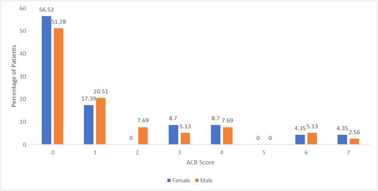
1Bishara D, Harwood D, Sauer J, Taylor DM — Anticholinergic effect on cognition (AEC) of drugs commonly used in older people. Int J Geriatr Psychiatry 2017; 32: 650-6.
2Mintzer J, Burns A — Anticholinergic side-effects of drugs in elderly people. J R Soc Med 2000; 93(9): 457-62.
3Tune LE — Anticholinergic effects of medication in elderly patients. J Clin Psychiatry 2001; 62(suppl 21): 11-4.
4Nishtala PS, Salahudeen MS, Hilmer SN — Anticholinergics: theoretical and clinical overview. Expert Opin Drug Saf 2016; 15: 753-68.
5Wu YH, Wang CJ, Hung CH — Association between using medications with anticholinergic properties and short-term cognitive decline among older men: a retrospective cohort study in Taiwan. Geriatr GerontolInt 2017; 17: 57-64.
6Kalisch Ellett LM, Pratt NL, Ramsay EN, Barratt JD, Roughead EE — Multiple anticholinergic medication use and risk of hospital admission for confusion or dementia. J Am Geriatr Soc 2014; 62: 1916-22.
7Panula J, Puustinen J, Jaatinen P, Vahlberg T, Aarnio P, Kivela S — Effects of potent anticholinergics, sedatives and antipsychotics on postoperative mortality in elderly patients with hip fracture. Drugs Aging 2009; 26: 963-71.
8Boustani M, Campbell N, Munger S — Impact of anticholinergics on the aging brain: a review and practical application. Aging Health 2008; 4(3): 311-20.
9Carnahan RM, Lund BC, Perry PJ — The Anticholinergic Drug Scale as a measure of drug-related anticholinergic burden: associations with serum anticholinergic activity. J Clin Pharmacol 2006; 46(12): 1481-6.
10Rudolph JL, Salow MJ, Angelini MC, McGlinchey RE. The anticholinergic risk scale and anticholinergic adverse effects in older persons. Arch Intern Med 2008; 168(5): 508-13.
11Fox C, Richardson K, Maidment ID — Anticholinergic medication use and cognitive impairment in the older population: the Medical Research Council Cognitive Function and Ageing Study. J Am Geriatr Soc 2011; 59(8): 1477-83.
12Bottiggi K, Salazar JC, Yu L — Long-term cognitive impact of anticholinergic medications in older adults. Am J Geriatr Psychiatry 2006; 14(11): 980-4.
13Carrière I, Fourrier-Reglat A, Dartigues JF — Drugs with anticholinergic properties, cognitive decline, and dementia in an elderly general population: the 3-City Study. Arch Intern Med 2009; 169(14): 1317-24.
14Perry EK, Kilford L, Lees AJ — Increased Alzheimer pathology in Parkinson’s disease related to antimuscarinic drugs. Ann Neurol 2003; 54(2): 235-8.
15Vinogradov S, Fisher M, Warm H — The cognitive cost of anticholinergic burden: decreased response to cognitive training in schizophrenia. AmJPsychiatry 2009; 166(9): 1055–62.
16Lönnroos E, Gnjidic D, Hilmer SN — Drug Burden Index and hospitalization among community-dwelling older people. Drugs Aging 2012; 29(5): 395-404.
17Lechevallier-Michel N, Molimard M, Dartigues JF, Fabrigoule C, Fourrier-Réglat A — Drugs with anticholinergic properties and cognitive performance in the elderly: results from the PAQUID study. Br J Clin Pharmacol 2005; 59: 143-51.
18Kersten H, Molden E, Tolo IK, Skovlund E, Engedal K, Wyller T B — Cognitive effects of reducing anticholinergic drug burden
in a frail elderly population: a randomized controlled trial. J. Gerontol. A Biol Sci Med Sci 2013; 68: 271-8.
19Chew ML, Mulsant BH, Pollock BG, Lehman ME, Greenspan A, Mahmoud RA, et al — Anticholinergic activity of 107 medications commonly used by older adults. J Am Geriatr Soc 2008; 56: 1333-41.
20O’Mahony D, O’Sullivan D, Byrne S, O’Connor MN, Ryan C, Gallagher P — STOPP/START criteria for potentially inappropriate prescribing in older people: version 2. Age Ageing 2015; 44: 213-8. doi: 10.1093/ageing/af u145
21Sathienluckana T, Unaharassamee W, Suthisisang C, Suanchang O, Suansanae T — Anticholinergic discontinuation and cognitive functions in patients with schizophrenia: a pharmacist-physician collaboration in the outpatient department. Integr. Pharm Res Pract 2018; 26: 161-71.
22Bishara D, Harwood D, Sauer J, Taylor DM — Anticholinergic effect on cognition (AEC) of drugs commonly used in older people. IntJGeriatrPsychiatry 2017; 32: 650-6. doi: 10.1002/ gps.4507
23Roe CM, Anderson MJ, Spivack B — Use of anticholinergic medications by older adults with dementia. J Am Geriatr Soc 2002; 50: 836-42.
24Swami S, Cohen RA, Kairalla JA, Manini TM — Anticholinergic drug use and risk to cognitive performance in older adults with questionable cognitive impairment: a cross-sectional analysis. Drugs Aging 2016; 33: 809-18.
25Perry EK, Kilford L, Lees AJ, Burn DJ, Perry RH — Increased Alzheimer pathology in Parkinson’s disease related to antimuscarinic drugs. Ann Neurol 2003; 54: 235-8.
26Risacher SL, McDonald BC, Tallman EF, West JD, Farlow M R, Unverzagt FW, et al — Alzheimer’s disease neuroimaging initiative. Association between anticholinergic medication use and cognition, brain metabolism, and brain atrophy in cognitively normal older adults. JAMA Neurol 2016; 73: 72132.
27Chuang YF, Elango P, Gonzalez CE, Thambisetty M — Midlife anticholinergic drug use, risk of Alzheimer’s disease, and brain atrophy in community-dwelling older adults. Alzheimers Dement 2017; 3: 471-9
28Campbell N, Maidment I, Fox C, Khan B, Boustani M Te.2012 update to the anticholinergic cognitive burden scale. J Am Geriatr Soc 2013; 61(No. S1): S142–S143
29Boustani M, Campbell N, Munger S, Maidment I, Fox C — Impact of anticholinergics on the aging brain: a review and practical application. Aging Health 2008; 4: 311-20
30Samajdar SS, Mukherjee S, Paul J, Tripathi SK, Joshi SR, etal — Deprescribing for Better Patient Outcomes in Chronic LongTerm Care and Role of Clinical Pharmacological Review. Case Series. J Assoc Physicians India 2021; 69(11): 90-1.
31Samajdar SS, Mukherjee S, Saha D, Jumani D, Tripathi SK — Drug-Induced erectile dysfunction: Two interesting cases. J Pharmacol Pharmacother 2021; 12: 177-9.
32Samajdar SS, Mukherjee S, Joshi S, Tripathi SK — Reversal of Diabetes Autonomic Neuropathy with Intense Treatment Monitoring — A Case Study. JIndianMedAssoc 2020; 118(12): 69-71.
Background : Despite the yearly increase in budget and improved health infrastructure, the improvement in the health indices is not parallel to them. It indicates that there are other factors influencing the health indices like morbidity and mortality.
Objective : This study highlights the socio-demographic factors and their importance in child health care.
Methods : A single-centre hospital-based cross-sectional study was done at a tertiary health care centre in central India. One thousand cases were enrolled over 3 years and evaluated for the association of socio-demographic parameters and child health status indicators.
Results : Of the 1000 cases, immunization status in children had a significant association with mother’s education status (p=0.005), father’s education status (p=0.001), and religion (p<0.001) but not associated with socio-economic status (p=0.254) and place of residence (urban versus rural) (p=0.916). The pallor was significantly associated with the mother’s education status (p=0.001), father’s education status (p=0.005), socio-economic status (p=0.001) but not associated with the sex of the child (p=0.934), place of residence (p=0.807) and religion of the participant child (p=0.812).
Conclusion : Immunization status of the child was significantly associated with the educational status of parents and religion while pallor was associated with the educational status of parents and economic status. The educational status of the parent is significant as for as child health care is concerned.
Indian Med Assoc 2022; 120(10): 44-7]
Key words :Child health care, Sociology of child health care, Socio-demography of child health.
The health of an individual and population is strongly influenced by social determinants. The impact of social inequalities on health is the most neglected area. The state of Kerala in India is well studied, highlighting the relationship between its phenomenal reduction of socio-economic inequalities in the last 40 years and improvements in the health status of the population as a whole, thereby improving the health indices comparable to the developed nations. Government with its Social, Political and Economic wings operate at every level of human life let it be household, village, municipality, nation, or globe, and influences the health of an individual, families, and communities1
Social determinants of health have a strong effect
1MBBS, MD (Ped), PhD, FCPS, Associate Professor, Department of Paediatrics, Dr S C Government Medical College, Nanded, Maharashtra 431606
2MBBS, DNB (Ped), DCH, MD (Micro), Assistant Professor, Department of Paediatrics, Government Medical College, Aurangabad, Maharashtra 431001
3MBBS, MD (Ped), Associate Professor, Department of Paediatrics, Government Medical College, Jalgaon, Maharashtra 425001
4MBBS, MD (Ped), Associate Professor and Head, Department of Paediatrics, Sri Bhausaheb Hire Government Medical College, Dhule, Maharashtra 424001 and Corresponding author
Received on : 27/09/2022
Accepted on : 01/10/2022
Editor's Comment : Educational level of parents and literacy rate in the community are most important of all the socio-economic parameters influencing child health care in the community. Parents educational status will definitely help to improve Immunization status & amp; nutrition well being of children. All Government departments should work together to attain socio-economic equality and improvement in child health care of the community.
on health-promoting behaviours in the community. Health equity is possible only with equal distribution of social determinants among the population. The richest countries in the world also have socioeconomic inequalities in health between the rich and the poor and because of these inequalities unable to achieve the health indices to 100%. Nanded district of Maharashtra state has more than 70% rural households, providing a sufficiently large size of sample for reliable estimates of most of the health and health-related demographic indicators for the rural population as many health care programs are designed considering the rural population at its centre.
Bhutan is almost 90% covered with provision for safe drinking water, it helped in the eradication of anaemia in women by reducing the exertion faced by women in the collection of potable drinking water. It depicts the importance of good social policies to
Sociology of Child Health Care Practices in Nanded District of Maharashtra, Central India — A Single Centre Hospital-based Crosssectional Study
Kishor G Rathod1, Mohd Ubaid ur Rahman Mohd Azam2, Balasaheb B Suroshe3, Neeta K Hatkar4
improve not only social status but even medical conditions like anaemia.
In Nepal, current health system arrangements can reduce health inequalities in child malnutrition by 4 percent while the current health system in Sri Lanka can help in the reduction of 10 percent of inequalities in child malnutrition2
Analysis of health system access and other social determinants in Nepal and Sri Lanka for malnutrition in children2.
Socio-economic Socio- Intermediary
The Gadchiroli district of Maharashtra has the best population per doctor but still has a very low proportion of utilization of health care facilities and very low coverage of immunization of children (46.4%). This scenario highlights that the mere availability of health infrastructure may not be sufficient for a better outcome though it is a must for improving the health system3.
the increasing yearly budget and improvement in the health system, from the above scenario it is obvious that there are many factors apart from the health system and mere availability of services, which influence the uptake of child health care practices by the parents or community. Hence this study is an attempt to highlight these socio-demographic factors and their role in child health care practices ie, social causes of poor health and avoidable inequalities.
It was a hospital-based single centre prospective cross-sectional study undertaken to determine the association of socio-economic factors with child health care practices among study subjects in the Nanded district of Maharashtra State of India from January 2017 to December 2019 followed by analysis, interpretation, and write-up.
Inclusion and Exclusion Criteria :
(i) Parents who gave consent for enrolment, for patients with age group from newborns to completed 12 years and resident of Nanded District. (ii) Only one participant (the child) from one household was enrolled to avoid the repetition of demographic data. Critical patients (children) were excluded from the study to avoid delay in the treatment. (iii) Patients visiting the Outpatient Department who were not residents of the Nanded district were not enrolled in the study.
Procedure The prototype of modern child health care practice taken into consideration was immunization status in children and the health status indicator taken into consideration was the presence
of pallor as it is easy to examine for presence or absence of anaemia without subjecting them to further investigations. The patients who visited the investigator for treatment at the Outpatient Department were enrolled in the study.
SampleSize— According to DLHS-4- (District Level Household and Facility Survey) Nanded Fact-Sheet4 the children aged 12-23 months having full immunization with BCG, Measles and 3 doses of Polio and DPT was 51.1%. This DLHS -4 data series was taken into account for sample size calculation as it was the survey done in the year 2012-13. Considering this percentage the sample size was calculated as follows:
N= 4 x p x q / e2 = 4 x 51.1 x 48.9 / 52 = 9995.16/ 25 = 399.81 ~ 400. Thus sample size is 400. To make the study substantial, the sample size taken was 1000.
Data collection process — A validated semistructured questionnaire was used in the initial stage of data collection. The mother/caretaker of the participant child was interviewed to collect all information as follows:
(i) For demographic details religion, address, and rural/urban status was inquired, (ii) Individual information on history included the participant child’s age, sex, and birth order, (iii) For socio-economic parameters; per capita income, education of parents were inquired. The socio-economic status was classified according to modified BG Prasad’s classification5, (iv) In child health care immunization status and presence and absence of pallor were taken into account. The data regarding vaccination covered was BCG, OPV, IPV, DPT, Pentavac, Measles, and MR which were supplied by the Government of India free of cost under the Universal Immunization Programme.
Ethical issues — The methodology, the Pre-tested questionnaire, Patient Exam form, evaluation procedure and informed consent form were approved by the Institutional Ethics Committee of Dr Shankarrao Chavan Government Medical College, Nanded, Maharashtra, India. The parent or caretaker was informed about this study. Sufficient time was given to decide on enrolment into the study. After voluntary written consent of the parent/caretaker, the child was enrolled and the questionnaire was filled out after the interview. Counselling about further investigations and treatment was done according to the diagnosis wherever necessary. The critical patients were not enrolled in the study to avoid delay in further treatment.
Data entry — Data entry was done by using MS Office-Microsoft Excel -2007 Software. Statistical Analysis was carried out with the help of IBM-SPSS statistical software (V.16.0; SPSS Inc, Chicago, Illinois, USA). Proportions were tested by the Chi-square test. Associations in variables with p- values less than 0.05
were considered significant while variables with p-values less than 0.001 were considered highly significant. Bivariate analyses were carried out to examine the level and trends. The Chi-square test is used to examine the significant association between categorical variables (outcome variables and socioeconomic indicators). The patients not completed the age-appropriate vaccination were counselled about the importance of the vaccination and referred to the immunization Outpatient Department for a catch-up vaccination.
The anaemic patients were subjected to further investigations and appropriate treatment after counselling and the consent of the parent/caretaker.
Out of 1000 participants, the youngest patient was 5 days old and the eldest was 12 years. The most common age group was between 1 to 5 years. The percentage of children under 5 years of age in this study was 63. The male to female ratio in our study is 1.3:1. Forty-nine percent were Hindus, 27.5% were Buddhist, 22.6 % were Muslims, 0.2% (2 participants) were Christian and 0.1% (only 1) was Sikh. The sociodemographic characteristics of the participants and the association with immunization and pallor are shown in Tables 1,2 and 3.
According to Census 20116, the male: female ratio of the Nanded District is 1.06:1 (1, 730075 males and
Table 3
ParametersChi-square
Sex0.020.93
Mother’s education 20.630.001
Father’s education 18.380.005
Economic status 19.280.001
Place of residence 0.060.81
0.420.81
1631217 females) and the sex ratio at birth is 888 females for 1000 males (1.13:1). In our study it is 1.3:1. It is well-known fact that male child is cared better than female in India because of the patriarchal system of descendence. (Table 1).
According to Census 2011, there were 74.4% Hindus, 14.4% Muslims and 10.5% Buddhists. In our study lesser percentage of Hindus and more percent of Muslims and Buddhist patients availed of the services because later two availed the Government facilities more than Hindus (Table 1).
Place of residence (Rural versus Urban) :
According to Census 20116, 72% of households are rural and 28% are urban residents but in our study (38.5% of urban residents) more percent of urban patients visited the facility as the health care centre is located near the urban area ie, district place.
Anaemia :
More percent (81.7%) of anaemic patients in this study (76.1% according to NFHS-57) (National Family Health Survey) may be due to the ill children visiting the health facility and the families from middle and lower socio-economic strata (where the prevalence of anaemia is more) visiting the government health care facilities.
Immunization :
(11.1)
>1
(43.3)
373 (37.3)
SexMales569 (56.9)
431 (43.1)
ReligionHindu496 (49.6)
(27.5)
(22.6)
(61.5)
(38.5)
(81.7)
(18.3)
526 (52.6)
(43.7)
(3.7)
The percentage of children fully immunized was 52.6% in our study. According to NFHS-5 7 the percentage of fully immunized 12-23 months children was 75.5%. It is well-known fact that as age advances immunization decreases and our study has taken into account of children from the newborn age group to 12 years of age.
Immunization and Socio-economic factors :
Table
ParametersChi-square
Sex3.1520.207
Father’s
Our study showed a significant association of immunization with parental education and religion but no significant association with the sex of the child, place of residence and economic status. It may be because the majority of the patients were from rural areas where the immunization sessions are held regularly at primary health centres, sub-centres and Anganwadis. Like many other studies, in our study Hindus availed more immunization services as compared to Buddhists and Muslims. (Table 3). In a
by Hossain, et al8 the Odds Ratio for full immunization and educated mother was 10.21 (CI:
4.10-25.37) and for educated father 8.71 (CI: 4.0318.80). In a study by Bettampadi D, et al9 in 2021, the Adjusted Odds Ratios for full vaccination in the 3 surveys were 1.08, 1.10 and 1.08 for Hindu versus other religions respectively, indicating an association between full vaccination and Hindu religion. In a study by Adenike, et al10 household income and place of residence were not significantly associated with immunization status (p>0.05) and 60% of rural and 69% of urban residents had full immunization and the difference was not significant (p=0.165). In a study by Priyanka Dixit, et al11 sex of the child was not significantly associated with immunization (p=0.207) but there was a small degree of discrimination favouring boys in medical treatment for common symptoms of infection.
Pallor (Anaemia) and Socio-economic factors : Pallor was significantly associated with parental education and economic status because knowledge about a balanced diet and affordability are important to prevent anaemia. But anaemia was not associated significantly with the sex of the child, place of residence and religion. In a study by Priyanka Dixit, et al11 there was no significant difference in anaemia in boys and girls. In a systematic review and meta-analysis of anaemia among children of Ethiopia by Gebrie A, et al12, low literacy of families (OR 1.3 (95% CI:1.4-1.9), low socio-economic status (OR-1.9 ; 95% CI 1.1-3.0) and rural residence (OR-3.3, 95% CI 1.7-6.1) were significantly associated with anaemia. In a study by Dey S, et al13 all types (mild, moderate, and severe) of anaemia were more prevalent among Hindu children (OR-2.97; p=0.000) but mild and moderate anaemia was more prevalent among Muslim children (OR-1.85; p=0.001).
Parental education plays a major role in the decision-making of families regarding childhood vaccination and preventing anaemia in children. Parental education is the most important of all the socio-economic parameters. Hence education is the key to improving child health and child healthcare. Social Determinants of Health (SDH) should be placed on high priority of all Government Departments and not limited to the Ministry of Health alone. In a nutshell, improving the educational level of parents will improve the child health care of the community.
The present study has many aspects of strengthThis is the first of its kind study in this region which has taken detailed account of the personal, social, economic, and educational background of the families in the community regarding child health care. The sample size of the study is substantial enough that it
has permitted to test of many associations of variables with sufficient statistical power.
National datasets like Census 2011, NFHS, DLHS, and RSOC14 (Rapid Survey on Children) can be used for the comparison of various parameters.
The exact timing of vaccination and categorization of socio-economic indicators was dependent on the history provided by family members of study participants.
Funding : None.
Conflict of interest : None.
1Health care delivery system, Park’s Textbook of Preventive and social medicine, 23rd edn. Banarasidas Bhanot Publishers, Jabalpur, India 2015; 907-913.
2Social Determinants of Health. Report of a regional consultation, Colombo, Sri Lanka, 2-4 October 2007. World Health Organization; Regional Office for South-East Asia, New Delhi, India.
3Maharashtra Human Development Report 2012. Yashwantrao Chavan Academy of Development Administration, Pune, India.2012.
4District Level Household and Facility Survey-4 Nanded FactSheet. 2012-13. Ministry of Health and Family Welfare, Govt. Of India, New Delhi, India. 2013.
5Sharma R — Modified BG Prasad’s classification. An online interactive calculator of Prasad’s Social Classification is available at: www.prasadscaleupdate.weebly.com. Modified as per the All India Consumer Price Index of April 2015.
6Census of India. Ministry of Home Affairs, Office of the Registrar General and Census, Commissioner of India. Social and Cultural Tables, accessed on 12 Aug 2017. From https// censusindia.gov.in/Tables Published/CSeries/c series tables 2011.aspx.
7National Family Health Survey-5, 2019-21.India Report. International Institute of Population Sciences, Deonar, Mumbai, India. 2021.
8Hossain MM, Sobhan MA, Rahman A, Flora SS & Irin ZS — Trends and determinants of vaccination among children aged 06-59 months in Bangladesh: a country representative survey from 1993 to 2014. BMC Public Health, 21(1), 1578. https:/ /doi.org/10.1186/s12889-021-11576-0
9Bettampadi D, Carlson BF, Mathew JL — Impact of Multiple Risk Factors on Vaccination Inequities: Analysis in Indian Infants Over 2 Decades. American Journal of Preventive Medicine 2021; 60(1 Suppl 1): S34–S43. https://doi.org/ 10.1016/j.amepre.2020.10.001
10Adenike OB, Adejumoke J, Olufunmi O, & Ridwan O — Maternal characteristics and immunization status of children in North Central of Nigeria. The Pan African Medical Journal, 2017, 26,159. https://doi.org/10.11604/pamj.2017.26.159.11530.
11Dixit P, Cleland J, James KS — Sex differences in child health and health care: A reappraisal for India. Popul Stud (Camb) 2020; 74(3): 379-98. https://doi.org/10.1080/ 00324728.2020.1807042
12Gebrie A, Alebel A — A systematic review and meta-analysis of the prevalence and predictors of anemia among children in Ethiopia. African Health Sciences 2020; 20(4): 2007-21. https://doi.org/10.4314/ahs.v20i4.59
13Dey S, Goswami S, Dey T — Identifying predictors of Childhood Anaemia in North-East India. J Health Popul Nutr, 2013;31(4):462-470. https://doi.org/10.3329/jhpn.v31i4.20001
14Rapid Survey On Children, 2013-14. India Fact Sheet, Ministry of Women and Child Development, Govt. Of India, New Delhi, India. 2014.
Background and Objectives : Routine surveillance and monitoring studies pose a constant need to update clinicians on prevalent pathogens and rational and empirical treatment in Urinary Tract Infection (UTI). Escherichia coli (E coli) is the most commonly isolated uropathogen globally. Extended-Spectrum β-Lactamase (ESBL) production and β-Lactamase Inhibitor Resistance (BLIR) among these pathogens together with their uro-virulence determinants further complicate treatment approaches. This study investigated the clinico-microbiological pattern of UTI and determined the antibiotic sensitivity pattern, the phylogenetic background, and virulence determinants of E coli, the most commonly isolated uropathogen.
Methods : Uropathogens isolated by urine culture from community and hospitalized patients were biochemically speciated. Antibiotic susceptibility was tested by Kirby-bauer disk diffusion method. Phylogenetic background and virulence determinants of E coli isolates were identified by PCR. SPSS 16.0 was used for statistical interpretation.
Results : 45% of the urine samples showed growth positivity. 44% amongst them were E coli. All isolates were multidrug-resistant. 50% and 40% were ESBL producers and BLIR respectively. Former showed highest resistance to quinolone, fluoroquinolones, cotrimoxazole, and latter were resistant against all drugs tested except nitrofurantoin. Significant correlation existed between the β-lactams, quinolone, fluoroquinolones, cotrimoxazole (p<0.05) resistance pattern. BLIR and ESBL E coli recorded highest prevalence of pathogenic phylogroup B2 and D respectively. Varied prevalence of fimbrial (fimH, papC, papEF, papG, GII) and toxin genes (iroN, hlyA, cnfI, i ucD, cdtBU) in ESBL, BLIR and non-ESBL isolates were observed. Their distribution was statistically significant (p=0.05).
Interpretation and Conclusions : Nitrofurantoin is the drug of choice in empirical treatment of uncomplicated UTI. Aggressive and consistent investigation and health education are highly recommended for effective clinical management in UTI.
[J Indian Med Assoc 2022; 120(10): 48-53]
Key words :Antibiotic resistance, Phylogenetic background, Virulence factor, Uropathogenic E coli , Urinary Tract Infection.
Urinary Tract Infections (UTIs) pose a major public health issue globally. In everyday practice, at least for the first 72 hours, treating UTI is empirical. The known risk factors associated with UTI primarily constitute recent hospitalization, history of urinary instrumentation or Urological surgery, Neurogenic infections, Prostate cancer, benign hyperplasia of the prostate and bladder. Additionally, other potential
Department of Biochemistry and Medical Biotechnology, School of Tropical Medicine, Kolkata 700073
1M Tech, Research Scholar
2PhD, Associate Professor
3MD, Assistant Professor, Department of Tropical Medicine
4MSc, Research Scholar
5MD, Senior Resident, Department of Tropical Medicine
6PhD, Research Associate, Department of Laboratory Medicine
7MD, Associate Professor, Department of Tropical Medicine and Correspondence Author
Received on : 01/07/2022
Accepted on : 23/07/2022
ESBL producing E coli being the most frequent isolate from complicated and uncomplicated UTI, correct choice of antibiotics in empirical treatment is very important.
Nitrofurantoin is a good option for treating uncomplicated UTI and BL-BLI for complicated cases. Further decision could be taken after getting the urine CS (Culture & Sensitivity) report in hand.
dangers include diabetes, HIV, and immunosuppressive therapies.
Escherichia coli, Klebsiella pneumoniae, Proteus mirabilis, Enterococcus faecalis, and Staphylococcus saprophyticus were frequently identified micro organisms in UTI1. 80-90% of all UTIs were caused by E coli designated as uropathogenic E coli (UPEC) to differentiate the pathogenic species from their commensal types in the intestinal flora. UPEC strains cause 75-95% of uncomplicated and 40-50% of
Clinico-microbiological Profile of Urinary Tract Infection with Special Reference to Uropathogenic E coli : Antibiotic susceptibility Pattern, Phylogenetic Background and Virulent Factor Distribution from West Bengal, India
Snehashis Koley1, Mandira Mukherjee2, Prantiki Halder3, Ambar Bose4, Dushyant Lahre5, Sumi Mukhopadhyay6, Sudeshna Mallik7
Earlier, extended-spectrum cephalosporins (ceftriaxone, ceftazidime, cefotaxime,) contributed to the UTI treatment arsenal3. Unfortunately, the advent of Extended-Spectrum Lactamases (ESBLs) in E coli rendered resistance to these antibiotics to complicate treatment options4,5.The genes encoding ESBLs were frequently harbored on plasmids with genes encoding resistance to aminoglycosides and sulphonamides. Moreover rampant use of quinolones, fluoroquinolones, aminoglycosides and sulphonamides in UTI also resulted in the selection and dissemination of multidrugresistant bacteria6.Many Enterobacteriaceae spp. reported mutations that exhibited high quinolone and fluoroquinolone resistance. Therefore advent of multidrug resistance complicated UTI therapy and posed a serious health risk7.
UPEC exhibits varied Virulence Factors (VFs) to establish UTI. Essential VFs were broadly classified into two categories; cell surface and secretory types. Adhesins encoded by type I fimbriae and pap operon assisted in adhesion to host cell surface, an important event in the establishment of infection. Toxins and siderophores supported bacterial colonization and helped in persistence against host defense mechanism. Reports revealed that Horizontal Gene Transfer (HGT) enabled the evolution of UPEC from non-pathogenic strains by the acquisition of different VFs8. Moreover, studies on the phylogenetic background of pathogenic UPEC isolates indicated an incidence of phylotypes that belonged to commensal E coli strains9
Appropriate clinical management in UTI demands information on UTI incidence concerning Gender, Age, Socio-economic status, Clinical symptoms, associated risk factors in the propagation of the illness, and characteristics of causative microorganisms and antibiotic exposures.This study was undertaken to investigate the clinico-microbiological aspects of UTI from Kolkata City in Eastern India with an insight into the antibiotic susceptibility pattern and uro-virulence determinants among E coli, the most commonly isolated uropathogen to administer appropriate therapeutics according to the severity of infection.
A total of 100 urine samples were collected from outdoor patients of the School of Tropical Medicine Kolkata and patients admitted to Carmichael Hospital of Tropical Disease, Kolkata, with clinical features of UTI. The case history and the clinical investigation data were collected from the hospital records. Urine culture positivity was tested for 24-48 hours. Samples that yielded significant monomicrobial growth (>105cfu/ml)
were subjected to gram staining and biochemical analysis10. The study protocol was approved by the Institutional Ethics Committee. Antibiotic Susceptibility and Extended-Spectrum β β β β β-Lactamase Production :
Kirby-bauer disk diffusion assay was conducted using antibiotics: Ampicillin(AMP; 10mcg), Cefelexin (CN;30mcg), Ceftriaxone(CTR;30mcg), Ceftazidime (CAZ;30mcg), Cefotaxime(CTX;30mcg), Cefoxitin (CX;30mcg), Nalidixic acid(NA;5mcg), Ciprofloxacin (CIP;5mcg), Levofloxacin(LE;5mcg), Amikacin (AK;10mcg), Gentamicin(GEN;10mcg), Tobramicin (TOB;10mcg), Chloramphenicol (C;30mcg), Cotrimoxazole (COT;25mcg), Nitrofurantoin (NIF;300mcg), Meropenem (MR;10mcg). Isolates resistant to CAZ, CTR and/or CTX were subjected to ESBL confirmatory test11
Bacterial total DNA isolation, Phylogenetic Background and Virulence gene determination :
Total DNA was released from whole bacterial cells using the boiling method. The phylogenetic background and virulence factors (fimH,papC,papEF,papGI,GII, GII, iroN, hlyA, cnfI, iucD, cdtBU) were identified by PCR using gene-specific primers on the total DNA as template12.
Data were analyzed by One-way ANOVA, the Bonferroni test and Pearson’s correlation coefficient for two-tailed bivariate correlation using SPSS 16.0 software13. A p<0.05 was considered to be statistically significant.
45 out of 100 urine samples showed significant microbial growth after 24 hours of culture. The remaining samples were culture-negative even after 48hour incubation. Samples collected from females (60%) revealed higher culture positivity rate than males (40%). Moreover highest (51.1%) culture positivity was reported in community-acquired uncomplicated UTI cases. The occurrence of UTI was mostly reported in the elderly population (>60 years, 28.88%), followed by age group; 41-50 years(26.66%) and 51-60 years (20%). 62.22% of the culture-positive patients belonged to a low socio-economic class with 64.44% without any addiction history towards alcohol or tobacco, 86.66% reported fever, 55.55% dysuria, 51.11% urinary frequency, 31.11% burning micturition, 26.67% suprapubic/flank pain and 6.66% reported urgency. 62.22% had a normal total WBC count (4000-11000), followed by 24.45% having raised WBC count (>10000). A very few patients (13.33%) reported a raised serum urea (>40mg/dl) and serum creatinine
level(>1.2mg/dl). 33.33%, 8.89%, and 51.11% patients reported albuminuria, glycosuria, and pyuria respectively. 86.6% had associated one or more risk factors including diabetes mellitus (73.33%), history of recent hospitalization (26.66%), antibiotic intake (20%), urinary catheterization (13.33%), benign prostatic hyperplasia (11.1%), immunocompromised state (6.66%), renal stone (4.44.%), and history of past UTI(4.44%).
The biochemically speciated pathogens were; Escherichia coli (20, 44.4%), Pseudomonas aeruginosa (11;24.4%), Enterobacter spp.(6;13.3%), Klebsiella pneumoniae (5;11.1%), Staphylococcusaureus (1;2.2%), Staphylococcusepidermidis (1;2.2%) and Enterococcus spp.(1;2.2%). The highest prevalence of Ecoli(31%) in community-acquired uncomplicated UTI cases and P aeruginosa(11%) and Kpneumoniae(8.8%) in hospitalacquired uncomplicated UTI cases were observed. Moreover comparable incidence of Ecoli (4.4%, 2.2%), P aeruginosa (4.4%, 2.2%) and Enterobacter spp. (4.4%, 4.4%) were reported from the community and hospital-acquired complicated UTI cases respectively. E coli isolates were further characterized for antibiotic sensitivity and the occurrence of virulence determinants.
Fig 1 — Drug resistance among the uropathogenic E coli isolates (n=20) against various antibiotics. AMP; ampicillin (10μg), CN; cefelexin (30μg), CTR; ceftriaxone (30μg), CAZ; Ceftazidime (30μg), CTX; Cefotaxime (30 μg), CX; Cefoxitin (30μg), NA; Nalidixic acid (30μg), CIP; Ciprofloxacin (5μg), LE; Levofloxacin (5μg), AK; Amikacin (10μg), GEN; Gentamicin (10μg), TOB; Tobramycin (10μg), COT; Cotrimoxazole (25μg), NIF; Nitrofurantoin (300ìg), MR; Meropenem (10μg). All assays were done in triplicate with each pathogenic isolate.
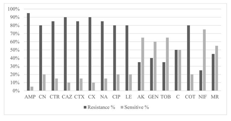
Ecoli isolates revealed high resistance to β-lactams; AMP (95%), CN (80%), CX (90%), CAZ (90%), CTR (85%), CTX (85%), quinolone; NA (85%), fluoroquinolones; CIP (80%), LE(80%) and trimethoprimsulfamethoxazole; COT (80%). The highest sensitivity was observed against NIF(75%) followed by AK (65%), TOB (65%), GEN (60%), C(50%), and MR(55%) respectively (Fig 1). Overall 14 discrete patterns of resistance were observed among the 20 isolates against the 16 antibiotics tested (Table 1). Furthermore, a significant correlation in the antibiotic resistance pattern was observed amongst the different antibiotics for CTR, NIF and MR respectively (Table 2).
Two out of the 20 E coli isolates were sensitive to all three third-generation cephalosporins (3-GC; CTR, CAZ, CTX) however the remaining 18 isolates were resistant to either one/all 3-GCs. ESBL confirmatory test revealed that 10 out of 18 were ESBL producers and 8 were BLIR. A discrete difference in the antibiotic resistance pattern was observed amongst the ESBL and BLIR isolates to the 16 antibiotics tested (Figs 2A,B).
The overall predominance of the pathogenic phylogroup D followed by pathogenic phylogroup B2 and non-pathogenic or commensal phylogroup B1 was observed among ESBL isolates. However, the BLIR
isolates primarily belonged tophylogroup B2 followed by B1 and D. Moreover the 2 non-ESBL isolates belonged to phylogroup B2 and B1 respectively (Fig 3). Distribution of virulence gene fimH (adhesin gene) from the type I fimbriae operon, papGI, papGIII from pap-operon and toxin gene cdtBU were higher in BLIR isolates compared to their incidence in ESBL producers. Furthermore, the distribution of other pap-operon genespapC, papEF, and papGII was higher in ESBL isolates in comparison to BLIR isolates. However, the distribution of the toxin genes, iroN, cnf1, iucD were comparable. Additionally, it was observed that the papoperon genes papC, papEF, papGII, and toxin genes iroN, hylA, cdtBU were absent among non-ESBL producers. (Fig 4). The overall gene frequency of the virulence genes was statistically significant (p=0.05) among the non-ESBL, BLIR, ESBL isolates (Fig 4).
Table 1 — Resistance pattern of the E. coli isolates (n=20) Resistance PatternNo of isolates
AMP, CAZ, CTX, NA, CIP, COT1 AMP, CN, CTR, CAZ, CTX, CX, NA, CIP, COT1 AMP, CN, CTR, CAZ, CTX, CX,1 AMP, CN, CTR, CAZ, CTX, CX, NA, CIP, LE, AK, GEN, TOB, C, COT, MR5 AMP, CN, CTR, CAZ, CTX, CX, NA, CIP, LE, C, COT, NIF2 AMP, CX1 AMP, CN, CTR, CAZ, CTX, CX, NA, CIP, LE, C, COT, NIF, MR1 AMP, CN, CTR, CAZ, CTX, CX, NA, CIP, LE, AK, GEN, TOB, COT, NIF, MR1 AMP, CN, CTR, CAZ, CTX, CX, NA, CIP, LE1 AMP, CN, CTR, CAZ, CX, NA, CIP, LE, COT, NIF, MR1 AMP, CN, CTR, CAZ, CTX, CX, NA, CIP, LE, C, COT1 AMP, CTR, CAZ, CTX, CX, NA, CIP, LE, GEN, C, COT1 AMP, CN, CTR, CAZ, CTX, CX, NA, CIP, LE, AK, GEN, TOB, COT, MR1 AMP, CN, CTR, CAZ, CTX, CX, NA, CIP, LE, COT1
CPearson
T correlation .546* .840**1.793**.608**.793**.608**.608**.840** .308.343.308.420.490*.243.343 RSig. (2-tailed) .013.000-.000.004.000.004.004.000.186.139.186.065.028.303.139
Sum of squares & cross products .8502.4002.5501.7001.5501.7001.5501.5502.4001.0501.2001.0501.5001.400.7501.200
Covariance.045 .126.134.089.082.089.082.082.126.055.063.055.079.074.039.063
N20202020202020202020202020202020
NPearson
I correlation .132.289.243.192-.081.192.243.243.289-.182 -.236-.182.115.2891.000 FSig. (2-tailed) .578.217.303.416.735.416.303.303.217.444.317.444.628.217-1.000
Sum of squares & cross products .2501.000.750.500-.250 .500.750.7501.00-.750-1.00-.750 .5001.0003.750.000 Covariance.013 .053.039.026-.013.026.039.039.053-.039 -.053-.039 .026.053.197.000
MPearson
R correlation .187.408.343.272.057.272.343.343.408 .685**.583**.685** .408.408.0001 Sig. (2-tailed) .429.074.139.246.811.246.139.139.074.001.007.007.074.0741.000
Sum of squares & cross products .4001.6001.200.800.200.8001.2001.2001.6003.2002.8002.8002.0001.600.0004.800 Covariance.021 .084.063.042.011.042.063.063.084.168.147.147.105.084.000.253
*Correlation is significant at the 0.05 level (2-tailed)
Community-or healthcare-acquired UTIs is clinically divided into complicated or uncomplicated and this classification determines the selection of antibiotics for treatment. Empirical therapy accentuated the emergence of antibiotic resistance, especially in uncomplicated UTI. This study documented information on the various aspects associated with UTI from Kolkata, an Eastern region in India.
Studies from different parts of India revealed a varied urine culture positivity rate ranging from 9.753.82%14-21. Our study reported an overall high culture positivity rate(51.1%) among community-acquired uncomplicated UTI cases in hospitalized patients with a higher prevalence among females(60%) than males (40%). The latter observation was consistent with findings from other studies15,18,20. Highest occurrence of UTI was reported among the elderly population14,15,17 which was very similar to the observation stated in this study which consisted of a mean age group of 51.95 years that reflected the association between age and complicacy in UTI.
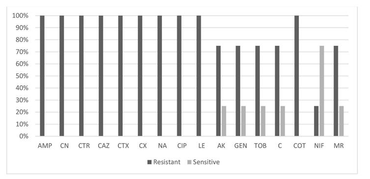
is significant at the 0.01 level (2-tailed)
Fever and dysuria were the most common clinical manifestations among the urine culture-positive patients in our study in concurrence to observations from other studies 14,22. Moreover several studies reported increased frequency to urinate as the commonest symptom in acute uncomplicated UTI patients 14,23. A similar symptomatology was also observed in our study although the symptoms were not always strong enough to conclude about the predictability of UTI.
Diabetes Mellitus (DM) affects the genitourinary system and frequently causes diabetic nephropathy. It also affects the immune system and enhances the chances of acquiring infection of the urinary tract. In our study, DM posed the most common risk factor associated with UTI (73.33%), very similar to the findings registered in another study14. Our study also indicated that clinical manifestations and associated risk factors may not always rightly predict the occurrence of UTI, reaffirming the fact that urine culture is the only confirmatory test to diagnose complicated and uncomplicated UTI.
coli isolates (n=20)
Gram-negative bacilli constituted 93.4% of the uropathogens isolated in this study. Our study also demonstrated that E coli was the leading uropathogen being responsible for UTI (44.4%) in Kolkata an Eastern region in India which was in concurrence with reports from other studies15,17,18,20. However on the contrary several reports also revealed S aureus, Paeruginosa and Klebsiella spp. to be the most predominant uropathogen detected respectively19,24,25.
The choice of antimicrobials for the treatment of UTI is often based on local resistance profiles of the uropathogen.The increasing pattern of resistance is emerging as a universal threat. In this study, the antibiotic susceptibility was conducted on the most frequent isolate E coli. The highest resistance was documented against the 3-GC (CTR, CAZ, CTX) as well as the second generation of fluoroquinolones (CIP, LE) and COT. The highest sensitivity was seen against NIF as well as the aminoglycosides (AK, GEN, TOB). This result was very similar to previous studies conducted in the different States of India15,17,18.
A significant correlation in the resistance pattern against CTR and CAZ, CTX, CX, CIP, LE, COT and MR and AK, GEN, TOB respectively further delineated the fact that resistance to the cephalosporin (CTR, CX, CAZ, CTX) group of the drug was accompanied by the fluoroquinolone (CIP, LE) group and COT and resistance against aminoglycosides (AK, GEN, TOB) was accompanied by MR respectively. Therefore our
study suggested that NIF should be an ideal choice for this population. However orally acceptable NIF formulation has drawbacks and was often not advised for severe upper UTI or individuals with systemic involvement and in such cases, aminoglycosides were the best choice. Therefore routine monitoring and intricate analysis of drug sensitivity patterns among uropathogens pose an absolute necessity to develop proper prescription policies. Our study also successfully detected the ESBL producers (50%) among E coli isolates resistant to third-generation cephalosporins (CTR, CAZ, CTX) which was much higher than that reported from other studies 14 . Incidence of BLIR was also observed in 40% of the E coli isolates. The ESBL and the BLIR isolates were highly resistant towards the fluoroquinolones in consistent with an Indian study26. Our study also indicated that the BLIR isolates were more difficult to treat as they revealed resistance to all drugs tested except NIF. In this study AK,GEN,TOB was found to be effective against the ESBL producers together with NIF, MR and C Siddaramappa, et al26 in his study stated that at least 70% of UTI-associated ESBLproducing E coli isolates could be sensitive to chloramphenicol and it may be an appropriate choice to treat the ESBL producers.
UPEC strains that routinely cause infections have been shown to belong to pathogenic phylogroups B2 and D27. In our study, 50% of the ESBL producers belonged to phylogroup D followed by B2 (40%) which was in agreement with another previous Indian study28 Moreover, for the first time, our study also showed that 62.5% of the BLIR isolates belonged to phylogenetic group B2 followed by B1 (25%) which was distinctly different from the phylogenetic background of the ESBL producers isolated in this study. Furthermore, our study also revealed a varied prevalence of virulence genes (fimH, papC, papEF, papGII, hlyA, iroN, cnf1, iucD, cdtBU) across the ESBL and BLIR isolates with the absence of some other virulence genes; papC, papEF, papGII, iroN, hylA, cdtBU among the nonESBL isolates very similar to another study that showed a high incidence of multiple virulence genes among the multidrug-resistant ESBL E. coli28. An earlier study reported that the presence of papC, cnf1 and hlyA in UPEC isolates played an important role in recurrent infections of the urinary tract27. Therefore the incidence of papC(50%), cnf1(25%) and hlyA(25%) in E coli isolated in this study indicated their possibility to cause recurrent infection.
Our study had some limitations. This work relied on very few UPEC isolates from a single hospital. Studies using many isolates from hospitals from different parts of West Bengal could provide broader
insights into the clinico-microbiological aspects of UTI from this Region in India. Future studies of such kind may be very useful to design appropriate clinical management strategies.
Therefore in the present study, we wanted to highlight the importance of investigating the clinicomicrobial aspects of UTI together with intricate analysis on the antibiotic sensitivity pattern and distribution of urovirulence determinants among the most frequent uropathogen E coli. Moreover increasing resistance against 3GCs together with the presence of β lactamase inhibitor resistance indicated that Cephalosporins, as well as their inhibitor combination drugs, must not be recommended in treating UTI. However, nitrofurantoin can be started as an empiric antibiotic, which can later be altered according to the drug susceptibility pattern of the uropathogen.
Conflict of interest : The authors declare that they have no conflict of interest.
1Bien J, Sokolova O, Bozko P — Role of Uropathogenic Escherichia coli Virulence Factors in Development of Urinary Tract Infection and Kidney Damage. IntJNephrol 2012; 2012: 681473. doi: 10.1155/2012/681473. Epub 2012 Mar 8.
2Tan CW, Chlebicki MP — Urinary tract infections in adults. Singapore Med J 2016; 57(9): 485-90.
3Bush K — The impact of beta-lactamases on the development of novel antimicrobial agents. Curr OpinInvestig Drugs 2002; 3(9): 1284-90.
4Rupp ME, Fey PD — Extended spectrum beta-lactamase (ESBL)-producing Enterobacteriaceae: considerations for diagnosis, prevention and drug treatment. Drugs 2003; 63(4): 353-65.
5Shah AA, Hasan F, Ahmed S, Hameed A — Extended-spectrum beta-lactamases (ESbLs): characterization, epidemiology and detection. Crit Rev Microbiol 2004; 30(1): 25-32.
6Bader MS, Loeb M, Brooks AA — An update on the management of urinary tract infections in the era of antimicrobial resistance. Postgrad Med 2017; 129(2): 242-58.
7Vellinga A, Cormican M, Hanahoe B — Antimicrobial management and appropriateness of treatment of urinary tract infection in general practice in Ireland. BMC Fam Pract 2011; 108(12).
8Tarchouna M, Ferjani A, Ben-Selma W, Boukadida J — Distribution of uropathogenic virulence genes in Escherichia coli isolated from patients with urinary tract infection. Int J Infect Dis 2013; 17(6): e450-3.
9Mukherjee M, Koley S, Mukherjee S, Basu S, Ghosh B, Chakraborty S — Phylogenetic background of E. coli isolated from asymptomatic pregnant women from Kolkata, India. J Infect Dev Ctries 2015; 9(7): 720-4.
10Mukherjee M, Basu S, Majumdar M — Detection of blaTEMandblaCTXM genes by multiplex polymerase chain reaction amongst Uropathogenic Escherichia coli strains isolated from hospitalized patients in Kolkata, India. Int J Biosci 2011; 1(6): 64-9.
11Mukherjee M, Basu S, Mukherjee SK, Majumder M — MultidrugResistance and Extended Spectrum Beta-Lactamase Production in Uropathogenic E. Coli which were Isolated from Hospitalized Patients in Kolkata, India. JClinDiagnRes 2013; 7(3): 449-53.
12Basu S, Mukherjee SK, Hazra A, Mukherjee M — Molecular Characterization of Uropathogenic Escherichia coli: Nalidixic Acid and Ciprofloxacin Resistance, Virulent Factors and Phylogenetic Background. J ClinDiagn Res 2013; 7(12): 2727-31.
13Liu Y, Huang H, Gao R and Liu Y — Dynamic Phenotypesand Molecular Mechanismsto Understand the Pathogenesisof Diabetic Nephropathy in TwoWidely Used Animal Models of Type 2Diabetes Mellitus. Front Cell Dev Biol 2020; 8: 172.
14Eshwarappa M, Dosegowda R, Aprameya IV, Khan MW, Kumar PS, Kempegowda P — Clinico-microbiological profile of urinary tract infection in south India. Indian J Nephrol 2011; 21(1): 30-6.
15Sood S, Gupta R — Antibiotic resistance pattern of community acquired uropathogens at a tertiary care hospital in jaipur, rajasthan. Indian J Community Med. 2012; 37(1): 39-44.
16Akram M, Shahid M, Khan AU — Etiology and antibiotic resistance patterns of community-acquired urinary tract infections in J N M C Hospital Aligarh, India. Ann Clin Microbiol Antimicrob 2007; 6: 4.
17Dash M, Padhi S, Mohanty I, Panda P, Parida B — Antimicrobial resistance in pathogens causing urinary tract infections in a rural community of Odisha, India. J Family Community Med 2013; 20(1): 20-6.
18Sahay S — Prevalence of Urinary Tract Infection and the Antibiotic Sensitivity Pattern of the Most Common Uropathogen from a Tertiary Care Hospital of Jamshedpur. Int J Med Res Prof 2020; 6(1): 51-3.
19Ghosh S, Bandyopadhyay, Lahiri A, Adhya S, BhattacharjeeA — Pattern of antibiotic sensitivity in clinically suspected cases of urinary tract infections: An observation from a tertiary care hospital in west Bengal. J Med Sciclin Res 2017; 5(8)
20Mehta M, Bhardwaj S, Sharma J — Screening of Urinary Isolates for the Prevalence and Antimicrobial Susceptibility of Enterobacteria other than Escherichia coli. InternationalJournal of Life Science and Pharma Research 2013; 3(1): 100-4
21Prakash D, Saxena RS — Distribution and antimicrobial susceptibility pattern of bacterial pathogens causing urinary tract infection in urban community of meerut city, India. ISRN Microbiol 2013; 2013: 749629.
22McNulty CA, Richards J, Livermore DM, Little P, Charlett A, Freeman E, et al — Clinical relevance of laboratory-reported antibiotic resistance in acute uncomplicated urinary tract infection in primary care. J Antimicrob Chemother 2006; 58(5): 1000-8.
23Little P, Merriman R, Turner S, Rumsby K, Warner G, Lowes JA, et al — Presentation, pattern, and natural course of severe symptoms, and role of antibiotics and antibiotic resistance among patients presenting with suspected uncomplicated urinary tract infection in primary care: observational study. BMJ 2010; 340: b5633.
24Ehinmidu JO, Bolaji RO, Adegboye EEA — Isolation and antibiotic susceptibility profile of Neisseria gonorrhoeae isolated from urine samples in Zaria, northern Nigeria. Journal of Phytomedicine and Therapeutics 2003; 8-11: 20-4.
25Aboderin OA, Abdu AR, Odetoyin BW, Lamikanra A — Antimicrobial resistance in Escherichia coli strains from urinary tract infections. J Natl Med Assoc 2009; 101(12): 1268-73.
26Siddaramappa S, Pullela K, Thimmappa B, Devkota R, Bajaj R, Manivannan B, etal — Characterization of blaCTX-M sequences of Indian origin and thirteen uropathogenic Escherichia coli isolates resistant to multiple antibiotics. BMC Res Notes 2018; 11(1): 630.
27Chakraborty A, Adhikari P, Shenoy S, Saralaya V — Molecular characterisation of uropathogenic Escherichia coli isolates at a tertiary care hospital in South India. Indian J Med Microbiol 2017; 35(2): 305-10.
28Padmavathy K, Padma K, Rajasekaran S — Multidrug Resistant CTX-M-Producing Escherichia coli: A Growing Threat among HIV Patients in India. J Pathog 2016; 2016: 4152704.
There is a general notion that glycemic control has deteriorated since the onset of COVID-19 pandemic. This study aimed to compare the glycemic status of a very large cohort of Persons with diabetes (PWD) prior to COVID-19 outbreak with that of a similar cohort post onset of the outbreak.
Materials and methods : In this retrospective study, entire data of glycated hemoglobin (HbA1c)% available in the hospital database from 3rd October, 2017 till 31st May, 2020 were collected and segregated into two cohorts namely on or prior to (ie, pre COVID-19) and after (ie, post COVID-19) 15th March, 2020 respectively.
Results : Total 20575 HbA1c values (12081 in the pre COVID-19 arm and 8494 in the post COVID-19 arm) were available for analysis. Mean (±SD) and Median HbA1c% in the pre COVID-19 arm (7.74±1.33, 7.5) was significantly (P<0.05) lower compared to those of the post COVID-19 arm (8.28± 1.9, 7.8). Such a difference was mainly driven by significantly higher numbers in the subgroup of HbA1c >10% (P<0.05). The Mean (±SD) and Median HbA1c% of the subgroups namely; males, females, age <65 years and age >65 years in the post COVID-19 arm (8.33±1.9, 7.8; 8.21±1.9, 7.7; 8.23±1.9, 7.7 and 8.13±1.8, 7.4 respectively) were significantly higher than the pre COVID-19 arm (7.73±1.3, 7.4; 7.75±1.2, 7.5; 7.72±1.2, 7.5 and 7.9±1.5, 7.6 respectively).
Conclusion : Onset of COVID-19 pandemic has adversely impacted glycemic control amongst PWD in general.
[J Indian Med Assoc 2022; 120(10): 54-7]
Key words :COVID-19, Diabetes mellitus, HbA1c, Glycemic status, Hyperglycemia.
The first cases of COVID-19 in India were reported on 30 January, 2020 in three towns of Kerala1. By March 11, 2020 the disease had spread in most countries and therefore was declared as a pandemic by the World Health Organization (WHO)2. Lockdowns started in different countries; in India it was announced in Kerala on 23 March and in the rest of the country on 25 March. The Ministry of Health and Family Welfare reported that due to scaling up of tests, which led to prompt identification and treatment, India’s positivity rate had fallen to 8% by October, 20203. The first case of COVID-19 in the state of Assam was registered on 31st March, 2020; a 52 year old person detected in Silchar Medical College4
The COVID-19 scenerio led to discontinuation in routine follow-up, treatment delays with treating physicians which were replaced by virtual consultations, except for emergencies. This situation mostly affected the people with chronic illnesses like diabetes, chronic kidney disease, hypertension etc 5-7 . Thus, the
Apollo Excelcare Hospital, Guwahati 781033
1DM (Endocrinology), Consultant Endocrinologist, Department of Endocrinology and Corresponding Author
2MSc, Research Associate, Department of Endocrinology
3MD, Consultant Biochemist and Laboratory Director, Department of Laboratory Medicine
4 MBBS, Chief Medical Administrator cum Medical Superintendent, Department of Hospital Administration
Received on : 22/08/2022
Accepted on : 01/09/2022
There are few indispensable factors like diet, exercise, stress management and treatment adherence for maintenance of glycemic status in patients with diabetes. The COVID-19 pandemic has posed a global threat, with collateral impact going beyond the direct outcome of infection, supposedly worsening the glycemic status of patients with diabetes in general.
This study provides concrete evidence from a very large cohort of almost 20,000 patients' data. The findings of this study will serve as an important 'reference point' for impact of the COVID-19 epidemic on diabetes- both for our country and the whole world as well.
aforementioned lockdown situation brought restrictions to people’s normal activities as only essential activities were allowed during the period 7,8. Even outdoor exercise was restricted9.
Diabetes is a metabolic condition which is in the increasing trend globally10. The predominant factors for management of chronic disease like diabetes includes life style modifications with proper diet and exercise and routine follow up visits with treating doctors. After the outburst of COVID-19 pandemic and social restrictions associated with it proper life style management was hindered, which is an important aspect to maintenance of glycemic status11. A report by the WHO, based on a survey in May, 2020 demostrated an interference in diabetes treatment in 155 Countries6. An earlier study in India reported that
Impact of COVID-19 Pandemic on the Glycemic Status of a Large Cohort in Patients with Diabetes
glycemic status worsened in diabetic patients after 3 weeks of lockdown 12 . Another study in Japan demonstrated a worsened glycemic status in terms of HbA1c values after declaration of state of emergency by the Government13. However, we have contrasting reports from different countries14-16
Hence aim of this study was to compare and analyze the values of HbA1c in a large cohort available in the hospital database during two specified periods; one prior to COVID-19 pandemic and another after the onset of COVID-19 pandemic.
In this registry based retrospective study, entire data of HbA1c since the day of commission of our hospital ie, from 3rd October, 2017 till 31st May, 2020 were included in the pre COVID-19 arm and HbA1c data from 1st June till 14th December 2021, were included in the post COVID-19 arm. Our institution became a Government registered COVID-19 care centre from 1st June onwards. Due to Government restrictions our hospital did not deal with any COVID19 infected patient till 31st May, 2020. Hence, we took 31st May as our cut off for pre COVID-19 arm. The lowest cut -off for including an HbA1c value in either arm was kept at 6% so as to avoid biases like haemoglobinopathy, pre diabetic or non- diabetic status etc. Each arm was divided into 5 subgroups based on HbA1c categories such as (6 6 6.9) %, (7 6 7.9)%, (8 6 8.9)%, 9 6 9.9)% and >10%, and head to head comparison with regard to distribution of patients was performed between identical sub-groups from each arm. Additionally baseline characteristics and certain other attributes like sex and age (above and below 65 years) of the pre COVID-19 arm were compared with those of the post COVID-19 arm.
Microsoft Office Excel 2007 17,18 was used to perform descriptive statistical analysis, and to generate graphs and tables. Continuous measurements are expressed as Mean (±SD) and categorical measurements are expressed as percentages. Variance and mean difference were calculated to perform z- test for p value analysis using Microsoft Office Excel 2007.
Data of subjects were collected and used for the study after necessary permission from the hospital authority.
HbA1c reports of 20575 subjects were included for final analysis; with 12081 in the pre COVID-19 arm and 8494 in the post COVID-19 arm. Mean age of the subjects in the pre and post COVID-19 arms were 53.7
and 54 respectively. Majority of subjects in the both the pre and post COVID-19-arms were males (58.45 and 61.43) % respectively.
Characterizations of Subjects based on Sex and Age of Pre and Post COVID-19 HbA1c status :
The Mean (±SD) and Median HbA1c (%) of male subjects, female subjects, subjects above and below 65 years of age and all subjects irrespective of age and sex in the post COVID-19 arm were significantly higher than the comparable categories of subjects in the post COVID-19 arm (Fig 1).
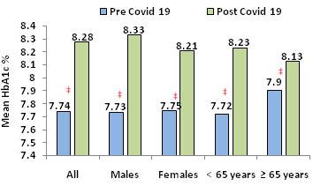
Sequential Hb1Ac Distribution of Pre and Post COVID-19 Subjects :
Analysis of the distribution of subjects into 5 sequential HbA1c subgroups such as (6 to 6.9)%, (7 to 7.9)%, (8to 8.9)%, 9 to 9.9)% and >10% revealed that there was a significantly higher proportion falling in HbA1c >10% sub group in the post COVID-19 arm (Fig 2). In the other sub-groups the difference between
Fig 1 — Histogram showing the comparison of Mean HbA1c values in the pre and post COVID-19 sub-groups [all, males, females, subjects <65 years and >65 years. The difference of means in all the sub-groups was found to be statistically significant. (P<0.05). HbA1c, Glycated haemoglobin
Fig 2 — Histogram showing the distribution of subjects in 5 different (6-6.9, 7-7.9, 8-8.9, 9-9.9 and >10) pre and post COVID-19 sub-groups based on HbA1c%. In the HbA1c >10 % sub-group, the distribution of subjects was statistically significant. P<0.05 is considered significant. ‡p=<0.05, †p=NS
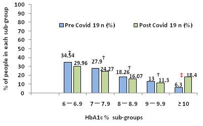
pre and post COVID-19 arm were insignificant (Fig 2) After segregation of subjects based on sex and age (below and above 65years), or irrespective of age and sex, in both pre and post COVID-19 HbA1c >10% subgroup, we observed a statistically significant differential distributuion in all the groups (p<0.05) (Table 1, Fig 3).
‘Lockdown’, an important decision taken globally to control and prevent the spread of COVID-19 disease, had an impact on glycemic control in diabetic patients as they weren’t able to continue with their routine activities and follow up with physicians. Therefore, we conducted this study to evaluate the effect of COVID19 pandemic related lockdown on glycemic status in PWD.
Our study used a huge data of subjects inclusive of pre (n=12084) and post COVID-19 (n=8490) diabetic patients. These subjects had HbA1c status of >6% segregated into 5 sub-groups who were attending a hospital in North East India from 3rd October, 2017 till 31st May, 2020. Higher Mean HbA1c values in the post COVID-19 subgroups suggested a correlation between poor glycemic status of subjects with COVID-19 pandemic (Fig 1). Possible reasons could be a discontinuity in the routine care of patients in the post
Table 1 — Total subjects in 5 different categories of HbA1c. Significance of each category is indicated in terms of p values
HbA1c (%) Pre COVID-19 Post COVID-19 P value categoryn (%)n (%)
6 - 6.94170 (34.54)2542 (29.96)0.3 (NS)
7 - 7.93368 (27.9)2061 (24.27)0.4 (NS)
8 - 8.92204 (18.26)1365 (16.07)0.2 (NS)
9 - 9.91565 (13)959 (11.3)0.4 (NS)
(6.3)1562 (18.4)< 0.05
COVID-19 period. Our study and others suggested that the lockdown situation worsened the glycemic status of PWD12,13. Most couldn’t even continue with their daily exercise schedules as outdoor movement was restricted except for emergency situations9. It was also described by other researchers that the forced nationwide lockdowns could produce acute panic, anxiety and stress which may also lead to worsened glycemic status19,20. The pandemic situation became quite scary as there were deaths all over and most people preferred to stay at home rather than contract the disease.
The segregation of subjects into 5 different subgroups based on HBA1c categories ie, (6 to 6.9)%, (7 to7.9)%, (8 to 8.9)%, (9 to 9.9)% and >10% gave us an opportunity to minutely look at influence of COVID-19 pandemic on trends of Hyperglycemia. Interestingly the trends in the lower HbA1c subgroups in the post COVID-19 cohort were not significantly different from the pre COVID-19 situation. In contrast, a clinically relevant and hugely impactful worsening trend was observed by way of significantly higher numbers in the HbA1c >10% sub group of post COVID19 sub-group, irrespective of age and sex. (Table 1, Fig 2). Hence, it can be speculated that a COVID-19 pandemic had a global adverse impact on the glycemic control of diabetic patients. In other words, the above pattern of distribution of the subjects confirms that the glycemic status deteriorated during the COVID-19 pandemic leading to increased PWD in the highest HbA1c category in the post COVID-19 group. Thus, regular monitoring of HbA1c values, stringent treatment adherence with routine follow up visits with treating physicians and continued effort for lifestyle modifications with proper diet and exercise are important aspects for maintenance of glycemic status which was interrupted due to various factors during COVID-19 pandemic and associated lockdown.
Fig 3 — Histogram showing the distribution of subjects with HbA1c >10% in the pre and post COVID-19 sub-groups based on sex [males, females, <65 years, >65 years and all irrespective of age and sex. ‡p=<0.05. HbA1c, Glycated haemoglobin
Although we analyzed the pre and post COVID-19 glycemic status of PWD they were not exactly matched for age and sex distribution or presence or absence of comorbidities, ongoing treatments, socioe-conomic background, dietary and lifestyle factors etc. which may have significant bearing on the glycemic status. This remains a major limitation of our study. However, it would not have been possible to invite each and every patient who attended the hospital during pre COVID19 period for evaluation during the pandemic. However the sheer number of available data for analysis represents two different situations namely; pre and post COVID-19 is definitely a strength of our study and justifies our effort to generate a hypothesis that COVID-
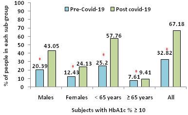
19 pandemic has adversely impacted the glycaemic status of diabetic patients in our country. This study provides some original data for reference with respect to COVID-19 pandemic situation in our country.
Onset of COVID-19 pandemic has adversely impacted glycemic control amongst PWD in general. Such an impact was seen in all sub categories irrespective of age and sex. Concerted effort with execution of alternative strategies for overall management of Diabetes Mellitus could possibly prevent such a worsening during the pandemic situation.
Authors’ contributions : MPB, Conceptualization and medical writing; SB, Data analysis, medical writing; ARB, Data extraction, initial analysis and SKS, Medical records, data extraction.
1Andrews MA, Areekal B, Rajesh KR, Krishnan J, Suryakala R, Krishnan B, et al — First confirmed case of COVID-19 infection in India: A case report. Indian J Med Res 2020; 151(5): 490-2. doi: 10.4103/ijmr.IJMR_2131_20.
2Cucinotta D, Vanelli M — WHO Declares COVID-19 a Pandemic. Acta Biomed 2020; 91(1): 157-60. doi: 10.23750/ abm.v91i1.9397.
3MoHFW. “With very high COVID-19 testing, India’s positivity rate fallen below 8%: MoHFW.” https://economictimes. indiatimes.com/news/politics-and-nation. 18 October 2020. The Economic Times.
4First Corona Case in Assam: 52-year-old Tested Positive In Silchar Medical College.https://www.barakbulletin.com. March 31 2020. Barak Bulletin.
5Chudasama YV, Gillies CL, Zaccardi F, Coles B, Davies MJ, Seidu S, et al — Impact of COVID-19 on routine care for chronic diseases: A global survey of views from healthcare professionals. Diabetes Metab Syndr 2020; 14(5): 965-7. doi: 10.1016/j.dsx.2020.06.042.
6Dyer O — COVID-19: Pandemic is having “severe” impact on non-communicable disease care, WHO survey finds. BMJ. 2020 Jun 3; 369:m2210. doi: 10.1136/bmj.m2210.
7Eberle C, Stichling S — Impact of COVID-19 lockdown on glycemic control in patients with type 1 and type 2 diabetes mellitus: a systematic review. Diabetol Metab Syndr 2021; 13 (1): 95. doi: 10.1186/s13098-021-00705-9.
8Shaki O, Gupta TP, Rai SK — COVID-19 pandemicEnvironmental perspective of COVID-19 and a primer for all of us. J Family Med Prim Care 2021; 10(1): 48-55. doi: 10.4103/jfmpc.jfmpc_1055_20.
9Ghosh A, Arora B, Gupta R, Anoop S, Misra A — Effects of nationwide lockdown during COVID-19 epidemic on lifestyle and other medical issues of patients with type 2 diabetes in
north India [published online ahead of print, 2020 Jun 2]. Diabetes Metab Syndr 2020; 14(5): 917e20. https://doi.org/ 10.1016/ j.dsx.2020.05.044.
10Saeedi P, Petersohn I, Salpea P, Malanda B, Karuranga S, Unwin N, et al — IDF Diabetes Atlas Committee. Global and regional diabetes prevalence estimates for 2019 and projections for 2030 and 2045: Results from the International Diabetes Federation Diabetes Atlas, 9th edition. Diabetes Res Clin Pract 2019; 157: 107843. doi: 10.1016/ j.diabres.2019.107843.
11Mei C, Kang Y, Zhang C, He C, Liao A, Huang D — C-Type Natriuretic Peptide Plays an Anti-Inflammatory Role in Rat Epididymitis Induced by UPEC Front Cell Infect Microbiol 2021; 11: 711842. doi: 10.3389/fcimb.2021.711842.
12Khare J, Jindal S — Observational study on Effect of Lock Down due to COVID 19 on glycemic control in patients with Diabetes: Experience from Central India. Diabetes Metab Syndr 2020; 14(6): 1571-4. doi: 10.1016/j.dsx.2020.08.012. Epub 2020 Aug 20.
13Tanji Y, Sawada S, Watanabe T, Mita T, Kobayashi Y, Murakami T, et al — Impact of COVID-19 pandemic on glycemic control among outpatients with type 2 diabetes in Japan: A hospitalbased survey from a country without lockdown. Diabetes Res Clin Pract 2021; 176: 108840. doi: 10.1016/ j.diabres.2021.108840.
14Fernández E, Cortazar A, Bellido V — Impact of COVID-19 lockdown on glycemic control in patients with type 1 diabetes. Diabetes Res Clin Pract 2020; 166: 108348. doi: 10.1016/ j.diabres.2020.108348.
15Ruissen MM, Regeer H, Landstra CP, Schroijen M, Jazet I, Nijhoff MF, et al — Increased stress, weight gain and less exercise in relation to glycemic control in people with type 1 and type 2 diabetes during the COVID-19 pandemic. BMJ Open Diabetes Res Care 2021; 9(1): e002035. doi: 10.1136/ bmjdrc-2020-002035.
16Potier L, Hansel B, Larger E, Gautier JF, Carreira D, Assemien R, et al — Stay-at-Home Orders During the COVID-19 Pandemic, an Opportunity to Improve Glucose Control Through Behavioral Changes in Type 1 Diabetes. Diabetes Care 2021; 44(3): 839-43. doi: 10.2337/dc20-2019.
17Divisi D, Di Leonardo G, Zaccagna G, Crisci R — Basic statistics with Microsoft Excel: a review. J Thorac Dis 2017; 9(6): 1734-40. doi: 10.21037/jtd.2017.05.81.
18Svetlana Todorova, 2019. “Statistics for Data Analysis Using Microsoft Excel,”Izvestia Journal of the Union of ScientistsVarna. Economic Sciences Series, Union of Scientists - Varna, Economic Sciences Section, vol. 8(2), pages 68-74, August.
19Dubey S, Biswas P, Ghosh R, Chatterjee S, Dubey MJ, Chatterjee S, et al — Psychosocial impact of COVID-19. Diabetes Metab Syndr 2020; 14(5): 779-88. doi: 10.1016/ j.dsx.2020.05.035. Epub 2020 May 27.
20Zandifar A, Badrfam R — Iranian mental health during the COVID-19 epidemic. Asian J Psychiatr 2020; 51: 101990. doi: 10.1016/j.ajp.2020.101990. Epub 2020 Mar 4.
Cancer is one of the leading causes of morbidity and mortality worldwide & cancer pain is experienced by patients with advanced, metastatic and terminal disease. Buprenorphine, a partial µ-receptor agonist and antagonist at the kopioid receptor, shows no clinically relevant accumulation of active metabolites, and pharmacokinetics remain unchanged in renal insufficiency. In elderly cancer patients, the use of opioids for control of cancer pain is a therapeutic challenge, as these group of patients often associated with renal and hepatic comorbidity that limited the use of strong opioids like morphine.
Methods : A retrospective observational study was conducted in elderly patients to estimate the efficacy of transdermal buprenorphine patch for controlling of cancer pain as well as to assess the safety of the patch. For pain control Numerical Rating Score (NRS) was used & for safety assessment Grade 3 or 4 toxicity were recorded.Hepatic & renal toxicity were measured at baseline, at 1st month & at 3rd month of treatment & lastly at 6th month of treatment.
Results : Majority of the patients showing good to excellent global satisfaction with Buprenorphine patch and 57% of patients suffered from constipation along with 38% nausea & vomiting. It was found that there was a significant reduction in pain intensity from baseline with a p value of <0.05. There was no significant hepatic or renal toxicity found in the study.
Conclusion : Transdermal buprenorphine patch is effective and safe in elderly cancer patients for pain control. Further studies should be performed in order to find safe and effective opioid methods necessary to give greater insight into the difficult balance between analgesia and toxicity.
[J Indian Med Assoc 2022; 120(10): 58-61]
Key words :Buprenorphine, Cancer pain, Elderly.
Cancer is one of the leading causes of morbidity and mortality worldwide, responsible for 19.2 million new cases and 9.9 million deaths in 2020 globally (Globocan 2020). InIndia, more than one million new cases of cancer are diagnosed each year and it is estimated that the cancer burden in India will almost double during the coming 20 years1
Pain is experienced by 55% of patients undergoing anti-cancer treatment and by 66% of patients who have advanced, metastatic and terminal disease (WHO). It was also shown that over 38.0% of all cancer patients experienced moderate-to-severe pain 2 . With the advances in preventive, diagnostic& therapeutic measures, the life expectancy of cancer patients have increased considerably. With high prevalence of advanced & metastatic disease in our country the prevalence of acute & chronic pain amounts to almost 70% of cancer patients.
Department of Radiotherapy, R G Kar Medical College & Hospital, Kolkata
1 MBBS, MD (Radiotherapy),
2MBBS, 3rd
3MBBS, MD (Radiotherapy),
on : 08/10/2022
:
Cancer related pain is the most common cause of morbidity in cancer patients. Among various opioids buprenorphine is one of the most common & efficacious method to control pain evident by various study. In our study it showed similar results in elderly population. So we may conclude that buprenorphine transdermal patch can effectively reduce the pain and alleviate quality of life, though a large scale prospective study may be needed.
The World Health Organization (WHO) recommended ‘a three-step analgesic ladder’ for management of cancer pain based on pain intensity. A wide variety of medications ranging from acetaminophen & NSAIDs to strong opioids like morphine, buprenorphine is available now. The use of morphine may be complicated by accumulation of morphine metabolites, leading to dose reduction or opioid rotation in renally impaired, elderly or high-dose patients 3 . By contrast Buprenorphine shows no clinically relevant accumulation of active metabolites4, and pharmacokinetics remain unchanged in renal insufficiency5. Buprenorphine therefore might be used as an alternative in these compromised patients6
Buprenorphine, a partial µ-receptor agonist and antagonist at the κ-opioid receptor7, has been available
Efficacy and Safety of Transdermal Buprenorphine Patch for Cancer Pain Relief in Elderly Patients : A Retrospective Study from a Tertiary Care Centre of Eastern India
since 1981 in sublingual and parenteral formulation, and has a well-established efficacy and safety profile8
A transdermal patch is a medicated adhesive patch, is placed on the skin to deliver a specific dose of medication through the skin and into the blood stream. The advantage of transdermal drug delivery route over other types of medication delivery route like oral, topical, intra-venous or intra-muscular is that the patch provides a controlled release of medication into the patient. Buprenorphine transdermal formulation was introduced in Europe in 2001, and the patches of different releasing rates are available now.
Transdermal patch is non-invasive, effective & well accepted formulation accepted by cancer patients who have gastrointestinal problems and difficulties in oral medication (esophageal,gastric, intestinal, maxillofacial cancer) either due to cancer or due to side effects of oral or parenteral concomitant medications9
In elderly cancer patients, the use of opioids for control of cancer pain is a therapeutic challenge, as these group of patients often associated with renal and hepatic comorbidity that limited the use of strong opioids like morphine. Transdermal buprenorphine has been found to be effective and safety for elderly patients. However, there is a lack of sufficient data or literatures on this specific topic specially in eastern part of India. So, a study was conducted to find out the safety and efficacy of transdermal buprenorphine patch in elderly cancer patients.
We have conducted a retrospective observational study & have retrieved medical records of 120 elderly cancer patients from Dept. ofRadiotherapy, R G Kar Medical College & Hospital, Kolkata, registered from January 2018 to December 2020. All the patients received Buprenorphine Patch of ‘20µg’ strength,as only patches of that strength were supplied in our institute at that period of time. The inclusion criteria were cancer patients with age 65 years or more. The patients who used concomitant other analgesic medications were excluded from the study.
The primary end point of the study was to estimate the efficacy of transdermal buprenorphine patch for controlling of cancer pain as well as to assess the safety of the patch for the elderly patients.
To estimate the efficacy of pain control, we looked at the Numerical Rating Score (NRS) and Visual Rating Scale (VRS) of the patients suffering from cancer pain at different time interval specifically at the beginning of treatment, at the 1st month ,3rd month and lastly
at the 6th month of treatment.
To assess the safety of the drug for elderly patients we looked at the number of patients suffering from grade 3/4 toxicity or adverse events during their treatment with transdermal buprenorphine patch.
The retrieved data were then tabulated and analysis made accordingly to assess the efficacy and safety of the drug.
We have collected data of a total of 120 patients for this study. Out of them 68 were male and 52 were female.Most of the cases were either in ECOG PS 3 or 2 in our study.
• Age Distribution :
The median age of the population was 69 years with a range from 65 years to 86 years.
• Primary site of the tumors (Fig 1):
It was observed that in majority of the cases, the primary was in breast followed by lung & prostate.
• Metastatic Sites :
our patients’ data sheet, it was seen that majority of them had skeletal metastasis (Table 1).
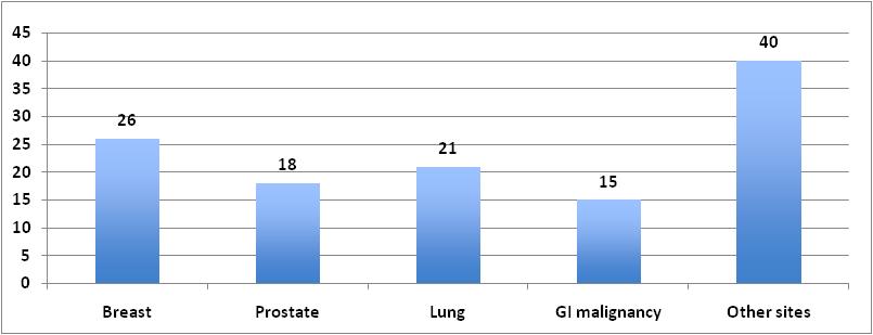
• Adverse events :
All patients were treated with buprenorphine transdermal patch 20 µg/hour in our study. Out of them constipation being the most common adverse event experienced and motor and cognitive impairment being the lowest (Fig 2).
• Global Satisfaction with treatment :
were categorized according to their global satisfaction with the prescribed treatment with
buprenorphine transdermal patch into 5 distinct categories, having “excellent” as the best outcome & “poor” as the worst outcome (Table 2).
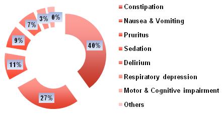
Renal toxicity was measured by using creatinine clearance level at beginning, at 1st month, at 3rd month & at 6th month on buprenorphine transdermal patch. There is no significant renal toxicity found with using buprenorphine transdermal patch in our study (Table 4).
Table
TimelineNo PatientsNo PatientsNo PatientsNo Patients
Crclhaving Crclhaving Crclhaving Crcl >80ml/min 50-80ml/min30-50ml/min <30ml/min
At beginning958467
1st month856488
3rd month7534812
6th month7544712
Pain intensity was measured using Numerical Rating Scale of all the participated patients at baseline, at 1st month & at 3rd month of treatment & lastly at 6th month of treatment. Only patients with moderate and severe pain were considered for the patch and on follow up most of the patients shifted from higher pain score to lower pain score or became painless. The patients were scored ‘0’ having no pain; ‘1-3’ having mild pain; ‘4-6’ having moderate pain & ‘7-10’ having severe pain (Table 3).
Hepatic toxicity was measured according to liver dysfunction level at beginning, at 1st month, at 3rd month & at 6th month on buprenorphine transdermal patch (Table 5).
Table 5 — Hepatic
Buprenorphine
TimelineNo PatientsNo PatientsNo PatientsNo Patients havinghavinghavinghaving no livermild livermoderate liver severe liver dysfunctiondysfunctiondysfunctiondysfunction
At beginning823062
1st month803172
3rd month773193
6th month7630122
There is no significant liver dysfunction found with using buprenorphine transdermal patch in our study.
Strong opioids are recommended for treating severe cancer pain in the advanced stages of the disease. Few data are available concerning the efficacy of buprenorphine in cancer pain. The European Medicines Agency (EMEA) guidelines on chronic
Among 120 patients having moderate to severe pain 60 of them had mild or no pain after six months of use. None of the patient suffered from increase in Pain Intensity Score during the treatment period. So, from the above table it has been found that there is a significant reduction in pain intensity from baseline with a p value of <0.05.
of
of
is a therapeutic
as these group of patients often associated
with renal and hepatic comorbidity that limited the use of strong opioids like morphine. Transdermal buprenorphine has been found to be effective and safety for elderly patients.
It has been observed that majority of the cases primary was in breast followed by lung & prostate in our study. It was also noted that 62 patients out of 120 elderly patients showed global satisfaction as “excellent” or “very good” after using transdermal buprenorphine patch which was also evident in various international papers.
Finally, when we compare pain intensity of those patients from beginning, at 1st month, at 3rd month & at 6th month using numerical rating scale we have found a significant trend towards reduction of pain intensity with a p value of 0.01. Similar data was found by research work done by Poulain Philippe, et al which showed a significant difference in the number of treatment responders was observed: 70 BUP TDS (74.5%, 65.7-83.3) versus 47 placebo (50%, 39.9-60.1) (P =0.0003)10.
It has also observed there is no significant hepatic or renal impairment found when using buprenorphine transdermal patch in our study, which makes itto be an easier convenient method to reduce pain intensity in geriatric population.
In conclusion, we think that further studies should be performed in order to find safe and effective opioid methods necessary to give greater insight into the difficult balance between analgesia and toxicity. It is also important to consider individual variables, such as psychological distress in cancer patients, as these are important as prognostic factors since they affect the therapeutic results.
1.There was no comparator arm in our study from which the efficacy of buprenorphine patch was ascertained.
2.This is a single institutional study.
3.Sample size was small.
4.There was provision for subjective bias as the study didn’t follow any blinding methods.
Disclosure : The authors report no conflict of interest in this work.
1Mallath MK, Taylor DG, Badwe RA, Rath GK, Shanta V, Pramesh CS, et al — The growing burden of cancer in India: Epidemiology and social context. Lancet Oncol 2014; 15: e205-12.
2Van den Beuken-van Everdingen MH, Hochstenbach LM, Joosten EA, Tjan-Heijnen VC, Janssen DJ — Update on prevalence of pain in patients with cancer: Systematic review and meta-analysis. JPainSymptomManage 2016; 51: 107090.e9.
3Dean M — Opioids in renal failure and dialysis patients. J Pain Symptom Manage 2004; 28: 497e504.
4Mercadante S, Arcuri E — Opioids and renal function. J Pain 2004; 5: 2e19.
5McQuay HJ, Moore RA, Bullingham RES — Buprenorphine kinetics. Adv Pain Res Ther 1986; 8: 271e278.
6Filitz J, Griessinger N, Sittl R — Effects of intermittent hemodialysis on buprenorphine and nor- buprenorphine plasma concentrations in chronic pain patients treated with transdermal buprenorphine. Eur J Pain 2006; 10: 743e748.
7Budd K — Buprenorphine: a review. Newmarket: Hayward Medical Communications, 2002.
8Johnson RE, Fudala PJ, Payne R — Buprenorphine: Considerations for pain management. J Pain Symptom Manage 2005; 29: 297-326.
9Vallerand AH — The use of long-acting opioids in chronic pain management. Nurs Clin North Am 2003, 38: 435-45.
10Efficacy and Safety of Transdermal Buprenorphine: A Randomized, Placebo-Controlled Trial in 289 Patients with Severe Cancer Pain Poulain, Philippe et al. Journal of Pain and Symptom Management, Volume 36, Issue 2, 117-25.
The information and opinions presented in the Journal reflect the views of the authors and not of the Journal or its Editorial Board or the Publisher. Publication does not constitute endorsement by the journal.
JIMA assumes no responsibility for the authenticity or reliability of any product, equipment, gadget or any claim by medical establishments/institutions/manufacturers or any training programme in the form of advertisements appearing in JIMA and also does not endorse or give any guarantee to such products or training programme or promote any such thing or claims made so after.
Objective : To find out the status of Malaria in Odisha.
Methods : By studying the most recent data available on the National Vector Borne Disease Control Programme (NVBDCP) website.
Results : Out of the 30 districts of Odisha, Malaria is highly concentrated in mainly Malkangiri District.
Conclusions : If an approach of universal diagnosis and radical treatment like that which was used in the “Malaria-mukt Bastar” campaigns of Chhattisgarh is adopted in Malkangiri District, it is possible that the Annual Parasite Incidence (API) may come down there quickly.
Indian Med Assoc 2022; 120(10): 62-3]
Key words :Malaria, Odisha, Malkangiri.
Odisha is a state located in the Eastern part of India and bordered by Chhattisgarh on the West and the Bay of Bengal in the East. It consists of 30 Districts.
Methods :
According to the most recent data available on the National Vector-Borne Disease Control Programme website (data for the year 2018), the Annual Parasite Incidence (API) for Odisha is 1.482. However, by going through the data for Odisha state, it is seen that the Malaria problem is not equally distributed in the Districts; it is focal as can be seen from the following information2 (Tables 1 & 2).
Results :
So, it is seen that out of the 30 districts, Malaria is highly concentrated in mainly Malkangiri District. From the map given in Fig 1, this District shares its borders with Chhattisgarh on the west and Andhra Pradesh on the East.
It may be further useful to study what was the trend of the APIs in Malkangiri District over the years. For this, the website of the National Vector-Borne Disease Control Programme was referred to and the following findings observed:
It is observed that there is a nearly three-fold decrease in the API between 2017 and 2018. Whether this trend continued into 2019 is not known because the APIs for 2019 have not been published by NVBDCP
1MD(Community Medicine), MPH (UNC-CH, USA), MPH (Biosecurity, NZ), Scientist ‘E’, Department of Epidemiology and Environmental Biology, National Institute of Malaria Research, Sector
8 Dwarka, New Delhi 110077
Received on : 22/11/2021
Accepted on : 07/02/2022
Editor's Comment : Malkangiri District in Odisha has the highest API followed by Gajapati and Phulbani Districts. If an approach of universal diagnosis and radical treatment like that which was used in the “Malaria-mukt Bastar” campaigns of Chhattisgarh is adopted in these and other Districts, it is possible that the overall API may come down quickly in the State.
yet. However, in Chhattisgarh, during 2020 and 2021, four rounds of “Malaria-Mukt Bastar” took place wherein every person living in each of the villages in the Bastar region had their finger pricked and a drop of blood drawn which was examined for the Plasmodium antigen using Rapid Diagnostic Kits. These campaigns detected the Malarial antigen in both febrile persons and asymptomatic carriers and the most recent round was held from June 15, 2021, till July 31, 2021. If the diagnosis was P vivax, Chloroquine and Primaquine were given to the patient. If it was P falciparum, Artemisinin-based Combination Therapy (ACT) and Primaquine was provided. Mixed infections were treated by ACT and Primaquine4,5. As a result, though in the one year preceding till November 2019 there were 5272 cases of Malaria in the Bastar region, during the following year till November, 2020 there were only 2696 cases ie, there was a drop of about 49% in the number of cases6. That means there was some useful effect of these campaigns in that the reservoirs of the Malarial parasite ie, the humans were effectively treated thereby reducing the number of those persons who could be sources of infection to the female Anopheline mosquitoes.
Conclusion : If an approach of universal diagnosis and radical
treatment like that which was used in the “Malaria-mukt Bastar” campaigns
adopted in Malkangiri District, it is possible that the API may come down further and more quickly in Odisha State, especially if it must reach the target of zero cases of Malaria by 2027. This would enable the country to receive the certification of Malaria elimination in 2030.
1Registrar General of India 2011. Map of Odisha. Available at https:/ /censusindia.gov.in/DigitalLibrary/ Data/Census_2011/Map/Orissa/ 00_Orissa.pdf. Accessed on 16 November 2021.
2Annual Report 2018. National Vector-Borne Disease Control Programme. Available from: https:/ /nvbdcp.gov.in/Doc/AnnualReport-2018.pdf. Accessed on 25 August 2021.
Anjul0.48 Balasore 0.05 Bargarh0.44 Bhadrak 0.06 Bolangir 0.32
Boudh0.53 Cuttack 0.04 Deogarh 0.50 Dhenkanal 0.19 Gajapati 6.23 Ganjam 2.43
Jagatsinghpur0.04
Jajpur 0.14
Jharsuguda0.35 Kalahandi 5.17 Phulbani (Kandhamal) 5.65
Kendrapada 0.03 Keonjhar (Odisha) 1.42 Khurda (Khordha) 0.14 Koraput 4.58 Malkangiri 13.85 Mayurbhanj 0.81 Nawarangpur 2.37 Nayagarh 0.49 Nuapada 0.86 Puri 0.05 Rayagada 4.73 Sambalpur 0.32 Sonepur (Subarnapur) 0.66 Sundergarh1.87 Odisha 1.48
3Annual Report 2017. National Vector-Borne Disease Control Programme. Available from: https://nvbdcp.gov.in/Doc/AnnualReport-2017.pdf. Accessed on 31 August 2021.
4https://theprint.in/health/while-covid-raged-chhattisgarhcovered-over-6000-villages-under-malaria-mukt-bastarproject/537481/. Accessed on September 16, 2021.
5https://www.patrika.com/raipur-news/fourth-phase-ofbastar-free-malaria-campaign-against-malaria-anemia6905880/. Accessed on September 16, 2021.
6https://nhm.gov.in/New_Updates_2018/Innovation_summit/ 7th/DCP/DCP-%20PPTs%20%287%29/CGBest%20Practices%20MMB1.pptx. Accessed on 20 September 2021.
The novel coronavirus disease (COVID-19) caused by SARS-CoV-2 has turned the world topsy-turvy since its emergence. Although COVID-19 is mostly associated with respiratory pathology, it can also result in several extrapulmonary manifestations. Multisystem Inflammatory Syndrome in Adults (MIS-A) seems to be a new addition to the ever expanding COVID-19 puzzle and warrants extensive research to familiarize the phenotype, formulate a definitive treatment and prognosticate accordingly. This article highlights the case definition, pathogenesis, clinical features and treatment modalities of this new entity with a concise review of available literature at present.
[J Indian Med Assoc 2022; 120(10): 64-7]
Key words :COVID-19, Multisystem Inflammatory Syndrome, Kawasaki disease.
In April 2020, Multisystem Inflammatory Syndrome in Children (MIS-C) was first described in the context of COVID-19 in United Kingdom and Italy. These reports described a shock-like illness in children resembling Kawasaki disease or Toxic Shock Syndrome1. Similar reports soon came in from most parts of the World and the Centers for Disease Control and Prevention (CDC) published an alert (HAN00432) regarding the same on May 14, 2020. While several reports and studies have been published regarding MISC since then, there is comparatively paucity of literature on Multisystem Inflammatory Syndrome in Adults (MIS-A)2. The authors attempt to review the existing literature and discuss this new clinical entity so as to facilitate early recognition of the condition by healthcare providers to ensure prompt treatment and improve the prognosis.
The CDC defines a case of MIS-A as a patient aged at least 21 years who meets the following clinical and
Department of Internal Medicine, Medical College and Hospital, Kolkata 700073
1MBBS, Junior Resident
2MD (Internal Medicine), MRCP(UK), Assistant Professor and Corresponding Author
3MD (Dermatology, Venereology and Leprosy), DNB, Senior Resident, Department of Dermatology, Venereology and Leprosy, RG Kar Medical College and Hospital, Kolkata 700004
4MD (Internal Medicine), Senior Resident, Department of Neurology, Bangur Institute of Neurosciences, IPGME&R and SSKM Hospital, Kolkata 700020
5MD, FNB (Critical Care Medicine), MRCP(UK), FRCP(London); Professor in charge of Critical Care Medicine division, RG Kar Medical College and Hospital, Kolkata 700004
Received on : 14/06/2022
Accepted on : 17/06/2022
Multisystem Inflammatory Syndrome in Adults (MIS-A) seems to be a new addition to the ever expanding COVID-19 puzzle. MIS-A patients more likely present with myocarditis, cardiac dysfunction, arterial thrombosis, pulmonary embolism, and/ or deep vein thrombosis; than MIS-C.
The manifestations of MIS-A may overlap with COVID-19 and make the diagnosis challenging.
laboratory criteria, in the absence of any other likely alternative diagnosis3 (Table 1).
The clinical features of MIS-C encompass shock, cardiovascular decompensation, abdominal pain with markedly elevated inflammatory markers. Since June 2020, a similar syndrome with gastrointestinal, cardiovascular, dermatologic and neurologic symptoms in absence of severe respiratory ailment in adults has surfaced up in literature. Significant overlap between symptoms of acute COVID-19 per se and MIS-A makes the diagnosis of the latter a tad challenging4. Most common organ system involved seems to be the cardiovascular system4. There have also been reports of young adults presenting with the full spectrum of Kawasaki disease manifestation5. It is also important to exclude respiratory involvement resulting in severe pulmonary disease as tissue hypoxia could lead to organ dysfunction with similar features as that of MISA. Clinical features of MIS-A are described in Table 26.
The pathophysiology of MIS-A is poorly elucidated. As per reports received by CDC, 30% adults and 45% children with Multisystem Inflammatory Syndrome had
Patient aged >21 years admitted for >24 hours, or with an illness resulting in death, meeting the following clinical and laboratory criteria in absence of a more likely alternative diagnosis for such illness
Subjective or documented fever (at least 38.0 C) for at least 24 hours prior to hospitalisation or within the first 3 days of hospitalisation* and at least 3 of the following clinical criteria in the same time frame* (at least one being a primary clinical criterion)
(1) Severe cardiac illness - myocarditis, pericarditis, coronary artery dilatation/aneurysm, or newonset right or left ventricular dysfunction (LVEF<50%), 2nd/3rd degree A-V block, or ventricular tachycardia.
(2) Rash and non purulent conjunctivitis
(1) New onset neurological signs and symptoms- encephalopathy, seizures, meningeal signs, or peripheral neuropathy (including Guillain-Barré syndrome)
(2) Shock or hypotension not attributable to medical therapy (sedation, dialysis)
(3) Abdominal pain, vomiting, diarrhoea
(4) Thrombocytopenia (<150,000/mcl)
(1) Elevated levels of at least 2 of the following - CRP, ferritin, IL-6, ESR, procalcitonin
(2) Positive SARS-CoV-2 test for current or recent infection by PCR, serology, or antigen detection
* These criteria should be met by the end of hospital day-3, where the date of hospital admission is considered as day-0.
negative PCR for SARS-CoV-2 and positive antibodies, suggesting a postinfectious pathophysiology. However, in contrary to moderate and severe COVID-19, usually accompanied by respiratory failure there has been an unexplained paucity of respiratory symptoms and signs in MIS-A 7 . In adults, COVID-19 is typically characterized by hyperactivation of the inflammatory cascade. Increasing evidence suggests that tissue damage in COVID-19 is mostly mediated by the host innate immunity8.This disease is characterized by a cytokine storm resembling that of macrophage activation seen in viral-induced haemophagocytic lymphohistiocytosis. Immune dysregulation, malfunction of the renin-angiotensin-aldosterone axis, endothelial injury and thromboinflammation are the proposed mechanisms for MIS-A. However, blockade of type I and type III interferon responses resulting in unchecked viral proliferation may be also attributable9 In children, studies have shown reduced and/or ineffective titres of neutralizing SARS-CoV-2 antibodies in patients with MIS-C compared to patients with mild to severe COVID-1910. However, similar studies are yet to explore MIS-A. Cheng MH et al. also highlights the mechanism by which SARS-CoV-2 spike protein binds to T cell receptors like a superantigen similar to staphylococcal exotoxin B11
Published Literature on MIS-A :
A recent systematic review documented the clinical characteristics of 221 patients with MIS-A. Around 70% of these patients had experienced a symptomatic COVID-19 like illness with complete recovery prior to onset of symptoms of MIS-A12. The median lag period (from COVID-19 like symptom onset to MIS-A symptom manifestation) was a month (28 days). Fever
was the most presenting feature followed by hypotension, cardiac dysfunction, respiratory distress and diarrhoea in most cases. Kawasaki-like presentation was found in 11% MIS-A cases. The common organ systems involved were haematological, cardiovascular, gastrointestinal, and respiratory. Among laboratory parameters, elevated levels of atleast one coagulopathy and/or inflammation marker – interleukin-6 (98%), ferritin (91%), fibrinogen (91%), C-reactive protein (90%), N-terminal pro BNP (90%), and B-type natriuretic factor (74%)were documented. Treatment modalities in MIS-A ranged from anticoagulants (heparin, enoxaparin), corticosteroids, intravenous immunoglobulin, and immune modulators (tocilizumab). The illness was severe in around half the patients; vasoactive medications required for shock (51%), intensive care admission (57%), respiratory support including mechanical ventilation (47%). The median hospital stay was 8 days while fatal outcomes were encountered in 7% cases.
On comparing with patients with MIS-C10,13,14, MISA patients were more likely to have documented previous COVID-19 infection, present with Myocarditis, cardiac dysfunction, Arterial thrombosis, Pulmonary embolism and/or deep vein thrombosis. On the other hand, MIS-C patients were found to report more mucocutaneous manifestations and received intravenous immunoglobulin as treatment compared to MIS-A patients. With respect to outcomes, patients with MIS-A had longer hospital stay, higher proportion needed mechanical ventilation, and more reported deaths.
Based on prior benefits on the use of steroids in
Table 2 — Clinical manifestations of multisystem inflammatory syndrome in adults
System involvedSymptom(s)Clinical findings#
Cardiovascular Chest pain, pressurePericardial effusion and/or tightnessMitral and tricuspid regurgitation
PalpitationsShock(hypovolemic,vasoplegic,cardiogenic) with diaphoresisHeart block (complete, 1st and 2nd degree atrioventricular)
Atrial fibrillation / flutter with rapid ventricular response
ST segment changes
Arrhythmias
Myocarditis
Elevated troponin levels
Global wall hypokinesis
Coronary artery dilatation
Left or right ventricular dysfunction with reduced ejection fraction
Enlargement of the main pulmonary artery without pulmonary embolus
Elevated pulmonary artery pressure
Gastrointestinal*Throat painAbdominal free fluid
Odynophagia
Hepatic steatosis
Abdominal painGallbladder wall edema
Profuse diarrhoeaPeripancreatic fat stranding
Vomiting
Perinephric fat stranding
Retropharyngeal and parapharyngeal edema
Hypoalbuminaemia Transaminitis
NeurologicalHeadache (occipital)Stroke (large vessel)
Bilateral tinnitus
Dermatological PruritusMucositis / glossitis (mucocutaneous)Rash
Diffuse exanthema / maculopapular rash
Edema / firm induration of hands and feet
Palmar erythema
Periorbital edema
OcularPain, redness, irritation Non-exudative conjunctivitis Dimness of visionUveitis/ Conjunctivitis
Respiratory Shortness of breath Pneumonia CoughPleural effusion Atelectasis
Bronchial wall thickening Acute respiratory distress syndrome
Reticulo-Joint pain (polyarthralgia)Lymphadenopathy (supraclavicular/ endothelial cervical/ anterior mediastinal)
Bilateral enlarged parotid glands Throbbing neck pain
Kawasaki-like disease
Renal Dark urineAcute renal failure
Hematological Weakness, easy fatigability Anaemia Petechial rash, Thrombocytosis mucocutaneous bleeding Leucocytosis
ConstitutionalFever, rigor, chills, malaise
laboratory and radiological work-up findings
neckspace findings in Otorhinolaryngology
management of COVID-19 pandemic, initiation of treatment with moderate-dose steroids have shown dramatic improvement in shock and end-organ failure in MIS-A15,16. Patients who were critically ill required
inotrope or vasopressor support, intubation or mechanical ventilation, anticoagulants, or even convalescent plasma therapy. Other studies have highlighted the role of IVIG and the IL-1 receptor antagonist anakinrain the management of MIS-A. Due to its influence on regulatory T cells, which help to suppress the hyperinflammatory response, IVIG is considered first line therapy (with steroids as adjunct) in cases of distributive shock 17,18 . Anakinra has ignited interest due to its rapid onset of action, short half life, and large therapeutic window (especially in comparison to IL6R inhibitor tocilizumab) 19 These therapies are based on guidelines published by the American College of Rheumatology for treatment of MIS-C 20 . Randomized controlled trials in adults with MIS-A are awaited.
MIS-A is a relatively nascent entity manifesting in the postinfectious period in association with COVID-19. The manifestations of MIS-A may overlap with COVID-19 and make the diagnosis challenging. As per treatment options are concerned, immuno-modulation is the cornerstone of therapy. The spectrum of MIS-A warrants further research on a large scale and clinicians must be vigilant of such an entity for prompt recognition and management.
Funding : Nil
Competing interests : No, there are no competing interests for any author
Acknowledgement : Nil
1Verdoni L, Mazza A, Gervasoni A, Martelli L, Ruggeri M, Ciuffreda M, et al — An outbreak of severe Kawasaki-like disease at the Italian epicentre of the SARS-CoV-2 epidemic: an observational cohort study. Lancet 2020; 395(10239): 1771-8. doi: 10.1016/S0140-6736(20)31103-X. PMID: 32410760; PMCID: PMC7220177.
2Morris SB, Schwartz NG, Patel P, Abbo L, Beauchamps L, Balan S, et al — Case Series of Multisystem Inflammatory Syndrome in Adults Associated with SARS-CoV-2 InfectionUnited Kingdom and United States, March-August 2020. MMWR Morb Mortal Wkly Rep 2020; 69(40): 1450-6. doi: 10.15585/mmwr.mm6940e1. PMID: 33031361; PMCID: PMC7561225.
3https://www.cdc.gov/mis/mis-a/hcp.html [Official site of Centers for Disease Control and Prevention Accessed on 28th December 2021]
4Davogustto GE, Clark DE, Hardison E, Yanis AH, Lowery BD, Halasa NB, et al — Characteristics Associated With Multisystem Inflammatory Syndrome Among Adults With SARSCoV-2 Infection. JAMANetwOpen 2021; 4(5): e2110323. doi: 10.1001/jamanetworkopen.2021.10323. PMID: 34009351; PMCID: PMC8134998.
5Faller E, Barry R, O’Flynn O, Kearney P, Sadlier C — Kawasakilike multisystem inflammatory syndrome associated with SARS-CoV-2 infection in an adult. BMJCaseRep 2021; 14(7): e240845. doi: 10.1136/bcr-2020-240845. PMID: 34301692; PMCID: PMC8311323.
6Hajra K, Chakraborty U, Chatterjee K, Chandra A, Halder S — Multisystem inflammatory syndrome in adults (MIS-A): a new addition to COVID-19 puzzle. J Eur Acad Dermatol Venereol 2021. doi: 10.1111/jdv.17841. PMID: 34839549.
7Gupta A, Madhavan MV, Sehgal K, Nair N, Mahajan S, Sehrawat TS, et al — Extrapulmonary manifestations of COVID-19. Nat Med 2020; 26(7): 1017-32. doi: 10.1038/ s41591-020-0968-3. PMID: 32651579.
8Chandra A, Chakraborty U, Ghosh S, Dasgupta S — Anticoagulation in COVID-19: current concepts and controversies. Postgrad Med J 2021 Apr 13:postgradmedj2021-139923. doi: 10.1136/postgradmedj-2021-139923. Epub ahead of print. PMID: 33850011.
9Rowley AH, Shulman ST, Arditi M — Immune pathogenesis of COVID-19-related multisystem inflammatory syndrome in children. J Clin Invest 2020; 130(11): 5619-5621. doi: 10.1172/ JCI143840. PMID: 32870815; PMCID: PMC7598032.
10Riollano-Cruz M, Akkoyun E, Briceno-Brito E — Multisystem inflammatory syndrome in children related to COVID-19: a New York City experience. J Med Virol 2021; 93(1): 424-33.
11Cheng MH, Zhang S, Porritt RA, Noval Rivas M, Paschold L,
Willscher E, et al — Superantigenic character of an insert unique to SARS-CoV-2 spike supported by skewed TCR repertoire in patients with hyperinflammation. Proc Natl Acad Sci USA 2020; 117(41): 25254-62. doi: 10.1073/ pnas.2010722117. PMID: 32989130; PMCID: PMC7568239.
12Patel P, DeCuir J, Abrams J, Campbell AP, Godfred-Cato S, Belay ED — Clinical Characteristics of Multisystem Inflammatory Syndrome in Adults: A Systematic Review. JAMA Netw Open 2021; 4(9): e2126456.
13Belay ED, Abrams J, Oster ME — Trends in geographic and temporal distribution of US children with multisystem inflammatory syndrome during the COVID-19 pandemic. JAMA Pediatr 2021; 175(8): 837-45.
14Kaushik S, Aydin SI, Derespina KR — Multisystem inflammatory syndrome in children associated with severe acute respiratory syndrome coronavirus 2 infection (MIS-C): a multiinstitutional study from New York City. J Pediatr 2020; 224: 24-9.
15Villar J, Confalonieri M, Pastores SM, Meduri GU— Rationale for Prolonged Corticosteroid Treatment in the Acute Respiratory Distress Syndrome Caused by Coronavirus Disease 2019. Crit Care Explor 2020; 2(4): e0111. doi: 10.1097/CCE.0000000000000111. PMID: 32426753; PMCID: PMC7188431.
16Heming N, Sivanandamoorthy S, Meng P, Bounab R, Annane D — Immune Effects of Corticosteroids in Sepsis. Front Immunol2018; 9: 1736. doi: 10.3389/fimmu.2018.01736. PMID: 30105022; PMCID: PMC6077259.
17Ahmad F, Ahmed A, Rajendraprasad SS, Loranger A, Gupta S, Velagapudi M, et al — Multisystem inflammatory syndrome in adults: A rare sequela of SARS-CoV-2 infection. Int J Infect Dis 2021; 108: 209-11. doi: 10.1016/j.ijid.2021.05.050. PMID: 34044140; PMCID: PMC8142712.
18Jiang L, Tang K, Levin M, Irfan O, Morris SK, Wilson K, et al— COVID-19 and multisystem inflammatory syndrome in children and adolescents. Lancet Infect Dis 2020; 20(11): e276-88. doi: 10.1016/S1473-3099(20)30651-4. PMID: 32818434; PMCID: PMC7431129.
19Mehta P, Cron RQ, Hartwell J, Manson JJ, Tattersall RS — Silencing the cytokine storm: the use of intravenous anakinra in haemophagocytic lymphohistiocytosis or macrophage activation syndrome. Lancet Rheumatol 2020; 2(6): e35867. doi: 10.1016/S2665-9913(20)30096-5. PMID: 32373790; PMCID: PMC7198216.
20Henderson LA, Canna SW, Friedman KG, Gorelik M, Lapidus SK, Bassiri H, et al — American College of Rheumatology Clinical Guidance for Multisystem Inflammatory Syndrome in Children Associated With SARS-CoV-2 and Hyperinflammation in Pediatric COVID-19: Version 1. Arthritis Rheumatol 2020; 72(11): 1791-805. doi: 10.1002/art.41454. PMID: 32705809; PMCID: PMC7405113.
We herein present a case of 48 years old female patient presented with fever, bloody diarrhea followed by palpable purpuric rash over the body along with recurrent oral and genital ulceration. These were associated with history of symmetric polyarthralgia. On examination moderate anemia, signs of anterior uveitis were found. In blood parameters thrombocytopenia along with elevated Erythrocyte Sedimentation Rate, C-Reactive Protein were noted. On further investigations the serological tests were found to be negative for Dengue, Chikungunya, HIV, HBV, HCV. Complement C3 found to be low. Colonoscopic biopsy is diagnostic of Indeterminate Crohn’s Disease with IgA, G, M, C3, Fibrinogen immunostaining in skin biopsy. ANA, P-ANCA, C-ANCA were found to be nonreactive for the patient. All of the above mentioned points were pointing towards Behcet’s disease. For confirmation, Anti Saccharomyces Cerevisae Antibody was found to be positive . Skin pathergy test was positive. So, we diagnosed this case as behcet’s disease.
[J Indian Med Assoc 2022; 120(10): 68-70]
Key words :Intestinal behcet’s disease, Skin pathergy test, HLA-B51.
Behcet disease, an autoimmune disease, affects each and every systems of our body. The entity was first described by a Turkish dermatologist Hulusi Behçet in 1937 as a syndrome having genital and oral ulcerations and inflammation of eye1,2. It is more prevalent in Turkey (80-370 cases per 100,000 inhabitants) which is followed by Asia and Middle Eastern countries3. Intestinal Behcet disease shares several common characteristics with Crohn’s disease and it seems difficult to differentiate between the two. There are many factors in pathogenesis of these two conditions among which genetic, immunological and environmental factors are most important. In this article, we are reporting a challenging case of intestinal Behcet’s (Figs 1-3).
A 48 years old female patient presented with chief complains of (1)Fever for last 45 days.
(2)Rash for last 35 days with oral and genital ulceration.
(3)Pain in eye, blurring of vision for last 25 days.
(1)Fever was for last 45 days which was insidious in onset and gradually progressive , intermittent , high grade and without any chill and rigor.
(2)Rash appeared on the 11th day of fever in the lower limb and gluteal region and then progressed to all over the body. Rashes were palpable and pupuric, nonpruritic.
1MBBS, Junior Resident, Department of Anaesthesiology and Critical Care Unit, Nil Ratan Sircar Medical College and Hospital, Kolkata 700014 and Corresponding Author
2MBBS, MD (Medicine), Senior Resident, Department of General Medicine, Nil Ratan Sircar Medical College and Hospital, Kolkata 700014
Received on : 30/07/2022
Accepted on : 01/08/2022
Editor's Comment :
Intestinal manifestation of behcet’s disease is rare. It is a differial diagnosis of inflammatory bowel disease and iliocaecal tuberculosis. Our case had a favourable outcome because of appropriate and timely diagnosis.
There were appearance of painful perioral, perineal and vulval ulcers.
(3)For 25 days she had redness, pain and blurring of vision of both eyes which was Insidious in onset and associated with intolerance to light.
These were associated with Sudden onset, Progressive bloody diarrhea which did not respond to normal medications. She also developed insiduous onset , gradually progressive joint pain involving knee, elbow joint without any swelling, redness and morning stiffness. There was no history of any orificial bleeding, Cough, haemoptysis, breathelessness, weight loss.
(1)Infective causes: viral exanthematous fever, disseminated gonococcal infection, ileocecal TB
(2)HIV/AIDS
(3)Autoimmune causes : Systemic lupus erythematosus, vasculitis, other connective tissue disorder,
(4)Malignancy
Examination :
(1)On examination patient was conscious, alert and co-operative.
(2)Vitals :
Pulse Rate - 90 beats per minutes
Blood Pressure - 128/70 mm of Hg.
(3)Pallor – (+)
(4)Rash - Over gluteal region, lower limb, upper limb, back and Trunk.
Fig 1 — Purpuric, non pruritic, non blnchable rash
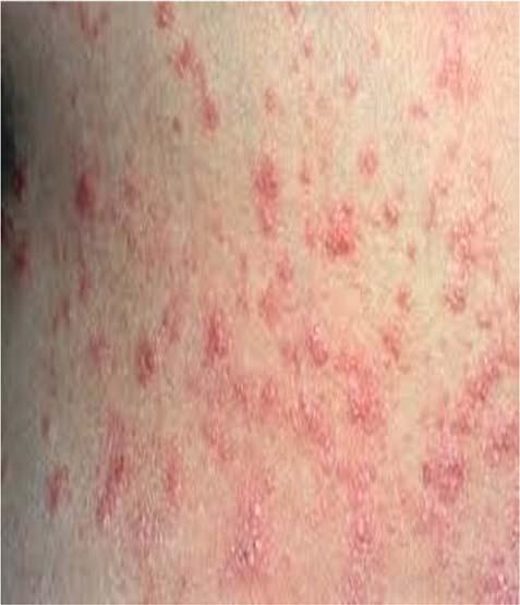
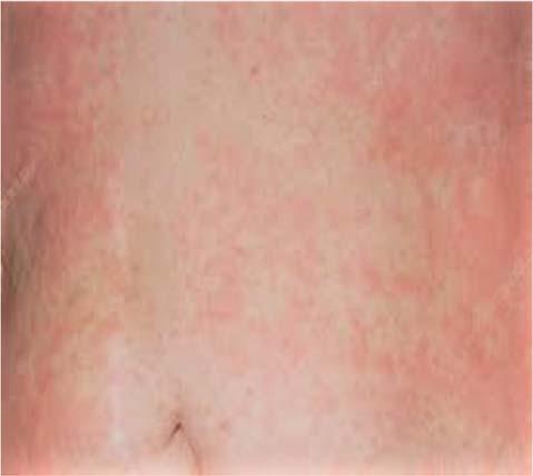
(5)Aphthous ulceration - in buccal mucosa, tongue, inner aspect of lips, genital area specifically over vulval region.
(6)Ocular examination reavealed bilateral mobile hypopyon with circumciliary congestion.
(7)Lymph nodes - Not palpable.
(8)Joint examination- no swelling and no deformity.
(9)Per-rectal examination – no hemmorrhoid, fissure, perianal fistula.
(10) Other Systemic examination - WNL
Differential Diagnosis :
(1)Infective causes: viral exanthematous fever, ileocecal TB
(2)Autoimmune causes : Systemic lupus erythematosus, vasculitis, other connective tissue disorder
Investigation :
CBC: Hb-9.0 gm% TLC-8800/mm3 PLT 35000/ mm3 ESR 40mm in 1 hour
LFT: Bil T -0.6mg/dl Bil D -0.2mg/dl Bil I -0.4mg/dl
– 3.2/3.0 gm/ dl
FBS/PPBS – 76/142 mg/dl
Urea/Creatinine – 17.5/0.71 mg/dl Na+ /K+-135/3.7 mmol/l
CRP-98.5 mg/l
MP,MPDA – negative
Denhue IgM, IgG- non reactive Leptospira IgM – non reactive Typhidot IgM- nonreactive HBsAg/Anti-HCV/HIV1,2 – non reactive Gastric lavage, sputum for AFB, CBNAAT - negative
On colonoscopy guided biopsy it showed focal area of erosion over colonic mucosa. The glands showed variable distortion and mild mucin depletion. The lamina
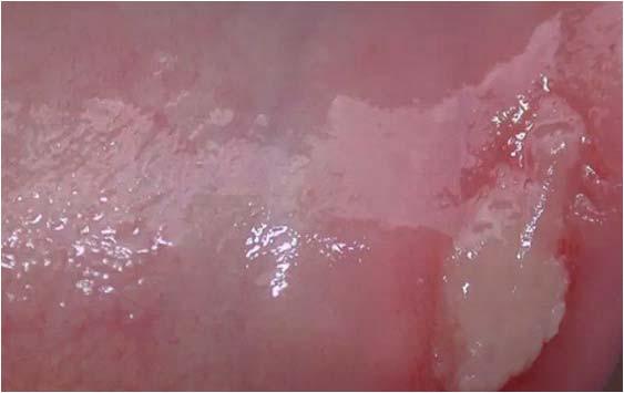
Fig 2 — Purpuric, non pruritic, non blnchable rash
propria is densely infiltrated by lymphocytes, plasma cells and a few eosinophils. Cryptitis and crypt abscess were present. There was no granuloma. No evidence of dysplasia was observed. Biopsy was suggestive of indeterminate Inflammatory bowel disease.
Skin biopsy findings- superficial and deep perivascular infiltrates of lymphocytes and neutrophils. There were features of vasculitis with endothelial cell swelling.
On immunofluorescence tyest – IgA, IgG, IgM, C3, Fibrinogen deposits were present in skin biopsy.
Autoimmune profile- ANA(-) / P-ANCA(-)/C-ANCA(-) Serum C3- 52.4 mg/dl which is below normal range
From above these results we were suspecting the case to be a Behcet’s disease. To confirm we perform the following tests.
Fig 3 — Painful, aphthous oral ulcer in the inner aspect of mouth
Anti Saccharomyces Cerevisiae Antibody – (+)
Skin pathergy test - reactive.
HLA-B51 : (+)
Provisional Diagnosis :
This is a case of Intestinal Behcet’s Disease.
Treatment and Follow Up :
She was treated with IV Hydrocortisone injection along with Azathioprine and other supportive treatment. After resolution of all symptoms she was discharged. Now she is doing well.
Behcet’s disease is a type of inflammatory disorder which can affect multiple systems of our body. Most commonly it presents as skin manifestations. Male and female have almost same preponderance towards developing this disease. But males generally develop severe forms of Behcet’s. There is a 1990 International Study Group Criteria for clinical diagnosis of Behcet’s. It includes presence of recurrent oral ulceration with two of the following4
Recurrent genital ulceration
Uveitis
Typical skin lesions
Skin Pathergy Test. Our case report also fulfilled this criteria.
In Behcet’s the oral ulcers are generally characterised by painful, recurrent and aphthous ulcers having necrotic base and they heal without scarring. In contrary the genital ulcers heal with scarring. Apart from theses it involves musculoskeletal system by developing non erosive arthritis of small joints which is collaborating with our case report. Uveitis is most common ophthalmological finding. But gastrointestinal Behcet’s like our case report is rare. Only 1-2% of cases having ulcers in GI tract5 mostly in iliocecal region. Behcet’s colitis mimics Inflammatory Bowel Disease specially Crohn’s. The exact cause of Behcet’s is still unknown. It is believed that there may be immunogenetic and inflammatory cytokines are two components of it’s pathogenesis. The autoimmunogenetic association with this disease shows
presence of T lymphocytes in lesions. TH1 cells along with Interferon-gamma, IL-12, IL-2 mediates cell mediated autoimmunity. Autoreactive T cells with anti HSP-60 function cause pathogenesis of this. HLA-B51 is more frequently associated with this disease. Due to lack of specific tests, Behcet’s is diagnosed clinically. Here elevated ESR, CRP are the nonspecific finding. But ASCA antibody test and skin pathergy tests are the few which can streamline the diagnosis to a certain extent. Apart from them it is associated with HLA-B51, HLA-B5 positivity. Perianal fistula, fissure , etc go in favour of crohn’s disease. In our case the mentioned complications were absent. So, the case is more in favour of intestinal behect’s rather than crohn’s. Steroids are the mainstay of treatment. In serious cases Azathioprine is added for speedy recovery.
Intestinal BD and inflammatory bowel disease specifically crohn’s have significant overlap in terms of sign, symptoms, pathological findings. For these clinicians always face difficulty in distinguishing the two. In both cases Steroids and Immunomodulators are the mainstay of treatment with decreased mortality and morbidity. Many researches are still ongoing to solve this dilemma to streamline the diagnosis. Understanding the mechanism properly will be important for appropriate treatment and prognosis of the disease.
1Baumgart DC, Sandborn WJ — Crohn’s disease. Lancet. 2012;380:1590–605. doi:10.1016/S0140-6736(12)60026-9. PMID: 22914295.
2Behçet H — Rezidivierende aphthose, durch ein virus verusachte geschwure am auge und an den genitalien. Dermatol Wochenschr 1937; 105: 1152-7.
3Bayraktar Y, Ozaslan E, Van Thiel DH — Gastrointestinal manifestations of Behcet’s disease. J Clin Gastroenterol 2000; 30: 144-54.
41990 International Study Group Criteria; Lancet 1990; 335: 1078-80.
5Kasahara Y, Tanaka S, Nishino M, Umemura H, Shiraha S, Kuyama T — Intestinal involvement in Behcet’s disease: review of 136 surgical cases in the Japanese literature. Dis Colon Rectum 1981; 24: 103-6.
Background : Common cold is an acute, self-limiting viral infection of the upper respiratory tract involving the nose, sinuses, pharynx, and larynx. According to various studies, the combination of analgesics, decongestants, and antihistamines provides better relief for multiple symptoms in the common cold. Fixed dose combination of Paracetamol as an analgesic, anti-inflammatory, and antipyretic, Chlorpheniramine maleate, an anti-histaminic, and Phenylephrine as a nasal decongestant is primarily used in the treatment of the common cold. Hence the present post-marketing surveillance study was planned to find any unwanted adverse effects and efficacy of commercially available combination in treating the common cold in children.
Methodology : The prospective, single-arm, multicenter, post-marketing clinical study included 224 children from four different study sites, of which 204 completed the study. Subjects were given this fixed dose combination for three days and then monitored for the next six days. During the study, the efficacy was evaluated using VAS score changes from the beginning to the end of the treatment. Incidence of Adverse Events (AE) and Serious Adverse Events (SAE) was assessed. The product's safety was also evaluated using blood biomarkers such as Hemoglobin, Platelet count, SGOT, SGPT, and creatinine level.
Results : The reduction in symptomatic score of common cold and flu syndrome was observed after 3rd followup visit [(0.384±0.168 (visit 1) to 0.001±0.009 (Visit 3), (p<0.001)]. No intervention-related or serious adverse events (SAE) were observed in the study or follow-up period. The study found no major changes in the levels of haemoglobin, platelets, SGOT, SGPT, and creatinine.
Conclusions : Fixed-dose combination of Paracetamol (125 mg), Phenylephrine HCL (5 mg), Chlorpheniramine Maleate (1 mg) and Sodium Citrate (60 mg/5 mL) is safe and effective in treating children's common cold and flu syndrome.
[J Indian Med Assoc 2022; 120(10): 71-5]
Key words :Common cold, Flu syndrome, Children.
The common cold is one of the highly prevalent illnesses worldwide1. It is an acute, self-limiting viral infection of the upper respiratory tract involving the nose, sinuses, pharynx, and larynx2. Children experience a high rate of incidence, which creates a significant economic and social burden1,3. Around 156 million new cases of respiratory infections occur worldwide every year, and about 1.56 million young children die because of such diseases4. Symptoms of the common cold in children typically reach peak intensity shortly after the onset of illness. Regardless of severity, the most prevalent symptoms among children with the common cold are runny nose, stuffed-
1Wallace Pharmaceuticals Pvt Ltd,
2ICBio Clinical Research Pvt Ltd,
*Corresponding Author
Received on : 28/03/2022
Accepted on : 10/04/2022
up nose, dry cough, sore throat, and sneezing 5 Diagnosis of the common cold can be problematic in young children and infants who cannot communicate their symptoms6
The flu syndrome is typical of sudden onset and is characterized by fever, cough, sore throat, myalgia, headache, nasal congestion, weakness, and loss of appetite. Antiviral agents are available for the treatment of flu. Still, they are ineffective against any other causes of upper respiratory infections. Thus, there is considerable interest in the early clinical diagnosis of influenza instead of the common cold7,8
Common colds and flu are syndromes of familiar symptoms caused by a viral infection of the upper respiratory tract. It is difficult to define the syndromes precisely because of the significant variation in the severity, duration, and types of symptoms 9
A Prospective, Interventional, Multicentre, Post-marketing Clinical Study of a fixed-dose combination of Paracetamol (125 mg), Phenylephrine HCL (5 mg), Chlorpheniramine Maleate (1 mg) and Sodium Citrate (60 mg/5 mL) for the Treatment of Common Cold and Flu Syndrome in Children
Pankaj Kumar1*, Rashmi Menezes1, Vinay Pinto1, Deepak Arora1, Karunraj Jayseela1, Sumit Kumar1, Dipeshh Rajdeo1, Harish S2, Vinda Z1, Tapas D1
Rhinoviruses account for 30-50% of all colds, and coronaviruses are the second most common agent, accounting for 10-15% of cold. Influenza viruses account for 5-15% of cold, and respiratory syncytial viruses are responsible for much flu-like illness, demonstrating much overlap in etiology and symptomatology of common cold and flu syndromes9,10
Currently, no antivirals are available to treat the common cold; therefore, symptomatic relief should be the primary focus for treating the common cold. Single drug therapy is not adequate to treat all the symptoms of the common cold, so multiple drug combinations are mainly used for symptomatic relief from the various symptoms of the common cold11,12
The current post-marketing clinical study evaluated the safety and efficacy of fixed-dose combination of Paracetamol (125 mg), Phenylephrine HCL (5 mg), Chlorpheniramine Maleate (1 mg) and Sodium Citrate (60 mg/5 mL)[Flucold Syrup], in the treatment of common cold and flu syndrome in children.
The study was a prospective, interventional, multicenter, post-marketing trial with 224 participants.
The study followed the ICMR guidelines, New Drugs and Clinical Trial Rules 2019 India, & the Declaration of Helsinki (Brazil 2013) & the ICH E6, R2, "Guidance on Good Clinical Practice" (GCP). Besides, the trial was approved by Royal Pune Independence Ethics Committee, HCG NCHRI- Institutional Ethics committee, MAVENS institutional Ethics committee, Basaveshwara Medical College, and Hospital Institutional Ethics Review Committee ( ICBio/CR/ WPPL/0309/108).
A total of 224 subjects were enrolled with common flu or cold and were treated with fixed-dose combination of Paracetamol (125 mg), Phenylephrine HCL (5 mg), Chlorpheniramine Maleate (1 mg) and Sodium Citrate (60 mg/5 mL) from which 204 subjects completed the study. The total duration of the study for the patient was nine days (three days treatment with six days of follow-up). The study included patients between the age of 2 to 12 years with recent onset of symptoms not less than 72 hours, such as common cold ( with symptoms such as sneezing, rhinorrhea, nasal congestion, headache, discomfort in the throat ) & flu syndrome (with symptoms such as high-grade fever, headache, chest discomfort, dizziness). Patients with known hypersensitivity, seasonal perennial allergic rhinitis, a recent history of influenza vaccination,
severely immune-compromised patients were excluded from the study.
The patient can be withdrawn from the study by the investigator for any of the following reasons: the occurrence of an adverse event associated with the administration of the IP, necessitating its cancellation; the emergence of any diseases or conditions during the study that worsens the patient's prognosis and makes it difficult for the patient to continue participating in the clinical research; the need for a prohibited concomitant therapy; research protocol violations; improper inclusion of a patient who did not fulfill the inclusion criteria and met the applicable exclusion criteria; other serious protocol violations, according to the investigators; The withdrawal of the assent form by the patient's representative. Twenty patients were removed from the study due to absenteeism during the follow-up.
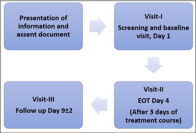
Suitable subjects who agreed to participate in the study were recruited from 4 sites (Jyothi Multispecialty Clinic, Abhinav Multispecialty Hospital, MAVEN's Hospital, Basaveshwara Medical College, and Hospital Chitradurga). Each site recruited participants whose parents willingly provided a written assent form for participation in the study.
All subjects were treated with fixed-dose combination of Paracetamol (125 mg), Phenylephrine HCL (5 mg), Chlorpheniramine Maleate (1 mg) and Sodium Citrate (60 mg/5 mL) for three days and follow up performed for the next six days.
Primary outcome measure —
To evaluate the safety of fixed-dose combination
(Paracetamol 125 mg + Phenylephrine HCL 5 mg + Chlorpheniramine maleate 1 mg + sodium citrate 60 mg per 5 mL) in the treatment of common cold or flu syndrome in the children. The incidence of adverse events (AEs) and serious adverse events (SAEs) was reviewed during the study.
Secondary outcome measures — Evaluation of the efficacy fixed-dose combination of (Paracetamol 125 mg + Phenylephrine HCL 5 mg + Chlorpheniramine maleate 1 mg + sodium citrate 60 mg per 5 mL), in the treatment of common cold and flu syndrome in children.
Data analysis was performed using ANOVA & χ2 test & SAS version 9.1 Inc, CARY; the USA used during the study. Efficacy analysis was performed for the perprotocol (PP) population. Primary efficacy was based on PP patients' samples.
A total of 224 patients was enrolled in the study from 4 sites, out of which 20 patients withdrew from the study.
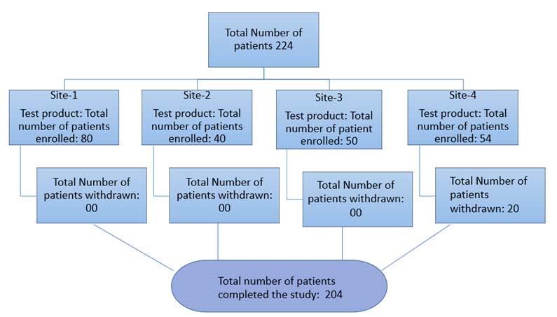
The mean age of the subjects enrolled in the study was 5.877±2.820 years, whereas average weight & height was 19.130±7.978 kg & 101.839±26.169 cm, respectively. The average BMI calculated during the study was found to be at 17.965±6.480 kg/m2
Age5.877 ± 2.820 years
Weight19.130 ± 7.978 kg
Height101.839 ± 26.169 cm
BMI17.965 ± 6.480 kg/m2
During the study, no intervention-related adverse events were observed. Moreover, no Severe & treatment-related Adverse Events (SAE) were observed during the investigation and follow-up period.
Reduction in total symptom score from day 1 to day 4 and during follow-up was assessed using a 4point scale (0- no symptom, 1-Mild, 2-Moderate, 3severe). Study results indicated that use of this SYRUP in children significantly reduced total symptom score from 0.384±0.168 (visit 1) to 0.001±0.009 (Visit 3), (p<0.001)(Fig 3).
The severity of the flu was assessed using visual analog score (VAS) changes in children. Treatment in children leads to significantly (p<0.05) change in VAS score from 4.172±1.668 (Visit 1) to 0.015±0.156 (Visit 3).(Fig 4).
Tolerability was analysed during the study in each subject & a tolerability scale was used during the analysis. It was well tolerated in children, as no side effects were observed (Table 2).
Additionally, the product's safety was evaluated by
analysing biomarker levels such as haemoglobin, platelet, liver, and kidney function test at the end of the study. fixed-dose combination of Paracetamol (125 mg), Phenylephrine HCL (5 mg), Chlorpheniramine Maleate (1 mg) and Sodium Citrate (60 mg/5 mL) did not significantly affect haemoglobin (11.841±0.976) and platelet (261.613±60.773) count during the study. Liver
biomarkers such as SGOT (23.496±14.138) and SGPT (26.957±19.729) levels were found to be normal, indicating the safety in children. Similarly, no elevation was observed in serum creatinine levels (0.665±0.194) at the end of the study (Table 3).
Table
Haemoglobin11.841 + 0.976
Platelet count261.613 + 60.773
SGOT/
23.496 + 14.138
SGPT/ALT 26.957 + 19.729
Serum
0.665
0.194
The study was the first prospective, interventional, multi-centred, post-marketing clinical trial to demonstrate the safety and efficacy of fixed-dose combination of Paracetamol (125 mg), Phenylephrine HCL (5 mg), Chlorpheniramine Maleate (1 mg) and Sodium Citrate (60 mg/5 mL) in treating common cold and flu syndrome in children. However, various clinical studies conducted on adults showed that this combination treats the common cold and flu syndrome.
The findings showed a reduction in the symptomatic score of common cold and flu syndrome from baseline (0.384±0.168) to the end of the treatment (0.001±0.009). A similar study conducted by Picon et al. also revealed that after treatment with a fixed-dose combination of chlorpheniramine maleate, Paracetamol and Phenylephrine for ten days reduced symptom score from 14.09 to 3.5412.
Similarly, a phase IV open-labelled multicentre study conducted in 159 patients found that mean TSS reduced from 6.62 (Day 1) to 0.69 (Day 5). Most patients included in the study had more than 50% reduction in total symptom score at visit 3, and 58.49% of patients had complete relief from the symptoms13 The results of both the study were in line with the current study12,13.
Visual Analogue Scale (VAS) score analysed symptomatic relief of common cold and flu syndrome. A significant change was observed in VAS scores from baseline (4.172±1.668) to EOT (0.0015±0.156).
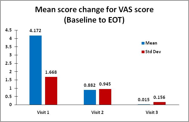
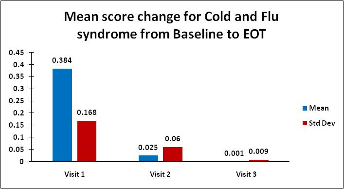
Biomarker evaluation showed no effect on liver and kidney function (Sr Cr = 0.665±0.194 parameters) at the end of the study. No major changes were observed in haemoglobin (11.841±0.976) and platelet count (261.613±60.773) after administration of this SYRUP. These outcomes support that combining multiple
formula provides the patient the convenience of
treating multiple symptoms with a single product and may promote improved compliance to the treatment. It may also help in both patient safety and the optimal efficacy of the medicines14,15
IP Tolerability (ie, is, are there any side effects observed or any changes in treatment) data reported and analysed to check the tolerance of the investigational report. There were no side effects reported during the study. The efficacy analysis was performed to check incidence rates of TreatmentEmergent Adverse Events (TEAEs) and Serious Adverse Events (SAEs). Based on reported results, no treatment-emergent or serious adverse event was observed during the study. Five subjects reported mild AE, which is unlikely related to the Other all subjects found no Treatment-emergent adverse events. In contrast to this, a study conducted by Kiran M, et al reported six non-serious adverse drug reactions in the study duration of 5 days16
Studies conducted by Janin, et al and Picon, et al with a fixed-combination formulation to treat common cold reported a good safety profile and an excellent tolerance. The rationale for the treatment is that multiple symptoms commonly co-occur and that a combination medicine provides a simplification of therapy compared to the use of monotherapies12,17
This study was performed to evaluate the efficacy and safety of combinations (analgesics, decongestants, and antihistamines) in treating common cold and flu syndrome.
After four days of treatment with a fixed-dose combination of Paracetamol (125 mg), Phenylephrine HCL (5 mg), Chlorpheniramine Maleate (1 mg) and Sodium Citrate (60 mg/5 mL)[Flucold Syrup], there was a significant improvement in symptomatic relief of common cold and flu syndrome. The safety results revealed that the Flucold Syrup is safe and welltolerated in children when administered orally.
Considering the results and outcomes, it is evident Paracetamol 125 mg + Phenylephrine HCL 5 mg + Chlorpheniramine maleate 1 mg + sodium citrate 60 mg per 5 mL was effective for treating cold and flu syndrome in children.
The Author would like to thank ICBio Clinical Research Pvt Ltd for carrying out Clinical studies .
Funding : The Study was funded by Wallace Pharmaceuticals Pvt. Ltd.
1Fendrick AM, Monto AS, Nightengale B, Sarnes M — The economic burden of non-influenza-related viral respiratory tract infection in the United States. Arch Intern Med 2003;
163(4): 487-94.
2Allan GM, Arroll B — Prevention and treatment of the common cold: making sense of the evidence. CMAJ 2014; 186(3): 190-9.
3Monto AS, Sullivan KM — Acute respiratory illness in the community. Frequency of illness and the agents involved. Epidemiology & Infection 1993; 110(1): 145-60.
4Liu L, Johnson HL, Cousens S, Perin J, Scott S, Lawn JE, et al — Global, regional, and national causes of child mortality: an updated systematic analysis for 2010 with time trends since 2000. Lancet 2012; 379(9832): 2151-61.
5Troullos E, Baird L, Jayawardena S — Common Cold Symptoms in Children: Results of an Internet-Based Surveillance Program. J Med Internet Res 2014; 16(6): e144.
6Heikkinen T, Järvinen A — The common cold. The Lancet 2003; 361(9351): 51-9.
7Monto AS, Gravenstein S, Elliott M, Colopy M, Schweinle J — Clinical signs and symptoms predicting influenza infection. Arch Intern Med 2000; 160(21): 3243-7.
8Monto AS — Studies of the Community and Family: Acute Respiratory Illness and Infection. Epidemiologic Reviews 1994; 16(2): 351-73.
9Vesa S, Kleemola M, Blomqvist S, Takala A, Kilpi T, Hovi T — Epidemiology of documented viral respiratory infections and acute otitis media in a cohort of children followed from two to twenty-four months of age. The Pediatric Infectious Disease Journal 2001; 20(6): 574-81.
10Simonsen L — The global impact of influenza on morbidity and mortality. Vaccine 1999; 17: S3-10.
11Dicpinigaitis PV, Eccles R, Blaiss MS, Wingertzahn MA — Impact of cough and common cold on productivity, absenteeism, and daily life in the United States: ACHOO Survey. Curr Med Res Opin 2015; 31(8): 1519-25.
12Picon PD, Costa MB, da VeigaPicon R, Fendt LCC, Suksteris ML, Saccilotto IC, et al — Symptomatic treatment of the common cold with a fixed-dose combination of Paracetamol, chlorphenamine and Phenylephrine: a randomized, placebocontrolled trial. BMC Infect Dis 2013; 13: 556.
13Kiran MD, Vakharia MP, Pawaskar LJ, Sheikh SN — Efficacy and safety of a fixed dose combination of Paracetamol, chlorpheniramine maleate and Phenylephrine in treatment of common cold: a phase IV, open-labelled, multi-centric study. International Journal of Basic & Clinical Pharmacology 2018; 8(1): 34-8.
14Hartley V — Overdose of cough and cold remedies: VICTORIA HARTLEY advises on the risk of toxicity when cough and cold remedies are ingested in accidental or intentional overdose. Emergency Nurse 2003; 10(9): 20-5.
15Eccles R, Fietze I, Rose U-B — Rationale for Treatment of Common Cold and Flu with Multi-Ingredient Combination Products for Multi-Symptom Relief in Adults. OJRD 2014; 4(3): 73-82.
16Kiran, Mayuresh, Pawaskar L, Sheikh A, Waghambare P — Post-marketing Surveillance Study to Substantiate The Efficacy and Safety For The Combination of Paracetamol, Phenylephrine and Chlorpheniramine Maleate In Indian Patients Of Common Cold. International Journal of Medical Science And Diagnosis Research 2021; 5(7):
17Loose I, Winkel M — Clinical, double-blind, placebo-controlled study investigating the combination of acetylsalicylic acid and pseudoephedrine for the symptomatic treatment of nasal congestion associated with common cold. Arzneimittelforschung 2004; 54(9): 513-21.
A 38-year-old female presented with Headache and Blurring of Vision since 4 month.
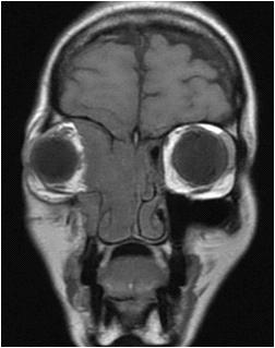
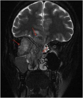
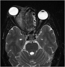
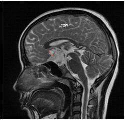
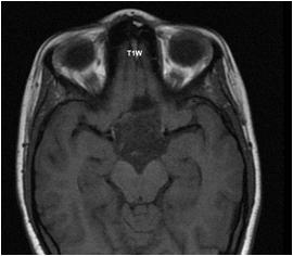
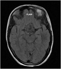
(1)What is the Diagnosis?
(2)What are the other common locations of this lesion?
(3)What are the differential Diagnosis?
Answers :
(1)Well defined lobulated altered signal intensity lesion is seen the suprasellar cistern separate from the pituitary gland. Lesion appears isointense to CSF on T1W and T2W images, shows heterogeneous / dirty signal on FLAIR and shows restriction on DWI images. Findings are suggestive of suprasellar epidermoid cyst.
(2)The common locations of epidermoid cyst are cerebellopontine angle (40-50%), suprasellar cistern (10-15%), fourth ventricle (17%), middle cranial fossa and interhemispheric.
(3)The differentials for an epidermoid cyst are :
(A)Arachnoid cyst – follows CSF signal on all sequences.
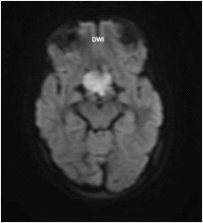
(B)Dermoid cyst – often fat density and located along the midline.
(C)Cystic tumours – solid component is usually seen.
A 28-year-female presented with Proptosis of Right Eye and Nasal Blockage.
(1)What
(2)What
(1)An aggressive soft tissue intensity lesion is seen with its epicentre in the superior olfactory recess involving ethmoid air cells, extending superiorly and infiltrating into anterior cranial fossa. Laterally the lesion extends into right orbit, encasing the extra-ocular muscles and causing proptosis of right eyeball. Biopsy of the lesion was done, whichconfirmed the diagnosis of olfactory neuroblastoma (esthesioneuroblastoma).
(2)The common differentials of such lesion are :
A)Olfactory neuroepithelioma
B)Olfactory groove meningioma
C)Sinonasal carcinoma – usually in older patients while olfactory neuroblastoma shows bimodal peaks in 2nd decade and 5th-6th decade.
D)Nasopharyngeal carcinoma – usually older patients
Department of Radiology, Picture This by Jankharia, Mumbai, Maharashtra 400004
1MD, DNB (Radiology)
2MD, DMRD (Radiology)
This is part of an extremely rare document :
The question paper of Medicine of Final M.B. examination, 1934 of Calcutta University. As the readers can see, Dr Upendranath Brahmachari was the convenor and under him, Dr B.C.Roywas examiner. There were two examiners from the Indian Medical Service (IMS). There is no possibility that anyone who had appeared in this examination is still alive in 2022. But the questions given in this paper are all still very relevant and may be used for an examination in 2021 also. As noted here, all questions were compulsory. There were just 2 questions (essay type) in each half. Just imagine an MBBS student facing Dr U Brahmachari and Dr BC Roy as examiners!!
 1Consultant Physician, Kolkata
[The Editor is not responsible for the views expressed by the correspondents]
SIR, — When we listen to the word “DOCTOR”, we have this image of one soft, kind person in formal dress with a white apron and stethoscope in hand1.2. Doctors are medical practitioners, physicians or clinicians. In people’s minds, doctors had hospitals and treated the patients. Various specialties other than physicians or surgeonslike epidemiologists, pathologists, microbiologists, and radiologistsare there, they may/may not be treating the patient in the hospital but they play a very important role in diagnostic and public health maintenance3. As of now, we can say they are the public servants who save human lives using their knowledge of the human body and aid those with permanent physical or psychological impairments. Doctors save human lives not only by treating or healing but also by educating people for the future.
Indian Medical Association (IMA) start celebrating 1st July as National Doctors' Day in 1991 to honour Dr. Bidhan Chandra Roy on his birth and death anniversary for his contribution tothe freedom fight and medical education. He was the chief minister of West Bengal from 1948 to till his death (1962)4
Every year IMA and the whole country celebrate 1st July to highlight the contribution of the doctors in humanitarian services and public health. The medicalfraternity’s contribution during the current COVID-19 pandemic from 2020 to till now, cannot be described in a few words or sentences. From local medical practitioners to super specialties and research scientists, all contributed to saving human lives by sacrificing their time and family and sometimes by their own lives4. IMA celebrates this day with a newer theme every year. This year’s theme is “Family Doctors on Front line” to highlight the support of doctors who are caring for families and communities5
Till today we have faced almost 3 waves of COVID-19 pandemic and now these days, cases are increasing maybe this is the starting of the 4th wave of COVID-196,7. In the last 3 waves, many private practitioners andfamily doctors took care of their patients because the government and private hospital doctors were not able to consult and care for all the patients during the pandemic. Other healthcare workers also care but doctors’ roles cannot be forgivable. In 3rd wave, symptomatic patients were treated easily with standard precautions as compared to the previous two pandemics6. Other days like mother’s day, women’s day, and children’s day celebration is done with the newer theme along with giving some appreciating specific benefit plans, schemes, or discounts in specific things while only Doctors' day is celebrated giving extra responsibility, instead of any appreciation to doctors.
This year’s theme again accelerates the medical fraternity specifically family doctors, to manage the COVID-19 upcoming cases. Upcoming waves may be easy to handle or worst, no one knows. But this year’s theme is definitely to prepare the doctors for future pandemics, not for appreciating the doctor’s past work.
Doctor’s profession is made only to serve society and humanity and they will accept this responsibility however they have to sacrifice their time, family, or own lives.
1Article writing on: A doctor by Admin, edumantra. Available at https://edumantra.net/learn-english/1-article-writing-on-adoctor/ last access in 5th July 2022.
2Steiner-Hofbauer V, Schrank B, Holzinger A. What is a good doctor? Wien Med Wochenschr. 2018 Nov;168(15-16):398405. doi: 10.1007/s10354-017-0597-8.
3What Are the Different Types of Doctors? By Kathryn Whitbourne, Health Insurance and Medicare, web MD. Available at http s://www.webmd.com/health-insurance/ insurance-doctor-types last access in 5th July 2022
4Pal M. Indian contribution in medical science—Modern medicine (Part 1). J West Bengal Univ Health Sci. 2020; 1(2):1-5
51st July 2022: National Doctors Day significance, wishes, quotes, and heartfelt messages to share by economic times available at https://economictimes.indiatimes.com/news/newupdates/1st-july-2022-national-doctors-day-significancewishes-quotes-and-heartfelt-messages-to-share/ articleshow/92599980.cms?from=mdrlast access in 5th July 2022
6Lemire F, Slade S. Family physicians and the COVID-19 third wave. Can Fam Physician. 2021;67(7):550.
7Covid-19 Is India heading towards a fourth wave? How should we prepare? Doctors explains by Livemint. Available at https:/ /www.livemint.com/news/covid19-is-india-headingtowards-a-fourth-wave-how-should-we-prepare-doctorexplains-11650759504098.html last access in 5th July 2022
Department of Microbiology, Sahjid Mukhida1, Dr D Y Patil Medical College, Hospital Chanda Vyawahare2, and Research Centre, Pune 411018 Nikunjakumar Das3, 1MBBS, PG Resident and Corresponding Author Vallari Jadav4 2MD, Professor 3MD, Associate Professor 4MBBS, PG Resident, Department of Community Medicine
SIR, — The epilepsies affect children, men and women of all ages, races and ethnic groups. It is the fourth most common neurological disease. Nearly 12 million people with Epilepsy are expected to reside in India; which contributes to nearly one-sixth of the global burden8
Epilepsy can be treated medically and surgically.
Epilepsy surgery must NOT be a LAST RESORT as a treatment option. If a patient being treated by a general physician/paediatrician/ neurologist for epilepsy, not responding to medication( DRE—drug resistant epilepsy) and continuing to have seizures impairing the
QOL must be referred for pre-surgical evaluation for determining the cause of intractability and hence a possible surgical treatment.(1)
Rationale for Epilepsy Surgery — Clinical experience and scientific data provide some compelling reasons for considering surgery for drug resistant epilepsy. Surgical candidates have a much higher chance of attaining surgical freedom as compared with patients receiving medical treatment for DRE (58 versus 8%, respectively1,2
Unfortunately, in our country, it is said that it is better to live with epilepsy and ruin quality of life rather than having surgery and enjoy a good quality of life.
Epilepsy itself is associated with a mortality of about 0.5% per year (including all causes, eg, sudden unexplained death due to epilepsy, from accidents, etc).
Thus, in a person with epilepsy for 2 years, the risk of surgery roughly equals that of mortality from the epilepsy itself (1%)3. After this, the risk of death from epilepsy becomes more than the surgical treatment.
Pediatric epilepsies should be investigated much earlier as uncontrolled seizures during infancy and early childhood are more likely from symptomatic etiologies.
The delay in identifying surgical candidates leads to severe irreversible changes in the developing brain, consequently to arrested or delayed development4, inducing a “catastrophic epilepsyinduced encephalopathy”3
It has been well demonstrated world over, including in India, that epilepsy surgery is safe and is associated with very low mortality and morbidity.
In summary, epilepsy surgery should not be withheld in wellindicated cases as it offers the patient the best chance to get seizure freedom and should be offered early in the course of the disease (and should not be considered as an option of last resort).
Indications for Epilepsy Surgery1 — Patients are said to have DRE(drug resistant epilepsy) if they have failed two or more AEDs used in their appropriate, adequate dosage, combinations and in appropriate indications after an adequate duration of treatment (not more than 2 years) in adult patients (16 years and above).
In pediatric patients, diagnosis of DRE should be made much earlier (sometime even within weeks of onset of seizures), particularly if they present with epileptic encephalopathy, infantile spasms, catastrophic onset of epilepsy, seizure frequency of >1 month and disabling seizures
When a patient does not respond to medical treatment and is defined as having Drug Resistant Epilepsy (DRE) as per the criteria mentioned above, he/she should then be investigated for a possibility of epilepsy surgery.
Investigations for Epilepsy Surgery1 — Before surgery, a careful presurgical evaluation is mandatory. The purpose is to delineate the epileptogenic zone, defined as “the area of cortex indispensable to the generation of epileptic seizures.”
The localization of the “epileptogenic zone” cannot be performed by any single investigation. It has to be localized by multiple investigations, which are of four broad categories:
(1) structural imaging : MRI as per the epilepsy protocol, (2) electrical localization : EEG, long-term Video-EEG5,6 (3) functional imaging : PET, SPECT, fMRI. [Wada test (an invasive test) is being replaced by fMRI.],
(4) the need for invasive investigations—When standard investigations are discordant for substrate-negative pathologies and dual pathologies —
The following are examples of instances that may require invasive intracranial monitoring (Depth, grid and strip electrodes are implanted either through a stereotactic frame or by an open craniotomy)
(a)Seizures are lateralized but not localized (eg, a left-sided, widespread frontal–temporal onset). Seizures are localized but not lateralized (eg, ictal EEG patterns that appear maximally over both temporal lobes).
(b) Seizures are neither localized nor lateralized (eg, stereotyped complex partial seizures with diffuse ictal changes or initial changes obscured by artifact).
(c) Seizure localization is discordant with other data [eg, EEG ictal scalp data discordant with neuroimaging (MRI, PET, SPECT) or neuropsychological data].
(d) Relationship of seizure onset to functional tissue must be determined (eg, seizures with early involvement of language or motor function).
(e) Relationship of seizure onset to lesion must be determined (eg, dual pathology or multiple intracranial lesions).
(f) If seizures are clinically suspected but video-EEG is inadequate for defining them [eg, simple partial seizures with no detectable scalp EEG ictal discharge or suspected epileptic seizures with unusual semiology that suggests psychogenic seizures (pseudo-pseudo seizures)].
(5) Neuropsychological testing7
Formal neuropsychological testing is important as a preoperative baseline, as a predictor of possible cognitive loss with surgery, and as an additional aid for localization. For example, patients with temporal lobe epilepsy tend to have memory deficits. Those with dominant TLE (usually left sided) have more prominent deficits in verbal memory compared with visual memory. Patients with average or above average memory function prior to temporal lobectomy have a higher risk of memory decline, especially with left (dominant) temporal lobectomy.
It is important to establish its relationship of epileptogenic zone with eloquent cortex as the surgery should not result in a new deficit.
The minimum investigations required are specialized MRI sequences, video EEG, documenting a minimum of three concordant and habitual seizures.
In cases where the MRI and video EEG are discordant or there exists a dual pathology, or MRI is negative, advanced investigations like ictal SPECT (with SISCOS/SISCOM), PET and invasive video EEG are required.
Functional MRI is required to identify speech, language, motor, memory like areas.
Magnetoencephalography (MEG) is an important tool for source localization.
In summary, presence of a “concordant” epileptogenic focus in a patient with DRE would form an indication for “resective” epilepsy surgery.
If such a focus is not detected, the patient may still be evaluated by advanced techniques.
Patients who have syndromes like Lenox Gestaut syndrome or
intractable disabling seizures without delineation of an epileptogenic zone may be candidates for “palliative” surgery, such as corpus callosotomy, multiple subpial transaction or vagal nerve stimulation. Absence of a lesion even on specialized MR images does not exclude that the person will not benefit from epilepsy surgery.
Types of surgical interventions7 — The surgical interventions may be broadly divided into — (1) Resection surgery (a)—temporal —Anteromedial temporal lobectomy with amygdalohippocampectomy: A surgical procedure where the anterior and the medial part of the temporal lobe resected along with hippocampus, amydala, uncus and the mesial structures. This is mostly indicated for epilepsies arising from the medial temporal lobe.
Selective amygdalo-hippocampectomy: A more technically demanding surgical procedure where only the mesial structures, like hippocampus, amydala and uncus, are removed, leaving the lateral temporal lobe intact. Its role over the earlier described procedure is not certain.
(b) Extra-temporal resection surgeries
(2) Disconnection surgery—Corpus callosotomy
Multiple subpial transection: A surgical procedure coming under the category of “palliative” procedure where the aim is to reduce the seizure burden only rather than to eliminate them completely. It is usually performed on an eloquent cortex like the mortor cortex so as to avoid producing any deficit. Here, the gyrus is divided into small blocks of 1 × 1 cm using a special instrument.
Hemispherotomy: A complex surgical procedure where the entire affected hemisphere (in conditions like Rasmussen’s syndrome) is disconnected from the opposite hemisphere. This is much less invasive than the procedure, like the earlier hemispherectomy (where the hemisphere is disconnected and then physically removed). The latter procedure has now been given up due to the higher incidence of complications, like blood loss, hemosiderosis, etc.
(3) Neuromodulation — vagal nerve stimulation, anterior thalamic deep brain stimulation
Electrocorticography : — An investigation to determine when different sizes of electrodes (strips, grids) should be placed on the surface of the brain to localize the “epileptogenic” focus before resection in all patients’ neocortical temporal and extra-temporal locations with concordant investigations. It is also to be used in tailored resections in hippocampal sclerosis
Conclusion — Epilepsy surgery should be considered early and liberally whenever anti-epileptic drug fails to control it adequately as mentioned above.
1Sarat CP, Tripathi M — Epilepsy surgery: recommendations for India. AnnalsofIndianAcademyofNeurology 2010; 13(2): 87-93. doi:10.4103/0972-2327.64625
2Wiebe S, Blume WT, Girvin JP, Eliasziw M — Effectiveness and Efficiency of Surgery for Temporal Lobe Epilepsy Study Group. A randomized, controlled trial of surgery for temporallobe epilepsy. N Engl J Med 2001; 345: 311-8.
3Korkman M, Granström ML, Kantola-Sorsa E, Gaily E, Paetau R, Liukkonen E, et al — Two-year follow-up of intelligence after pediatric epilepsy surgery. Pediatr Neurol 2005; 33: 173-8
4Nolan MA, Redoblado MA, Lah S, Sabaz M, Lawson JA, Cunningham AM, et al — Intelligence in childhood epilepsy syndromes. Epilepsy Res 2003; 53: 139-50.
5Alving J, Beniczky S — Diagnostic usefulness and duration of the inpatient long-term video-EEG monitoring: findings in patients extensively investigated before the monitoring. Seizure 2009; 18(7): 470-3. doi: 10.1016/ j.seizure.2009.04.005. Epub 2009 May 9. PMID: 19428271.
6Tatum WO, Mani J, Jin K, Halford JJ, Gloss D, Fahoum F, et al Minimum standards for inpatient long-term video-EEG monitoring: A clinical practice guideline of the international league against epilepsy and international federation of clinical neurophysiology. Clin Neurophysiol 2022; 134: 111-28. doi: 10.1016/j.clinph.2021.07.016. Epub 2021 Dec 13. PMID: 34955428.
7Bromfield EB, Cavazos JE, Sirven JI, editors. An Introduction to Epilepsy [Internet]. West Hartford (CT): American Epilepsy Society; 2006. Chapter 4, Epilepsy Surgery. Available from: https://www.ncbi.nlm.nih.gov/books/NBK2514/ 8Amudhan S, Gururaj G, Satishchandra P — Epilepsy in India I: Epidemiology and public health. Ann Indian Acad Neurol. 2015; 18(3): 263-77. doi:10.4103/0972-2327.160093)
DNB (Neurosurgery), Consultant, Amit Kumar Ghosh Department of Neurosurgery, Institute of Neurosciences, Kolkata
Laboratory Work and Analysis of the Biochemical components of Human Umbilical Cord Blood and Adult peripheral blood SIR, — As we previously discussed, “A comparative study of the Biochemical components of Human Umbilical Cord Blood and Adult peripheral blood”(JIMA, July, 2022). Now we have done some laboratory work and analysis of the Biochemical components like TC, DC, ESR, and the IL6.
At first the Informed Consent Form was signed by the patient through which the patient was confirmed to give the umbilical cord blood sample to the student for the betterment of Research and development for humankind.
Then, the sample was collected and stored in EDTA vials.
After that, the cord blood sample was preserved in an Ice pack within a box and quickly transferred to the laboratory for the analysis of biochemical components.
The umbilical cord blood sample was tested for TC, DC, ESR and IL-6
The results are as follows :
Umbilical Cord Blood Sample
Hematological Findings :Total Count :
Hemoglobin is 15.1 gm/dl
RBC count is 4.63 million/cumm
WBC count is 15530/cumm
Platelet count is 277000/cumm
Differential Count: Neutrophils are 90%
Lymphocytes are 43%
Monocytes are 3%
Eosinophils are 4%
Basophils are nil
Pack cell volume (PCV) is 48.3%
Mid cell volume (MCV) is 110 fl
Mean corpuscular hemoglobin concentration (MCHC) is 31.2%
Red cell distribution width (RCDW) is 18% Erythrocyte Sedimentation rate (ESR) is 12mm/hr.
General Blood Picture - WB-EDTA
RBC – MILD MACROCYTOSIS
WBC – NO ABNORMAL CELL SEEN
Platelet – MORE THAN ADEQUATE
*The sample is rich in hemoglobin i.e. 15.1 gm/dl
*The White blood cell(WBC) count is 15530/cumm, much higher than the normal range.
*By the differential count we get to know that the amount of lymphocyte i.e. 43% and PCV, MCV and MCHC are slightly different from the normal range.
Immunological Findings: - Immunoassay:IL-6 is 3.21 pg/ml
*The sample is tested for Interleukin-6 (IL-6) immunoassay.
*The result of this immunoassay IL-6 is found to be 3.21 pg/ml which is much less than the inflammatory range.
*Hence it acts as an anti-inflammatory cytokine.
It is a well-known fact that Cord blood is a pregnancy specific biological substance. We enumerated the clinical, biochemical and other parameters of cord blood.
Now, we compare the peripheral blood of the same patient i.e. the mother.
The adult peripheral blood sample was also tested for TC, DC, ESR and IL-6.
The results are as follows:
Adult Peripheral Blood Sample Hematological Findings: Total Count: Hemoglobin is 9.3 gm/dl
RBC count is 4.39 million/cumm
WBC count is 12000/cumm
Platelet count is 1,93,000/cumm
Differential Count: Neutrophils are 51%
Lymphocytes are 07% Monocytes are 2% Eosinophils are nil
Basophils are nil
Pack cell volume (PCV) is 30.5%
Mid cell volume (MCV) is 65.9 fl
Mean corpuscular hemoglobin concentration (MCHC) is 30.5%
Red cell distribution width (RCDW) is 16%
Erythrocyte Sedimentation rate (ESR) is 10mm/hr.
General Blood Picture - WB-EDTA
RBC – NO ABNORMAL CELL SEEN WBC – NO ABNORMAL CELL SEEN
Platelet – Adequate
*The hemoglobin level of the sample is very low i.e. 9.3 gm/dl
*White blood cell(WBC) count is 11000/cumm, in the normal range.
*By the differential count we get to know that the amount of lymphocyte i.e. 7% and PCV, MCV and MCHC are in the normal range.
Immunological Findings: -
Immunoassay : IL-6 is 3.78 pg/ml
*The sample is tested for Interleukin-6 (IL-6) immunoassay.
*The result of this immunoassay IL-6 is found to be 3.78 pg/ml which is much less than the inflammatory range.
*Hence it acts as an anti-inflammatory cytokine.
By comparing the two blood samples we have finally reached a discussion that.
I.Cord blood is enriched with fetal hemoglobin i.e. present in great quantities.
II.The number of Red blood cells (RBC) present in cord blood is higher than the adult blood.
III. The number of White blood cells (WBC) present in cord blood is a massive amount which indicates that it is immunologically very much beneficial for use to cure a large number of diseases.
IV.The number of platelets also present in cord blood in large figures.
V.It is also enriched with anti-inflammatory cytokines and growth factors to serve as a repair and regeneration type pregnancy specific biological substance.
So, we conclude that Cord Blood is a pregnancy specific biological substance which is free from contamination and has an immense potential to be the true blood substitute. Maybe our future lies within it. It should be served for the betterment of mankind in the upcoming years through the world.
1Ammanuel Angelo, G. D. (2021, June 11). national library of medicine. From NIH: https://www.ncbi.nlm.nih.gov/pmc/ articles/PMC8194248/
2Sameer Timilsina, S. K. (2018). Correlation between maternal and umbilical cord blood in pregnant women in Pokhra valley.
BMC Pregnancy and Childbirth, 01-05.
3Vassilios Katsares, P. Z.-A. (2009). Reference Ranges for Umbilical Cord Blood. LABMEDICINE, 437-439.
4Yu-HsunChangabShang-HsienYangaTso-FuWangcTengYiLindKuo-LiangYangeShu-HueyChen. (2011, June 03). ScienceDirect. From sciencedirect.com: https:// www.sciencedirect.com/science/article/pii/ S1875957211000386
1MBBS(Cal), DPH(Cal), Amitabha Bhattacharya1, M. PHIL RMTS, Honorary Past Associate Editor, JIMA 2B Sc. Microbiology, Anwesh Ghosh 2 , M Sc Forensic Science, M PHIL RMTS Part-I
Ifyouwanttosendyourqueriesandreceivethe responseonanysubjectfromJIMA,pleaseuse the E-mail or Mobile facility.
For
For
For
For
For
For
: +919477493033
Mobile : +919477493027
Mobile : +919477493037
jimamkt@gmail.com Mobile : +919477493036
journalaccts@gmail.com Mobile : +919432211112