















The clinical profile of communicable diseases is changing with the passage of time and malaria is no exception. The WHO has taken up the challenge of Health for All by 2030 which includes elimination of malaria. The global targets in the coming years are - reduction of mortality of malaria along with decrease of case incidence by 90%. Country-level elimination and prevention of re-establishment of malaria is top priority through investment, innovation and implementation.
In 2020, about 5.12 lakh cases of malaria were reported in South East Asia. India accounted for 36.4% of the reported cases in the region as well as 63% of deaths. However, between 2000 to 2019 percentage drop of cases has been 71.8% and of death, 73.9% which is considerable. India has significantly reduced malarial burden and contributed to the largest drop in cases region-wide. But the pandemic has led to disruption of health services and wreaked havoc on health infrastructure and manpower. Literature has reported a worldwide increase in malaria cases & 47,000 more deaths in 2020 compared to 2019 due to disruptions to services during the pandemic though not in India.
Elimination requires early diagnosis, prompt treatment, radical cure, vector control and optimal surveillance. However, it is being observed that the clinical presentation of malaria is largely atypical now-a-days and the diagnosis of malaria is becoming increasingly elusive to the treating physician. This has a direct implication on the morbidity and mortality patterns of the disease.
Symptoms like lack of taste, sore throat, cough are being reported simulating an influenza like illness. The usual manifestations of falciparum malaria such as cerebral malaria, black water fever and algid malaria seen in the past are being replaced with unusual complications like haemolyticanaemia, severe anaemia, thrombocytopenia, pancytopenia and adult respiratory distress syndrome. Severe malaria is being increasingly reported in P vivax malaria with complications like cerebral malaria, severe anemia, hepatic dysfunction, acute lung and kidney injury, acute respiratory distress syndrome, severe thrombocytopenia, bleeding, as well as shock.
It is important to evaluate the underlying factors responsible for the predominance of vivax infections as well as complications in recent times. The foremost reason is the high proportion of recurrent infections attributable to relapse, which are associated with transmissible gametocytemia. This leads to appearance of sexual-stage parasites
prior to clinical presentation or start of antimalarial treatment and easy transmissibility. It has been documented that P vivax can be transmitted at lowlevel parasitemia.
Secondly, there is a reduced rate of detection of P vivax due to low sensitivity of rapid diagnostic tests to peripheral P vivax parasitemia. Moreover, the high proportion of asymptomatic carriers harboring hypnozoites in the liver, goes undetected and untreated.
Radical cure is of prime importance in P vivax malaria and it is hindered by multiple impediments. Mixed-species infections are often misreported as P falciparum mono infection and radical cure is not offered to them, thus the relapse of vivax infections can not be prevented.
There is also the issue of nonadherence to a 14day radical cure treatment regimen. At times suboptimal primaquine regimens are recommended, due to concerns about severe haemolytic reactions caused by G6PD deficiency which can not be tested in remote areas.
The vector longevity and behavior has undergone much differentiation and the global climate change is responsible for the alteration of the spatial and temporal distribution of malaria. Climate change is expected to have a profound effect on the mosquito’s life cycle and development of malaria parasites in the vectors. There is rise in malaria transmission particularly in areas which used to be disease free due to extremes of temperature. But now even districts in the Himalayan
foot hills are likely to experience new foci of infection and transmission for 1-3 months more due to increase in temperature and prolongation of monsoon.
As of now, the transmission suitability improves from July onwards and withdraws by October, but by 2030, this suitability window will extend up to November, thus making these regions vulnerable to prolonged malaria transmission.
Drug resistance is another issue specially in the North eastern parts of the country and physician awareness and training programs and optimal supply of drugs needs to be ensured.
To summarize, the face of malaria, its transmission dynamics, complication profile and resistance patterns are altering. Knowledge about this will enhance prompt diagnosis and treatment. But it must be remembered that effective surveillance and reporting coupled with infrastructural backup, trained manpower and commitment is warranted to achieve our goal by 2030.
1Shital N Rathod, Arvind Chavan, Shilpa Sharma, Tushar Rathod, Nihal Khan, Koustubh Bavdhankar — Changing clinical profile of malaria at a tertiary care hospital. International Journal of Advances in Medicine 2018; 5(3): 510-13.
2Katyal VK, Singh H, Siwach SB, Jagdish, Lahri S, Basu M — The changing profile of Plasmodium falciparum malaria. Indian J Med Res 1997; 105: 22-6. PMID: 9029831.
3Sharma A, Khanduri U — How benign is benign tertian malaria? J Vector Borne Dis 2009; 46(2): 141-4. PMID: 19502694.
Introduction : In recent decades, as a result of Urbanization, has caused many sanitation problems, including a lot of garbage, dirty streets and blocked drains which greatly increase the Sanitation Workers’ workload. India generates approximately 1,33,760 tonnes of municipal solid waste per day. In Tamilnadu 14,659 tons per day. Occupational health risks among Sanitary Workers occur at every step in the process, from the point of collection at homes, during transportation and at the sites of recycling or disposal. As a result of these hazards, they suffer from Skin diseases, Accidental injuries, Gastroenteritis, Diarrhoea, Dysentery, Hepatitis (Hep B vaccines), HIV, Meningitis, Tetanus, Respiratory Diseases, Eye & Ear Infections. This study was conducted to assess the Knowledge, Attitude and Practices of Sanitary Workers on use of Personal Protective Equipment in Erode Corporation, Tamilnadu.
Materials and Methods : Cross-sectional study was conducted among Sanitary Workers (1404) working in four administrative zones of Erode Corporation were considered for study purpose. Among them 244 study subjects were selected by using Probability proportionate sampling and studied for 2 months period from Jun-July 2018 by using pre tested questionnaire. Data were analysed by using SPSS software version 16.
Results : Among the 243 participants, 182 (74.9%) Male, 61 (25.1%) were Female. Most of them were belonging to 35-44 years of age. among the Male participants 57.7% and Female 65.6% received Tetanus Toxoid Vaccine. Only 0.4% received Hepatitis B vaccine. Among male workers 84.1% received training on Personal Protective Equipments (PPE) and 88.5% received PPE, likewise in Female the proportions were 90.2% & 91.8% respectively. All the participant had adequate knowledge and positive attitude on usage of Personal Protective Equipments (PPE). But among them more than half of them (52.2% & 57.4) were only regularly using glove while working, 11% & 9.8% of male & female never use it. Majority of the participants were using other PPE like mask, boots etc while working. Among the total participants
9.9%, 14.8%, 12.3% & 8.2% didn’t use Mask, Boots, Reflector and not wearing clean uniform respectively.
Conclusion : The result of this study is revealed that the overall level of the Knowledge and Attitude of the Sanitary Workers is very high about the benefits of using PPE in their working stations but the magnitude of using PPE among them was moderately low.
[J Indian Med Assoc 2023; 121(5): 14-8]
Key words : Personal Protective Equipments (PPE), Occupational Health Risk, Sanitary Workers.
In recent decades, Urbanization has become a widespread trend in developing countries with rapid economic development. As a result of Urbanization, the large population concentrated in cities has caused many sanitation problems, including a lot of garbage, dirty streets and blocked drains which greatly increase the Sanitation Workers’ workload1. These Sanitation
1MD, Assistant Professor, Department of Community Medicine, Government Erode Medical College, Erode 638053 and Corresponding Author
2MBBS, MD, Assistant Professor, Department of Community Medicine, Government Medical College Krishnagiri 635115
3 MD, Professor, Department of Community Medicine, Government Erode Medical College, Erode 638053
4MBBS, MD, Assistant Professor, Department of Community Medicine, Government Erode Medical College, Erode 638053
Received on : 05/04/2022
Accepted on : 02/05/2022
Editor's Comment :
Overall level of the knowledge and attitude of the sanitary workers is very high about the benefits of using personal protective equipments in their working stations but the magnitude of using PPE among them was moderately low.This raises a public health concern. Periodic health education and continuous motivation and monitoring of the availability PPE and its usage are to be ensured.
Workers form the backbone of the civic cleaning system of any society2. In India, we have nearly 1.2 million sanitation workers and with limited resources most of the cleaning process in urban localities remains manual3
As per 2013 report from the International Solid Waste Association (ISWA), the total garbage generation in 2013 was 1.84 billion tonnes per year
around the World. The countries creating the highest amounts of waste were China, followed by the United States and India4. According to data given by World Bank, around the World waste generation rates are rising. In 2016, the Worlds’ cities generated 2.01 billion tonnes of solid waste5
India generates approximately 1,33,760 tonnes of municipal solid waste per day6. In Tamilnadu 14,659 tons per day7 & Chennai itself every day around 5400 MT of garbage is collected from the city8. Amongst the total 1,33,760 tonnes of waste generated in India, Uttar Pradesh tops the list with 19,180 tonnes per day of waste followed by Maharashtra (app 17000) and Tamil Nadu (app 14532)4
Sanitation Workers are responsible for cleanliness of the city. Occupational health risks among Sanitary Workers occur at every step in the process, from the point of collection at homes, during transportation and at the sites of recycling or disposal 9 . Sanitation workers are exposed to occupational health and accident risks related to the content of the materials they handled, emissions from those materials, and the equipment’s being used. Several studies have reported that these workers have got the high risk to occupational health hazards10.
With improper segregation of waste materials at the source and all types of garbage being disposed on the street these workers are exposed to dirt, infective organisms and other hazardous chemicals, harmful gases, animal excreta and sharp objects and injury during garbage handling. As a result of these hazards, they suffer from Skin Diseases, Accidental Injuries, Gastroenteritis, Diarrhoea, Dysentery, Hepatitis (Hep B Vaccines), HIV, Meningitis, Tetanus, Respiratory Diseases, Eye & Ear Infections11,12
A study conducted in Pudukottai, revealed that lack of awareness of health-oriented protective methods while handling the garbage resulted in various diseases 13 . Most of the above diseases are preventable. These burden can be minimized by educating the sanitary workers and train them to use appropriate personal protective equipment like Rubber Boots, Face Masks, Gloves, Goggles, High Visibility
Clothing and Reinforced Trousers and take recommended vaccines like Tetanus Toxoid & Hepatitis B Vaccine11,12. The Sanitary Workers need adequate education and training about the Personal Protective Equipment (PPE) in order to protect their own health.
There is a paucity of studies regarding Knowledge, Attitude and Practices on use of PPE among Sanitary Workers in India. Therefore this study was conducted
to assess the Knowledge, Attitude and practices of Sanitary Workers on use of PPE in Erode Corporation, Tamilnadu. This would facilitate to plan Health Education to Sanitary Workers on use of PPE. Objectives of the Study :
To assess the Knowledge, Attitude and Practices on usage of PPE among Sanitary Workers in Erode Corporation.
Study Design : Cross-sectional study
Study Area : Erode Corporation
Study Population : Sanitary Workers (1404) working in four Administrative Zones of Erode Corporation were considered for study purpose. Among them 244 study subjects were selected by using Probability Proportionate sampling.
Probability Proportionate Sampling :
Multiplication factor x = 244 / 1404 = 0.174
Zones Ward PermanentTotalTotal workers*Study SanitationSanitation multiplication populaWorkersWorkers factor (x) tion
Zone 11 to 15120293293*0.17451
Zone 216 to 30 197427427*0.17474
Zone 331 to 45 145340340*0.17459
Zone 446 to 60 155344344*0.17460
Total606171404 1404*0.174244
Based on the study conducted among municipal solid waste management workers by Marhatta, et al9 in which about 45.8% of participants used PPE, the sample size for the present study was calculated.
Formula Used: Sample size = 4pq/d2
p= 45.8
q= 54.2
d= 7 = 4 * 45.8 * 54.2 / 49 = 203
20% of non-responders= 203 *20/ 100 = 41
Total sample size= 203 + 41 = 244
Inclusion criteria : Sanitary Workers both Male and Female working for more than one year
Exclusion criteria : Sanitary Workers taken leave (absent) during the day of interview.
Study Period : 2 months (Jun – July, 2018)
Sampling Method : The required number of Sanitary Workers in each zone was selected according to Probability Proportionate sampling from the 4 zones
Study Tool : pre tested questionnaire
Data Collection Method : Data was collected by the investigator through interview method
Risk and Benefits : There is no risk for participants and they get benefits from knowing importance of use of PPE and thereby benefitting from less exposure to occupational health hazards.
Ethical Issues : Written informed consent was taken from the study participant. The information collected was used only for the purpose of study and strict confidentiality was maintained throughout the study.
Data was coded and entered in Microsoft excel software. Analysis were done with SPSS-22 version. Percentage of Sanitary Workers having adequate Knowledge, favorable attitude and regular use of PPE were calculated.
In this study 182 Men & 61 Women totally 243 people were included to determine the Knowledge Attitude & Practice on use of PPE.
Table 1 shows, among the 243 participants, 182 (74.9%) Male, 61 (25.1%) were Female. Most of them were belonging to 35-44 years of age. The mean age of the study subjects was 35 years, which varied from 17 to 65 years. Among the Females 57% were illiterate whereas among Male 21% were illiterate, one third of them have completed high school. Interestingly 3.8% were graduates. 86.3% of male and 54.1% of female
were married. It is important to notice that 39.3% were widow among women.
Table 2 depicts, among the Male participants 57.7% and among Female 65.6% received Tetanus Toxoid Vaccine. Only 0.4% (one woman) received Hepatitis B Vaccine among total participants.
Table 3 explains, among male 23% & 29% had habit of Smoking & Alcohol and among female 1.6 % had habit of Smoking & Alcohol.
Table 4 explains that all the participant had adequate knowledge on usage of Personal Protective Equipments.
Table 5 shows that among male workers 84.1% received training on PPE and 88.5% received PPE, likewise in female the proportions were 90.2% & 91.8% respectively.
Table 6 explains that all the participant had positive attitude on usage of PPE.
Table 7 shows, among Male and Female Sanitation Workers, more than half of them (52.2% & 57.4) were regularly using glove while working, 11% & 9.8% of Male & Female never use it. Majority of the participants were using Mask, Boots, Wearing reflector and wearing clean uniform while working. Among the total participants 9.9%, 14.8%, 12.3% & 8.2% didn’t use
reduce damage to respiratory organs182(100)61(100)243(100)
Wearing Rubber Chapels/Boots will reduce damage to feet
Wearing Reflector will reduce accidents.
182(100)61(100)243(100)
more prone for injuries and needle prick in their working environment .Therefore, emphasis should be given for the TT and Hepatitis B Vaccination to all the Workers. The vaccine coverage is low, compared to a study done in Nepal reported 85.3% and 71.5% of the Sanitary Workers received tetanus toxoid and hepatitis vaccine9 In a study done in Ethiopia showed that only 49 solid waste handlers had tetanus toxoid vaccine14
182(100)61(100)243(100)
182(100)61(100)243(100) Working with washed clean Uniform can Prevent Skin Diseases
Table 7 — Practice of Personal Protective Equipment in the last 1 month
In this study, we observed about 17% (Male 23.1% & Female 1.6%) of total Sanitation Workers reported smoking habit and 22.2% (Male 29.1% & Female 1.6%) reported alcohol consumption and about 11% (Male 6.6% & Female 24.6%) workers reported tobacco/ betel nut chewing habits. These findings seems to be low when compared to a study done in Colombo 72% were alcoholics, 64% had smoking habit and 2% had betel chewing habit15. In study done in Nepal reported 40.6% of the total sanitary workers were alcoholics, 20.3% of them were smokers and 9.9% had Tobacco Chewing habit9
In this study 85% of the workers were trained on PPE and around 90% received PE. Among them, 65% of the workers were regularly using PPE. This findings is in contrast with a study done by Chellama, et al, only 18% regularly used PPE11. It is also reported that only about 42.6% of them received PPE like Glove, Facemask, Boots and Apron in a study done in Ethiopia and also revealed 55% of them were using PPEs14
Mask, Boots, Reflector and not wearing clean uniform respectively.
182 Men & 61 Women totally 243 people were participated in this study. Majority of them were male (74.9%). This is in contrast with a study done by Marahatta, et al where majority of the participants were female (61.4%). In our study one third (30%) of the workers were illiterate and among Male 21% & among Female it was 57% which is comparable with the study by Marahatta, et al9 where 77.7% of the workers were illiterate.
In our study reported that, 59.7% of Sanitation Workers received Tetanus Toxoid (among Male 58% and female 66%) and only 0.4% (1 Female only) of them received Hepatits B Vaccine despite they are
In our study, 100% of the participants had adequate knowledge and positive attitude on prevention of occupational health risks by using PPEs. This finding is comparatively high with a study done by Marahatta, et al they reported that only 31.5% had knowledge about PPE9. In another study done in ethopia showed that 73.9% of the waste handlers had knowledge on PPEs usage, majority (75.9%) of the study participants had favorable attitude14. The present study shows comparatively high level of knowledge and positive attitude towards PPE usage.
The present study revealed that 89% (217), 90% (219), 85% (207), 87.7% (213), 91.7% (223) respondents used PPEs like Gloves, Masks, Boots, Reflector and wore clean uniform respectively but not on regular basis. Among them 53.5% &, 56.4% workers regularly using Glove & Mask, 69.5%, 60.5% & 86.8% were regularly using Boots, Reflector and Clean Uniform. This finding is comparatively high with a study done in Nepal ie, 32.7% respondents used PPEs like Gloves, Masks 47% and Boot 3.1%9. And 43.6% of respondents used PPE mentioned in a study done by Bogale, et al16. In another study in Ethiopia revealed 55% of them were using PPEs14
In spite of 100% of the participants had adequate knowledge & positive attitude towards PPE, only 53.5% & 56.4% workers were using Glove & Mask and 69.5%, 60.5% & 86.8% were using Boots, Reflector and clean uniform on regular basis. Hence, periodical Health Education, Continuous Motivation and Monitoring of the workers is very much needed from their supervisors and their concerned Administrators of the Corporation to enhance the usage of the PPE to the workers.
The result of this study is revealed that the overall level of the Knowledge and Attitude of the Sanitary Workers is very high about the benefits of using PPE in their working stations but the magnitude of using PPE among them was moderately low.
This raises a public health concern that despite of their high level of Knowledge and positive attitude towards usage of PPE,the usage was moderately low.
The interventions including periodic Health Education and continuous motivation and monitoring of the availability PPE and its usage are to be ensured.
Municipal Corporation officials should give more emphasis to the importance of tetanus toxoid & Hepatitis vaccination during their training period and monitor them that they have received Vaccine and getting booster every 3 years as per National Immunization Shedule.
1Yan Y, Wang X, Wu J, Xu Li — Occupational skin diseases and prevention among sanitation workers in China. Afri Health Sci 2015; 15(3): 768-75. [assessed on 2008 Oct 20]. Available from: http://dx.doi.org/10.4314/ahs.v15i3.10
2Tiwari RR — Occupational health hazards in sewage and sanitary workers. Indian Journal of Occupational and Environmental Medicine 2008; 12(3): 112.
3Zaidi A — India.s shame. Frontline 9-22 September 2006.
43 Million Truckloads Daily: India’s Real Trash Problem. [online] December 12, 2014. [cited on 2018 Oct]. Available from: http:/ /archive.indiaspend.com/cover-story/3-million-truckloadsdaily-indias-real-trash-problem-68539
5World Bank. Urban development. Solid Waste Management. [online] September 20, 2018. [cited on 2018 Nov]. Available
from: http://www.worldbank.org/en/topic/urbandevelopment/ brief/solid-waste-management
6Kumar S, Smith SR, Fowler G, Velis C, Kumar SJ, Arya S, R, et al — Challenges and opportunities associated with waste management in India. R Soc Open Sci 2017; 4: 160764. Available from: http://dx.doi.org/10.1098/rsos.160764
7Annual Report 2016-2017 — Implementation of Solid Waste Management Rules, 2016. CENTRAL POLLUTION CONTROL BOARD. Ministry of Environment, Forests & Climate Change. GOI. [online] 2017. [cited on July 2018]. Available from: http:/ /cpcb.nic.in/uploads/hwmd/MSW_AnnualReport_201617.pdf
8Soid Waste Management Department — Greater Chennai Corporation. [online] [cited on 2018 Oct]. Available from: http:/ /www.chennaicorporation.gov.in/departments/solid-wastemanagement/index.htm
9Marahatta SB, Katuwl D, Adhikari S, Rijal K — Knowledge on occupational health hazard and safety practices among the municipal solid waste handler. JournalofManmohanMemorial Institute of Health Sciences 2017; 3(1): 56-72.
10Aminuddin MS, Rahman HA — Health Risk Survey for Domestic Waste Management Agency Workers: Case Study on Kota Bharu Municipal Council (MPKB), Kelantan, Malaysia. International Journal of Environmental Science and Development 2015; 6(8): 629.
11Chellamma P, Sudhiraj VA — Morbidity profile of sanitary workers in Thrissur Corporation, Kerala. J Evol Med Dental Sci 2015; 54(89): 15468-9.
12Occupational health issues. [online] [assessed on 2018 Oct]. Available from: http://niohenvis.nic.in/newsletters/vol10_ no2_Occup_Health_Iss_of_Sewage_and_Sanitary_Workers.pdf.
13The Hindu. Sanitary workers urged to be aware of health hazards. [online] Aug 2015 [cited on 2018 Sep]. Available from: https://www.thehindu.com/news/national/tamil-nadu/ sanitary-workers-urged-to-be-aware-of-health-hazards/ article7524242.ece
14Gebremedhin F, Debere MK, Kumie A, Tirfe ZM, Alamdo AG — Assessment of Knowledge,Attitude and Practices Among Solid Waste Collectors in Lideta Sub-city on Prevention of Occupational Health Hazards, Addis Ababa, Ethopia. Science Journal of Public Health 2016; 4(1): 49-56.
15Mulalige OC, Dharmathilake AD — Health Problems Among Colombo Municipal Council Workers. Faculty of medicine, University of Colombo 2000; 15-9.
16Bogale D, Kumie A, Tefera W — Assessment of occupational injuries among Addis Ababa city municipal solid waste collectors: a cross-sectional study. BMC Public Health 2014; 14: 169.
Introduction : Seizure patients are challenging to physicians in terms of prompt management need and for determining further follow up plan in accordance with relevant etiology. Early etiological work up of first Seizure presentation in adult age, is backbone for best outcome in these group of patients.
Materials and Methods : This study is an Observational, Descriptive, Cross-sectional, Non-randomized, Single Center Study for a span of one year, over sixty-five adults above eighteen years of age, with first onset of Seizure in adult age, admitted in Medicine and Neurology emergency, at a tertiary care hospital in Kolkata. Cases are evaluated by detailed history, clinical examinations and relevant investigations. The etiological diagnosis is ascertained. Seizure was classified according to International League Against Epilepsy (ILAE), 2017 criteria.
Observation and Results : Middle aged adult male patients between 30-60 years are the most vulnerable Category of people for first onset Seizure at adult age with Generalized Tonic Clonic Seizure (GTCS) is the most common type of Seizure. The most common Seizure etiologies are metabolic derangements, Central Nervous System (CNS) Infection and inflammation and intracerebral SOL. Epileptogenic foci detected by Computerized Tomography (CT) scan of Brain is more frequent presentations in older adults compared to younger counterpart. Correlation between abnormal Neuroimaging and abnormal EEG is strong to reach to an etiological diagnosis.
Conclusion : The usual etiology of new onset Seizures in middle age are Dyselectrolytemia, CNS infection/ inflammation and intracerebral structural lesion. CT Scan brain is a useful investigation in elderly as structural lesion is most common etiology. Abnormal EEG and Neuroimaging has strong correlation to detect the etiology.
[J Indian Med Assoc 2023; 121(5): 19-22]
Key words : Seizure, Neuroimaging, EEG.
Seizure patients attending medical departments are challenging to physicians. A variety of factors influence the incidence of seizure. About 5-10% of the population have recorded to have at least one Seizure in lifetime with highest incidence in early childhood and late adulthood amongst which 2 to 3% go on to develop Epilepsy1. A first Seizure episode can be terrifying and it immediately raises questions about the underlying cause and immediate prognosis as well as the chances of recurrence. Neuroimaging by Computerized Tomography (CT) scan and MRI as well as EEG along with routine investigations can well dictate diagnosis and prognosis. Although some studies on adult-onset Seizure have been done in India no such study has been recorded in the north-eastern part of the country invoking us a study of this kind.
Department of General Medicine, NRS Medical College and Hospital, Kolkata 700014
1MD (General Medicine), Senior Resident
2MD (General Medicine), Professor
3MD (General Medicine), DM (Cardiology), Associate Professor and Corresponding Author
Received on : 13/05/2022
Accepted on : 22/07/2022
Editor's Comment :
First onset seizure in adult affects middle age group belonging to 30 to 60 years usually due to Dyselectrolytemia, CNS infection or inflammation, whereas structural brain lesion is the predominant cause in the elderly.
It is a single center, Institution based Non randomized, Cross sectional Observational Descriptive Study over 65 patients attending general medicine and Neuromedicine emergency ward at NRS Medical College, Kolkata, India for a duration of one year (April, 2019 to March, 2020). All patients above eighteen years of age with new onset seizure attended to the aforementioned venue included in the study. Patients with first seizure onset earlier before 18 years age and established cases of movement disorder has been excluded from the study. The objectives of our study are to detect etiological causes of first onset seizure above 18 years of age, along with their Neuroimaging, EEG findings and to detect a correlation between Neuroimaging and EEG of these patient population. After proper explanation about the study, informed
consent was taken from the guardians of the patients. Detailed history, clinical examination and relevant investigation done. Classification of Seizure made using International League Against Epilepsy (ILAE), 2017 Guidelines. Lab investigations included Complete Blood Count (CBC), Liver Function Test (LFT), Renal Function Test (RFT), Random Blood Sugar, Serum Electrolytes along with CT scan/MRI brain, EEG, Cerebro Spinal Fluid (CSF) study, Chest X-ray (CXR), ECG, sputum for Acid Fast Bacilli (AFB)/Cartridge Based Nucleic Acid Amplification Test (CBNAAT) etc. Patients were followed up in the indoor till they are stable clinically. Data analyzed with SPSS 18 statistical software. Ethical clearance taken from Institutional Ethics committee of NRS Medical College, Kolkata vides memo no: No/NMC/10024 dated 08/01/ 2019.
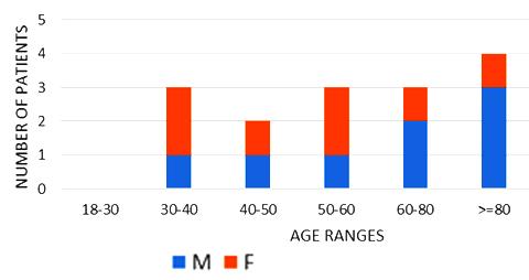
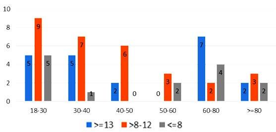
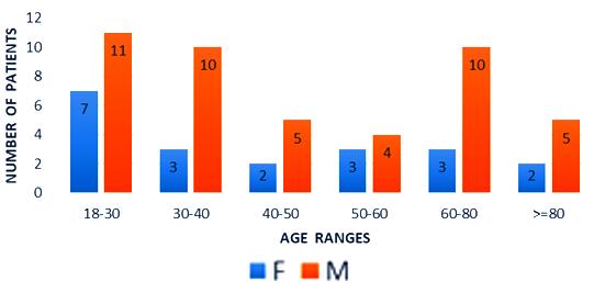
In our study, out of 65 patients 20(31%) were Female and 45 (69%) were Male. The minimum recorded age of the patients are 18 years and maximum are 93 years. The mean age was 46 years among female and 47.5 years in male and 47 years overall. The most affected age group belongs to the range of 18-30 years comprising 27.69% (n=18, of which 11 are Males and 7 Females) of study population, next affected category is 30-40 age group comprising 20% (n=13, comprising 10 males & 3 females). The other age categories of 40-50, 50-60 and > 80 years comprises 10.77% each. As per level of consciousness, GCS score >13 observed in 32.31%, GCS score (8-12) found in 46.15% and GCS < 8 detected in 21.54% of cases. Among 14 patients presenting with poor GCS score <8, six patients (ie, 42.86%) belonged to elderly age group (>60 years) and 5 (ie, 35.71%) patients falls in younger age category (18-30 years). Risk factor for Seizure in the form of old Cerebral Vascular Accident (CVA)/Stroke, recent/remote head injury, Alcohol/substance abuse, past CNS TB/Infection, history of developmental delay in childhood and family history of epilepsy present in 24 (36.92%) and not found in 41(63.08%). In our study majority (63.08%, 41out of 65) of the patient population presented with Generalized Tonic Clonic Seizure (GTCS), followed by Focal Seizure with impaired consciousness in 23.38% cases. Only one patient had generalized Myoclonic Seizure (1.54%). So, 64.62% of the study population presented with generalized and 35.38% cases with Focal Seizure type. Of the total 65 patients, metabolic abnormalities are present in 23.08% (n=15) cases, not found in 79.62% (n=50).
Among the metabolically abnormal patient’s population 20% (n=3) had Hypoglycemia, 40%(n=6) presented with Dyselectrolytemia, 26.67% (n=4) had Uremia, 13.33% (n=2) presented with Hepatic encephalopathy. Overall, 31 patients underwent Lumber puncture, of which CSF report shows abnormality in 11 (35.48%) cases and normal in 20 patients (64.51%). Abnormal CSF report suggesting viral meningoencephalitis found in 46% (n=5), bacterial meningitis in18% (n=2), Tuberculous Meningitis in 27% (n=3), Aseptic meningitis in 9% (n= 1). All 65 subjects underwent CT scan brain among which MRI Brain also done in 55 cases. CT findings is normal in 44.62% (n=29), abnormal in 55.38% (n=36). The most common CNS lesion on CT scan was infarction in 10.77% followed by ring lesion in 9.23% and intracerebral Hemorrhage in 7.96%. We observed cerebral infarction is causative factor of Seizure in female (15%) but intracerebral hemorrhage and ring lesion are most common findings in male, comprising 11.11% patients at each category. The CT scan in new onset Seizure of elderly patient population revealed Infarction in 28.57%, Gliosis in 19.04% and cerebral atrophy in 14.28%. The breakup of findings in MRI Scan of 55 patients shows Hyperintensities on T2, indicating Demyelination / Inflammation/edema in12.31% followed by infarction in 10.77% and ring lesion in 9.23%. In the female population, hyperintensities on T2 MRI found in 15% and Infarction in 15% whereas among the males intracerebral Hemorrhage, Ring lesion and hyperintensities on T2 found at similar occurrence rate @11% in each of these three types of lesions. EEG was done on 80% patients (n=52) of whom 22 patients has abnormal discharge in EEG but 30 patients have normal presentation. Among 65 patients one female expired due to Acute Demyelinated Encephalomyelitis (ADEM) and two males expired one due to Hemorrhage, another one with CNS infection when undergoing treatment indoor.
We found a strong positive correlation between abnormal EEG and abnormal Neuroimaging with Correlation coefficient of 0.902, (P- Value 0.0138) but almost no correlation found between normal EEG with normal Neuroimaging (Figs 1-4).
After analysis we found that Seizure incidence is more common in Male compared to Female in a ratio of 2.25: 1. The majority of patients (41.53%) presented at middle age range (30-60 years) with mean age of the study population being 47 years. These findings corroborate with study by V muralidhar2. In our study,
majority (46%) of Seizure patients presented with GCS score (8-12). We also observed that most patients with poor GCS score (<8) belonged to elderly age group (>60 years). Among the multiple risk factor predisposing to seizure most common was Alcohol and substance use, comprising about 12.31% of study population and old CVA is present in 9.23% of patients. We found most common Seizure type to be GTCS (63.08%) followed by Focal Seizure (Motor in type) with impaired consciousness (23.08%) which corroborates with another few studies3. In our study status epilepticus occurred in 7.69% patients.
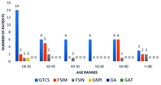
About 23% of patients with adult onset first seizure has metabolic derangement and the most common etiology was Dyselectrolytemia. The most common Central Nervous System (CNS) Infection as detected by CSF study was viral meningoencephalitis followed by Tuberculous Meningitis. The most common CNS lesion detected by CT studies as well as by MRI Brain was infarction and ring lesion, the two Neuroimaging studies corroborated in 75.38% of Seizure patients. Abnormal discharge in EEG found in34% cases. In majority patients EEG report was normal. EEG abnormality was more common in patients with Focal Seizure rather than GTCS patients. Thus, the most common etiology of first Seizure onset at adult age over 18 years were metabolic derangements (16.92%), CNS Infection or Inflammation (15.38%) and intracerebral Space occupying lesion (15.38%), usually ring lesion, tumors and Metastasis. Idiopathic Seizure found in 7.69% patients. The Correlation between abnormal Neuroimaging and abnormal EEG is strongly positive and statistically significant (P -Value = 0.013) but the same is not true in cases of normal Neuroimaging and normal EEG findings.
Limitations of the study : Our study has few constrains in extrapolating our observation to general population as our study population is a small size of convenient patient population attending in a Tertiary Care Centre in Kolkata, West Bengal, India over one year. Reliance has been given on history delivered by lay people and relatives of patients who may not be efficient enough to detect exact episode, prior episode of Seizure, nor able to detect
any manifestation of sensory Seizure. Inability to perform EEG in all patients happened due to huge work pressure of Neuro electrophysiology department.
New onset Seizure in adult is more common in male belonging to middle age group of 30-60 years. Most usual causes are Dyselectrolytemia, Viral meningoencephalitis, CNS infection or Inflammation, Cerebral Infarction and Ring Lesion. CT Scan Brain is the most useful tool of investigation in elderly population to detect an epileptogenic focus as compared to young. CT Scan brain and MRI is nearly had equal value to detect structural lesion in Seizure patients. Abnormal EEG and abnormal Neuroimaging has strong positive correlation to reach to a diagnosis.
We acknowledge the participation of students and faculties of postgraduate Medicine and Neurology as well as related laboratory personnel in conducting the study. We hugely acknowledge the immense contribution from the Scholars whose article are cited and included in reference of this manuscript. We are also grateful to Authors, Editors, Publishers of those journal and books from where the article is reviewed and discussed.
Conflict of Interest : Nil
Source of Funding : Nil
Ethical Clearance : Taken from Institutional Ethics Committee vide memo No/NMC/10024 dated 08/01/ 2019.
1Harrison Principles of Internal Medicine, 20th Edition, Vol 2, 2018.
2Muralidhar V, Venugopal K — New onset seizures: Etiology and co-relation of clinical features with computerised tomography and electroencephalography. Journal of the Scientific Society 2015; 42(2): 82-7.
3Kaur S, Garg R, Aggarwal S, Singh SPC, Pal R — Adult-onset seizures: Clinical, etiological, and radiological and radiological profile; Journal of Family Medicine and Primary Care 2018; 7(1): 191-7.
4Sinha S, Satishchandra P, Kalband BR, Bharath RD, Thennarasu K, — Neuroimaging observation in a cohort of elderly manifesting with new onset seizures: Experience from a university hospital. Ann Indian Acad Neurol 2012; 15(4): 273-80.
5Amudhan S, Gururaj G, Satishchandra P — Epilepsy in India I: Epidemiology and public health. Annals of Indian Academy of Neurology 2015; 18(3): 263-77.
6Gavvala JR, Schuele SU — New-Onset Seizure in Adults and Adolescents, A Review. JAMA 2016; 316(24): 2657-68
7Debicki DB — Electroencephalography after a single unprovoked seizure. Seizure (European Journal of Epilepsy) 2017; 49(7): 69-73.
8Newton CR, Garcia HH — Epilepsy in poor regions of the world. The Lancet 2012; 380(9848): September 29, 1193201.
9Santhosh NS, Sinha S, Satishchandra P — Epilepsy: Indian perspective. Annals of Indian Academy of Neurology 2014; 17 (Suppl 1): S3-S11
10van Donselaar CA, Schimsheimer RJ, Geerts AT, Declerck AC — Value of the Electroencephalogram in Adult Patients with Untreated Idiopathic First seizures. ArchNeurology1992; 49(3): 231-7.
Background : The efficacy of primary Percutaneous Coronary Intervention (PCI) as well as Pharmaco-invasive strategy for revascularization in ST-Elevation Myocardial Infarction (STEMI) patients has been well proven. But in developing countries most of the patients are unable to undergo Angiography/PCI within first 24 hours of presentation. In such patients studies assessing the efficacy of revascularization through extended Pharmaco-invasive therapy (fibrinolysis followed by elective PCI between >24 hours to 2 weeks of fibrinolytic administration) are sparse.
Aims and Objectives : To observe change in Left Ventricle (LV) function as assessed by Global Longitudinal Strain (GLS) at 3 months and at 6 months in cohort of STEMI patients undergoing extended Pharmaco-invasive therapy.
Materials and Mehods : This is a prospective observational study conducted in SCB Medical college, Cuttack. 65 patients who presented with STEMI were included in the study from November, 2020-2021. All the patients underwent thrombolysis with various fibrinolytic agents followed by elective PCI within 24 hours to 2 weeks. LV function was assessed through Echocardiography (Simpson’s Biplane and GLS) at preprocedure, 3 months and at 6 months.
Results : Mean age of the sample population was 60.18±8.289 years. All of the patients were thrombolysed within a mean window period of 7.57±2.744 hours. The mean time to revascularization was 69.35±22.97 hours after thrombolysis. The mean GLS were -11.5±3.3, -13.2±4.9 and -15.1±3.1 at pre-procedure, at 3 months follow-up and at 6 months follow-up respectively. When change in mean GLS at 6 months from baseline was compared with respect to the time to PCI, mean change in GLS at 6 months was found to have strong linear correlation with time to PCI after thrombolysis (r=0.773, p24 hours) still outcomes are better in those in whom PCI was done early ie, within 48 hours (by -4.50) than those in whom PCI was done after 96 hours (by -2.63) of Thrombolysis.
Conclusion : Extended Pharmaco-invasive strategy is a reasonable option if PCI cannot be performed within the first 24 hours. This strategy is thus likely to widen the window between Thrombolysis and PCI. There is a significant improvement of LV function as assessed by GLS at short-term follow-up of 6 months.
[J Indian Med Assoc 2023; 121(5): 23-7]
Key words :LV GLS, STEMI, Pharmaco-invasive Strategy.
Timely re-perfusion in patients presenting with ST-Elevation Myocardial Infarction (STEMI) leads to myocardial salvage and improvement in Left Ventricle (LV) function. The preferred means for reperfusion in STEMI is primary Percutaneous Coronary Intervention (PCI) within 24 hours of symptom onset, provided, it can be performed within 120 minutes of first medical contact1. But unfortunately, in developing countries like India a large subset of patients reach PCI capable centres only after 24 hours of the onset of chest pain. These subsets of patients may potentially benefit from
Department of Cardiology, SCB Medical College and Hospital, Cuttack, Orissa 753007
1MD (General Medicine), Resident and Corresponding Author
2MD, DM, FACC, FSCAI, FESC, Associate Professor
3MD, DM, Associate Professor
4MD, DM, FACC, FSCAI, FESC, Professor and Head
Received on : 25/05/2022
Accepted on : 03/06/2022
Editor's Comment :
There is significant improvement in LV function as assessed by GLS and LVEF, in patients undergoing extended Pharmaco-invasive therapy in whom primary pci within 24 hours was not possible. However GLS serves as a better tool for LV assessment as compared to LVEF especially in patients with post MI.
revascularization. Studies assessing the efficacy of extended Pharmaco-invasive therapy (fibrinolysis followed by elective PCI after 24 hours) are sparse. The current study postulates that extended Pharmacoinvasive therapy is a reasonable option for STEMI patients presenting late to a PCI capable centre.
To assess the LV function in such patients we would utilize various echocardiographic parameters such as Ejection Fraction by Simpson’s biplane method, end diastolic volume, end systolic volume, stroke volume and Global longitudinal strain through Speckle
tracking2. Strain is a sensitive tool that correlates well with other measures of cardiac function, and detect changes in myocardial contractility, both normal and abnormal, across a wide range of ischemic syndromes3.
This prospective observational study was conducted in the Department of Cardiology, SCB Medical College and Hospital, Cuttack from November, 2020-2021. In 65 patients were included in the study.
Inclusion criteria :
The patients included in the study were those who fulfilled the criteria of ST Elevation Myocardial Infarction as established by The American College of Cardiology, American Heart Association, European Society of Cardiology, and the World Heart Federation committee and presented within a window period of 12 hours from the onset of chest pain.
Exclusion criteria :
•AMI patients who already have coronary stent implanted for any indication.
•Patients who had previous episodes of MI.
•Left main or multi-vessel coronary artery disease mandating coronary artery bypass graft.
•Patients who do not wish to follow up at 6 months.
•Patients with cardiogenic shock.
•Renal failure
•Mechanical complications
All patients received fibrinolytic therapy and then underwent PCI in accordance with the current standard of practice. The minimum time to PCI was 24 hours while the maximum time to PCI was 2 weeks after the completion of Thrombolysis. PCI was defined as successful when Thrombolysis in MI (TIMI) flow grade was 2/3 post procedure and less than 20% residual stenosis.
All patients underwent 2D-TTE before coronary angiogram and PCI. The LV ejection fraction was assessed by Simpson’s biplane method, and GLS was estimated by speckle-tracking echocardiography. The following Echocardiographic parameters were also analysed: End Diastolic Volume, End Systolic Volume, Stroke Volume, Mitral Regurgitation.
All patients were followed-up at 3 months and at 6 months after discharge. A repeat 2D-TTE assessing all the above-mentioned parameters was performed at 3 months and at 6 months.
The collected data were analysed using the Statistical Package for the Social Sciences (IBM SPSS for Windows version 23.0). Categorical variables were
presented as frequency and percentage while continuous variables were described as mean value ± standard deviation. Paired t-test was used to compare values at baseline, 3months and at 6 months. Pearson’s correlation was used to establish statistical difference between groups. P<0.05 was considered to indicate statistically significant difference.
In 65 patients who fulfilled the inclusion criteria were included in the study. The mean age of the patients was 60.18 ± 8.3 years. 60% of patients were Male while rest 40% of patients were Female. In 39 patients were Diabetic, 30 patients were Hypertensive while 25 patients were smoker. 32 patients had no risk factors or a single risk factor. 33 patients had two or more risk factors. Mean window period for Thrombolysis was 7.57±2.7 hours. STREPTOKINASE was used in 35 patients (53.85%) for Thrombolysis while TENECTEPLASE and RETEPLASE each was used in 15 patients (23.08 %) for thrombolysis. The mean time to revascularization was 69.35± 22.98 hours after Thrombolysis. In 34 (52.31%) patients PCI was done in single vessel; in 26 (40%) PCI was done in 2 vessels, while in 5 (7.69%) of patients PCI was done in 3 vessels. In Multivessel PCI all vessels were addressed in the same setting.
The mean LV ejection fraction were 46.54±8.8%, 47.9±8.6% and 51.12±7.6% at preprocedural, at 3 months follow-up and at 6 months follow-up respectively. The mean increase in Left Ventricular Ejection Fraction (LVEF) was 1.3% at 3 months follow up and 4.6% at 6 months follow-up. The mean Global Longitudinal Strain (GLS) were -11.5±3.3, -13.2±4.9 and -15.1±3.1 at preprocedural, at 3 months follow-up and at 6 months follow-up respectively. The mean increase in GLS was -1.66 at 3 months follow up and -3.6 at 6 months follow-up from baseline.
Paired T-test was used to correlate LVEF and GLS at baseline, at 3 months and at 6 months. The correlation was found to be significant at p-value of less than 0.01.
Mean Left Ventricular End-Diastolic Volume (LV EDV) were 83.6±12.1 ml, 86.42±10.8 ml and 88.12±9.9 ml at preprocedural, at 3 months follow-up and at 6 months follow-up respectively. Mean Left Ventricular End Systolic Volume (LVESV) were 44.3±9.4 ml, 44.91±9.9 ml and 42.8±8.4 ml at preprocedural, at 3 months follow-up and at 6 months follow-up respectively. Mean LV SV were 39±9.7 ml, 41.4±8.9 ml and 44.6±7.9 ml at preprocedural, at 3 months follow-up and at 6 months follow-up respectively.
Paired T-test was used to correlate these values at baseline, 3 months and at 6 months follow-up. EDV and SV were found to increase linearly with time following intervention which was found to be significant with p-value <0.01. ESV was found to decrease at 6 months with a significant p-value <0.05 which may be due to more increase in SV than the simultaneous lesser increase in EDV, suggesting improvement in LV function.
When the mean change in GLS at 3 months and at 6 months follow up from baseline was correlated with respect to the number of risk factors present, a significant moderate linear correlation (r=0.513 at 6 months, p<0.01) was found inferring better improvement in GLS in patients with no risk factors or 1 risk factor (by -2.91, -2.64 at 3 months and -4.92, -4.28 at 6 months) than those with all 2 or 3 risk factors (by -1.78, -0.69 at 3 months and -2.85, -1.53 at 6 months).
When the mean change in LVEF at 6 months follow up was compared with that from baseline with respect to the number of risk factors, a low correlation (r=0.35 at 6 months, p=0.015) was found, inferring better improvement in LVEF in patients with no risk factors (by 5.20%) as compared to those with 2 or 3 risk factors (by 3.88% and 1.57% respectively). No significant difference in change in mean LVEF was found at 3 months follow up when compared with that at baseline with respect to the number of risk factors.
When change in mean GLS at 6 months with that at baseline was compared with respect to the time to intervention (PCI), mean change in GLS at 6 months was found to have strong linear correlation with time to PCI after thrombolysis (r=0.773, p<0.01) inferring that though delayed (>24 hours.) still outcomes are better in those in whom PCI was done early ie, within 48 hours. (by -4.50) than those in whom PCI was done after 96 hours (by -2.63) of thrombolysis. There was no significant difference noted at 3 months follow up in GLS.
When change in mean LVEF at 3 months and 6 months follow up was compared with that at baseline with respect to time to intervention, no significant difference was found; though LVEF improved more in whom the PCI was done within 48 hours. (by 5.42%) than in those whom PCI was done after 96 hours (by 3.68%) (Tables 1-4).
In developing countries and remote areas of many developed countries, Pharmaco-invasive strategy is
now the most commonly used approach in the management of patients presenting with STEMI because despite all efforts, there are many patients who reach PCI capable centers >24 hours after Thrombolysis. The current study clearly demonstrates the fate of such patients with respect to their LV function as assessed by Echocardiography.
This study shows that there is significant increase
in both LVEF and GLS both at short term follow up of 3 months and at 6 months duration. LV EDV and SV were found to increase linearly with time following intervention. ESV was found to decrease at 6 months which may be due to more increase in SV than the simultaneous lesser increase in EDV, suggesting improvement in LV function. Also, the increase in mean LVEDV at 6 months from baseline was 5.4%, this is well below 20% increase in LVEDV, as seen in patients with post MI LVAR (Left Ventricular Adverse Remodeling) which is an independent factor leading to Heart Failure4
A significant moderate linear correlation was found when mean change in GLS was compared with the number of risk factors present both at 3 months and at 6 months follow up, inferring better improvement in GLS after revascularization in patients with no risk factors than in those with all 2 or 3 risk factors. Such correlation was found only at 6 months with LVEF. The lesser improvement may be due to sub-clinical myocardial disease process in patients with multiple risk factors where Global Longitudinal Strain measurements appeared to be sensitive indicators for subclinical diseases as compared to LVEF5
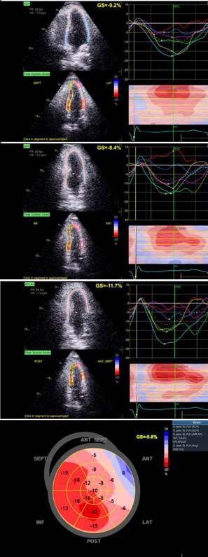
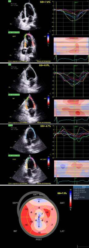
When correlated with time to intervention after PCI this study also clearly shows that the improvement in post-PCI LV function, as assessed through GLS, are better in those in whom PCI was done early (ie, <48 hours in this study) than in those whom PCI is still delayed (ie, >96 hours). Such a change could not be detected through LVEF. Thus, the assessment of LV function after PCI in patients with STEMI was superior with GLS when compared to 2D LVEF. As strain imaging is an inexpensive tool, it can be applied easily to assess LV function in the large subset of population (Figs 1A, 1B & 2).
The following conclusions can be drawn from the study:
•Extended Pharmaco-invasive strategy is a reasonable option if PCI cannot be performed within the first 24 hours. This
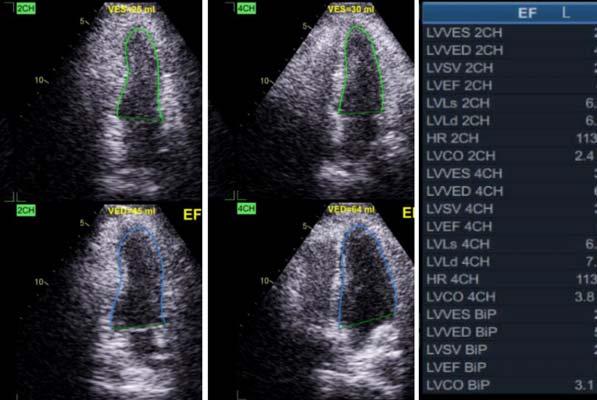
strategy is thus likely to widen the window between Thrombolysis and PCI and broaden the subset of patients eligible for coronary angiogram plus revascularization after Thrombolysis.
•There is a significant improvement of LV function as assessed by TTE when measured through GLS and with ejection fraction at short-term follow-up of 6 months.
•Improvement in LV function was significantly more in patients with no or single risk factor as compared to those with 2 or more risk factors as assessed by GLS and LVEF at 6 months follow-up.
•GLS is a better tool for LV assessment as compared to 2D LVEF especially in post-MI patients.
1Mamas MA, Anderson SG, O’Kane PD — Impact of left ventricular function in relation to procedural outcomes following percutaneous coronary intervention: insights from the British Cardiovascular Intervention Society. Eur Heart J 2014; 35: 3004-12.
2Mistry N, Beitnes JO, Halvorsen S — Assessment of left ventricular function in ST-elevation myocardial infarction by global longitudinal strain: a comparison with ejection fraction, infarct size, and wall motion score index measured by noninvasive imaging modalities. Eur J Echocardiograph. 2011; 12: 678-83.
3Mistry N, Beitnes JO, Halvorsen S — Assessment of left ventricular function in ST-elevation myocardial infarction by global longitudinal strain: a comparison with ejection fraction, infarct size, and wall motion score index measured by noninvasive imaging modalities. Eur J Echocardiograph. 2011; 12: 678-83
4Bolognese L, Neskovic AN, Parodi G, Cerisano G, Buonamici P, Santoro GM, etal—Left ventricular remodeling after primary coronary angioplasty: patterns of left ventricular dilation and long-term prognostic implications. Circulation 2002; 106(18): 2351-7.
5Yu CM, Sanderson JE, Marwick TH, Oh JK — Tissue Doppler imaging a new prognosticator for cardiovascular diseases. J Am Coll Cardiol 2007; 20(3): 234-43.
Ifyouwanttosendyourqueriesandreceivethe responseonanysubjectfromJIMA,pleaseuse the E-mail or Mobile facility.
Website:https://onlinejima.com
For Reception:Mobile : +919477493033
For Editorial:jima1930@rediffmail.com
Mobile : +919477493027
For Circulation:jimacir@gmail.com
Mobile : +919477493037
For Marketing: jimamkt@gmail.com
Mobile : +919477493036
For Accounts: journalaccts@gmail.com
Mobile : +919432211112
For Guideline:https://onlinejima.com
Background : Continuous Positive Airway Pressure has emerged as one of the main modality of treatment in Respiratory Distress Syndrome in Preterm Neonates. Early CPAP therapy has been shown to be successful in many clinical trials in the management of RDS, though studies from India are scarce.
Aim : The aim of the study was to determine whether introduction of Early CPAP results in improvement in terms of survival, need for mechanical ventilation and complications in early Preterm Neonates.
Methods : The study was a Prospective Observational study. All statistical analysis were performed using SPSS software version 24.0. P values <0.05 were considered significant.
Results : 126 newborns with gestational age 28-32 weeks were included in the study. 94 babies were given Early CPAP (74.6%) whereas 32 were not (25.4%). Among 94 babies who were put on Early CPAP, 91 babies did not need mechanical ventilation whereas 3 babies needed it (3.12%). Among the babies who were not put on Early CPAP (32), 26 did not need mechanical ventilation but 6 babies needed it (18.8%). P value is 0.003 ie, statistically significant. Among 94 babies who were put on Early CPAP, 4 babies died (4.3%) whereas 32 babies were not put on Early CPAP and 5 among them died (15.6%). P value is 0.031 ie, statistically significant.
Conclusion : Early institution of CPAP in the management of RDS in premature Neonates can significantly reduce the need for Mechanical ventilation and Mortality, with minimum associated complications.
[J Indian Med Assoc 2023; 121(5): 28-31]
Key words :Early Continuous Positive Airway Pressure (CPAP), Outcome, Preterm Neonates, Respiratory Distress Syndrome.
Respiratory Distress Syndrome (RDS) is the commonest cause of neonatal mortality in preterm babies. It is an acute illness, developing within 4-6 hours of birth, characterized by a rapid respiratory rate (>60 breaths/min), intercostal, subcostal and sternal retraction or indrawing, expiratory grunting and cyanosis.
(1) To assess the outcome of early CPAP therapy in Preterm Neonates with 28-32 weeks of gestation in a Tertiary Care Hospital in terms of survival and need for mechanical ventilation.
(2) To assess the incidence of various adverse effects in neonates with 28-32 weeks of gestation undergoing CPAP therapy.
Study Design : Prospective Observational Study
1MD, Senior Resident, Department of Pediatrics, Calcutta National Medical College and Hospital, Kolkata 700014 and Corresponding
Author
2MD, Professor and Head, Department of Pediatrics, KPC Medical College and Hospital, Kolkata 700032
Received on : 20/05/2022
Accepted on : 06/06/2022
Editor's Comment :
Early institution of CPAP is a cost-effective intervention for Preterm babies with RDS in a 3rd world country like India, where resources are scarce in many areas. Early CPAP can significantly reduce the mortality, need for invasive ventilation and its associated complications.
Place of Study : Neonatal Intensive Care Unit (NICU) in the Department of Paediatric Medicine, R G Kar Medical College and Hospital, Kolkata.
Period of Study : 2017-2019
Study Population : Preterm babies with gestational age 28-32 weeks born in R G Kar Medical College and Hospital
Sample Size : For calculation of sample size, a formula for Descriptive studies was used. The formula is as follows-
N = (Zα/2)² × p × q
L²
Where N = Sample size
Zα/2 = Standard Normal Deviate and its value would be 1.96 considering 95% Confidence Interval.
p = Expected proportion of CPAP failure obtained from a study carried out by RV Jeya Balaji, et al in a Tertiary Care Teaching Hospital in Kochi.
q = (100-p)
L = Precision in absolute terms and it will be taken as 8 percentage.
An observation period of one month showed that an average of 1 newborn babies are put on CPAP every day in the inborn Neonatal Intensive Care Unit of R G Kar Medical College & Hospital. So the yearly average is around 360 babies who are put on CPAP. Keeping the Precision level of our study at 8, the required sample size was 126. Systematic random sample technique was taken as sampling technique for this study.
Thus, (Zα/2)² = 3.84
p = 30%
q = 70%
L = 8
N = 3.84 × 2100 64 = 126
Inclusion Criteria :
Preterm babies born in R G Kar Medical College and Hospital with gestational age 28-32 weeks having respiratory distress.
Exclusion Criteria :
(1) Babies with Congenital anomalies
(2) Needs mechanical ventilation at birth
(3) Congenital anomalies affecting ventilator function (Diaphragmatic hernia)
(4) Infants with Severe Early Onset Sepsis or Compromised pulmonary blood flow (PPHN)
(5) Infants with low APGAR scores (Birth asphyxia) and responding poorly to resuscitation efforts
(6) Premature babies <28 weeks of gestation or birth weight <1000 gm
(7) Progressive atelectatic disease
Outcome Measures and Parameters :
• Primary Outcome Measures : Need for Mechanical Ventilation and Mortality
• Secondary Outcome Measures: Complications like Nasal trauma, Hypotension, Intraventricular hemorrhage, CPAP belly, Oliguria, Metabolic acidosis, Necrotising Enterocolitis, Bronchopulmonary Dysplasia (BPD), Pneumothorax, Pulmonary Hemorrhage
Statistical Methods : Quantitative variables were presented as mean and standard deviation. Categorical variables were presented as frequency and percentages and were compared using Pearson c2 or Fisher’s exact tests. Continuous variables were compared using Student t-test. P values <0.05 were
The total number of study population was 126, of which 76 were boys (60.3%), 49 were girls (38.9%) and 1 baby had ambiguous genitalia (0.8%).114 babies were Appropriate for gestational age (90.5%), 9 babies were Small for gestational age (7.1%) and 3 babies were Large for gestational age (2.4%). Among 126 deliveries, 59 were delivered by Lower Segment Caesarean Section (46.8%) and 67 were delivered by Normal Vaginal Delivery (53.2%) (Table 1).
Among all the babies, 106 (84.1%) had RDS soon after birth (within 30 minutes) whereas 20 had developed RDS later (15.9%). Among 126 babies, 94 were given Early CPAP (74.6%) whereas 32 were not given (25.4%)(Table 2).
Measures :
Among the babies who were put on CPAP due to RDS, 9 had CPAP failure and had to put on Mechanical ventilation (7.1%). Among the babies who were given CPAP therapy, 117 survived (92.9%) and 9 babies died (7.1%).
Among 126 babies, 94 were put on Early CPAP, among which 91 babies did not need mechanical ventilation whereas 3 babies needed mechanical ventilator (3.12%). Among the babies who were not put on Early CPAP (32), 26 did not need mechanical
ventilator but 6 babies needed it (18.8%). P value is 0.003 i.e. statistically significant.
Association of Early CPAP with Mortality : 94 babies were put on Early CPAP among whom 4 babies died (4.3%) whereas 32 babies were not put on Early CPAP and 5 among them died (15.6%). P value is 0.031 ie, statistically significant (Table 3).
Descriptive analysis of Secondary Outcome Measures :
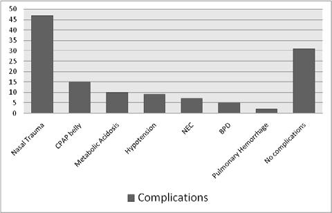
Among the babies on CPAP, 37.3% had Nasal trauma (commonest complication in our study), followed by CPAP belly 11.9%, Metabolic acidosis 7.9%, Hypotension 7.1%, NEC developed in 5.5% cases, BPD in 3.9%, 1.5% developed Pulmonary haemorrhage. 24.6% babies had no complications (Fig 1).
In 1971, Gregory, et al first described the method of delivery of CPAP in treating RDS. Since then many studies have been published evaluating the effectiveness of CPAP. But studies evaluating the effectiveness of Early CPAP is scarce in Indian literature. In our study, 126 Preterm Neonates born at
RGKMCH were selected depending upon the inclusion and exclusion criteria. 94 of the babies were put on Early CPAP (within 30 minutes) as they developed RDS soon after birth, according to protocol. 32 babies were put on CPAP later when they developed respiratory distress and needed FiO2 >60%.
•In our study, among all the babies, 106 (84.1%) developed RDS soon after birth (within 30 minutes) whereas 20 had developed RDS later (15.9%). In a study conducted by R V Jeya Balaji, 84.3% babies developed RDS soon after birth whereas 15.7% babies developed RDS later1
•In our study, Among 126 babies, 94 were given Early CPAP (74.6%) whereas 32 were not given (25.4%). Among the babies who were put on CPAP due to RDS, 9 had CPAP failure and had to put on Mechanical ventilation (7.1%). In a study conducted by R V Jeya Balaji, the incidence of CPAP failure was 30% (95% CI 19.3% to 40.7%) 1 . Another study conducted by Sunil B, et al showed that the incidence of CPAP failure who require Mechanical ventilation was 22.1% (95% CI 14.27-32.54%)2. In another study by Tapia, et al, incidence of CPAP failure was 29.8%3
•Among the 126 babies who were given CPAP therapy, 117 survived (92.9%) and 9 babies died (7.1%). In the study conducted by R V Jeya Balaji, et al, proportion of neonates, who met with mortality was 7.1% (1.1% to 13.2%) ie, same with our study1. In another study by Sunil B, et al, percentage of babies who met with mortality was 6.5% (10.14-26.77%)2 Another study by Tapia et al showed that mortality in babies given CPAP therapy was 8.4%3. All the studies showed similar results.
•Among the babies who were put on CPAP, 37.3% had Nasal trauma (commonest complication in our study), followed by CPAP belly 11.9%, Metabolic acidosis 7.9%, Hypotension 7.1%, NEC developed in 5.5% cases, BPD in 3.9%, 1.5% developed Pulmonary haemorrhage. 24.6% babies had no complications. In the study conducted by R V Jeya Balaji showed that Nasal Trauma, Hypotension, Intra Ventricular Hemorrhage and CPAP belly were the most common complications, occurring in 80% (70.6% to 89.4%), 11.4% (4% to 18.9%) and 10% (3% to 17%) of neonates each respectively. The other complications observed were CPAP belly, oliguria, septal injury, metabolic acidosis etc. No case of pulmonary hemorrhage was reported in the study1. Another study by Tapia, et al found that Incidence of Pneumothorax was 3.1% in CPAP group, BPD 6.9%, PDA 34.4%, IVH 25.2%, NEC 15.3%, and Nasal trauma in 8.4% cases3. In Another study named COIN trial by Colin J
Morley showed that Complications were Pneumothorax 9.1%, Pulmonary interstitial emphysema 5.5%, Intraventricular haemorrhage grade III or IV 8.9%, Cystic periventricular leukomalacia 2.9%, Necrotizing enterocolitis grade II or III 3.9%, Retinopathy of prematurity 53.1% and Patent ductus arteriosus 32.4%4
•Our study shows that babies who were put on Early CPAP needed less Mechanical Ventilation. Among 126 babies, 94 were put on Early CPAP, among which 91 babies did not need Mechanical Ventilation whereas 3 babies needed Mechanical Ventilator (3.12%). Among the babies who were not put on Early CPAP (32), 26 did not need Mechanical Ventilator but 6 babies needed it (18.8%). P value is 0.003 ie. statistically significant. Study conducted by R V Jeya Balaji, et al showed that the incidence of CPAP failure was 30% (95% CI 19.3% to 40.7%) in study population ie, 30% babies needed Mechanical Ventilation 1 Another study by Sunil B, et al showed that the incidence of CPAP failure who require Mechanical Ventilation was 22.1% (95% CI 14.27-32.54%) in study population2. Another study by Tapia, et al showed that need for Mechanical Ventilation in Early CPAP group was 29.8%3.
•Our study showed that there is strong correlation between Early CPAP and survival. Among 126 babies in total, 94 babies were put on Early CPAP among whom 4 babies died (4.3%) whereas 32 babies were not put on Early CPAP and 5 among them died (15.6%). P value is 0.031 ie, statistically significant. In the study conducted by R V Jeya Balaji, et al, proportion of babies who met with mortality was 7.1% (1.1% to 13.2%)1. Another study by Sunil B, et al showed that babies who were put on Early CPAP and met with mortality was 6.5% (10.14-26.77%)2 . In
another similar study by Tapia, et al, mortality among babies put on Early CPAP was 8.4%3
We conclude from our observational study that:
•Early institution of CPAP in the management of RDS in Premature Neonates can significantly reduce the need for Mechanical Ventilation and Mortality.
•Early institution of CPAP can also reduce the incidence of BPD, with minimum associated serious complications.
•Early use of CPAP is a low-cost, simple and non-invasive option for a country like India, where most places cannot provide Invasive ventilation and Surfactant.
Limitations :
•No Control group was taken for comparative analysis of the efficacy of Early CPAP with Late CPAP.
•Preterm babies with gestational age <28 weeks and >32 weeks were not taken in our study, which limits the generalizability of the results.
•The role of many confounding factors could not be evaluated because of the limited sample size.
1Jeya Balaji RV, Rajiv PK, Patel VK, Kripail M — Outcome of Early CPAP in the Management of Respiratory Distress Syndrome (RDS) in Premature Babies with <32 Weeks of Gestation, A Prospective Observational Study. DOI: IJNMR/ 2015/13361.2035
2Sunil B, Girish N, Bhuyan M — Outcome of preterm babies with respiratory distress syndrome on nasal CPAP. Int J Contemp Pediatr 2017; 4: 1206-9.
3Tapia JA, Urzua S, Bancalari A, Meritano J, Torres G, et al South American Neocosur Network. Randomized Trial of Early Bubble Continuous Positive Airway Pressure for Very Low Birth Weight Infants. J Pediatr 2012; 161: 75-80.
4Morley CJ, Davis PG, Doyle LW, Brion LP, Hascoet JM, Carlin JB — Nasal CPAP or Intubation at Birth for Very Preterm Infants. N Engl J Med 2008; 358: 700-8.
Purpose : To determine the effectiveness of intralesional injection triamcinolone acetonide in treatment of chalazion. Material and Methods : All patients meeting the inclusion criteria were included in the study through OPD irrespective of their Age and Gender. Chalazion was diagnosed on the basis of presence of painless and nontender nodule in the eyelid. Under strict aseptic technique 0.1 to 0.2 ml of Triamcinolone Acetonide (40mg/ml) was injected intralesionally. Follow up visit was done at 1 week, 2 weeks and 4 weeks to determine effectiveness in term of reduction in size of chalazion.
Results : overall success rate of the study was 97.50% with complete resolution while 2.5% lesions failed to show any response.
Conlusion : Intralesional injection of Triamcinolone Acetonide is quick, safe, cheap, convenient highly effective and acceptable method in treatment of Chalazion. Most lesions resolve with one or two injections and higher effectiveness is seen in chalazia of size less than 6mm.
[J Indian Med Assoc 2023; 121(5): 32-5]
Key words : Chalazion, Intralesional Triamcinolone Acetonide, Meibomian Glands.
Eyelid being one of the important ocular component subserves the protective function for the globe. Focal swelling of the eyelid is a common complaint that is seen in Out Patient Department (OPD). Actual meaning of chalazion is a hail stone. Chalazia are the most common inflammatory lesions of the eyelid. They are focal non infective chronic lipogranulomatous inflammatory lesion of the Meibomian Glands, caused by retained Sebaceous 1 . Meibomian glands are modified Sebaceous Glands present in the tarsal plate of upper and lower eyelids. These glands secretes the outer lipid layer of the tear film. Chronic inflammation of these glands causes retained secretions inside the ducts and cause Chalazion.
It is a common condition of lids affecting Males and Females equally2, presenting as chronic gradually enlarging painless rounded nodule over the lid. The chief effects are cosmetic disfigurement and discomfort to a variable degree. The diagnosis of Chalazion is usually done with the help of history and clinical examination.
The standard treatment of this lesion has been incision and curettage, which although a minor
Department of Ophthalmology, Dr. Vaishampayan Memorial Government Medical College, Solapur, Maharashtra 413003
1MS (Ophthalmology), Resident and Corresponding Author
2MS (Ophthalmology), Professor and Head
3MS (Ophthalmology), Assistant Professor
Received on : 13/05/2022
Accepted on : 22/05/2022
Editor's Comment :
Intralesional Triamcinolone Acetonide is an effective and safe method for the treatment of Chalazion. Intralesional injection of Triamcinolone Acetonide can be used as first line therapy for primary Chalazion without any significant complications.
procedure often causes some distress and discomfort to the patient.
However, intralesional corticosteroid therapy has been described as more convenient and less expensive alternative method of treatment. This method was found to be safe and effective alternative to incision and curettage3
The purpose of the present study was to evaluate the safety and efficacy of Intralsional Triamcinolone Acetonide in 40 cases.
This study was conducted at OPD of Ophthalmology , Tertiary Care Center, during the period of September, 2019 to October, 2021. Patients coming with complaints of swelling over eyelid or mass lesion over eyelid irrespective of their age and gender were thoroughly examined noting the detailed history and examination.
Exclusion Criteria :
•Patients with Infected Chalazion
•Multiple chalazia
•Those with a history of prior treatment for
chalazion whether surgical or corticosteroid injection intralesionally
•Uncontrolled diabetes mellitus
•Immunocompromised patients
Chalazion was diagnosed on the basis of presence of painless and non tender nodule in the eyelid. The size of nodule was measured with Castravejo’s caliper. The purpose and benefits of the study were explained to all patients.
In the present study total 40 patients of Chalazion were examined and treated with intralesional injection of Triamcinolone Acetonide after taking their consent.
In this study Triamcinolone Acetonide suspension was used for the intralesional injection. It is manufactured and sold under the market name of “KENACORT”. It is available in the concentration of 40 mg/ml vial (1ml).
Method : Under strict aseptic conditions, 0.1-0.2 ml of injection Triamcinolone Acetonide 40mg/ml was used for intralesional injection on conjunctival side. Patient was kept under observation in the OPD for 30 minutes. Antibiotic eye drops installed into the conjunctival sac. Bandage or eyepatch was not applied in any patient following injection. Patients were re examined at 1 week, 2 weeks and 4 weeks interval.
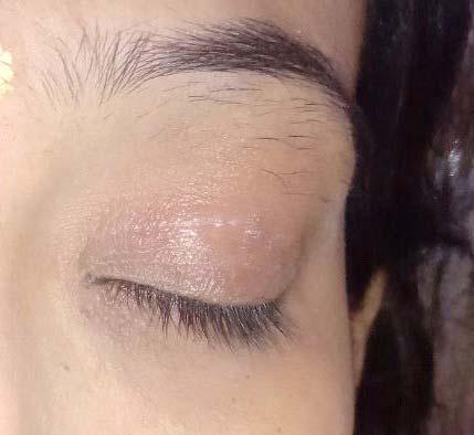
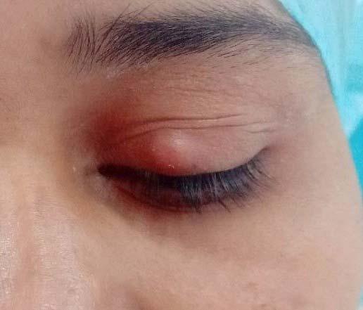
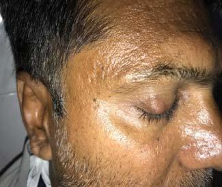
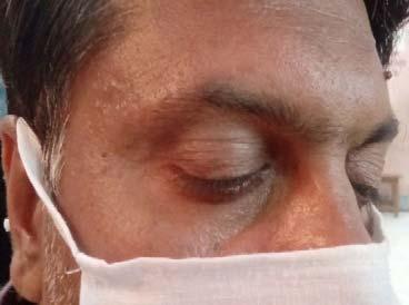
If Chalazion has not decreased significantly in size ie, half of its original in 2 weeks, a second injection was given. Patients were photographed during each visit. During follow up special attention was paid to note size of Chalazion, any pigmentation and white subcutaneous deposits at the site of injection and for infection (Figs 1-4).
In this study, 40 patients with Chalazion had been included, in which 18 (45%) were Male and 22 (55%) were Females. Patients age was divided in different categories out of which most presented in age group of 11-30 years (42.50%) (Table 1).
In the study, 72.50% of chalazia were of moderate size (3-5mm), 17.50% were of small
size (upto 2mm)(Table 2).
31 out of 40 chalazia had the duration less than 6 months. Out of which 19 chalazia showed resolution after 1 st injection while 12 chalazia resolved after 2 nd injection (Table 3).
Overall success rate of the procedure is 97.50% while failure rate was found to be 2.50% (Table 4).
Present study was conducted in Tertiary Care Center in the period of September, 2019 to October, 2021.
All patients coming to OPD with complaints of painless swelling over eyelid or mass lesion of one or more eyelid were thoroughly examined. The detailed history was noted and thorough external examination including slit lamp biomicroscopy was done.
Diagnosis of Chalazion was made on clinical examination differentiating it from other lid swellings. Patients with infected , recurrent and multiple chalazia were excluded from this study.
Each patient was examined and studied as per proforma. In this study, we have studied a series of
patients of chalazion. Their Age, Sex, duration of Chalazion, size of Chalazion, site of Chalazion was noted.
Triamcinolone acetonide drug was choosen as the drug of choice for reasons that it is insoluble in nature and there by is able to remain at the local site for longer duration. Also, its effectiveness is proved in treatment of chalazion.
Total number of patients studied in this study was 40 and total no. of chalazia studied were 40. In this study, 5 (12.50%) patients were in the age group of 1 to 10 years, 7 (17.50%) patients were in the age group af 11 to 20 years, 10 (25%) patients were in 21 to 30 years age group, 6 (15%) in 31 to 40 years, 8 (20%) in 41 to 50 years, 3 (7.50%) in 51 to 60 years, 1 (2.50%) in 61 to 70 years respectively. It was found that incidence of Chalazion is more in the age group of 11 to 40 years (Table 2)
Similarly, according to Pizzarello, mean age for the patients with Chalazion was 36 years with a range of 20 to 54 years4
A cross sectional observational multicentric study conducted by AV Das and TV Dave between 2010 and 2019 also showed that the commonest age group affected with Chalazion was the third decade of life13 Similarly, To Otulana, et al found that most of the cases of Chalazion in their study were in the age group of 2130 years14
We have found that 29 out of 40 (72.50%) chalazia
were of moderate size, 7 out of 40 (17.50%) were of small size and 4 out of 40 (10%) were of larger size.
Majority of the cases had the chalazia for 1 to 3 months duration ie, in 19 out of 40 (47.50%) patients. 13 out of 40 (32.50%) patients had a history of 4 to 6 months, 6 out of 40 (15%) patients had a history of over 6 months and 2 out of 40 (5%) patients were having history of 1 year or more.
In the present study, it was observed that chalazia which were of short duration ie, less than 6 months responded well to the treatment with a resolution rates of nearly 100% after 1 month of intralesional Triamcinolone injection.
Similarly, in the study done by Pizzarello, et al, in all cases the lesion had lasted from six weeks to one year with a mean duration of 6 months4
40 cases of chalazia were treated by intralesional injection of 0.1-0.2 ml of 40 mg/ml suspension of Triamcinolone Acetonide. The same concentration of Triamcinolone Acetonide was used in previous studies by MYY Wong, Gordon SK, et al5, Guy J Ben Simon, Lynn Huang, et al6 and Nabie R, et al7
In all patients the injection was given from conjunctival side to avoid the complication like pigmentation which was found to occur when injection was given from skin side. Jain and Misuria8, Jacob, et al9 and Watson, et al2 had given injections by same approach.
Thus the percentage of number of chalazia which resolved with one or two injections was 97.50% which is consistent with the results of Nilawar10 97.5% , 95.83% in the study done by Iradier, et al12 and 95% in the study done by Yagu and Mentes11.
One Chalazion did not resolve in spite of reinjection had history of more than 6 months duration. Percentage of no. of chalazia which did not resolve inspite of reinjection was 2.50%. in study done by Pizzarello4 two lesions out of 17 did not subside at all. While in the study done by Jain and Misuria8 it was 12%.
In our study, 2 out of 40 chalazia showed recurrence after complete resolution following steroid injection. 2.22% chalazia recurred after complete resolution in the study done by Nilawar10. Pizzarello4 in his study has mentioned that two lesions out of 17 which subsided after injection either recurred or a second neighbouring lesion occurred.
Complications like yellow deposits, tissue atrophy, hypopigmentation and increased intraocular pressure were not noted in any case. This is in agreement with the study conducted by Harminder Singh Dua, et al10 who observed no complications in their study of 90 cases of chalazia which were treated with Intralesional
Triamcinolone acetonide injections. No complication have been reported by Jain and Misuria8
The study was designed to determine the efficacy of intralesional triamcinolone acetonide injection in the treatment of Chalazion. The study proved that the Intralesional Triamcinolone Acetonide is ad effective and safe method for treatment of Chalazion. It is highly effective among patients with Chalazion of size <6mm and duration of <6 months. So from this study the conclusion can be drawn that for primary Chalazion <6mm size, the intralesional injection of Triamcinolone can be used as first line therapy without any significant complications.
1Sahu SK, Sen S, Poddar C — Sebaceous gland carcinoma of lid: Masquerading as a recurring chalazion. International Journal of Applied and Basic Medical Research 2021; 11(2): 117.
2Watson AP, Austin DJ — Treatment of chalazions with injection of a steroid Suspension. Br J Ophthalmol 1984; 68: 833-5.
3Mohan K, Dhir SP, Munjal VP, Jain IS — The use of intralesional steroids in the treatment of chalazion. Annals of Ophthalmology 1986; 18(4): 158-60.
4Pizzarello, Fredrick Hofeldt, A.J. : Amer J Ophthal 1978; 85: 818.
5Wong MY, Yau GS, Lee JW, Yuen CY— Intralesional triamcinolone acetonide injection for the treatment of primary chalazions. International Ophthalmology 2014; 34(5): 104953.
6Simon GJ, Huang L, Nakra T, Schwarcz RM, McCann JD, Goldberg RA — Intralesional triamcinolone acetonide injection for primary and recurrent chalazia : is it really effective? Ophthalmology 2005; 112(5): 913-7.
7Nabie R, Soleimani H, Nikniaz L, Raoufi S, Hassanpour E, Mamaghani S, et al — Aprospective randomized study comparing incision and curettage with injection of triamcinolone acetonide for chronic chalazia. Jr of Current Ophthalmology 2019; 31(3): 323-6.
8Jain PK, Misuria V — Recent non surgical approach in the treatment of chalazion. Ind Jr of Ophthal 1988; 36: 34.
9Jacobs, Thaller and Wong. British Jr of Ophthalmology 1984; 68: 836-37.
10Dua H, Nilawar DV — Non surgical therapy of chalazion. Amer J Ophthal 1982; 94(3): 424-5.
11Yagu, Mentes : Turk ophthalmol, Gaz 17,9-13, Jan-March 1987.
12Iradier M, Strada C, Quinzamos AG — ArchSocEspophthalmol 54: 399-409 Feb 1988.
13Das AV, Dave TV. Demography and clinical features of chalazion among patients seen at a multi-tier eye care network in India: Clinical ophthalmology (Auckland, NZ).2020;14:2163
14Otulana TO, Bodunde OT, Ajibode HA. Chalazion, a benign eyelid tumour- the sagamu experience. Nigerian Journal of Ophthalmology 2008; 16(2).
The information and opinions presented in the Journal reflect the views of the authors and not of the Journal or its Editorial Board or the Publisher. Publication does not constitute endorsement by the journal.
JIMA assumes no responsibility for the authenticity or reliability of any product, equipment, gadget or any claim by medical establishments/institutions/manufacturers or any training programme in the form of advertisements appearing in JIMA and also does not endorse or give any guarantee to such products or training programme or promote any such thing or claims made so after.
— HonyEditorShobana Sundaram1, Vasudevan Devanathan2, Vinoth Kanna2, Sathish Kumar3, Sangeetha Jayaraman4
Background : Seizure in elderly is quite frequent and potentially problematic to identify the cause. New Onset Seizure (NOS) is defined as the first Seizure or the first cluster of Seizures within a 24-hour period ever experienced by the patient. With increasing age, the prevalence and incidence of epilepsy and Seizures increases correspondingly. New-onset Epilepsy in elderly people often has the following etiology, including Cerebrovascular Diseases (Ischemic Stroke, Hemorrhagic Stroke & Cerebral Venous Sinus Thrombosis), Intracerebral Tumors and Traumatic Head Injury. In addition, an acute symptomatic Seizure cannot be considered Epilepsy, which presents usually as a common symptom secondary to metabolic, infection or toxicity causes in older people, which needs extreme attention and treatment. Adult-onset Seizure disorder is a major public health concern in terms of burden of disease, nature of illness and its impact on individual, family and community. This study was done to assess the clinical and etiological profile causing new onset Seizure in patients above 60 years of age and to find opportunities to control epileptic Seizures.
Methodology : This study was carried out in Saveetha Medical College Hospital and Research Institute for 100 patients with New Onset Seizure (NOS) who are more than 60 years of age. It is a Prospective, Observational hospital based clinical study conducted from August, 2020 to August, 2021. We excluded the patients with known case of Childhood Epilepsy, Movement Disorder, Pseudo Seizure, Hyperventilation, or Narcolepsy and those who did not give consent. Neurological examination, laboratory, neuroimaging, and electroencephalogram investigation was done in all the patients to find out the etiology.
Results : A hundred patients were divided into three groups as group A (aged 60-65 years), group B (66-70 years), and group C (aged above 70 years). Out of 100 patients, 61(62.4 %) were males and 39 (37.5%) were females. Stroke was the most common etiology of NOS (40%), followed by idiopathic (26%) metabolic causes like hyperglycaemia, hypoglycaemia, hyponatremia, and hypocalcemia (24%), traumatic brain injury (4%), neoplasm and infection 3%. Among these cases, NOS was found in 45% of cerebrovascular disease, 25% in TBI, 67% in neoplasm, 38% in Idiopathic. In our study, frequency of epilepsy was <2 in 67% and >2 in about 33%. The most common type of Seizure was focal to bilateral tonic-clonic Seizure (70%), followed by focal Seizure (18%) and generalized Seizure (12%). Nearly 64% had abnormal neuroimaging, Hypertension was the most common co-morbidity (33%).
Conclusion : The present study proposes that Epilepsy in the elderly patients have etiological relationship with Stroke, Metabolic, Neoplasm and Idiopathic. The incidence of first ever Seizure among elderly is as high as among young children and mortality of untreated seizure among the elderly is remarkably higher. The natural history and outcome of new onset seizure among elderly has been the subject of many recent studies. Epileptic seizures in the elderly are recognized as frequent but potentially difficult and burdensome to diagnose. Their clinical features and relevant diagnostic problem remain poorly investigated in hospital populations outside the setting of tertiary referral centers. This study was undertaken to determine the clinical characteristics and etiology of NOS's/Epilepsy in the elderly in a university hospital setting. [J Indian Med Assoc 2023; 121(5): 36-40]
Key words :New Onset Epilepsy, Seizures, Cerebro Vascular Disease, Idiopathic, Elderly.
Seizures are most common disease affecting 10% of the general population. In contrast to earlier findings that Seizures being a disease of children, data from geriatric population found increased occurrence
Department of Neurology, Saveetha Medical College, Hospital and Research Institute, Chennai 602105
1DM (Neurology), Assistant Professor and Corresponding
Author
2DM (Neurology), Professor
3DM (Neurology), Assistant Professor
4DM (Neurology), Senior Resident
Received on : 23/05/2022
Accepted on : 07/06/2022
Editor's Comment :
New onset seizure above 60 years is not very uncommon. One should be extra precautions while taking history and ruling out all structural and metabolic causes before diagnosing idiopathic seizures in elderly.
of New Onset Seizure (NOS)1. 1 to 3 per 1000 geriatric person per year has NOS and this is 2-6 times greater than young adults2 and mortality of untreated Epilepsy among the Elderly is remarkably higher. Care of the Elderly is an emerging health concern as the number
of Elderly persons will rise approximately 140 million by 20213. Though Seizure research is common, very few were done on the etiological profile of NOS4. NOS is defined as the first Seizure or the first cluster of seizures within a 24-hour period ever experienced by the patient5
NOS is manifested by two or more recurrent unprovoked Seizures. It may be of other secondary origin due to cerebral neoplasm, systemic disorder and infection which needs special treatment6. Seizures in the elderly are recognized as frequent but potentially difficult and burdensome to diagnose. The aim of the present study is to rule out the cause and risk factors to diagnose and treat the geriatric population with NOS at the earliest.
This study was an observational hospital-based study carried out in tertiary health care center. Geriatric subjects above 60 years with NOS were included in this study. Subjects with Movement Disorder, Hyperventilation, Pseudo Seizure, Narcolepsy and those who did not give consent were excluded from our study 100 patients were included and divided into three groups as group A (aged 60-65 years), group B (66-70 years) and group C (>70 years). Prior to the data collection Institution Ethical Committee approval was obtained. Patients and eyewitness were interviewed, history elicited and clinical examination was done. Complete Blood Count, Urea, Serum Creatinine, Blood Glucose Level, Serum Electrolytes Like Sodium, Potassium and Calcium were done. Routine investigations and Special investigations like EEG and Neuroimaging was also done.
Statistical Analysis :
Statistical analysis was done using SPSS 21. All continuous variables were expressed as mean ±SD. Qualitative data were expressed as percentage significant.
In 50% of our study population belonged to 60-65 years age, 31% between 66-70 years and 19% of them were above 70 years age group. 62% of our study population was males (Table 1).
We have observed that the commonest form being focal to bilateral tonic-clonic Seizures affecting 63% of our subjects, 21% of them had focal Seizures and 16% had generalized Seizure. 67% of our study population had less than two episodes of Epilepsy. Only a minority of elderly individuals had fever (3%), Headache (26%) and Vomiting (17%). 33% had past
history of hypertension and 62% of them had no personal history of smoking and alcohol (Table 2).
The pattern of etiology in males and females were similar in this study. Most common etiology in males was CVA (38.7% & 42.1%)in which major contributor was ischemic stroke (29%& 31.6%) followed by hemorrhage stroke (08.1% & 05.3%) and CVT (1.6% & 05.3%). Metabolic seizures comprised about 17% and 18.4% respectively. Idiopathic origin was seen in 24.2 % of males and 18.4% of females, followed by neoplasm (0.32% & 2.6%) and infection (01.6% & 5.3%). 12.8 % had alcohol withdrawal Seizure (Table 3).
Stroke contributes around 40% of NOS, followed by Idiopathic (26%) and Metabolic derangement (24%). Traumatic brain injury contributes only 4% and 3% were due to infection and neoplasm (Fig 1).
Among CVA, majority were due to infarct (30%), followed by Hemorrhage (7%) and Cerebral Venous Thrombosis (3%). Among Metabolic Seizures (24%) 11% were of hyperglycaemiac Seizures, 6% of Hypoglycemic Seizures and 1% of cases had Hyponatremia and Hypocalcemia respectively. In 4%
GenderAge groupTotal χ2 p-value 60-6566-70>70N (%)
Male31 (62)16 (51.6)15 (78.9)622.4540.293
Female19 (38)15 (48.4)04 (21.1)38
Total503119100
Clinical History Frequency
Seizure : Focal21
Focal to bilateral tonic -clonic seizure63 Generalized16
Number of Episode : < 267 >233
Fever : Present03 Absent97
Headache : Present26 Absent74
Vomiting : Present17 Absent83
History : No Complication32 Hypertension33
Diabetes mellitus15
Hypertension & Diabetes20
Personal History : No bad habit62
Smoking, Alcohol, Tobacco17
Smoking + Alcohol + tobacco21
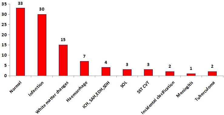
From Fig 4 we noted the maximum conversion of seizure into Epilepsy on Neoplasm (67%) followed by CVD (45%), Idiopathic (38%), and TBI (38%) (Fig 4).
There are only few prospective incidence studies exists to find out the cases with first Seizures in the adult population7. So, we intended to do the current study aimed on etiological profile of NOS in Elderly population.
According to 2010, ILAE Commission, classified Seizures into three major categories based on etiology (symptomatic, Metabolic and Idiopathic)8 We have evaluated data of 100 geriatric patients with NOS, in which males were 62% and females were 38%. Kaur S, et al and Chalasani S, et al also reported similar result in gender wise distribution5,7. Joshi M, et al reported that Seizures were more common in age group of greater than 60 years9. In our study, we found that NOS were common in age group of 60-65 years resulting in 62% of cases.
Patients with the past medical history of Hypertension were significantly associated with unprovoked Seizures. In our study, 33% of study population had the past medical history of Hypertension and 62% of the study population had no personal habit of smoking and alcohol.
The most common type of Seizure in this study was focal to bilateral tonic-clonic Seizure (70%) followed by focal (18%) and generalized (12%), in contrast to a study done by Hart YM etal, where they concluded that focal seizures are the most common type10.
We observed that the major cause for Seizure was cerebrovascular accident which accounted for 40% in
of the cases the cause for seizures was alcohol withdrawal. Neoplasms (Cerebral Metastasis) and infections (Tuberculoma, Meningitis) were the other minor causes of Seizures (Table 4).
64% of our subjects had abnormal neuroimaging findings. 30% had ischemic infarcts, 7% had hemorrhage and 4% of patients had ICH, SAH, SDH and EDH (Fig 2).
Among our subjects, 81% patients had normal EEG, while abnormalities of EEG were found in 19%. Focal delta activity Frontal spike, focal theta activity occupies the top three position (Fig 3).
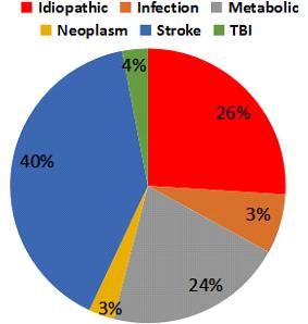
which ischemic stroke accounted for 30% of cases, 7% were hemorrhagic stroke and 3% were cerebral vascular thrombosis. This result is similar to Roberts MA, et al12
Seizures are associated with alteration in metabolic homeostasis such as Hypoglycemia, Hyperglycemia, Hyponatremia and Hypocalcemia (13,14). We had 21% of cases with metabolic causes with 11% of hyperglycemic, 6% of Hypoglycemia and Hyponatremia and hypocalcemia accounted for 1% of cases.
Seizure is age dependent in developed countries. Among children, 20% are Symptomatic, 50% are Metabolic and 30% are Idiopathic. In Geriatrics, 55% are remote symptomatic and 45% are Idiopathic15. We reported that idiopathic cause is comparatively less (26%) and metabolic cause with 24%, whereas stroke occupies 40% when compared to the other studies15. This study showed that in 4% of the cases had TBI and 3% of the patients had Neoplasms and Infections.
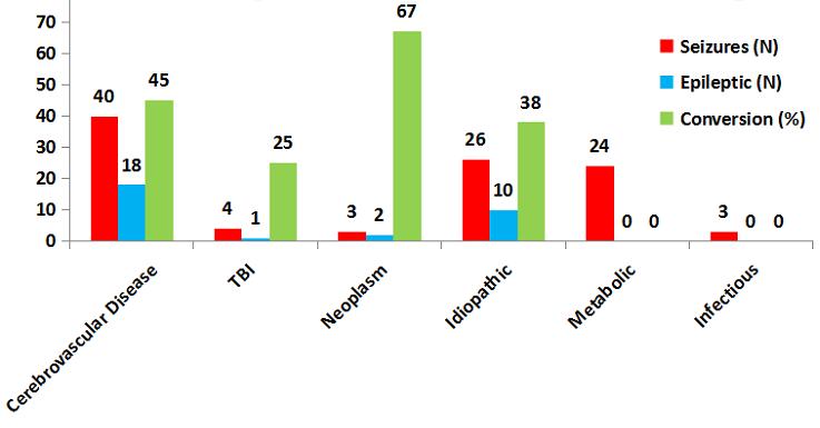
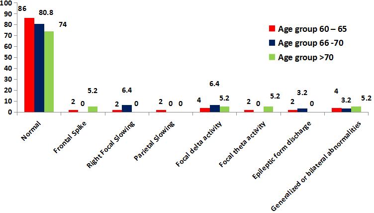
In our study nearly 64% had abnormal Neuroimaging findings. Ischemic infarcts were the common finding (30%) followed by hemorrhage (7%). These finding are in concordant with study done by Kaur, et al in which18% had ischemic infarcts and 8% had brain tumors5
EEG abnormalities with intermittent focal slowing are commonly seen in older patients even without Seizure16. Our study is similar to other studies done by Drury, et al, which showed EEG was normal in majority of the patients with Seizure, irrespective of age17. 81% of patients had normal EEG, while abnormal EEG was found among 19%. Frontal spike, focal theta activity and epileptic form discharge were detected in 2% and 5% with focal delta activity. Right focal slowing (03%) and generalized or bilateral abnormalities (04%) were detected in marginal level.
We observed that NOS are common among the age of 60-65 years males. Cerebrovascular disease and Metabolic Diseases are the common etiology and
idiopathic Epilepsy is the predominant etiology among them.
CONCULSION
NOS among elderly patients are more prevalent in males with age group of 60-65 years. Stroke and the Metabolic Diseases are the major etiological profile for Seizure in geriatric population. Idiopathic Epilepsy is the predominant etiology among age of 60-65 years males.
Prior Publication – Nil
Conflicts of Interest – Nil
Permissions – Nil
1Lily Chi V, Piccenna L, Kwan P, O’Brien TJ — New-onset epilepsy in the elderly. Br J ClinPharmacol 2018; 84(10): 2208-17.
2Choi H — Predictors of incident epilepsy in older adults: The Cardiovascular Health Study. Neurology 2017; 88(9): 870-7.
3Das SK — Situation analysis of the elderly in India; 2011 jun. 1-3.
4Ashwin T, Tumbanatham A, Green S, Singh K — Clinico etiological profile of seizures in adults attending a tertiary care hospital. Int J Adv Med 2017; 4(2): 490-6.
5Kaur S, Garg R, Aggarwal S, Chwawla SPS, Pal R — Adultonset seizures: Clinical, etiological, and radiological profile. J Fam Med Prim Care 2018; 7(1): 191-7.
6Shorvon SD, Andermann F, Guerrini R — The causes of epilepsy. Cambridge University Press, Cambridge. Epilepsia 2011; 52(6): 1033-44.
7Chalasani S, Kumar MR — Clinical Profile and Etiological Evaluation of New Onset Seizures after Age 20 years. IOSRJDMS 2015; 14(2): Ver. VII: 97-101.
8Berg AT, Berkovic SF, Brodie MJ, Buchhalter J, Cross JH, Boas WVE, et al — Revised terminology and concepts for organization of seizures and epilepsies: report of the ILAE Commission on Classification and Terminology, 2005-2009. Epilepsia 2010; 51(4): 676-85.9
9Joshi M, Bhargav B — A study of evaluation of etiology and clinical profile of new onset seizure in adults.Sch J App Med Sci. 2017;5(2E):620–5.in our study, we found that NOS were common in older age group(60-65 years),resulting 62% of cases.
10Luhdorf K, Jenson LK, Plesner AM — Etiology of seizures in elderly. Epilepsia 1986; 27(4): 458-63.
11Hart YM, Sander JW, Johnson AL, Shorvon SD — National General Practice Study of Epilepsy: recurrence after a first seizure. Lancet 1990; 336: 1271
12Roberts MA, Godfrey JW, Caird FI — Epileptic seizures in the elderly: I. Aetiology and type of seizure. AgeAgeing 1982; 11: 24.
13Beghi E, Carpio A, Forsgren L, Hesdorffer DC, Malmgren K, Sander JW, et al — Recommendation for a definition of acute symptomatic seizure. Epilepsia 2010; 51(4): 671-5. https:// doi.org/10.1111/j.1528-1167.2009.02285.x.
14Nardone R, Brigo F, Trinka E — Acute Symptomatic Seizures Caused by Electrolyte Disturbances. J Clin Neurol 2016; 12(1): 21-33. https://doi.org/10.3 988/jcn.2016.12.1.21.
15Hauser WA, Annegers JF, Kurland LT — Incidence of epilepsy & unprovoked seizures in Rochester,Minnesota 1935-1984 Epilepsia 1993; 34: 453-68.
16McBride AE, Shih TT, Hirsch LJ — Video-EEG monitoring in the elderly: a review of 94 patients. Epilepsia 2002; 43: 1659.
17Drury I, Beydoun A — Interictal epileptiform activity in elderly patients with epilepsy. Electroencephalogr Clin Neurophysiol 1998; 106: 369-73.
Ruchi Kabra1, Hemaxi Desai2, Krupali Rao3, Saloni Shah4, Riddhi Gajjar5, Akanksha Patel6, Ronak Bhanat7
Introduction : Pseudomyopia is a temporary form of myopia and presents as a rather short-sighted subject Refraction compared to the corresponding objective refraction.
Objectives : The objective of this study is to evaluate and diagnose Pseudomyopia patients visiting Ophthalmology OPD with asthenopic symptoms.
Methodology : It is a prospective cross-sectional study of 177 patients (354 eyes) presenting with asthenopic symptoms like Headache, Eye ache, Watering and Eye strain from January 2021 to June 2021. These patients were subjected to a detailed ophthalmic examination that included best-corrected visual acuity, cycloplegic retinoscopy, subjective correction, post mydriatic test, slit-lamp biomicroscopy and fundus examination.
Results : Of the 177 asthenopic patients, 18% emmetropic, 32% myopic, 4% hypermetropic, 24% astigmatic and 22.5% pseudomyopic patients were identified. Asthenopia is found to be significantly associated with Pseudomyopia (P-0.02). Apart from refractive errors, Pseudomyopia is also an important cause of Asthenopia.
Conclusion : Cycloplegic refraction is required to identify these patients and subsequently treat them with ocular exercise, counselling and lifestyle modifications.
[J Indian Med Assoc 2023; 121(5): 41-3]
Key words :Pseudomyopia, Asthenopia, Cycloplegic Refraction.
Pseudomyopia is a temporary form of Myopia and presents as a rather short-sighted subject Refraction compared to the corresponding objective refraction1 Patients with Pseudomyopia have dimness of vision for distance, which generally worsens after tedious close-up work as a result of the ciliary muscle contraction 2 . Psuedomyopia can also generate asthenopic symptoms, ie, Eyestrain, Watering, Headache, Fatigue and other Non-specific complaints due to an imbalance in the autonomous nervous system that is responsible for regulating accommodation2 Pseudomyopia like presentation can occur in certain conditions like extreme close up work, uncorrected Hypermetropia, emotional stress and conditions leading to accommodative spasms1,3,4 . Treatment modalities for Pseudomyopic patients include is cycloplegics, eye exercises and glasses of lower power. If the result is not obtained with cycloplegics, then an extensive course is advocated5,6.
Department of Ophthalmology, GCS Medical College, Hospital & Research Centre, Ahmedabad, Gujarat 380025
1MBBS, MS (Ophthalmology), Associate Professor
2MBBS, DO, MS (Ophthalmology), Professor and Head of the
Department and Corresponding Author
3MBBS, MS (Ophthalmology), Assistant Professor
4MBBS, MS (Ophthalmology), Senior Resident
5MBBS, Second Year Resident
6MBBS, Second Year Resident
7MBBS, Third year Resident
Received on : 10/06/2022
Accepted on : 06/09/2022
Editor's Comment :
Pseudomyopia is also a kind of refractive error which is associated with asthenopic symptoms so it should not be ignored.
All patients must be examined with cycloplegic refraction based on their age as pseudomyopia is a major type of under diagnosed refractive error.
If diagnosed, it is easy to treat with simple treatment like ocular exercise, glasses, counseling and lifestyle modification.
•To find out the prevalence of underdiagnosed Pseudomyopia from patients having asthenopic symptoms
•To create awareness and treat Pseudomyopia
The present prospective cross-sectional study was organized in the Department of Ophthalmology at GCS Medical College, Hospital and Research Centre, Ahmedabad for a period of 6 months. 354 eyes of 177 patients were included in the study. Institutional Ethics Committee approval was obtained and informed consent was taken.
The inclusion and exclusion criteria for our study are as follows.
All the subjects with asthenopic symptoms in the age group 5-35 years were selected for the study.
Participants with extremes of age (<5 years and >70 years) were not included in this study.
Participants with Pseudophakia, Presbyopia, Strabismus, Amblyopia or any other established Ophthalmic pathology were excluded from this study. Subjects with any known systemic illness or immunocompromised states were not included in the study.
All the selected participants were evaluated in detail. Factors like occupation and previous medical, surgical and ocular history were noted. All the collected information was recorded in prestructured Performa. Following that, a detailed ocular examination was carried out after taking consent. Tests included unaided visual acuity (Snellen’s chart), best-corrected visual acuity (Snellen’s chart), post mydriatic test, NPC (Near Point of Convergence), FPS (Far Point of Convergence), Anterior segment examination done by slit-lamp biomicroscopy, Dilated retinoscopy and Posterior segment examination by indirect ophthalmoscope using 20D. After these assessments, Pseudomyopic patients were diagnosed.
The sex distribution and refractive error data are documented in Fig 1. There was no statistically significant difference noted in gender among all asthenopic patients (Chi-square test P>0.05).
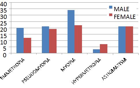
Relation of age to asthenopic symptoms is mentioned in Fig 2. There was a statistically significant difference noted in the age group of 18-22 among all asthenopic patients (Chi-square test P<0.02)(Table 1).
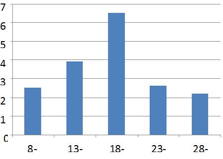
Out of 177 patients with asthenopic symptoms like Headache, Eye ache, Watering and Eye strain Pseudomyopic patients were having significant Asthenopia in comparison to emmetropic patients (P=0.0001) and the same observation was noted
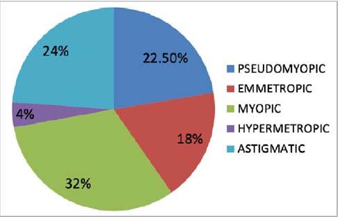
between Pseudomyopia and other refractive errors like Myopia, Hypermetropia and Astigmatism (P=0.0001) (Fig 3).
Table 2 showing the relationship of refractive error with post mydriatic test. There was a statically significant difference noted in Pseudomyopic patients among all Asthenopic patients (Chi-square test P0.006).
The current study aimed to assess the number of
underdiagnosed cases of Pseudomyopia with Asthenopic symptoms in OPD. In our study prevalence of Pseudomyopia was 22.50% and it was found most commonly in the age group 18-22 years which was comparable to Sweden’s study (prevalence – 34.7%, age group – 6-10 years)9 No association was found between Asthenopia and sex, skin colour, or economic status in our study which was similar to Sweden’s study9. In our study, the majority of the subjects were in the working-age group of 20 to 32 years. Though accommodation is known to be more powerful at a younger age, this overactive accommodation could be the cause of Pseudomyopia in the working-age group, depending on their working distance and reading requirements. Khalid K, et al, in their study, showed that active accommodation is the main causative factor in Pseudomyopia , Activities that are visually more demanding like reading or using the screen for long hours, especially in the younger age group (20-30 years) play a significant role in Pseudomyopia10 A variation of >2D in the readings between subjective refraction and retinoscopy is an unusual clinical finding and is generally suggestive of Pseudomyopia. In the current study, retinoscopy values were found to have a mean ± SD difference of 1.33 ± 0.59 D in comparison to readings subjective acceptance. Also, it was noted that subjective correction values were consistently more myopic. Our findings were is comparable to Jameel, et al study in which the mean ± SD difference was 1.87 ± 0.12 D11
There is statistically significant Asthenopia associated with Pseudomyopia as compared to Emmetropia and also with other refractive errors. Hence, Pseudomyopia remains under diagnosed in OPD during the routine ophthalmic examination. Cycloplegic refraction is required to identify these patients and
subsequently treat them with glasses, ocular exercise, counselling and lifestyle modifications.
Limitation :
The main limitation of this study is the assessment of the study population drawn from a specific environment that is (eye OPD of Urban Health tertiary centre). Also, the sample size was small and did not include the pediatric age group. Thus, generalization of the finding for the entire population is not possible.
Support : NIL
Conflicts of Interest : NIL
1Rutstein RP, Daum KM, Amos JF — Accommodative spasm: A study of 17 cases. J Am Optom Assoc 1988; 59: 527-38.
2J Neuroophthalmol ; 22(1): 15-7.doi:10.1097/00041327200203000-00005.
3Rutstein RP, Marsh-Tootle W — Acquired unilateral visual loss attributed to an accommodative spasm. Optom Vis Sci 2001; 78: 492-5.
4Faucher C, De Guise D — Spasm of the near reflex triggered by disruption of normal binocular vision. Optom Vis Sci 2004; 81: 178-81. .
5Hussaindeen JR, Mani R, Agarkar S, Ramani KK, Surendran TS — Acute adult onset comitant esotropia associated with accommodative spasm. Optom Vis Sci 2014; 91: S46-51.
6 Hussaindeen JR, Ramani KK — Comparison of cycloplegic refraction between retinoscopy, closed field and open field autorefraction in pseudomyopia. Invest Ophthalmol Vis Sci 2013; 54: 570-6.
7Carnevali T, Southaphanh P — A retrospective study on presbyopia onset and progression in a Hispanic population Optometry 2005; 76: 37-46.10.1016/S1529-1839(05)702530
8Asharlous A, Hashemi H, Jafarzadehpur E, Mirzajani A, Yekta A, Nabovati P — Does astigmatism alter with cycloplegia? 131-36.10.1016/j.joco.2016.05.00327579457
9Abdi S — Asthenopia in schoolchildren. [Thesis] Stockholm: Karolinska Institutet; 2007. 59.
10Ip JM, Robaei D, Rochtchina E — Prevalence of eye disorders in young children with eyestrain complaints. AmJOphthalmol 2006; 142(3): 495-7 .
11Rizwana HJ, Mithra A, Viswanathan S, Krishna Kumar R, Peter A — Indian Journal of Ophthalmology 2018; 66(6): 799-805.
The incidence of inappropriate behavior, malpractice, medical errors and medical professional being sued in Court of Law has increased recently. According to Graduate Medical Regulations 2019 by NMC one of the roles assigned to Indian Medical Graduate (IMG) is to be Professional. To inculcate professionalism amongst the IMG, it has to be included in Curriculum and effective teaching-learning methods are used to teach professionalism to undergraduate and Postgraduate Medical Students. Various teaching learning methods are mentioned in literature. PubMed search engine was used extensively to study the articles relevant to teaching learning methods to teach professionalism in medical students. A total of 7 articles were selected for critical analysis, of which 2 were excluded as full articles were not available. Role model and reflection are the most effective teaching learning method to teach professionalism. Professionalism is to be taught at the beginning of the curriculum ie, in 1st professional year. During their clinical postings in 2nd Professional year, the students keenly observe the professional behavior of the teachers and follow them as the teachers are their role model. In reflective writing, the student introspects their own Professional Behavior and make the necessary changes. The student learns about the various attributes of Professionalism and will become a successful Indian Medical Graduate.
[J Indian Med Assoc 2023; 121(5): 44-6]
Key words : Professionalism, Medical students, Teaching and Learning.
Nowadays, it is seen that there is increased incidence of Medical Professionals being sued in the Court of Law. Although, the cases of inappropriate behaviour, malpractice, medical errors are few in number, but it maligns the entire Medical Profession. Previously Doctors were granted autonomy by the society but the altered public perception of the role of doctor has defied it.
In one of the studies, it was found that students who demonstrate unprofessional behaviour during UG and PG education are more likely to be found guilty of unprofessional behaviour actions by monitoring boards 1 . Even Abraham Flexner considered Professionalism for reconstruction of Medical Education 2 . Thus, the need to include teaching Professionalism in curriculum for UG and PG has been globally acknowledged.
The word Professionalism is derived from Latin “Professio” or public declaration. Professions are occupations which are granted a special status by the society so that the Professionals can deal with
1MD (Pharmacology), Professor, Department of Pharmacology, Datta Meghe Medical College, Nagpur, Maharashtra 440016 and Corresponding Author
2MDS (Periodontology), Assistant Professor, Department of Periodontology, Government Dental College and Hospital, Nagpur, Maharashtra 440003
Received on : 30/04/2022
Accepted on : 10/05/2022
Editor's Comment :
Professionalism needs to be introduced early in undergraduate medical education.
One of the identified role of Indian Medical Graduate is to be professional.
Professionalism can be most effectively taught by role play and reflection.
needs that are valued by the community they serve. Professionalism is a competence or skill expected of a Professional. It is said to be the social bridge between Medicine and Society3
According to Graduate Medical Education Regulations 2019, one of the roles assigned to Indian Medical Graduate is to be Professional ie, committed to Excellence, Ethical, Responsive and accountable to Patient, Community and Profession.
The core attributes of Professionalism are:
(1) Altruism, (2) Confidentiality, (3) Commitment, (4) Caring, compassion, (5) Competence, (6) Insightful and Self-awareness, (7) Integrity, Honesty, Trustworthiness, (8) Morality and Ethics, (9) Responsibility to Profession, (10) Responsibility to Society, (11) Teamwork
To critically review the literature related to professionalism and analyse the various methods for achieving professionalism.
Guraya, et al in a study entitled “The legacy of teaching Medical Professionalism for promoting Professional practice: A systematic review ‘’ conducted systematic review according to PRISMA guidelines of all the studies related to Medical Professionalism and found 48 articles about how to teach Professionalism. But according to the author, there was a lack of a unified model for teaching Professionalism. The author mentioned about the use of interactive lecture, small group discussion, case vignettes, role play, experiential learning as different methods to teach professionalism. The author identified 4 teaching methods and they are Role modelling, mentoring, The hidden curriculum and reflective practice. But how to implement these methods in the institute is lacking. The author shifted the onus of implementation to the individual institution. The discussion in the study is limited to the use of the 4 teaching learning methods in detail4.
“Teaching and assessing Professionalism in the Indian context” is a study by Modi JN, et al. It is a Review article. There is a dearth of studies related to Professionalism in Indian context. All studies are in US and western countries. In this particular article, the author highlights the importance of Professionalism in Indian context. The Teaching Learning Methods and Assessment of Professionalism in various professional years is also discussed. According to authors, interactive lectures, brain storming sessions, role play and observation of doctors at work followed by discussion in small group can be used during I Professional year. Role model (sensitization of faculty members), case-based discussion in small groups, reflective thinking Teaching Learning Method can be used during II Professional year and in addition to above, group discussion based on clinical scenarios and case vignettes during III Professional year can be used. Although, the authors have proposed their view about implementation of Professionalism but, actual studies have to be conducted. The methods have to be standardized and later it can be validated that a particular method is effective in our setting1
The study of Hatem CJ entitled “Teaching approaches that reflect and promote Professionalism” is limited to residents only. He mentioned 2 teaching initiative – Residents as Teacher program and Bedside teaching which was developed by the author in 1980 and followed in Mt. Auburn Hospital, Cambridge, Massachusetts. The author is of the view that Professionalism can be better taught during clinical training. But the concept should be taught the moment students enters the medical institute. If the students
are made aware about Professionalism in the beginning, then Professionalism can be better learned during clinical posting5
Aziz AB and Ali SK conducted a study entitled “Relationship between level of empathy during Residency Training and perception of Professionalism climate “. It included 70 Residents of OBGY and Paediatrics Department of Tertiary Care Hospital in Pakistan. The study was conducted to assess level of empathy and perception of Professionalism. No significant difference regarding professionalism was found among medical students, residents and faculty. This study was conducted in a private medical college and the sample size is small and moreover it is limited to the Department of OBGY and Paediatrics only. The study included Residents and their view about the professional behaviour of Medical students, Fellow residents and Faculty is assessed. Thus, it reflects the view of residents of OBGY and Paediatrics and hence cannot be generalized6
Altirkawi K in Review article entitled “Teaching Professionalism in Medicine: what, why and how?” mentioned about the roadmap to teach Professionalism. The author defined the steps to teach Professionalism in general which are setting the expectations, performing assessments, remediating inappropriate behaviour, preventing inappropriate behaviour and implementing a cultural change. The author did not mention a particular method to be used during a professional year of curriculum. This study is lacking about the implementation of teaching learning methods in Professionalism7
By teaching Professionalism students will know the importance of Professionalism in Healthcare. They will know what constitutes professional and unprofessional behaviour. Their idea about ethics and malpractice will be clear. Learning Professionalism will help to develop a better doctor-patient relationship. Students can develop empathy towards the patients. Moreover, better communication will be developed between treating Doctor and Patient which will avoid most of the problems. A better understanding will be developed between them.
During the Professional years of curriculum more importance is given on cognitive and psychomotor domain according to Bloom’s Taxonomy. By teaching Professionalism, the students learn about the affective domain also. Even, one of the roles assigned to Indian Medical Graduate by GME Regulations 2017, will be learned by teaching Professionalism. The students will learn that to be a good Professional one needs to be
clinically competent with good communication skills. The core attributes of Professionalism which were discussed earlier will be learned by the students. The students will also be sensitized about the behaviour towards the differently abled person. Thus, the students become aware about the concept of compassion, working in a healthcare team, gender sensitivity, cultural and language diversity.
Proposed-Implementation Plan/Translatory Component :
As per the Graduate Medical Education Regulations 2019, one of the major components is Professional development including Ethics which is allotted a total teaching hour of 40 in the Foundation course. This will be further continued as AETCOM module after foundation course which is presently followed.
However, the professionalism among the Medical Students can be assessed by “Climate of Professionalism instrument’’ developed by Quaintance, et al. It comprises of 12 statements on behaviour associated with Professionalism. Students will require to mark their response on a four-point Likert scale ie, 4=mostly, 3=often, 2=sometimes, 1=rarely for a total of 36 items. The score ranges from 36 to 144 and higher score will denote more professional behaviour.
Teaching Professionalism is important as Professionalism is integral to Medical Profession and it is explicitly mentioned in medical Curriculum. There are various teaching-learning methods to teach professionalism to the students. The most effective are Role model and Reflection. Thus, learning Professionalism will lead to the Professional development of the students.
Author's Reflection :
The importance of Professionalism in Medical Education came to be known. Most important, it
created awareness that the teaching medical faculty can play important role in inducing the concept of Professionalism among the students. It has created the insight that the faculty members must behave responsibly in front of students. It also came to know that Role modelling play important role and for the student, Teacher is the Role model. To inculcate Professional behaviour among the students, it is important that the teacher’s own behaviour must be professional since the students will follow their teacher. The teacher’s behaviour with the patient is keenly observed by the students during clinical posting. Apart from this, the students also observe how the teacher communicate with the Patients.
The earlier perception was that teaching medical students means the future doctor should treat patient efficiently. The roles assigned to future Indian Medical Graduate according to GME regulations 2017 came to be known and one of the roles is Professionalism and the teaching faculty can only teach Professionalism better.
Conflict of Interest : None
1Modi JN, Anshu, Gupta G, Singh T — Teaching and assessing professionalism in the Indian context. Indian Pediatr 2014; 51: 713-7.
2Morris PM — Reinterpreting Abraham Flexner’s speech, “Is social work a profession?”: Its meaning and influence on the field’s early professional development. JSTOR 2008; 82(1): 29-60
3Spiwak R, Mullins M, Isaak C, Barakat S, Chateau D, Sareen J — Medical students’ and postgraduate residents’ observations of professionalism. Educ for Health 2014; 27(2): 193-8.
4Guraya SY, Guraya SS, Almaramhy HH — The legacy of teaching medical professionalism for promoting professional practice: A Systematic review. Biomed & Pharmacol J 2016; 9(2): 809-17.
5Hatem CJ — Teaching approaches that reflect and promote professionalism. Acad Med 2003; 78: 709-13.
6Aziz AB, Ali SK — Relationship between level of empathy during residency training and perception of professionalism climate. BMC Med Educ 2020; 20: 320
7Altirkawi K — Teaching professionalism in medicine: what, why and how? Sudan J Paediatr 2014; 14(1): 31-8.
Cancer is a serious public health problem that affects people worldwide. The onset and the progression of cancer are influenced by several factors, viz internal/external environmental factors, as well as genetic factors, which can play a role in the emergence as well as the progression of many malignancies. Diet and Nutrition may play a role in the prevention or treatment of different types of Malignancies. A great number of studies have found that certain dietary patterns can help in preventing cancer and/or slowing the progression of tumors in patients. In addition to this, the literature suggests that a variety of dietary substances, including Curcumin, Green Tea, Folate, Selenium, and Soy Isoflavones, have anti-cancer potential. It has been demonstrated that these chemicals could be employed for cancer chemoprevention and therapy by targeting a series of cellular and molecular pathways. In this review, we have tried to summarise the impact of food patterns on cancer prevention.
[J Indian Med Assoc 2023; 121(5): 47-52]
Key words :Cancer, Diet, Phytochemicals, Anti-cancer, Curcumin, Green Tea.
Cancer is regarded as one of the most serious public health issues that affect people all over the world1 The identification of several elements involved in cancer pathogenesis has resulted from the evaluation of various dimensions of this disease. These discoveries may aid in the prevention and treatment of many cancers2. To present, researchers who have a better grasp of the molecular/cellular pathways involved in various Malignancies have been able to devise a variety of treatments and regimens for both before and after cancer. Lifestyle is one of the most important variables in the onset and progression of cancer. Human dietary habits may have a variety of effects on human health. Appropriate nutrition has a significant impact on human health3. It was discovered that adequate intake of different Vitamins and Lipids could have beneficial impacts on disorders including cancer4,5. Several studies have suggested that a healthy diet and eating patterns can help prevent or possibly treat cancer6
A plant-based diet with limited red meat consumption has been related to a lower risk of breast cancer7. Dietary chemicals are well-known therapeutic agents that can alter different kinds of cellular and molecular pathways8,9. Consumption of diverse fruits and a diet rich in antioxidants may aid in cancer prevention. Various dietary ingredients, such as green
Department of Respiratory Medicine, King George's Medical University, Lucknow, UP 226003
1MD, Professor and Corresponding Author
2PhD, Research Associate, Department of Nuclear Medicine, Sanjay Gandhi Postgraduate Institute of Medical Sciences, Uttar Pradesh 226014
3MSc, PhD Scholar
Received on : 25/06/2022
Accepted on : 24/01/2023
Editor's Comment :
Processed meat, fried food, refined products, carbonated & alcoholic beverages and packaged food increases the risk of cancer.
Dietary chemopreventive agents are long chain polyunsaturated fatty acids ,carotenoids ,curcumin ,green tea ,polyphenols, glucosinolates, vitamins (vitamin D, folate),minerals(calcium, selenium),etc.
In nutrigenomics, we study the impact of dietary components on genetic variations as well as the effect of nutrients and bio active food ingredients in gene expression, individually. Life style is one of the most important variables in the onset and progression of cancer.
tea, carotenoids, selenium, curcumin and vitamins, have been shown in numerous studies to aid in cancer prevention and treatment. Inflammation is one of the most important contributors to the development of many malignancies5,7. The use of appropriate dietary components, including antioxidants, may affect cancer development and prevention5. As a result, it appears that including them in one’s diet could be beneficial for cancer prevention and treatment.
Foods that can make you more likely to get cancer “You are what you eat,” as the cliche goes, is especially true when it comes to improving your health and adopting healthy eating habits. One will feel more energetic and fit if one eats healthily. Sluggishness and health problems are unavoidable if the food on your plate is high in trans fat and processed ingredients. Poor dietary choices and unhealthy eating habits can contribute to a variety of liver, renal and cardiac illnesses. While some can be treated if caught early enough, others, such as cancer, can be fatal. Cancer is one of the most common causes of mortality in the
world, but it can be avoided by making appropriate lifestyle modifications. Cancerous cells proliferate in the body due to variety of causes, one of them is an unhealthy diet. Other factors that may play a role include a lack of physical activity, smoking, obesity, alcohol and UV-exposure. There is mounting evidence that eating habits might either increase or decrease cancer risk. So, to keep your health in tip-top shape, here are 5 items to stay away from10.
(A) Processed Meat :
Meat, poultry, fish and eggs are all good if cooked properly and eat them in moderation. Taking any animal-based goods that have been preserved by smoking and salting is unhealthy and can lead to a variety of health problems, including weight gain and cancer. Meat processing produces a chemical that is potentially carcinogenic, increasing the risk of colorectal and stomach cancer. Cook your meat instead of buying processed meat like hot dogs, salami and sausage11
(B) Fried Food :
Excessive consumption of fried meals can also promote the growth of malignant cells in the body. A chemical called acrylamide is generated when foods like potatoes or meat are fried at high temperatures. According to studies, this chemical is carcinogenic and can even destroy DNA. Furthermore, fried foods can raise oxidative stress and inflammation in the body, both of which are associated with cancer cell proliferation. Look for alternate healthier cooking ways to replace frying12.
(C) Refined Products :
Refined flour, sugar, and oil, for example, all have the potential to cause malignant cells to form. According to studies, eating a lot of highly processed sugar and carbohydrates raises the risk of oxidative stress and inflammation in the body, which can lead to cancer. Ovarian, breast and endometrial (uterine) cancers are more common in people who eat a diet heavy in refined goods. Make healthy substitutions to reduce your intake of processed items. Substitute jaggery or honey for sugar, whole grain for refined carbs, and refined oil for mustard oil and clarified butter10
(D) Carbonated Beverages and Alcoholic Beverages :
Alcohol and carbonated beverages both include a lot of refined sugar and calories. Excessive intake of either fluid can lead to an increase in free radicals in the body, which can lead to inflammation. Alcohol also hinders your immune system’s ability to recognize and
target precancerous and cancerous cells, making it more difficult for your body to detect and target them11
(E) Foods in Cans and Packages :
In India, the consumption of canned and packaged goods is continuously increasing. The market aisle is brimming with pre-packaged foods that can be cooked and severed in a matter of minutes. There are many different types of packaged foods to choose from, such as instant poha, noodles, idli, upma and spaghetti. Though it makes cooking more convenient, it also increases the risk of cancer. Bisphenol A (BPA, a plasticizer) is a chemical that is used to line most ready-to-cook food packages. When this molecule is dissolved in food, it can induce hormonal abnormalities, DNA changes and cancer13
Eat less
Eat more
Saturated fatsFruits and vegetables
Red meat
Food rich in Vitamin A, C and E
Processed meatWhole grains
Food with preservativesGreen leafy vegetables
Alcoholic beverages
Salt-cured and smoked food
Table 1 shows list of food that reduces the risk of cancer. This table shows what food an individual should take more in amount and what should they have to eat less so that the risk of cancer will be reduced.
In Table 2 different types of cancer and foods as a risk factor, probable mechanism behind this, as well as food and nutrients which are responsible for minimizing/avoiding the cancer is shown. Cancer of the oral cavity and pharynx (nasopharyngeal cancer, oral and pharyngeal cancers), oesophageal cancer (squamous cell carcinoma and adenocarcinoma), stomach cancer, colorectal cancer, liver cancer, pancreatic cancer, lung cancer, breast cancer, and prostrate cancer are dominant cancers, across the globe14. Obesity, overweight and alcohol consumption raise the chances of the several forms of cancer; these are just the most nutritional elements contributing to the global cancer burden14. Mutagen-containing foods can cause cancer, salted fish can cause nasopharyngeal cancer15 and aflatoxin-contaminated foods can cause liver cancer16
According to the World Cancer Research Fund report-2018, neither fruits nor vegetables are convincingly or likely associated with the risk of any cancer 14 . Evidence suggests that it may protect against some cancers and the risk may increase at very low intakes. Specific elements of some fruits and
vegetables may have antioxidant properties. Other than this, vegetarians do not consume meat or fish and typically consume more fruits and vegetables than nonvegetarians. Vegetarians and vegans may have a slightly lower risk of all cancer sites combined than non-vegetarians, however and observations for individual cancers are unclear. Hence, maintaining a healthy body weight and blood sugar levels by eating a varied diet high in fruits and vegetables and minimize the intake of red and processed meats and processed
sugar is beneficial17. It is advised that try to get your nutrients and vitamins from your diet rather than supplements.
Cancer Chemoprevention and Dietary Chemicals: Cancer chemoprevention is defined as a method of reducing or suppressing the development and progression of cancer using natural or synthetic substances9,18 . The use of dietary components is appealing because of their unique qualities, such as minimal toxicity as compared to conventional
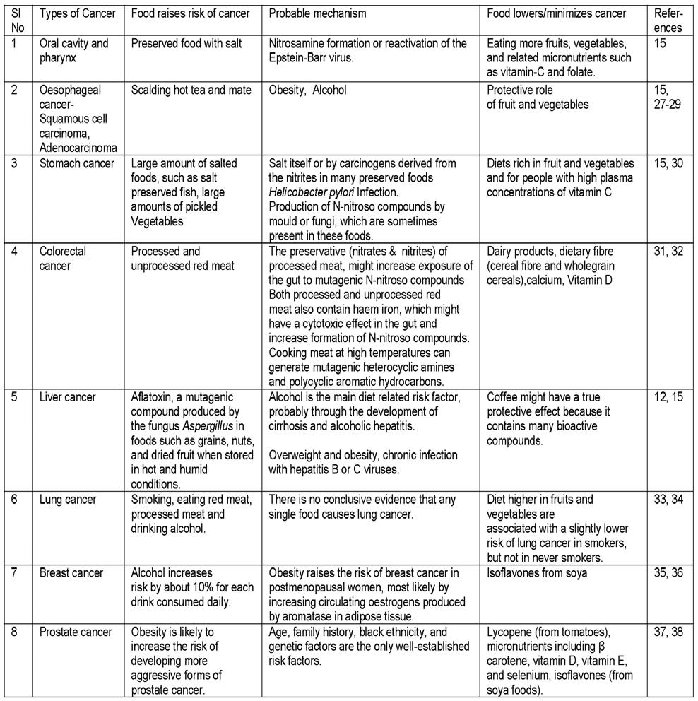
medications. It has been demonstrated that dietary chemicals can be used as a cancer chemoprevention treatment agent. Multiple lines of evidence suggested that a wide range of dietary chemo-preventive agents, including long-chain polyunsaturated fatty acids, carotenoids, curcumin, green tea polyphenols (ie, catechins)19, glucosinolates/isothiocyanates, vitamins (ie vitamin D and folate), and minerals (ie, calcium and selenium), could be introduced into clinical application for cancer therapy8
Curcumin is a fascinating phytochemical with a variety of anti-cancer activities19. Curcumin exerts its therapeutic benefits by targeting various cellular and molecular pathways, and may also alter several cellular and molecular targets, including microRNAs (miRNAs), COX-2 NF-B, MMPs, AP-1, cyclin D1, EGFR, Akt, -catenin, adhesion molecules and TNF4,19.
Green tea is another natural component that has been linked to a variety of medicinal benefits20. Green tea is thought to have anti-cancer qualities via inhibiting tumor growth by targeting cellular and molecular factors involved in cell proliferation and angiogenesis. One of the main targets that this component may influence is VEGFs 20 . Proteins (including enzymes), carbohydrates, lipids, amino acids, vitamins (B, C, E), and minerals (ie, Ca, Mg, Cr, Fe, Zn, F, K) have all been found to be abundant in this plant21. The presence of polyphenols, particularly flavonoids, has been linked to the medicinal effects of green tea in several studies. Catechins (flavan-3-ols) are flavonoids found in green tea leaves, Including epicatechin (EC), epicatechin-3gallate (ECG), gallocatechin (GC), epigallocatechin (EGC), and predominant epigallocatechin-3-gallate (EGCG)9
Nutrigenomics or Nutritional Genomics : Role in Nutrition and Medical Science
Nutrigenomics investigate the effect of food on our genes and the reaction of specific genes to nutrients absorbed via different foods22. It is linked to the molecular interaction between genes and nutrients in the body (nutrigenetics) and these factors that influence the transcript profiles (transcriptomics), metabolites (metabolomics) and proteomics 22 Nutrigenetics and transcriptomics, in conjunction with “omic” technologies such as metabolomics and proteomics, are accountable for the great variation in cancer prevention as well as risk among persons22-24
Nutrigenomics is defined as the impact of dietary components on genetic variations as well as the effect of nutrients and bioactive food ingredients on gene expression individually23,24. The principles of nutritional genomics includes- metabolomics, transcriptomics,
proteomics and epigenetics22.
In cancer chemoprevention, epigenetic mechanisms are significant pathways that can be influenced by dietary components 24-26 . DNA methylation, histone changes, chromatin remodelling, miRNAs are some of the processes that serve as epigenetic regulators. Dietary chemicals have been suggested to affect cancer prevention by targeting epigenetic pathways25,26. Bioactive phytochemicals have been shown in numerous studies to be able to alter the expression of a range of oncogenes and tumor suppressor genes by targeting epigenetic pathways involved in Cancer start and development 25,26 . Furthermore, the use of bioactive phytochemicals, either alone or in conjunction with other natural or synthetic medicines, may be linked to considerable anti-cancer effects26. Omega-3 fatty acids (found in fatty and greasy fish, and flaxseed oil), dietary polyphenols or polyhydroxy phenols [epigallocatechin (green tea), turmeric acid (cinnamon), resveratrol (grape, wine, blueberries and mulberries) and curcumin (spices turmeric and curry)], apigenin, a plant flavone [present in many fruits and vegetables, parsley, celery and dried chamomile flowers], genistein is isoflavones [soyabeans], and have role in cancer prevention.
The Eatwell Guide outlines the guidelines for eating a healthy and well balanced diet44. The guide outlines the various foods and beverages you should consume and in what proportions on a daily or weekly basis. The Eatwell Guide categorises the foods and beverages we consume into five major groups :
• Vegetables and fruits — The majority of us are still not eating enough fruits and vegetables. They should account for more than a third of the food we consume each day. They are good source of vitamins, minerals and fibre.
• Bread, potatoes, rice, pasta and other starchy carbohydrates — These are good source of energy as well as chief source of different kinds of nutrients.
• Legumes, pulses, fish, eggs, meat and other protein sources — These foods are high in protein, vitamins and minerals. Pulses, such as beans, peas, and lentils, are excellent meat substitutes because they are lower in fat and greater in fibre and protein. Select lean cuts of meat and mince and consume less red and processed meats such as bacon and sausages.
• Dairy and other fluids — The guidelines suggest 6-8 glasses of water per day. Water, low-fat milks, and low-sugar or sugar-free beverages, such as
tea and coffee, all comes in this category.
• Oils and spreads — Unsaturated fats, which include vegetable, rapeseed, olive, and sunflower oils, are healthier fats. Remember that all types of fat are high in energy and should be consumed in moderation.
These recommendations implement to the vast majority of people, regardless of their weight, ethnic origin, dietary restrictions or preferences. If one have special dietary or medical requirements, consult a registered dietician about how to best adapt this guide to one’s need.
Dietary habits are one of the most important risk factors for a variety of malignancies. A high intake of meat (red meat, processed meat, fish, and processed fish) or a sugary diet has been linked to an increased risk of cancer. Curcumin, green tea components, carotenoids, minerals and vitamins, among other dietary chemicals, may affect several cellular and molecular targets involved in cancer genesis and progression. As a result, it appears that incorporating them into varied dietary patterns could be beneficial for cancer prevention and treatment. Furthermore, several lines of evidence suggest that plant-based diets contain many antioxidant and anti-inflammatory components, such as vitamin E, that may be useful in the prevention or treatment of certain malignancies. The impact of diverse dietary patterns on the prevention or treatment of certain cancers is still unknown. As a result, additional research is needed to show whether they play a helpful or harmful function in cancer prevention.
Funding : None
Conflicts of Interest : Authors declare no competing financial interest.
1Ferlay J, Soerjomataram I, Dikshit R, Eser S, Mathers C, Rebelo M, et al — Cancer incidence and mortality worldwide: Sources, methods and major patterns in GLOBOCAN 2012. International Journal of Cancer 2014; 2014/10/09; 136(5): E359-E86.
2Mirzaei HR, Mirzaei H, Lee SY, Hadjati J, Till BG — Prospects for chimeric antigen receptor (CAR) ãä T cells: A potential game changer for adoptive T cell cancer immunotherapy. Cancer Lett 2016; 380(2): 413-23. PubMed PMID: 27392648. Epub 07/05. eng.
3Couto E, Boffetta P, Lagiou P, Ferrari P, Buckland G, Overvad K, et al — Mediterranean dietary pattern and cancer risk in the EPIC cohort. Br J Cancer 2011; 104(9): 1493-9. PubMed PMID: 21468044. Epub 04/05. eng.
4Banikazemi Z, Mirzaei H, Mokhber N, Ghayour Mobarhan M — Selenium Intake is Related to Beck’s Depression Score.
Iran Red Crescent Med J 2016; 18(3): e21993-e. PubMed PMID: 27247783. eng.
5Mayne ST, Playdon MC, Rock CL — Diet, nutrition, and cancer: past, present and future. Nature Reviews Clinical Oncology 2016 2016/03/08; 13(8): 504-15.
6Chen Z, Wang PP, Woodrow J, Zhu Y, Roebothan B, McLaughlin JR, etal— Dietary patterns and colorectal cancer: results from a Canadian population-based study. Nutr J 2015; 14: 8-. PubMed PMID: 25592002. eng.
7Catsburg C, Kim RS, Kirsh VA, Soskolne CL, Kreiger N, Rohan TE — Dietary patterns and breast cancer risk: a study in 2 cohorts. The American Journal of Clinical Nutrition 2015 2015/02/11; 101(4): 817-23.
8Chen C, Kong A-NT — Dietary cancer-chemopreventive compounds: from signaling and gene expression to pharmacological effects. Trends in Pharmacological Sciences 2005 2005/06; 26(6): 318-26.
9Langner E, Rzeski W — Dietary derived compounds in cancer chemoprevention. Contemp Oncol (Pozn) 2012; 16(5): 394400. PubMed PMID: 23788916. Epub 11/20. eng.
10Donaldson MS — Nutrition and cancer: a review of the evidence for an anti-cancer diet. Nutr J 2004; 3: 19-. PubMed PMID: 15496224. eng.
11Cena H, Calder PC — Defining a Healthy Diet: Evidence for The Role of Contemporary Dietary Patterns in Health and Disease. Nutrients2020; 12(2): 334. PubMed PMID: 32012681. eng.
12Aleksandrova K, Bamia C, Drogan D, Lagiou P, Trichopoulou A, Jenab M, et al — The association of coffee intake with liver cancer risk is mediated by biomarkers of inflammation and hepatocellular injury: data from the European Prospective Investigation into Cancer and Nutrition. The American Journal of Clinical Nutrition 2015; 102(6): 1498-508. PubMed PMID: 26561631. Epub 11/11. eng.
13Fiolet T, Srour B, Sellem L, Kesse-Guyot E, Allès B, Méjean C, et al — Consumption of ultra-processed foods and cancer risk: results from NutriNet-Santé prospective cohort. BMJ 2018; 360: k322-k. PubMed PMID: 29444771. eng.
14Key TJ, Bradbury KE, Perez-Cornago A, Sinha R, Tsilidis KK, Tsugane S — Diet, nutrition, and cancer risk: what do we know and what is the way forward? BMJ 2020; 368: m511m. PubMed PMID: 32139373. eng.
15Secretan B, Straif K, Baan R, Grosse Y, El Ghissassi F, Bouvard V, et al — A review of human carcinogens—Part E: tobacco, areca nut, alcohol, coal smoke, and salted fish. The Lancet Oncology 2009 2009/11; 10(11): 1033-4.
16Trakoli A — IARC Monographs on the Evaluation of Carcinogenic Risks to Humans. Volume 99: Some Aromatic Amines, Organic Dyes, and Related Exposures. International Agency for Research on Cancer. Occupational Medicine 2012 2012/04/01; 62(3): 232-.
17Appleby PN, Key TJ — The long-term health of vegetarians and vegans. Proceedings of the Nutrition Society 2015 2015/ 12/28; 75(3): 287-93.
18Pan M-H, Ho C-T— Chemopreventive effects of natural dietary compounds on cancer development. ChemicalSociety Reviews 2008; 37(11): 2558.
19Mirzaei H, Shakeri A, Rashidi B, Jalili A, Banikazemi Z, Sahebkar A — Phytosomal curcumin: A review of pharmacokinetic, experimental and clinical studies. Biomedicine & Pharmacotherapy 2017 2017/01; 85: 102-12.
20Rashidi B, Malekzadeh M, Goodarzi M, Masoudifar A, Mirzaei H — Green tea and its anti-angiogenesis effects. Biomedicine & Pharmacotherapy 2017 2017/05; 89: 949-56.
21McKay DL, Blumberg JB — The Role of Tea in Human Health: An Update. Journal of the American College of Nutrition 2002 2002/02; 21(1): 1-13.
22Nasir A, Bullo MMH, Ahmed Z, Imtiaz A, Yaqoob E, Safdar M, et al — Nutrigenomics: Epigenetics and cancer prevention: A comprehensive review. Critical Reviews in Food Science and Nutrition 2019 2019/02/07; 60(8): 1375-87.
23Riscuta G — Nutrigenomics at the Interface of Aging, Lifespan, and Cancer Prevention. JNutr2016; 146(10): 1931-9. PubMed PMID: 27558581. Epub 08/24. eng.
24Simopoulos AP — Nutrigenetics/Nutrigenomics. AnnualReview of Public Health 2010 2010/03/01; 31(1): 53-68.
25Hardy TM, Tollefsbol TO — Epigenetic diet: impact on the epigenome and cancer. Epigenomics 2011; 3(4): 503-18. PubMed PMID: 22022340. eng.
26Li Y, Saldanha SN, Tollefsbol TO — Impact of epigenetic dietary compounds on transgenerational prevention of human diseases. AAPS J 2014; 16(1): 27-36. PubMed PMID: 24114450. Epub 10/11. eng.
27Textbook of Cancer Epidemiology. Oxford Scholarship Online: Oxford University Press; 2018.
28Yu C, Tang H, Guo Y, Bian Z, Yang L, Chen Y, et al — Hot Tea Consumption and Its Interactions With Alcohol and Tobacco Use on the Risk for Esophageal Cancer: A Population-Based Cohort Study. Ann Intern Med 2018; 168(7): 489-97. PubMed PMID: 29404576. Epub 02/06. eng.
29Lubin JH, De Stefani E, Abnet CC, Acosta G, Boffetta P, Victora C, et al — Maté drinking and esophageal squamous cell carcinoma in South America: pooled results from two large
multicenter case-control studies. Cancer Epidemiol Biomarkers Prev 2014; 23(1): 107-16. PubMed PMID: 24130226. Epub 10/15. eng.
30Tsugane S — Salt, salted food intake, and risk of gastric cancer: Epidemiologic evidence. Cancer Science 2005 2005/ 01/13; 96(1): 1-6.
31Murphy N, Norat T, Ferrari P, Jenab M, Bueno-de-Mesquita B, Skeie G, et al — Consumption of dairy products and colorectal cancer in the European Prospective Investigation into Cancer and Nutrition (EPIC). PLoSOne2013; 8(9): e72715-e. PubMed PMID: 24023767. eng.
32Zhang X, Keum N, Wu K, Smith-Warner SA, Ogino S, Chan AT, et al — Calcium intake and colorectal cancer risk: Results from the nurses’ health study and health professionals followup study. International Journal of Cancer 2016; 139(10): 2232-42. PubMed PMID: 27466215. Epub 08/09. eng.
33Vieira AR, Abar L, Vingeliene S, Chan DSM, Aune D, NavarroRosenblatt D, et al — Fruits, vegetables and lung cancer risk: a systematic review and meta-analysis. Annals of Oncology 2016 2016/01; 27(1): 81-96.
34Pirie K, Peto R, Green J, Reeves GK, Beral V, Million Women Study C — Lung cancer in never smokers in the UK Million Women Study. International Journal of Cancer 2016; 139(2): 347-54. PubMed PMID: 26954623. Epub 03/31. eng.
35Grodin JM, Siiteri PK, Macdonald PC — Source of Estrogen Production in Postmenopausal Women1. The Journal of Clinical Endocrinology & AMP ; Metabolism 1973 1973/02; 36(2): 207-14.
36Dong J-Y, Qin L-Q — Soy isoflavones consumption and risk of breast cancer incidence or recurrence: a meta-analysis of prospective studies. Breast Cancer Research and Treatment 2010 2010/11/27; 125(2): 315-23.
37Oxford Textbook of Urological Surgery. Oxford Medicine Online: Oxford University Press; 2017.
38Kant S — Being Vegetarian : A boon for good health. Acta Scientific Gastrointestinal Disorders 2021; 7(4): 94-103.
Jithin T Chand1, George G Tharakan2, George M
Sebastian3, Anoop Paulose4Gastrojejunocolic Fistula is a rare and under-recognised complication following gastric surgery for Peptic Ulcer Disease or as part of a malignant process. It is associated with malnutrition as the food bypasses the small bowel into the Colon as well as the colonic contents entering the Jejunum leading to Malabsorption. A multi-stage repair was performed in the past to reduce mortality. However, currently a single stage repair is feasible because of prehabilitation and better surgical critical care. Here we are presenting a case which after adequate pre-operative optimisation with parenteral nutrition, the patient tolerated a single stage repair of the Gastrojsjunocolic Fistula. [J Indian Med Assoc 2023; 121(5): 53-5]
Key words :Gastrojejunocolic Fistula, Gastrojejunostomy, Incomplete Vagotomy, Fistula, Stomal Ulcer, Chronic Diarrhoea, Malabsorption.
Gastrojejunocolic Fistula (GJCF) is a rare complication following gastric surgery for Peptic Ulcer Disease in the yesteryears. It usually presents 20-30 years after the surgery. Its presentation may lead to a diagnostic dilemma as its incidence has come down drastically due to better medical management of Peptic Ulcer Disease. The treatment for the same was performed in two or three stages, in view of high mortality and morbidity due to the pathology itself1. Presently due to better surgical care, the repair can be performed in a single stage with much less morbidity for the patients2
Our patient, a 38-year-old former smoker, presented with foul smelling Diarrhoea and upper abdominal discomfort, 2 hours after food intake for a duration of one year. This was associated with weight loss of 20 kgs over 1 year. He gave a history of a loop Gastrojejunosotmy (GJ) for a suspected gastric outlet obstruction due to a Duodenal Ulcer at a primary health centre, the details of which were not available to us.
He was anorexic with a BMI of 13.15 kg/m2. Physical examination revealed an upper midline incision and pedal oedema. His pre-operative biochemical parameters are summarised in Table 1. An esophagogastroduodenoscopy (EGD) revealed a cicatrised duodenum and three openings at the anastomotic site. Colonoscopy revealed a fistulous opening at 60 cms from the anal verge (Fig 1). Biopsy from the gastric side was suggestive of moderate chronic
Department of Surgical Gastrointestinal, GI and HPB Oncosurgery, Amala Institute of Medical Sciences, Thrissur, Kerala 680555
1MS, DNB, FMAS, Senior Resident and Corresponding Author
2MBBS, Junior Resident
3MS, DNB GI, MRCS, FACS, FMAS, FALS, Assistant Professor
4MD, Senior Resident, Department of Medical Gastroenterology
Received on : 29/03/2022
Accepted on : 31/03/2022
Editor's Comment :
Gastrojejunocolic Fistula is a rare complication presenting as a triad of Diarrhoea, foul smelling flatus and weight loss, with the onset of said symptoms 20 years after the Primary Gastric Surgery.
gastritis.CECT abdomen showed a GJCF of size 2.3 cms in the posterior wall of gastric antrum to proximal jejunal loop and anterior wall of distal transverse colon with no obvious focal thickening (Fig 2). Barium meal follow through showed contrast leaking into jejunum and colon with gastric emptying of contrast into the duodenum, signifying that there was no gastric outlet obstruction at the time of presentation (Fig 3).
He was admitted for nutritional optimisation with parenteral nutrition and correction of dyselectrolytemia. He was on a high protein diet to enable him to tolerate the surgical ordeal awaiting him. Once we were confident that his body was up to the task, by tracking his serum albumin, electrolytes and physical condition which were towards the improvement trend, he was taken up for surgery after obtaining anaesthetic clearance. Entering the abdomen via an upper midline incision, an antecolicposterior GJ was noted in situ with a fistula of size 2 cms at the anastomotic site opening into the jejunum and transverse colon (Fig 4). We presume a vagotomy was not performed previously, as the planes seemed virgin. The fistula was divided with sharp dissection and after freshening the margins of the gastric, jejunal and colonic openings, primary closure of the gastrotomy, jejunotomy and colotomy in two layers was done transversely. We proceeded with primary closure as the stomach and duodenum showed no ulcers or suspicious lesions supplemented with our pre-operative barium meal report. The gastric margin on histopathology revealed foveolar type of epithelium with an oedematous lamina propria with lymphoplasmacytes neutrophils, eosinophils and congested vessels suggestive of acute
on chronic inflammation.
His postoperative period was uneventful with oral feeds being started on the fifth post operative day and was discharged by the tenth day. A review 30 days after the procedure brought a smile on the faces of all involved as his BMI was 19.73 kg/m2
Gastrojejunocolic fistula is an uncommon complication post gastrojejunostomy for peptic ulcer disease1,2. Stomal ulcer is the most common cause for GJCF as reported in the available literature (Table 2).
GJCF presents as a triad of diarrhoea (few hours following a meal), foul smelling flatus and weight loss as contrived by Marshal and Knud-Hansen3,4. There are also reports of passing undigested food in the stools if the size of the fistula is large3. Malnutrition is caused by
colonic contents entering the small bowel as well as food bypassing the small bowel leading to malabsorption5. An EGD usually reveals a fistulous opening into the jejunum and colon and sometimes faeculant material maybe present in the stomach. Barium enema has better sensitivity than a barium meal (95% versus 27%) 4 . In our case, EGD unveiled the fistula, which was solidified with CECT abdomen. Negative endoscopic findings should not alter the train of thought to the presence of a GJCF in a patient with prior history of gastric surgery along with the above-mentioned complaints 4
The operative mortality was around 40% in the former years. In the 1930s, the surgical procedure was performed in three stages with a colostomy followed by resection of the fistula and then finally with a colostomy reversal5. This brought down the mortality significantly. As with any surgical progress, this was slowly converted into a two staged procedure and by the 1960s to a single stage procedure 1. But this latter progression was due to improved intensive care and parenteral nutrition by which the patient could be optimised to tolerate the surgical stress 1,5,6 .
Gastrojejunocolic Fistula albeit an uncommon pathology, should ring a bell when a patient with prior
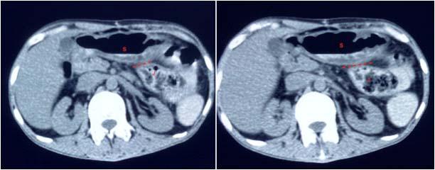
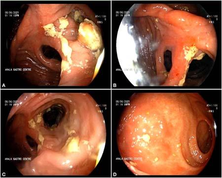
ParameterValue Range
Haemoglobin 8.1 g/dl13-17 g/dl
Total Count4.65 x 103/uL4.6-10.2 x 103/uL
SerumAlbumin 1.5 g/dl3.5-5 g/dL
Serum Sodium136 mmol/L136-145 mmol/L
Serum Potassium2.8 mmol/L3.5-4.5 mmol/L
Serum Calcium 7.6 mg/dl8.6-10.2 mg/dl
SerumMagnesium 1.8 mg/dl1.6-2.3 mg/dl
NSAID Anticoagulants Inadequate gastric resection Long afferent loop Incomplete Vagotomy
Personal Habits
SmokingPeptic Ulcer Disease
AlcoholMalignancy
gastric surgery presents with diarrhoea after food intake. A Computed Tomography usually delineates the Fistula. A negative Endoscopy finding should not deter us from the possibility of a Fistula. Surgical repair in a single stage is now possible with appropriate nutritional optimisation of the patient and adequate intensive care.
1Kece C, Dalgic T, Nadir I, Baydar B, Nessar G, Ozdil B, et al — Current Diagnosis and Management of Gastrojejunocolic Fistula. Case Rep Gastroenterol 2010; 4(2): 173-7.
2Treef WI — Non-neoplastic gastrojejunocolic fistula in 68year-old male patient. Eastern Mediterranean Health Journal 1999; 5(4): 623-6.
3Wu J-M, Wang M-Y, Lee P-H, Lin M-T — Gastrojejunocolic fistula after gastrojejunostomy: a case series. JMedCaseReports 2008; 2(1): 193.
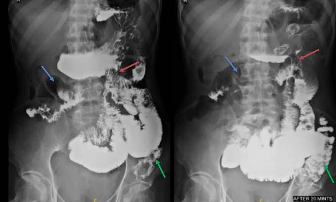
4Araaya GH, Desta KG, Gebremeskel WW, Wasihun AG — Gastrojejunocolic fistula after gastrojejunostomy in Ayder referral hospital Northern Ethiopia: A report of two cases. Annals of Medicine and Surgery 2015; 4(4): 448-51.
5Chung DP, Li RS, Leong HT — Diagnosis and current management of gastrojejunocolic fistula. Hong Kong Med J 2001; 7(4): 439-41.
6Kim KH, Jee YS — Gastrojejunocolic fistula after gastrojejunostomy. JKoreanSurg Soc 2013; 84(4): 252.
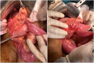
Zuber Ali Quazi1, Dilip Maheshwari2, Vijay Sardana3, Bharat Bhushan4
SARS-CoV-2 causing COVID-19 pandemic is well known for causing various acute and chronic complications affecting almost all the organs of body with unusual presentations. Here we report a case of 50 years old man with Type 2 Diabetes Mellitus presented with gradual involvement of unilateral palsy of left side sided II, III, IV, V, VI, VII, VIII, IX & X without direct invasion into brain parenchyma after typical symptoms of Rhinorbital Mucormycosis.
[J Indian Med Assoc 2023; 121(5): 56-8]
Key words :Pandemic, Diabetes Mellitus, SARS-CoV-2, Mucormycosis, Garcin Syndrome.
The 2019, Novel Coronavirus (2019-nCoV) or Severe Acute Respiratory Syndrome Coronavirus 2 (SARSC0V-2) first reported in Wuhan, Hubei province in China quickly spread to other parts of the world leading to pandemic1. A complex interplay of factors including preexisting diseases, use of immunosuppressive therapy and systemic immune alterations by COVID-19 infection leads to secondary infections and complications. Poorly controlled Diabetes Mellitus with or without diabetic ketoacidosis, prolonged use of corticosteroids, illicit intravenous drug use, malnourishment, severe neutropenia, Malignant Haematological Disease and iron overload are the known predisposing factors leading to Mucormycosis2. Mucormycosis is an aggressive, rapidly progressive and life threatening fungal infection. A hallmark of Mucormycosis infection is presence of extensive angioinvasion with resultant vessel thrombosis and tissue necrosis resulting in rhinorbital involvement3 Globally, the incidence rate of Mucormycosis varies between 0.005 and 1.7 per million population. Prevalence of Mucormycosis in India is much higher than that of developed countries, which is around 140 per million population 4 . Garcin Syndrome is the progressive involvement of the Cranial Nerves resulting in near total unilateral Paralysis of Cranial Nerves, absence of sensory or motor tracts involvement and not associated with raised ICT. Guillain-Alajouanine-Garcin described this in 1926 as “Syndrome Paralytique Unilateral Global Des Nerfs Crannies,” which consist of multiple Cranial Nerve Palsies (at least seven ipsilateral cranial nerves) without any evidence of long tracts signs or increased intracranial pressure5. Here we report a case of middle aged diabetic who developed Garcin syndrome as a complication of
Department of Neurology, Government Medical College, Kota, Rajasthan 324010
1MD (General Medicine), Senior Resident and Corresponding
Author
2MD (General Medicine), DM (Neurology), Head
3MD (General Medicine), DM (Neurology), Senior Professor, Principal and Controller
4MD (General Medicine), DM (Neurology), Assistant Professor
Received on : 23/02/2022
Accepted on : 22/07/2022
Editor's Comment :
COVID-19 infection is not only limited to respiratory tract involvement, but rather involves most of the organs of body. It not only manifests acutely but also has an implication in subsequent period as well with protean manifestations, requiring special attention and care to address the morbidity associated with it. Garcin syndrome is one of such kind for which one should be aware of in those with Rhinorbital Mucormycosis.
Post Covid Rhinocerebral mucormycosis.
A 50 years old gentleman poorly controlled diabetic, presented with 1½ month history of being treated for COVID-19 with the symptoms of Fever, Sore Throat, Cough and Breathlessness and later on followed by pain and swelling over left side of face, with pain more in left preauricular region, and restriction of movements in the left eye and diminution of vision in left eye for 20 days. Patient was then hospitalised in ENT ward where Computerized Tomography of the Paranasal Sinus (CT PNS) had revealed left Rhinorbital Mucormycosis and subsequently Amphotericin-B was administered. Few days later patient developed insidious onset gradually progressive hoarseness of voice with nasal regurgitation of liquids, requiring nasogastric tube insertion.
Examination revealed fixed non-reactive pupils, ptosis and absence of extraocular movement of left eye with visual acuity of finger counting of 2 metre in right eye and perception of light in left eye and decreased sensation over left half of face and Lower Motor Neurons (LMN) type of left facial palsy, deviation of uvula to right and with absent gag reflex on left side with preserved XI and XII nerve.
Among the investigations done, MRI of brain revealed left orbital Rhinosinusitis with bony erosions and myositis with enhancing dura over the anterior left frontal lobe and involvement of left cavernous sinus and left optic nerve with inflammation in left parapharyngeal space (Figs 1-4) as evident by the hyperintensity in T2 / STIR images.
Cerebrospinal Fluid (CSF) examination revealed 2 cells, with no malignant cells seen, protein was 65mg/
dl, sugar was 96mg/dl and Adenosine Deaminase (ADA) was 3.9 U/L with gram’s stain, z-n stain and India ink all being negative. KOH preparation did not show any fungal elements. Histopathological examination from the biopsy of the debrided mucosal tissue revealed Mucormycosis.
Patient was managed by doing endoscopic debridement of necrotic tissue followed by antifungals, antibiotics and other supportive measures, with good glycemic control and was discharged uneventfully on oral posaconazole after giving injectable Amphotericin B as per protocol.
Our patient was diabetic with poor compliance to
antidiabetic medications with history of steroid use for COVID-19 symptoms which further added to the preexisting immunocompromised state.
Patients with Rhinocerebral Mucormycosis present with Fever, Headache, Nasal Discharge, Orbital Pain, with Decreased Vision, Facial Numbness and sometimes Blackish Secretions from nasal and oral cavity 6 . The involvement of CN III, IV, V, and VI is known due to extension of disease into the retro orbital region and cavernous sinus 7 . In our case, there was additional unilateral involvement of CN VII, IX and X as also reported by Kazuo, et al8, Narayana, et al9 and Nagendra, et al10
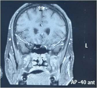
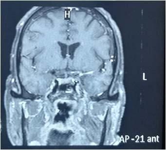
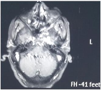
Our patient had similar involvement of the left sided II, III, IV, V, VI, VII, VIII, IX, X, XII as reported by Nagendra, et
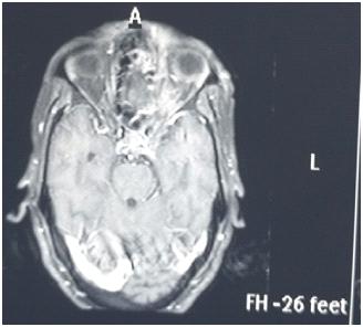
al10. The possibility of other causes leading to such multiple nerve palsies like neoplasms of posterior fossa, vascular malformations at the base of skull, trauma and brainstem infarct were ruled out. The MRI brain revealed the extension of infection to left cavernous sinus and left parapharyngeal space, resulting in LMN type of cranial palsies due to local invasion of nerve fibres by the infective pathology and associated neural and perineural edema as also reported by Nagendra, et al10. Mycelium growth along the cranial nerves or invasion of leptomeningeal blood vessels was reported by Narayana, et al9 as the cause for involvement of unilateral cranial nerve11
Factors indicating poor prognosis are delay in diagnosis, delay in initiating treatment, hemiparesis or hemiplegia and bilateral involvement of sinuses orbit and palate, impaired renal function, Septic shock and immunocompromised status as reported by Nagendra, et al6. Our patient had developed hypokalemia, which is a known side effect of Amphotericin B therapy, which was corrected in time.
Rhinorbital Mucormycosis presenting as multiple Cranial Nerve Palsies, beyond cavernous sinuses is not seen commonly. A high suspicion should be kept for involvement of Lower Cranial Nerves in Rhinorbital Mucormycosis especially in immunocompromised patients, who can present as Garcin syndrome. As Rhinorbital Mucormycosis presenting as Garcin syndrome is a known entity even in pre-Covid era, especially in patients with immunocompromised status, so one should be vigilant about possibility of developing Garcin syndrome in patients with Mucormycosis. So that, early diagnosis and initiation of treatment can decrease the morbidity by preventing the spread of mucormycosis.
We are thankful to staff of Neurology Department for meticulous efforts towards patient care.
Disclosure : All authors approved the final version to be published. The authors report no conflicts of interest in this work.
1Farnoosh G, Alishiri G, Zijoud SH, Dorostkar R, Farahani AJ— Understanding the severe acute respiratory syndrome coronavirus 2 (SARS-CoV-2) and coronavirus disease (COVID-19) based on available evidence-a narrative review. Journal of Military Medicine 2020; 22(1): 1-1.
2Yang H, Wang C. Looks like Tuberculous Meningitis, But Not: A Case of Rhinocerebral Mucormycosis with Garcin Syndrome. Front Neurol 2016; 7: 181.
3Desai EJ, Pandya A, Upadhya I, Patel T, Banerjee S, Jain V — Epidemiology, Clinical Features and Management of Rhino Orbital Mucormycosis in Post COVID 19. IndianJOtolaryngol Head Neck Surg 2021; 15: 1-5.
4Singh AK, Singh R, Joshi SR, Misra A — Mucormycosis in COVID-19: A systematic review of cases reported worldwide and in India. Diabetes Metab Syndr 2021; 15(4): 102146.
5Guillain R, Alajouanine Th, Garcin R — Le syndrome paralytique unilateral global des nerfs craniens. Bull Soc Med Hop Paris 1926; 50: 456.
6Riley TT, Muzny CA, Swiatlo E, Legendre DP — Breaking the Mold: A Review of Mucormycosis and Current Pharmacological Treatment Options. Ann Pharmacother 2016; 50(9): 747-7.
7Mehta MM, Garg RK, Rizvi I — The Multiple Cranial Nerve Palsies: A Prospective Observational Study. NeurolIndia 2020; 68(3): 630-5.
8Kazuo M — No to shinkei Brain and nerve 2004; 56(3): 2315.
9Narayana S, Panarkandy G, Subramaniam G, Radhakrishnan C, Thulaseedharan NK, Manikath N, et al — The “black evil” affecting patients with diabetes: a case of rhino orbito cerebral mucormycosis causing Garcin syndrome. Infect Drug Resist 2017; 10: 103-8.
10Nagendra V, Thakkar KD, Prasad Hrishi A, Prathapadas U — A Rare Case of Rhinocerebral Mucormycosis Presenting as Garcin Syndrome and Acute Ischemic Stroke. Indian J Crit Care Med 2020; 24(11): 1137-8.
11Petrikkos G, Skiada A, Lortholary O, Roilides E, Walsh TJ, Kontoyiannis DP — Epidemiology and clinical manifestations of mucormycosis. Clin Infect Dis 2012; 54(1): S23-S34.
Umakanta Mahapatra1, Debapratim Ganguly2, Sourav Sen2, Soutrik Ghosh2
Idiopathic Intracranial Hypertension (IIH) or Pseudotumor cerebri is an uncommon neurological diseases of unknown aetiology. It is characterized by elevated intracranial pressure without apparent aetiology and associated with normal Cerebrospinal fluid analysis without any structural lesion of the brain. Patients usually present with Headache, transient blurring of vision and occasional involvement of Cranial Nerve, most commonly 6th Cranial Nerve. But involvement of Multiple Cranial Nerves in Idiopathic Intracranial Hypertension is rare. We are presenting a case of a 20 years old female admitted with history of Headache, Blurring of vision and Diplopia, and was diagnosed as pseudotumor cerebri with Multiple Cranial Nerve Palsy (left sided 6th and right sided incomplete 3rd Cranial Nerve Palsy). Our patient improved on treatment with acetazolamide. Physician should be aware of Pseudotumor cerebri. Early diagnosis and treatment may improve patients’ vision. [J Indian Med Assoc 2023; 121(5): 59-61]
Key words :Idiopathic Intracranial Hypertension, Multiple Cranial Nerve Palsy, Rare Association.
Pseudotumor cerebri or Idiopathic Intracranial Hypertension (IIH) is an uncommon Neurological Disorder of unknown aetiology. It is characterized by features of elevated Intra Cranial Pressure (ICP) and high opening pressure of Cerebrospinal fluid without structural lesions of the brain1. Primarily it occurs in women with higher Body Mass Index (BMI), Obstructive Sleep Apnoea or Autoimmune Aetiology 2. The incidence rate in the general population is 1.56 cases / 100,000 / year and 11.9 / 100,000 / year in overweight and obese women3 The pathophysiology of IIH is not clear. Overweight or Obesity, Hypertension, Autoimmune Diseases, Uremia, Over Dosages of Drugs (eg, Vitamin A, Tetracycline, Minocycline, Nalidixic Acid, or Oral Contraceptive Pills), and chronic respiratory insufficiency are common associated risk factors of IIH. Most common presentation is headache (92%) and two-third of patients complains of transient blurring of vision4. 6th Cranial Nerve (CN) is commonly affected that was reported in 12% of adults and 9-48% of children. But involvement of multiple CNs in IIH is very rare5. We are presenting a case of IIH with multiple CN palsies (left sided 6 th and right sided incomplete 3rd Cranial Nerve Palsy).
A 20 years old female presented to the Emergency Department of Midnapore Medical College on 24 th December, 2021 complaining of severe holocranial headache for 7 days. Her headache was insidious onset and gradually progressive and was associated with
Department of General Medicine, Midnapore Medical College and Hospital, Midnapore, West Bengal 721101
1DCH, MD, DM, Assistant Professor and Corresponding Author
2MBBS, Postgraduate Trainee
Received on : 30/03/2022
Accepted on : 04/04/2022
Editor's Comment :
IIH should be suspected in overweight and obese patients presenting with headache and transient blurring of vision. IIHs usually presents with headache, blurring of vision and bilateral 6th cranial nerve palsy but involvement of multiple cranial nerves are not uncommon.
Fundoscopy should be routinely done in all patients presenting with sudden onset headache, as visual acuity may be normal initially.
Early diagnosis and timely commencement of treatment can save the vision of patient.
Vomiting, Weakness, Dizziness and Transient Blurring Of Vision. On 4 th day of her illness, horizontal diplopia with maximal separation of images on left direction of gaze had developed. She denied any history of Fever, Convulsion, Weakness of any Limb, Trauma or Tick Bite. She was normotensive and non- diabetic. There was no significant past medical history or family history of similar illness. On general physical examination patient was conscious, alert and cooperative, BMI was - 35.2 kg/m2, Blood Pressure - 130/80 mm of Hg, Pulse Rate - 74/min, Respiratory Rate - 18 / min without pallor, cyanosis, clubbing, oedema and icterus. She was not able to abduct her left eye (Fig 1) and there was limitation of extorsion of her right eye (Fig 2). Pupillary reaction was normal with a visual acuity of 6/6 in both the eyes and normal colour vision. Fundoscopy showed bilateral Papilledema (Fig 3). Rest of the neurological examination, including sensory and motor function, reflexes, coordination and gait was normal. The systemic examination was unremarkable.
Hemogram, Routine and Microscopic examination of Urine, Electrolytes, Liver Function Tests, Urea and Creatinine, T3, fT4 and TSH, autoimmune profile and Ddimer were within normal limits. Computed Tomography
Normal neuro-imaging supported the diagnosis of IIH as the most probable cause of the patient’s symptoms. The diagnosis was further supported by the marked improvement with acetazolamide therapy.
The patient was treated with oral Acetazolamide 500 mg twice daily and Paracetamol and discharged with the same medication. On follow-up after 1 month, the patient was symptomatically better without headache and visual problems (Table 1), and there was improvement of ocular movements (Fig 4) with partial resolution of papilledema.
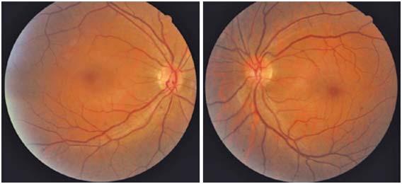
IIH is an uncommon Neurological Diseases with signs and symptoms of elevated ICP without structural lesions of the brain. In 1897, Quincke first described IIH6. IIH was named Pseudotumor cerebri in 1904 7 Headache and Transient Blurring of vision are the commonest presentation of
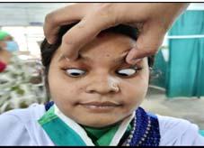
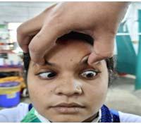
(non-contrast) was normal. Cerebrospinal fluid study reported normal except high opening pressure (265 cm of H20). Magnetic Resonance Imaging (MRI) of brain (Fig 4) and Magnetic Resonance Venogram were normal.
IIH is diagnosed by exclusion of other causes and neuro-imaging studies should be performed to rule out the structural lesions of the brain. Normal neuro-imaging of brain excluded the structural and vascular lesions in our case. Use of drugs like Tetracycline, high dose of Vitamin A, corticosteroid, lithium and Oral Contraceptive Pills were excluded in our patient. Only involvement of multiple CNs without other manifestation of Sarcoidosis and normal Leptomeninges on MRI make the diagnosis of Neurosarcoidosis unlikely. In absence of history of Tick Bite, Fever and abnormal CSF analysis, the diagnosis of Lyme Disease in our case is less likely.
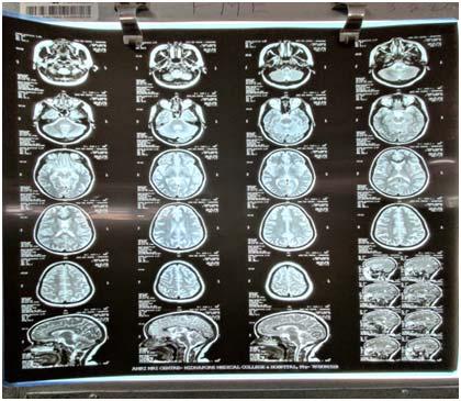
IIH8,9. Headache is the most frequent symptom. Females are most commonly affected. More than 90% patients with IIH are either overweight or obese and in child bearing age group. IIH is diagnosed by the presence of all the criteria (Friedman ’ s criteria) {normal neurological examination except involvement of CN (usually 6th CN), Papilledema, elevated Lumber Puncture (LP) pressure >250 cm H2O and normal CSF study without any structural abnormality of the brain}. It is often associated with nonlocalizing neurologic symptoms, other than unilateral or bilateral 6th CN palsy. Multiple CNs involvement in IIH was rarely reported in the literature. Our patient of IIH presented with left sided 6th and right sided incomplete 3rd Cranial Nerve Palsy. Modified Dandy criterion for IIH do not allow for any focal neurological deficit other than 6th Nerve Palsy but our case report showing Multiple Cranial Nerve Palsy indicating the need to revise it.
The pathophysiology of involvement of multiple CNs in IIH is not exactly known. It is a false localizing sign due to elevated ICP10,11. Due to its long intracranial course, 6th Cranial Nerve is usually susceptible to elevated ICP. Raised ICP, along with venous congestion of the brain parenchyma or Ocular Motor Nerves of 3rd and 4th CN, had been suggested as possible aetiologies12. But the possibility of coincidental association could not be completely refuted in our patient.
Agarwal, et al reported 3rd, 6th and 7 th CN involvement in IIH who improved with dexamethasone and LP 13
Soroken C, et al reported a case of IIH with multiple CNs palsies in a 13 years old girl who completely recovered within 3 weeks of treatment with acetazolamide14. A 16 years old girl presented with bilateral 6th, and left 3rd and 4 th CN Palsies secondary to IIH in primary antiphospholipid syndrome 15
The major complication of IIH is papilledema that may result in loss of vision. Acetazolamide decreases CSF production by inhibiting carbonic anhydrase and commonly prescribed to treat IIH.
In conclusion, IIH is an uncommon neurological
disorder. Loss of vision is main long term complication of IIH. Early diagnosis and treatment may improve patients’ vision.
1Boles S, Martinez-Rios C, Tibussek D, Pohl D — Infantile idiopathic intracranial hypertension: A case study and review of the literature. J Child Neurol 2019; 34(13): 806-14. https:/ /doi.org/10.1177/0883073819860393
2Rowe FJ, Sarkies NJ — The relationship between obesity and idiopathic intracranial hypertension. Int J Obes (Lond) 1999; 23(1): 54-9. doi: 10.1038/sj.ijo.0800758.
3Raoof N — The incidence and prevalence of idiopathic intracranial hypertension in Sheffield, UK. Eur J Neurol 2011; 18(10): 1266-8.
4Wakerley BR, Tan MH, Ting EY — Idiopathic intracranial hypertension. Cephalalgia 2015; 35(3): 248-61.
5Wall M, George D — Idiopathic intracranial hypertension. A prospective study of 50 patients. Brain 1991; 114(Pt 1A): 155-80.
6Corbett JJ, Savino PJ, Thompson HS, Kansu T, Schatz NJ, Orr LS, et al — Visual loss in pseudotumor cerebri. Followup of 57 patients from five to 41 years and a profile of 14 patients with permanent severe visual loss. Arch Neurol 1982; 39(8): 461-74. https://doi.org/10.1001/archneur. 1982.00510200003001
9Wall M, Hart WM, Burde RM — Visual field defects in idiopathic intracranial hypertension (pseudotumor cerebri). Am J Ophthalmol 1983; 96(5): 654-69. doi: 10.1016/S00029394(14)73425-7.
10Pseudotumor D, Ergebnisse J — Pseudotumor cerebri in childhood and adolescenceresults of a Germany-wide ESPEDsurvey. Klin Pa diatrie 2013; 225(2)
11Mollan SP, Spitzer D, Nicholl DJ — Raised intracranial pressure in those presenting with headache. BMJ 2018; 363: k3252.
12Kearsey C —. Seventh nerve palsy as a false localizing sign in benign intracranial hypertension. J R Soc Med 2010; 103(10): 412-4.
13Larner A — False localising signs. J Neurol Neurosurg Psychiatry 2003; 74(4): 415-8.
14Patton N, Beatty S, Lloyd IC — Bilateral sixth and fourth cranial nerve palsies in idiopathic intracranial hypertension. J R Soc Med 2000; 93(2): 80-1.
15Agarwal MP, Mansharamani GG, Dewan R — Cranial nerve palsies in benign intracranial hypertension. JAssocPhysicians India 1989; 37(8): 533-4.
16Soroken C, Lacroix L, Korff CM — Combined VIth and VIIth nerve palsy: Consider idiopathic intracranial hypertension! Eur J Paediatr Neurol 2016; 20(2): 336-8.
17Sun Young Shin, Jeong Min Lee — A Case of Multiple Cranial Nerve Palsies as the Initial Ophthalmic Presentation of Antiphospholipid Syndrome. Korean Journal of Ophthalmology 2006; 20(1): 76-8.
Dhaval Parekh1, Tattvam Gaurang Shah2, Vedanti Kavin Dave2
Background : Hypertension is a widely prevalent lifestyle disease. Hence it is important to incorporate nonpharmacological changes with pharmacological drugs to prevent and improve its outcomes. This study aims to analyse the relation of lifestyle changes with Hypertension.
Methodology : A questionnaire and consent form was distributed personally and online on Whatsapp amongst 177 people. A total of 125 agreed to participate. Data analysis was done using IBM SPSS. Correlation for MAP versus age, years since diagnosis and exercise frequency is done using Kendall’s Tau-b. Difference in MAP of those following dietary and exercise recommendation compared to those who are not is done using ANOVA.
Result : A slight positive correlation is present between age and years since diagnosis versus MAP. Adherence to exercise regimen and dietary restrictions successfully help lower MAP as per ANOVA. Kendall’s Tau-b test also shows a strong negative correlation between frequency of exercise and MAP. Walking and yoga are the most preferred forms of physical activity. Most common dietary restriction include reducing salt intake, avoiding packaged food and eating fresh fruits.
Conclusion : Non-pharmacological lifestyle habits play a huge role in control of hypertension. And this calls for promotion of healthy lifestyle through various means. At an individual level this includes following proper diet, exercise plans and maintaining a healthy body weight. At a community level we have to make lifestyle changes a part of medical curriculum, incorporating healthy habits at an early age and creating awareness about the prevention, control and treatment of Hypertension at a larger scale. People should be motivated and inspired to lead a healthy life along with encouraging their compliance to the medications.
[J Indian Med Assoc 2023; 121(5): 62-6]
Key words :Diet, Exercise, Hypertension, Lifestyle modification, Public health.
Hypertension is one of the most concerning and common health problems of current times1. Most people in their late 30s and 40s are at increasingly high risk for developing Hypertension2-5 and this makes it even more important to study the various factors that contribute to the development of this major health concern and its disease progression.
Hypertension is considered to be a lifestyle disease that is, a person with highly unhealthy lifestyle is more prone to develop it and that it can be prevented by developing healthy lifestyle habits6,7. A high number of lifestyle and genetic factors seem to influence the occurrence and the course of this disease and this gives us an opportunity to prevent it by understanding how each one of these factors is related to hypertension.
A family history of Hypertension makes one more likely to be hypertensive8,9. A strong family history for hypertension is also an indication for early intervention
1MBBS, MD, Assistant Professor, Department of Community Medicine, KPC Medical College and Hospital, Kolkata 700032 and Corresponding Author
2MBBS, Student, BJ Medical College, Ahmedabad, Gujarat
380016
Received on : 01/03/2022
Accepted on : 26/11/2022
to prevent the development of the disease or improve its outcome10. Stress, lack of sleep hygiene, unhealthy eating habits (excess consumption of fatty foods), Obesity, Diabetes, Addictions (Alcohol, Smoking, Drugs), Sedentary Life, Lack of Exercise etc, are huge contributors of hypertension. These factors not only contribute Hypertension but also worsen the course of the disease by causing complications 11,12 . Hypertension is a major risk factor for severe diseases like Vascular diseases, Myocardial infarction, Renal diseases, Ischemia and also it also leads to organ failures in cases of hypertensive emergencies13.
All the previously mentioned lifestyle factors except family history (due to genetic factors) are very much preventable. Thus, Hypertension is one of the most important healthcare problems that needs to addressed immediately in order to ensure a good health amongst the community. There is an alarming need to promote healthy habits like eating healthy foods, exercising, sleeping well, strictly avoiding heavy consumption of alcohol etc in order to prevent this deadly yet preventable healthcare problem.
This study aims at analysing how the various lifestyle factors contribute to development of Hypertension and its progression while promoting
various practices to prevent this major health problem in the world.
This study has been conducted in Ahmedabad, Gujarat in the months of August-October. The sampling frame aims to include people clinically diagnosed with Hypertension. The participants were asked whether they had a family history of hypertension. People who had a strong history of Hypertension with occurrence of inherited early onset Hypertension and Hypertension despite good dietary and lifestyle choices were not included in the study. This was done to make sure that the majority of the cases in this study are preventable/controllable cases of Hypertension. The questionnaire along with the consent forms were handed out personally as well as distributed online through WhatsApp. After explaining the aim of the study, assuring proper handling of data and addressing concerns the consent forms were signed and the questionnaire was asked to be filled and collected. No personal information has been asked for or used in this study. The questionnaire included questions about Gender, Age, Last recorded Blood Pressure, years since diagnosis of hypertension, dietary changes that have been advised and adhered to, and finally the exercise patterns and frequency followed by the participant. The forms were distributed amongst 177 participants from which 125 agreed to participate in the study, with a response rate of 70.6%.
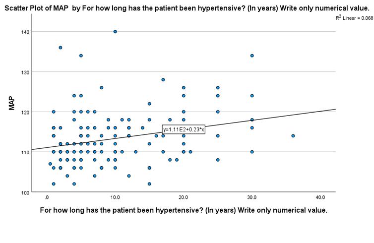
Data analysis is done using IBM SPSS. The last recorded blood pressure was taken as systolic/diastolic but for data analysis it has been converted to Mean
Arterial Blood Pressure (MAP = 2/3 diastolic + 1/3 systolic). Scatter plots of MAP versus Age and years since diagnosis are made (Figs 1&2) and Kendall’s Tau b coefficient is used to correlate while accounting for heteroskedasticity (Table 1). MAP versus Frequency of exercising is shown in Fig 3 with correlation done using Kendall’s Tau b coefficient in Table 2. Participants were asked about whether they have been following the advice to exercise regularly (Table 3). The MAP of participants who exercised compared to those who didn’t was analysed by ANOVA (Table 4). The participants who exercised were asked about the methods of exercise they preferred, shown in Fig 4 (multiple option selected so total is more than 125). Similarly, adherence to dietary changes was inquired (Table 5) and its effects on MAP analysed by ANOVA are shown in Table 6. Fig 5 shows the different types of dietary restriction implemented (multiple option selected so total is more than 125)
No personal information from the participants was
collected or included in this study. No funding was used.
Out of the 125 responses received in this study 47 (37.6%) are females while 78 (62.4%) are males. The average age of the respondents is 54.5 years with a minimum of 25 years and maximum of 79 years.
Figs 1 & 2 shows that age and years since diagnosis of Hypertension both have a slight positive correlation with Mean Arterial Pressure (MAP). This correlation is shown to be statistically significant as per Kendall’s Tau b test in Table 1 (p<0.05).
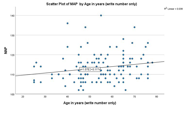
Fig 3 shows how MAP decreases with increasing frequency of exercising (per week), a negative correlation that is shown to be statistically significant as per Table 2. A slight upward rise in MAP is seen among participants who exercise 7 times a week, possible explanations could be adherence to regular exercise secondary to consistently high blood pressure OR they could simply be outliers and an aberration.
Table 3 and 4 shows that the 70 (56%) people who self-report to being adherent to advice of having an active lifestyle and exercising regularly had a
significantly better control over their MAP and consequentially their disease progression too. Fig 4 shows us what are the modes of exercise preferred, with walking being the most favoured one. It is important to note here that this graph shows the results to multiple answers allowed (hence the total is more than 125) question posed to the participants who claimed to exercise regularly.
Table 5 and 6 shows that 90 (72%) participants selfreported being adherent to dietary restrictions and were seen to have a significantly lower MAP compared to those who did not follow the dietary advice. Fig 5 shows the results to a multiple answers allowed question posed to participants following dietary restrictions about the changes that they have made regarding their diet. The most common changes were restricting high salt food, avoiding packaged food and eating fresh fruits.
Hypertension has a mortality rate of 13% and also significantly raises the mortality rate in numerous other ailments.
It is important to acknowledge that Hypertension is a vastly multifactorial disease. The parameters analysed in this study though are an important part of control and prevention of this disease. Non pharmacological advice and lifestyle changes to control Hypertension have been in the limelight since the last few decades partly because these are controllable risk factors and can be easily avoided, partly because it is very prevalent14,15 but also due to worsening dietary and lifestyle habits leading to exacerbation of the problem.
A healthy lifestyle with a regular exercise regimen, Yoga and Healthy Diet is highly beneficial in preventing and improving the outcomes of this disease as seen in this study. Similar results were also found in British Hypertension Society guidelines for Hypertension Management 2004 (BHS-IV): summary 16 , a pronouncement called Exercise and Hypertension by American College of Sports Medicine March 200417,
0.006. <0.001 N125125125
For how long has theCorrelation Coefficient 0.187** 0.637** 1.000 patient beenSig. (2-tailed) 0.004 <0.001 hypertensive? (In years)N125125125 Write only numerical value.
** Correlation is significant at the 0.01 level (2-tailed).
Hypertension is a menace to our society with a prevalence of 32-34% worldwide and 28-30% in India. Although it is relatively asymptomatic in most cases it can cause adverse effects on cardiovascular, renal and central nervous system with complications such as Stroke, Aneurysm, Heart Attack, Chronic Kidney Disease, Dementia.
MAPHow frequently does the patient exercise?
MAPCorrelation Coefficient 1.000 -0.400 Sig. (2-tailed) .<0.001 N125125
How frequently does Correlation Coefficient -0.400** 1.000 the patient exercise?Sig. (2-tailed) <0.001. N125125
** Correlation is significant at the 0.01 level (2-tailed).
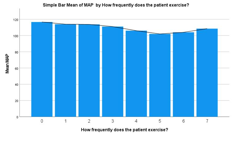
Is
step towards prevention of hypertension in normotensives and control of hypertension in Hypertensives. Salt intake shouldn’t exceed 2.3 grams (one teaspoon) in healthy adults. Although patients of Hypertension may need to decrease their intake further.
•Having a diet rich in fruits, vegetables, low fat dairy products and reducing intake of trans fat. Certain diet patterns help reduce BP and although the exact nutrients responsible for that is unknown it is recommended that the patients’ diet should be rich in fruits, vegetables and low-fat dairy products. Inclusion of nuts, fish and whole grains are also recommended. Red meat should however be avoided. Diet rich in Potassium, Magnesium and Calcium could help, but results of studies in this aspect have been inconclusive.
•Moderation in alcohol consumption. Many studies link high BP levels to heavy drinkers and it is
and research named Exercise as Hypertension therapy conducted in Columbia in August, 200118.
From the findings of this study and numerous others done before it, it is essential to take steps that act on these findings.
Firstly, let’s talk about the changes that can be made as an individual in our own personal habits. An article in the American Journal of Nephrology19 acts as a guide for the recommendations. These include -
•Regular physical activity of 30 minutes or more on most days of the week. The improvement in BP can be seen irrespective of intensity of exercise (hence high intensity weight lifting is just as effective as simple power walking) and whether the person has ideal body weight (both obese and healthy individuals attain similar gains from the exercise)

•Reduction in salt intake. A reduction in salt intake is an excellent
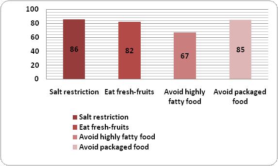
a = R Squared = 0.062 (Adjusted R Squared = 0.054)
Dietary
Dependent Variable : MAP
III
a = R Squared = 0.098 (Adjusted R Squared = 0.091)
recommended that males shouldn’t exceed 2 drinks a day and females shouldn’t exceed 1 drink per day.
•Maintaining a healthy weight has shown to reduce BP and is highly recommended for patients who are overweight or obese.
The various steps that can be taken to promote these habits at a community level are to encourage medical personnel to emphasize on giving nonpharmacological advice to their patients and explaining its importance to ensure patient compliance to both medications and the lifestyle changes prescribed. Nonpharmacological advice should be made a part of medical education so that the future medical personnel know its importance and incorporate the habit of emphasising on it along with prescribing conventional medications.
Making major lifestyle changes like eating fresh fruits and vegetables, avoiding packaged and junk foods can be difficult initially and so introducing these habits along with a regular exercise regimen and Yoga at a young age can be helpful and can be an important step in the primary prevention of the disease. This can be achieved by educating the children and the youth by including it in their curriculum and through promoting it in mass media too. These practices can be promoted by popular celebrities, social media influencers and role models of the current generation by holding various awareness events, celebrating Yoga weeks, organizing health drives and marathons.
In conclusion we can say that hypertension and its association with voluntary lifestyle choices is undeniable. With the life-threatening consequences of the disease, it is of utmost importance to take decisions which help prevent and control the disease. Working on an individual level is the first hurdle but people can also help others by increasing their awareness and providing scientifically sound advices.
1Ibrahim MM, Damasceno A — Hypertension in developing countries. The Lancet 2012; 380(9841): 611-9.
2Tzunoda K, Abe K, Goto T, Yasujima M, Sato M, Omata K, Seino M, et al — Effect of age on the renin-angiotensinaldosterone system in normal subjects: simultaneous measurement of active and inactive renin, renin substrate, and aldosterone in plasma. The Journal of Clinical Endocrinology & Metabolism 1986; 62(2): 384-9.
3Tuck ML, Williams GH, Cain JP, Sullivan JM, Dluhy RG — Relation of age, diastolic pressure and known duration of hypertension to presence of low renin essential hypertension. The American Journal of Cardiology 1973; 32(5): 637-42.
4Huang Z, Willett WC, Manson JE, Rosner B, Stampfer MJ,
Speizer FE, et al — Body weight, weight change, and risk for hypertension in women. Annals of Internal Medicine 1998; 128(2): 81-8.
5Vasan RS, Beiser A, Seshadri S, Larson MG, Kannel WB, D’Agostino RB, et al — Residual lifetime risk for developing hypertension in middle-aged women and men: The Framingham Heart Study. JAMA 2002; 287(8): 1003-10.
6Beilin LJ, Puddey IB, Burke V — Lifestyle and hypertension. American Journal of Hypertension 1999; 12(9): 934-45.
7Cléroux J, Feldman RD, Petrella RJ — Lifestyle modifications to prevent and control hypertension. 4. Recommendations on physical exercise training. Canadian Hypertension Society, Canadian Coalition for high blood pressure Prevention and Control, Laboratory Centre for Disease Control at Health Canada, Heart and Stroke Foundation of Canada. CMAJ: Canadian Medical Association Journal 1999; 160(9): S21.
8Stamler R, Stamler J, Riedlinger WF, Algera G, Roberts RH — Family (parental) history and prevalence of hypertension: results of a nationwide screening program. JAMA 1979; 241(1): 43-6.
9Munger RG, Prineas RJ, Gomez-Marin O — Persistent elevation of blood pressure among children with a family history of hypertension: the Minneapolis Children’s Blood Pressure Study. Journal of Hypertension 1988; 6(8): 647-53.
10van der Sande MA, Walraven GE, Milligan PJ, Banya WA, Ceesay SM, Nyan OA, et al — Family history: an opportunity for early interventions and improved control of hypertension, obesity and diabetes. Bulletin of the World Health Organization 2001; 79: 321-8.
11Neuhouser ML, Miller DL, Kristal AR, Barnett MJ, Cheskin LJ — Diet and exercise habits of patients with diabetes, dyslipidemia, cardiovascular disease or hypertension. Journal of the American College of Nutrition 2002; 21(5): 394-401.
12O’Keefe JH, Gheewala NM, O’Keefe JO — Dietary strategies for improving post-prandial glucose, lipids, inflammation, and cardiovascular health. Journal of the American College of Cardiology 2008; 51(3): 249-55.
13Meisinger C, Döring A, Löwel H — Chronic kidney disease and risk of incident myocardial infarction and all-cause and cardiovascular disease mortality in middle-aged men and women from the general population. European Heart Journal 2006; 27(10): 1245-50.
14Williams B, Poulter NR, Brown MJ, Davis M, McInnes GT, Potter JF, et al — British Hypertension Society guidelines for hypertension management 2004 (BHS-IV): summary. BMJ 2004; 328(7440): 634-40.
15Wolf-Maier K, Cooper RS, Banegas JR, Giampaoli S, Hense HW, Joffres M, et al — Hypertension prevalence and blood pressure levels in 6 European countries, Canada, and the United States. JAMA 2003; 289(18): 2363-9.
16Fryar CD, Ostchega Y, Hales CM, Zhang G, Kruszon-Moran D — Hypertension prevalence and control among adults: United States, 2015-2016.
17Pescatello LS, Franklin BA, Fagard R, Farquhar WB, Kelley GA, Ray CA — Exercise and hypertension. Medicine & Science in Sports & Exercise 2004; 36(3): 533-53.
18Kokkinos PF, Narayan P, Papademetriou V — Exercise as hypertension therapy. Cardiology Clinics 2001; 19(3): 50716.
19Appel LJ — Lifestyle modification as a means to prevent and treat high blood pressure. Journal of the American Society of Nephrology 2003; 14(suppl 2): S99-102.
A 2-year-old male child was brought by his mother to outpatient Department of Paediatrics with chief complains of pain in abdomen associated with fever which is on and off and loss of appetite for 3 months. On clinical examination, mass was felt in peri-umbilical region predominantly on left side which was associated with tenderness. For further investigation, the child was referred to the Department of Radiodiagnosis for Computed Tomography. On contrast enhanced Computed Tomography, ill-defined lobulated heterogenous solid-cystic lesion was noted in retroperitoneum in the left suprarenal region crossing the midline and showed heterogenous enhancement on contrast study. Multiple calcific foci were noted within this lesion. There was encasement of abdominal aorta with its branches and left renal vein by the lesion; However, no evidence of any thrombus or invasion noted. Also, the lesion was causing the right lateral displacement of IVC. Multiple enlarged heterogeneously enhancing lymph nodes were noted in the pre, paraaortic, pre-paravertebral and pelvic region likely metastasis. A diagnosis of Neuroblastoma with lymph nodal metastasis was made.
Classic radiological features : We got in our case in favor of neuroblastoma — (1) Age, (2) Location, (3) Calcification, (4) vessel encasement, (5) Midline crossing lesion, (6) Regional lymph-nodal metastasis.
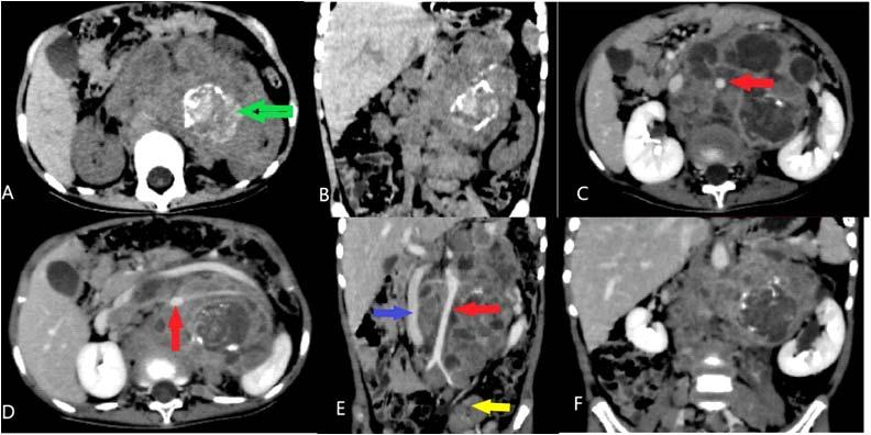
Fig 1 — (A & B) Axial & coronal non-enhanced abdomen CT scan showing ill defined lobulated heterogenous solid-cystic lesion in retroperitoneum in the left suprarenal region showing multiple areas of calcification (Green arrow);
(C & D) Axial enhanced abdomen CT scan showing heterogenous enhancement of the lesion and anterior displacement of abdominal aorta (Red arrow);
(E) Coronal enhanced abdomen CT scan showing encasement of abdominal aorta (Red arrow), right lateral displacement of Inferior vena cava (Blue arrow), Heterogeneously enhancing lymph node (Yellow arrow);
(F) Postcontrast coronal image showing the lesion is crossing the midline.
1MD, Senior Resident, Department of Radiodiagnosis, NKPSIMS and LMH, Nagpur, Maharashtra 440019 and Corresponding Author
2MD, Processor and Head, Department of Radiodiagnosis, NKPSIMS and LMH, Nagpur, Maharashtra 440019
Bhoomi Angirish1, Bhavin Jankharia2
Quiz 1
A 68-year-male presented with Haematuria since 1 Month.
Questions :
(1)What is the Diagnosis ?
(2)What are the complications of Bladder Diverticulum?
(3)What are the Differential Diagnosis?
Answers :
(1)A well defined outpouching (yellow arrow) is seen arising from the posterior wall of urinary bladder on right side suggestive of urinary bladder diverticulum. Within the diverticulum there is a well defined enhancing polypoidal lesion (red arrow) attached to its wall suggestive of intradiverticularurinary bladder carcinoma.

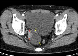
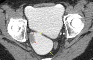
(2)The common complications associated with bladder diverticulum are intradiverticular carcinoma and calculi.
(3)The most common differential diagnosis of filling defect in urinary bladder is bladder clot, however it is mobile on changing position and does not show post contrast enhancement.
Quiz 2
A 57-year-old Lady Presented with Radicular Pain and Limb Weakness since 4 Months.
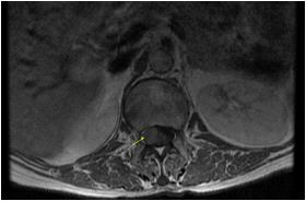
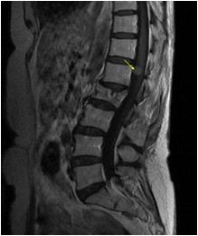
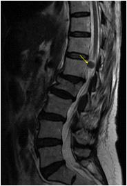
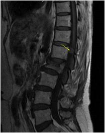
Questions :
(1)What is the Diagnosis ?
(2)What are the associations of this condition?
(3)What are the common Differential Diagnosis?
Answers :
(1)A well defined altered signal intensity intradural extramedullary lesion is seen at L1 vertebral level. Lesion appears hypointense on T1W and T2W images and shows homogeneous post contrast enhancement. These findings are in suggestive of meningioma.
(2)There is an increased incidence of spinal meningiomas in patients with neurofibromatosis type 2 (NF2).
(3)The common differential diagnosis of intradural extramedullary lesions are : Meningioma, Nerve Sheath Tumor (Schwannoma, Neurofibroma), Leptomeningeal Metastasis, Ependymoma, Paraganglioma.
Department of Radiology, Picture This by Jankharia, Mumbai, Maharashtra 400004
1MD, DNB (Radiology)
2MD, DMRD (Radiology)
Asian Indians exhibit a unique pattern of dyslipidemia characterized by elevated triglyceride (TG) levels and a greater incidence of hypertriglyceridemia (HTG). HTG refers to a medical condition characterized by elevated levels of triglycerides in the blood, which is strongly associated with an increased risk of cardiovascular disease (CVD). Thus, the management of hypertriglyceridemia becomes vital. Omega-3 fatty acid (O3FA) supplements containing both eicosapentaenoic acid and docosahexaenoic acid have been shown to reduce plasma TG levels through various mechanisms and play a significant role in the management of hypertriglyceridemia. Various national and international guidelines like Lipid Association of India, ESC/EAS guideline and ACC expert consensus recommend the use of O3FAs in the management of hypertriglyceridemia. O3FAs have also been found to play a significant role in the prevention of CVD, as evidenced by multiple clinical trials. O3FAs offer the other benefits like significant reduction in blood pressure in hypertensive subjects and patients with other CV risk factors such as hyperlipidemia or in patients treated with hemodialysis and TG levels in patients with antipsychotic-induced hypertriglyceridemia. This review article will emphasize the importance of maintaining the levels of TG in Indians and the role of O3FAs in the management of hypertriglyceridemia.
[J Indian Med Assoc 2023; 121(5): 69-74]
Key words :Omega-3 Fatty Acids, Hyperlipidemia, Cardiovascular diseases.
Dyslipidemia refers to either lipoprotein overproduction or deficiency, a consequence of abnormal lipoprotein metabolism. This leads to elevated total cholesterol (TC), low-density lipoprotein (LDL-C) cholesterol and triglyceride (TG) concentrations and a decrease in the high-density lipoprotein (HDL-C) cholesterol concentration in the blood. Dyslipidemia has been strongly associated with the pathophysiology of cardiovascular diseases (CVDs). It is a significant independent risk factor for coronary artery disease (CAD) and can lead to the development of atherosclerosis and associated cardiovascular (CV) events1. Patients with dyslipidemia have a 2-fold higher risk of developing CVD than those without dyslipidemia. Dyslipidemia is responsible for approximately 4 million CVD-related deaths globally2 Distinct pattern of Dyslipidemia in Indians : The pattern of Dyslipidemia observed in Indians is distinctive and characterized by elevated triglyceride (TG) levels, low levels of high-density lipoprotein cholesterol (HDL-C), moderately high levels of lowdensity lipoprotein cholesterol (LDL-C), and a higher
1MBBS, MAMS, MRSH, FICA, Dr Arun’s Health Centre, New Delhi
2MD, FCP, PGDHEP, Clinical Pharmacologist & Nutraceutical, Physician, Founder & CEO, IntelliMed Healthcare Solutions, Mumbai and Corresponding Author
Received on : 09/05/2023
Accepted on : 13/05/2023
proportion of small, dense LDL particles. This type of dyslipidemia is commonly referred to as “atherogenic dyslipidemia” and is more prevalent in South Asian populations3,4
According to the ICMR-INDIAB study, which included 16,607 subjects, dyslipidemia is highly prevalent among Indian adults, with 79% of the study participants having at least one lipid abnormality. Hypertriglyceridemia was observed in 29.5% of the individuals, decreased HDL-C levels in 72.3% of individuals, and elevated LDL-C levels in 11.8% of individuals5. The prevalence and pattern of concomitant cardiovascular (CV) risk factors that modify the impact of dyslipidemia on CV risk (eg, truncal obesity, metabolic syndrome, and diabetes) are also unique in Indians 4 . Although the cause of atherogenic dyslipidemia in Indians is unknown, genetic predisposition and characteristic body composition, such as excess truncal subcutaneous and intraabdominal fat, may be significant contributors3. Thus, Asian Indians have a distinctive pattern of dyslipidemia, which includes a higher incidence of elevated triglyceride levels, commonly referred to as hypertriglyceridemia (HTG).
The plasma TG level represents the concentration of TG-rich lipoproteins, includingvery-low-density lipoproteins (VLDL), chylomicrons and their remnants5.
Elevated plasma triglycerides result from excess triglyceride-rich lipoproteins of several different types, most commonly VLDLs but also intermediate-density lipoproteins (or VLDL remnants), chylomicrons, or chylomicron remnants6.
Pathophysiology of Hypertriglyceridemia : Under normal conditions, the liver packages endogenous lipids into very-low-density lipoprotein (VLDL) particles, which mainly contain triglycerides (TG), while dietary lipids absorbed in the intestine are incorporated into chylomicrons (Fig 1). Chylomicrons are quickly cleared as their TG content is hydrolyzed by lipoprotein lipase (LPL) at adipose and muscle tissue capillary beds, releasing free fatty acids (FFA) for cellular metabolic activities leaving behind chylomicron remnants. Similarly, VLDL is hydrolyzed by LPL, leaving behind remnants of VLDL particles in the bloodstream and intermediate-density lipoprotein (IDL) particles that are smaller in size and enriched in cholesteryl esters. The levels of TG-rich lipoproteins in circulation are determined by LPL activity. The liver primarily clears remnants through various receptors present on the surface of hepatocytes, including the LDL receptor, VLDL receptor, LDL receptor-related protein 1 (LRP1), and heparan sulfate proteoglycans (HSPGs), which facilitate the hepatic clearance of these lipoproteins. Another pathway for lipoprotein clearance involves syndecan 1, a core component of HSPG, which is essential for both the binding and
degradation of VLDL remnant particles. Any disruption that causes increased production of chylomicrons and/ or VLDL particles or a reduction in their metabolic breakdown will result in elevated levels of TG7
Need for management of Hypertriglyceridemia : Elevated levels of triglycerides in the blood have been linked to the presence of small, dense LDL particles and lower levels of HDL cholesterol, both of which are associated with an increased risk of CVD8 A study conducted on 3216 American Indians withoutany CVD at baseline reported a 32% higher risk for developing coronary heart disease (CHD) in patients with high TG and low HDL-C levels than those with normal TG and HDL-C levels over a follow-up period of 17.7 years. In patients with established CHD, it was found that patients with TG levels >500 mg/dl were at 68% increased risk of mortality as compared to those with TGs <100 mg/dl5. According to a study conducted by Miller and colleagues in patients who had experienced a myocardial infarction, a decrease of 10 mg/dL in TG levels was associated with a 1.8% reduction in the risk of experiencing a CV event. The management of HTG becomes crucial because of the higher prevalence ofelevated TG levels as a component of atherogenic dyslipidemia and by controlling TGs, the risk of developing CVD can be reduced7.
Role of Omega-3-fatty acids (eicosapentaenoic acid and docosahexaenoic acid) in management of hypertriglyceridemia :
Apo: Apolipoprotein, DGAT: diacylglycerol acyltransferase, IDL: Intermediate density lipoprotein, LDL: Low-density lipoprotein, LPL: Lipoprotein lipase, MTP: Microsomal triglyceride transfer protein, VLDL: Very low-density lipoprotein.
Introduction to Omega-3-fatty acids : Omega 3 ( ω 3 or n 3) is a structural descriptor for a family of polyunsaturated fatty acids (PUFAs). The size of the fatty acid chain affects its function, with short-chain fatty acids having less than six carbons, mediumchain fatty acids having six to 12 carbons, and very long-chain O3FAs having 12 carbons or more. The very long chain O3FAs include α-linolenic acid (ALA), docosahexaenoic acid (DHA) and eicosapentaenoic acid (EPA). ALA is an essential fatty acid because it cannot be synthesized in humans and, thus, must be consumed in the diet. ALA is a plantderived O3FA that can be converted to EPA and DHA in mammals. However, the conversion of ALA to EPA is modest, and the subsequent conversion of EPA to DHA is also very low. Thus, preformed EPA and DHA are best obtained through dietary sources such as fish or oily marine seafood9,10.
Prescribed O3FAs supplements have been shown to reduce fasting and non-fasting TG
levels by 20%-50% (Table 1) and increase the LDL particle size5,10. Nonprescription fish oil products are not interchangeable with prescription omega-3 products due to differences in O3FA content and bioavailability. As well as, prescription O3FA supplements have undergone more rigorous safety and efficacy evaluation than dietary supplements10,11
Several national and international guidelines such as Lipid Association of India (LAI) 2020 guideline,ESC/ EAS 2019 guideline, and ACC Expert Consensus 2021also recommend the use of O3FAs in the management of HTG5,11,12
Mechanism of Omega-3-fatty acids in hypertriglyceridemia :
Different mechanisms have been proposed and investigated to demonstrate the effect of O3FAs on lipoprotein metabolism (Fig 2). O3FAs reduce plasma triglycerides and offer CV protection by increasing the oxidation of fatty acids, resulting in decreased hepatic lipogenesis and subsequent VLDL production. They also enhance the lipolysis of increased triglyceriderich lipoproteins (chylomicron), have anti-inflammatory effects, and exert CV protection viapro resolving lipid
mediators. Additionally, O3FAs also reduce the risk of thrombosis.13
In a study conducted by Grevengoed, et al, a new mechanism was proposed for the reduction of plasma and liver TGs by O3FAs. The researchers discovered that O3FAs generate N-acyl taurines, which conjugate with bile acids and aid in the absorption of fats. The omega-3 fatty acid–derived N-acyl taurines (NATs) accumulated significantly in bile and plasma following O3FA supplementation. One of these NATs (DHA NAT) was found to inhibit intestinal triglyceride hydrolysis and lipid absorption, resulting in lower plasma triglycerides and protection against hepatic triacylglycerol accumulation in mice fed with a highfat diet13
Clinical evidence of Omega-3 fatty acids in the treatment of hypertriglyceridemia :
Table 2 presents the different clinical trials demonstrating the effect of EPA and DHA on TG and other blood lipid parameters in patients with HTG. Benefits of Omega-3-fatty acids in prevention of CVD : Clinical evidence
Large-scale epidemiological studies, clinical outcome trials, and meta-analyses have established the role of O3FAs in preventing atherosclerosis8. Table 3 presents the clinical trials demonstrating the effect of O3FAs in the prevention of CVD.
Other beneficial effects of Omega-3-fatty acids :
*High potency statins and higher doses of statins result in greater triglyceride reduction. Patients with higher baseline triglyceride levels achieve greater reduction with the same statin dose.
A high intake of O3FA has been associated with cardiovascular protective effects, improving endothelial function and reducing atherosclerosis through their beneficial effects on BP, lipid profile, platelet aggregation, and anti-inflammatory properties. Various epidemiological and clinical studies suggest that consumption of O3FA can reduce BP in hypertensive subjects and patients with other CV risk factors, such as hyperlipidemia or in patients treated with hemodialysis (Table 4)20
It has been demonstrated that Atypical antipsychotics (AAPs) frequently lead to marked elevation of serum triglyceride levels with modest alterations in other lipid profiles.
*Each 1-g capsule of omega-3 fatty acids contains eicosapentaenoic acid (EPA) 465 mg and docosahexaenoic acid (DHA) 375 mg.
Antipsychotic-induced dyslipidemia further increases the risk of developing metabolic complications and CVD. Despite its burden, antipsychotic-induced dyslipidemia is often left untreated. O3FAs supplementation reduces TG levels with minimal side effects, making it an excellent candidate for use in psychiatric patients. O3FA have been uniformly reported to lower TG levels in antipsychotic medications-induced hypertri-glyceridemia patients (Table 4)21-23
Safety information : O3FA have shown safe and relatively mild side-effect profiles. Omega-3 fatty acids (O3FA) have been found to have a safe and relatively mild side effect profile. Some
of the most commonly reported adverse reactions (with an incidence of over 3% and higher than placebo) include constipation, gastrointestinal disorders, vomiting, increased ALT and AST levels, pruritus, and rash. It is important to note that long-term use of O3FA supplements, particularly at higher dosages, may increase the risk of atrial fibrillation. Additionally, O3FA supplementation may also prolong bleeding time. Therefore, patients who take O3FA along with anticoagulants or other drugs that affect coagulation, such as antiplatelet agents, should receive periodic monitoring. O3FA supplementation of up to 1 g/day is generally well-tolerated and does not increase bleeding time, except for occasional dysgeusia27-29
Table 4 — Clinical evidence demonstrating the other beneficial effect of EPA and DHA
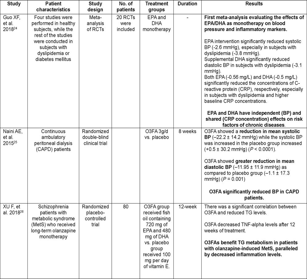
1Misra S, Lyngdoh T, Mulchandani R — Guidelines for dyslipidemia management in India: A review of the current scenario and gaps in research. Indian Heart J 2022; 74: 34150.
2Thongtang N, Sukmawan R, Llanes EJB, Lee ZV — Dyslipidemia management for primary prevention of cardiovascular events: Best in-clinic practices. Prev Med Rep 2022; 27
3Dyslipidemia in Asian Indians: determinants and significance - PubMed. https://pubmed.ncbi.nlm.nih.gov/15656049/.
4Chandra KS — Consensus statement on management of dyslipidemia in Indian subjects. Indian Heart J 2014; 66: S1.
5Puri R — Management of diabetic dyslipidemia in Indians: Expert consensus statement from the Lipid Association of India. J Clin Lipidol 2022. doi:10.1016/j.jacl.2022.11.002.
6Skulas-Ray AC — Omega-3 Fatty Acids for the Management of Hypertriglyceridemia: A Science Advisory From the American Heart Association. Circulation 2019; 140: E673–E691.
7Arca M — Hypertriglyceridemia and omega-3 fatty acids: Their often overlooked role in cardiovascular disease prevention. NutrMetab Cardiovasc Dis 2018; 28: 197-205.
8Watanabe Y, Tatsuno I — Omega-3 polyunsaturated fatty acids focusing on eicosapentaenoic acid and docosahexaenoic acid in the prevention of cardiovascular diseases: a review of the state-of-the-art. Expert Rev Clin Pharmacol 2021; 14: 79-93.
9Calder PC — Omega-3 polyunsaturated fatty acids and inflammatory processes: nutrition or pharmacology? Br J Clin Pharmacol 2013; 75: 645-62.
10Bays HE, Tighe AP, Sadovsky R, Davidson MH — Prescription omega-3 fatty acids and their lipid effects: physiologic mechanisms of action and clinical implications. Expert Rev Cardiovasc Ther 2008; 6: 391-409.
11Virani SS — ACC Expert Consensus Decision Pathway on the Management of ASCVD Risk Reduction in Patients With Persistent Hypertriglyceridemia: A Report of the American College of Cardiology Solution Set Oversight Committee. J Am Coll Cardiol 2021; 78: 960-93.
12Mach F — ESC/EAS guidelines for the management of dyslipidaemias: Lipid modification to reduce cardiovascular risk. Atherosclerosis 2019; 290: 140-205.
13Bornfeldt KE — Triglyceride lowering by omega-3 fatty acids: a mechanism mediated by N-acyl taurines. J Clin Invest 2021; 131
14Safety and efficacy of Omacor in severe hypertriglyceridemia - PubMed. https://pubmed.ncbi.nlm.nih.gov/9865671/.
15Davidson MH — Efficacy and tolerability of adding prescription omega-3 fatty acids 4 g/d to simvastatin 40 mg/d in hypertriglyceridemic patients: an 8-week, randomized, doubleblind, placebo-controlled study. Clin Ther 2007; 29: 1354-67.
16Roth EM — Prescription omega-3 fatty acid as an adjunct to fenofibrate therapy in hypertriglyceridemic subjects. J Cardiovasc Pharmacol 2009; 54: 196-203.
17Shearer GC, Pottala JV, Hansen SN, Brandenburg V, Harris WS — Effects of prescription niacin and omega-3 fatty acids on lipids and vascular function in metabolic syndrome: a randomized controlled trial. J Lipid Res 2012; 53, 2429-35.
18Bhatt DL — Cardiovascular Risk Reduction with Icosapent Ethyl for Hypertriglyceridemia. N Engl J Med 2019; 380: 1122.
19Marchioli R — Dietary supplementation with N-3 polyunsaturated fatty acids and vitamin E after myocardial infarction: Results of the GISSI-Prevenzione trial. Lancet1999; 354: 447-55.
20Cabo J, Alonso R, Mata P — Omega-3 fatty acids and blood pressure. Br J Nutr 2012; 107 Suppl 2
21Yan H, Chen JD, Zheng XY — Potential mechanisms of atypical antipsychotic-induced hypertriglyceridemia. Psychopharmacology (Berl) 2013; 229: 1-7.
22Kanagasundaram P — Pharmacological Interventions to Treat Antipsychotic-Induced Dyslipidemia in Schizophrenia Patients: A Systematic Review and Meta Analysis. Front Psychiatry 2021; 12
23Fetter JC, Brunette M, Green AI — N-3 fatty acids for hypertriglyceridemia in patients taking second-generation antipsychotics. Clin SchizophrRelat Psychoses 2013; 7
24Guo X, fei Li, K lei, Li, J mei, Li D — Effects of EPA and DHA on blood pressure and inflammatory factors: a meta-analysis of randomized controlled trials. Crit Rev Food Sci Nutr 2019; 59: 3380-93.
25Taheri S, Keyvandarian N, Mortazavi M, Hosseini S, Naini A — Effect of Omega-3 fatty acids on blood pressure and serum lipids in continuous ambulatory peritoneal dialysis patients. J Res Pharm Pract 2015; 4: 135.
26Xu F — Effects of omega-3 fatty acids on metabolic syndrome in patients with schizophrenia: a 12-week randomized placebo-controlled trial. Psychopharmacology (Berl) 2019; 236: 1273-9.
27Lovaza. USFDA PI. http s://www.accessdat a.fda.gov/ drugsatfda_docs/label/2019/021654s043lbl.pdf.
28Fish oils in cardiovascular prevention: new evidence, more questions. https://cardiologie-universitaire-geneve.ch/wpcontent/uploads/2022/01/ Baris_Gencer_JeudI_20_01_2022_Omega3_CV_Prevention.pdf.
29Kromhout D, Yasuda S, Geleijnse JM, Shimokawa H — Fish oil and omega-3 fatty acids in cardiovascular disease: do they really work? Eur Heart J 2012; 33: 436-43.
[The Editor is not responsible for the views expressed by the correspondents]
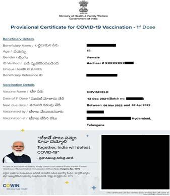
SIR, — Guillain-Barre syndrome (GBS), formerly known as Landry’s paralysis, is a condition in which the body’s immune system affects a portion of the peripheral nervous system. The precise causes of GBS are largely unresolved1. The disorder can be seen days or weeks after respiratory or digestive infection and can occur rarely after recent vaccination or surgery. The condition may result in acute flaccid paralysis of limbs and areflexia2,3 Complete recovery has been observed in most patients, and the mortality rate is about 5%4. The frequency of occurrence in men is one and a half times more than in women, and adults are more susceptible to GBS than children5. This study reports a 63-year-old female patient with Acute Motor Sensory Axonal Neuropathy (AMSAN) subtype of GBS, 10 days following the preliminary dosage of the ChAdOx1nCov-19 Coronavirus vaccine (COVISHIELDTM).
A 63-year-old female with a medical history of hypertension and type 2 diabetes mellitus on regular medication presented to the general medicine department with chief complaints of abrupt onset of aggravating generalized weakness in both upper and lower limbs, body pains, and back pain, difficulty to stand or walk without support, difficulty in rolling over the bed,chest pain, breathlessness, and loss of appetite, over the previous 10 days following the first dose of ChAdOx1nCov-19 Coronavirus vaccine (COVISHIELDTM) (Fig 1).
The patient had no previous medical history of trauma, weight loss, fever, or respiratory or gastrointestinal diseases.On physical examination, the patient was found to be afebrile, with blood pressure – of 130/90 mmHg, SpO2 – 96% on RA, Pulse Rate – 92 bpm, GRBS –149 mg/ dl. Neurological examination revealed acute areflexic quadriparesis of both upper and lower limbs. With normal bone density and tone in the upper limbs and decreased tone in the lower limbs, the power of the upper limbs was found to be 4/5, and power in the lower was found to be 3/ 5. The patient’s serum electrolytes revealed decreased sodium levels of 114 mEq/L, and decreased chloride levels of 81mEq/L respectively, CBP revealed leucocytosis of 15.04[10^3/µl], increased blood urea levels of 45.7 mg/dl.
Her nerve conduction investigations revealed sensory-motor axonal neuropathy in both the lower and upper limbs (Tables 1&2). In nerve conduction examinations, both motor and sensory nerves exhibited lower amplitude. On inspection, there was no evidence of cranial nerve involvement. The results of all other systemic exams were within normal ranges.
Electroneuromyography (ENMG) (Table 3) revealed the absence of both the sural nerve sensory nerve action potential (SNAPS) and a reduction in the tibial nerve compound muscle action potential (CMAP), as well as significantly elevated common peroneal CMAP distal latency and significantly lowered CMAP amplitude (with spatial and temporal dispersion). The F wave latency was within standard ranges.
Her treatment procedure was started with intravenous immunoglobulin (IVIg) at a dose of (2 g/kg/) five vials 24h
Nerve Dist. Lat CMAPCVF Wave
Left median 15.5/18.63.2/0.270NR
Right median 12.8/20.71.1/1.027NR
Left ulnar42/8.81.7/1.251NR
Right ulnar5.7/9.53.0/2.169NR
Left CPNNR---
Right CPN 8.3/18.81.0/0.934NR
Left PTNNR---
Right PTN 7.3/16.81.6/1.440NR
Dist. Lat – Distal latency, CMAP – Compound Muscle Action Potential, CPN – Common Peroneal Nerve, PTN – Posterior Tibial Nerve, NR – No response
velocityReduced Rt median, left CPN
SNAP – Sensory Nerve Action Potential,CMAP – Compound Muscle Action Potential, CPN – Common Peroneal Nerve, NR –No Response
infusion for five days with a close watch for respiratory failure. She responded well to IVIg therapy and remained stable, hence IVIg therapy was discontinued and physiotherapy has been advised.She is still being monitored at home and receiving regular assessments and follow-ups at the Neurology outpatient department.
Guillain-Barré syndrome is an uncommon autoimmune condition that causes inflammatory demyelination of nerve fibers and usually arises after infection 5 . Although there have been numerous case reports of post-vaccination GBS, the global incidence of GBS in society is estimated to be 17 cases per million people per year. An analysis of earlier post-vaccination periods (Swine flu vaccination campaigns in 1976/1977 and H1N1 vaccination programs in 2008/2009) indicated no increased prevalence of post-vaccination GBS3
Even though the fact that there is no proven link between the Covishield vaccination and GBS, the Food and Drug Delivery (FDA) has updated GBS warnings for another vaccine [GlaxoSmithKline’s SHINGRIXTM (Zoster Vaccine Recombinant, Adjuvanted)] as of 24 March 2021, after observing 3 additional cases of GBS per million within 42 days after administration of the vaccine1,2. The development of GBS was seen within 10-14 days of the covid 19 vaccination administration in every case where GBS development was linked to its administration. Although the pathophysiology of GBS caused by the administration of the COVID-19 vaccine is unknown, it is considered that when a COVID-19 mRNA vaccine is given, the mRNA enters human cells and instructs them to recognize the spike protein located on the surface of SARS-CoV-2, the virus that results in COVID-19. The spike protein is then identified by our bodies as an invader, which causes the development of antibodies to fight it. These antibodies are prepared to detect and eliminate the virus when it is encountered, blocking it from spreading and causing harm. This immune response
may trigger autoimmune processes in some patients, leading to the development of antibodies against myelin causing GBS6,7
In this case report, the GBS patient has been diagnosed with the condition despite having neither a history of the illness in her family nor a history of an infection that would have contributed to it. Therefore, it is feasible to hypothesize that in this case, the COVID-19 vaccine is what caused the GBS.
We present this case because a global vaccination drive is underway to control the pandemic. A correct analysis of the neurological drug reactions linked to the vaccination and the onset of occurrence can reduce morbidity and mortality through early disease detection. Using immunoglobin to treat GBS shows quicker results.
1National institute of neurological disorders and stroke. GuillainBarré Syndrome Fact Sheet | National Institute of Neurological Disorders and Stroke. Nih.gov. 2018https:// www.ninds.nih.gov/Disorders/Patient-Caregiver-Education/ Fact-Sheets/Guillain-Barr%C3%A9-Syndrome-Fact-Sheet
2da Silva GF, da Silva CF, Oliveira RE, Romancini F, Mendes RM, Locks A, Longo MF, Moro CH, Longo AL, Braatz VL. Guillain–Barré syndrome after coronavirus disease 2019 vaccine: A temporal association. Clinical and Experimental Neuroimmunology 2022; 13(2): 92-4.
3Maramattom BV, Krishnan P, Paul R, Padmanabhan S, Cherukudal Vishnu Nampoothiri S, Syed AA, et al — Guillain Barré syndrome following ChAdOx1 S/nCoV 19 vaccine. Annals of Neurology 2021; 90(2): 312-4.
4Vellozzi C, Iqbal S, Broder K — Guillain-Barre syndrome, influenza, and influenza vaccination: the epidemiologic evidence. Clinical Infectious Diseases 2014; 58(8): 114955.
5Hauser SL, Asbury AK — Guillain-Barre syndrome and other immune-mediated neuropathies. Harrisons Principles of Internal Medicine 2005; 16(2): 2513.
6Kripalani Y, Lakkappan V, Parulekar L, Shaikh A, Singh R, Vyas P — A Rare Case of Guillain-Barré Syndrome following COVID-19 Vaccination. European Journal of Case Reports in Internal Medicine 2021; 8(9).
7Waheed S, Bayas A, Hindi F — (February 18, 2021) Neurological Complications of COVID-19: Guillain-Barre Syndrome Following Pfizer COVID-19 Vaccine. Cureus 13(2): e13426. doi:10.7759/cureus.13426.
1Doctor of Pharmacy, Swathi Pagadala1, Keerthi N1, Department of Pharmacy, Sunil Kumar Behera1, CMR College of Pharmacy, Shweshikaa K1, Hyderabad, AP Ramya Bala Prabha1 JIMA
is now fully ONLINE and Publishes only ONLINE submitted Articles through
