History of Quarantine – Past, Present and future. Are we in Same Platform ?
[The same tradition continues uninterrupted ; nowhere has it changed]
— Bharatbarsa by S wajed Ali
History of the world has been intertwined with the impact of infectious disease over its population. Evidence of smallpox has been found in 3000 years old Egyptian Mummy. Hippocrates had clearly written that diseases spread by “air, fomite, and places”. Centuries after centuries, infectious diseases have influenced political, social and economic balance of many countries. Plague of Athens changed power equation between Athens and Sparta, ending the golden age of Athenian predominance. Alexander the Great defeated Puru, the great Indian Warrior but was helplessly defeated at the age of 33 by tropical fever. During age of exploration Europeans invaded different continents like Asia, Africa, Latin America and brought vectors and organisms to non-endemic parts of world. Thus the infectious disease became a global problem.
But, before discovery of the Germ Theory, advent of antimicrobials and vaccination, there was no definite way to defend against infectious disease. From ancient times, people practiced isolation of infected person from community and separating susceptible community from infected person. This practice was termed as Isolation and Quarantine respectively. In absence of definite medicine these methods were adopted as powerful tools centuries after century to reduce rapid spread of infection.
Evidence of isolation found in ancient literature.
An early mention of isolation occurs in Biblical book of Leviticus written in 700 BCE. The Islamic prophet Muhammad also advised quarantine: "those with contagious disease should be kept away from those who are healthy". In Hindu literature, isolation of 21days had been advised to get rid of diseases.
(Twenty one days isolation can remove poison from your body – Astanga Hriday Grantha, 65 no shloka.)Although the number “21” is not based on scientific evidence, still the spirit of this advice remains valid even today.
Isolation & Quarantine in Medieval Period :
Though practice similar to isolation and quarantine were practiced from even before the birth of Christ, but 1377 AD is considered as a watershed zone in Medieval history. In 1377, great council of Ragusa (modern Croatia) first enacted the law of isolation, which was enforced by the State. Initially, it was for 30 days for anybody trying to enter a city. In 1423, this method was adopted by Venice – quarantine of merchant ships (presuming sailors were carrying infectious disease from different country or continent). Gradually whole Europe adopted this practice. Then it was enhanced to 40 days – name adopted as Quarentina from Latin Quadraginta –referring to 40. Italy applied quarantine in the fifteenth century. Basically it was initially applied to ships coming from abroad to make sailors infection free before entering to country. A more detailed description of human response to pandemics can be found in the medical history section of this issue.
To utter surprise, Great Britain was reluctant to follow this practice in spite of repeated outbreaks. Ultimately, after 200 years, in 1665, during the “Great Plague of London”- Britain ruthlessly enforced this law. From 16th to 18th Century, France adopted isolation of people coming in Ships from abroad. Subsequently, US Supreme Court affirmed power to state to enact quarantine.
Quarantine in Nineteeth Century :
Quarantine was challenged in early nineteenth century by reformers as an outdated practice. Europe was in stage of renaissance and in dream of Industrial revolution. Germ theory was not established by that time. Reformers viewed that
Prof. (Dr.) Jyotirmoy Pal MD, FRCP, FRCP, FICP, FACP, WHO Fellow, Hony. Editor, JIMA
quarantine would be infringement of their personal freedom and contemporary economist and industrialists opined that commerce would be heavily affected by this century old practice. In 1830, when Cholera epidemic reached England, British government again switched over to Old practice, having no curative Medicine. Quickly it became unpopular. LANCET (1832): in one article called Cholera as “humbug got up for the destruction of Commerce”. Riot flared up in Liverpool in 1832 against quarantine. Debate continued between quarantine, economy, public health and personal liberty. Fortunately in mid –nineteenth century, Germ Theory was established by Louis Pasteur and nature of disease and its spread was defined and so again need of quarantine was warranted.
In 1851, in response to repeated epidemics, France held the first International Sanitary Conference at Paris to make a uniform practice guideline for containment of infection. But in spite of several meetings, Europe failed to formulate a consensus policy due to different economic and political agendas of European countries who were in race for colonization. Great Britain was a big blocker of quarantine policy in that time. Finally in 1893 (after Cholera pandemic in Europe in 1892) a ratified convention with act for compulsory notification was achieved. In the same year, US Congress also passed National Quarantine Act.
Quarantine in Twentieth Century :
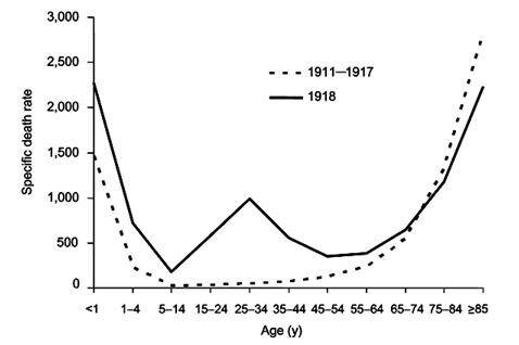
But history repeats itself. In 1911, Encyclopedia Britannica defined quarantine – “thing of past in UK and in majority of our states”. In 1914, Europe engaged in World War 1 and Spanish Flu struck the whole World. Again, Europe adopted the so called redundant policyQuarantine, Lockdown and Isolation. The World committed several mistakes during the Spanish flu. In war torn countries, media was censored (except in Spain). So, the actual extent of the epidemic was unknown to the public. Lack of awareness and transparency made it difficult to control disease and unregulated mixing particularly among soldiers took more lives than the preceding war. After first wave of Flu, lockdown was quickly withdrawn due to several reasons – to celebrate victory in war, reestablishment of economic activities and so on; as a result second wave came heavily with more mortality.
After 2nd World War, two remarkable milestones were: establishment of WHO in 1948 and CDC in 1967.
Quarantine in Twenty First Century :
At the beginning of 21st Century, there were outbreak of SARS, Ebola, avian influenza etc. and Health officials had to use the old preventive processes — Isolation and quarantine. With time, there have been remarkable advancement in Medical Sciences; but mankind is helpless before infectious disease. Still the World is grasping old
practice when there is sudden outbreak. So in 2003 CDC declared “ Quarantine is medically very effective in protecting public health from diseases”. But due to advent of knowledge of incubation period and pathogenesis, scientists can now clearly define the duration of quarantine, that differs from disease to disease . This has been widely applied in COVID-19 pandemic. This is probably the largest quarantine and isolation in the history of Medical sciences
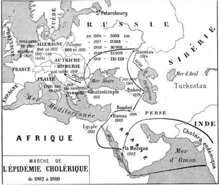
In spite of usefulness and indispensability even in 21st century, Quarantine is never without controversy. Controversy lies in its application. There are several examples of either ruthless application or liberal application. There are several examples, where quarantine or lockdown has not given desired benefit. Quarantine is often weighed against politics, economic, ethics, freedom, fundamental rights or emotions. Lack of balance had put the process under question in past. When applied ruthlessly as in Cholera epidemic in Jessore 1818, it ignored basic fundamental rights. When applied keeping emotions, freedom as priority, as in Spanish Flu, it invited surge of infections. Rulers either ignored economic priority of individual or given high priority on trade economy of their country.
Quarantine & Society in Colonial India:
Quarantine, isolation, lockdown is never accepted from heart by mass in British India. It was considered as imprisonment.
[Plague was dangerous, but quarantine was more dangerous : Rajendra Singh Bedi].
Famous Bengali Writter Saratchandra Chattopadhyay expressed feeling of quarantine in his famous book Srikanta:
[Doctor called me to the corner and said- Mr. Shrikanta, you shouldn’t have come without the letter. Taking people to the quarantine, they inflict pain more than that suffered by the cattle in the slaughter-house. Although, the poor may endure such pain, the rest succumb to such pain.]
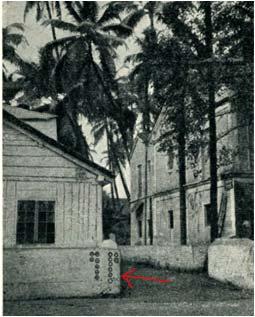
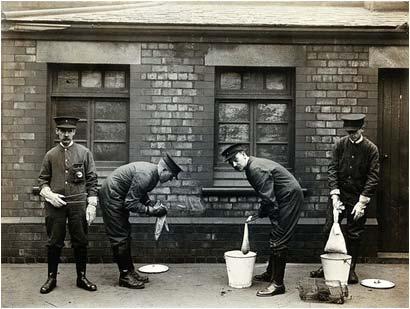
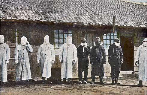
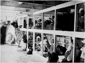
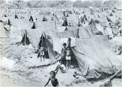
For successful quarantine, State has to impose restriction, which may raise many questions on fundamental rights. Bombay faced Plague epidemic in 1897
and British Government enforced Epidemic Act 1897. But this Act was not beyond criticism. Implementation of Act was discriminatory and disrespectful, ignoring emotion and rights of people. Adequate food, shelter, treatment were not ensured and all people put in same shelter without considering caste, gender, religion, (which was relevant at that time in India; the Hindu upper castes did not want to stay in the same tent with untouchables). Eminent British historian David Arnold in his book "Colonizing the body: state medicine and epidemic disease in nineteenth century India" - Epidemic Act 1897 was a product of the colonizing effort of IMS officials, which give them a forehead in exercising their whims. Natasha Sarkar, Indian historian has written in Journal of Indian History Congress, 2001 –British health committee invited criticism on quarantine policy. No notice was issued in advance. This caused great inconvenience to ordinary people, more to migrant labour. Mass resentment started in Bombay, Delhi and Kolkata. People started refusing quarantine. Riot started in Bombay. A British official was assassinated in Pune by Chapekar brothers.
So our question to the Public health experts, Where is the mistake? Where is the conflict?
Indian Response to COVID-19 :
In 2019 November, there was outbreak of Coronavirus infection in Wuhan province of China. Gradually it spread to almost all countries and in all continents. WHO declared this pandemic as a Health Emergency. Due to lack of specific therapy, sudden surge of infection and growing international travel WHO embraced 600 years old traditional practices – isolation, quarantine and lockdown.
[The same tradition continues uninterrupted; nowhere has it changed]
India’s response to pandemic was to some extent a make-shift arrangement. Most of the States were not prepared to gear up to combat pandemic. Our healthcare system had redirected resources – hospital beds, equipment, human resources from Non-Covid management to Covid management. As a result there is crisis in Non Covid area. So Government should build up separate infrastructure for quarantine, Isolation, ward and CCU for future epidemic or pandemic. Again, this time Government has utilized lot of Private infrastructure. But we should remember that the Public health issue is to be dealt by Public Health care system, not by Profit driven Private health Care system. Private health care system may not have same commitment as Public Sector. Only help on technological issues can be utilized.
Perception in Modern India :
There are several reports in last few months regarding refusal of quarantine, isolation, flee from hospitals, attack on health care workers (HCW) ( Indore and Chennai) and police, denying entry of HCWs in residential places (Kolkata and Delhi) and so on. These are out of fear, stigma, distance from family for prolonged period, loss of wages and loss of trust in public health care system. We committed the same mistakes as in the past. We imposed measures without taking people in confidence. Stigmatization, fear was integral part of contagious disease in the past. Poet John Donne suffered from severe infection in 1623. He immediately found himself alone even doctors deserted him. He wrote “as sickness is the greatest misery, so the greatest misery of sickness is solitude “. Rabindranath Tagore in his poem Puratan Bhritya expressed loneliness after contagious infections like smllpox.
[Where, alas, the damsels of Vraja, where the fabled woods, where was Hari
—The Gardener? Springtime? Accursed luck, dreaded smallpox, lethal and scary
Found me. One by one, every last room mate vacated the quarters of our dream
While, forlorn in my room lay I, even as pox lesions swamped my every limb.]
But in era of Internet, satellite, when we are moving towards the moon, frequent reports of resistance faced by HCWs in entering their own houses is definitely a redflag sign.
[The same tradition continues uninterrupted; nowhere has it changed]
— even after 400 years.
Widespread fear of disease, mistrust on authority, wrong popular belief (doctors killed patients for anatomical dissection) during cholera epidemic led to cholera riot in 1832 in Liverpool. Same mistrust was observed in the Bombay Plague in 1898; people thought plague was a conspiracy of British government to kill natives, particularly the downtrodden, who were pushed to unhygienic, poor quality shelters as a method of quarantine. After 125 years, still people have a belief that the Corona pandemic may be a conspiracy of China Government to restore supremacy over world. Mass hysteria, panic, what we see today, is
nothing new in pandemic. What happened in Indore (attack of Health Care workers) or Kolkata (Nurses denied entry in their housing complex) is nothing new, but the legacy of previous centuries. Only time changed, we have not changed much in our attitude or practice. For example, during the plague epidemic of Calcutta in the last decade of the Nineteenth century, people also had a lot of misunderstandings. Premankur Atorthi, in his book, “Mohasthobir Jatok” has given some descriptions of the public perception in that era:
[In the midst of the general public, such deadly rumors began to circulate regarding vaccines that people of this age would consider it a joke.
Some said, within ten hours of the day, people would go to the grave.
Others said, taking a penny sized piece of flesh from the stomach, seeds of plague were inserted.
The Plague Hospital was established in Mark’s Square of Mechhobazar. This further incited a riot.]
We can compare this attitude to the various rumours and public resistance faced by the administration during setting up of Covid hospitals in different places.
[The same tradition continues uninterrupted; nowhere has it changed]
Another unheard aspect is voice of migrant labourers centuries after centuries. If we cannot ensure their food, shelter more people will die of hunger rather than disease itself. Jobless, derouted people will increase social inequalities. In Mumbai Plague epidemic, sudden notice of Lockdown in 1898 made life of migrant laborers miserable. In the present pandemic, these people walked miles after mile to reach home. In spite of several schemes taken by both Central and State Government of India, the images of these people walking, walking & walking their hunger, clash with police for food, death on way tarnished the Nation’s shining Face. Great Poet Gulzar in his poem depicted
— Gulzar
[There was a great pandemic
All the workers, craftsmen, ran off to their homes. All the machines were shutting down in the city
This is what helped in the keeping the hands and legs working
Otherwise life was blissful in village only]
Controversy & Futuristic Approach :
Protecting health of community, combating fear psychosis and discrimination during epidemic period is really complex. This needs Planned programming on Health and behavioural education much before next outbreak of infectious disease. Dr Giridhari Babu, famous epidemiologist said “faith in the public health system cannot emerge immediately as a response to the pandemic”.
In Post-Independence era, Government of India has definitely taken several measures on Preventive health. With different Disease Control Programs, life expectancy have increased dramatically. But after the 90s GOVT policy moved more to Hospital based curative treatment, stress on Non communicable diseases and boosting of private and insurance based health Care System. As a result, public health care system, particularly preventive care was neglected. This weakness was revealed during Nipah virus outbreak (Kerala), Dengue outbreak and recent JE outbreak. Government of India’s prompt enforcement of lockdown was praised by WHO as “Tough and timely” but this has thrown several questions – particularly food insecurities of migrant labours. Also quarantine or containment provoked danger of stigmatization. Rumors in social media, fear, lack of political will, politicization of health issues, violence against health care workers, and transmission among health care workers made this challenge even more difficult.
After Pandemic or Epidemic, immediate challenge is to keep infection at a manageable level, ensure maximum tests and tracing of contacts, isolate patients, treat as per protocol and timely dissemination of proper information. Food securities for the poor and vulnerable section and prevention of Economic fallout, along with international commitment should be the key arena for Government of India. All efforts will go in vain if we cannot create vibrant, enlightened, committed health care workers – including Doctors, Nurses, Paramedical staffs, public Health
administrator a dedicated Public health Specialist with good remuneration (including insurance for death or disability), satisfaction and pride in profession. Separate Fund allocation on Public heath, building of infrastructure and Human resources should be a priority. There should be strong surveillance system that can exactly detect or predict outbreak. India has Integrated Disease Surveillance System (IDSP), but needs stronger commitment with legislation to meet any challenge. To reach the goal, the country needs upgraded Laboratory i.e. apex laboratory like National Institute of Virology and also state laboratories. Updated Epidemic Act should give doctors enough power even above bureaucracy to achieve clinical significance rather than statistical significance. Lack of transparency, rumors in public (today at social media), unbalanced media reporting hinder epidemic control in times of crisis. In words of famous cardiologist Prof. G S Wander, "we seem to have lost balance on the emotional to rational scale".
We should not repeat mistakes of the past and should be prepared with better epidemic Act that will incorporate human emotions, participation, preserved fundamental rights.
“ Pandemic provided us with a break from the past and enables the possibility for us to imagine an entirely new world”
— Arundhati Roy
Except technological improvement, psychologically and culturally we are in almost same platform as we were in last few pandemics in the past 200 years. We should make a trust based Public health system and new Pandemic act that include People’s sentiment, involvement and confidence suitable for an independent, democratic country which will not repeat the mistakes of colonial period. So, in my opinion, this pandemic has given us a wake-up call for a long walk to build a stronger and trust-based healthcare system in India.
“ He gives his harness bells a shake To ask if there is some mistake……… And miles to go before I sleep And miles to go before I sleep”
— Robert Frost
I AM CONFIDENT WE WILL BUILD STRONG, DEMOCRATIC, HEALTHY INDIA
Disclaimer
The information and opinions presented in the Journal reflect the views of the authors and not of the Journal or its Editorial Board or the Publisher. Publication does not constitute endorsement by the journal.
JIMA assumes no responsibility for the authenticity or reliability of any product, equipment, gadget or any claim by medical establishments/ institutions/manufacturers or any training programme in the form of advertisements appearing in JIMA and also does not endorse or give any guarantee to such products or training programme or promote any such thing or claims made so after.
— Hony Editor
Review Article Review Article Review Article
Role of Chest Radiograph (CXR) in COVID-19 Diagnosis and Management
Vimal Raj1
Coronavirus disease- 2019 (COVID -19) is a highly contagious disease and has been declared as a pandemic by the World Health Organization. COVID-19 presents with lower respiratory tract infectionrelated symptoms and many patients might be asymptomatic carriers. Reverse transcriptasepolymerase chain reaction (RT-PCR) test used for diagnosis is not robust and has limited availability. Chest radiograph (CXR) is an easily available test and universally used for assessment of patients with respiratory symptoms. In this review, we discuss the various imaging appearances of COVID-19 on a CXR. We also look at the role of CXR in the diagnosis/screening of COVID-19, the utility of artificial intelligence and highlight various guidelines on imaging in COVID-19. Practical aspects relating to infection control and quality control are also discussed.
Firstcases of pneumonia with unknown cause were reported to the World Health Organisation (WHO) on 31st December 2019 from Wuhan city. By 7th January, 2020, a novel coronavirus was identified as the cause for this and termed ‘2019-nCoV’. Subsequently, the virus was officially named as Severe Acute Respiratory Syndrome coronavirus 2 (SARS CoV-2)and the illness caused is termed COVID-19 (Corona Virus Disease 2019) by the WHO. On 30th January, 2020, COVID-19 was declared as a public health emergency of international concern and by 11th March, 2020 declared it as a global pandemic1 2
Since its discovery, COVID-19 has rapidly spread across the globe claiming many lives. At the time of writing, there are more than 40 lakhs of proven cases worldwide with a mortality of nearly 2.8 lakh3. In India, the disease has affected nearly seventy thousand subjects with more than two thousand deaths4. With lockdown restrictions being eased, it is likely that the numbers will see a further rise in the coming weeks to months.
COVID-19 has similar clinical profile as Severe Acute Respiratory Syndrome (SARS) and Middle Eastern Respiratory Syndrome (MERS) and mainly presents as lower respiratory tract infection5 6. COVID-19 diagnosis is reliant on identifying the virus in the respiratory samples using real-time reverse transcriptase-polymerase chain reaction (RT-PCR). There is limited availability of the test in different parts of the country and the turn around time
1FRCR, CCT (UK), EDM, PGDMLS, Department of Radiology, Narayana Hrudayalaya, Bommasansdra industrial area, Bangalore 560099, Karnataka
Received on : 25/04/2020
Accepted on : 15/05/2020
Editor's Comment :
Coronavirus Disease 2019 (COVID-19) is an infection caused by SARS CoV-2
Easy accessibility and low cost are the most important advantages of chest X ray in our country
Poor sensitivity and specificity are limitations of Chest X ray
Bilateral involvement, peripheral and lower lobe involvement increases the probability of COVID-19. It is helpful for triage.
for reports is also variable. The RT-PCR testing has also been reported to have variable sensitivity ranging from 37% to 71%7-9. All these factors make imaging critical in the assessment of suspected patients.
CXR’s are widely available and cost-effective imaging modality in the initial assessment of thoracic abnormalities. Frontline clinicians must be aware of the CXR findings in patients with COVID-19 and also its limitations. In this review, we demonstrate the typical and atypical presentations of COVID-19 on CXR. We also discuss the role of CXR in management of COVID, national and international guidelines on CXR imaging and certain practical aspects related to quality and infection control.
CXR findings in COVID-19 :
Most common findings seen on imaging of COVID-19 patients are ground-glass opacity and consolidation with a preferential involvement of lower lobes and bilateral disease5-7,10-13
Ground Glass Opacities (GGO):
On CXR, GGO appears as an area of hazy increased
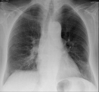
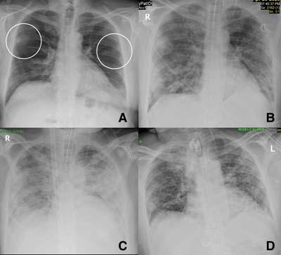
lung opacity within which margins of pulmonary vessels may be difficult to see14. These are much better seen on Computed Tomography (CT) and are less opaque compared to consolidation (Fig 1). When associated with reticular opacities, the detection becomes easier. Hazy opacities on CXR can also be diffuse making its identification challenging10 (Fig 2). In patients with proven COVID-19, GGO was seen in 20-33% of patients at presentation1113,15. Normal lung parenchyma may mimic areas of GGO in poorly taken films and/or due to overlying soft tissues such as prominent breast tissue13
Consolidation :
Consolidation is seen as an area of homogeneous opacification in the lung parenchyma with obscuration of the vessel and airway walls14. In COVID-19 and other viral pneumonias, there is multi-lobar and often bilateral involvement (Fig 3). This is in contrast to the typical unilateral lobar distribution of bacterial pneumonia16. One of the early studies from China had reported the presence of consolidation in all CXR’s at presentation17. On studies published subsequently, consolidation was found in varying frequency, ranging from 5-80%11-13 15
Distribution :
Classical distribution seen in most of the patients is that of bilateral involvement with lower lobe predominance. Peripheral distribution was more common than central involvement12,13,17 (Fig 4). In a more recent study by Weinstock et al15, lower lobe predominance and peripheral distribution was seen in about 35% of patients but bilateral involvement was only seen in 21% of cases. Diffuse distribution of lung opacities can also be seen as the disease progresses. The appearances are similar to Acute Respiratory Disease Syndrome (ARDS) patterns10 (Fig 5).
Atypical Findings :
Interstitial pattern of distribution has been reported apart from GGO and consolidation15. Pleural involvement is an atypical finding with pneumothorax and pleural effusions reported in some selective cases especially during disease progression/prolonged admission. Assisted ventilation related pathologies such as pneumomediastinum have also been reported18,19 (Fig 6). Nodular lesions have also been described and more easily recognized on CT13 (Fig 7).
Learning Points :
Ground glass opacification and consolidation are the most common findings on CXR of patients with COVID-19.
Bilateral involvement with lower lobe predominance and peripheral distribution is most likely.
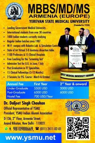
Fig 1 — CXR (A) and CT (B) images of a 45-year-old male who presented with fever and cough. He had hypoxia and leukopenia on examination and his nasal swab was positive for SARS COV-2. CXR shows bilateral blurred opacities with unclear vascular margins (white arrow) with corresponding ground glass changes in the CT (black arrows). Images reproduced with permission from Covid-19 Database of the SocietaItaliana di Radiologia Medica e Interventistica.
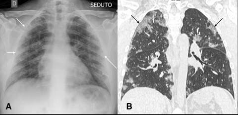
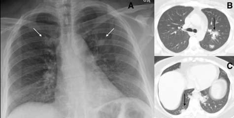
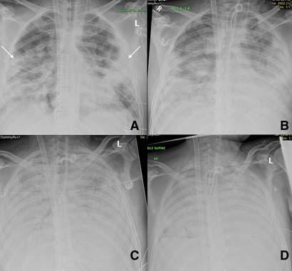
Fig 2 — CXR (A) and CT (B& C) images of a 50-year-old man with 6 days history of fever and dry cough. RT-PCR test was positive. CXR shows bilateral diffuse opacities, with a more opaque patch in the right lower zone (white arrow). The corresponding CT shows the true extent of the disease (black arrows). Images reproduced with permission from Covid-19 Database of the Fleischner Society.
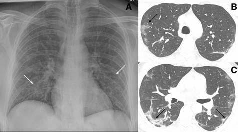
Fig 3 — CXR images from two different patients with COVID-19 showing peripheral areas of consolidation bilaterally in A and unilaterally in B (arrows). Images reproduced with permission from Covid-19 Database of the Societa Italiana di Radiologia Medica e Interventistica.
Pleural involvement at the time of presentation is not common.
Role of CXR in COVID-19
CXR in Diagnosis and Screening for COVID-19:
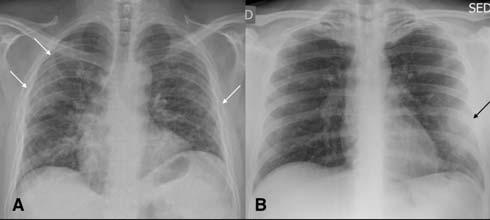
4 — CXR of a 71-year-old man with 4 days history of shortness of breath. Classical features of hazy opacities are seen in the lower lobes bilaterally in a peripheral distribution. Image reproduced with permission from Covid-19 Database of the Fleischner Society.
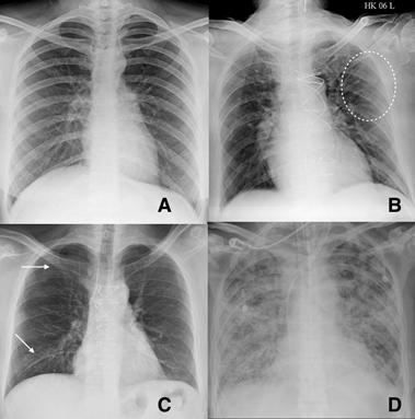
Fig 5 — CXR’s of different patients with proven COVID-19 demonstrating varied appearances at the time of presentation. ANo abnormalities could be seen on CXR and the corresponding CT (not shown) was also near normal. B- Ill-defined hazy peripheral opacities seen in the left upper zone. C- Multifocal opacities were seen in the right lung on CXR at presentation. D- CXR showing extensive parenchymal infiltrates in a patient who came to the hospital in very bad respiratory distress and was found to have COVID-19 on testing.
Two of the very early reports from China and Hongkong had shown high sensitivity of CXR abnormalities in patients testing positive for COVID-1911,17. Wong et al12 , showed a sensitivity of 69% of CXR compared to 91% of RT-PCR with CXR abnormalities preceding positive RTPCR testing in 9% of patients. With these results, it was proposed to consider CXR as a screening tool especially due to limited availability and sensitivity of RT-PCR testing12. The same performance of the CXR, however, could not be replicated as the disease spread wider and more continents were involved. A recent study from New York City looked at 636 patients (confirmed and
Fig 6 — Serial CXR’s of a patient with COVID-19 showing development of atypical findings during the admission. The admission radiograph (A) demonstrates multifocal peripheral opacities (white arrow), followed by the development of right pneumothorax (black arrow) on day 7 (B) of admission with improvement in parenchymal changes subsequently on day 15 of admission (C). He developed extensive left pneumothorax (black arrow) and surgical emphysema (star)(D)of the chest wall later in the course. Images courtesy of Dr Amrita Bajaj, Glenfield Hospital, Leicester.
Fig 7 — Atypical presentation of COVID-19 in the form of nodules (arrows) seen on the CXR (A) and the corresponding CT (B). Image reproduced with permission from Covid-19 Database of the Fleischner Society.
symptomatic COVID-19) presenting to urgent care. They found a normal CXR in 58.3% patients and up to 89% of patients had normal to near normal CXR15. A similar finding was also seen in a study published from Korea15. The described CXR findings are not specific for COVID-19 and may also be seen in other viral pneumonias such as SARS and MERS. Many GGO and consolidative changes visible on CT may not be seen on CXR making it a less sensitive technique 11 .
Learning Points :
CXR can be normal or near-normal in a large number of patients with COVID-19 and hence will not be a reliable test for diagnosis or screening.
Fig
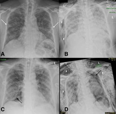
Fig 8 — Serial CXR examinations in a patient showing disease progression. Presentation (A) film had bilateral peripheral hazy opacities that increased on day 7 (B) and became confluent. Further worsening of parenchymal changes on day 11 with the patient requiring extracorporeal membrane oxygenation (ECMO) support (C) with improvement in clinical condition and persistent parenchymal fibrotic infiltrates on day 23 of admission (D). Images courtesy of Dr Amrita Bajaj, Glenfield Hospital, Leicester.
CXR abnormality can precede RT-PCR positivity. Patients with abnormal CXR and high suspicion for COVID-19 should undergo repeat RT-PCR testing.
CXR in assessing severity of COVID-19:
Imaging can play a vital role in assessing the severity of COVID-19 patients. To assess the extent of disease involvement, a simplistic radiographic scoring system was used by Wong et al12. Each lung was graded from 0-4 based on the extent of involvement (0- no involvement, 1- up to 25%, 2- 25-50%, 3- 50-75% and 4 >75% involvement). The scores of each lung were added to get a final score. The severity score of CXR varied over the time and peak severity was seen at 10-12 days from symptom onset (Fig 8). As the disease progresses the GGO are replaced by areas of consolidation that either resolves or worsens to give ARDS picture11 (Fig 9). Various CT severity scores have shown good correlation with clinical severity of disease20 21. The degree of lung inflation at the initial CT can also predict adverse outcomes in patients with COVID-1922
Learning Points :
CXR findings are at its worst at 10-12 days from symptom onset.
Simple CXR severity scoring can be used to assess the progression of disease.
CXR for Disease progression/ Discharge decision :
Can CXR be used to decide when to discharge the
Fig 9 — Serial CXR examinations in a patient who succumbed to the infection. This patient came in with severe respiratory distress and was put on ventilator support early in his admission. ECMO therapy was also started (A) with bilateral parenchymal consolidation. Patient kept deteriorating on ECMO (Day 13- B, Day 18 C and Day 27- D) and succumbed to the disease. Images courtesy of Dr Amrita Bajaj, Glenfield Hospital, Leicester.
patient? No, there is no clear evidence to support this. In the study by Wong et al, there was no statistical difference between the time taken for radiographic and virologic recovery12. About 42% of patients had shown recovery in CT findings before RT-PCR test getting negative while the remainder either showed worsening of findings or showed improvement after RT-PCR became negative7
Learning Points
:
CXR resolution cannot be used to decide the time to discharge.
Guidelines on Use of CXR in COVID-19 :
Multiple national and international societies have proposed guidelines on the use of different imaging modalities in the diagnosis and management of COVID19 6,23-25. Some of these have also taken into account resource constraints in their guidelines 9. None of the recommendations support the use of CXR for the diagnosis or screening of the patients. Imaging is recommended in patients with proven COVID-19 and worsening clinical features or patients with moderate to severe disease at presentation. Routine serial follow-up CXR is not recommended. In areas where RT-PCR testing is not available, imaging (either CXR/CT) can be utilized in medical triage of patients with suspected COVID-19 with moderate to severe features and high pre-test probability9. Most of the guidelines also recommend the use of dedicated portable/mobile equipment for performance of CXR. Specific reporting guidelines have also been proposed to
encourage structured reporting of findings, which will help in the assessment of disease severity and also in research studies.
Learning Points :
CXR imaging should not be used for screening purposes.
CXR should be used in patients with COVID-19 and worsening clinical condition.
In areas with lack of RT-PCR testing, imaging can be utilized for medical triage of patients with moderate to severe features and high pre-test probability of COVID-19.
Machine Learning/Artificial Intelligence in CXR :
There have been significant advances in the field of machine learning/artificial intelligence (ML/AI) in the field of imaging. Many commercially available products have been utilized in the interpretation of CXR and are effective, especially for tuberculosis (TB)26. Interpretation of CXR can be subjective, especially when there are subtle abnormalities. There are also resource constraints in the developing world with regards to the availability of radiologists around the clock 27 . ML/AI based CXR reporting may provide a viable solution which can interpret CXR accurately, quickly and round the clock. One of the ML/AI models achieved similar accuracy of 6 independent radiologists in detected COVID-19 related changes on a CXR with a sensitivity of 85%28. Many other products are available commercially and some of them are indigenously built in India.
Learning Points :
Artificial Intelligence-based CXR interpretation can help in early and accurate detection of COVID-19.
Practical Aspects :
Infection Control :
The SARS CoV-2 is a highly contagious virus and transmission via droplets and contaminated surfaces in radiology departments is known29. This was one of the reasons for not utilizing imaging in screening/diagnosis of COVID-19 patients. Patient care should not be compromised while maintaining staff safety 5 . Every imaging department should have a thorough standard operating procedure (SOP). Continued education and regular training should be provided to the staff regarding social distancing, hand hygiene and use of personal protective equipment (PPE). Wherever possible, portable radiographic equipment should be used to limit disease
transmission. If possible, radiographic equipment should be dedicated to isolation units/wards and should be stationed within the ward 29. Spontaneously breathing patients should wear a mask. When imaging proven patients or patients with suspected COVID-19, radiology technologists should use PPE according to their institution policy. A facemask, face shield, gloves, head-cover and a disposable isolation gown are standard recommendations5 29. Equipment should be thoroughly cleaned, with water and manufacturer-approved detergent, after each patient. Fumigation and ultraviolet rays are also other ways of cleaning the equipment post use.
Learning Points :
Dedicated portable equipment should be utilized whenever possible.
All radiology technologists, while performing CXR examination, should use appropriate PPE.
Equipment should be sanitized between two examinations.
Quality Control :
CXR abnormalities may be subtle and not easily recognizable. It is important to get the best quality images with appropriate exposure parameters and good inspiration. Computed Radiography (CR) is superior to conventional radiography in image quality and reducespatient’s radiation exposure30. Digital Radiography (DR) systems are faster and allow immediate visualization of the image at the bedside. This has a great advantage in the isolation wards as the equipment does not have to leave the ward and the images can be directly loaded into the hospital PACS (picture archiving and communication system) wirelessly. Physicians can also see the CXR images straightaway in their mobile phones/computers as per the institutional setup.
Learning Points :
CXR should be of high quality to detect subtle findings.
DR (digital radiography) technology is faster and better compared to conventional radiography.
Conclusion :
CXR is aneasily available and cost-effective imaging modality in the assessment of chest pathologies. COVID19 predominantly presents with lower respiratory tract infection-related symptoms. CXR is not sensitive in diagnosis/screening for COVID-19 and can be normal at the time of presentation. In areas with limited access to RTPCR testing, CXR/CT imaging can be utilized for medical triage of patients with moderate to severe features and
high pre-test probability of COVID-19. CXR can be utilized in assessing disease severity and monitoring its progress. Ground glass opacity and/or consolidation in a peripheral distribution with lower lobe/bilateral involvement are commonly seen. Portable bedside examination is recommended to restrict disease spread. Robust infection control and quality control policies should be set up and followed to ensure staff and patient safety.
Funding : None
Conflict of Interest : None
REFERENCES
1World Health Organisation — WHO Timeline- COVID -19 2020 [Available from: https://www.who.int/news-room/detail/ 27-04-2020-who-timeline—covid-19 accessed 12 May 2020.
3World Health Organisation — WHO Coronavirus Disease (COVID-19) Dashboard 2020 [Available from: https:// covid19.who.int/ accessed 12 May 2020.
4Ministry of Health and Family Welfare — COVID-19 India 2020 [Available from: https://www.mohfw.gov.in/ accessed 12 May 2020.
5Nasir MU, Roberts J, Muller NL — The Role of Emergency Radiology in COVID-19: From Preparedness to Diagnosis. Can Assoc Radiol J 2020:846537120916419. doi: 10.1177/ 0846537120916419
6Rodrigues JCL, Hare SS, Edey A — An update on COVID-19 for the radiologist - A British society of Thoracic Imaging statement. Clinical Radiology 2020;75(5):323-25. doi: 10.1016/j.crad.2020.03.003
7Ai T, Yang Z, Hou H — Correlation of Chest CT and RT-PCR Testing in Coronavirus Disease 2019 (COVID-19) in China: A Report of 1014 Cases. Radiology 2020:200642. doi: 10.1148/radiol.2020200642
8Li Y, Yao L, Li J — Stability issues of RT-PCR testing of SARS-CoV-2 for hospitalized patients clinically diagnosed with COVID-19. J Med Virol 2020 doi: 10.1002/jmv.25786
9Rubin GD, Ryerson CJ, Haramati LB — The Role of Chest Imaging in Patient Management during the COVID-19 Pandemic: A Multinational Consensus Statement from the Fleischner Society. Radiology;0(0):201365. doi: 10.1148/ radiol.2020201365
10Jacobi A, Chung M, Bernheim A — Portable chest X-ray in coronavirus disease-19 (COVID-19): A pictorial review. Clin Imaging 2020;64:35-42. doi: 10.1016/j.clinimag.2020.04.001
11Ng M-Y, Lee EY, Yang J — Imaging Profile of the COVID-19 Infection: Radiologic Findings and Literature Review. Radiology: Cardiothoracic Imaging 2020;2(1):e200034. doi: 10.1148/ryct.2020200034
12Wong HYF, Lam HYS, Fong AH — Frequency and Distribution of Chest Radiographic Findings in COVID-19 Positive Patients. Radiology 2019:201160. doi: 10.1148/radiol.2020201160
13Yoon SH, Lee KH, Kim JY — Chest Radiographic and CT Findings of the 2019 Novel Coronavirus Disease (COVID19): Analysis of Nine Patients Treated in Korea. Korean J Radiol 2020;21(4):494-500. doi: 10.3348/kjr.2020.0132
14Hansell DM, Bankier AA, MacMahon H — Fleischner Society: glossary of terms for thoracic imaging. Radiology
15Weinstock MB, Echenique A, Russell JW —l. Chest X-Ray Fidnings in 636 Ambulatory Patients with COVID-19 Presenting to an Urgent Care Center: A Normal Chest X-Ray Is no Guarantee. The Journal of Urgent Care Medicine 2020:13-18. [published Online First: 13 April 2020]
16Vilar J, Domingo ML, Soto C — Radiology of bacterial pneumonia. Eur J Radiol 2004;51(2):102-13. doi: 10.1016/ j.ejrad.2004.03.010
17Chen N, Zhou M, Dong X — Epidemiological and clinical characteristics of 99 cases of 2019 novel coronavirus pneumonia in Wuhan, China: a descriptive study. The Lancet 2020;395(10223):507-13. doi: 10.1016/s01406736(20)30211-7
18Salehi S, Abedi A, Balakrishnan S — Coronavirus Disease 2019 (COVID-19): A Systematic Review of Imaging Findings in 919 Patients. AJRAmJRoentgenol 2020:1-7. doi: 10.2214/ AJR.20.23034
19Sun R, Liu H, Wang X — Mediastinal Emphysema, Giant Bulla, and Pneumothorax Developed during the Course of COVID-19 Pneumonia. Korean J Radiol 2020;21(5):541-44. doi: 10.3348/kjr.2020.0180
20Lyu P, Liu X, Zhang R — The performance of chest CT in evaluating the clinical severity of COVID-19 pneumonia: identifying critical cases based on CT characteristics. Invest Radiol 2020 doi: 10.1097/RLI.0000000000000689
21Zhao W, Zhong Z, Xie X — Relation Between Chest CT Findings and Clinical Conditions of Coronavirus Disease (COVID-19) Pneumonia: A Multicenter Study. AJR Am J Roentgenol 2020;214(5):1072-77. doi: 10.2214/AJR.20.22976
22Colombi D, Bodini FC, Petrini M — Well-aerated Lung on Admitting Chest CT to Predict Adverse Outcome in COVID19 Pneumonia. Radiology 2020:201433. doi: 10.1148/ radiol.2020201433
23ACR Recommendations for the use of Chest Radiography and Computed Tomography (CT) for Suspected COVID-19 Infection, 2020.
24ACEP COVID-19 Field Guide- CXR and CT Imaging, 2020. 25Indian College of Radiology and Imaging — Imaging recommendations for COVID-19 patients. In: Chest Radiology Subspecialty Group, ed., 2020.
26Qin ZZ, Sander MS, Rai B — Using artificial intelligence to read chest radiographs for tuberculosis detection: A multisite evaluation of the diagnostic accuracy of three deep learning systems. Sci Rep 2019;9(1):15000. doi: 10.1038/ s41598-019-51503-3
27Mollura DJ, Culp MP, Lungren MP — Radiology in Global Health: Strategies, Implementation, and Applications: Springer 2018.
28Murphy K, Smits H, Knoops AJG — COVID-19 on the Chest Radiograph: A Multi-Reader Evaluation of an AI System. Radiology 2020:201874. doi: 10.1148/radiol.2020201874
29Kooraki S, Hosseiny M, Myers L — Coronavirus (COVID19) Outbreak: What the Department of Radiology Should Know. Journal of the American College of Radiology 2020;17(4):447-51. doi: 10.1016/j.jacr.2020.02.008
30Ozcete E, Boydak B, Ersel M — Comparison of Conventional Radiography and Digital Computerized Radiography in Patients Presenting to Emergency Department. TurkJEmerg Med 2015;15(1):8-12. doi: 10.5505/1304.7361.2014.90922
Review Article Review Article Review
Virology and Pathogenesis of COVID-19
Ashis Kumar Saha1, Goutam Biswas2
After the discovery of human coronavirus from the samples of human respiratory tract in 1960 by Dr June Almeida several years elapsed before epidemics occurred in China in 2002-2003 as SARSCoV and epidemics in Middle East countries in 2012-2014 as MERS-CoV. But recently in December, 2019 in Wuhan in China the novel coronavirus started its journey and ultimately spread worldwide to involve millions of people and took the life of more than 1.25 lakh of affected patients. There are recurrent antigenic changes in this virus, SARS-CoV-2, which has to be determined by the scientists all over the world to discover the definite medicine as well as vaccines for prevention.
[J Indian Med Assoc 2020; 118(5): 20-5]
Key words : MERS-CoV, SARS-CoV-2, Cytokines.
Discovery of Human Coronavirus :
History of human coronavirus,started in 1960 when Tyrrell and Bynoe found a virus in embryonic tracheal organ culture received from adult respiratory tract of a patient in the cold unit in Salisbury in Wiltshire. They sent several samples to virologist, June Almeida, who demonstrated the particles under electron microscope. She also saw this type of particle while investigating mouse hepatitis. She wrote a research paper but was rejected by one peerreviewed journal. In 1965, British Medical Journal published the new discovery of thevirus B814. The photograph of this B814 particle was exactly like that what Dr. Almeida demonstrated previously and ultimately her article was accepted and published two years later. Now she is no more (died in 2007 at the age of 77 years) but corona virus remains and responsible for this huge pandemic.
Taxonomy and description of onset of pandemic of COVID-19 :
Order Nidovirales has four families, namely Coronaviridae, Arteriviridae, Roniviridae. Coronaviridae, largest of all the above families has two sub-families— Coronavirinae and Torovirinae, former one is subdivided into four sub-groups – alpha, beta, gamma and delta coronaviruses. These viruses are divided according to the phylogenetic clustering. Coronaviruses are the main pathogen of human being and vertebrates, like birds, bats, mouse and many other wild animals attacking respiratory, gastrointestinal, nervous and hepato-biliary systems1,2,3 Since the primary reservoir of COVID-19 is bats, ICMR
1MD (Medicine) DTM&H (Cal), MNAMS (Ind), FRCP (Edin), FRCP (Glasgow), FACP (USA), FICP, Professor & Head, Mata Gujri Memorial Medical College, Kishanganj, Bihar
2MBBS, Post graduate trainee, Department of General Medicine, RG Kar Medical College and Hospital, Kolkata
Received on : 14/04/2020
Accepted on : 11/05/2020
Editor's Comment :
SARS-CoV-2 has a complex protein structure that helps in entry, incorporation into host cell and replication. Clinical outcome depends on cytokine activation, immune evasion and coagulopathy
Knowledge of structure and pathogenesis of SARS-CoV2 infection will help in devising therapy and preventive measures.
started to gather evidence of any presence of virus from different types of Indian Bats. Very recently ICMR reported there is presence of COVID-19 in two types of bat, one is Pteropas (Indian Flying Foxes and the other is Rousettus (Fruit Bats) collected from different regions of India. They have tested for COVID-19 in 508 flying foxes and 78 Rusetus and recovered the viruses from 21 flying foxes and 4 Rusetus.
The primary target of coronaviruses is respiratory system of human being. Almost 50 years ago coronaviruses started producing mild respiratory symptoms by the four coronaviruses. HCoV-229E and HCoV-NL63 are alpha-coronaviruses and HCoV-OC43 and HCoV-HKU1 are beta-coronaviruses responsible for producing respiratory symptoms. HCoV-229E and HCoDOC43 were isolated 50 years ago but the other two were identified in recent coronaviruses outbreak4,5,6,7,8. In 20032004 in Guandong province of China, a virus, SARS-CoV, was isolated from patients with severe respiratory tract infection, i.e. group 2b beta-coronavirus. It was responsible for 8098 cases with death of 774 having higher mortality rate of about 50% above 60 years of age and loss of 40 billion dollar activity. It started in a hotel in China and ultimately spread into two dozen of countries. During that time this SARS-CoV was originated in bats and Chinese horse shoe bats9
Again in 2012, another coronavirus was isolated from patients of Middle-East including Saudi Arabia and other
countries, who suffered from severe respiratory tract infections with mortality of nearly 50% at early stage—it was known as Middle-East respiratory syndrome Virus or MERS-CoV10. Though this outbreak decelerated in 2013 but again a small peak occurred in 2014 which gave rise to 200 cases with death of 40 patients – this resulted from seasonal increase in birth of camel, improved detection methods as well as good reporting. MERS-CoV is group 2c beta-coronavirus believed to be originated form bats but also camels in middle East as viral antibodies was detected in these animals11
Ultimately in last week of December, 2019, patients wereadmitted in hospitals with symptoms of respiratory tract infections of unknown etiology12. These patients were directly related to wet animal wholesale market in Wuhan, in the province of Hubei, China. On the same day International virus classification declared that the name of the new virus as Severe Acute Respiratory Syndrome Virus 2 (SARS-CoV-2)13. Within 18th to 29th December, 2019, total 5 patients were admitted with same infection and one of them died14. Again, according to a report, by 2nd January 41 patients admitted in hospitals with confirmed SARSCoV-2 positive respiratory tract infections half of them having comorbidities, like, diabetes mellitus, hypertension, cardiac diseases helped to come to a conclusion that these patients may be infected by nosocomial infection by unknown mechanism during hospital stay in various locations throughout the hospital rather than in a single hall15. It should be remembered also that during that time those who were clinically infected were tested but not mildly symptomatic or asymptomatic patients. Till 20th January, 2020,291 clinically and sequence analysis proved cases were recovered of which 270 were from Wuhan and rest 21 from Beijing, Shanghai and Guangdong. In addition four more cases were confirmed of which one from South Korea, one from Japan and rest two from Thailand, but all these patients went as visitor in Wuhan 2 weeks back. By 22nd January, 2020, 571 more cases were recovered from 25 Provinces covering districts and cities of China16. First 17 deaths were reported in detail by China National Health Commission, some of them had some comorbidities, like, cardiovascular diseases, renal dysfunction, liver disease and abdominal tumor. By 25th January, 2020 total confirmed cases were 1975 with total death of 56, where as in another report on 24th January, 2020total COVID -19 positive cases were 5502 17,18. Ultimately it spilled over the several countries worldwide to reach a recent pandemic stage. As per report of 30th January, 2020 total case cases from china was 7734 and from other countries, worldwide , 90 cases were recovered as COVID-19 positive with case fatality rate of 2.2%19
After recovery of the first case from United States,
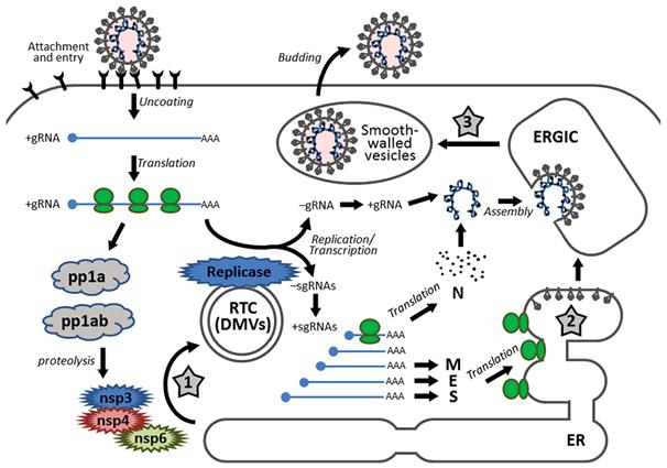
proper description of the illness came across, which was characterised by mild presenting symptoms, like, cough, fever followed by progression to pneumonia within 9 days of illness20. On 30th January, 2020 first case of human to human transmission was identified in United States. According to a report of 7th February, 2020, in Nature Journal total infected patients in China was 31161 with death of more than 630 (http://www.nature.com/articles/ d41586-020-00154). In 11th February, 2020 World Health Organization gave the new name of this corona virus as COVID-19 (Fig 1).
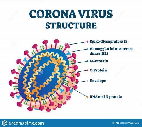
Structure :
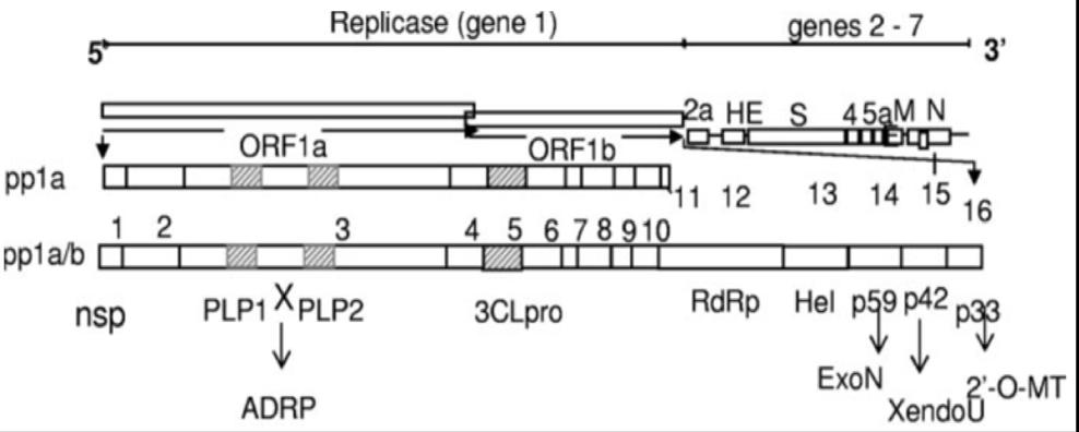
This virus is non-segmented positive sense single stranded RNA of 30 kb containing 5xþ cap structure and 3xþ poly tail. It has ten open reading frames; out of which first frame (ORF 1a/1b) contains two third of viral RNA of 20 kb which will be translated into two polyproteins, pp1a and pp1ab by the method of -1 frame shift between ORF1 and ORF2 which will be processed into 16 non-structural proteins (nsp1 – 16) leading to formation of replicase transcriptase complex21,22. These non-structural proteins rearrange the membrane starting from rough endoplasmic reticulum into double membrane vesicles23. Since the length of RNA is small as compared to DNA viruses hence the replication and mutation rate of the former is much higher. But human coronavirus being largest RNA virus (30 kb in length) maintains this genomic structure due to presence of unique RNA processing enzymes, like, 3xþ-5xþ exoribonuclease of non-structural protein 14 which provides proof reading function of replicase-transcriptase complex24 (Fig 2).
The main functions of nonstructural proteins are degradation of cellular RNA,inhibition of interferon signalling, cleaving of polypeptideand blocking of host innate immune response. They promote expression of cytokines and formation of double membrane vesicles25,26
There are four structural proteins. These proteins serve many functions. These are the following (Fig 3) :
(A)Spike protein(S) : These proteins are responsible for attachment to the host receptors.
(B)Membrane (M) protein :
1.It will give shape to the virions
2.It promotes the curvature of membrane of the virus.
3.It will bind to nucleocapsid.
(C)Envelope (E) protein :
1.It helps in assembling of the virus.
2.It will help in release of virus.
3.It will take part in pathogenesis.
(D)Nucleocapsid (N) protein: It has two domain which binds viral RNA genome through different mechanisms.
1.It can bind to nsp3 protein to help tether the genome
to replication-transcription complex.
2.It helps in encapsulating the genome into the virions.
3.It acts as antagonist of interferon as well as viral encoded repressor of RNA interference – it is beneficial for viral replication (Fig 3).
Pathogenesis of COVID-19 :
Entry of coronavirus and its replication:
Spike protein (S) is responsible for attachment to the host cell receptor27 that is the ACE2 receptor for SARSCoV, SARS-CoV-2 (COVID-19).
After entry of the virus there will be fusion between virus and plasma membrane followed by viral infectivity due the occurrence of a proteolytic cleavage at position 2xþof S protein 28,29. There is another process of entry of SARS-CoV2 through clathrin-dependent as well as clathrin-
Fig 1
Fig 2
independent endocytosis30,31. After gaining entry into the cells viral RNA is released in to the cell cytoplasm which will be translated into two polyproteins as well as structural proteins followed by viral genome replication32. Then newly formed envelope glycoproteins are inserted into endoplasmic reticulum and golgi apparatus and genomic RNA and nucleocapsid protein are combined to form nucleocapsid. Then small viral particle will germinate into the endoplasmic reticulum-golgi intermediate compartment and small vesicle containing viral particles will be formed. Lastly this vesicle will fuse with the plasma membrane followed by the release of full-blown virus into the circulation.
Presentation of Antigen in COVID-19
Infection :
After entry viral antigenic peptides will be presented to antigen presentation cells by major histocompatibility complex or human leukocyte antigen which will be subsequently recognized by virus-specific cytotoxic T lymphocytes, hence antigen presentation is of prime importance in pathogenesis as well as development of viral specific immunity. In case of SARS-CoV MHC I and to some extent MHC II are responsible for antigen presentation 33,34 . Again, genetic polymorphism of mannose binding lectin (MBL) are also related to risk of SARS-CoV infection. But there is no specific information regarding pathogenesis of COVID-19.
Different Types of Immunity :
As a result of antigen presentation T and B cells are stimulated leading to development of cellular as well as humoral immunity. Like other viral infection SARS-CoV develops IgM of acute phase response and IgG antibody
corresponding to chronic phase response. IgM develops within 5 to 7 days and persists for another 5 to 7 days followed by disappearance. On the other hand T and N protein SARS-CoV specific IgG antibodies persist for years which has protective role35,36. But as compared to humoral immunity cellular immunity is greatly depressed in SARSCoV-2 positive individuals as evidenced by severely decreased in number of CD4+ T and CD8+ T cells in acute phase response but its status is excessive activation as evidenced by high proportion of HLA-DR and CD38 double-positive fractions37
But there is increase in neutrophil count along with neutrophil/lymphocyte ration will be increased indicating severe form of disease with poor outcome38,39. In addition, in COVID-19 patients exhaustion markers, like, NKG2A present on cytotoxic T lymphocytes, natural killer cells, CD8+ T cells are up-regulated but on the other hand in convalescent or recovered patients thesecells will be normalized along with detection of SARS- specific antibodies in the blood.
In case of COVID-19 patients there are two phases of immune responses. In the incubation period i.e. in the nonsevere stage an adaptive response is required to prevent progression into the severe stage. So boosting of immune response by several means, like, pegylated interferon or anti-sera are required along with good health and good genetic background. But if the protective response is impaired COVID-19 virus will propagate, invade into different tissues mainly affecting those having high ACE2 receptors, like, intestine, kidney and destroy them. Damaged tissue produces innate inflammatory response mediated by inflammatory macrophages as well as granulocytes leading to severe respiratory disorder in severe stage. After discharge from the hospital some patients are unable to eliminate the Virus-eliminating immune response of SARS-CoV-2 from the body and in these patients, vaccine will not work as the immune system is probably very weak in these patients. Already recovered patients from the early non severe stage should be monitored for T/B cell response. (40,41)
Cytokine Response in COVID-19 :
In early stage of outbreak, amongst 41patients with COVID-19 six patients died of acute respiratory distress syndrome. The most common immunopathological event is cytokine storm, the uncontrolled systemic inflammatory response releasing large amount of pro-inflammatory cytokines, like, interferon-α, interferon-, interleukin-1β , interleukin-6, interleukin-12, interleukin-18, interferon-33, tumor necrosis factor- α , tumor growth factor- β and chemokines, like, CCL2, 3, 5, CXCL8, 9, 10 etc by effector immune cells in COVID-19 infection This storm ultimately triggers the immune system of the body to attack different
Fig 3
organ systems leading to multi-organ failure followed by death in COVID-19 infection as occurred in case of SARSCoV and MERS-CoV epidemic. Cytokine release syndrome in severe patients with leucocytosis with lymphopenia is mediated by leukocytes other than T cells.42
Immune Evasion by Coronavirus :
Like SARS-CoV or MERS-CoV, COVID-19 avoids immune response. Pattern recognition receptors (PPRs) recognize pathogen-associated molecular pattern, evolutionarily conserved microbial structure. But SARSCoV, MERS-CoV and COVID-19 are bound by doublemembrane vesicle thus host immune cells cannot detect microbial dsRNA .Interferon á and â are protective in coronavirus infection. But by the following methods coronavirus SARS-CoV, MERS-CoV prevent interferon from preventive actions:
(A)Accessary protein 4a blocks the induction of interferon in MERS-CoV infection at the level of MDA5 through direct interaction with double stranded RNA.
(B)Accessary proteins, like, 4a, 4b, ORF5, membrane protein of MERS-CoVprevents activation of interferon â promoter by inhibiting nuclear transport of interferon regulatory factor 3. 42,43
So, destruction of this evasion of immune system is a way by which one can treat COVID-19.
Effect on Coagulation and Heme
Metabolism :
It has been documented that SARS-CoV-2 causes intense epithelial viral cytopathic effects involving alveolar and small airway epithelium with variable number of small fibrinous thrombi in small pulmonary arterioles in areas of damaged and preserved lung parenchyma.Endothelial tumefaction (swelling) and large numbers of pulmonary megakaryocytes in pulmonary capillaries due to activation of coagulation cascade, and small foci of alveolar hemorrhage and pulmonary infarctions are seen. This supports the concept of hypercoagulative status, showing high frequency of pulmonary microthrombosis. The most common pattern of coagulopathy observed in patients hospitalized with COVID-19 is characterized by elevations in fibrinogen and D-dimer levels. This correlates with parallel rise in markers of inflammation (e.g. CRP). Unlike the pattern seen in classic DIC from bacterial sepsis or trauma, the degree of aPTT elevation is often less than PT elevation (likely due to increased factor VIII levels), the thrombocytopenia is mild (platelet count ~100 x109/L), and microangiopathy is not present.Some patients with severe COVID-19 infection can develop a coagulopathy meeting criteria for DIC per ISTH criteria with fulminant activation of coagulation and consumption of coagulation factors44.
Moreover ,ORF8 protein and surface glycoprotein of the virus bind to porphyrin respectively and Orf1 ab, ORF10, and ORF3a proteins attack the heme on the 1-beta chain of hemoglobin to dissociate the iron to form porphyrin. This reduces hemoglobin’s ability to carry oxygen and carbon dioxide.O2 dissociation curve shifted to right -> release of O2. But this hypothesis has been challenged on the grounds that RBCs have no DNA and it is unclear how SARS-CoV-2 would enter RBCs45.
Conclusion :
To conclude, knowledge about the structure and function of the virus as well as its complex interaction with host will hopefully help us to device new therapeutic and preventive strategies in the future.
Funding : None
Conflict of Interest : None
REFERENCES
1Wang LF, Shi Z, Zhang S, Field H, Daszak P, Eaton B — Review of bats and SARS. Emerg Infect Dis 2006; 12(12):1834-1840.
2Ge XY, Li JL, Yang XL — Isolation and characterization of a bat-SARS like coronavirus that uses the ACE2 receptor. Nature 2013; 503(7477): 535-538.
3Chen Y, Guo D — Molecular mechanisms of coronavirus RNA capping and methylation. 2016; 31(1):3-11.
4McIntosh K, Beaker WB, Chanock RM — Growth in sucklingmouse brain of “IBV-like” viruses from patients with upper respiratory tract disease. Proceedings of the National Academy of Sciences of the United States of America 1967; 85:2268-2273.
5Bradburne AF, Bynoe ML, Tyrell DAJ. Effects of a “new” human respiratory virus in volunteers. Br Med J 1967; 3:767769.
6Hamre D, Prockknow JJ — A new virus isolated from the human respiratory tract. Proceedings of the Society for Experimental Biology and Medicine Society for Experimental Biology and Medicine 1966; 121(1):190-193.
7Van der Hoek, Pyrc K, Jebbink MF, Vermeulen-Oost W, Berkhout RJ, Wolthers KC, et al — dentification of a new coronavirus. Nat Med 2004; 10(4):368-373.
8Woo PC, Lau SK, Chu CM, Chan KH, Tsoi HW, Huang Y, et al Characterization and complete genome sequence of a novel coronavirus, coronavirus HKU1, from patients with pneumonia. J of Virol 2005; 79(2):884-895.
9Lau SK, Woo PC, Li KS, Huang Y, Tsoi HW, Wong BH, et al Severe acute respiratory syndrome coronavirus like virus in Chinese horse shoe bats. Proceedings of the National Academy of Sciences of the United States of America. 2005; 102(39):14040-14045.
10Zaki AM, van Boheemen S, Bestebroer TM, Osterhaus AD, Fouchier RA — Isolation of a novel coronavirus from a man with pneumonia in Saudi Arabia. The New Eng J of Med. 2012; 367(19):1814-1820.
11Van Boheemen S, de Graaf M, Lauber C, Bestebroer TM, Raj VS, Zaki AM, et al — Genomic characterization of a newly discovered coronavirus associated with acute
JOURNAL OF THE INDIAN MEDICAL ASSOCIATION, VOL 118, NO 05, MAY 2020
respiratory distress syndrome in humans. mBio. 2012; 3(6).
12Bogoch A, Watts A, Thomas-Bachli C, Huber MUG, Kraemer L, Khan — Pneumonia of unknown etiology in Wuhan, China: potential for international spread via commertial air-travel. J Trav Med. 2020; https://doi.org/10.1093/jtm/tasa008.
13Zhao S, Lin Q, Ran J, Musa SS, Yang G, Wang W, et al — Preliminary estimation of the basic reproduction number of novel coronavirus (2019-nCoV) in China, from 2019 to 2020: a data-driven analysis in the early phase of the outbreak. Int J Infect. Dis. IJID: Off. Publ. Int. Soc. Infect. Dis. 2020; 92(214217.
14Ren LL, Wang YM, Wu ZC, Xiang L, Guo L, Xu T, et al — Manifestation of a nevel coronavirus causing severe pneumonia in human: a descriptive study. Chinese Med J. 2020.
15Huang C, Wang Y, Li X, Ren L, Zhao J, Hu Y, et al — Clinical features of patients infected with 2019 novel coronavirus in Wuhan, China. Lancet.2020; 395(10223)
16Lu H — Drug treatment options for the 2019-new coronavirus (2019-nCoV)Biosci. Trends. 2020;
17Wang W, Tang J, Wei F — Updated understanding of the outbreak of 2019 novel coronavirus (2019-nCoV) in Wuhan, China. J Med Virol. 2020; 92(4):441-447.
18Nishiura H, Jung SM, Linton NM, Kinoshita R, Yang Y, Hayashi K, et al — The extent of transmission of novel coronavirus in Wuhan, China, 2020. J Clin Med. 2020; 9.
19Bassetti M. Vena A, Roberto Glacobbe D — The novel Chinese coronavirus (2019-nCoV)infection: challenges for fighting the storm. Eur J Clin Invest. 2020; el13209.
20Holshue ML, DeBolt C, Lindquist S, Lofy KH, Wiesman J, Bruce H, et al — First case of 2019 novel coronavirus in the United States.NEng J Med. 2020;
21Masters PS — The molecular biology of coronaviruses. Adv Virus Res. 2006;66:193-292.
22Ziebuhr J, Snijder EJ, Gorbalenya AE. Virus-encoded proteinases and proteolytic processing in the Nidovirales. J Gen Virol. 2000; 81(Pt 4):853-879.
23Hussain S, Pan J, Chen Y — Identification of novel subgenomic RNA and noncanonical transcription initiation signals of severe acute respiratory syndrome coronavirus. J Virol.2005; 79(9):5288-5295.
24Eckerle LD, Becker MM, Halpin RA — Infidelity of SARS-CoV Nsp14-exonuclease mutant virus replication is replication is revealed by complete genome sequencing. PLOS Pathog. 2010; 6(5):e1000896.
25Tanuka T, Kamitani W, DeDiego ML, Enjuanes L, Matsuura Y— Severe acute respiratory syndrome coronavirus nsp1 facilitates efficient propagation in cells through a specific translational shutoff of host Mrna. J Virol. 2012; 86(20):1112811137.
26Shi P, Su Y, Li R, Liang Z, Dong S, Huang J— PEDV nsp16 negatively regulates innate immunity to promote viral proliferation. Virus Res. 2019; 265:57-66.
27Li W, Moore MJ, Vasilieva N — angiotensin-converting enzyme 2 is a functional receptor for the SARS coronavirus. Nature. 2003; 426:450-454.
28Wu F, Zhao S, Yu B et al. A new coronavirus associated with human respiratory disease in China. Nature. 2020; 29Simmons G, Reeves JD, Rennekamp AJ et al. Characterization of severe acute respiratory syndrome-
mediated viral entry. Proc. Natl. Acad. Sci. USA. 2004; 101:4240-4245.
30Belouzard S, Chu VC, Whittaker GR. Activation of the SARS coronavirus spike protein via sequential proteolytic cleavage at two distinct sites. Proc Natl. Acad. Sci. USA. 2009; 106:5871-5876.
31Wang H, Yang P, Liu K et al. SARS coronavirus entry into host cells through a novel clathrin- and caveolae-independent endocytic pathway. Cell Res. 2008; 18:290-301.
32Perlman S, Netland J — Coronaviruses post-SARS: update on replication and pathogenesis. Nat. Rev. Microbiol. 2009; 7:439-450.
33Liu J, Wu P, Gao F — Novel immunodominant peptide presentation strategy: a featured HLA-A*2402-restricted cytotoxic T-lymphocyte epitope stabilized by intrachain hydrogen bonds from severe acute respiratory syndrome coronavirus nucleocapsid protein. J Virol 2010; 84:1184911857.
34Keicho N, Itoyama S, Kashiwase K — Association of human leukocyte antigen class II alleles with severe acute respiratory syndrome in the Vietnamese population, Hum, Immunol. 2009; 70; 527-59-31.
35Wang SF, Chen KH, Chen M — Human-leukocyte antigen class I Cw1502 and class II DR 0301 genotypes are associated with resistance to severe acute respiratory syndrome (SARS) infection. Viral Immunol. 2011; 24:421426.
36De Wit E, van Doremalen N, Falzarano D — SARS and MERS: recent insight into emerging coronaviruses. Nat Rev. Microbiol. 2016; 14:523-534.
37Li G, Chen X, Xu A — Profile of specific antibodies to the SARS associated coronavirus. N Engl J Med 2003; 349:508509.
38Zhang B — Immune phenotyping based on neutrophil to lymphocyte ration and IgG predict disease severity and outcome for patients with COVID-19. Preprint at medRxiv 2020. https://doi.ogr/10.1101/2020.03.12.20035048
39Chen X — Restoration of leukomonocyte count is associated with viral clearance in COVID-19 hospitalized patients. Preprint at medRxiv. 2020. https://doi.org/10.1101/ 2020.03.03.20030437.
40Fan YY, Huang ZT, Li L et al. Characterization of SARS-CoVspecific memory T cellsfrom recovered individuals 4 years after infection. Arch. Virol. 2009; 154:1093-1099.
41Tang F, Quan Y, Xin ZT — Lack of peripheral memory B cell responses in recovered patients with severe acute respiratory syndrome: a six year follow-up study. J Immunol. 2011;186:7264-7268.
42Zhao J, Li K, Wohlford-Lename C — Rapid generation of a mouse model for Middle East respiratory syndrome. Proc. Natl. Acad. Sci. USA. 2014; 111:4970-4975.
43Qin C — Dysregulation of immune response in patients with COVID-19 in Wuhan, China. Clin. Infect Dis 2020; https:// doi.org/10.1093/cid/ciaa248.
44Thachil J — ISTH Interim Guidance on Recognition and Management of Coagulopathy in COVID-19. Journal of Thrombosis and Haemostasis 2020. [Epub Ahead of Print]
45Beta Chain of Hemoglobin and Captures the Porphyrin to Inhibit Human Heme Metabolism” ChemRxivPrepring 2020 [Epub Ahead of Print
V V V V Voice of the Expert oice of the Expert oice of the Expert oice of the Expert oice of Expert
Professor Roman Jaeschke is the Professor of Medicine and Department of Health Research Method, Evidence and Impact at the McMaster University in Canada. He is actively involved in Critical Care Medicine and is the lead author of the world-famous McMaster Textbook of Medicine. On behalf of JIMA, Dr Rudrajit Paul and Dr TanukaMandal conducted an online interview with Dr Jaeschke about the current coronavirus pandemic in the first week of May, 2020.
Dr Roman, welcome to JIMA, the oldest medical journal in India. On behalf of this journal, we will be asking you a few questions on COVID-19 pandemic. We thank you for your valuable time.
Dr.Roman, we were going through the online McMaster perspective series. It is certainly useful. But there are a few more queries which we would like to discuss with you.
(1)In Belgium, it has been reported that doctors are sometimes doing retrospective diagnosis. For example, if a patient has already died and the doctor is told that he/she had fever and dyspnoea, the case is categorized as coronavirus. Is this approach correct? Or will it falsely increase the mortality figures?
(a)The mortality rate from COVID-19 is not clear. The ‘right’ percentage requires that both numerator and denominator are known and accurate. Yet without widespread testing or serological examination, the number of people who went through the infection is unclear. Same for cause of death: it is likely that some of the deaths categorized in Belgium as ‘COVID-19 related’ were in fact not due to this virus, thus increasing perceived mortality. But, requiring more for diagnosis would miscategorise and miss some of the real cases of COVID-19. Both ways have problems, and both may be manipulated. There are obviously possibilities of geographically different either virus mutation or genetic predisposition. Time will show.
(2)You have said that Remdesivir has very little benefit compared to the cost. So are you using it for your patients? If so, are the insurance people covering its cost?
(a)It was the person I was interviewing who said so. The cost-benefit ratio is in the eyes of person obtaining benefit and incurring cost. Let’s assume the cost of drug is really 4,500 US $ per treatment. Let’s assume that the mortality reduction is in the ‘reported’ range (around 4%). That means that we would need to treat 25 patients to
prevent one from dying. This will translate into about 110,000 US $ per averted death. In the world where some of the diabetic or heart failure medication cost 10,000 $ per year, and some biologic drugs cost 20,000-40,000 $ per year, cost of 110,000$ per life (say, with 1-510-20 more years to live) does not seem out of range. I suspect this one-time cost will be widely accepted if scale of benefit is confirmed.
(3)Smokers are protected from COVID. This is just an observational data. Should smokers be less concerned during the epidemic? If this is so, why is female mortality less than male? Females are usually less likely to be smokers (at least in India). Also is this observation true only for smoking or for any form of tobacco?
(a)I would not pay much attention to this finding. Certainly not enough to start smoking! I understand some trials of nicotine replacement therapy (patches) are being conducted. In the meantime anything else I say is a speculation. Except: smoking kills.
(b)The reason for gender differences is not clear. Possibly related to estrogen level.
(4)Have you any experience with auto proning of patients?
(a)No. We are starting an RCT of doing so. Plenty of experience with proning, which became a norm looking at major gains in oxygenation. As of today (May12) I have seen a report from New York about self proning in emergency department with striking improvement in oxygenation. Something to at least start thinking about, if not doing. Editor’s note :
Professor Roman Jaeschke
(5)Someone mentioned vasoplegia as a pathophysiology of COVID illness. Can you please elaborate?
(a)The lungs are usually very efficient in matching ventilation and perfusion. If part of the lung is not ventilated, vessels auto-regulate (constrict) and blood is diverted away from that region. In COVID-19 it appears this is not the case, and blood continue to circulate through non-ventilated areas resulting in refractory hypoxia.
(6)There were no drugs in the recent two corona epidemics. Will there be anything this time? What does the trend suggest ?
(a)Predicting the future is notoriously difficult. Yes, there will be treatment – the obvious question is when, and how effective it will be (pneumococcus pneumonia kills despite great antibiotics, after all). The rest is still guessing - If I had a free rein, I would like to have an option to use remdesivir and convalescent plasma. I would be happy to use them still in clinical trial. I would prefer platform trial, where my patient would likely get ‘something’.
(7)Hydroxychloroquine Sulfate study in Brazil was stopped due to cardiac side effects. What is the status of other similar trials?
(a)Over 100 trials of HCQS are registered. We need to wait, probably another 4-8 weeks. In the meantime, we are not using antimalarials. Need data convincing of benefit.
(8)How common is sepsis in COVID patients? Is sepsis the main cause of death?
(a)My limited experience tells me that sepsis (as defined currently) occurs in minority of patients. In our hospital about 10% of patients admitted with COVID-19require life support. Those who survive have prolonged and refractory hypoxia with complications of long term ventilation.
Editorial note : Indiscriminate use of antibiotics in Covid-19 patients is not needed.
(9)You have said that false hope generated by the media is often causing the relatives to pressure physicians into using doubtful remedies like steroids. How are you overcoming this situation in your hospital ?
(a)We are in the centre which for years prides itself with rational approach. As health care professional we
support each other and have quite clear pattern of practice. Being on one page with your colleagues is crucial. Being convinced that you would do the same for your relative or want for yourself is helpful. In the end, if you spend enough time explaining that we do all what seems reasonable, people will almost always accept it. It is difficult, though – it is much easier to give ‘something’. But please keep in mind that giving oxygen and fluids and antipyretics is already ‘something’.
(10) How many health care persons are affected in your set up?
(a)We had less than 10 cases in our hospital. That, taking into account that we have 2,000 workers, is not likely excessive. But, we are quite lucky – our hospital has relatively few COVID-19 patients. I have just seen a data which showed that 1 metre physical distancing decreases odds of being infected 5 folds, and adding another meter cuts it in half. Good eye protection is very effective (fold odds decrease) and so are masks.
(11) What is effect of Heparin in COVID? What is Prophylactic or therapeutic dose and duration of useof Heparin?
(a) This is clearly evolving and moving towards higher doses. People are looking for reasons to give more. RCTs awaiting. Some anticoagulate fully if D-Dimer is (markedly) elevated. Personally I am ‘migrating’ towards increasing prophylactic anticoagulation dose by 50%; some of my colleagues advice to double it. Unfortunately, this is still opinion based. But, data will be coming soon.
Editor’s note : In CCU, the care pathway of Covid-19 patients should include heparin prophylactically.
(12) What is the use of CT pulmonary angiogram in COVID-19?
(a)We are really trying to limit transfer of patients. I would send person to CT almost exclusively to rule out large PE. But, if hypoxia is resistant, I may anticoagulate anyway. This is clearly opinion based. Transfer only if whatever I see will change management.
Editor’s note: Transfer of Covid-19 patients for investigations increases the chance of spread of the infection. So, such investigations should be minimized.
(13) What are the pros and cons of use of Mechanical ventilation vs High flow nasal cannula?
(a)This is evolving. Original advice was to intubate early. Survival was so low after intubation, that I see people moving away from it. No clear data, so it is frequently institution based pattern of practice. HFNC and possibly CPAP will be my choice if there is no need for immediate intubation.
(14) What is the Role of D-dimer in COVID-19? How much you are using it?
(a)Time will show. I suspect we will measure and follow it in all patients, and anticoagulate (unless strong contraindication) all with elevation. How much elevation? Twice / three times upper limit of normal? Five times? The higher the D-dimer, more likely to require anticoagulation. At least until data show this is wrong (hopefully right).
Editor’s note: In hospitals managing Covid-19 patients, D-dimer testing should be available.
(15) What is the Treatment policy for asymptomatic patients?
a.Essentially no treatment. Self isolation.
(16) How much is Lockdown acceptable to people?
(a)It depends on the people. And the country. And the culture. And the politics. And if you had older relatives. And if somebody in your family takes immunosuppressant. Or is pregnant. Or is about to lose the job. Or has no means to survive without work.
(17) How are you using the Risk stratification or prognostication tools in COVID-19?
(a)I assume you are asking for predictions of death. As a biostatistician I have very healthy respect for determining the population risk – by that I mean that I can predict that from 100 people ‘scoring something’ 25 will die. As a clinician I know, however, that I am not sure which 25. So, as of now, I rely on clinical acumen which simply says – ‘the sicker you are, the less likely you are to survive’ and ‘it is better to be young and otherwise healthy than old and already unwell before’. Mind you, all scores are doing essentially the same, adding points for age, diabetes, CV disease, limited mobility, malfunctioning kidneys or liver, etc.
If those prognostications tool are to be ever used to decide arbitrarily on treatment versus palliation, they have to be applied equally and uniformly. This would require same criteria applied by the same people (?group of people) to all patients. I hope this will not be needed; neither in Canada, nor in India.
Dr Roman, thank you for your time.
We are sure our readers will be delighted to know your viewpoint. As you said, the Covid situation is now evolving and we will have a lot more alterations in the management protocol in the coming days. Hopefully, we will be talking with you again in the future.
V V V V
Voice of the Expert oice of the Expert oice of the Expert oice of the Expert oice of Expert
Professor Vajira H W Dissanayake is a legendary physician in Sri Lanka. He is the past president of the Sri Lanka Medical Association. He is presently attached to the Medical Genetics unit of the University of Colombo. We thought that Dr Dissanayake would be an ideal person to consult regarding the Covid-19 situation in Sri Lanka. So, in the first week of May, 2020 Prof Jyotirmoy Pal and Dr Rudrajit Paul conducted an online interview with Prof Dissanayake regarding the Covid-19 epidemic in Sri Lanka. Dr Dissanayake, on behalf of the Journal of the Indian Medical Association, we welcome you to this interview. The whole world is now battling the Covid-19 pandemic and doctors are in the frontline. At this juncture, our readers are eager to know how our neighbouring country is coping with the epidemic. Hence, on behalf of our readers, we would like to ask you a few questions on this topic. We thank you for your valuable time.
(1)How many cases of Covid-19 have been reported from Sri Lanka till now?
Current data from the country dashboard :Till 18/5/2020, Sri Lanka has 981 cases with 9 fatalities. Maximum number of cases is in Colombo. After a peak in the last week of April, the daily incidence has decreased now.
Are the cases clustered in specific regions or are they widespread?
Clusters, There is no community spread.
(2) What was the testing strategy in Sri Lanka? Did you go for mass testing or only contact testing? Who were the priority groups for testing?
Please see http://www.epid.gov.lk/web/images/pdf/Circulars/ Corona_virus/final_draft_of_testing_strategy.pdf
To summarize, Sri Lanka is using the RT-PCR as the
main mode of diagnosis. There is both active and passive case finding. There is another strategy called sentinel surveillance. 35 hospitals throughout the country are designated as covid-19 sentinel sites. Patients coming to those hospitals are tested randomly (up to 10 per day). There is also random sampling from communities like market places or urban slums.
(3) Did you use antibody testing?
No, we did not use it.
Editor’s note: In India, antibody testing was proposed in some cases, especially after 7 days of illness.
(4) Did the physicians of Sri Lanka try hydroxychloroquine (HCQ)?
No, we did not use this drug.
Editor’s note : In India, the ICMR had proposed a prophylactic course for HCQ for physicians. Many physicians engaged in Covid care used HCQ in the dose 400 mg BD on day 1 followed by 400 mg weekly for 7 weeks.
(5) How did you arrange for isolation and quarantine of suspected contacts?
All PCR positive, symptomatic and asymptomatic, people have been hospitalized. All primary contacts of PCR positive people have been sent to quarantine centres run by the Army, and quarantined for 14 days.
All returnees from abroad are quarantined for 14 days in the same centres or if they can afford, in hotels allocated for that purpose.
Editor’s note: In India, primary contacts were often quarantined at home. The policy was similar for returning travellers. For healthcare workers with exposure, quarantine is also now arranged in home.
(6) What was the common presentation of COVID-19 patients in your place?
The majority are asymptomatic.
(7) Did you get a lot of SARI in your patients?
No, we did not.
(8) Did you use anti-coagulants in your patients? No.
Editor’s note: - In many studies it has been shown that one of the pathologies in Covid-19 patients is thrombosis, especially pulmonary vascular thrombosis. Thus, anticoagulants may have a role. However, this is still an area of active research.
(9) Was there a complete social lockdown in your area? How did the government enforce the lockdown?
Yes, the airport was closed and the country was put on a long term curfew for nearly two months. Curfew still continues in main areas such as Colombo. It is slowly being eased now.
(10) What were the comorbid conditions associated with death in your experience?
Diabetes
Hypertension
Renal Failure
(11) How did you screen patients for fever in your hospitals and clinics?
Fever patients are seen in separate clinics and wards in hospitals
Editor’s note: In India too, many hospitals have opened separate clinics for fever and SARI patients. In most city hospitals, patients are screened at entry.
(12) Did you get any unusual clinical presentation of Covid-19 in your area? If so, please discuss No, we did not.
(13) Were there infections among healthcare workers in Sri Lanka? If so, which category of workers was more affected?
No, as far as I am aware only one physician contracted COVID from a patient during an outpatient consultation. The patient and the physician recovered.
Editor’s note: By contrast, in India, there were a lot of positive cases among physicians, nurses and other healthcare staff. There were also reports of fatalities among doctors all over the country.
(14) Did you get pregnant women with covid-19? What was the pregnancy outcome?
Yes.One woman, ended up with IUD.
(15) Since the coronavirus is likely to remain for the next one to two years, how are you planning to maintain social distancing in the future?
This strategy is being worked out by the government now.
(16) What are the special precautions for doctors?
Provision of PPE is the main method.
(17) What was overall outcome in cases of elderly individuals with COVID-19?
Favourable
(18) What was the age distribution of COVID-19 in Sri Lanka?
Majority, young, asymptomatic people
(19) What were the Causes of death in COVID-19 patients both with and without co-morbidities?
Dr Dissanayake, we thank you again for the time. We are sure that our readers will love to know the situation in your country. We hope to speak with you again in the future.
Original Article Original Article Original Article Original Article Article
Clinical Characteristics of Hospitalized Patients with 2019 Novel Coronavirus Infection In Tertiary Care Centres of Three States of India
Since the end of January, 2020 when the first case of coronavirus disease 2019 (Covid-19) was detected in Thrissur, Kerala and rapidly spread throughout India over a short span of time, there has been an ardent need of data on the clinical presentations of the affected patients.This study has been done by extracting data from 95 patients with laboratory-confirmed Covid-19 admitted in different hospitals of Assam, Chhattisgarh & West Bengal from 1st May to 15th May,2020.The median age of the patients was 44 years; 62.1% of the patients were male.The most common symptoms were fever (69.47%) followed by cough(50.52%). Diarrhoea was less common (7.36%). Among the other atypical manifestations, anosmia was found in 3 patients & 2 patients developed cerebrovascular accident (CVA) during hospital stay. 24 patients had associated comorbidities (like hypertension, diabetes, hypothyroidism etc.). Our findings suggest that patients with Covid 19 may often present without fever and some patients may present with atypical features as well. [J Indian Med Assoc 2020; 118(5): 31-3]
Key words : Covid-19; fever; cough; anosmia; comorbidities.
Coronaviruses (CoVs) are spherical or pleomorphic enveloped, positive-sense, single-stranded RNA viruses (size ranges from 60 to140 nm in diameter) with distinctive club shaped spikes on their surface giving the appearance of “solar corona”.1 CoV was first recognized in the mid- 1960s. Only four strains were identified which caused mild diseases such as cough, sore throat, malaise, and fever. In 2002, Severe Acute Respiratory Syndrome coronavirus (SARS-CoV), a new strain of coronavirus was identified in China which spread to the East Asian countries claiming the lives of nearly 900 individuals (mortality rates of 10%)2-4. In 2012, Middle East Respiratory Syndrome Coronavirus (MERS-CoV), another new strain of
1MD (Medicine), DNB (Med), MRCP (UK); Assistant Professor, Department of General Medicine, R.G.Kar Medical College and Hospital, Kolkata
2 MBBS; Post graduate trainee, Department of General Medicine, R.G.Kar Medical College and Hospital, Kolkata.
3 DCH, MD (Medicine); Assistant Professor, Department of General Medicine, R.G.Kar Medical College and Hospital, Kolkata and Corresponding Author
4 MD (Pulmonary Medicine), FICP, Professor and Head, Department of Chest Medicine, College of Medicine and Sagore Dutta Hospital, Kolkata
5 MD (Medicine), FRCP; Associate Professor, Department of General Medicine, Silchar Medical, College and Hospital, Silchar 6MD (Medicine) MIP, FCCP, FICP, FCSI, FCGP, FIAMS, FICA, PG Dip (Diabetology), Associate Professor, Department of General Medicine, Chandulal Chandrakar Memorial Hospital, Chattisgarh
Received on : 20/05/2020
Accepted on : 25/05/2020
Editor's Comment :
Covid 19 is an infectious disease caused by SARS Cov2 virus.
Fever is usually the most common symptom of Covid 19, followed by cough and shortness of breath.
Atypical presentation like anosmia have been seen in early disease or mild cases.
Covid 19 has been associated with a prothrombotic state, which is an indicator of severe illness and patients may also have thrombotic manifestations like cerebrovascular accident.
coronavirus was identified in Saudi Arabia with a higher mortality rate than SARS taking a toll of 750 lives (mortality rates of 37%).5-6 Another strain of coronavirus was isolated from Wuhan, China on December 31, 2019 which presented with pneumonia of unknown cause.7 The never before identified strain of coronavirus in man was named as “novel coronavirus (2019-nCoV)”. The infection has now been named as “coronavirus disease 2019 (COVID-19)”. The new illness continued to spread in such a large proportion affecting several countries that WHO declared it as a pandemic on March 11, 2020. The first confirmed case of COVID-19 pandemic was reported in India by end of January 2020. Since then, more than 1.3 lakh cases of COVID-19 have been reported in India, including over 3900 deaths (as on May 24th , 2020).8
Clinical features of patients with COVID-19 demonstrate that the SARS-CoV-2 infection can cause clusters of severe acute respiratory illness with clinical presentations simulating SARS- CoV, leading to intensive
care unit (ICU) admission and considerable mortality.9 During the last 6 months, several studies have been published on COVID-19 describing the clinical features, laboratory findings and diagnostic evaluation of individuals suffering from this disease.
In this study, we performed a comprehensive evaluation of clinical characteristics of 95 patients with COVID-19 admitted in different hospitals of Assam, Chhattisgarh & West Bengal. Our aim was to study the clinical characteristics of those patients. These findings may help us extend our understanding of the various clinical presentations associated with the SARS-CoV-2 infection.To the best of our knowledge, there are no such studies before from this area on this topic.
MATERIALS & METHODS
We obtained the medical records of laboratory-confirmed Covid-19 patients admitted from 1st May to 15th May, 2020 to different hospitals of Assam, Chhattisgarh & West Bengal. A confirmed case of Covid-19 was defined as a positive result on real-time reverse-transcriptase– polymerase-chain-reaction (RT-PCR) assay from oropharyngeal and nasopharyngeal swab specimens. RT-PCR assays were performed in accordance with the protocol established by the WHO. Patients' demographical data, history of presentation & history of any comorbidities were recorded. Continuous variables were expressed as medians and interquartile ranges or simple ranges, as appropriate. Categorical variables were summarized as counts and percentages. No imputation was made for missing data. Because the cohort of patients in our study was not derived from random selection, all statistics are deemed to be descriptive only.
OBSERVATIONS
We obtained data regarding clinical characteristics of 95 patients admitted in to different hospitals of Assam, Chhattisgarh & West Bengal from 1st May to 15th May, 2020 (Tables 1&2).
The median age of the patients was 44 years. 62.1% of the patients were male. Fever was present in 69.47% of the patients. The second most common symptom was cough (50.52%); followed by shortness of breath(36.84%). Desaturation (Spo2<94%) was present in 21 patients (22.10%). Diarrhoea was less commonly present (7.36%). Among the overall population, 25.2% had at least one coexisting illness (eg, hypertension, diabetes mellitus, obstructive pulmonary disease, chronic kidney disease etc.). Among all the comorbid illnesses, hypertension & diabetes were most common.
DISCUSSION
Fever was found as the most common clinical feature (69.47%) in our study followed by cough (50.52%) & shortness of breath (36.84%). Diarrhoea was present in only 7 out of 95 patients.These findings are in accordance to one of the first published studies on COVID-19 by Chen et al. on January, 2020 from Wuhan, China, where more
Table 1 — The summary of baseline characteristics & clinical presentations of the patients (n=95)
VariablesFreq
Table 2 — Showing Summary of the Associated Comorbidities (Number of Patients with comorbidities-24)
Comorbidity Frequency
Diabetes Mellitus16
Hypertension19
Chronic Kidney disease4
Obstructive airway disease4
Hypothyroidism6
Malignancy3
Chronic Liver Disease2
than 80% study population had fever and cough 10 Dyspnea on admission was found in one third of the study population. Diarrhoea was present in less than 10% of their patients. Most of them (about 90% of the patients) had more than one symptom. The next study from Wuhan, published in the first week of February also revealed thatfever was seen to be the most common symptom (99%)11. Dry cough was reported in about 60% of the cases.Another important finding reported in this study was that, 10% of the study population presented with nausea & diarrhoea 1–2 days before onset of fever.
The first case series from Europe described 5 patients of COVID-19 from France,where three of them (60%) had fever at presentation12. In a case series from South Korea, fever and sore throat were reported in around 30% each. About 64% developed pneumonia after admission.13 In another study from China, data were extracted from 1099 laboratory-confirmed Covid-19 patients from 552 hospitals in 30 provinces of mainland China.14 The most common symptoms were fever (43.8% on admission and 88.7% during hospitalization) and cough (67.8%). Diarrhoea was uncommon (3.8%).
Rodriguez-Morales et al. analyzed 19 different studies in a meta-analysis and reported that fever, cough and dyspnoea were the most common manifestations among the 656 COVID-19 patients15. A study by Huang et al. analyzed 41 patients of Covid-19 where fever (98%) & cough (76%) were the common symptoms. Dyspnea was found in 55% of the patients, while diarrhoea was present in only 3%.9
Another study conducted by Bhatraju et al. studied 24 patients admitted to ICU with confirmed COVID-19 revealed that cough (88%) & dyspnea (88%) were the commonest symptom, while fever(50%) was infrequent.1658% of the patients had diabetes mellitus as co-morbidity.
A comparison of some salient clinical features in patients with Covid-19 has been made between our study and already published literature in Table 3.
Table 3 — Comparison of salient clinical features in patients with Covid-19
Bhatraju et al. 2020508888 Not mentioned Our study69.47 50.5236.847.36
In most of the published studies, it has been seen that fever is present in more than 80% of the patients. In our study, though fever is the commonest symptom, it has a comparatively lesser frequency. This may be due to the fact that, many of our patients were minimally symptomatic or asymptomatic.
Another two interesting findings in our study was presence of anosmia in 3 patients and development of Cerebrovascular accident (CVA) in 2 patients during hospitalization. Anosmia as an atypical presentation of Covid-19 patients has been based on many anecdotal reports, but some studies have revealed loss of smell with or without dysgeusia has been found in early stage of the disease, specially in patients with none or minimal symptoms.17A multinational group has suggested that on evaluating patients with acute-onset loss of smell or taste, particularly in the context of a patent nasal airway, there should be a high index of suspicion for concomitant
SARS-CoV-2 infection.18 Avula, Akshay et al. reported a series of four Covid-19 patients with acute stroke as a presenting symptom.19 The pathophysiology of stroke in Covid-19 is debated. Some studies have suggested a prothrombotic state in patients with Covid-19, while some studies have demonstrated hypercoagualibilty precedes or coincides with severe illness.20
Therefore our study finding suggests that proper clinical assessment and regular monitoring should be done in all patients infected with CoV. However, further studies are needed in this aspect.
2Drosten C, Gunther S, Preiser W, et al — Identification of a novel coronavirus in patients with severe acute respiratory syndrome. N Engl J Med 2003; 348: 1967–76.
3Ksiazek TG, Erdman D, Goldsmith CS, et al — A novel coronavirus associated with severe acute respiratory syndrome. N Engl J Med 2003; 348: 1953–66.
4WHO — Summary of probable SARS cases with onset of illness from 1 November 2002 to 31 July 2003. Dec 31, 2003. https://www.who.int/csr/sars/country/table2004_04_21/en/
5de Groot RJ, Baker SC, Baric RS, et al — Middle East respiratory syndrome coronavirus (MERS-CoV): announcement of the Coronavirus Study Group. J Virol2013; 87: 7790–92.
7Zhu N, et al — A novel coronavirus from patients with pneumonia in China, 2019. N Engl J Med.2020;382(8):727–733.
8COVID-19 Tracker | India [Internet]. Covid19india.org. 2020.https://www.covid19india.org/.
9Huang C, et al — Clinical features of patients infected with 2019 novel coronavirus in Wuhan, China. Lancet 2020;395(10223):497–506.
10Chen N, Zhou M, Dong X, Qu J, Gong F, Han Y, et al Epidemiological and clinical characteristics of 99 cases of 2019 novel coronavirus pneumonia in Wuhan, China: a descriptive study. Lancet. 2020;395:507-13.
11Wang D, Hu B, Hu C, Zhu F, Liu X, Zhang J, et al — Clinical Characteristics of 138 Hospitalized Patients With 2019 Novel Coronavirus Infected Pneumonia in Wuhan, China. JAMA. 2020;323:1061-9. Available from: https://www.ncbi. nlm.nih.gov/pubmed/32031570.
12Lescure F, Bouadma L, Nguyen D, Parisey M, Wicky PH, Behillil S, et al — Clinical and virological data of the first cases of COVID-19 in Europe: a case series. Lancet Infect Dis. 2020;pii: S1473-3099(20)30200-0. Available from: https:// www.ncbi.nlm.nih.gov/pubmed/32224310.
13COVID-19 National Emergency Response Center— Epidemiology and Case Management Team, Korea Centers for Disease Control and Prevention. Early epidemiological and clinical characteristics of 28 cases of coronavirus disease in South Korea. Osong Public Health Res Perspect. 2020;11(1):8-14. Available from: https://www.ncbi.nlm.nih.gov/ pmc/articles/PMC7045878/.
14W. Guan, Z. Ni, Yu Hu, W. Liang, C. Ou, J. He, L. Liu, H. Shan et al— Clinical Characteristics of Coronavirus Disease 2019 in China. N Engl J Med2020;382:1708-20. DOI: 10.1056/ NEJMoa2002032
15Rodriguez-Morales A, Cardona-Ospina J, Gutie´rrezOcampo E, Villamizar-Pen˜a R, Holguin-Rivera Y, EscaleraAntezana J, et al — Clinical, laboratory and imaging features of COVID-19: a systematic review and meta-analysis. Travel Med Infect Dis. 2020. https://doi.org/10.1016/ j.tmaid.2020.101623
16Bhatraju PK, Ghassemieh BJ, Nichols M, Kim R, Jerome KR, Nalla AK, et al — COVID-19 in critically ill patients in the seattle region case series. N Engl J Med. 2020. Available from: https://www.nejm.org/doi/full/10.1056/ NEJMoa2004500.
17Giacomelli A, Pezzati L, Conti F et al — Self-reported olfactory and taste disorders in SARS-CoV-2 patients: a crosssectional study.Clin Infect Dis. 2020; (published online March 26.)DOI:10.1093/cid/ciaa330
18Xydakis, Michael S et al — Smell and taste dysfunction in patients with COVID-19. The Lancet Infectious Diseases, Volume 0, Issue 0. DOI: https://doi.org/10.1016/S14733099(20)30293-0
19Avula, Akshay et al — “COVID-19 presenting as stroke.”Brain, behavior, and immunity, S0889-1591(20)30685-1. 28 Apr. 2020, doi:10.1016/j.bbi.2020.04.077
20Tan CW, Low JGH, Wong WH, Chua YY, Goh SL, Ng HJ — Critically Ill COVID-19 Infected Patients Exhibit Increased Clot Waveform Analysis Parameters Consistent with Hypercoagulability.Am J Hematol. 2020.
Case Series Case Series Case
Covid in Disguise – A Series of Neurological Presentations
We present three patients of COVID19 who presented to the emergency with neurological derangements. On admission fever, cough, sorethroat or contact history were notably absent in the first two that led to initial confusion about the diagnosis. We hereby stress on keeping SARSCoV2 infection in the differential diagnosis if patients present during this pandemic with neurological symptoms.
[J Indian Med Assoc 2020; 118(5): 34-6]
Key words : COVID19, Neurological Derangements, Differential Diagnosis.
The ongoing COVID19 pandemic has overwhelmed the medical fraternity with its expanse and virulence. The novel coronavirus is classically said to present as a febrile illness involving the respiratory tract predominantly. However,neurological presentations with or without typical features are being encountered especially in the elderly1
Case 1 :
A 72 year old hypertensive, nondiabetic male presented with the history of headache and insidious onset of drowsiness for the last two days. There was no history of seizures vomiting, limb weakness, head trauma, addictions or known liver, kidney or pulmonary disease. On admission, his relatives denied any history of fever, cough, sorethroat or respiratory distress.
On examination at admission —
The patient had a GCS of E3M3 V2. There was mild anaemia, no jaundice, edema cyanosis clubbing lymphadenopathy. Pulse was 100/min, BP 150/90mm Hg, respiratory rate 20/min, oxygen saturation 96% in room air. There was neck rigidity along with a positive Kernig’s sign. No cranial nerve palsies, tone and reflexes were normal. Power and sensation could not be tested. Other systemic examinations were non-contributory.
Preliminary investigations reveal Hb% 11gm/dl, TC 12000/cu mm DC N 92% L6 %E 1%M1%, ESR 70mm/hr, platelet- 70,000/ cu mm, Na 123meq/litre, K 4.5 meq/liter,urea30 mg/dl creatinine 0.9mg/dl, LFT- bilirubin -1mg/dl, ALT64meq/l, AST50meq/l . ALP, Albumin, Globulin were normal. CT Scan Brain came out to be normal. CSF was sent for evaluation.
After six hours —
The patient had deteriorated. GCS E1M1V1, pulse rate 130/
1MD, FICP, Professor, Department of Medicine, IPGMER / SSKM Hospital, Kolkata
2MD, MACP, Senior Resident, Dept of Medicine, RG Kar Medical College Hospital, Kolkata
3Post Graduate Trainee, Department of Medicine, IPGMER/ SSKM Hospital, Kolkata and Corresponding author
4Post Graduate Trainee, Department of Medicine, IPGMER/ SSKM Hospital, Kolkata
5MD, FRCP, FRCP, FICP, FACP, WHO Fellow, Professor, Dept of Medicine, RG Kar Medical College Hospital, Kolkata, & Hony. Editor, JIMA
Received on : 16/04/2020
Accepted on : 10/05/2020
Editor's Comment :
COVID 19 patients with severe disease may manifest neurological features like stroke.
During the pamdemic of COVID-19, patients presenting with neurologic manifestations, should prompt clinicians to consider SARS-CoV-2 infection as a differential diagnosis.
min, BP 90/70, respiratory rate 34/min and, saturation 68% on oxygen. He was febrile, breathless and unconscious.Chest examination revealed bilateral scattered crepitations. Other findings were similar as before.
ABG revealed a Type 1 respiratory failure. Chest X ray was done, showing bilateral interstitial infiltrates. CT Chest bilateral ground glass appearance. The CSF demonstrated a cell count of 8 (all lymphocytes) and protein 84mg/dl, other parameters being normal. Prothrombin time, D Dimer and RT PCR for SARSCoV2 were sent. All of the reports were abnormal i.e the patient was diagnosed to be suffering from COVID19 with a presentation resembling acute encephalitic syndrome (AES) in the absence of preceding history of fever, cough or dyspnoea.
A 66 year old lady presented to the emergency with sudden onset weakness of right half of the body and slurring of speech since morning . There was no history of hypertension, diabetes, ischaemic heart disease or dyslipidaemia. In view of ongoing pandemic h/o fever, cough, SOB was taken. No such significant history or h/o Travel or contact. On examination the patient was conscious, with GCS of E4M5V3. Blood Pressure – 160/90 mm Hg , P/R – 110/min, R/R – 32/min. There was evidence of UMN type of facial palsy of the right side. The power on right upper and lower limbs was 4/5 and 3/5 respectively. Tone and jerks were normal on both sides, sensation could not be tested. Plantar was extensor in right side. Patient was sent for CT Scan Brain. CT brain revealed Left MCA territory infraction. Before admission Resident medical officer checked saturation by Pulse oximeter as per Hospital Protocol. Saturation was 76% at room air but no dyspnoea. Immediately Xray Chest PA view was done. X-ray Chest showed – bilateral infiltrates. She was admitted, but in same night she had developed shortness of breath. On further enquiry, it was evident that she had been suffering from malaise, anorexia, and bodyache for a last few days but no fever. Investigation revealed : Hb% - 11.4 gm%. TLC5600/dl , ABG – PO2- 66mm Hg , PCO2- 34mm Hg, ECG – normal. Random Blood sugar 112mg/dl . CRP, D-dimer was high . RT-PCR for SARSCoV2 was positive.
Case 3 :
A male patient of 65 years came from a red zone with a history off ever for 5 days, cough for 4 days and shortness of breath for 2 days. He had no h/o hypertension, diabetes mellitus, dyslipidaemia, travel or contact . On examination the following findings were noted: pulse rate – 120/min, BP – 140/92 mmHg, respiratory rate– 24/ min, oxygen saturation – 86% at room air. Examination of chest revealed few crepitation in both lung bases. Patient was admitted . On investigation it was found that Hb- 12.4bg/dl, TLC was 4500/ dl. ABG showed PO2 – 60mmHg, PCo2 – 32mm Hg. Renal function was normal. X-ray Chest showed B/L infiltrates. HRCT thorax showed ground glass opacities. Patient tested for COVID-19 and was positive. Patient was put on treatment as per protocol. But on 4th day of admission patient became drowsy E3M3V2and the physician noticed decreased movement of left side of body. There was decreased tone in left upper and lower limb. Power 1/5 in all limbs. Plantar was extensor in left side. CT scan brain advised and revealed large infract in right MCA territory.
DISCUSSION
The typical clinical features of COVID 19 are fever cough dyspnoea or diarrhoea. However, it is being increasingly recognized that patients may present with atypical variants involving other organ systems. About four fifth of the patients infected by the SARSCoV2 are said to be asymptomatic2
The elderly are more prone to develop atypical clinical features. These include dizziness, lethargy, delirium, syncope and falls, nausea vomiting, abdominal pain,hemoptysis, hypotension,
conjunctivitis, loss of sense of smell or taste3-6.
Neurological presentations of COVID 19 may include acute cerebrovascular disease, necrotising hemorrhagic encephalopathy and muscle injuries. It has been documented that these are seen more in elderly and patients of severe disease1,7
The underlying pathogenesis put forward is that ACE2 was identified as the functional receptor for SARS-CoV-2, which is present in multiple human organs, including nervous system and skeletal muscles. The expression and distribution of ACE2 may indicate that the SARS-CoV-2 may cause some neurologic manifestations through direct or indirect mechanisms8
SARS-CoV-2infection is said to produce a prothrombotic state causing venous and arterial thromboembolism and elevated D-dimer levels. Severe COVID-19 leads to abundance of proinflammatory cytokines which induce endothelial and mononuclear cell activation with expression of tissue factor leading to coagulation activation and thrombin generation. Circulation of free thrombin, uncontrolled by natural anticoagulants, can activate platelets and lead to thrombosis. Although, ischaemic stroke has been recognised as a complication of COVID-19 (usually with severe disease)1, the mechanisms are not yet understood. All patients had large-vessel occlusion; in one ischaemic stroke occurred 4 days after Covid-19 symptom onset, and in the other, during the presymptomatic phase, suggesting that COVID-19 associated ischaemic stroke can occur both early and later in the course of the disease9.
It is also recently being reported that multiple vascular territories may simultaneously get involved and young individuals (less than 40 years) are also presenting with cerebral strokes.
It has been suggested that COVID-19 might stimulate the production of antiphospholipid antibodies (aPL) as a mechanism of ischaemic stroke, although post-infection aPL are usually transient and not associated with thrombosis10
In a Chinese study with 214 patients, it was found that seventyeight patients (36.4%) had nervous system manifestations: CNS [24.8%], PNS [8.9%], and skeletal muscle injury [10.7%]. In patients with CNS manifestations, the most common symptoms were dizziness [16.8%] and headache [13.1%]. In patients with PNS symptoms, the most common reported symptoms were taste impairment [5.6%] and smell impairment [5.1%]1. It was also found that patients with nervous system involvement had severe disease.
Patients with severe infection had multiple organ involvement, such as serious liver (increased lactate dehydrogenase, alanine
Fig 2 — (A) CT Scan brain shows left MCA territory infarct in case 2, (B) CT S Thorax shows bilateral ground glass opacities in case 3
aminotransferase, and aspartate aminotransferase levels), kidney (increased blood urea nitrogen and creatinine levels), and skeletal muscle damage (increased creatinine kinase levels).
It is documented that patients with severe infection are significantly older or have comorbidities, especially hypertension and have fewer typical symptoms of COVID-19 such as fever and dry cough. Some patients with fever initially negative for SARSCoV2, several days later, may develop typical COVID-19 symptoms such as cough, throat pain, lower lymphocyte count, and ground-glass opacity appearance on lung CT and have positive test result1,11
Autopsy of patients with COVID-19 have demonstrated that the brain tissue to be hyperemic and edematous and some neurons degenerated12. Neurologic injury has been also found in infection of other CoVs such as in SARS-CoV and MERS-CoV. SARS-CoV nucleic acid was detected in the cerebrospinal fluid of those patients and also in their brain tissue on autopsy13,14
These cases presented as AES and CVA respectively. All the patients were elderly, and developed severe disease. The point to be highlighted here is that during the epidemic period of COVID-19, if patients present with neurologic manifestations, clinicians should consider SARS-CoV-2 infection as a differential diagnosis. Early diagnosis will entail rapid isolation, break of the transmission chain and better management .
REFERENCES
1Mao L, Wang M, Chen S, He Q, Chang J, Hong C, et al — Neurological Manifestations of Hospitalized Patients with COVID-19 in Wuhan, China: a retrospective case series study. medRxiv. 2020 Feb 25;2020.02.22.20026500.
2Day M — Covid-19: four fifths of cases are asymptomatic, China figures indicate. BMJ [Internet]. 2020 Apr 2 [cited 2020 Apr 5];369. Available from: https://www.bmj.com/content/369/ bmj.m1375
3Shi F, Yu Q, Huang W, Tan C — 2019 Novel Coronavirus (COVID-19) Pneumonia with Hemoptysis as the Initial Symptom: CT and Clinical Features. Korean Journal of Radiology [Internet]. 2020 Feb 26 [cited 2020 Apr 5];21. Available from: https://doi.org/10.3348/kjr.2020.0181
4Pan L, Mu M, Yang P, Sun Y, Yan J, Li P, et al — Clinical characteristics of COVID-19 patients with digestive symptoms in Hubei, China: a descriptive, cross-sectional, multicenter study. :25.
5Hopkins C, Kumar N — Loss of sense of smell as marker of COVID-19 infection.pdf [Internet]. ENT UK. 2020 [cited 2020 Apr 5]. Available from: https://www.entuk.org/sites/default/files.
6Wu P, Duan F, Luo C, Liu Q, Qu X, Liang L, et al — Characteristics of Ocular Findings of Patients With Coronavirus Disease 2019 (COVID-19) in Hubei Province, China. JAMA Ophthalmol [Internet]. 2020 Mar 31 [cited 2020 Apr 5]; Available from: https://www.ncbi.nlm.nih.gov/pmc/ articles/PMC7110919/
7Poyiadji N, Shahin G, Noujaim D, Stone M, Patel S, Griffith B — COVID-19–associated Acute Hemorrhagic Necrotizing Encephalopathy: CT and MRI Features. Radiology. 2020 Mar 31;201187.
8Zhao Y, Zhao Z, Wang Y, Zhou Y, Ma Y, Zuo W — Singlecell RNA expression profiling of ACE2, the putative receptor of Wuhan 2019-nCov. bioRxiv. 2020. doi:10.1101/ 2020.01.26.919985Google Scholar
9Tang N, Li D, Wang X, et al — Abnormal coagulation parameters are associated with poor prognosis in patients with novel coronavirus pneumonia. J Thromb Haemost 2020;18:844–7.doi:10.1111/jth.14768 PubMedGoogle Scholar
10Zhang Y, Xiao M, Zhang S, et al — Coagulopathy and antiphospholipid antibodies in patients with Covid-19. N Engl J Med 2020 (published Online First: 2020/04/09).
11Huang C, Wang Y, Li X — Clinical features of patients infected with 2019 novel coronavirus in Wuhan, China. Lancet. 2020;395(10223):497-506. doi:10.1016/S01406736(20)30183-5PubMedGoogleScholar
12National Health Commission of the People's Republic of China. Diagnosis and treatment of the novel coronavirus pneumonia (Trial version 7) [D]. Published 2020. Accessed March 3, 2020. http://www.nhc.gov.cn/yzygj/s7653p/202003/ 46c9294a7dfe4cef80dc7f5912eb1989/files/ ce3e6945832a438eaae415350a8ce964.pdf
13Marc D, Dominique JF, Élodie B, et al — Human Coronavirus: Respiratory Pathogens Revisited as Infectious Neuroinvasive, Neurotropic, and Neurovirulent Agents. CRC Press; 2013:93-122.
14Arabi YM, Balkhy HH, Hayden FG, et al — Middle East Respiratory Syndrome. NEnglJMed.2017;376(6):584-594. doi:10.1056/NEJMsr1408795PubMedGoogle ScholarCrossref
If you want to send your queries and receive the response on any subject from JIMA, please use the E-mail or Mobile facility.
Know Your JIMA
Website:https://onlinejima.com
For Reception: Mobile : +919477493033
For Editorial: jima1930@rediffmail.com Mobile : +919477493027
For Circulation:jimacir@gmail.com Mobile : +919477493037
For Marketing:jimamkt@gmail.com
Mobile : +919477493036
For Accounts:journalaccts@gmail.com Mobile : +919432211112
For Guideline:https://onlinejima.com
Happy hypoxia
Case Discussion In Medicine Case Discussion In Medicine Case Discussion In Medicine
Case Discussion In Medicine Case Discussion In Medicine
Key words : Silent Hypoxia, COVID-19, Lung compliance, Pulse Oximeter.
Case 1 : 28 yr male came to a Fever Clinic with h/o fever for 7 days. He also have h/o cough for same duration. Fever was remittent and moderate in grade without chill and rigor. Cough was mostly dry with little expectoration. No h/o haemoptysis. Patient was not dyspneic. While patient was in OPD waiting area, suddenly became dyspneic. Physician rushed to patient. Patient was in gasping condition. BP-100/60 mmHg. P/R – 130/min, R/R –46/min. Chest – diffuse crepititon and rhonchi. Saturation (SPO2) 56%. He was admitted at CCU. Investigations : ABG – type 1 respiratory failure. Xray Chest PA view – B/ L infiltrates. HRCT thorax – ground Glass Opacities. COVID 19 nasopharyngeal swab RT-PCR was positive.
Case 2 : Patient 46 yr female came to an OPD with fever for 5 days with cough and sputum for same duration. Patient was examined . BP-136/86 mm Hg. P/R – 100/min, R/ R – 18/min. saturation not seen by resident. Chest bilateral VBS, no crepitation, no rhonchi. Patient was discharged with oral medicines and advised to come to OPD after 2 days with relevant investigations. After 2 hrs patient came back with acute breathlessness. Patient was in gasping situation. R/R- 60/min, BP 110/60 mmhg, saturation (SPO2) 60%. Patient was intubated and admitted in CCU. Again in this patient COVID-19 nasopharyngeal swab RT-PCR was positive.
Issues :
(1)Why apparently clinically stable patient become gasping ?
(2)Is it unique in COVID-19 patients ?
(3)Is it preventable ?
Entity is named as “Silent Hypoxia “ or “happy Hypoxia”. It is severe hypoxemia without dyspnea and poorly responsive to supplemental O2. It is not a new entity, seen in different physiological and pathological situations,
1Professor, Department of General Medicine, R.G.Kar Medical College and Hospital
2Post graduate trainee, Department of General Medicine, R.G. Kar Medical College and Hospital
Received on : 18/04/2020
Accepted on : 12/05/2020
Editor's Comment :
Silent Hypoxia common in COVID 19 patients
Can falsely give sense of wellbeing in patient and in physician
Overlooking this entity can result in late prersentation to health care facilities
Routine use of Pulse-oximeter can detect silent hypoxia in early stage of disease.
but in COVID era we are relooking to this entity as lack of awareness of this entity leading to confusion in decision making in COVID patients attending in OPD and emergency and leading to more catastrophic result.
Patients with COVID-19 may present to hospitals and emergency with an atypical form of ARDS (acute respiratory distress syndrome)1.
The COVID-19 pneumonia and these disease spectrum is a specific disease with some peculiar phenotypes. The main characteristic features is the dissociation between hypoxemia severity and the maintenance of good respiratory mechanics. The median respiratory system compliance is around 50 ml/cmH2O2
Hypoxia Physiology :
Hypoxia is a condition in which the body or a region of the body is deprived of adequate oxygen at tissue level. Normal arterial oxygen is approximately 75-100 mm Hg and normal pulse oximeter reading ranges from 94-99%.
Hypoxemia
Generally occur in two ways
(1)Ventilation – perfusion mismatch
(2)Right to left shunt – either intracardiac or intrapulmonary
CO2 Clearance
It depends on how much gas enter and leave lung and remove CO2 in process.
CO2 level = CO2 production in body
(in blood)
Respiratory rate x (Tidal volume- dead Space)
So body can remove CO2 either by increasing tidal volume i.e depth or by increasing rate.
Respiratory Chemotactic Centre :
Body have two Chemosensor. Central (primary) chemoreceptor located in Brainstem and stimulated by body’s level of CO2. Peripheral Chemoreceptor (secondary) located in carotid bulb of Internal carotid artery and stimulated by level of O2. Peripheral chemoreceptors are sensitive to changes in mostly O2, but less to CO2 and pH. Central chemoreceptor’s are sensitive to changes in pCO2 and pH.3 Body primarily response to level of CO2 in blood. If there is Hypercapnia respiratory depth or rate will be increased and CO2 will be eliminated. If there is hypocapnia; respiratory chemosensors will not be activated.
Work of Breathing :
Dyspnea related to work of breathing. Patient becomes dyspneic if work of breathing increases. Work of breathing strongly related to drive to clear CO2 from body. Airhunger caused by primarily hypercapnia /acidosis, whereas hypocapnia/alkalosis decrease ventilator drive. Only when Po2 falls below 60mm Hg then hypoxia becomes stimulus
for ventilator drive.
Work of breathing related to a) Tidal Volume – Dead Space b) Lung Compliance c) airway resistance. If tidal volume decreases body will compensate hypoxia and will eliminate CO2 by increasing rate of respiration, thus work of breathing will increase. If dead Space increases Tidal Volume – Dead space will be low, and CO2 will accumulate, thus body will compensate by increasing Tidal volume that is increasing depth of respiration (as depth of respiration not related to work of breathing so patient will not be dyspneic). If depth of respiration not adequate then rate of respiration will increase and person will be dyspneic.
Silent Hypoxia :
It is a remarkable discrepancy in a patient with gross hypoxaemia yet without proportionate sign of respiratory distress due to well preserved lung compliance but compromised gas exchange.
Refractory hypoxemia with a normal work of breathing can occur :- if there is a shunt (right to left) and if there is
not excessive dead space or if lung compliance and resistance are normal. Lungs are reasonably normal. It is a form of hypoxia, difficult to detect as patient appear less in distress. Unless Po2 become significantly low person not become symptomatic. This is a common entity in aviation medicine. In COVID era we are giving a relook on it.
Generally, lower lobe consolidation will cause shunt of deoxygenated blood through the collapsed lung, thereby causing hypoxia with a normal work of breathing; causing happy hypoxia.
Experiment in hypobaric chamber revealed that hypocapnic hypoxia is not usually accompanied by airhunger, paradoxically have feeling of well being. Physiology of hypocapnic hypoxia has been extensively studied in aviation medicine.4
While climbing in high altitude in a short time (environment of Low pressure oxygen) Po2 frequently low in blood. But body compensate initially by increasing depth of respiration, thus eliminating CO2. As depth is increased not rate person does not become dyspnic. Air hunger does not occur much but suddenly person suffers from blackout when Po2 becomes dangerously low. This is a example of Silent Hypoxia.
What happens in COVID-19 patient —
(A) Corona virus attach more to Pneumocytes of lung alveoli as it has more ACE2 receptor. Pneumocyte 1 responsible for forming lining of alveoli and Pneumocyte2 for production of surfactant, responsible for compliance of lung. In mild to moderate stage of Corona infection alveoli are inflamed and filled with exudates,so there is impairment of gas exchange.5 At this stage Lung compliance is normal. Diffusion capacity of CO2 is 20 times more than O2. But CO2 diffuses out but O2 can not mix properly with blood, so hypoxia occurs without hypercapnia. As CO 2 is responsible for work of breathing, in spite of hypoxia, breathlessness does not occur. This is called Silent Hypoxia and unawareness of this entity will create confusion among physicians regarding admission of COVID patients. In late stage due to fall of surfactant lung compliance decreases, so work of breathing increases and so patient become dyspneic.
(B) In other scenario, mainly in lobar pneumonia due to covid, the consolidated lung tissue may causes hypoxia and as the remaining lung tissue was normal, allowing the patients to clear CO2;
(C) Another cause may be dysfunctional hypoxemic vasoconstrictionwhich explanation for these severe hypoxemia in compliant lungs due to a loss of lung
perfusion regulation and also hypoxic vasoconstriction.6
(D) Intrapulmonary shunting.7
Conclusion :
(1) Understanding of this entity and application of Pulse oximeter in Suspected or confirmed Covid patient in OPD or emergency will helphysicians in early diagnosis of hypoxic patients and brought in appropriate management protocol. Identification of silent hypoxia in Fever patients during pandemic period give a clue to investigate for COVId-19 and admission in health care facilities rather than discharging with oral medications. This knowledge can limit mortality.
(2) In patient with hypocapnic hypoxia an increase in PaCO2 will lead to right shift of oxyhemoglobin dissociation curve resulting in abrupt fall of saturation and resulting circulatory collapse.
(3) In community mild Fever and cough often ignored by individuals. During door to door surveillance in red zone or containment area use of Pulse oximeter by community health workers can identify hypoxia in apparently clinically healthy persons with fever and cough and identification of hypoxia can be clue to COVID-19 infection and can be brought to health care facilities and early investigation and treatment can be initiated before becoming gasping.
REFERENCES
1Zhou F, Yu t, Du r — Clinical course and risk factor for mortality of adult inpatients with COVID-19 in Wuhan, China : a retrospective cohort study. Lancet 2020; 395 : 1054-62.
2Gattinoni L, Chiumello D, Rossi S — COVID-19 pneumonia: ARDS or not?.Crit Care 24, 154 (2020). https://doi.org/ 10.1186/s13054-020-02880-z
3Patel S, Miao JH, Majmundar SH — Physiology, Carbon Dioxide Retention. [Updated 2020 Feb 12]. In: StatPearls [Internet]. Treasure Island (FL): StatPearls Publishing;2020Jan-. Available from: https://
www.ncbi.nlm.nih.gov/books/NBK482456/
4William Ottestad, Signe Sovik. COVID-19 patients with respiratory failure : what can we learn from aviation medicine ? ; British Journal of Anaesthesia, doi : 10.1016/ j.bja.2020.04.012.
5Bombardini, Tonino, and Eugenio Picano — “AngiotensinConverting Enzyme 2 as the Molecular Bridge Between Epidemiologic and Clinical Features of COVID-19.” The Canadian journal of cardiology vol. 36,5 (2020): 784.e1784.e2. doi:10.1016/j.cjca.2020.03.026
6Gattinoni L, Coppola S, Cressoni M, Busana M, Rossi S, Chiumello D — Covid-19 Does not lead to a “Typical” acute respiratory distress syndrome. Am J Respir Crit Care Med. 2020. https://doi.org/10.1164/rccm.202003-0817LE.
7Lang M, et al — Hypoxaemia related to COVID-19: vascular and perfusion abnormalities on dual-energy CT. TheLancetInfectious Diseases S1473309920303674 (2020). https://linkinghub.elsevier.com/ retrieve/pii/ S1473309920303674.
APictorial CME Pictorial CME Pictorial CME
Pictorial CME Pictorial
A case of Painful Black Hands
Sibabrata Banerjee1, Tanuka Mandal2, Raja Dhar3, Rudrajit Paul4
Covid-19 positive patient was admitted with fever and dyspnoea. On the third day after admission, he developed pain in the left hand (Figure 1). There was no i.v. cannula or arterial line in that hand and there was no history of trauma. He did not have any comorbidities like diabetes, cancer or hypertension.
1.What may be the cause of this presentation?
2.What is the pathophysiology of this condition?
3.What is the treatment of this condition?
Answers: -
1.There is significant edema of the hand with areas of necrosis in distal fingers, more in the thumb. Clinical diagnosis is vascular occlusion around the wrist, most probably in radial artery supply area.
2.Severe Covid-19 infection is associated with vascular changes and this increases the propensity for thrombosis. One main pathologic mechanism of thrombosis in this infection is complement activation, with deposition of C5b-9 in the endothelium. Other pathophysiological mechanisms could be direct effect of the virus on the endothelium and hypoxia mediated pro-coagulant state. This condition is sometimes referred to as COVID-19-associated coagulopathy (CAC), which is a syndrome distinct from DIC. Other thrombotic episodes that have been reported in Covid-19 infections till now are pulmonary artery thrombosis and CVA. But such peripheral thromboses, like the present case, are reported rarely.
4MD, DNB, MRCP (UK), Associate Professor, Department of Critical Care Medicine, IPGMER & SSKM Hospital, Kolkata 700020
treatment. Dosing is the same as used in other thrombotic episodes. The doses are to be adjusted based on renal status. Daily D-dimer levels may be done for follow up. At this moment, there is no guideline on the use of oral anticoagulants. If the vascular thrombosis is at a critical site like pulmonary artery, emergency thrombolysis may be considered. But for such peripheral vascular sites like the present case, thrombolysis is not used. Along with anti-coagulation measures, anti-edema measures and analgesics should also be used. If there is widespread gangrene, the hand may require amputation.
REFERENCE
1.Cuker A, Peyvandi F. Coronavirus disease 2019 (COVID-19): Hypercoagulability. UpToDate [Internet]. [Updated 2020 May 11; Cited 2020 May 13]. Available online from https://www.uptodate.com/contents/coronavirusdisease-2019-covid-19-hypercoagulability
Drug Cor Drug Cor Drug Cor Drug Cor
Remdesivir in the
Horizon
Chandan Chatterjee1
The pandemic of corona virus disease 2019 presents an unprecedented challenge to identify drugs for prevention and treatment. Remdesivir, a broad spectrum antiviral drug has been authorized by the U.S Food and Drug Administration (FDA) under Emergency Use Authorization (EUA) for treatment of hospitalized patients with severe disease on 1st May 2020.
[J Indian Med Assoc 2020; 118(5): 42-4]
Key words : COVID-19, SARS-CoV-2, Remdesivir.
The Corona Virus Disease 2019 (COVID 19) pandemic declared by WHO on 11th March 2020, is posing a veritable threat to the existence of mankind1. No specific drug has been proven fully effective for treatment of patients with COVID 19 infection. Remdesivir, a prodrug (GS-5734), is an adenosine triphosphate analogue first described in the literature in 2016 as a potential treatment for Ebola virus infection. This antiviral drug has shown inhibitory effects on pathogenic human and animal coronavirus infection,that includes Middle East Respiratory Syndrome, Severe acute respiratory syndrome corona virus 2 in vitro and SARSCoV-1 and SARS CoV2 in animal models2.
Remdesivir has been authorized by the U.S Food and Drug Administration (FDA) under Emergency Use Authorization(EUA) for treatment of hospitalized patientswith severe disease on 1st May 20203. It was originally invented to manage Ebola virus and Marburg virus infections4. This drug is given via intravenous route5.
Indications :
Treatment of COVID 19 under EUA for treatment of hospitalized adult and paediatric patient with severe disease.
Severe disease is defined as patients with an oxygen saturation (SpO2 <94%) in room air or requiring supplemental oxygen or mechanical ventilation or extracorporeal membrane oxygenation (ECMO)
1 MD (Pharmacology), Associate Professor, Department of Pharmacology, ESI-PGIMSR, ESIC MCH, JOKA
Received on : 18/04/2020
Accepted on : 12/05/2020
Editor's Comment :
Remdesivir, a prodrug (GS-5734), converted to its active metabolite which acts by inhibiting the action of RNA dependent RNA polymerase.
Remdesivir can be used in both adult and pediatric age group. It can also be used in pregnancy. Nausea, vomiting, diaphoresis and shivering and increase in liver enzymes are common adverse drug reactions of this preparation.
Remdesivir is contraindicated only in patients with known hypersensitivity to the drug.
Dose Recommendations for Treatment7 :
Adult Patients : The FDA Emergency Use Authorization suggests a loading dose of 200mg I.V(infused over 30 to 120 mins) in patients >40 kg followed by a maintenance dose of 100mg I.V(infused over 30 to 120 mins) once daily. Patients not needing invasive mechanical ventilation or extracorporeal membrane oxygenation (ECMO) should be treated for 5 days (including the loading dose on day 1). It can be extended up to 10 days if they do not show improvement. Patients requiring invasive mechanical ventilation or ECMO should be treated for 10 days7
Paediatric patients : If bodyweight of child is more than 40 Kg, we will follow same protocol as mentioned above for adult patients.
If bodyweight is between 3.5 kg to 40 kg remdesivir is to be used as lyophilized powder preparation for injection, 5mg/kg of bodyweight (infused over 30 to 120 mins) in day 1 followed by 2.5 mg/kg of bodyweight (infused over 30 to 120 mins). Patients not needing invasive mechanical ventilation or extracorporeal membrane oxygenation (ECMO) should be treated for 5 days (including the loading dose on day 1). It can be extended up to 10 days if they do
not show improvement. Patients requiring invasive mechanical ventilation or ECMO should be treated for 10 days.
Storage : Diluted Remdesivir solution for infusion may be kept up to 4 hours at room temperature (20oC to 25oC) or 24 hours at refrigerated temperature (2oC to 8oC).
After infusion is complete, to flush with at-least 30 ml of 0.9% saline.Discard any remaining reconstituted Remdesivir lyophilized powder and dilute solution.
Mechanism of Action :
Remdesivir is a nucleoside analogue and a prodrug having broad spectrum antiviral activity.It is converted to its active metabolite adenosine nucleotide triphosphate analogue8. This metabolite(GS-441524) acts by inhibiting the action of RNA dependent RNA polymerase. It incorporates into RNA and terminates RNA transcription. But viruses with mutations in RNA polymerase may develop partial resistance to Remdesivir.
SARS-Cov2 is an RNA virus. It is dependent on an RNA polymerase enzyme to grow the RNA chain. Remdesivir substitutes this RNA polymerase enzyme.Hence this RNA cannot develop so the virus cannot replicate itself.
Adverse Drug Reactions :
Nausea, vomiting, diaphoresis and shivering Increased liver enzyme levels in blood that may indicate possible liver damage.
Hypoalbuminemia
Hypokalaemia
Pregnancy : Remdesivir can be used in pregnancy if the potential benefit justifies the potential risk for the mother and the foetus. It is the risk-benefit ratio that justifies use of this drug in this special group7.
Monitoring :
Complete hemogram, electrolytes, liver function test, renal function test.
Drug Interactions :
Co-administration of other drugs mayaffect Remdesivir concentration in blood.This drug is partially metabolized by Cytochrome P-450 system (CYP3A4, CYP2D6). Enzyme inducer(CYP 450) drugs like rifampicin, carbamazepine and phenobarbitone will reduce therapeutic concentration of
Remdesivir6. Remdesivir itself is not believed to affect any other medication.
Contraindications: Remdesivir is contraindicated in patients with known hypersensitivity to the drug. It should be used cautiously in associated liver disease. No information is available related to use of this drug in paediatric patients less than 3.5 kg body weight.
Remdesivir was used as a treatment option against Ebola Virus and adequate data is not available to use it as drug of choice in COVID-19. Much information in this article were taken from recently published small trials2,8 where Remdesivir in patients with severe COVID-19 was used. Hence it is important to discuss the relevance and limitation of thesestudies from clinicians perspective.
These studies providing us initial information of safety and efficacy of Remdesivir in SARS-CoV2 had a limited number of study population. They are mostly underpowered e.g statistical power 58% instead of 80% in one trial 2 with more subjects with invasive mechanical ventilation placed in placebo group in comparison to study group. Relatively delayed initiation of investigational drug in study subjects compared to animal study was seen. Study was uncertain about the effect of other drugs (lopinavir/ritonavir, INF- α 2b) on Remdesivir pharmacodynamics. In another study8 though they have shown relative effectiveness with compassionate use of Remdesivir, inadequate sample size (out of 61 data analysed only in 53 patients) and funding by company marketing the drug minimised the credibility of the drug.
Ideally some study subjects should be sampled for plasma drug monitoring to confirm that this therapeutic regimen is sufficient to achieve desired plasma/bronchial lavage fluid drug concentration. Also with the genetic variability of the virus, we need to confirm drug response with strain subtypes in a particular region.
More randomized trials with larger sample size8,9 and more stringent study designs are required to substantiate Remdesivir as a significantly effective weapon against SARS-CoV2.
REFERENCES
1Wu Z, McGoogan JM — Characteristics of and important lessons from the coronavirus disease 2019 (COVID-19) outbreak in China: summary of a report of 72 314 cases from the Chinese centre for disease control and prevention. J Am Med Assoc 2020 Feb 24.
2Wang Y, Zhang D, Du G, Du R, Zhao J, Jin Y — Remdesivir in adults with severe COVID-19:a randomised, doubleblind,placebo-controlled multicentre trial. The Lancet 2020, April 29.
3“‘Solidarity’ clinical trial for COVID-19 treatment”. World Health Organization. 27 April 2020. Retrieved 1 May 2020.
4Scavone C, Brusco S, Bertini M, Sportiello L, Rafaniello C, Zoccoli A, et al. — “Current pharmacological treatments for
COVID-19: what’s next?”. British Journal of Pharmacology April 2020.
5“Remdesivir”. Drugs.com. Retrieved 30 April 2020.
6”Summary on Compassionate Use: Remdesivir Gilead” (PDF). European Medicines Agency. Retrieved 1 May 2020.
7FDA: Fact Sheet For Health Care Providers EUA of Remdesivir(GS-5734)
8Grein J,Ohmagari N,Shin D,Diaz G,Asperges E,Castagna A, et al — Compassionate Use of Remdesivir for Patients with Severe Covid-19.NEJM,2020 April 10
9James M Sanders,Marguerite L Monogue, Tomaz Z Jodlowski, James B Cutrell — Pharmacologic Treatments for Coronavirus Disease 2019(COVID-19) A Review. JAMA 2020 April 13.
Learning Points :
Remdesivir, a prodrug (GS-5734), is an adenosine triphosphate analogue first described in the literature in 2016 as a potential treatment for Ebola virus infection
Remdesivir has been authorized by the U.S Food and Drug Administration (FDA) under Emergency Use Authorization (EUA) for treatment of hospitalized patients with severe disease on 1st May 2020
Remdesivir is a prodrug converted to its active metabolite which acts by inhibiting the action of RNA dependent RNA polymerase
Remdesivir can be used in both adult and pediatric age group. It can be used in pregnancy considering risk-benefit ratio for the mother and the foetus.
Nausea, vomiting, diaphoresis and shivering and increase in liver enzymes are common adverse drug reactions of this preparation.
Introduction:
A Brief his A Brief his A Brief his A Brief his A Brief history
of Pandemics tory of Pandemics tory of Pandemics tory of Pandemics tory
Rudrajit Paul, Jyotirmoy Pal
Keywords : Pandemic; Europe; Plague; History; Mortality; Global texts.
Pandemics or epidemics are a recurrent scourge of the human civilization. The battle between the humans and the microbial world has always been a difficult proposition. Just as humans have invented ways to beat the plagues, the microbial world has also responded with changes to bypass the human armamentarium and invade the society repeatedly. As the recent coronavirus pandemic has shown, invasion by the microbes can always test the limits of human resourcefulness. Each epidemic leaves its indelible mark on the collective consciousness of a country and impacts the culture. Thus, the study of epidemics is an indispensable part of the study of human history and lessons learnt in the past can be invaluable for the future. In this treatise, we will describe the history of five major diseases that have afflicted human civilization: Plague, Small pox, Cholera, Influenza and HIV. The discussion will cover the timelines of these epidemics/pandemics, the approximate mortality and the significant changes that these epidemics ushered in the contemporary society. Thus, this article will describe not only the epidemics but also how the contemporary humans, physicians and nonmedicos alike, responded to those catastrophes.
Plague :
Plague, caused by the gram negative bacilli, Yersinia pestis, was once a great menace of the human civilization. The causative organisms are carried by fleas, which reside on the skin of rodents like mouse and Marmots. The history of the human civilization is the history of food grain production, grain storage and infestation of that storage with rats. Thus, man and rodents have always been in close contact throughout history and consequently, the zoonotic disease, plague has also been lurking in the wings of human society. While innumerable localized outbreaks have occurred in all parts of the world, plague have also often crossed continents and given rise to mayhem. Plague was not unknown in ancient India. In Susruta Sanhita, in the chapter Nidansthana, there is mention of a disease called agnirohini . The symptoms are described as “deep hard swellings in the armpit, violent fever like burning fire. It kills the patient in seven or ten days.” It is described as an incurable disease (asadhyamsannipatatas). But there is no mention of any epidemic in ancient Indian
Bubonic plague has had three pandemics till now in recorded history. The first was the Justinian plague (541542 C.E.), which was mainly concentrated in and around Europe. Its main devastating effect was on the Byzantine Empire and the port cities around the Mediterranean Sea. The contemporary emperor was Justinian I and historians named the epidemic after him. Justinian himself was said to have contracted the disease during the epidemic, but survived after a protracted illness. But the epidemic did not end after two years. Frequent recurrences of the disease were recorded in Europe upto the eighth century. Recently, skeletons of Justinian plague victims were excavated in Germany and the DNA of Yersinia pestis was isolated from those remains. Later genomic analysis from those remains suggests that the Justinian plague may have originated in Central Asia.
The plague may have arrived in Constantinople, the capital of the Byzantine Empire, via ships carrying grain from Egypt. As the Byzantine Empire expanded, North Africa became its main source of food-grain, ivory, slaves and oil. The weather in southern Italy and surrounding regions in that period was unusually harsh and cold, leading to severe crop failure. This led to more import of food grains from North Africa and with this, the black rats carrying the fleas also travelled to Europe. Procopius was a famous Byzantine historian, who recorded details of the epidemic in 541 C.E. and said that at its peak, the disease was killing around 10000 people daily in the city (figure 1). However, this figure is thought to be an exaggeration and is hotly debated; the true estimate may never be known. Whatever may be the death toll, the Justinian plague caused huge political and economic impact in Europe. It was a crucial factor in weakening of the Byzantine Empire and rise of independent forces like the Goths in Western Europe.
Procopius also described the symptoms and signs of the plague (figure 2). He described that the victims suffered from delirium, hallucinations, nightmares, fevers and swellings in armpits, groins and behind the ears. Many people died immediately after the onset of symptoms (probably Septicaemic plague). The disease continued to spread along with the soldiers of the empire. However, neither northern Europe (Scandinavia north of Denmark)
nor the countryside was affected. This led to the conclusion by modern historians that probably the black rat, a species which is concentrated near ports and docks, was the sole reservoir of the disease and not the usual country rats. The plague is said to have killed between 25 and 100 million humans over two to three centuries. That would equate to one-third to half of the population of Europe at that time. In many places there was no space to bury the dead and corpses were thrown into the sea.The emperor Justinian had to arrange for special boats to take the corpses deep into the sea. Based on descriptions of the illness by contemporary writers, modern medical historians think that all three forms of plague, bubonic, pneumonic and septicemic were present, although the bubonic plague was the predominant form.
The medical system of Europe was not prepared for the pandemic and physicians had almost nothing to offer. Most famous physicians of that era were trained at the famed Alexandria medical school but there was hardly any knowledge of infectious diseases back then. Thus, they tried to treat the disease with water, vinegar or bloodletting. Some tried lucky charms, witchcraft or other similar remedies. Some physicians tried lancing the buboes. It was thought that if pus and blood could escape through the wound, then the disease would leave the body. Sometimes a mixture of tree resin, flowers and human faeces would be applied to the wound. Some others made a concoction consisting of roasted shells of newly laid eggs, treacle, ale and marigold petals. Patients were asked to drink this mixture every morning. Dust or soil touched by a holy person or a hermit was thought to be a remedy or lucky charm. Amulets bearing the image of biblical king Solomon were worn as protection against the disease.
Some communities were thought to be responsible for this epidemic and the emperor enacted laws against these communities. This included Jews, Samaritans, pagans and heretics. The movement of these people in public places was restricted. Such racist measures were naturally failures but this attempt by Justinian may be considered an early example of isolation.
Public health measures like isolation were known in 6th century Europe (the term quarantine had not yet been coined). During the Justinian plague, this was practiced by many local administrators. However, since the mode of spread of the disease was unknown, effective isolation was not possible and man-animal contact remained high around the granaries and ports. Constantinople had large hospitals. But those centres soon became overcrowded and patients mostly waited for death in their crowded wards. The streets were deserted and people went out of their homes with name tags on their body. As many patients of plague had sudden death, these name tags were used to
identify the corpses on the streets in the morning.
Figure 2 : “Secret history”: The book by Procopius which gives detailed accounts of the Justinian plague The second pandemic of plague was the most infamous; it was called the Black Death .
This pandemic swept over a wider area than the Justinian plague, including Europe, central Asia, West Asia and Africa. Origin of this pandemic is a matter of debate. Modern scientific analysis has revealed that the epidemic probably originated in Asia. The exact date of origin of the pandemic is unknown but historical estimates have put the date of commencement somewhere around the beginning of fourteenth century in Asia. By 1340, the disease had spread to China, India, Persia and Egypt. Probably, the epidemic started from the Gobi desert with Mongol invasion of China. As Mongol invaders blazed
Figure 1: A medieval painting showing mass burial of corpses during the Justinian plague
their way into the steppes of Central Asia, they may have unleashed the epidemic from some natural focus. The invasion caused high levels of human migration and along with this, the rodents also made their way into human settlements. But the same epidemic may also have heralded the end of Mongol rule in china.
The Egyptian scholar Al-Mazriqi has claimed that numerous tribes of central Asia were completely wiped out by the epidemic without trace. The silk route from China was a convenient route of spread of the disease throughout Asia. In 1335, the Mongol ruler of Persia died from plague. IssykKul is a lake in present Kyrgyzstan. It was an important stopover point in the silk route. Recent excavations in the shores of that lake have revealed evidences of plague in 1338 and 1339. Different countries in central Asia may have suffered mortality ranging from 40—70% of the population.
In 1344, the Mongol rulers of the Golden Horde laid siege to Kaffa, a port city of Crimea which was under control of Genoese traders from Italy. The siege lasted till 1347. But then, as new soldiers coming from the east along with reinforcements and food supply joined the Mongol lines in Kaffa, they brought the plague with them. Thousands of soldiers started dying. One military leader then ordered the corpses to be placed on catapults and thrown over the walls into the city. This is probably the first recorded instance of biological warfare in history. Whether this warfare had its intended effect is not known but as the siege continued, many people boarded ships from Kaffa and fled to Europe. These refugees may have been the source of the subsequent epidemic in Europe which we will describe next.
But before starting the description of Europe, we need to mention the chronicles of one more person. IbnBatuta was one of the most famous travellers and historians of the middle ages. He travelled extensively in Asia and recorded his findings. In 1348, he was travelling across Syria. He has recorded details of the plague epidemic in Damascus, where more than 2000 people died per day. Another famous historian in Damascus at that time was IbnKathir. He has also documented the plague in Damascus in vivid details. On a particular day, July 21, 1348, the religious leaders asked the people of the city to fast for three days and pray to god for deliverance from the plague. IbnBatuta continued his journey to Cairo, where he chronicled an even higher death rate. He subsequently went to Mecca where there was death all around. Thus, different parts of West Asia suffered endlessly in the epidemic. But instead on going into further descriptions of the Asian epidemic, we will now describe the situation in Europe.
When did the black plague arrive in Europe? The exact
date is unknown but there is one account of October 1347 when 12 ships from the black sea docked at the Sicilian port of Messina. People at the docks noted that most people aboard the ships were dead and those who were still alive had horrible black boils oozing blood. The authorities immediately ordered the ships out of the harbour but it was probably too late by then. The disease had already spread to the people on land. The Italian poet Giovanni Boccaccio wrote that,
“…..at the beginning of the malady, certain swellings, either on the groin or under the armpits…waxed to the bigness of a common apple, others to the size of an egg, some more and some less, and these the vulgar named plague-boils.”
Sicily was ravaged by the epidemic. Ships from Sicily took the illness to the nearby trading towns like Sardinia. Then, along the trading routes and the routes of travellers, the disease spread to nearly half of Europe by June 1348. Ships from Kaffa, the city that had been infected with plague through biological warfare, carried the illness to Genoa, France and Valencia of Spain.
North Africa was also affected via the port of Tunis. In Italy, the disease was creating havoc. By February 1348, it had spread to Pisa, Florence and Rome. The Archbishop of Milan took a horrible measure to contain the pestilence. When the first cases were reported in Milan, the first three houses where cases were reported were just walled up with all the inhabitants inside and they were left to die. But due to this drastic measure and other factors, Milan suffered much less compared to other Italian cities. As Florence was affected, the famous poet Boccaccio wrote the “Decameron” which describes a group of people fleeing the city from the plague. He describes the swellings that appeared on the body of the victims (plague boils) and once black patches developed on the skin, it was a certain sign of death. Physicians were powerless to cure any of the patients.
In France, the plague entered through the port city of Marseilles in February 1348, where the Genoese ships had docked. By March, the disease spread to Avignon and by June, it spread to Paris. The pope, the spiritual leader of Christendom, was staying in Avignon at that time. To keep him alive, he was completely isolated and was made to sit between two raging fires throughout the day. People were dying very quickly and even the priests were dead; thus there was no one to perform the last rites. The disease also arrived in the South of England by June 1348. In the war at Granada in Spain, soldiers of both sides were struck down with plague. King Alfonso of Spain, who was leading the siege of Gibraltar, died of plague in 1350.
After the first year of mayhem, the rate of infection began to slow by 1349. The wealthy people in Europe fled
to the countryside. But others, who were living in crowded places like Paris, were dying in large numbers. By this time, the disease spread to northern Europe. As per anecdote, the disease spread to Norway via a ship full of wool from Britain. The ship set sail from Britain and by the time it reached Norway, all the sailors on board were dead. The ship ran aground at Bergen and the local people went up to see. Thus, they got infected.
By 1349, the disease had spread to Ireland and by 1350, it spread to the Baltic states and Russia. Between 1347 and 1352, somewhere between 24 and 30 million people in Europe is said to have died. Many worst affected cities lost more than 50% of their population to the plague. In absence of rational scientific explanations for the disease, rumours and myths were ubiquitous (figure 3). In some places, Jews were accused of polluting the waters, in some places the disease was thought to be a result of air over the swamps and many people also thought this to be a divine punishment.
In the UK, the disease entered through the port of Dorset in 1348. It ravaged cities and villages alike throughout Britain. By September 1348, it had reached London. The disease devastated the length and breadth of the country before dying out in December 1349. Many affected villages lost 80-90% of their population. However, the term “black death” was not used then. The epidemic was known as “the great mortality”. The term “black death” was coined later, only in the 17th century.
According to the contemporary British physicians, major symptoms included blotches on the skin, lymphadenopathy and dementia. Major treatment modalities were sweating, bloodletting and forced vomiting. Usually, bloodletting was done on the same side as appearance of the buboes. The patients were made to sweat with medicines like Venice-Treacle and bezoar water. Another method used was rupturing the buboes. Once the glands became red and fragile, they were pierced with a feather from a young pigeon’s tail. Then sometimes, a young pigeon would be cut from breast to back and the innards would be applied over the swelling. This was also known as pigeon therapy. Since the Christian clergy were the main caregivers in most communities, they had a high rate of mortality. Some estimates put the mortality among clergy at 50%.
Throughout Europe, the government officials and administrators hired people known as plague doctors (Figure 4). These were not always qualified physicians but sometimes even lay persons. They were paid huge amounts of salary by the state to attend plague victims. In absence of a proper medical knowledge, they often failed to cure patients. Sometimes they even offered false cures. These doctors often wore a special costume with a beak-like
projection over the face. But this costume was designed in France only in 1630 and thus, it was not present during the Black Death period. This beak like projection contained aromatic herbs and essential oils to purify the air. The plague doctors usually carried a stick to examine patients and had no direct patient contact. These “doctors” were required to count and document the number of deaths and perform autopsies.
Towards the end of this pandemic, in 1377, the republic of Rasuga in modern Croatia enacted a law that required the newcomers to stay outside the city for thirty days before they were allowed to go inside. This was called trentino. During this period, they would be observed for appearance of any symptoms of bubonic plague. This public health measure was done at the suggestion of the famous physician, Jacob of Padua. There were four tenets of this law: -
•No one to enter the city until after 1 month of isolation
•No one from the city could visit anyone in the isolation area
•No one to bring food to the isolated persons, except those appointed by the city council
•Whoever broke these rules would be subject to isolation for 1 month
In 1448, Venice of Italy prolonged this period of isolation to 40 days. This gave rise to the term” quarantine” in Italian language (meaning 40).Now the question is why 40? The origin of this particular number is probably based on biblical inspiration. Many of the major events in the Bible, like the great flood of Noah or Moses’ stay on Mount Sinai lasted for forty days. Usually, small islands near the port city would be demarcated for temporary stay of the travellers. A similar measure would be used by the British administration much later (1897) in Rangoon port of Burma for immigrant labourers from India.
Now, another question which has dogged scholars for long is the fate of India during this epidemic. It is known that the second plague epidemic started from central Asia and spread all over the world. But was India affected? Many European scholars are of the opinion that millions of people died in India from this epidemic. But there is no convincing proof. The period of Black Death was the time of reign of Muhammad bin Tughlaq (reign 1325-51). There is no mention of any great epidemic in this period. He was an atrocious ruler who completely destabilized the kingdom. But he encouraged scholarship and had written accounts; and there is no proof that plague was present in his time. IbnBatuta has described the daily life of India in great details. He had also described the great famines. But there is no mention of the plague during his stay in India. Thus the European view of Indian plague epidemic is probably
erroneous.
Although the main wave of death from plague was between 1347 and 1352, the disease lingered on in Europe for the next 300 years and caused frequent outbreaks including the great London plague of 1665. The total number of deaths in this pandemic over the whole period was close to 200 million.
One particular incident which has an important lesson for current pandemic is the plague of Marseilles in 1720. The second pandemic lingered on in Europe after the Black Death period. In 1720, a merchant ship named Grand-SaintAntoine with plague outbreak on board came to the port of Marseilles. The ship was promptly placed in quarantine. But the ship had a valuable cargo of silk and cotton, which the city merchants wanted for business. Overruling the public health caveats, these merchants forced the city administration to deliver the goods for their profit. With that, the plague spread into the city, killing 50000 over the next two years. A further 50000 died in the surrounding areas. The streets were just filled with heaps of dead bodies and all public health efforts failed. This historical anecdote is a warning for anyone who wants to open the country for business too soon in the current pandemic.
In Russia, Plague struck Moscow in 1770. This led to the plague riots of 1771. The archbishop of Moscow was killed by rioters. The outbreak continued till October 1771. A total of more than 100 000 people died in the epidemic.
The third plague pandemic is a contemporary event, beginning in China around 1855 and continuing till 1960. Unlike the other two pandemics, India was massively hit by this pandemic. In fact, as the subsequent discussions will show, the lion’s share of global mortality was from India and China. Due to advancements in education and printing technology, this was also the best documented plague epidemic.
To understand the origin of this third pandemic, we have to delve a little into the history of China in the nineteenth century. The Yunnan province in china was opened up for mineral exploration (mainly copper) in the middle of nineteenth century. By 1850, the population had risen to almost 7 million in that province. The indigenous rats and other rodents of Yunnan were already zoonotic reservoirs of Plague but in absence of significant human contact, it remained confined. But this sudden increase in human activity caused the disease to spread among humans quickly. The people brought the disease back to the growing urban areas and coastal settlements. Another factor which contributed to quick spreading of the disease was the growing opium trade in China which picked up momentum after 1840. Finally, the Panthay rebellion (185673) in Yunnan caused a lot of civil unrest, human migration and movement of imperial troops in the region. This also led to quick spread of the disease.
The disease slowly spread from Yunnan to the surrounding provinces. By 1894, the disease had spread to Canton, killing around 60000 people within a few weeks. Canton had regular water traffic with HongKong and the plague quickly spread to HongKong. The first case in HongKong was reported in May 1894. The patient was a clerk at the Hong Kong national hospital. From May to October 1894, more than 2000 people died in the city and a large part of the population fled. There was another reason for quick spread of the disease in Hong Kong. In April, the Chinese Han people celebrate the Qing Ming festival when they go to the countryside to sweep the tombs of their ancestors. That year, in 1894, the countryside was already having local plague epidemics. The disease was transmitted to visitors from Hong Kong, and when they returned to Hong Kong, they took the disease back with them. In 1894, Dr AlexandreYersin identified the plague bacillus in Hong Kong.
Hong Kong was an important maritime business centre and from there, the disease spread via merchant ship all over the world. However, the country where it really caused widespread destruction was the neighbouring country of India. The first place of India where plague struck was Bombay. Probably, the disease came with rats in opium merchant ships from HongKong. In September 1896, the first case was detected in Mandvi of Bombay by Dr
Figure 3: The “dance of death”: a medieval illustration inspired by events of the Black Death
Figure 4: The “plague doctor”: from a seventeenth century roman copper engraving
AcacioViegas. Then, the disease was reported from other parts of the city rapidly and death toll was recorded at 1900 people per week for the rest of 1896. The population of Bombay in 1891 was 820000. But in the census of 1901, the population was 780000. However, this decrease in population was not only due to death from the disease but also mass emigration of people out of the urban area. The British government took drastic measures for control of the epidemic like random police searches, forced evacuation of some residential areas and detention of travellers. This led to considerable resentment among the common Indians who found the measures offensive. There were many protests against the British government, culminating in the murder of WC Rand, the British chairman of the special plagues committee, in Pune by the Chapekar brothers.
In 1896, Bombay was a city of thousands of migrant workers living in “chawl”s, which were thatched roof houses. These houses attracted the rats and fleas. These densely populated communities already had high levels of other infectious diseases like typhus and malaria. So, when the fever of plague first started, it was mistaken for these other diseases. Later, appearance of swelling in groin and armpits and quick death (usually within 48 hours) led to proper identification of the epidemic. Mortality rate was close to 60%. When normal public health measures failed, the colonial government enacted a highly authoritarian act: the Epidemic Diseases Act of 1897. This act gave the colonial government sweeping powers to do anything to stop the epidemic. The government also set up a plague research committee consisting of, among others, Dr WaldemarHaffkine from Ukraine. The committee first started working in JJ hospital of Bombay and then moved to another building in Parel. However, initial attempts at finding a drug were unsuccessful. Then, Dr Haffkine started his work on a vaccine. He produced a vaccine but initially, many people rejected this new vaccine. The first experiments were conducted among prison inmates at Byculla(probably at that time, medical experiments among prisoners was not unethical) and the vaccine was shown to be highly effective. However, some serious side effects were also reported. By 1900, millions of people were vaccinated throughout India.
Waldemar Wolff Haffkine was a Jewish scientist from Ukraine. Due to the anti-Jewish sentiments in Russian government, he was forced to leave his country and started working first in Geneva and then in Paris, at Pasteur institute. He is credited with discovering vaccines for both cholera and Bubonic plague. It is said that he tested the first vaccines on himself. He came to India in 1893 and worked till 1914. The last years of his life were spent in Calcutta. He was conferred the knighthood by the Queen and in 1925, the plague laboratory in Bombay was renamed,
the Haffkine institute.
The government in Bombay also took other measures like demolition of “infected” houses, washing streets with lime, pumping sea water through the sewage channels and advising people to expose household objects to sunlight daily. Disposal of dead bodies was also planned and carbolic powder was sprinkled over corpses. Quarantine camps were set up to move the household members of the patients. Due consideration was given to keep the various castes of the Hindu society in separate camps. But the government measures often enraged the people severely (Figure 5). For example entry of soldiers inside houses were thought to be an affront to the modesty of women and people shifted to quarantine camps often found their houses looted on return. In some places, government doctors would strip suspected patients in public to check for buboes. Moreover, the government efforts were not very successful. More than 50000 people died in Bombay but the British officials often suppressed those records to present a favourable picture of their colony to the world (Figure 6).
From Bombay, the disease spread to other parts of north and west India. Punjab and United provinces were hard hit. Later, it also spread to Bengal and Burma. However, the mortality in eastern India was much lower. By 1905, more than 1 million people in India had died from plague. Over the next thirty years, there were repeated outbreaks with more than 10 million more deaths.
Figure 5:A document showing the directions given to “searchers” during plague epidemic in Pune, 1897
However, the plague epidemic also gave rise to mass movements in India, which would later metamorphose into anti-colonial revolts. In 1897, Balgangadhar Tilak published a his opinion against the draconian measures taken by the government in his paper, Kesari. This may have influenced the Chapekar brothers in Pune to retaliate against the actions of WC Rand. After hanging of the Chapekar brothers, the British government started a country wide crackdown. Many people were interred in the name of “libel”, an infamous colonial act. BalGangadharTilak was also imprisoned and he famously said during his trial, “Swaraj is my birth right”. This sentence is considered a milestone in Indian freedom movement. Thus, it was an epidemic which gave rise to the first nationalist movement in colonial India. An epidemic was a more important catalyst (compared to literature or religion) in giving rise to the independence movement in India.
Figure 6 : A house in Bombay during the plague pandemic showing (red arrow) number of dead and number of infected with symbols
The pandemic also spread to other parts of the world. In Europe the earliest known cases occurred in September 1896, when two sailors in a ship from Bombay died at the London port. Compared to the earlier two pandemics, the number of cases in Europe was much less. It mainly was concentrated in the port cities. The Nordic countries reported no cases. Total number of reported deaths in Europe up to 1947 was only 457. One reason for this paucity of cases in Europe in this period was the phenomenal improvement in public health measures. There were regular sanitary conferences. Just after the report of cases in London in 1896, a sanitary conference was held in Venice in 1897 where specifically the spread of plague was discussed. Also, from 1899, regular public health reports
were published in Europe which compiled the cases and deaths from plague. This led to a good alert system for all countries. Also, many European ports introduced a “certificate of health” for ships coming from Asia. Before being allowed to dock, the ships were inspected for any suspected human case or unexplained rat mortality. Also, in port areas, there were special “rat catchers”. These workers were required to catch rats and dip them in petrol to kill fleas (Figure 7).
: Liverpool port rat catchers dipping rats in buckets of petrol before killing them
Also, if plague broke out in a city, the local authorities would examine local rats for evidence of carriage of the bacilli. This method was recorded in Glasgow in 1900 C.E. On the Eastern front, Russia was also affected by the epidemic. In 1899, plague was reported in areas ruled by the Astrakhan government. According to current reports, it was probably pneumonic plague and spread quickly among close contacts of the first victim. Many of them suffered from hemoptysis. Although number of cases was small, mortality was 96% and most people died by day three of onset. The government took public health measures like covering the ground with lime and cordoning off the villages with reported cases. Houses with plague cases were sealed off and later, burnt.
An offshoot of this third pandemic was the Manchurian plague of 1910-11. This was a devastating epidemic of pneumonic plague which killed around 60000 people. Mortality was almost 100%. This plague was spread through marmot hunting activity in north east China. Dr Wu Lien Teh was a Chinese physician who used quite revolutionary measures to control this epidemic within a very short time. He was the first to advocate for the use of cloth face masks by healthcare workers while attending patients. The Chinese government also took the help of foreign doctors. Dr Teh’s promotion of face mask use by
Figure 7
healthcare workers was an important milestone in public health and this would later become more popular during the influenza pandemic of 1918. This also gave rise to the concept of hazmat suits (figure 8). After this third pandemic, the intensity of plague outbreaks has been very low till now. But the disease is by no means eradicated. As a later section of this article will show, local plague epidemics are still reported frequently.
Figure 8 : Plague workers wearing protective clothing during Manchurian plague of 1910
Small Pox :
Small pox was one of the major causes of widespread mortality throughout the human history. It is one of the oldest known killers of mankind. The earliest known example of small pox victim was probably Pharaoh Ramesis V of Egypt, who died in 1157 B.C.E. Examination of his preserved mummy has revealed skin lesions similar to small pox. However, other researchers have refuted this claim as it was impossible to isolate the virus from the skin of those mummies. Ancient Chinese texts have also described small pox like the text of Wan Quan (1495-1580). He is known to have written at least ten treatises, two of which deal with small pox. He described the contagious nature of the disease and its treatment.
In SushrutaSamhita (compiled between 100-400 C.E.), there is mention of skin diseases “Masurika” which modern researchers equate with small pox. But other Vedic Scholars like Y.L. Nene assert that small pox was known in India much before that time. In Rigveda book 7, chapter 50, verse 4, there is mention of a disease called “Shipada”. Many researchers argue that this was a reference to pox. Madhavakara, writing in the eighth century C.E. has also described small pox in great details.
In Europe, small pox was probably brought to Greece in 430 B.C.E. during a war. The prosperous city state of Athens was devastated with estimated mortality of 75 000100 000. This is known as the plague of Athens. There
were two further epidemics in 429 and 426 B.C.E. The leader of Athens, Pericles perished in the epidemic. Thucydides, a contemporary historian, has described the disease symptoms and the social upheaval resulting from the epidemic (figure 9). In his words,
“..the body externally not so hot to the touch, nor yet pale; a livid color inkling to red; breaking out in pustules and ulcers.” The pain in the skin was so intense that people preferred to lie naked or be immersed in cold water.
People started ignoring the law and dead bodies were dumped in mass graves. Even the carrion eating birds refused to feed on the dead bodies. Now, recently historical scholars have cast a doubt whether this epidemic was small pox. They have suggested that this epidemic may also have been due to typhus, measles or even an ancient form of Ebola. Description of symptoms of the illness are varied and Titus Lucretius, writing in the 1 st century BCE described that the victims had bloody discharges from bodily orifices. Some scholars have also suggested that this disease may have resulted from a virus which is now extinct. Some authors have suggested that this epidemic may be the result of two simultaneous pathogens. Whatever may be the reason, this epidemic dealt a severe blow to the city state of Athens which could never recover its previous glory.
Figure 9: Manuscript of Thucydides on the Peloponnesian War, which describes the small pox epidemic of Athens [Dorieo, Wikimedia Commons (License CC-BYSA 4.0)]
A similar epidemic struck Rome in 167-68 CE, called Antonine plague. The term “plague” here is used in the generic sense to mean an epidemic. The epidemic continued till 180 CE. The famous Greek physician, Galen lived in Rome at that time and he has given detailed descriptions
of the epidemic. The disease probably came to Rome along with the troops returning from the East after invasion of the city of Seleucia. At that time in Rome, there were two emperors (co-regents): Lucius Verus and Marcus Antoninus. The former died from the illness in 169 CE. The disease was named after Antoninus. The epidemic came in a second wave nine years later. The mortality in Rome was recorded at around 2000/day and a large part of the population perished. The Roman army also suffered heavy losses. Indo-Roman trade relations in the Indian Ocean were severely reduced. Total number of deaths is estimated at 5 million.
Galen has described the disease symptoms as “fever, diarrhoea, pharyngitis and skin eruptions”. Most scholars believe that this illness was small pox although some historians are of the opinion that this was an epidemic of measles. Galen also described:
“Of some of theses which had become ulcerated, that part of the surface called the scab fell away and then the remaining part nearby was healthy and after one or two days became scarred over. In those places where it was not ulcerated, the exanthem was rough and scabby and fell away like some husk and hence all became healthy.”
He said that those who survived got well by roughly two weeks. Also the survivors developed lasting immunity to the infection and thus, cared for subsequent victims.
Modern historians argue that the Antonine plague, along with similar other disease outbreaks was one of the factors which initiated the downfall of the Roman Empire. There is archaeological evidence that the roman government invested heavily in building places of worship during the epidemic. If this epidemic started the downfall, the Justinian plague, which was described earlier, dealt a death blow to any hope of Roman comeback (Figure 10).
As the army became depleted and the farmlands went uncultivated, the emperor Antoninus freed slaves and gladiators to fill the army. Also, he invited outsiders from Germany or Gaul to settle inside the empire for cultivation.
VOL 118, NO 05, MAY 2020
Small pox did not break out as a distinct pandemic but there were several devastating epidemics in all the continents throughout history. For example, in Africa there was smallpox epidemic in South Africa in 1713 and 1755 and multiple epidemics in Ethiopia and Sudan throughout the nineteenth century. Sometimes whole tribes were wiped out in some parts of Africa. But the part of the world where small pox really changed history for good was the Americas. As invaders and travellers came to the Americas from the “old world” they brought with them the deadly diseases to which the indigenous people were not exposed. The chief among them was small pox. The disease destroyed two of the greatest empires of the Americas:Aztec and Inca and also caused high mortality among the Cherokee Indians and other indigenous North American tribes.
The small pox virus was introduced into the Aztec empire of Mexico by Spanish soldiers. At first the Aztecs chased the Spaniards away. But by 1520, the disease had spread and Aztecs were dying in millions. Their army was in ruins and administration was effectively paralyzed. Thus, when Hernan Cortes returned in 1521, he easily captured the Aztec city of Tenochtitlan. The Spanish soldiers found heaps of dead bodies inside the city and streets were full of small pox victims. Toribio, a Spanish Monk has described the epidemic thus:
“It became such a great pestilence among them throughout the land that in most provinces more than half the population died; in others the proportion was less. They died in heaps, like bedbugs.”
It is estimated that around half the population in Central America died from the illness within a very short time. Many of the military leaders were dead and thus, the borders were effectively left unguarded(figure 11). This small pox was the “secret weapon” which enabled a group of around 500 Spanish conquerors to capture the Aztec empire of more than 16 million people.
Figure 10: Angel of death at a door in Rome during the Antonine plague: Medieval painting
Figure 11:Aztec small pox victims, Medieval painting. An inhabitant of Tenochtitlan has described the disease:
“It began to spread…striking everywhere in the city and killing a vast number of our people. Sores erupted on our faces, our breasts, our bellies; we were covered with agonizing sores from head to foot. The illness was so dreadful that no one could walk or move. The sick were so utterly helpless that they could only lie on their beds like corpses, unable to move their limbs or even their heads. They could not lie face down or roll from one side to the other. If they did move their bodies, they screamed with pain.”
Further south, in the Inca Empire, the effects were even more devastating. Small pox killed the Inca emperor HuaynaCupac suddenly in 1524, leaving his prosperous kingdom in tatters. Among the common people, more than 200 000 died quickly. This left the empire considerably weakened and Francisco Pizarro was able to conquer the empire in Peru with less than 200 soldiers. Small pox arrived in Brazil in 1563 with Portuguese colonizers and devastated many indigenous tribes. Thus small pox wiped out whole segments of the human civilization in three places: Mexico, Brazil and Africa.
Small pox was also raging in Europe at that time. Queen Mary II of England died of small pox in 1694. In some cities of Britain, mortality from only small pox was one-sixth of the birth rate. It is estimated that small pox caused around 400 000 deaths per year in Europe and was responsible for one-third to half of all blindness. Among the other royalty who succumbed to the disease were Peter II of Russia and Joseph I of Germany in the eighteenth century. In 1721, in Boston, out of 10700 citizens of that city, 5889 contracted the disease and 855 died (figure 14). During the FrancoPrussian war of 1870-71, more than 23,000 French soldiers died from small pox. In America, there is account of the British soldiers using small pox as a bio-weapon. Local Indian chiefs would be gifted with clothes earlier used by small pox victims. These clothes were the source of the virus and many tribes were simply wiped out. As Europe increased the rate of vaccination despite religious antagonism (figure 12), incidence of the disease decreased but other parts of the world still suffered violently.
India was another country where small pox had devastating effects on the population. Small pox was present during the mediaeval times. The 11th century text of “kitab-ul-Hind” by Al-Beruni mentions this disease. The contemporary Indians believed that the disease was caused by a particular wind blowing from the south. The Mughal prince Khurram, who would later become famous as Shah Jahan, was affected by small pox in his childhood. But he survived. Medieval India was a time when slaves were sent to various parts of the world by European colonial powers. In such an account, it is written that out of 600 slaves who reached Batavia (present Jakarta) from Masulipatnam, 135 died on the way due to small pox. Thus, such localized epidemics were frequent. Contemporary Dutch records have documented small pox epidemics in Cochin and Malabar in 1718-1726. Stavorinus, a Dutch naval commander in Chinsura, recorded a severe epidemic in the city in 1770.
There is very little mention of treatment, except fresh food, rest or isolation. In 1722, the Mughal physician Muhammad Akbar Arzani tried to treat patients by piercing their blisters with gold needle. Gold dust and clove were also used in medicines for small pox.
During the British colonial rule, there are numerous accounts of small pox epidemics in different parts of India. in Bengal, small pox epidemics occurred in 1832-33, 183738, 1843-44, 1849-50, 1878-79 and 1894-95. While in 183233, smallpox killed 2,814 in sixteen months in Calcutta, in 1837-38 it killed 1548, in 1843-44, there were 2949 deaths, and in 1849- 50 it killed 6,100 (Report of Smallpox Commissioner, 1850). This record includes only urban Calcutta and the mortality is rural areas was largely unrecorded. In 1849, nearly 13% of deaths in Calcutta were due to small pox. In 1875, the death from small pox peaked between February and April with the highest death rate in March (6.5 per 1000). Most of the dead were locals with very few of the European immigrants suffering from the disease. Between 1868 and 1907, there were 4.7 million small pox deaths in India. In the nineteenth century, 75% of the blindness in India was due to small pox. The British surveyors often recorded the death rate of small pox as “per mile”. Thus, between 1869 and 1879, small pox death rates were between 10—32 per mile per year in many Indian villages. In 1895, the deaths from small pox in Calcutta ranged from 140-230 weekly. As the rate of vaccination rose, the mortality from small pox decreased proportionately. The table 1 below gives an account of small pox mortality and vaccination coverage in British India (excluding native states) : -
Table 1 : Table showing mortality from small pox in British India over 80 years
Figure 12: Sermon against small pox vaccination in London, 1722
Period Small pox deaths/Vaccination year coverage(in million)
1868-771436890?
1878-8714608904.75
1888-979614246.75
1898-1907 8321658.75 1908-178519999.5
1918-2783247714.5 1928-3776327919.1
*Adapted from Rogers L., 1944
In Kochi, the modern Mattancherry Ayurveda hospital was then an isolation hospital in an island away from the main city. The bodies of the dead were laid out on banana leaves and very few people agreed to perform the last rites.
In 1944, there was another small pox epidemic in Calcutta. Contemporary physicians felt that inadequate vaccination was the main reason as large sections of the population still resisted vaccination. 40% of the mortality was in children 1—5 years of age, and this indicated lack of vaccination coverage. In Calcutta city hospitals, a total of 120 beds were demarcated for small pox patients. Also, one author in “Indian medical Gazette” notes that many convalescent patients of small pox were allowed to roam in the crowded streets, thereby increasing the risk of spread. Goddess Sitala was worshipped by many citizens in the hope of warding off the disease (figure 15).
In 1974, the worst small pox epidemic of the 20th century occurred in India (figure 13). There were 61482 cases in India between January and May 1974. Over 15000 people died. The main focus of infection was the three eastern states of West Bengal, Bihar and Orissa. The highest case load was in the first week of May, 1974 with 11000 new cases in one week. A lot of people were permanently blinded. The case load represented almost 90% of the total number of cases all over the world. But this was the final epidemic before small pox was eradicated from India in May, 1975.
14
Figure 15: A Kalighat pat of 19th century depicting goddess Sitala, the reigning goddess of pox
A recent archaeological find has cast doubt on the origin of small pox. In 2016, from the body of a mummified child in Lithuania, researchers extracted the DNA of small pox. DNA analysis reveals this to be the oldest specimen of small pox and those researchers are of the opinion that modern small pox, which was eradicated in 1980, was probably originated sometime in the 16th century. The earlier descriptions of the disease may be some different virus which is not present now. However, this view is not widely accepted.
Cholera :
Figure 13: A poster distributed by the Indian government during small pox eradication campaign in 1974 (public domain document)
The disease which caused frequent pandemics within a very short period of time was cholera. The disease was probably present from ancient times but it generally caused localized outbreaks.The quick succession of pandemics occurred only in the nineteenth and twentieth centuries with improvement in ocean trade and European colonization.
In the nineteenth and twentieth centuries, seven cholera pandemics occurred and there were numerous other local epidemics. The dates of the seven pandemics were 181724, 1829-37, 1846-60, 1863-75, 1881-96, 1899-1923 and 196175. As these dates make clear, the pandemics were not short-lived but each one lingered on for ten to twenty years. Some authors are of the opinion that the seventh pandemic is still in vogue.
Figure
: Signs posted in front of houses with small pox patients in the USA (before 1924)
The first pandemic of cholera started in 1817 in British presidency of Bengal and spread to rest of India by 1820. The disease then spread to China, East Asia and Asia Minor upto the Caspian Sea. The epidemic is thought to have started from Jessore in East Bengal. Then it spread quickly to Calcutta and from there, to other ports of India and Sri Lanka. By 1820, the disease had spread to Thailand, Indonesia and Philippines. By 1821, it had spread to the Muscat in the Persian Gulf along with the British troops and Astrakhan port in southern Russia; by 1822, it reached Japan. The notorious slave trading post of Zanzibar in Africa was also affected via trade routes from the Persian Gulf. It raged on till 1823-24 after which the severe winter may have killed the bacteria in water supplies. From this first Pandemic, the British colonists named the disease “Asiatic Cholera” or “Indian cholera” although the bacteria were the same as earlier European strains. Thus, Bengal gained infamy as the birthplace of cholera, although this was mainly a colonial construct.
Now the question is, was cholera present in India before this pandemic? Definitely. In 1781-83, the new British settlement of Calcutta was severely affected by a cholera epidemic. In 1814, there was an epidemic of cholera among the European troops in Fort William. In contrast to small pox epidemics where native population was far more affected than the European immigrants, cholera killed both the demographic groups equally.
In 1817, a government doctor in Jessore reported high mortality among the townspeople from a diarrhoea like illness. Jessore at that time was the centre of textile and indigo trade and a lot of migrant labourers were employed in the town. These labourers may have been the source as well as vehicles of the epidemic. At first the epidemic in Jessore did not raise much alarm. But after March 1817, it spread to Calcutta and within two months, there were 727 deaths in Fort William. This first raised alarm among the British administrators.
The disease then spread to the native population and between September 1817 and July 1818, there were 36, 945 cases in Calcutta and its surroundings. From august 1818 to February 1819, there were 24, 227 cases in Bombay. In Calcutta, there was a register at the KashiMitraGhat for Hindu cremation. There, it is seen that out of 3559 bodies brought there in 1817, 1323 (37%) were due to cholera and a further 1269 (36%) were due to “diarrhoea”, which may or may not mean cholera. This data is from one cremation site of one religion in one city. Thus the actual extent of the mortality was much higher. People started fleeing the epidemic hotspots. Charles Chapman, the magistrate of Jessore wrote a letter to Calcutta describing the exodus from the town of both European and Indian officials. Similar report was sent by the magistrate of Balasore. The epidemic was seen by the common people in Bengal as divine
punishment and worshipping of Ola Bibi and goddess Kali increased manifold. On 17 September, 1817, the magistrate of Calcutta wrote of the disease that:
“….of late been far more fatal than at any former period within the recollection of the oldest inhabitants, running its course generally in a few hours and sometimes in a few minutes.”
Since the germ theory of disease was still unknown, the medical men of that age did not attribute the disease to water and thought it was not contagious. In fact, when the first news of the Jessore epidemic came, it was put down to seasonal illness. Many of them thought that since Calcutta was not situated in the humid part of Ganges delta, cholera epidemic would not occur here. Dr Tytler, the assistant surgeon in Jessore in 1817, thought the disease was the usual seasonal outbreak and treated it with calomel and opium. He, along with the local people, thought that the disease has resulted from consumption of newly harvested rice (morbusoryzeus). When the disease broke out in Travancore in 1818, the traditional physicians or vaidyas just fled the city and could not offer any hope. However, the Europeans studied the Indian texts to find any mention of treatment of the disease. As both European and traditional Indian physicians failed to provide any remedy, the common people in many places tried to find divine or supernatural explanations. For example, in one place of Bundelkhand, the outbreak was attributed to killing of cow by the British. In some places, some people in the society were demarcated as witches responsible for the disease. The total mortality from this pandemic may never be known. In India, the total mortality over 8 years was somewhere around 8.75 million. But famous historian David Arnold thinks this to be an over-estimation and in his estimate, the total mortality was around two million all over India. Outside India, around 30000 people may have died in Bangkok and more than 100 000 in Java. In Basra, around 18000 people perished and in Mauritius, around 6000 (mostly slaves). Figure 16 shows a rare map of the pathway of spread of cholera in 1817 (an early example of medical cartography)
The second pandemic occurred between 1829 and 1837. The second pandemic is also thought to have started from the Ganges delta. However, some historians are of the opinion that vestiges of the first pandemic lingered in Indonesia and Philippines till 1830 and from this focus, the second pandemic arose. Whatever may be the source, the disease reached Japan by 1831. By 1829, it had already reached the Ural mountains. At Orenburg, a city at the border of modern Kazakhstan, there were 3500 cases in 1829. The disease then spread to whole of Russia by 1831 and in February 1831, the Russian soldiers brought the disease to Poland. More than 100, 000 deaths occurred in Russia alone.
Figure 16 : The first cholera pandemic in map
By December 1831, the disease reached Britain. In London, there were 6536 deaths, in Paris 20, 000 and in Belgium around 8000 deaths. The disease spread via ships to Canada and America. In America, the disease spread from Atlantic to the Pacific coast (figure 17). A little later, the Scandinavian countries of Norway and Sweden were ravaged. In 1831, Cholera reached Mecca, killing about 12, 000 pilgrims. Pilgrims returning from Mecca carried the disease back to Egypt and Tunisia. While the disease died down somewhat in India after 1835, there were two more violent recurrences in lower Bengal in 1837 and 1840.
Figure 17: Public health notice in New York, 1832 (showing inadequate understanding of the disease)
Scientists were still trying to establish the cause of cholera. The French thought that the disease was a result of habits of poor communities. In America, the disease was associated with Irish immigrants. In Edinburgh, Dr Thomas Latta first established intravenous saline drip and showed that this drip could improve the condition of
cholera patients. However, sadly, Dr Latta himself died from the disease. But before death, he published his observations in the Lancet journal in 1832 (figure 18). Extracts from that article are given below:
“Shortly after the commencement of the injection the pulse which was not ok, gradually returns; the eyes, which were sunk and turned upwards, are suddenly brought forward, and the patient looks round as if in health, the natural heat of the body is gradually restored, the tongue and breath, which were in some cases at the temperature of 79 and 80, rise to 88 and 90, and soon become natural, the laborious respiration and oppression of weight of the chest are relieved ...”
Figure 18: Lancet article of 1832 by Dr.Thomas Latta, which is the first description of intravenous fluid therapy in cholera (since 70 years have passed after death of the authors, this work is copyright free)
The third pandemic continued for a long one, from 1846 to 1860. According to Sticker (1912), it continued till 1864. This time also, Europe suffered heavily, with more than a million deaths in Russia and more than 50000 deaths in England and Wales. But the most remarkable feature of this epidemic was a discovery by Dr John Snow. In 1853-
54, Cholera claimed 10, 739 lives in London. On 31st august 1854, cholera outbreak started in Broad Street of London. Over the next three days, 127 people in the area died. Over 10 days, more than 500 people were dead. Florence Nightingale started working in the nearby Middlesex hospital to help with the epidemic. Dr John Snow was working in the area and after talking with the local people he realized that a public water pump on Broad Street was the chief source of the outbreak. He did chemical and microscopic analysis of water from the pump but did not find anything to suggest a cause of the disease. But still he was convinced about the source of the disease and persuaded the St. James parish authorities to remove the handle of the pump. This action caused a significant decline in cholera cases in the area. Although the germ for cholera would be discovered much later, this event is considered as a milestone in public health in the world (Figure 19 and 20).
There was one more observation. None of the workers in the nearby Broad street brewery contracted cholera. Dr Snow saw that the beer was boiled for adding hop. This step may have killed the germs. Thus, it was proven that boiling water can kill germs of Cholera. In Tokyo the epidemic killed between 100, 000 and 200, 000 people. In this pandemic, the remote islands like Puerto Rico and Gran Canaria were also affected by marine routes. Between November 1855 and December 1856, more than 25000 people in Puerto Rico were killed.
Just as Irish immigrants were blamed for the second pandemic in the Americas, in the third pandemic they blamed the African-Americans. There was a lot of mortality from Cholera during the American civil war. Still the etiology of the disease was unknown and it was blamed on miasma, wrath of God or filthy living habits.
Figure 19: A newspaper caricature showing the broad street pump as source of cholera(since 70 years have passed after death of the authors, this work is copyright free)
Figure 20 : The map by Dr John Snow showing clustering of cases around that water pump
So, till the third pandemic, there was only rudimentary medical knowledge about cholera and people were still groping in the dark. In 1854, an Italian doctor named FilippoPacini did histological study of intestine of a patient dying from cholera. In that intestinal mucosa, he noticed certain comma shaped organisms, which he called “vibrio”. He published his work in 1854 itself but because of the prevailing belief in the “miasma” theory, this research was ignored in his lifetime (figure 21). It would be another three decades before this bacteria would be recognized as the cause of cholera and Pacini’s contribution would be recognized. Pacini published further observations on the cholera organism in 1865, 1866 and 1871.
So, in hindsight, the cholera bacilli were discovered
Figure 21: The 1854 publication by Pacini in Italian Medical Gazette(since 70 years have passed after death of the authors, this work is copyright free)
during the third pandemic, although its importance would be realized much later.
The fourth pandemic occurred between 1863 and 75. This one also originated in Bengal and spread elsewhere. Calcutta was recognized as the epicentre of the outbreak and in 1866, an article in Indian Medical Gazette called the Calcutta port the “Maelstrom of death”. Although the “poison” of Cholera was still unknown, the pollution of Ganges River has been discussed and there is some mention of the connection between human sewage and the disease. It is documented that out of all the cholera patients in Calcutta medical College, half were sailors from the port area. Drinking of river water by sailors in the ships docked at the Calcutta port is discussed as a reason for frequent outbreak of the disease among the sailors.
With a bunch of Muslim pilgrims from Bengal, the disease spread to Mecca and it killed around 90,000 pilgrims in the first year of its arrival. Large religious congregations were always a source of the cholera epidemic, as we will see later in more examples from India. From Mecca, the disease spread to other cities in the Middle East. Iran was a highly prosperous kingdom at that time and cholera attacked its cities like Tehran and Shiraz (figure 22). In the 1870s, the epidemic killed around 50000 people in North America and it spread along the inland waterways. A region, which was newly affected this time, was sub-Saharan Africa and more than 70,000 people were killed at Zanzibar alone. In Italy between 1865 and 1867, there were 1,13,000 deaths from Cholera. Physicians in Italy tried several bizarre remedies. Alcohol and distilled water were injected into the veins of patients. One doctor injected strychnine (Faustino Gamba, 1867). Dr Rodolfi tried oral ammonium citrate and even intravenous air. Such experiments were often conducted on “mental” patients in asylums. Cholera also wreaked havoc in Hungary in 1872-3 and Syria in 1875. However, the effect on England was much less, mainly due to the earlier work of John Snow.
Figure 22: An Iranian medical script from 1866, which was a translation of an Indian book on cholera
VOL 118, NO 05, MAY 2020
The fifth and sixth pandemics occurred in the periods 1881-96 and 1899-1923 respectively. The fifth pandemic is notable for the first official characterization of the germ, Vibrio cholerae, by Robert Koch. Dr Koch also established that the disease was spread by faeces of the infected person, in water and that it was contagious. The origin of the fifth pandemic is doubted but probably it originated in Lahore area, from where pilgrims carried it to Mecca. From Mecca it spread to Egypt, where is claimed more than 58 000 lives. In 1884, the disease spread to Europe. Italy had already started the quarantine measures, which were highly successful in limiting the epidemic in all places except Naples. Spain suffered severely in the summer of 1885 with more than 60 000 deaths. Great Britain was able to contain the epidemic successfully and in 1887, when the disease appeared in New York, quick testing and isolation also succeeded in containing the spread.
However, the disease came to Afghanistan and from there, via Persia, it went to Russia, where more than 800, 000 people were killed in the epidemic. In one German city, Hamburg, the disease became particularly severe. The American author, Mark Twain, was in Hamburg during the epidemic. He has described that at the height of the epidemic in 1892, the poor people with the disease would be forcibly taken from their houses to isolation camps and most of them died unseen. He also found that the local newspaper often downplayed the death toll. There were many reasons for the severity of the epidemic in Hamburg. Firstly, it was a prosperous port and the local administration valued the economy over human lives. Thus, they did not ensure proper isolation. Secondly, although many European cities like London were investing in water purification and showing good results, Hamburg authorities refused to invest in water filtration systems. Finally, when 10 000 people died within six weeks, they had to call in Dr Robert Koch for advice.
The sixth pandemic began in 1899 from Calcutta, Bombay and Madras presidencies. This pandemic was particularly severe in India, while in Europe or USA, its effect was much less. This was a triumph of western public health system (figure 23). But other areas of the world suffered haplessly.
In Russia, more than 500, 000 people died between 1900 and 1925. In Philippines, more than 200, 000 people died in three years, including their first prime minister. In India, more than 800, 000 people died. There were repeated epidemics in two major religious gatherings, Kumbh Mela and Mecca. The table 2 below gives the mortality at Kumbh Mela in this sixth pandemic
Figure 23: A French map of 1911 showing the pathway of the pandemic from India
Table 2 —Table showing cholera mortality at Kumbh Mela during sixth cholera pandemic
As the readers have realized, the sixth pandemic overlapped with the First World War. There was a lot of cholera mortality among the soldiers and prisoners of War. But other diseases like Typhus and of course, the Influenza, were far greater killers.
In India, cholera was already rampant after 1900, but during the war years, it became even more virulent, probably due to lack of doctors in the country, all of whom had gone for war service (table 3).
Table 3 — Table showing annual cholera mortality in India in select years
In the decade starting 1890, there was a lot of mortality
among the pilgrims of Mecca from cholera. So, returning pilgrims were kept at a quarantine station at Jebb el Tor in Egypt before being shipped back. The pilgrims were kept in tents widely separated in the desert and there was no flowing river nearby. Infected parties were kept separate from others (figure 24).
Figure 24: Mecca pilgrims in quarantine camp, early 1900s
The seventh pandemic was a recent phenomenon. In started somewhere around 1961 and continued till 1975. It started from Indonesia and spread to India and East Pakistan. Then, it spread to west Asia up to Turkey. Whether it involved the former Soviet Union is unknown. In 1971, total worldwide cases was 155, 000. This pandemic was different from those before it because the strain of bacteria was new: El Tor. However, by this time, public health measures were far advanced and local outbreaks were quickly controlled.
One important historical event in this period was the Bangladesh war of liberation. During the war, an influx of refugees occurred along the border into India and in these refugee camps, there were frequent outbreaks of Cholera. Many people perished overnight (figure 25). But this epidemic also saw the first large scale use of the ORS solution for dehydration and it was highly successful.
Figure 25: A refugee camp at Bongaon; similar camps were the epicentres of cholera outbreaks: Source: Bangladesh Genocide Archive
Influenza :
Influenza was known as a mild illness for many centuries. In the middle ages in Europe, there are accounts of epidemics of respiratory infections. However, there is no way to be sure whether this was influenza.In 1580, the Italian doctor, Buoninsegni coined the term “una influenza di freddo” to mean that all respiratory infections result from cold. This gave rise to the term “influenza” in English and the notion among the common public that cold weather is the cause of respiratory infections. In 1889-90, there was a large scale outbreak of influenza, known as the “Russian Flu”, which killed around one million people. However, later research has cast doubt on the aetiology of this epidemic and some researchers are of the opinion that it may have been a strain of coronavirus also. But in 1918 there was no doubt: a new mutated strain of the influenza virus caused a pandemic and led to large scale loss of human lives.
Flu pandemic of 1918 :
The 1918 influenza pandemic lasted between 1918 and 1920. Earlier, it was estimated that between 20 to 50 million people were killed. But recent historical revision puts the mortality figure close to a 100 million worldwide. The disease had high secondary attack rate and a quick stormy course. More than 40% of the world population were infected including population in remote places like Greenland and Pacific Islands. Mortality was very high.
The pandemic is known as the “Spanish flu” although this was a misnomer. The disease did not originate in Spain. The disease is likely to have originated among the troops of the First World War. But the countries kept the news a secret to avoid breaking the morale of the countrymen and the military. Since Spain was a neutral country during that war, the Spanish media was free to report on any public health event. Thus, the Spanish media was the first European source of news about the epidemic. In this way, the term “Spanish Flu” was coined in Europe and America. The king of Spain, Alfonso XIII also got infected and this increased the frenzy of coverage in the media. While the rest of Europe thought that the flu had originated in Spain, the Spanish people called it the “French Flu”.
But where did the flu pandemic actually originate?
Scientists have been divided in their opinion with potential suggestions being Britain or Russia. But recent data suggest that the pandemic may have originated from the USA. On March 4, 1918, more than 100 soldiers at Fort Riley in Kansas reported to the hospital with fever, headache and sore throat. These are now thought to be the first cases of the flu. As American soldiers crossed the Atlantic to join the battlefield in France, they carried the disease with them. The disease affected both sides in the conflict with numerous cases among both British and
German troops. In fact, the flu epidemic may be a major reason for the eventual defeat of the Germans. The disease spread with the returning soldiers to all parts of the world. In the USA, the disease affected nearly 30% of the population. The cause of the disease was still unknown. In the December 28, 1918 issue of JAMA, there is mention of attempts to isolate the “influenza bacillus”. In the same issue, there is detailed description of clinical course of a patient, given by an US army doctor. He describes that the patient was admitted with fever, body ache and cough. Soon, the chest was full of ronchi and the patient had severe tachypnea. By day 5, the patient was cyanotic and had clinical features of bilateral lobar pneumonia. Within three days, the patient passed away. In Omaha, following the Aksarben festival, there was a sudden outbreak of the flu. Similar outbreaks occurred in many US army camps. There was high mortality among the young adult and again, very old age groups (figure 26).
Figure 26: The age-wise mortality in 1918 flu pandemic (solid line) vs usual flu (dotted line) in USA
The mortality of the disease varied from region to region but in many places it was above 10%. A letter from a physician in US army makes the face of the disease clearer: “These men start with what appears to be an ordinary attack of LaGrippe or Influenza, and when brought to the Hosp. they very rapidly develop the most vicious type of Pneumonia that has ever been seen … and a few hours later you can begin to see the Cyanosis extending from their ears and spreading all over the face, until it is hard to distinguish the colored men from the white. It is only a matter of a few hours then until death comes…. It is horrible.”
In Europe, the picture was no different. But at first many physicians in Europe did not understand the nature of the illness. Even after the pandemic had started, doctors in Italy and Britain were arguing that this was not influenza.
On July 13, 1918 an Article in the Lancet was published where Little et al opines that this was a mild illness. There were a lot of unusual features of the illness which made initial identification difficult. In words of one author, “One of the most striking of the complications was hemorrhage from mucous membranes, especially from the nose, stomach, and intestine. Bleeding from the ears and petechial hemorrhages in the skin also occurred..” Another one wrote, “paresis or paralysis of either cerebral or spinal origin … impairment of motion may be severe or mild, permanent or temporary … physical and mental depression. Intense and protracted prostration led to hysteria, melancholia, and insanity with suicidal intent.”
The disease was particularly virulent in pregnant women with high rates of death and miscarriage. In isolated places like islands, the disease was even more virulent. In Fiji, it killed 14% of the population; in Alaska, it killed around 30% of the native population. According to US army records, more than half the deaths were due to “atypical pneumonia”, a term which has changed over the years to mean ARDS in modern times.
The scientists and physicians tried everything they knew but to no avail. They tried to develop vaccines against Hemophilus influenza as it was initially thought to be the aetiological agent. Sera from recovered patients showed some benefit only. The public tried various home remedies. Public health measures like isolation were successful, only if started early and implemented strictly. Such success stories included Gunnison, a town in Colorado, Fairbanks in Alaska and the American Samoa. The disease came in three waves. Doctors and nurses fell ill in large numbers and later in the epidemic, many patients went unattended.
The management of the epidemic was utterly chaotic in USA and Europe. During the First World War, the USA government had put a lid on freedom of speech and the press obeyed. Thus, news about the epidemic was at first hidden from the public. The public health officials often gave the people false assurance, every day they put out the news that the worst was over and they often said that it was just mild seasonal influenza. This attitude is best exemplified by the words of Chicago public health commissioner, “It is our duty to keep the people from fear. Worry kills more people than the epidemic” (Robertson, 1918). However, some public health measures were taken. In Philadelphia, the city closed all schools, churches, theatres and other public places. The city of San Francisco implemented strict lockdown and made mask wearing in public compulsory. But this was during the first wave of the flu. By November 1918, the restrictions were relaxed and the second wave of the pandemic hit the city with more than 3000 victims. This story makes it clear that putting down the guard prematurely can be
counterproductive.
In other parts of Europe, the situation was similar. In July 1918, Sir Arthur Newsholme of UK had written a public guideline urging the sick people to home and avoid public places. But the government buried the memo. Thus, there was free mingling of people, especially with the troops returning from war and the disease spread quickly. The messages from the government in UK were confusing. Some people used masks in public places but there was no central lockdown on pubs or football matches. There were a lot of half-baked ideas in the press like this one from November 1918:
“…wash inside nose with soap and water each night and morning; force yourself to sneeze night and morning, and then breathe deeply. Do not wear a muffler; take sharp walks regularly and walk home from work; eat plenty of porridge.”
Buses and public places were sprayed with antiseptic solutions.
In India, the disease struck viciously. The disease probably entered India with British troops returning to Bombay port. Police constables and clerks at the telegraph office of the port were the first victims. This was quickly followed by rapid spread of the epidemic among the population. At its peak, the flu was killing around 200 people per day in Bombay. From there, the epidemic spread to north and east India. After the monsoon season, the disease came back in a second wave, killing even more. In India, women were more affected than men. In many cases, whole families were wiped out. The GangaRiver was full of dead bodies. This is the situation similar to Philadelphia, where corpses were just loaded in carts without coffin and dumped in mass burial grounds.
Dr JA Turner, working in Bombay at that time, wrote that,
“Bombay during the month of June may be compared to a huge incubator with suitable media already prepared for the insemination of germs of disease; the temperature, moisture and material in suitable conditions, an overcrowded city with a large working class population living in conditions which lend themselves to the rapid spread of disease, either insect-borne or from personal contact, should it be introduced.”
In Bombay, the secondary attack rate was so high that the incubation period was thought to be hours, not even days. Physicians, both Indian and British, had no remedies to offer. They tried unconventional drugs like Thymol with guaiacol. Many traditional Indian herbal remedies were tried. At Shantiniketan, the famous poet, Rabindranath Tagore advised everyone to drink a concoction made of five bitter herbs. An Indian doctor writes thus:
“If infection reached a certain house all the inmates
were down in twenty-four to forty-eight hours, leaving the household in a helpless condition;”
Many physicians were still trying to isolate the “influenza bacillus” from clinical specimens and there were many Indian publications on characteristics of the “bacilli”. Common complications of the disease included proteinuria and septicaemia. Autopsy of the lung showed involvement of all lobes in different stages of evolution of the pneumonia. The spleen was red and enlarged.
Treatment consisted of placing patients in wellventilated rooms away from the draught. They were given liquid diet. They were to drink plenty of water but avoid cold water. Aspirin was used. A mixture of salicylate with ipecac, digitalis and ammonium citrate was used. Some physicians used quinine mixture. The chest had to be massaged with some liniments.
There is a record of cases admitted at SambhuNathPandit hospital in Calcutta. There, it is seen that majority of cases were between 10 and 40 years of age. Out of 710 admitted cases, 214 died. The disease was more lethal during the second wave. After recovery, there were sequelae like debility and prostration for a long time. Treatment was based on local protocol (figure 27).
Figure 27: Treatment Protocol in Calcutta during flu pandemic of 1918 (Ind. Med. Gazette)
The flu pandemic of 1918 killed more than 10 million Indians and led to a marked fall in GDP.
Other influenza pandemics:
Although this “Spanish Flu” pandemic was the largest and most lethal one, there have been other influenza pandemics in the last 100 years. These were not as virulent as the 1918 one but still, there were considerable upheavals. A brief mention will be made here of these subsequent pandemics.
In 1957-58, there was the “Asian Flu” pandemic which originated from China and killed around 1 million people globally. It was caused by H2N2 strain. In February 1957, the virus originated, probably from Geese virus in China
and by April, it had spread to Hong Kong and Singapore. By May, it reached Taiwan and by June, India was hit very hard. In contrast to the Spanish flu, case fatality rate in Asian flu was less than 1%. The vaccine was available very soon and it helped contain the epidemic. But still, in the USA, total mortality was close to 100, 000. In South America, the disease also caused a lot of mortality. Similar to the current coronavirus epidemic, this Asian flu also caused excess mortality in elderly and those with preexisting conditions.
The virus soon entered a latent phase but it did not go away. By 1968, it had mutated and come back to cause the 1968 pandemic. The strain responsible was H3N2. The first case probably was in Hong Kong on 13 July, 1968. Thus, the moniker coined for this flu was “Hong Kong Flu”. By end of the same month, extensive outbreaks were reported in Vietnam and Singapore. Vietnam was important in the geological spread of the virus as the Vietnam War was going on. The American troops carried this virus back home. Total number of deaths globally was around 1 million. This flu was also more deadly in the elderly population. In the USA, one of the most iconic cultural events of modern history, The Woodstock festival, occurred during the pandemic. The organizers kept a few doctors ready for an outbreak, which thankfully, did not happen. The clinics in USA were overcrowded but most people recovered without complications. There was no public lockdown although many industries were affected due to sickness of the workers. Doctors did not offer any specific medicine although many remedies were advertised by pharmacies (figure 28).
Figure 28: An advertisement in South China Morning Post about miracle cures for the Hong Kong Flu
Compared to these previous epidemics, the 2009 Swine flu pandemic was much milder. This was again caused by the H1N1 strain, similar to the 1918 pandemic. This pandemic was known as “swine flu”. But in spite of being so recent an episode, there is a lot of confusion regarding the number of cases and number of deaths. Number of cases could have been anywhere between 1.6 million and 700 million. Similarly, number of deaths could have been between 18449 and 284, 000. Where did the virus originate? The source was definitely pigs. At first it was thought that the epidemic started from factory farms in Mexico. Then some researchers said that the virus came from pigs in Asia. But again in 2016, it was found that the virus probably originated from pigs in central Mexico. Mexico City was put under lockdown and public were instructed to wear masks. But elsewhere in the world, public lockdown was not done. Since it was called “swine flu” there was a general antipathy towards eating pork. But the virus did not spread by food. It came from virus strains found in pigs. But it is a respiratory virus and is spread by droplets only. Also, during this pandemic, Oseltamivir was recommended for the first time. Countries like China imposed travel restrictions. On 10 August 2010, WHO officially declared end of the pandemic. Vaccine became available very soon and vaccine guidelines were also published.
HIV pandemic :
The last important pandemic of the twentieth century was the HIV pandemic. Unlike the other epidemics, which began either in Asia or Europe, the HIV pandemic started from Africa. When did the HIV epidemic start? It is very difficult to point at one particular date when the first case was recorded. But generally it is believed that the first cases of HIV infection occurred in Kinshasa of Congo sometime around 1920. The virus probably jumped species from chimpanzee to humans in this period. But for the first few decades, the virus remained confined to some of the communities in Africa and clinical features of those cases are not known. From Africa, the disease probably spread to Haiti and then to the western world. The disease entered USA sometime in 1968 but for the next decade, it was largely unknown. In 1976, a Norwegian family died of the illness (diagnosed later). But it was only in 1980 that the infection first caught the attention of scientists and the public in the Western world.
In 1980, a number of events occurred simultaneously. On April 24, in man in San Francisco reported to the CDC with Kaposi’s sarcoma. In Copenhagen, a 36 year old man passed away with pneumocystis pneumonia. On October 31, a Brooklyn schoolteacher died of an unknown illness. And in Paris, a woman died, again of Pneumocystis
pneumonia. The new virus had arrived.
In May 18, 1981 the New York Native, a newspaper dedicated to the gay community, ran a headline, “Disease Rumors Largely Unfounded”. In this article, it was mentioned that a “gay cancer” was being talked about in the society but it assured the readers that the rumours were false. So, this reporter missed the chance to be the first to report HIV in the world. On July 3, 1981, the New York Times ran a headline, “Rare Cancer seen in 41 homosexuals”. The first reported cases were from New York and California. It was reported that eight of these 41 men died within 2 years of diagnosis of the cancer. At first, it was thought to be a disease of homosexual males only. The condition was thought to be non-contagious. At around the same time, the CDC also reported Pneumocystis pneumonia in five gay men in Los Angeles (figure 29). It was commented in this report that this infection in extremely rare in healthy persons.
Figure 29 : The CDC weekly report of June 5, 1981, mentioning pneumocystis pneumonia
The same year (1981), the disease was reported from UK and Spain. By the end of the year, 337 people were reported to have this disease with 130 of them dead. At first the disease was called GRID (Gay related immunodeficiency). But in 1982, the CDC proposed the term AIDS. By 1982, the disease had been reported from South America, Canada and Australia.
By 1983, the retrovirus had been discovered in Paris. It was called by various names like HTLV-III before being finally named as HIV in 1986. By 1984, the first case in Asia was reported (Philippines). By this time, the PCR was available and widespread HIV testing was started.
Death of prominent people like Rock Hudson, Michael Foucault and Freddie Mercury made the HIV a well-known entity all over the world. HIV is a slow but steady epidemic. By 1983, the number of cases in USA was 3064 and in 1984, it was 7699. By 1985, all regions of the world had reported at least one case of HIV infection and by 1986, there were a total of 38, 000 cases reported globally.
However, the region where AIDS caused a disaster was sub-Saharan Africa. Countries like South Africa and Botswana suffered pathetically. In 1997, 50% of all deaths in Botswana were due to HIV while for South Africa, this was 13%. Later, in 2010, still 50% of all deaths in Botswana were due to HIV while in South Africa, this percentage had risen to 43%. In Mozambique, in 2012, 32% of all deaths were due to HIV. Kenya reached the same figures in 2004. In absolute numbers, the infection rate was also staggering. In South Africa, 20% of all adults are HIV infected. Up to 2000 C.E., half a million people were getting newly infected each year in that country. Now, the annual figures have reduced somewhat to half that number. In Botswana, there are around 400 000 people living with HIV presently.
However, now with better awareness and use of effective ART, the number of new infections has reduced a lot. In most of Europe, the incidence is on the decline except Russia. Russia now has over one million PLHA. In India, HIV infection is mainly limited to certain key population groups. Globally, till now, about 32 million people have died from HIV.
Epidemics of the 21st century :
So, after describing all the epidemics of the last two millennia, we have now come to the twenty first century. Science has progressed a lot, new drugs have been discovered and public health measures are also in place in most of the world. So, we would like to think that epidemics are a thing of the past and something to be read only in
4 — Epidemics after 2000 C.E.
DiseaseTime
Regions affected
history books. But we would be wrong. Epidemics are as common as before.
The table below (Table 4) will mention some of these recent disease outbreaks. As this table makes clear, various types of diseases, from vector borne (like Plague) to contact-dependent infections (like Ebola) have been the scourge of mankind in this century. So, has things changed for better or for worse? Epidemics are flaring up with ominous regularity at some corner or another. Earlier, such disease outbreaks remained localized. But with marked improvement in international travel, a disease outbreak anywhere in the world can now spread within days to remote corners. So, now is the time to remain extra vigilant and never lower the guard against these microbes. Is climate change to blame? Is increased consumption of exotic meat the reason? We are still speculating (Table 4).
Conclusion :
As this article makes clear, epidemics have struck mankind periodically with lethal force. The more we try to get rid of these bugs, the more they find ways to circumvent all human innovations and sneak into our bodies. The coronavirus pandemic is just another event in the long history of human struggle against microbes and this struggle will continue for ever. While mutation in the microbial world is a natural phenomenon and can cause new disease outbreaks at any time, man-made calamities like climate change are also important factors in causing epidemics.
Number affected Casualties
SARS 2002-326 countries including China 8098774
MERS201227 countries including UAE and Korea 2494858
Ebola 2014-16West Africa, including Liberia 28, 652 11325
Swine Flu 2009-10 Global1.6 million+ 18449
Plague 2017 Madagascar 2119171
Cholera 2010 Haiti 665, 000 8183
Dengue 2006India3163+50+
Coronavirus 2020 GlobalMore than 4.2 million 293 000
(Till May 13/2020)
Disclaimer : All images in this article have been taken from Wikimedia commons or similar free sources and are copyright free. The authors have tried utmost to verify that the images did not infringe any copyright issues.
Table
JOURNAL OF THE INDIAN MEDICAL ASSOCIATION, VOL 118, NO 05, MAY 2020
JOURNAL OF THE INDIAN MEDICAL ASSOCIATION, VOL 118, NO 05, MAY 2020
JOURNAL OF THE INDIAN MEDICAL ASSOCIATION, VOL 118, NO 05, MAY 2020
Mediquiz Mediquiz Mediquiz Mediquiz Mediquiz
Series - 4
Investigation of a Covid-19 outbreak, Part-1
(1)In a recent report, a cluster of cases of fever with Dyspnoea has been found in a slum area. What is the definition of a cluster?
a.An aggregation of similar cases in an area over a period of time
b.An aggregation of similar cases in the same place and time which may or may not be greater than the expected incidence
c.An aggregation of similar cases in the same place and time which are far greater than the expected occurrence
d.Similar cases found in one area with one defined point source
(2)A hospital has introduced a new test for coronavirus infection. After that, over the next one week, 52 cases were identified. Normally, the expected number of new cases in that area per week is 30-35. What is this phenomenon called?
a.Outbreak
b.Cluster
c.Pseudo-outbreak
d.Epidemic
(3)A hospital reported two new cases of Covid-19 among the staff. An outbreak management team was set up and it was found that there were lapses at various levels. The hand hygiene practice had gone lax, patient screening at the emergency was missing and bronchoscopy was done by a first year resident. What is this multiple level failure better known as?
a.Swiss cheese model
b.Haddon matrix
c.Administrative failure
d.Cream cheese model
(4)In a nursing home, it is seen that there are cases of hepatitis A every month. It started around six months ago and every month, there are 10-15 cases. Later, the outbreak investigation team found that one drinking water filter in the kitchen was contaminated. What is the type of epidemic?
a.Propagated epidemic
b.Common source intermittent exposure
c.Common source continuous exposure
d.Mixed type
(5)Hospitals often have infection outbreaks. Based on literature, what is the commonest infection outbreak in healthcare?
a.Skin and soft tissue
b.Surgical site
c.Pneumonia
d.Bloodstream infections
(6)When an outbreak occurs, the main duty is to break the chain of transmission. Healthcare workers are trained on the methods of asepsis for central venous catheter insertion. Which link in the chain of transmission does this address?
a.Reservoir
b.Portal of entry
c.Portal of exit
d.Method of transmission
(Answer : next page)
Rudrajit Paul Quiz Master
Answer : Mediquiz
1.B
A cluster, for the purpose of outbreak investigation, is a number of similar cases grouped in place and time. The total number of cases may or may not be greater than the expected incidence for that population. It does not define the source of infection. Occurrence of a cluster is an indication for public health action. For example, a slum area normally has 10-12 cases of measles every quarter. Now, in the first week of April, there are 10 cases of measles in that slum. This is still not greater than the expected incidence. But this is a cluster and it needs investigation.
2.C
The introduction of new diagnostic method or new test kit may lead to a sudden increase in the number of detected cases. This may be due to higher sensitivity of the new test. This may also happen if the case definition is changed. This is called a pseudo-outbreak. If this happens, new baseline incidence rates will have to be established.
3.A
In epidemiology, as in other industries, it is seen that a catastrophe does not occur due to a single point of carelessness. Rather, there are systemic failures at multiple levels which cause the eventual disaster. This is called the Swiss cheese model. Each layer of the Swiss cheese has holes at different levels. Only when the holes of all layers line up, there is a catastrophe. Thus, the key to preventing a disaster is to ensure that multiple failures do not add up.
4.C
This is an example for common source epidemic with continuous exposure. In such cases, the case number reaches a plateau and continues in that plateau till the source is removed. In common source intermittent exposure, there are intermittent peaks coinciding with the exposure. For example, if the nursing home allows outside food twice a month and the outbreaks coincide with the days of outside food, then it is intermittent exposure.
5.D
Based on published surveys, blood stream infections are the commonest healthcare associated infection outbreaks. It occurs in about 35-40% of cases.
6.B
Any procedure on a patient is a potential source of entry of infections into the body of a susceptible host (patient). Thus, strict asepsis is needed to break this link (portal of entry) in the chain of transmission. The “method of transmission” link is broken by hand hygiene.
Letters to the Editor Letters to the Editor Letters to the Editor Letters to the Editor Letters to the Editor
[The Editor is not responsible for the views expressed by the correspondents]
Covid-19 – Gastrointestinal & Liver Effects
SIR, — We read with great interest the article published in your journal on COVID-19 disease. We note that gastrointestinal (GI) and liver manifestations of COVID-19 have not been given their due importance. Hence, we would like to highlight some important facts.
GI manifestations such as anorexia, nausea, vomiting, diarrhoea, abdominal pain and loss of taste are seen in 30-50% of patients with COVID-19 infection. Diarrhoea is the commonest GI manifestation seen in 30% of patient with GI involvement. It may occur even in the absence of respiratory symptoms. Fecal RNA has been identified in 50% patients and about 25% of such patientshave prolonged fecal viral shedding even after resolution of respiratory illness. Hence fecal-oral transmission of COVID-19 is possible and may last longer than the respiratory illness.
In such patients, a more prolonged isolation may be considered esp. if fecal RNA is identified. In addition, apart from standard measures like isolation, social distancing and hand hygiene, certain other measures to prevent transmission must also be emphasized, such as closing the toilet lid before flushing, proper sanitizing of commode button/handle and toilet door handles, and avoiding unnecessary use of PPI (higher gastric pH may increase risk of infection). COVID19 is more likely to infect or have more severe disease in certain GI conditions such aspatients with severe inflammatory bowel disease on steroids/ immunomodulators. If such patients develop COVID-19 infection, drug modifications are required as per current guidelines.
Hepatic involvement in COVID-19 occurs in about 50% patients with mild non-specific transaminitis which is of no clinical significance. Higher transaminase levels, however,are associated with more severe COVID 19 infection. Liver conditions such as NAFLD, autoimmune liver disease, liver cirrhosis and liver transplant candidates/ recipients are at increased risk of COVID-19 complications. COVID-19 patient presenting with acute hepatitis has also been described and we have recently encountered a COVID19 patient with acute liver failure. Hence, it would be prudent to advise such patients to avoid routine hospital visits, obtain tele consultation opinion and avoid agents that may cause liver toxicity such as alcohol, NSAIDs, and certain antibiotics. Overall management of cirrhosis and its complications remains same as per guidelines. Endoscopic procedures are aerosol generating and fecal transmission may also occur during colonoscopy, thus increasing the risk of transmission to the health personnel. All routine endoscopic procedures during this pandemic should be withheld and limited to emergencies such as GI bleeding, cholangitis or other life threatening conditions.
1Senior Director & HOD Rajesh Upadhyay1, 2Associate Consultant, Department of Ankit Gupta2 Gastroenterology & Hepatology, Max Superspeciality Hospital, Shalimar Bagh, Delhi
Dear Chief Editor Dr. Jyotirmoy Pal,
01.05.2020, Guziliamparai
Greetings !!! I hope that this mail finds you in good health. Kindly take care of your personal protection and your associated Heath care worker's protection during this pandemic of COVID-19. I am writing this to extend my appreciation for your extensive contribution for the JIMA April 2020 issue. I found the topics included were very relevant for the present scenario, especially when it comes in handling COVID-19 cases. I found that the topic describes on the practical aspects of personal protective equipments, especially addressing the home made masks as there is a real scarcity of mask existing now. The article has clearly given criteria for diagnosing COVID-19 and the various laboratory investigations, their interpretation and the dosage of drugs that can be safely used specifically addressing the candidates for drug therapy. It has also given a clear insight about the usage of all the drugs that were tried in many centers all over the world. India being the pioneer in controlling Infectious diseases, COVID -19 has also now been a greater challenge for all the practitioners in treating and equally addressing the personal protection for Health care workers and many thanks for including this in the present issue. Few other areas where I had pleasure in reading were the topic on CARDIORENAL SYNDROME , where there was special mention on the role of Aldosterone in the pathogenesis and its specific reversal associated with Aldosterone antagonist as per RALES and EPHESUS trial. A special mention about the prescription pattern on MIGRAINE where they the included a variety of drugs and there was individualized preference in drug prescription, although there is no much difference in the clinical presentation of the disease as per the data. It was interesting to read about the Blood group analysis in the heterogeneous origin of people from Nepal and India as this has given an insight of the diversity of ethnic communities in Tarai region. Panhypopituitarism generally presents to a general practitioner as a shock state or with decreased sensorium. The topic had clearly given an enlightment on the early diagnosis of the same. Last but not the least to be enjoyed was the history of origin of the word QUARENTINE along with the INFLUENZA pandemic during the last century where we should reread the history to develop new ways of controlling the present pandemic of COVID-19. It was a wholesome pleasure in reading April 2020 issue where many of us were relatively free from their day today stressful life.
With Warm Regards,
MD, FICP, Dr Palaniappen
Tamil Nadu State API Hon. General Secretary 2018-2020, National API Governing Council Member 2019-2022 & 2014- 2017.
Scientific Committee Chairman TAPICON 2020, 2018&2011, Managing Director, Dr. V. Palaniyappen’s Diabetes Specialities Centre & Sri Sakthi Vinayakar Multispeciality Hospital, Guziliamparai – 624703. Dindigul (Dt.) Tamilnadu State
Dear Editor,
Doctors bear the greatest responsibility in fighting any pandemic, leading the team of other healthcare workers (HCWs). And Covid-19 is no exception. Therefore, at least from public health relevance perspective, it makes sense to take all possible measures and strategies to protect doctors and HCWs from contracting the infection. One such strategy is prophylactic therapy. In India, the Indian Council of Medical Research (ICMR) recommended for oral intake of hydroxychloroquine as prophylaxis against the disease. Hydroxychloroquine (HCQ) is an age-old antimalarial that is currently used in rheumatoid arthritis, SLE and diabetes. There are some safety concerns associated with its use, like ventricular arrhythmias resulting from QT prolongation. The ICMR advisory seemingly had varying impacts on the medical professionals – from frank non-acceptance to blind compliance ignoring the cautions flagged. While some have questioned the basis of the advisory, pointing to its low level of evidence, and thus have refrained from consuming HCQ, others preferred to embrace it too uncritically, paying little attention to the risks associated with its use. While the nation is struggling to contain the disease, the number of Covid-19 deaths among doctors and HCWs in the last five months, is worrisome. The ICMR advisory initially recommended HCQ intake for ‘at-risk’ doctors and HCWs for seven weeks. A further notification has recommended to continue the intake beyond seven weeks, until the risk of exposure continues. However, there has been a fresh wave of confusion around the risk-benefit ratio of HCQ use in Covid-19, following the recent Lancet publication reporting a large, multi-nation registry-based study. Even the World Health Organization (WHO) has responded to this publication by suspending the HCQ arm in the Solidarity Trial.
In view of this, we propose that from the JIMA Editor’s Desk, a questionnaire-based KAP study is launched to assess the status and impact of the HCQ prophylaxis in Covid-19 among doctors in India. We are in the process of designing the online data collection tool that can be accessed using a Google Form (docs.google.com/forms). The JIMA readers who wish to participate are encouraged to just send an Expression of Interest what’s app message to the number: ………………. where the Google Form link shall be available.
1MD, DM, Professor and Head;
Dr Santanu K Tripathi1 2MD, DM (Clinical Pharmacology) Resident Dr Shambo S Samajdar2 Dept of Clinical & Experimental Pharmacology Calcutta School of Tropical Medicine, Kolkata