Basal insulins are an essential part of diabetes therapy. In the last 20-25 years, significant advances have been made in this area resulting in the development of long-acting basal insulin analogues. These have prolonged and less variable action profiles, better efficacy, safety and reduced incidence of nocturnal hypoglycaemia. Despite these improvements, adherence to therapy remains much lower than desired, mainly due to concerns related to glycaemic variability, hypoglycaemia and the burden of daily injectable therapy.
An ideal basal insulin should simulate endogenous insulin profile as closely as possible. It should effectively and continuously control basal glucose production by mimicking the hepatic/peripheral insulin gradient seen with endogenous insulin. Thus, overin sulinization at periphery will be prevented, lowering the risk of hypoglycaemia. Moreover, it should be predictable with minimum day-to-day variability and should have a simple dosing schedule which will improve adherence to therapy. Unfortunately, the presently available daily basal insulins have their share of limitations. As they are injected into the subcutaneous spaces, the hepatic/peripheral insulin gradient seen with endogenous insulin is absent. This leads to under insulinization in the liver and Hepatic Glucose Production (HGP) is not effectively controlled. The other shortcoming is that once-daily basal insulins are affected by absorption differences between different subcutaneous sites, the physical state of insulin and the frequency of dosing. This leads to significant glycaemic variability.
The once-weekly insulins in the late stages of clinical development are expected to offer several advantages over and above the daily basal insulins currently in use. The stable and predictable Pharmaco-kinetic (PK) profile of a once-weekly basal insulin is expected to reduce treatment burden of daily injections and the other hassles of current insulin therapy. These ultra-long-acting insulins would provide more flexibility in the timing of the dosing and would overlook dosing errors or skipped doses once the insulins reach a steady state. Additionally, the flat PK profile of once-weekly insulins will lead to a steady basal insulin coverage over days, particularly during the night and will control HGP more efficiently. The need for bolus therapy is also likely to reduce. The flatter PK profile of once-weekly insulins is expected to result in a decrease in day-to-day glycaemic variability, thereby reducing the burden of unpredictability with insulin therapy, particularly the fear of hypoglycaemia.
Insulin Icodec (Novo Nordisk) and Insulin Efsitora Alfa (Eli Lily and Company) are two such insulins designed for once weekly administration and have the potential to further basal insulin replacement. Icodec is an acylated insulin analogue with three amino acid changes to enhance stability and reduce Insulin Receptor (IR) binding. A C20 icosane diacid is added with a spacer which leads to strong and reversible Human Serum Albumin (HSA) binding to prolong plasma half-life. Icodec absorption from subcutaneous tissue is delayed by hexameric dissociation and binding of monomers to HSA. Efsitora is an IR agonist which is composed of a novel single chain variant of insulin fused to a human IgG2Fc domain. The absorption shifts from subcutaneous site to the lymphatic system. There are amino acid changes to reduce IR affinity and reduce post receptor clearance. Once injected, circulating Efsitora binds to FcRn within the endothelial cells. There it is protected from degradation and recycled back to the cell surface creating a reservoir of insulin. The recycling system is controlled by pH switching.
The mode of action of these two once-weekly basal insulins is almost alike, using similar strategies to extend basal activity. They create a circulating reservoir of insulin from which active insulin is released in a sustained manner which then acts on IR. Both molecules have large hydro-dynamicsizes and have reduced IR affinity compared to native insulin. Thus, internalization and IR-mediated clearance is limited. These properties decrease transport across capillary endothelium, activity is limited and time-action profile is prolonged making once-weekly administration feasible. Apart from the difference in binding property of the two insulins the other differences lie in their halflives, which are approximately 8 days for Icodec and approximately 17 days for Efsitora.
Icodec and Efsitora phase 2 clinical trials, as well as data from the phase 3 Icodec programme indicate thatonce-weekly insulins provide glycaemic control which is comparable to once-daily analogues, with a similar risk of hypoglycaemia. Studies were carried out in T2D patients, both insulin naïve and insulin treated. T2D patients with renal and hepatic impairment were included in the studies. T1D patients were also included in the studies.
Several concerns have naturally arisen related to the use of once-weekly insulins. Several major differences in dosing regimens between once-daily andonce-weekly insulins are anticipated. Firstly, since an entireweek’s basal insulin dose will be administered at onetime, there will be the fear that the dose is too large and will be stressful both for the patient and health careprovider. Secondly, to shorten the time toreach a steady-state concentration, a one-time loading (or starting) dose may be indicated which is likely to be unique for each once-weekly basal insulin. The concept of a loading dose will be a cause of greater concern. Fortunately, data from 2 Icodecphase 3 studies on switch from once-daily basal insulin to Icodec showed that such switches did not worsen glycaemic control or lead to more hypoglycaemic episodes when a loading dose was administered.
Risk of hypoglycaemia remains one of the main concerns with once-weekly insulins. The duration of a hypoglycaemic episode and the chance of recurrence are the two vital areas we need to be clear about. Studies were conducted with double or triple doses of Icodec versus IGlarU100 in a crossover study on T2D patients. Since both the hypoglycaemia scores and counter regulatory responses were similar with Icodec and IGlarU100, it is likely that hypoglycaemia recognition and acute treatment would be similar. However, the risk of hypoglycaemia recurrence may be increased with once-weekly insulin and calls for intensive monitoring after a single episode of hypoglycaemia.
Glycaemic monitoring with once-weekly insulins is emerging as a grey area. Keeping in mind the long duration of action of once weekly insulins and with the increasin gavailability of Continuous Glucose Monitoring (CGM) technology, Time in Range (TIR) may be the appropriate parameter for monitoring the responseto therapy. A lack of concordance between reduction of Fasting Blood Glucose (FBG)and reduction of HbA1c has been shown in Icodec trials. These findings raise some important issues as to whether FBG is the ideal way to monitor response to therapy with weekly insulins and whether the FBG targets that are applicable for once-daily basal insulins would also be appropriate for weekly insulins. Perhaps,
the actual response to therapy will be better assessed with CGM since it would provide more details on glycaemic trends. Another approach that may be considered is the widening of the FBG targets beyond the treat-to-target goals of 80 to130 mg/dL and these could be used with once-weekly insulins, even in the absence of CGM.
Use of once-weekly may be challenging in specific scenarios. In long standing T1D there is not only the lack of endogenous insulin production, the counter regulatory responses are also inadequate. Then again, because of the slow onset of action, onceweekly insulins may not always be the best initial basal insulin in recently diagnosed TIDM. These insulins are not appropriate either, to initiate in patients hospitalized with acute illness, since they can take weeks to achieve glycaemic control. Basal insulin with amore rapid onset of action is preferred in these situations.
Combination of once-weekly insulins with GLP1-RA may simplify the treatment of T2D further and improve adherence. Guidelines recommend GLP1RA as first- line agents because of their marked CV benefits beyond glycaemic control. Guidelines also recommend that if insulin is to be used, it combination with GLP-RAs is preferred for better efficacy and durability. Once-weekly basal insulins may be integrated with once weekly incretin therapies either as separate injections or as one combined fixed-dose preparation. One such fixed-dose combination of Icodec and Semaglutide (IcoSema), is currently in phase 3 studies.
Looking forwards, Icodec has completed an extensive phase 3 program (ONWARDS trials) and has applied for regulatory review. The first decisions are expected in 2024. Efsitora has commenced phase 3 trials (QWINT trials). Both the trials are designed for once-weekly use with an initial one-time loading dose. However, education about the new dosing regimens for once weekly insulins, will be needed for their safe and effective use.These would include the need for an initial one-time loading dose, the need for transition from once-daily to once weekly insulins, management for missed doses or dosing errors and management during hospitalizations, surgery and exercise.
To conclude, data available so far indicate that both the insulins are as efficacious as once-daily insulins. Overall frequency of hypoglycaemia is low and major hypoglycaemic events are not significantly different from once-daily basalinsulins in people with T2D. In people with T1D, however, thereis reason for caution until additional data is available. We are still in the learning curve and further data along with longer evaluation in clinical practice will be informative. However, these insulins do offer endless possibilities and have the potential to become “game changers” in the management of diabetes. We eagerly look forward to improved acceptance, adherence and persistence on insulin therapy because of several advantages including the significant reduction in injection burden. Overall, these molecules are empowered to bring about a sea change in basal insulin therapy.
FURTHER READINGS
1Rosenstock J, Juneja R, Beals JM — The Basis for Weekly Insulin Therapy: Evolving Evidence with Insulin Icodec and Insulin Efsitora Alfa. Endocr Rev 2024; 45(3): 379-413. doi: 10.1210/endrev/bnad037.
2Philis-Tsimikas A, Bajaj HS, Begtrup K — Rationale and designof the phase 3a development programme (ONWARDS 1-6 trials) investigating once-weekly insulin icodec in diabetes. Diabetes Obes Metab 2023; 25(2): 331-41. doi: 10.1111/ dom.14871. Epub 2022 Oct 14.
3Bajaj HS, Ásbjörnsdóttir B, Carstensen L — 804-P: similar hypoglycemia duration with once-weekly icodec vs. degludec or glargine U100 in insulin-treated T2D—a post hoc CGM analysis from ONWARDS 2 and 4. Diabetes 2023; 72(Supplement_1): 804-P.
4Rosenstock J, Bain SC, Gowda A — Weekly icodec versus dailyglargine U100 in type 2 diabetes without previous insulin. N EnglJ Med 2023; 389(4): 297-308.
5Bajaj HS, Bergenstal RM, Christoffersen A — Switching to once-weekly insulin icodec versus once-daily insulin glargine U100 in type 2 diabetes inadequately controlled on daily basal insulin: a phase 2 randomized controlled trial. Diabetes Care 2021; 44(7): 1586-94.
6Skyler JS — Weekly insulin becoming a reality. Diabetes Care 2021; 44(7): 1459-61.
1Hony Editor, JIMA Sanjoy Banerjee1 2DCH, MD, DM (Endo) Moutusi Raychaudhuri2 Consultant Endocrinologist, Narayana Hospital - RN Tagore Hospital, Mukundapur, Kolkata, West Bengal 700099 Head, Pediatric Endocrine Unit, Institute of Child Health, Kolkata 700017
Original Article
Evaluation of Handson Workshop in AETCOM Modules for Faculty in a Teaching Medical College
Pavani Gandham1, Swetha Goka2, Rekha Arcot3
Background : Graduate medical regulation guidelines and NMC has made it mandatory for MBBS students to be trained in Attitude, Ethics and Communication domains. With the introduction of Attitude, Ethics and Communication (AETCOM) classes in the Medical curriculum of an Indian Medical Graduate, there is a need for medical teachers to get trained in AETCOM modules.
Materials and Methods : A one day workshop was conducted for medical faculty on planning and execution of phase 3 and phase 4 AETCOM modules. Analysis of feedback taken from the faculty delegates was done.
Results : 73.3 % to 83.3 % of delegates opined that content was adequate and informative. 70-83.3% of delegates felt that AETCOM topics were well delivered. The Use of AV aids was appropriate as per the perception of majority of the faculty delegates. Majority of delegates were impressed by group teaching.
Conclusion : AV aids play an effective role in enhancing the quality of workshop especially a day long workshop in medical education. Group teaching is a complimentary method for the training of medical faculty in AETCOM workshops
Key words : AETCOM Modules , Workshop, Feed back.
Faculty development programmes and workshops have always proven to be pivotal in improving faculty teaching skills, whether be it in medical subjects or medical education1. The new MBBS Curriculum has a course called Attitide, Ethics and Communication (AETCOM), which runs across various phases of the curriculum. Our GMR guidelines emphasise that the Indian medical graduate, on completion of MBBS course should have been well trained in Attitude, Ethics and Communication domains. The New NMC regulations also mandate not only teaching of AETCOM competencies through 27 modules but also assessment in these modules for Medical students2,3
With the introduction of AETCOM classes in the Medical curriculum of an Indian Medical Graduate,it has become mandatory for medical teachers to be trained in the AETCOM module formulated by MCI. The faculty teachers trained in medical education before 2017 were not trained in Attitude Ethics and Communication domains. Hence, we felt the need to conduct a hands on workshop for the teaching faculty in our Medical College in AETCOM.
MATERIALS AND METHODS
A one day AETCOM workshop was conducted in
Department of Microbiology, Apollo Institute of Medical Sciences and Research, Hyderabad, Telangana 500033
1MD, Professor and Corresponding Author 2MD, Assistant Professor
3MS, Dean, Department of General Surgery
Received on : 01/05/2023
Accepted on : 06/09/2023
[J Indian Med Assoc 2024; 122(6): 15-6]
Editor's Comment :
Quality content and good delivery of content are the requisites for the conduction of AETCOM workshops. Appropriate use of AV aids and group teaching can be regarded as complimentary methods for training medical faculty in AETCOM modules.
AIMSR in April, 2023 which included an exposure of the faculty delegates to the phase 3 and phase 4 AETCOM modules of MCI. The topics covered in the AETCOM workshop workshop were “Introduction to AETCOM”,”Why AETCOM”, “ Teaching methodologies and assessment in AETCOM”, “Use of narrative reflections.”,”Planning and execution of phase 3 modules” and “ Planning and execution of phase 4 modules”. A Feedback questionnaire was administered to 30 delegates of AETCOM workshop .
The faculty perception regarding content, delivery, Handson method of the workshop, use of AV aids and general comments about the workshop were noted and analysed.
RESULTS
When the content of the workshop was analysed 83.3%of the delegates felt that the content pertaining to planning and execution of phase 3 module was sufficient 80% of delegates felt that the content of the topics. “Teaching methodologies and assessment in AETCOM" and "Planning and execution of phase 4 modules" was adequate. 76.6% of the delegates felt that the content of "Introduction to AETCOM" and "Use of narrative reflections" was apt. 73.3% felt that the
122, No 06, June 2024Journal
content of "Why AETCOM" was informative.
The study of the delivery of the topics in the workshop revealed that 83.3% of the delegates felt that the delivery of "Planning and execution of phase 3 modules" was delivered excellently. In 70 to 73.3% of the delegates felt that the delivery of "Teaching methodologies and assessment in AETCOM", "Use of narrative reflections", "Planning and execution of phase 4 modules" was impressive. 76.6% felt that the topics "Why AETCOM" was delivered well.
When the hands on method for the various topics of the workshop was assessed, 70-76% of the faculty felt that the method was useful for all the topics in the workshop.
The faculty perception of use of AV aids in the workshop revealed 60 to 73.3% felt that the use of AV aids was appropriate and effective .
General comments of the workshop included that the workshop was well planned, executed, interactive,practical, enthusiastic,engaging, amazing, explicit, elaborate and would form an effective base for planning future AETCOM sessions . Majority of faculty were impressed by group teaching.
DISCUSSION
The success of a Medical Education workshop depends mainly on its content and delivery. In our study about 73.3 to 83.3% of delegates felt that the content in the 6 topics of AETCOM workshop was apt, informative, and adequate and in about 76.6 to 83.3% of delegates felt that all the topics were well delivered and the delivery was so impressive that they lost track of time4 . Delivering content in Medical education workshop by a resource person requires resource person to be enthusiastic able to engage listeners and start discussions and have proper knowledge on the topics of the workshop. It is also required that the resource person makes the group of learners feel comfortable, using good humours and Ice breakers appropriately. Moreover ,the facilitator should support learners for their active participation in completing their task in the workshop5. Since the delivery and content of the topics were well appreciated by the delegates ,we can extrapolate that our workshop had been successful in training our faculty delegates.
The purpose of Faculty development programs/ workshops in medical education is to develop strength and skills6. Hands on workshop especially goes a long way in improving the teaching skills particularly pertaining to AETCOM. Our study reveals that 7076% of the delegates found that the hands on methods used in the workshop very useful for their future AETCOM classes 7 . Participants in the study of Shaifaly, et al also opined that Hands on workshops
are very effective in Medical education. Hence, we infer that an effective handson workshop would be very useful for faculty for conduction of AETCOM classes.
The faculty perception on the use of AV aids in the workshop revealed that 60-73.3% of the delegates felt that the AV aids were appropriate and effective AV aids improve critical and analytic thinking which in turn improves the interest in the presentations8 and also clarifies the topics and engages the trainees. . Prem Sunder, et al’s study also put forth that any content can be made more relevant , effective and easy to understand by use of AV aids and play an effective role in enhancing the quality of workshop especially a day long workshop9. So we infer that the workshop was interesting and effective.
In our study the delegates felt that the workshop was very interactive, enthusiastic, engaging, amazing, explicit because of group teaching. Niharika, et al also opined that group teaching is an effective method for an interactive and engaging experience to the learners with similar learning needs to master the content and skills covered in a particular topic10. We recommend group teaching as a complimentary method for the training of medical faculty in AETCOM workshops.
CONCLUSION
Hands on workshop especially goes a long way in improving the teaching skills particularly pertaining to AETCOM. Group teaching can be used as an effective tool for the training of medical faculty in AETCOM workshops.
REFERENCES
1Ashraf MF Kamel — Role of faculty development programs in improving teaching and learning 2016; 3(2): 61-8.
2Medical Council of India. Attitude, Ethics and Communication (AETCOM) competencies for the Indian Medical Graduate. Dwarka, New Delhi: Medical Council of India; 2018.
3Medical Council of India. Competency Based Undergraduate Curriculum for the Indian Medical Graduate. Vol. 1. Dwarka. New Delhi: Medical Council of India; 2018.
4Hiberet Tessema Belay, Brían Ó Ruairc & Allys Guérandel. Workshops: an important element in medical education. B J Psych Advances 2019; 25(70): 13.
5Brooks-Harris JE, Stock-Ward SR — Workshops: Designing and Facilitating Experiential Learning. SAGE Publications, 1999.
6Carkhuff MH, Crago MG — Advanced organisers: a frameworkto implement learning readiness insupport of broadscale change.
7Rustagi SM, Verma N — Participant Perception of a CME cum Hands-on Training Workshop on Small Group Teaching Methodologies at a North Indian Medical College. J ournal of Medical Academics 2019; 2(2): 39-438.
8Mishra SK, Yadav B — Audio-Visual Aids & The Secondary School Teaching. Global Journal of human-social science 2004; 1: 15.
9Sunder P — The Effectiveness of Audio-Visual Aids in TeachingLearning Process. IJCRT 2018; 6(1): 2320-882.
10Gautam N — Importance of group learning and its approaches in teacher education. JETIR 2018; 5(4): 1.
Original Article
Role of Fetal Kidney Length in Estimation of Gestational Age : Second
Trimester versus Third Trimester
Neelu Luther1
This study aims to evaluate the reliability of Fetal Kidney Length (FKL) measurement in determining the Gestational Age (GA) of the fetus in second and third trimester to compare its accuracy with other fetal biometric indices like Biparietal Diameter (BPD), Femur Length (FL), Head Circumference (HC) and to study the change in efficacy of GA by combining the FKL with other fetal biometric indices.
[J Indian Med Assoc 2024; 122(6): 17-20]
Key words :Fetal Kidney Length, Gestational Age, Femur Length, Bi-parietal Diameter, Head Circumference.
An accurate estimation of Gestational Age (GA) plays a vital role in excellence maternal care such as to observe the growth of developing fetus and to plan the delivery date. Any wrong estimation possibly will result in perinatal morbidity and mortality attributable to iatrogenic pre-or post maturity1
GA is the time elapsed since the first day of the Last normal Menstrual Period (LMP) and is approximately 280 days or 40 weeks2
An exact knowledge of GA of fetus plays a fundamental role in obstetrics care for diagnosis of growth disorders, especially in case of wrong dates or forgotten dates and also to plan the delivery either by induction or caesarean section. Above all it is important in complicated pregnancies like severe Preeclampsia, hypertension, severe IUGR, central placenta previa, sensitized Rh- negative mother etc. where early termination may become necessary once the fetus achieves maturity3
About 30% of women however, do not remember their correct LMP or misinterpret early pregnancy bleeding as normal menses. Inaccurate estimation in pregnancy dating may occur because of delayed ovulation due to hormone therapy or improperovulation1. Even if menstrual history is correct, the exact time of ovulation, fertilization and implantation is not known exactly.
Since the introduction of diagnostic ultrasound, more reliable methods to date the pregnancy have been developed. In the first trimester, these are gestational sac diameter and volume and Crown Rump Length (CRL) measurement4-6. Estimation of GA in the second and third trimester is accomplished by measuring multiple parameters like Bi-parietal Diameter (BPD), Head Circumference (HC), Abdominal Circumference (AC) and
1MBBS, MD, Assistant Professor, Department of Anatomy, Christian Medical College & Hospital, Ludhiana 141008 and Corresponding Author
Received on : 16/04/2023
Accepted on : 23/09/2023
Editor's Comment :
This study showed that Fetal Kidney Length can be used as an alternative and additional method for estimation of gestational age especially in rural communities of India where accurate menstrual history is difficult to obtain. Also, in women who present in late trimester where other parameters like BPD & HC are difficult to measure due to descent of fetal head in the pelvis.
Femur Length (FL) and some other non-traditional parameters that have been used to estimate the GA are transverse cerebellar diameter, foot length, antero posterior thigh diameter and Fetal Kidney Length (FKL)4 FKL is one such non-traditional parameter under study for estimating GA. It correlates well with GA, this has been demonstrated on MRI as well3,4
Hence, the present study is undertaken to evaluate the reliability of FKL for estimation of GA in second trimester and third trimester and to compare its accuracy with that of other gold standard fetal biometric indices such as BPD, FL, AC and HC also to find out the change in efficacy of gestation age if measured alone by FKL or when combined with other standard biometric indices.
MATERIALS AND METHODS
The study was carried on women with singleton uncomplicated pregnancies attending the Outdoor Patient Department (OPD) for routine ultrasound fetal biometry using two dimensional ultrasonography. This was a prospective study for 18 months, commenced after the approval of ethical and research committee. Women with multiple pregnancies or women with known pregnancy complications like eclampsia, preeclampsia, gestational diabetes, polyhydramnios, oligohydramnios etc. were excluded from the study.
Fetus with pre-diagnosed chromosomal abnormalities, congenital anomalies and intrauterine growth retardation were excluded from the study.
The selective fetal biometric indices (BPD, FL, HC, AC) were measured along with FKL by using Ultrasound
122, No 06, June 2024Journal
machine HD-11x E (Philips Medical Systems, USA) With convex array transducer in the second and third trimesters. The maximum renal length was measured from the upper pole to lower pole of both the kidneys in the longitudinal section of the fetus in the sagittal plane. The data was then analyzed using software SPSS Version 21. To predict GA by using FKL alone and by other fetal biometric indices, univariate and multivariate linear regression analysis was performed based on the ultrasonography. GA was taken as dependent variable whereas fetal biometric indices as an independent variables.
New models were constructed included BPD, HC, FL, AC and KL (average) in various combinations. Determination of the best model was based on Akaike Information Criterion (AIC), r2 (Coefficient of Determination) and SE (standard Error of Estimation in days) was also calculated. The left kidney and the right kidney length were compared between 20-24 weeks and 34-38 weeks based on ultra-sonogram,its significance was assessed by usage of paired T-test. The coefficient of correlation between GA according to USG and fetal biometric parameters including Mean Kidney Length (MKL) was also calculated.
RESULTS
The present study evaluated the role of FKL measurement in the estimation of gestational age alone and when combined with and compared with that of routinely used gold standard parameters like (BPD, FL, AC and HC) in second trimester 20-24 weeks and in third trimester 34- 38 weeks . In all the cases the FKL was easily visualized with little manipulation of transducer position and angle insonation relative to kidney plane.
The youngest women in the study was 18 years old and the oldest was 42 years .503 women were enrolled and underwent fetal biometric ultrasound in the second trimester and out of which only 424 women came for third trimester follow up ultrasound ,79 women dropped out of study in the third trimester due to certain reasons (Table 1).
Table 2 shows that the left kidney and right kidney lengths based on ultrasound showed slight but significant variation in 2nd trimester when compared using paired sample T test with P value <0.0005. The left kidney length was longer than the right.
The MKL estimated by ultrasound increased with increase in gestation with mean of 18.87±1.60 mm in 20 weeks to 23.03±1.89 mm in 24 weeks (Table 3).
The Univariate regression shows that the
standard error of BPD is lowest so the BPD is the most accurate in predicting GA with standard error of 2.709 days followed by AC, HC, FL and MKL (Table 4).
Table 5 model shows that accuracy of precision for GA estimation is best when kidney length is combined with BPD, HC,FL and AC showed standard error of just 2.025 days.
The left and right kidneys showed significant variation with p value <0.0005 by using paired sample T test. The left kidney was seen longer than the right. (Table 6).
The MKL increased with the advancement of gestation in third trimester with mean of 33.75±0.9 mm in 34 weeks to 38.25±1.32 weeks at 38 weeks of gestation (Table 7).
The standard error AC is lowest so AC is the most accurate in predicting GA with SE of 2.589 days followed by HC, BPD, FL and KL(Table 8).
Table 9 model derived from regression equation shows accuracy of estimation is best when kidney length is combined with standard fetal parameters with standard error SE of just 2.063 days.
Table 1 — Frequency distribution table of the cases, age range and trimester
TrimesterAge DistributionNumber of Cases Second 18-42503 Third 18-42424
Table 2 — Gestational Age according to USG in second trimester and its relation with the Kidney Length GA weeksLKRK DifferenceP value acc to USGMean±SD(mm)Mean±SD(mm)
2019.19 ± 1.5718.55 ± 1.69 0.638<0.0005
2119.84 ± 1.2219.28 ± 1.23 0.558<0.0005
2220.39 ± 1.519.77 ± 1.56 0.618<0.0005
2321.33 ± 1.4420.92 ± 1.39 0.404<0.0005
2423.33 ± 1.8622.72 ± 1.99 0.610<0.0005
Table 3 — Gestational Age according to USG in second trimester and its correlation with Mean Kidney Length (MKL) in second trimester
Gestational Sample Mean Kidney Length (MKL) Age in Weeks size Mean+-SD(mm)Median(mm)
Table 4 — UnivarIate Regression on GA on USG in second trimester ParametersR2STD Error RegressionP value Correction Coefficient ofof Estimate Equation coefficient (r) determination (days)
GA is calculated with precision by measuring ultrasonic fetal parameters like BPD, AC, HC and FL in 2nd trimester. However, these parameters are not reliable in the late trimester of pregnancy where growth discrepancies are obvious. In certain circumstances these parameters may not be reliable like femur length in achondroplasia, similarly BPD and HC becomes unreliable in altered skull growth like microcephaly, macrocephaly etc. These two parameters are also difficult to measure in the late third trimester when the head descents deep down the pelvic cavity4,7.
Taking into consideration the disparities of the third trimester scan, various non-traditional methods are under study. FKL is one such non-traditional parameter which is easy to measure and correlates well with GA especially in unbooked women who presents late in the third trimester itself. Although all fetal organs are affected by growth variations and in fetal kidney, these
Table 7 — Gestational age accordingly to USG and its correlation with Mean Kidney Length (MKL) in Third Trimester
Table 8 — Univariate Regression on GA on USG in third trimester
appear to predominantly affect the anterior-posterior and transverse diameters but FKL is not affected by growth variations. However, in practice all these are not common methods of dating pregnancies8,10,11
The present study evaluated the role of FKL measurement in the estimation of GA alone and was combined and compared with that of routinely used gold parameters like BPD, FL, AC and HC in second trimester (24-28 weeks) and third trimester (34-38
Table 9 — Multivariate Regression on GA on USG in Third trimester Parameters Residual
weeks). In all the cases the FKL was easily visualized with a little manipulation of transducer position and angle insonation relative to kidney plane which is in agreement with Konje, et al and Luther, et al5,10
Fetal parameters like BPD, FL, AC and HC were measured in 503 cases along with both kidney lengths in 2nd and 3rd trimester. 480 women were sure of their LMP (Table 1).
The study showed that the left kidney was significantly longer than the right kidney in both trimesters (Tables 2,3,6) These findings were in agreement to the studies done earlier which also found that the left kidney was longer than the right5.
This study revealed a strong correlation of GA with KL and that MKL increased linearly with the advancement of GA in both trimester (Tables 2,6). These findings were supported by the studies done earlier12,13
The study also found that accuracy of GA estimation is best when MKL is added to routinely used parameters (BPD, HC, AC and FL) in both trimesters on multivariate regression models (Tables 5,9). The results obtained were in agreement with the studies done by Gupta, et al13-15. Whereas AC was the best parameter in the third trimester; this result was in concordance with the study done by Toosi, et al1
This study evaluated that KL when combined with the routinely used parameters estimated the GA with more precision in both the trimesters but however the accuracy of prediction of GA was more in the third trimester and therefore KL can be incorporated in models where other parameters are unreliable or difficult to obtain14-19.
Additionally, FKL is easy to measure and is least affected by the discrepancy of the third trimester and is therefore is useful parameter even in fetuses with growth retardation. Moreover, it can be used as an alternative and additional parameter for estimation of GA particularly in rural communities of India where illiteracy rate is higher and accurate menstrual history is difficult to obtain. As early antenatal check-ups is not a routine practice in such communities FKL can therefore, be used in women who presents in late trimester where other parameters are difficult to measure especially (BPD &HC) due to descent of foetal head in the pelvis. KL does not get affected by the discrepancy of late trimester or growth retardation where the foetal parameters can get affected which is predominantly seen in undernourished women who belong to the low socio-economic strata.
However, a further research is warranted to find the reliability and correlation of FKL in the estimation of gestational age between the nourished and the undernourished women.
REFERENCES
1Toosi FS, Delui HR. Evaluation of the normal foetal kidney length and its correlation with gestational age. Acta Med Iran 2013; 51(5): 303-6.
2Cunningham FG, Gant NF, Leveno KJ, Gilstrap LC, Hauth JC, Wenstorm KD — Fetal growth and development. William Obstetrics. 21st ed.Mcgraw-Hill: Medical Publishing Division; 2001. P. 130.
3Kaul I, Menia V, Anand AK, Gupta R — Role of fetal kidney length in the estimation of gestational age. JK Sci 2012; 14(2): 65-9.
4Butt K, Lim K — Determination of gestational age by ultrasound. J Obstet Gynaecol Can 2014; 36(2): 171-81.
5Luther — Fetal Kidney Length as a Paramater for Determination of Gestational Age in Second Trimester. J Int Med Sci Acad 2018; 31(2)
6Gupta K — Measurement of fetal parameters. In : Ultrasound in Obstetrics and Gynecology. Malhotra N, Kumar P, Dasgupta S, Rajan R,editors .3rd ed. New Delhi: Jaypee Brothers Medical Publishers (P) Ltd; 2001. p93.
8Gayam S, Bai G, Paul S — Fetal Kidney Length for Determining Gestational Age in Third Trimester. J Obs and Gyane 2018; 3(4): 2581-4389.
9Bardhan J, Ghosh SK, Sarkar KN, Sarkar M — Fetal kidney length as a parameter for gestational age determination and its comparative evaluation with other fetal biometric indices. IAIM 2016; 3(8): 36-44.
10Konje JC, Bell SC, Morton JJ , de Chazal R, Taylor DJ — Human fetal kidney morphometery during gestation and the relationship between weight , kidney morphometry and plasma active renin concentration at birth. Cin Sci (Lond)1996; 91(2): 169-75.
11Kwon JY, Park IY, Wie JH, Choe S, Kim CJ, Shin JC — Fetal biometry in the Korean population :reference charts and comparison with charts from other populations. Prenat Diagn 2014; 34(10): 927-34.
12Zaghloul AS — Evaluation of ultrasonographic fetal kidney length for gestational age detection in late second and third trimesters. JWHC 2020; 3(4).
13Gupta DP, Gupta HP, Zaidi Z, Saxena DK, Gupta RP — Accuracy in estimation of gestational age in third trimester by fetal kidney length in Indian women. Indian Journal of Clinical Practice 2013; 24(5): 459-63.
14Naga MK, Vandana K, Vijaya Lakshmi K — Fetal Kidney Length and Circumference as Parameters for Determination of Gestational Age in Pregnancy by Ultrasonography after 30 Weeks of Gestattion. Research International Journal of Recent Scientific Research 2019; 10(2): 30940-42.
15Konje JC, Abrams KR, Bell SC, Taylor DJ — Determination of gestational age after the 24thweek of gestation from fetal kidney length measurements. Ultrasound Obstset Gynecol 2002; 19(6): 592-7.
16Falatah HA, Awad IA, Abbas HY, Khafaji MA, Alsafi KGH, Jastaniah SD — Accuracy of ultrasound to determine gestational age in third trimester. Open J Med Imag 2014; 4: 126-32.
17Bhargavi M — Ultrasonographic evaluation of fetal kidney length for assessment of gestational age and its comparison with other conventional parameters. IJSR 2019; 8(10): 2277-9.
18Das — Correlation of Gestational Age with Fetal Renal Length in Third Trimester Pregnancy. JMSH 2018; 4(1).
19Gupta S, Gupta V, Mahajan M — Fetal Kidney Length as a parameter for determination of Gestational Age from 20th weeks to term in healthy women with uncomplicated pregnancy. MSCR 2019; 7(!): 635-39.
Original Article
Drug Utilization Pattern of Proton Pump Inhibitors at a Tertiary Care Hospital in South India
Objectives : Evaluate Proton Pump Inhibitors (PPI) utilization in a tertiary care hospital using questionnairebased patient self-assessment of symptom control, Quality of Life (QoL), and PPI use and safety.
Methods : This cross-sectional study included inpatients with a diagnosis of acid-peptic disease, Gastroesophageal Reflux Disease (GERD), or oesophagitis and those who were on oral or parenteral PPI therapy. Inpatients were categorized as those receiving long-term PPI therapy and those started on PPI therapy after admission. Demographic data from the inpatients,diagnosis, and comorbidities, were recorded. Self-administered questionnaires for symptom control, PPI use, adverse effects and QoL were completed by all patients. Details of prescribed drugs, their brands, dosage, and the route of drug administration were also recorded.
Results : The study included 228 inpatients with ages ranging between 16-88 years. A total of 215 (94.30%) patients were on 40 mg Pantoprazole and received an intravenous administration once a day (132, 57.89%). Among patients who were prescribed PPI upon admission, moderate levels of abdominal and epigastric pain were reported. The mean QoL score among inpatients was 137.54 (34.78%). Most of the patients with heartburn or burning sensation in the chest were prescribed PPI (11, 47.83%).
Conclusion : In the present study, patients were appropriately prescribed PPI for acid reflux or regurgitation and other acid-related symptoms. PPI was not overutilized as most of the patients maintained the prescribed dose of once every day and most of the patients did not report any adverse events. The patients also reported improved QoL with PPI use.
[J Indian Med Assoc 2024; 122(6): 21-7]
Key words :GERD, Peptic Ulcer, Prescription, Proton Pump Inhibitors, Quality of Life.
Since its introduction 25 years ago, Proton Pump Inhibitors (PPIs) are the mainstay for treating acidrelated disorders and further have been the first line of treatment for conditions such as esophagitis, Gastroesophageal Reflux Disease (GERD), peptic ulcer, Non-steroidal Anti-inflammatory (NSAID)-induced ulcer and for part of Helicobacter pylori infection treatment1,2. PPIs have also been used as an adjunct for patients at risk of gastrointestinal bleeding, functional dyspepsia and eosinophilic esophagitis during antiplatelet therapy1-3
PPI reduces the acid secretion from the parietal cells of the stomach, by binding irreversibly to the sulfhydryl groups of cysteines in the H+/K+- ATPase enzyme or the proton pump and inactivates this proton pump responsible for acid secretion2,4. In general, few adverse events have been reported, therefore making
Department of Pharmacology, PSG Institute of Medical Sciences and Research, Coimbatore, Tamil Nadu 641004
1MD, Professor and Corresponding Author
2MD, Professor, Department of Medicine
3Pharm D, Intern
4MBBS, Resident, Department of Medicine
Received on : 13/09/2023
Accepted on : 23/09/2023
Editor's Comment :
Proton Pump Inhibitors (PPIs) are commonly prescribed and safe for acid-related disorders, improving patients’ Quality of Life.
PPIs are mostly used appropriately and prescribed for heartburn and acid reflux, with low adverse events. Excessive and inappropriate long-term PPI use can lead to risks like pneumonia, chronic kidney disease, and dementia.
it easily available as an over-the-counter drug in many countries4
An expert review from India recommends the use of PPI for GERD and acid-related disorders for a minimum of 12 weeks and a total of up to 48 weeks for symptom control 5 . PPIs are among the most prescribed drugs in the world, as it is prescribed for various conditions6. Use of PPIs ranged from 46% to a whopping 90% across India, among which 40%-70% of the cases are improperly used or prescribed7,8 Literature confirms that PPIs are used excessively and 25%-70% of the times due to inappropriate indications9
Excessive and long-term use of PPIs increases the risk of pneumonia, chronic kidney disease, Clostridium difficiles infection and even dementia compared to patients with less or no exposure 9
122, No 06, June 2024Journal
Depending on their interactions with other biological mechanisms, PPIs are also known to increase the risk of cardiovascular diseases like endothelial dysfunction4
The prevalence of GERD in India ranges between 5%-28.5%10. With PPI being the mainstay of treatment for these patients 5 , there are very few studies 8 identifying the usage pattern and overutilization in south India. Therefore, we aimed to evaluate the utilization of PPIs among inpatients of a tertiary care hospital in south India,using questionnaire-based patient’s selfassessment of symptom control, Quality of Life (QoL), and parameters of prescribed PPI use and safety.
MATERIALS AND METHODS
This was a cross-sectional study conducted at the Department of General Medicine and Gastroenterology of a tertiary care teaching hospital in south India. The study was approved by Institutional Human Ethics Committee (IHEC) with approval ID PSG/IHEC/2018/ Appr/Exp/148 dated June 04, 2018.
The study included adults above 18 years of age, who provided written informed consent to participate. Inpatients with a diagnosis of acid-peptic disease, GERD, or oesophagitis, on oral or parenteral PPI therapy were included in the study. Patients admitted in critical care units, with renal or liver failure, with known Gastrointestinal (GI) tumors or malignancies, diagnosed with Helicobacter pylori administered PPI as a combination in a multi-drug regimen and pregnant and lactating women were excluded from the study.
Inpatients were categorized as those receiving longterm PPI therapy and those started on PPI therapy after admission. Demographic data from the inpatients, including age, sex, body weight, Body Mass Index (BMI), socio-economic status, diagnosis and comorbidities, were recorded. Pre-validated questionnaires for symptom control11, PPI use, adverse effects12 and QoL13 were administered to all patients by two trained clinical pharmacists. Details of prescribed drugs, their brands, dosage and route of administration were also recorded.
Based on World Health Organization’s (WHO) guidelines for drug utilization study, following relevant WHO prescribing indicators were included14 :
(1) Average number of drugs per encounter determined by dividing the total number of different drug products prescribed, by the number of encounters surveyed.
(2) Percentage of drugs prescribed by the generic name obtained by dividing the number of drugs
prescribed by the generic name by the total number of drugs prescribed and expressed as a percentage.
(3) Percentage of drugs prescribed from essential drugs list was calculated by dividing the number of products prescribed which were on the essential drugs list divided by the total number of products prescribed and multiplied by 100.
Other definitions as per WHO as given below14:
The Anatomical Therapeutic Chemical (ATC) classification and the Defined Daily Dose (DDD) are defined by WHO for each drug for specific routes of administration and DDD as the average maintenance dose per day which is used as a comparable unit for use in drug utilisation studies. Prescribed Daily Dose (PDD) is defined as the average dose prescribed according to a representative sample of prescriptions. The PDD measures the average daily amount of a drug that is actually prescribed and PDD is not always equal to DDD. PDD/DDD ratio provides an account of the discrepancies in the prescribed and defined daily doses, thereby reflecting the dosing patterns of prescribed drugs.
The Drug Utilization percentage (DU%) indicator ranks the drugs in a specific pharmacological group and utilization of a specific drug as a total percentage of its therapeutic class can be helpful to represent its clinical use in patient population.
For patients who were started on PPI therapy after admission, the GI symptom questionnaire along with the details of PPI therapy in addition to the demographic and drug-related details was collected. GI symptom questionnaire assessed the symptom severity in the past four weeks and was scored on a scale of 1-7, 1 being no symptom to 7 being unbearable11. The symptoms in patients with GERD were assessed using the Frequency Scale for the Symptoms of GERD (FSSG) questionnaire that is used for both diagnosis and assessing treatment effectiveness15. Scoring was done on a scale of 0-4, where 0 means no symptoms with4 being always.
The QoL was assessed using the 25-item QoL in Reflux and Dyspepsia (QoLRAD) questionnaire13 where emotional, sleep disturbances, vitality, difficulties with food/drink and physical or social functioning were assessed using a 7-point Likert scale. A higher total score indicated better functioning, while lower scores meant poor QoL.
Statistical Analysis :
Data were analyzed using Microsoft Excel 2021 (Office 365, Microsoft Corporation). A descriptive analysis of inpatients receiving PPI therapy with definitive indication and empirical therapy was done.
122, No 06, June 2024Journal
The data were summarized as mean (standard deviation, SD) in the case of continuous variables and as frequency and percentages for categorical data.
RESULTS
The study included 228 inpatients with ages ranging between 16-88 years (mean± SD, 48.55±15.92 years).
Most of the patients were women (143, 62.72%). Other demographic characteristics of the inpatients are enlisted in Table 1. A majority of the patients had a history of alcohol intake (173, 75.88%) and missing a
meal (198, 86.84%), while few of them had a history of smoking (31, 13.60%), consuming Tea or Coffee (35, 15.35%), Spicy food (34, 14.91%) and delayed or untimely meal (46, 20.18%). A total of 215 (94.30%) patients were prescribed 40 mg Pantoprazole. The remaining patients were prescribed40mg Esomeprazole (5, 2.19%), 20mg Rabeprazole (5, 2.19%) and 20mg Esomeprazole (4, 1.75%). With respect to the route of administration, most of them received an intravenous administration once a day (132, 57.89%), followed by 83 (36.40%) patients who were administered the drug orally, once a day.
It was found that a total number of drug products (n=1247) had been prescribed in the 228 patient encounters and thus, the average number of drugs per prescription was 5.47. Only a minimal proportion of patients (n=7) received a fixed-dose combination of pantoprazole with prokinetic drug domperidone. With respect to generic prescribing, the study recorded a very low percentage of 13.31% indicating that the current prescribing pattern of clinicians favoured prescribing by brand names rather than using generic ones. It was interesting to find that among the 1247 drug products prescribed, an overall 86.26% adherence to the Essential Drugs List (EDL) was evident (Table 2).
The presentation of different symptoms among patients who were prescribed PPI at admission has been listed in Table 3. Moderate levels of abdominal and epigastric pain were reported by all the patients. Stomach rumbling was severe in all of them. Most of the patients did not have any co-morbidities (102, 44.74%), except a small fraction. Few of them had hypertension (29, 13.6%), diabetes mellitus (38, 16.67%), hypothyroidism (11, 4.82%) observed as significant comorbidities.The mean± SD symptom scores of patients with GERD are mentioned in Table 4. High mean scores were reported for heartburn (2.25±0.70), bloating (1.37±0.74), and heartburn after meals (1.25±0.7).
The mean QoL score among inpatients was 137.54 (34.78). A split of scores among different items of the 25-item QoL questionnaire is provided in Table 5. A majority of the patients did not show any impairment when assessed for the ability to bend (101, 44.3%), GI symptoms due to eating or drinking (115, 50.44%),
Table 2 — Prescribed Daily Dose/Defined Daily Dose ratio for PPI use
Note : The total may not be 100% due to the presence of missing data(#)ormorethanonetypeofmedicationincertainparticipants (*). GERD-Gastroesophageal Reflux Disease, PO-Per Orally, IV-Intravenous, OD- Once a Day, BD- two times a day, PPIProton-Pump Inhibitors.
Table 3 — Presentation of symptoms among inpatients started on PPI use at admission
SymptomsType of presentation in the IP
Abdominal pain (General)Moderate
Abdominal pain (postprandial)
Moderate
Abdominal pain (Fasting)Moderate
Abdominal pain (doesn’t decline after defecation) Moderate
Epigastric pain (general)Moderate
Epigastric pain (daytime)Moderate
Epigastric pain (night/asleep) Moderate
Heart burn Mild
Regurgitation
Rumbling
BloatingNone
Empty feeling None
Nausea Mild
Vomiting None
Loss of appetite Mild
Postprandial fullness None
BelchingNone
Dysphagia (liquid food)None
Dysphagia (solid food)None
Symptoms related to stool formationNone
Pain analog scale (0 as no complaints and 10 as unbearable pain)Score 8
Note : All the patients showed same level of presentation with respect to each symptom
ability to spend time with family or friends (121, 53.07%), eating foods they liked (118, 51.75%), waking up well rested in the morning (120. 52.63%), etc.
Among 228 inpatients, 23 of them had a history of long-term PPI therapy. Almost 96% (n=22) of patients in this category were under PPI prescription every day. Most of them took the drug before the manifestation of symptoms (21, 91.30%). Most of the patients with heartburn or burning sensation in the chest were prescribed PPI (11, 47.83%) and seven of them received it as prophylaxis (30.43%).All except one patient did not experience any adverse events after PPI therapy. The history of the use of PPI and its safety among these patients are given in Table 6.
DISCUSSION
PPIs, while considered a safe medication of choice for several acid-related disorders, are overutilized and prescribed without proper indications3. The present
Feel frustrated because the exact cause of your symptoms is not known2611.407934.6512253.51
122, No 06, June 2024Journal
Table 6 — History of PPI use and safety among inpatients with long-term PPI therapy Variable
How often do you useEvery day22
the prescribed PPI?4 to 6 days per week1
On those days, how manyOnce a day16
times a day do you take?Twice a day5
Three or more times a day2
How often did your doctorOnce a day17
tell you to take it?Twice a day5
Three or more times a day1 4.35
When you usually use theOnly before your symptoms start21
prescribed PPI?Only after your symptoms start1
Do you typically take PPI Morning23
at the same time of day?
Are there any breakthroughYes9
symptoms?No14
symptoms do you get?
At which time do you getNil16
breakthrough symptoms?#
condition
conditions did the doctor
How satisfied are you0-40
with PPI?51
(“10” means feeling61
extremely safe and72
“0” means feeling87
not safe at all)99
Have you ever takenYes0
any non-prescriptionNo
remedies such as antacids? Have you taken aYes00.00 non-prescription remedyNo
and PPI during the same day?
Have you experienced anyYes1 4.35 of these adverse effects No2295.65 after being started on PPI therapy?
Note : # more than one presentation in certain patients. The adverse effect reported was pain abdomen about 2 to 6 times per week
study evaluates the utilization pattern of PPIs among inpatients of a tertiary care hospital in south India. All the patients in the present study were undergoing PPI therapy for various acid-related
disorders and almost 95% of them were on 40 mg of Pantoprazole. On assessing the pattern of utilization of PPIs in these patients, we observed that the majority of them used a PPI once daily, as prescribed by the physician.Most of the patients in the present cohort took the medication before the onset of symptoms. Overall, the majority of the patients were satisfied with the PPI therapy prescribed.The pattern of utilization of PPI was previously studied by Tadvi N, et al 2014, in Andhra Pradesh, India7 Drawing similarities to the current study, Tadvi N, et al 2014, reported Pantoprazole as a commonly prescribed PPI and a once-daily frequency of PPI use by a majority of the patients 7 . While in the present study, there was no inappropriate indication of PPI as it was predominantly prescribed for heartburn and acid reflux conditions, a study conducted on hospitalized patients reported an inappropriate indication in about 61.5% of the patients16. However, inappropriate prescription volume varies across geographies, as observed by different studies conducted in Saudi Arabia, Shanghai, Canada,etc., where PPI was inappropriately prescribed in 6.5%, 50% and 20.3%, respectively 17-19 . We also observed that the PDD/DDD ratio was close to 1 indicating an adequate prescription as there was no difference in the actual dosage and recommended dosage. However, different studies, including global trends and practices report a higher prescribed dose, ie, a dose higher than DDD18,20
Generic medicines play a key role in providing costeffective health care. Generic medicines account for over 80 per cent of medicines prescribed in countries like USA, UK, China and Australia. In India, however,
122, No 06, June 2024Journal
generic prescribing is often lesser than 50 per cent. The primary advantage of unbranded generics is their lower cost. But the major concern is the lack of confidence of physician and patients in their quality21 Enforcement of strict generic prescribing without a parallel stringent system to ensure quality of generic medicines can impact patient safety and care and hence this has been voiced by the doctor fraternity through the Indian Medical Association (IMA)22. In the current study, prescription adherence to National List of Essential Medicines (NLEM) was 86.26% which was relatively higher in comparison to a recent study evaluating the extent and pattern of prescribing drugs not included in NLEM at 13 tertiary care hospitals across India in which about a third 31.12% of the total drugs prescribed were not included in the NLEM23
In the present study, a group of patients who were not under long-term use of PPI and were prescribed the same at the time of admission, presented with severe rumbling, moderate abdominal and epigastric pain, mild heartburn, regurgitation, nausea, appetite loss, which are typical symptoms of peptic ulcer disease 24. Among the eight inpatients who were already under PPI therapy and diagnosed with GERD, heartburn, bloating, the feeling of heaviness after meals, and heartburn after meals were commonly reported symptoms. A mean FSSG score of 10(SD=3.29) was observed in the present cohort. This was similar to a study conducted in Japan which reported a mean (SD) FSSG score of 9(7.3) among PPI users15
Regarding adverse effects of PPI, almost 74% of the patients did not report any breakthrough symptoms and 95.65% of the patients did not experience any adverse events. This re-emphasizes the excellent tolerability of PPIs with fewer side effects and this trend of very few to no adverse symptoms could also be attributed to the rational prescription of PPI among the patients in the present study. There is evidence to support the occurrence of adverse events like hypomagnesemia, pneumonia, or even fractures among patients inappropriately prescribed PPI therapy19
In the present study, a mean QoL score of 137.54±34.78 indicated better QoL among the PPI users.Patients reported mild to no impairment emotionally, in daily routine activities, physical distress, sleep disturbances, etc. A study conducted in the Turkish population reported a mean QoLRAD score of 96.08±34.76 in patients who had stopped PPI use for 10 days. They confirm that the use of PPI considerably improved the symptoms and hence the QoL25
Strengths and Limitations :
The present study is the only study in South India that describes various factors among PPI users including QoL, symptom control and adverse events. However, as it was a single-centre drug-utilization study, this could have induced bias. A cost analysis was not captured in the present study and the study design was cross-sectional rather than a prospective one to compare the effects on outcomes pre- and post PPI therapy. Further studies warrant a comparison of PPI and non-PPI users and symptomatic and nonsymptomatic patients with respect to the QoL and other factors.
CONCLUSION
In the present descriptive study, patients were appropriately prescribed PPI for heartburn or regurgitation and other acid-related symptoms. PPI was not overutilized as most of the patients maintained the prescribed dose once every day.In consensus with the safety of PPI, most of the patients did not report any adverse events. The patients in fact had improved QoL with PPI use.
REFERENCES
1Strand DS, Kim D, Peura DA — 25 Years of Proton Pump Inhibitors: A Comprehensive Review. Gut Liver 2017; 11(1): 27.
2Ahmed A, Clarke JO — Proton Pump Inhibitors (PPI). Stat Pearls 2022 Jul 25;
3Schnoll-Sussman F, Niec R, Katz PO — Proton Pump Inhibitors: The Good, Bad, and Ugly. Gastrointest Endosc Clin N Am 2020; 30(2): 239-51.
4Tanus-Santos JE, Pinheiro LC — Proton pump inhibitors: New mechanisms of action. Basic Clin Pharmacol Toxicol 2019; 125(2): 87-8.
5Bhatia S, Shukla A, Johnson D, Thatte U, Lawate P, Babu S, et al — An Expert Review and Recommendations on the Rational Use of Proton Pump Inhibitors: Indian Perspective. J Assoc Physicians India 2019; 67(4): 88-96.
6Tosetti C, Nanni I — Use of proton pump inhibitors in general practice. World J GastrointestPharmacol Ther [Internet] 2017 [cited 2023 Apr 11]; 8(3): 180. Available from: https:// pubmed.ncbi.nlm.nih.gov/28828196/
7Nousheen, Tadvi N, Shareef S — Use of proton pump inhibitors in general practice: Is it rationale? International Journal of Medical Research & Health Sciences 2014; 3(1): 37.
8M ZA, Lavu A, Ansari M, Raviraj Acharya V, Vilakkathala R — A Cross-Sectional Study on Single-Day Use of Proton Pump Inhibitors in Tertiary Care Hospitals of South India. HospPharm 2021; 56(2): 109.
9Jaynes M, Kumar AB — The risks of long-term use of proton pump inhibitors: a critical review. Ther Adv Drug Saf 2019; 10.
Vol 122, No 06, June 2024Journal of the Indian Medical Association
10Rai S, Kulkarni A, Ghoshal UC — Prevalence and risk factors for gastroesophageal reflux disease in the Indian population: A meta-analysis and meta-regression study. Indian J Gastroenterol 2021; 40(2): 209-19.
11Bovenschen HJ, Janssen MJR, Van Oijen MGH, Laheij RJF, Van Rossum LGM, Jansen JBMJ — Evaluation of a gastrointestinal symptoms questionnaire. Dig Dis Sci 2006; 51(9): 1509-15.
12Robinson M, Shaw K — Proton Pump Inhibitor Attitudes and Usage: A Patient Survey. Vol 27. 2002.
13Kulich KR, Madisch A, Pacini F, Piqué JM, Regula J, Van Rensburg CJ, et al— Reliability and validity of the Gastrointestinal Symptom Rating Scale (GSRS) and Quality of Life in Reflux and Dyspepsia (QOLRAD) questionnaire in dyspepsia: A six-country study. Health Qual Life Outcomes 2008 Jan 31; 6.
14World Health Organization. How to set up a Drug Utilization Study [Internet]. [cited 2023 Sep 8]. Available from: https:// www.who.int/tools/atc-ddd-toolkit/how-to-setup-study
15Yamamichi N, Mochizuki S, Asada-Hirayama I, Mikami-Matsuda R, Shimamoto T, Konno-Shimizu M, et al — Lifestyle factors affecting gastroesophageal reflux disease symptoms: A cross-sectional study of healthy 19864 adults using FSSG scores. BMC Med 2012 May 3; 10.
16Gamelas V, Salvado V, Dias L — Prescription Pattern of Proton Pump Inhibitors at Hospital Admission and Discharge. GE Port J Gastroenterol 2019; 26(2): 114.
17Al-Dosari BS, Binafeef BM, Alsolami SA — Prescribing pattern of proton pump inhibitors among patients admitted to medical ward at King Abdulaziz University Hospital, Jeddah, Saudi Arabia: A retrospective study. Saudi Med J 2021; 42(12): 1313.
18Liu Y, Zhu X, Li R, Zhang J, Zhang F — Proton pump inhibitor utilisation and potentially inappropriate prescribing analysis: insights from a single-centred retrospective study. BMJ Ope 2020; 10(11): e040473.
19Nguyen PVQ, Tamaz R— Inappropriate Prescription of Proton Pump Inhibitors in a Community Setting. Can J Hosp Pharm 2018; 71(4): 267.
20Shanika LGT, Reynolds A, Pattison S, Braund R — Proton pump inhibitor use: systematic review of global trends and practices. Eur J Clin Pharmacol 2023; 79(9): 1159-72.
21Roy V, Rana P — Prescribing generics: All in a name. Indian J Med Res 2018; 147(5): 442-4.
22Malhotra S — Doctors in India raise concerns about new mandate to prescribe generically or risk suspension. BMJ 2023; 382: p1930.
23Jhaj R, Banerjee A, Kshirsagar N, Sadasivam B, Chandy S, Bright H, et al — Use of drugs not listed in the National List of Essential Medicines: Findings from a prescription analysis by the Indian Council of Medical Research-Rational Use of Medicines Centres Network in tertiary care hospitals across India. Indian J Pharmacol 2022; 54(6): 407-16.
24Narayanan M, Reddy KM, Marsicano E — Peptic Ulcer Disease and Helicobacter pylori infection. Mo Med 2018; 115(3): 219.
25Hançerlioðlu S, Yýldýrým Y, Bor S — Validity and reliability of the Quality of Life in Reflux and Dyspepsia (QoLRAD) questionnaire in patients with gastroesophageal reflux disease for the Turkish population. The Turkish Journal of Gastroenterology 2019; 30(6): 511.
Original Article
Cross-sectional Observational Study to Determine Morphology of the Hippocampus in Seizures Disorder by Magnetic Resonance Imaging in a Tertiary Care Hospital
Vishwaprem Raj D R1, Khan Mohd Shahid Mohd Salim2, Naveen D3, Srinivasa Babu C R4, Mallikarjunappa B5
Background and Objective : Approximately 2-5% of the World’s population experiences a seizure in their lifetime and underlying cause which may be revealed by brain MRI. Hippocampus is a structure frequently involved in epilepsy and its morphological imaging is crucial in the diagnostic management of epileptic patients. The objective is to assess the MRI findings in hippocampus with seizures disorder patients.
Materials and Methods : The current study is a cross sectional observational study. All patients referred to the Department of Radio-diagnosis with clinically suspected seizures were evaluated with 1.5T Siemens Magnetom Essenza MRI. MR Imaging protocol of seizures was used for evaluation. The changes in hippocampus were evaluated.
Conclusion : With its high spatial resolution, excellent inherent soft tissue contrast, multiplanar imaging capability and lack of ionizing radiation, MR imaging has emerged as a versatile tool in the evaluation of patients with seizures. In our study, MRI shows Partial Loss of Hippocampal Striations (PLHS) in 14 cases (23.33%) out of total 60 cases included. MRI also revealed hippocampal atrophy and secondary signs of hippocampal sclerosis in 11 patients (18.33%) out of 60 patients. Hence, we conclude that in our study PLHS in adult population is much more common than the classic signs of hippocampal sclerosis (increased signal intensity and volume loss).
Aseizure is a paroxysmal change in neurologic function brought on by abnormally high levels of neuronal electrical activity 1 . Epilepsy is chronic condition characterized by recurrent seizures by an acute systemic or neurologic insult1 . An epileptic seizure explained as a clinical manifestation of abnormal, excessive neuronal activity arising in the gray matter of the cerebral cortex2. The incidence of epilepsy is approximately 0.3 to 0.5% and prevalence of epilepsy is estimated to be 5 to 10 persons per 10003. The incidence is higher in children and elderly persons than in young adults.
Because of its excellent soft tissue contrast, ability to depict anatomy in detail, freedom from the beamhardening artefact that occurs with CT and capability for multiplanar imaging, MR imaging has emerged as
Department of Radiodiagnosis, Sapthagiri Institute of Medical Sciences & Research Centre, Bengaluru, Karnataka 560090
1MBBS, MD (Radiodiagnosis), Associate Professor
2MBBS, MD, Postgraduate Trainee,
3DMRD, DNB, Associate Professor, Department of Radiology and Corresponding Author
4MBBS, DMRD, MD (Radiodiagnosis), Professor
5MBBS, MD (Radiodiagnosis), Professor and Head
Received on : 27/07/2023
Accepted on : 28/11/2023
Editor's Comment :
Mesial Temporal Sclerosis (MTS) is the most common cause of medically intractable partial complex epilepsy in adults. Identifying MTS is crucial because surgery is the most effective treatment option.
MR imaging, with its high spatial resolution, excellent soft tissue contrast, multiplanar imaging and absence of ionizing radiation, has become a versatile tool for evaluating seizure patients.
This study suggests that in adults, Partial Loss of Hippocampal Striations (PLHS) is more prevalent than the classic signs of hippocampal sclerosis, such as increased signal intensity and volume loss.
the most valuable tool for preoperative localization of epileptogenic focus 1 . Several studies have been reported with 72% to 90% sensitivity and 75% to 100% specificity to detect MR abnormality in Temporal Lobe Epilepsy (TLE).
The most frequent cause of focal epilepsy is known to be Temporal Lobe Epilepsy (TLE). In 70% of cases, TLE is accompanied with hippocampus (mesial temporal) sclerosis, which on pathology results in neuronal death and gliosis1
Hippocampal sclerosis is best seen on MRI using thin coronal sections on T2-weighted or T2 Fluid Attenuated Inversion Recovery (FLAIR) sequences.
122, No 06, June 2024Journal
Atrophy and a high signal intensity that is localised to the hippocampus are classical signs of hippocampal sclerosis. Temporal lobe volume loss, choroidal fissure dilatation, narrowing of collateral white matter, forniceal asymmetry and atrophic mamillary body are secondary MR findings. A highly sensitive characteristic of hippocampal sclerosis was recently proposed as Partial Loss of Hippocampal Striation (PLHS)1.
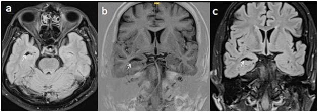
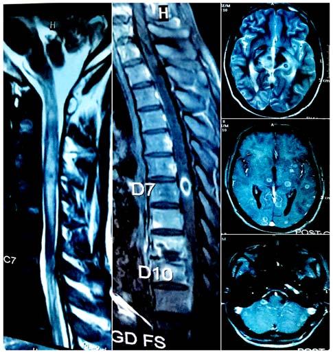
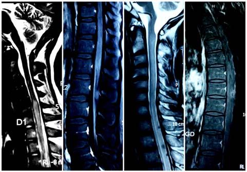
This study has been undertaken to study the hippocampal changes in seizure disorder by MRI (Figs 1-4).
AIMS AND OBJECTIVES
(1)To assess the spectrum of MRI findings in hippocampus of brain in patients with seizures.
(2)To determine the proportion of Partial Loss of Hippocampal Striations (PLHS) in case of seizures disorder and its diagnostic value.
MATERIALS AND METHODS
Source of Data :
Study place : Sapthagiri Institute of Medical Sciences and Research Centre, Bengaluru.
Study subjects : Patients presenting with seizures and in the age group of 18years to 65 years were enrolled in this study after obtaining written informed consent.
Study design : Cross-sectional observational study.
Imaging Protocol :
Visualization of hippocampal gray matter is important for diagnosis of hippocampal sclerosis. So, the accurate distinction of gray matter from white matter and of gray matter from CSF, is essential. Inversion-recovery (IR) images [3500/26 (TR/TE); inversion time: 300 ms, section thickness: 5 mm] in tilted axial and coronal planes give optimal anatomical definition of the hippocampal gray matter. The IR sequence was chosen because it provides details of internal structure of hippocampus and demonstrates decreased signal on T1W images in gliotic areas.We used an asymmetrical field of view with a 256 X 128 matrix to reduce scanning time to 7.5minutes per sequence17
Sample size :
Using the simple following formula to calculate sample size, n= 4pq/ d2
Where- n= sample size
P= expected prevalence or proportion d= margin of error at 15% (standard value of 0.05) q=100-p
Hence, sample size for the study=4*33*67/ (15)2= 8844/225=39.30
According to the prevalence by using the above formula calculated sample size is 39.3. 60 patients were taken up for the study.
Statistical Analysis :
Data analysis will be carried out using statistical software called SPSS V.20. The results were expressed in the form of descriptive and inferential statistics.
RESULTS
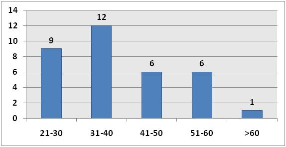
A total of 60 patients were taken up for study. In that 42 patients were Male and 18 were Females. 30% of the patients (18 patients) were in the age group of 21-30 years. 53 of the 60 patients presented with generalized tonic clonic seizures with 3 patients presenting with focal seizures and 2 patients presenting with
Fig 1 — (a) Axial FLAIR, (b) T1 oblique Coronal, (c) FLAIR oblique coronal showing loss of striations on the right side with loss of volume in right hippocampus
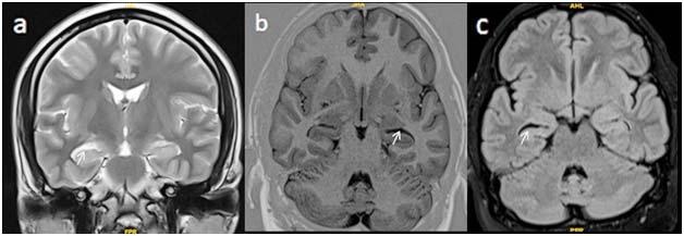
Fig 2 — (a) T2 coronal, (b) T1 inversion recovery oblique Coronal, (c) FLAIR oblique coronal showing loss of striations on the right side with loss of volume in right hippocampus
absence seizures. Partial loss of hippocampal striations were seen in 23.3% of cases (14 patients). Hippocampal atrophy was seen in 18.3% of cases (11 patients). Secondary signs of mesial temporal sclerosis were seen in 18.3% of cases (11 patients). Classic signs of Mesial Temporal Sclerosis (MTS) was seen in 21.6% of cases (13 patients).
DISCUSSION
Mesial Temporal Sclerosis (MTS) is most common cause of medically intractable partial complex epilepsy in adults. MTS can be bilateral in up to 3-10%. Pathologically, it is characterized by hippocampal gliosis and neuronal loss. Different postulates have been put forward. One hypothesis holds that in individuals with a genetic predisposition, prolonged febrile seizures cause hippocampal injury. However, difference between cause and effect is not clear, because a child may have prolonged febrile seizures due to MTS secondary to a prenatal / perinatal insult or genetic predisposition.
MRI findings can be divided into primary, secondary signs and changes in other structures.
Primary Signs :
•Increased T2 / FLAIR signal intensity in the hippocampus.
•Partial loss of hippocampal striations.
•Hippocampal atrophy and volume loss.
•Loss of the internal architecture of the hippocampus (Interdigitations).
Secondary Signs (Temporal lobe findings) :
•Dilatation of the ipsilateral temporal horn
•Temporal lobe atrophy
•Collateral white matter atrophy.
Changes in Other Structures:
•Increased signal intensity and/or atrophy of the ipsilateral amygdale
•Atrophy of the ipsilateral mammillary body
•Atrophy of the ipsilateral fornix
•Atrophy of the contralateral cerebellar hemisphere
•Atrophy of the ipsilateral entorhinal area
Identification of MTS is important as surgery is only treatment option with a good outcome. MTS can be bilateral in upto 3-10% of cases although symptoms may be caused by an unilateral disease2. Patients presenting with seizures can have wide range of MR imaging abnormalities depending upon etiology. MRI can reliably identify and localize the intracranial abnormality so that further management can be planned accordingly.
MTS is a common structural abnormality seen in association with temporal lobe epilepsy. Coronal oblique T2W/FLAIR sequences obtained perpendicular to the long axis of the hippocampus is required for optimal evaluation of hippocampus.
MR images were examined for presence of unilateral hippocampal atrophy in all patients. Moreover, loss of internal architecture and increased T2 signals were only revealed in the sclerotic hippocampus.
The normalized volumes of the hippocampus were compared, and the results were made based on prominent volume decrease in sclerotic hippocampus8-10.
In a study by, Anitha Sen, et al3, the author opines that PLHS may be an easy technique for early detection of hippocampal sclerosis. PLHS is a sensitive indicator of hippocampal sclerosis. Classic signs of
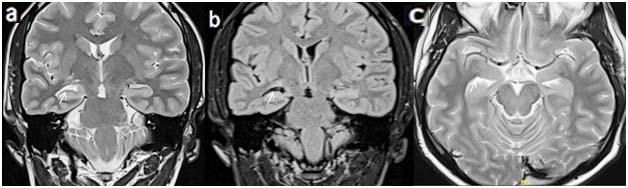
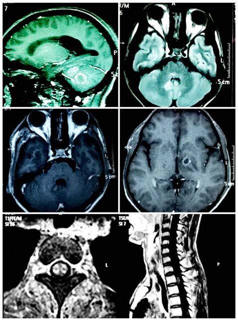
Fig 4 — (a) T2 coronal, (b) FLAIR oblique Coronal, (c) T2 axial showing loss of striations on the right side with loss of volume in right hippocampus
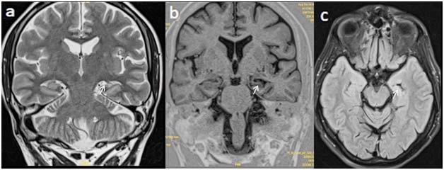
Fig 3 — (a) T2 coronal, (b) T1 inversion recovery oblique coronal, (c) FLAIR showing loss of striations on the left side with loss of volume in left hippocampus
hippocampal sclerosis was seen in 6% cases in this study whereas in our study classic signs were seen in 21.6% of cases.
In a study by, Paramdeep Singh, et al4, where authors opine right hippocampal volume was slightly more than left with no effect of age or gender and concluded that quantitative techniques are more sensitive to diagnose bilateral and mild unilateral hippocampal abnormalities. In our study also there was no significant age or gender related changes in hippocampus.
In a study by, Graeme D, Jackson, et al5, authors had proposed optimal imaging parameters and MR features of hippocampal sclerosis. Hippocampal sclerosis was diagnosed alone in 64% of patients (23.3% in our study). Hippocampal atrophy was seen in 83% (18.3% of cases in our study) and disruption of the internal hippocampal structure was seen in 89% (21.6% in our study). Our study results were less compared to the findings in this study.
In study by, Dongyan Wu, et al6 , the authors demonstrated that Mesial Temporal Lobe Epilepsy (MTLE) is most common form of focal epilepsy, which is frequently characterized by hippocampal sclerosis. Volume quantitative analysis in the hippocampus was conducted and group related volumetric difference was assessed. The results of their study are comparable to the results of our study.
In a study by, Yiran Duan, et al7, author opines that Mesial Temporal Lobe Epilepsy is a neurological disorder associated with hippocampal atrophy. In this study they analyzed the morphologic patterns of hippocampal atrophy to better understand the underlying pathological and clinical characteristics of the condition. They observed significant reduction in unilateral hippocampal volume with a mean volume reduction of 28.38% as compared with healthy controls (p<0.05). In our study, volume reduction was seen in 18.3% of cases.
Limitations of the study : This study represents a limited experience from a single tertiary center.
CONCLUSION
Assessment of the patient presenting with seizure disorder is a common problem in clinical practice. Generalized seizures are the most common seizure and generalized tonic-clonic seizures are the most common seizure in sub-classification.
With its high spatial resolution, excellent inherent soft tissue contrast, multiplanar imaging capability and lack of ionizing radiation, MR imaging has emerged as a versatile tool in the evaluation of patients with seizures.
This study was carried out in 60 patients presenting with seizures by subjecting them to Magnetic Resonance Imaging to evaluate the spectrum of hippocampal abnormality.
In our study, MRI shows PLHS in 14 cases (23.33%) out of total 60 cases included.
MRI also revealed hippocampal atrophy and secondary signs of hippocampal sclerosis in 11 patients (18.33%) out of 60 patients.
Hence, we conclude that in our study PLHS in adult population is much more common than the classic signs of hippocampal sclerosis (increased signal intensity and volume loss).
REFERNCES
1Pearce JM. Ammon’s horn and the hippocampus. Journal of Neurology, Neurosurgery & Psychiatry 2001; 71(3): 351-5.
2Dekeyzer S, De Kock I, Nikoubashman O, Bossche SV, Van Eetvelde R, De Groote J, et al — Unforgettable–a pictorial essay on anatomy and pathology of the hippocampus. Insights into Imaging 2017; 8(2): 199-212. 15.
3Sen A, Sankaran S — Detection of partial loss of hippocampal striation at 1.5 Tesla magnetic resonance imaging. Insights into Imaging 2019; 10(1): 1-7.
4Singh P, Kaur R, Saggar K, Singh G, Kaur A — Qualitative and quantitative hippocampal MRI assessments in intractable epilepsy. BioMed research international 2013; 25.
5Jackson GD, Berkovic SF, Duncan J, Connelly A — Optimizing the diagnosis of hippocampal sclerosis using MR imaging. American Journal of Neuroradiology 1993; 14(3): 753-62. 21.
6Wu D, Chang F, Peng D, Xie S, Li X, Zheng W — The morphological characteristics of hippocampus and thalamus in mesial temporal lobe epilepsy. BMCNeurology2020; 20(1): 1-9. 22.
7Duan Y, Lin Y, Rosen D, Du J, He L, Wang Y — Identifying Morphological Patterns of Hippocampal Atrophy in Patients With Mesial Temporal Lobe Epilepsy and Alzheimer Disease. Frontiers in Neurology 2020; Jan 23.
8Keller SS, Richardson MP, O’Muircheartaigh J, Schoene-Bake JC, Elger C, Weber B — Morphometric MRI alterations and postoperative seizure control in refractory temporal lobe epilepsy. Hum Brain Mapp 2015; 36: 1637-47.
9Barron DS, Fox PM, Laird AR, Robinson JL, Fox PT — Thalamic medial dorsal nucleus atrophy in medial temporal lobe epilepsy: a VBM meta-analysis. Neuroimage Clin 2012; 2: 25-32.
10Keller SS, O’Muircheartaigh J, Traynor C, Towgood K, Barker GJ, Richardson MP — Thalamotemporal impairment in temporal lobe epilepsy: a combined MRI analysis of structure, integrity, and connectivity. Epilepsia 2014; 55: 306-15.
Original Article
Histogenesis of Urinary Bladder in Human Fetuses
Yanglem Elizabeth Devi1, N Saratchandra Singh2, Ch Rajendra Singh2, Laishram Cindy3
Background : The Urinary Bladder starts functioning during fetal life, It fills every hour with minimal increase in pressure (5-10 cm H2O) and empties by contraction over a few seconds. The proper functioning of an organ depends on its histological maturation. There are only few literatures of study on microstructure of the development of Urinary Bladder. For this reason, this study was done to look into the histological development of urinary bladder in different human fetuses.
Materials and Methods : Fifty fetuses of different gestational ages ranging from 15 weeks to 40 weeks were studied after staining with Haematoxylin and eosin stain, Van Gieson, Masson Trichrome and Verhoeff stain.
Results : In the specimen of 15 weeks, Urinary Bladder wall consists of 3 layers ie, inner mucosa comprising of lining epithelium and lamina propria, middle muscular layer and an outer adventitia. Mucosa layer appears thickest comprising about half of the thickness of the bladder wall. The lining epithelium assumed adult urothelium by 22 weeks.
Conclusion : Human fetal bladder undergoes a series of vital developmental changes during 15-40 weeks of gestation both morphologically and histologically finally acquiring the typical adult shape with a typical urothelial lining and well developed muscle coat.
[J Indian Med Assoc 2024; 122(6): 32-5]
Key words :Urinary Bladder, Urothelium, Lamina Propria, Detrusor Muscle Layer.
The Urinary Bladder is a temporary reservoir of urine brought from the kidneys by the ureter1. In a normal adult, internal surface of the bladder is lined with urothelium,deep to this is the lamina propria and underneath the lamina propria is the detrusor muscle layer2
The Urinary Bladder starts functioning from fetal life3 The impermeability of transitional epithelium, particularly during early fetal development is possibly a function of umbrella and intermediate transitional cells4.
MATERIALS AND METHODS
Fifty fresh human fetuses without any gross anatomical abnormality, ranging from 15 to 40 th Gestational Weeks (GWs) were procured from Obstetrics and Gynaecology Department, RIMS Imphal. Permission for the study on fetuses was obtained from the Institutional Ethics Committee.The age of the fetuses were assessed from the obstetrical history, Crown Rump Length (CRL) and gross features before fixation. The fetuses were kept immersed in 10% formalin for 10 to 15 days.
Department of Anatomy, Regional Institute of Medical Sciences, Imphal, Manipur 795004
1MD (Anatomy), Senior Resident and Corresponding Author 2MD (Anatomy), Professor 3MD (Paediatrics), Senior Resident, Department of Paediatrics
Received on : 05/02/2022
Accepted on : 23/09/2023
Editor's Comment :
Functional maturity of an organ largely depends on its histological maturity.
In case of urinary bladder also, its primary function as urinary storage organ and its impermeability depends on the proper development of urothelium and detrusor muscle layer.
The fetuses were categorized into different age groups according to their gestational age as follows for easier study and observation :
Group I: Upto 15 weeks
Group II: 16 – 20 weeks
Group III: 21 – 25 weeks
Group IV: 26 – 30 weeks
Group V: 31 – 35 weeks
Group VI: 36 – 40 weeks
After proper fixation (for about 2 weeks), the fetuses were dissected, the Urinary Bladder was identified. Appearance of the Urinary Bladder in situ was studied in detail and then isolated. Sections of these specimens were fixed in the neutral buffer formalin for 10-15 days. After proper fixation and trimming, the tissue was processed for parafin sections. Serial sections of 7 micrometer thickness were cut with Leika RM 2125 RT rotary microtome. The prepared sections were stained with Haematoxylin and Eosin stain. Special staining was done with Van Gieson’s stain, Masson’s Trichrome for differentiation between the collagen fibres, muscle fibres and Verhoeff’s stain for elastic fibre. Aii the stained slides were studied under trinocular compound light research microscope and
122, No 06, June 2024Journal
microphotography was done with USB camera. All the digital microphotographs were stored and analysed.
RESULTS AND OBSERVATIONS
Group I : 15 Weeks
At 15 GW, Urinary Bladder wall consists of an outermost adventitia, middle muscular layer and an innermost mucosa layer comprising of lining epithelium and lamina propria. Connective tissue cells are present in the Lamina Propria with some undifferentiated mesenchymal cells. Few blood vessels have developed in the Lamina Propria. Lots of mucosal foldings are seen indicating that the epithelial growth is faster than the rest of the wall. Mucosa layer appears thicker than muscular layer. Muscular layer consists of three indistinctly formed layers (Fig 1).
Group II : 16 - 20 Weeks
Well formed lumen is seen. Lining epithelium looks like stratified cuboidal and basal cells are present. Intermediate polygonal cells are present (Fig 2). Superficial cuboidal cells are darker stained than the underlying cells (Fig 4). Lamina Propria is well developed with presence of mesenchymal cells and fibroblasts. Smooth muscle fibres are well developed with varying thickness at different sites and are oriented in multiple directions as longitudinal, circular and oblique muscle fibres. More blood vessels have developed in the Lamina Propria than in the previous group (Fig 3). Blood vessels are also present in between the muscle fibers. Adventitious coat consisting of collagen fibers is seen.
Group III : 21 - 25 Weeks
By this time, epithelium is four to six cell layered. Surface cells are darker stained going towards adult epithelium, some of them are binucleate. Lamina Propria is well developed, muscle layer have grown
Note :
Note : (a) Superficial cells stained darker, (b) Basement membrane is ill defined (c)Fibers element starts predominating cellular element in the lamina propria
more so it appears to dominate in thickness. Fibroblast cells dominate in the Lamina Propria whereas mesenchymal cells are less. Mesenchymal cells are seen to be clustered more near the basement membrane (Fig 5). Elastic fibres couldn’t be seen in the Lamina Propria though it is visible in the wall of arteriole in sections stained with Verhoef’s stain (Fig 6).
Fig 1 — Photomicrograph of urinary bladder from a 15 weeks fetus (H&E x 10)
Note : Lamina propria thicker than the muscle layer
Fig 3 — Lamina propria in a 18 weeks urinary bladder showing laying down of collagen fibers by the fibroblasts
Cellular elements predominate fibers element
Fig 2 — Lining epithelium and part of lamina propria of 18 weeks urinary bladder (H&E x 40)
Fig 4 — Cytoarchitecture of a 20 weeks urinary bladder (H&Ex40)
Group IV : 26 - 30 Weeks
There is spurt of growth so that the cells are compressed and the rounded intermediate cells assume polygonal in shape. Signs of apoptosis are also seen. In the Lamina Propria, both fibroblasts and fibrocytes are present, as the fibroblasts mature into fibrocytes nuclear shape gradually changes from the earlier tadpole form to spindle shape. Collagen fibres being laid down by the fibroblasts are seen. Mesenchymal cells are also present which are differentiated from the fibroblasts by their oval nucleus and are not associated with collagen fibres. Mesenchymal cells are seen more near the basement membrane and possess oval nucleus. Muscle wall has also increased in thickness. Elastic fibers start appearing in the Lamina Propria (Fig 7). Abundance of collagen fibres are also visible in the Lamina Propria (Fig 8).
Group V : 31 - 35 Weeks
Bladder by this time has assumed the adult cytoarchitexture. Lamina Propria is well developed with
more fibrous element than the cellular element. Subepithelial capillaries are also visible. Mesenchymal cells are still present in the Lamina Propria. Muscle wall is well developed but some connective tissue elements are present in between the muscle layers.
Group VI : 36 - 40 Weeks
In this series, well developed Lamina Propria is seen which is more compact in the subepithelial zone and is loose close to the muscle layer, fibrous element predominates the cellular element. Detrusor muscle layer is well formed and is thicker than the Lamina Propria.
DISCUSSION
The epithelial lining of the urinary bladder is derived from the endoderm of the vesical portion of the urogenital sinus, while the Lamina Propria of the bladder mucosa, the muscle coat and the adventitia all develop from the adjacent splanchnic mesenchyme. Initially the entire vesico-urethral anlage is lined by a single layer of low cuboidal to tall columnar epithelium with prominent vesicular characteristics.
Above the openings of the ureters this single layer becomes bi or trilayered by the end of the 6th week according to Felix (1912) or by the 7th week (Gray and Skandalakis, 1972). Below the ureteric orifices it becomes bi- or trilayered and later, by the 9th
Fig 5 — Photomicrograph of a 22 weeks old urinary bladder showing the lining epithelium assuming the adult urothelial form (Verhoeff’s stain x 40)
Fig 6 — Photomicrograph showing the elastic fibers in the internal elastic lamina of an arteriole present in the adventitia of a 22 weeks urinary bladder (Verhoeff’s stain x 40)
Fig 8 — Cytoarchitecture of a 28 weeks urinary bladder showing abundance of collagen fibers in the lamina propria (Masson’s Trichrome stain x 10)
Fig 7 — Photomicrograph of 28 weeks urinary bladder showing the first appearance of elastic fibers in the lamina propria (Verhoeff’s stain x 40)
week, 4 to 5 cells thick (Felix 1912). According to Felix the urothelial characteristics appear in the 11th week embryo while according to Gray and Skandalakis (1972) they appear in the 12th week . While according to Newman J, et al the typical ultrastructural urothelial characteristics appeared by 21 weeks5
According to Hoyes AD, in the 4th month, the bladder epithelium was bilaminar and in the 5th month, a third layer of cells was sometimes present. Differentiation of the luminal plasma membrane was evident by the end of the 3rd month6. During second trimester, the epithelial lining of the bladder becomes transitional in type7
In our present study, we observed the adult urothelial characteristics by 22 weeks as defined by the presence of specific features – large binucleated umbrella shaped surface cells with prominent darker staining luminal plasma membrane, two to three layers of intermediate polygonal shaped cells and a basal layer of cuboidal cells lying over an ill defined highly undulating basement membrane.
The musculature of the bladder wall begins to develop at the 13th week and is very prominent by the 16th week8. Muscle thickness progressively increased and the relative collagen content in the muscle decreased during gestation. The ratio of thick-to-thin collagen fibers also decreased, whereas elastic fibers increased9.
In our present study, in the earliest available fetus of 15 weeks, muscular layer consists of three indistinctly formed layers which are infiltrated with abundant connective tissue elements in between the muscle fibers. The muscle layer can be more or less differentiated into inner longitudinal, middle circular and outer longitudinal layers by 18 weeks. This detrusor muscle layer gradually thickened as age advances and is associated with the progressive decrease in the connective tissue elements infiltrating the muscle fibers.
The highest level of expression of RNA products of elastic fiber genes occurred in the urothelial – Lamina Propria fraction during the late second – early third trimester9. In the present study, Verhoeff’s elastic tissue stain was used to identify the elastic fibers, internal elastic lamina in the wall of arteriole could be identified by 22 weeks, but elastic fibrers are not yet visible in the Lamina Propria. By 28 weeks, few elastic fibers are identifiable in the Lamina Propria and the elastic fibers distribution gradually increases as age advances and at term plenty of elastic fibers could be seen in the Lamina Propria.
CONCLUSION
On histological examination, in the early specimen of 15 weeks, urinary bladder wall consists of 3 layers ie, inner mucosa comprising of lining epithelium and lamina propria, middle muscular layer and an outer adventitia of which mucosa layer appears thickest comprising about half of the thickness of the bladder wall. By this time, in the Lamina Propria there is abundance of mesenchymal cells and fibroblast cells which have started laying down few collagen fibers. By 18 weeks, lining epithelium appears stratified cuboidal with the superficial cells more darker stained than the underlying cells. The lining epithelium assumed adult urothelium by 22 weeks.
There is lack of literature regarding development of Lamina Propria, available literatures just mention that there is increase in fibers element with increase in the gestational age but specific age of this change has not been mentioned. In our study, in the Lamina Propria, there is gradual change in the proportion between the cellular element and fibers element. In the earlier week of fetus less than 20 weeks, cellular element predominates the fibers elements whereas in the later fetal life, fibers element predominates the cellular element. This is associated with the increase in both elastic and collagen fibers of which the collagen fibers appear earlier. Elastic fibers could be seen in the wall of arteriole by 22 weeks while it appears in the Lamina Propria by 28 weeks.
REFERENCES
1Dyson M — Urinary system. In : Williams PL, Bannister LH, Berry MM, Collins P, Dyson M, Dussek JE, et al, editors. Gray’s Anatomy. 38th ed. New York : Churchill Livingstone; 1995. 1837-9.
4Rosette J, Smedts F, Schoots C, Hoek H, Laguna P — Changing patterns of Keratin expression could be associated with functional maturation of the developing human bladder. J Urol 2002; 168(2): 709-17.
5Newman J, Antonakopoulos GN — The fine structure of the human fetal urinary bladder . Development and maturation. A light,transmission and scanning electron microscopic study. J Anat 1989; 166: 135-50.
6Hoyes AD, Ramus NI, Martin BG — Fine structure of the epithelium of the human fetal bladder. J Anat 1972; 111(Pt 3): 415-25.
7Rahilly RO, Muller F — Human embryology and teratology.2nd ed. New York : Wiley-Liss, Inc; 1996.
8Hoyes AD, Ramus NI, Martin BG — Fine structure of the epithelium of the human fetal bladder. JAnat 1972; 111 (Pt 3): 415-25.
9Kim KM, Kogan BA, Massad CA, Huang YC — Collagen and elastin in the normal fetal bladder. J Urol 1991; 146{2(Pt. 2)}: 524-7. 35
Original Article
A Non-randomized Retrospective Study of Applicability of ClavienDindo Classification of Surgical Complications to Emergency Laparotomies
Background : In developing countries, emergency laparotomy have very high morbidity and mortality rates. The aim of the present study was to evaluate and find out various determinants for safe outcome in emergency laparotomy in terms of decreased morbidity and mortality.
Materials and Methods : A non-randomized retrospective study with collected data from 300 patients with emergency laparotomy. Data from various preoperative and operative factors, surgical technique and complications were recorded. The output was measured as better and worse, as per the Clavien–Dindo classification.
Results : The study enrolled 300 patients with a male:female ratio of 4.5:1. The majority of patients presented with the history of abdominal pain (98%), abdominal distension (62%) and altered bowel habit (62%). Perforation peritonitis constituted 231 cases followed by intestinal obstruction in 60 cases, rupture liver abscess with peritonitis in 9 cases. Using the Clavien–Dindo classification, 30% patients had no complications 27% had Grade I, 12% had Grade II, 16% had Grade III, 11% had Grade IV and 4% had Grade V complication rates.
Conclusion : Postoperative complications increase patient morbidity and mortality and are target for quality improvement programs. Many complications may be prevented by thorough preoperative evaluation preceded by adequate aggressive resuscitation, broad spectrum antibiotics, correction of electrolyte imbalances, early minimum & sound surgical technique and careful follow-up care. The Clavien–Dindo classification can be adapted to assess the severity of postoperative complications following emergency laparotomy.
Laparotomy is a most common surgical procedure done by routine surgical team. Postoperative complication may be defined as any negative outcome as perceived by the surgeon or by the patient. It may occur intraoperatively in the immediate postoperative period, or later on. They are the chief weakness of surgeon’s craftsmanship on operation table. Commencing as a seemingly minor disturbance and if allowed to persist they can jeopardize the patient recovery and even result in a fatal outcome. Post operative complication may occur after laparotomy whether elective or emergency. Various factors like
1MS (General Surgery), Professor and Head, Department of Surgery, RVRS Government Medical College, Bhilwara, Rajasthan 311001 and Corresponding Author
2MS, Assistant Professor, Department of Obstetrics and Gynaecology, Mahatma Gandhi Medical College, Jaipur, Rajasthan 302022
3MS, Senior Resident, Department of Neurosurgery, SMS Medical College, Jaipur 302004
4MS, Senior Professor, Department of General Surgery, JLN Medical College, Ajmer, Rajasthan 305001
5MS, Assistant Professor, Department of General Surgery, RVRS Government Medical College, Bhilwara, Rajasthan 311001
Received on : 31/01/2022
Accepted on : 11/02/2022
Editor's Comment :
The Clavien–Dindo classification system serves as a valuable tool for assessing postoperative complications in emergency laparotomies. It categorizes outcomes into grades, with Grades I and II denoting minimal problems or none at all and Grades III, IV and V denoting more serious complications. This classification is based on the therapy required to address a specific complication, ensuring an objective and reproducible ranking of complications.
proper resuscitation, meticulous surgical technique, age, any co-morbid condition, anaesthesia technique and postoperative care contribute to final result. More accurately gauging the quality of care may come from adverse occurrences closely linked to the processes of care, like postoperative complications, rather than from death rates or other intermediate outcomes. Therefore, early detection and proper intervention can reduce the morbidity and mortality related with complication1
The assessment of surgical operations has been significantly impacted by the lack of agreement on the definition and grading of unfavourable postoperative complications. Clavien, et al2 1992 suggested a new classification of complications, which they defined as
122, No 06, June 2024Journal
any variation from the ideal postoperative course that is not inherent in the treatment and does not include a failure to cure. A condition that doesn’t improve after surgery isn’t a result of a problem; rather, it indicates a failure to heal. There was not much guidance provided on how this categorization should be applied to patients experiencing emergency surgery because it was created and verified with patients undergoing elective general surgery.
The current study’s objectives are to assess the Clavien-Dindo classification (Table 1) of surgical complications in patients undergoing emergency laparotomies and investigate pre-operative variables for risk assessment that have to be recorded in a surgical complications database3
MATERIALS AND METHODS
A non-randomized prospective study of postoperative complications in 300 patients who had undergone emergency laparotomy in JLN Medical College and Hospital, Ajmer under various surgical units, during the period of December, 2016 to September, 2018.
Inclusion Criteria :
All patients (>15 years) presenting at emergency or surgical OPD and had undergone emergency laparotomy.
Exclusion Criteria :
•Children < 15 years.
•Patient with history of trauma.
•Redo laparotomy
•Pregnant women
Type of Study :
Non randomized prospective Time-Bound Hospital Based study.
Method of Collection of Data :
Based on inclusion and exclusion criteria, all abdominal cases admitted to the surgical ward for emergency laparotomy and surgery were assessed using a comprehensive clinical examination, co-morbid conditions and history. All patients underwent routine investigations and based on the provisional diagnosis and the patient’s needs, specialized investigations such as CT, USG and X-rays were performed.
Intravenous fluids were used to revive all patients before to surgery and the electrolyte imbalance was addressed. For each patient, urethral catheterization and Ryle’s tube aspiration were performed. All patients received wide spectrum antibiotics prior to surgery. Patients with anemia needed blood transfusions. Under general anaesthesia, all patients underwent exploratory
Table 1 — Classification of surgical complication proposed by Clavien –Dindo et al4
Grades Definition
Grade I :Any deviation from the normal postoperative course without the need for pharmacological treatment or surgical, endoscopic and radiological interventions.
Allowed therapeutic regimens are: drugs as antiemetic, antipyretics, analgesics, diuretics and electrolytes and physiotherapy. This grade also includes wound infections opened at the bedside.
Grade II :Requiring pharmacological treatment with drugs other than such allowed for grade I complications.Blood transfusions and total parenteral nutrition are also included.
Grade III : Requiring surgical, endoscopic or radiological intervention
Grade III-a :Intervention not under general anaesthesia
Grade III-b :Intervention under general anaesthesia
Grade IV :Life-threatening complication (including CNS complications)* requiring IC/ICU-management
Grade IV-a :Single organ dysfunction (including dialysis)
Grade IV-b :Multi organ dysfunction
Grade V :Death of a patient
laparotomy through a midline incision. The entire intestine was examined and the results were recorded concerning the volume and kind of fluid, the pathology of the gut, the area of the gut affected and the state of the intestine next to it.
A number of parameters were recorded, including the surgical technique, intraoperative findings, length of the operation, length of the recovery time, postoperative complications and their management. Postsurgical complications were then assessed and classified using the Clavien-Dindo classification.
Following surgery, all patients were monitored in the ward or Intensive Care Unit (ICU). Each patient’s medication regimen was different. The necessity for postoperative blood products, complete parenteral nutrition, ventilator support and complications such anastomosis leak, wound infection, ruptured abdomen, and respiratory issues were all documented. The complications were graded (Grades I–V), as per the classification proposed by Clavien– Dindo 4 .
•No complications
•Grade I : Wound infection
•Grade II : Respiratory complications
•Grade III : Burst abdomen, anastomosis leak
•Grade IV : Septicaemia
•Grade V : Death
Better (Grades I, II and No problems) and worse (Grades III, IV and V) outcomes were used to gauge the outcome. For the purpose of this study, a predesigned proforma was used to collect and record data. The Statistical Package for the Social Sciences (Version 20.0 for Windows, Developer(s) IBM
Table 2 — Pre-operative Data
VariableNo of patientsPercentage
Age and sex distribution :
Age < 60 year21672%
Age > 60 year8428%
Male24983%
Female5117%
Clinical sign & symptom :
Pain abdomen29498%
Abdominal distension18662%
Altered bowel habbit18662%
Nausea and vomiting13846%
Fever10836%
Respiratory distress8428%
Shock7224%
Diarrhoea31%
Tenderness28896%
Guarding25284%
Distension21672%
Rigidity21070%
Positive findings on investigations
Pneumoperitoneum21672%
Air fluid level6923%
Increase Total Leucocyte Count16254%
Raised serum creatinine15351%
Electrolyte imbalance7224%
Raised serum total bilirubin6923%
Anemia4214%
Associated comorbidity
H/o Tuberculosis279%
DM217%
Hypertension186%
Previous H/O surgery186%
Time for resuscitation
< 24 hour14749%
> 24 hour15351%
Corporation, Armonk, New York) was the program used for data analysis. The chi square test and the analysis of variance (ANOVA) test are suitable for data analysis. Every test was used with a 95% significance level.
RESULTS
From this study two peaks in age distribution were found in emergency laparotomies. First in age group 20-40 years patients and second in age group 40-60 years. The oldest patient was eighty-six years old, and the youngest was eighteen. The ratio of men to women was 4.5:1.
The majority of patients (98%), Abdominal Distension (62%), Abnormal Bowel Habits (62%), Nausea or Vomiting (46%), Fever (36%) and Shock (24%), caused by Septicaemia and Dehydration, were among the symptoms that they reported having experienced in the past. The majority of patients (96%) reported having abdominal pain, which was followed by Guarding (84%), Distension (72%) and Stiffness (70%).
In our study patients present within 3 days of onset of symptom were 153 and after 3 day were 147 patients.
In all patient flat plate abdomen X-ray was done and found gas under diaphragm in 216(72%), dilated bowel loop with air fluid level 69(23%), rest of patients X-ray were normal. Gas under diaphragm was present in 216 patients of perforation out of 231 patients of perforation (93.5%). Pre-operative investigative data included increase Total Leucocyte Count (>10000) in 54%, raised creatinine( >1.2 mg/dl) in 51%, electrolyte imbalance in 24%, raised serum total bilirubin (>1.2mg/dl) level in 23% and Anaemia (Hb<10gm/dl) in 14%.
In 48% of the cases, the time needed for resuscitation, diagnosis, and patient optimization for surgery was less than 24 hours, while in 52% of the cases, it was more than 24 hours.
Table
3 — Operative Data
VariableNo of patientsPercentage
Indication of Laparotomy : Perforation Peritonitis
Gastroduodenal perforation16555%
Small bowel perforation5418%
Large bowel perforation62%
Appendicular perforation31%
Gall bladder perforation31%
Intestinal obstruction
Band adhesion279%
Malignancy155%
Sigmoid volvulus93%
Stricture62%
Tuberculosis31%
Rupture liver abcess93%
Peritoneal fluid : <1000 ml15351% >1000 ml13846%
Nil93%
Bilious17157%
Feceal5418%
Serous4515%
Purulent217%
Surgical procedure : Gastroduodenal perforation
Omentopexy14448%
Figure of eight217%
Small bowel perforation
Primary repair with omental patch3612%
Resection anastmosis124%
Resection anastmosis with ileostomy62%
Appendisectomy31%
Cholecystectomy31%
Large bowel perforation
Resection anastmosis with ilieostomy62%
Intestinal obstruction
Release of band with adhesionlysis279%
Resection anastmosis155%
Resection anastmosis with ileostomy93%
Right hemicolectomy with ileostomy62%
Left hemicolectomy with ileostomy31%
Peritoneal toileting93%
Duration of surgery :
< 2 hour15953%
> 2 hour14147%
122, No 06, June 2024Journal
Most cases involved acute abdomen/peritonitis. There were 231 cases of perforation peritonitis, 60 cases of intestinal obstruction and 9 cases of ruptured liver abscess with peritonitis. On exploration 171 patients have bilious peritoneal fluid, faecal containing peritoneal fluid in 54 cases, serous peritoneal fluid in 45 cases, purulent fluid in 21 cases and no peritoneal fluid in 9 cases. In 153 patients amount of peritoneal fluid >1000ml, in 138 patients amount of peritoneal fluid were <1000ml and 9 patients have no free fluid in peritoneal cavity.
Gastroduodenal Perforations (165) were treated with omentopexy in 144 patients and figure of eight repair in 21 patients. Small bowel perforations (54) were managed according the condition of the gut primary repair of perforation with omental patch (36), resection anastomosis (12) and resection anastomosis with stoma (6). Large bowel perforation (6) patients were managed by resection anastomosis with stoma (6). Intestinal obstruction (60) was managed by release of band and adhesion-lysis (27), resection anastomosis (15), resection anastomosis with ileostomy (9), right hemicolectomy with ileostomy (6), extended left hemicolectomy with ileostomy (3). In 6 patients of rupture liver abscess simple peritoneal lavage with drain was done. Right hemicolectomy with ileostomy and peritoneal lavage was done in 3 patients. Three patients of appendicular perforation were managed by appendicectomy. Three cases of GB perforation were treated by cholecystectomy.
Postoperative complications recorded were wound infection 81(27%), respiratory complications 36(12%), septicaemia 33(11%), burst abdomen 30(10%),
anastomosis leak/faecal fistula 18(6%) and Death in 12(4%). A total of 300 patients (30%) had no issues, 81 patients (27%) had Grade I complications, 36 patients (12%), 48 patients (16%) had Grade III complications, 33 patients (11%) had Grade IV complications, and 12 patients (4%), had Grade V complications. These data were based on the ClavienDindo classification.
DISCUSSION
Emergency laparotomy is one of the most commonly performed major surgical procedures in emergency. It becomes imperative to assess the risk of morbidity and mortality in each of such patients undergoing emergency laparotomy, as these patients are often brought to the hospital in morbid conditions. Risk assessment prior to surgical procedure not only allows proper allocation of resources and psychological preparedness about outcome among relatives but also helps surgeons in medico legal suits. In our study, Median age of patient was 45 year, 72% patients were below the age of 60 years. This is consistent with other studies as reported by Jignesh A Gandhi, et al5. In our study majority of patients were males (249 males and 51 females) and the male-to-female ratio was 4.5:1. Another study done by A. Singh, et al6 reported male to female ratios of 3.95:1 also showed that there were more male patients going for emergency laparotomy. Patients’ clinical presentations differed depending on the pathogenic state and location. Individuals who experienced a perforation of the duodenum typically had a brief history of upper abdominal or epigastric pain. Most patients with ileocecal pathology had a
Table 4 — Various Surgical Procedures according to Pathology and Outcome in Relation to Clavien-Dindo Classification
PathologyNo of Percen-Surgical procedure n(%) BetterW
Perforation peritonitis
patients tage outcomeoutcome
Gastroduodenal perforation16555%Omentopexy 144(87.27%)111(77.08%) 33(22.92%) Figure of eight 21(12.73%)18(85.71%)3(14.29%)
Small bowel perforation5418%Primary repair with omental patch 36(66.67%)27(75%)9(25%)
Resection anastmosis 12(22.22%)6(50%)6(50%)
Resection anastmosis with ileostomy6(11.11%) 3(50%)3(50%)
Large bowel perforation62% Resection anastmosis with ilieostomy 6(100%) 0(0%) 6(100%)
Right hemicolectomy with ileostomy 3(33.33%) 0(0%) 3(100%)
122, No 06, June 2024Journal
history of nausea or vomiting, changed bowel habits, abdominal pain and distension. Individuals who had suffered a small bowel typhoid perforation also had a history of fever, which was followed by an abrupt start of abdominal pain. Patient of intestinal obstruction were present with abdominal distension, colickypain, frequent vomiting and altered bowel habit. Patient of rupture liver abscess present with pain initially in right hypochondrium followed by generalised pain abdomen and vomiting.
In our study, the commonest sign was abdominal tenderness (96%) followed by abdominal guarding (84%), distension (72%) and rigidity (70%). In a study done by Jain R, et al7 the commonest sign is abdominal tenderness (99.9%) followed by abdominal guarding/rigidity (61.2%).
Plain abdominal radiography remains an important diagnostic tool if it is restricted to certain surgical conditions, especially those pertaining to intestinal obstruction and hollow viscus perforation8. In our study Plain X-ray abdomen was done in all patient and found gas under diaphragm in 216 patients of perforation peritonitis with diagnostic sensitivity 93.5% and specificity 100%, these findings were contradictory to study done by Sarah L Gans, et al 9 show sensitivity 60%. When intestinal obstruction is suspected in a patient, imaging should reveal the location, reason, and severity of the obstruction 10. As a standard diagnostic modality, plain abdominal radiography is recommended for the identification of intestinal obstruction11,12. In our study 69 patients showed dilated bowel loop and multiple air fluid level in plain X-ray abdomen out of which 54 had intestinal obstruction, 6 patients of intestinal obstruction have normal X-ray abdomen with sensitivity 78% and specificity 90%.
Gastroduodenal perforations are a frequent surgical complication that can result from gastric cancer13, NSAID usage, or Peptic Ulcer Disease (PUD). Our analysis shows that 57% of the patients have a history of using NSAIDs. Perforation makes up fewer than 1% of the complications associated with gastric cancer. Our study found that simple closure of the perforation using an omentopexy, adequate antibiotic treatment, and adequate hydration all greatly reduced the death rate, which was 77.08% in the better result group. Omental patching has also been advised in cases of gastroduodenal perforations by Singh A, et al6
Alternative therapies for perforated peptic ulcers, such figure of eight repair, have demonstrated very positive and satisfactory outcomes. In our study, figure of eight repair was performed on 21 patients, with 18 (85.71%) of those patients in the better outcome group14. A study by Singh A, et al15. also produced results that were
similar. Billroth I, Billroth II, truncal vagotomy and drainage procedures are the remaining treatment options for perforated peptic ulcers; however, definitive acid reduction and bypass surgery were not feasible for these patients because of their compromised physiological status and septicaemia, which seems to increase the risk of death and morbidity without significantly improving the long-term outcome 16 Laparoscopic repair of the perforated gastroduodenal ulcer by running suture with or without omental patch is an option for stable physiological state and early presentation of case17
Typhoid and TB were the causes of small intestinal perforations that we observed in our investigation. Even in healthy, young patients, it can be lethal. In underdeveloped nations, the most terrible consequence of enteric fever is intestinal perforation, which can result in widespread peritonitis18. It is more prevalent in young boys who are in their economically productive years to have the condition and its sequelae. The number of perforations, the patient’s overall health and the state of the gut all affect how tubercular ileal perforations are managed. Ileocecal tuberculosis was managed by right hemicolectomy with or without stoma, perforation along with multiple stricture with strictureplasty or resection anastomosis, and a covering stoma or only stoma19,20 Typhoid enteric perforations were managed by simple primary double layer repair, primary double layer repair with free omental sheet graft, primary double layer repair with stoma, primary double layer repair with Ile transverse anastomosis, and depending on the condition of the gut with multiple perforations, it was also managed by resection and anastomosis with and without stoma. Primary double layer repair with free omental sheet graft of the typhoid perforation is a safe and effective treatment; as seen in our study, 36 patients of ileal perforation were managed by primary repair with free omental sheet graft, 27(75%) were in the better outcome group and 9(25%) in worse outcome group. An alternative procedure like resection and anastomosis were done in 12 patients where there were multiple perforations, 6(50%) patients in better outcome and 6(50%) were in worse outcome group. In 6 patients of enteric perforation where the gut was not healthy enough or with multiple perforations or there was excessive soiling, exteriorization of gut was done, in better outcome group were 6(100%) and in worse outcome group was 0.
A rare cause of perforation peritonitis, observed in 2% of patients, is colorectal perforation. In our setup, malignancy is an uncommon cause of perforation peritonitis; compared to its western counterpart21, only 40
1% of cases had peritonitis as a result of malignancy. Treatment choices for rectal perforations are determined by the surgeon’s experience level, the patient’s age, general health and the severity of peritonitis. Rectal perforations have a significant morbidity and fatality rate. When treating acute left-sided colon perforation without faecal peritonitis22, primary anastomosis and protective ileostomy are better to the Hartman method. In our study, 6 patients have large bowel perforation were managed with resection anastomosis and ileostomy, 6(100%) patients were in worse outcome group. Study done by Singh, et al15 shows similar result. Appendicular and GB perforation were managed with appendicectomy and cholecystectomy respectively.
Intestinal obstruction was another common surgical emergency that was associated with a high mortality if the diagnosis was delayed or managed inappropriately. In our study small bowel obstruction constitutes about 70% of all intestinal obstructions, bands and adhesions being the most common cause. Study done by Jaiswal NK, et al 23 also show band adhesion was the most common cause of intestinal obstruction. An intestinal blockage that was strangulated required immediate surgical attention. The administration of analgesics, intestinal decompression, intravenous antibiotics, fluids and electrolytes, and, if necessary, surgery24, are the fundamentals of managing intestinal obstruction. Conservative therapy is effective in up to 75% of patients with adhesive small intestinal obstruction; hence, it was initially tried in all patients who did not exhibit obvious indications of strangling. If, after 48-72 hours of conservative therapy, the obstruction in the small intestine does not clear up on its own without surgery, this is an uncommon occurrence.
In our study, 60 patients presented with intestinal obstruction, which on laparotomy was found to be most commonly due to bands and adhesions in 27(45%) patients. Release of bands and adhesiolysis was done in all case, 21(77.77%) patients belong to the better outcome group and 6(33.33%) patients in worse outcome group. Patients presenting with gangrenous small bowel obstruction were subjected to resection and anastomosis. On exploration growth were found in 15 patients’ resection of growth with anastomosis weredone in 6 patients, right hemicolectomy with ileostomy were done in 6 patient and extended left
hemicolectomy with ileostomy was done in 3 patients, belonging to better outcome group were 3(50%), 3(50%), 0(0%) respectively and 3(50%), 3(50%), 3(100%) respectively in worse outcome group. Six patients presented with intestinal stricture, in which resection anastomosis were done, 3(50%) in better and 3(50%) in worse outcome group. Sigmoid volvulus were present in 9 patients for which resection anastomosis with ileostomy performed, in better outcome group 6(66.67%) and in worse outcome group 3(33.33%).
Rupture liver abscess with peritonitis were present in 9 patients, in 6 patient exploratory laparotomies with peritoneal lavage done, 3 (50%) in better outcome group and 3(50%) patients in worse outcome group. Three patients of rupture liver abscess present with slough out caecum and ascending colon for which right hemicolectomy with ileostomy was performed and it belong to worse outcome group 3(100%).
Postoperative complications recorded in our study were wound infection 81(27%), respiratory complications 36(12%), septicaemia 11(11%), burst abdomen 30(10%), anastomosis leak/faecal fistula 18(6%) and Death in 12(4%). Using the Clavien–Dindo classification complication were graded, 90 out of 300 (30%) patients had no complications, 81(27%) had Grade I complication, 36(12%) had Grade II complications, 48 (16%) had Grade III complications, 33 (11%) had Grade IV complications, and 12(4%) had Grade V complication rates. Singh A, et al15 found similar result of complication 38.28% patients had no complications, 18% had Grade I complication, 16.57% had Grade II complications, 9.14% had Grade III complications, 7.14% had Grade IV complications, and 10.85% had Grade V complication rates.
Despite waiting longer to seek treatment, the mortality rate in our study was 4%; however, the global
Fig 1 — Postoperative outcome according to Clavien- Dindo classification
No 06, June 2024Journal
literature on the subject indicates that the mortality rate following emergency laparotomies surgery ranges from 6% to 27%25. The existence of septicaemia was one of the major factors in our study that contributed to mortality. As a result, contamination plays a major role in perforation peritonitis patients, and infection is linked to death. For optimal results that minimize morbidity and mortality26 , adequate preoperative resuscitation, correction of electrolyte imbalances and an early surgical intervention to remove the source of infection and limit future contamination are essential.
CONCLUSION
This study shows that explorative laparotomy has to be done most commonly as an emergency procedure in case of hollow viscous perforation. Peptic ulcer perforation is the most common cause of perforation peritonitis. Intestinal obstruction was the next most common cause for emergency exploratory laparotomy. “Postoperative complications increase patient morbidity and mortality and are a target for quality improvement programs .” Many complications may be prevented by thorough preoperative evaluation preceded by adequate aggressive resuscitation, broad spectrum antibiotics , correction of electrolyte, imbalances early minimum & sound surgical technique and careful follow-up care.
“The Clavien–Dindo classification can be adapted to assess the severity of postoperative complications in emergency surgeries in view of easy understanding and applicability and assessment of different surgical therapies.”
Conflicts of Interest : All authors have no conflicts of interest.
REFERENCES
1Chauhan S, Chauhan B, Sharma H — A comparative study of postoperative complications in emergency versus elective laparotomy at a tertiary care centre. International Surgery Journal 2017; 4(8): 2730-5.
2Clavien PA, Sanabria JR, Strasberg SM — Proposed classification of complications of surgery with examples of utility in cholecystectomy. Surgery 1992; 111(5): 518-26.
3Mentula PJ, Leppäniemi AK — Applicability of the ClavienDindo classification to emergency surgical procedures: a retrospective cohort study on 444 consecutive patients Saf Surg 2014; 8: 31. Published online 2014 Jul 26. doi: 10.1186/ 1754-9493-8-31PMCID: PMC4114794
4Dindo D, Demartines N, Clavien PA — Classification of surgical complications: a new proposal with evaluation in a cohort of 6336 patients and results of a survey. Annals of Surgery 2004; 240(2): 205-13.
5Gandhi JA, Shinde PH, Digarse RD — Evaluation of abdominal wall closure technique in emergency laparotomies at a tertiary care hospital. International Surgery Journal 2016; 3(4): 1796801.
6Singh A, Gora N, Soni ML — Comparative Study of Free Omental Sheet Graft and Other Operative Procedures of
7Jain R, Gupta V — A prospective study of epidemiology and clinical presentation of nontraumatic acute abdomen cases in a tertiary care hospital of central India. Int Surg J 2017; 4: 242-5.
8Gupta H, Dupuy DE — Advances in imaging of the acute abdomen. Surg Clin North Am 1997; 77: 1245-63.
9Gans SL, Stoker J, Boermeester MA — Plain abdominal radiography in acute abdominal pain; past, present, and future. Int J Gen Med 2012; 5: 525-33. Published online 2012Jun13. doi: 10.2147/IJGM.S17410
10Lameris W, Van Randen A, Van Es HW — Imaging strategies for detection of urgent conditions in patients with acute abdominal pain: diagnostic accuracy study. Br Med J 2009; 338: b2431.
11Royal College of Radiologists. Referral guidelines for imaging. 2000.
12American College of Radiologists. Practice guideline for the performance of abdominal radiography. Radiography 2006: 1-5.
13Gupta S, Kaushik R, Sharma R, Attri A — The Management of Large perforations of Duodenal Ulcers. BMC Surg 2005; 5: 15.
14Gupta SP — A Safer Technique of Closure of Peptic Ulcer Perforation. Indian J Surg 2011; 73: 361-2.
15Singh A, Porwal R, Gupta HP, Sharma AK, Kumawat G — Determinants of outcome in gastrointestinal perforations with special reference to clavien–dindo classification of surgical complications: Experience of a Single Institute in Central Rajasthan. Arch Int Surg 2016; 6: 170-5.
16Jhobta RS, Attri AK, Kaushik R, Sharma R, Jhobta A — Spectrum of perforation peritonitis in India - Review of 504 consecutive cases. World J Emerg Surg 2006; 1: 26.
17Koninger J, Bottinger P, Redeeke J, Butters M — Laproscopic repair of perforated gastroduodenal ulcer by running suture. Langenbecks Arch Surg 2004; 389: 11-6.
18Rehman A — Spontaneous ileal perforation: An experience of 33 cases. J Post Grad Med Inst 2003; 17: 105-10.
19Yadav D, Garg PK — Spectrum of perforation peritonitis in Delhi: 77 Case Experience. Indian J Surg 2013; 75: 133-7.
20Ara C, Sogutlu G, Yildiz R, Kocak O, Isik B, Yilmaz S, et al — Spontaneous small bowel perforation due to intestinal tuberculosis should not be repaired by simple closure. J Gastrointest Surg 2005; 9: 514-7.
22Breitenstein S, Kraus A, Hahnloser D, Decurtins M, Clavien PA, Demartines N — Emergency left colonre section for acuteper- foration: Primary anastomosis or Hartmann’s procedure? A case matched control study. World J Surg 2007; 31(11): 2117-24.
23Jaiswal NK, Shekhar S, Ranade P — Study of clinical spectrum and management of acute intestinal obstruction. Int Surg J 2018; 5(4): 1310-4. DOI: http://dx.doi.org/10.18203/23492902.isj20181060.
25Oheneh-Yeboah M — Postoperative complications after surgery for typhoid ileal perforation in adults in Kumasi. West Afr J Med 2007; 26: 32-6.
26Ahmad MM — Spectrum of perforation peritonitis in Kashmir: A prospective study at our tertiary care centre. Int Surg J 2015; 2: 381-4.
Original Article
Knowledge, Attitude and Practices Regarding Tuberculosis among adult Residents of Urban Field Practice Area of Shri B M Patil Medical College, Vijayapura
Santosh D Patil1, Praveen Ganganahalli2, Sandeep Yankanchi3
Background : Tuberculosis (TB) remains a severe public health burden across the globe, including India. India alone accounts for approximately 24% of all incidents TB cases globally. According to a recent WHO report, approximately one million deaths occur due to TB every year globally, of which 0.24 million were Indians. A high awareness of Tuberculosis (TB) is very important for its prevention and control in the community.
Aims and Objective : To study the Knowledge, Attitude and Practices regarding TB among adults of urban field practice area.
Materials and Methods : A cross-sectional study was conducted among adult residents of urban field practice area of Vijayapura. The information was collected about Knowledge, Attitude and Practices regarding TB (its signs and symptoms, mode of spread, cause, investigations, treatment and prevention) among adults. Chi-square test was applied to study the effect of socio-demographic characteristics on KAP regarding TB
Results : A total of 390 subjects were interviewed for the survey. About 28.46% of the subjects were in the age group 19-28 years. The median age of the study subjects was 42 (SD±14) years. Of these, 170 (43%) were male subjects and 220 (56%) were female. Based on the knowledge variables, 31.28% felt that pollution is the cause followed by 22.56% as certained the cause to habits like Smoking and Tobacco use. About 16.67% said bacteria can cause TB and 11.54% did not have any knowledge about the cause of TB. About 60.52% of the patients said that TB is a very serious disease and 3.33% said that they did not know about the seriousness of the disease
Conclusion : The study participants had good knowledge of TB but there were several misconceptions regarding its cause. Community-based awareness programs on TB need to be increased among residence of urban field practice area.
[J Indian Med Assoc 2024; 122(6): 43-6]
Key words :Knowledge, Attitude, Practice, Tuberculosis, Urban Community.
Tuberculosis (TB) is a very ancient disease; its description has been found in Buddhist and Chinese writings. Even Egyptian mummies as old as 500 BC show evidence of a man suffering from TB. In India, it is known by many names as ‘Kshaya Rog,’ Tapedik, and Rajayaakhsma, whereas in the Western world, it is known as Phthisis, Roman as Tabes and Greek as consumption1.
Tuberculosis is one of the leading global focuses of infectious disease. As per the statistics by the World Health Organization in the year 2017, the incidence of TB was approximately 10 million people, with a mortality of 1.6 million. India accounts for 27% of the incidence and 29% of the mortality. The Government of India (GOI) has attempted to control TB for the past five decades with significant advances2. More recently,
Department of Community Medicine, BLDE (DU), Shri B M Patil Medical College, Vijayapura, Karnataka 586103
1MD, Associate Professor and Corresponding Author
2MD, Associate Professor
3MD, Assistant Professor
Received on : 03/12/2022
Accepted on : 13/11/2023
Editor's Comment :
The study highlights that while participants generally have a good understanding and attitude towards TB, there is a significant gap in their knowledge about its causes and preventive measures.
Despite government initiatives like free treatment and the DOTS program, awareness of these resources is low. To address this, both the government and the healthcare community need to enhance efforts in spreading TB knowledge, especially through better utilization of mass media.
Improving public awareness about TB symptoms and the importance of early diagnosis and treatment is crucial for effective disease control.
GOI has declared TB as one of the infections of national interest and planned to eliminate TB before 20253
Numerous lifestyle determinants, such as patient compliance, patient attitude, standard of living, quality of life, healthcare delivery and policies, contribute to the festering of this disease. These need to be improved in developing countries. The knowledge of the patient, his attitude toward the disease and compliance with treatment are critical factors in the management of
122, No 06, June 2024Journal
the disease. India is one of the major TB-affected countries of the World with prevalence rates of 211 per 10000 population and incidence rates of 171 per 10,000 populations per year4
Ending the TB epidemic by 2030 is one of the sustainable development goals. Countries, mostly high TB burden areas, must be on track to meet this sustainable development goal5 . Awareness of individual impacts of TB transmission and early screenings for TB could help to end TB6. Factors contributing to the progression of latent TB to active TB were :
•Poor nutritional status.
•Smoking.
•HIV infection.
•Re-infection, which could increase the load of the bacilli.
•Poor socio-economic status7.
Individuals with a low economic class, having no access to health facilities and lack knowledge about the disease as well as a mode of transmission were determinants for contracting the disease. Individual attitude is also crucial in the control of Tuberculosis transmission8
In such a community where TB, a highly communicable infectious disease, is so prevalent and where the social stigma attached to the same is so high, the knowledge, attitude and practice of a TB patient gain tremendous importance. This study is, therefore, directed toward evaluating the patient’s understanding and subsequent response to the disease and treatment. This will give us an insight into why TB, even after being preventable and curable, is still the second most common cause of death attributable to infectious disease.
MATERIALS AND METHODS
A community-based cross-sectional study was conducted among adults aged 19 years and above in the urban field practice area Chandabowdi, under the Department of Community Medicine, Shri B M Patil Medical College, Vijayapura. The study period was from June, 2021 to December, 2021. Based on the assumption of a 95% confidence interval, 5% margin of error and the proportion of awareness about TB 54.4%9, a total sample of 390 was needed.
After obtaining informed oral consent from the study subject, a pre-tested and pre-structured questionnaire was used to record data. Individuals who stayed as guests in the selected households, those who were <19 years of age, not willing to participate and who had chronic illnesses were excluded from the study. The information obtained from patients included sociodemographic characteristics, knowledge regarding TB,
including its signs and symptoms, mode of spread, cause, investigations and treatment, prevention methods and choice of treatment facilities for TB. Before starting the interview, each respondent was explained in brief about the need and purpose of the study. The importance of their cooperation for the study’s success and possible benefits to the community through its findings were emphasized. The Institutional Ethical Committee approved the study protocol and questionnaire. Data were entered and analyzed using SPSS.
RESULT
A total of 390 subjects were interviewed for the survey. 28.46% of the subjects were in the age group 19-28 years and only 23.59% of the subjects were in the age group > 60 years. The median age of the study subjects was 42 (SD±14) years. Out of these, 170 (44%) were male subjects and 220 (56%) were female. Majority of the study subjects were married 72.31% and 14.10% of the study participants were Divorced/ separate/widowed. About 39.49% were illiterates whereas among literates 25.90% studied up to primary school and 7.17% were graduates and postgraduates. In our study, 24.1% were professional, doing business and working as clerks. 21.02 were unskilled workers and only 13.59% was skilled workers and remaining 22.57% were home maker, 18.72% were unemployed (Tables 1&2).
Table 1 — Socio-demographic characteristics of the Participants (n=390)
Based on the knowledge variables, 31.28% felt that pollution is the cause followed by 22.56% ascertained the cause to habits like smoking and tobacco use. About 16.67% said bacteria can cause TB and 11.54% did not have any knowledge about the cause of TB. About 51.33% said that symptoms of TB were cough, 43.34% said it was weight loss, 30% said it was loss of appetite, 37.33% said that it was fever and about 8% did not have any idea of the symptoms of TB. About 58.46% believed that it spreads from person to person and 41.54 people who said it not spread by person to person. About the treatment of TB, 75.38% medicines given by health institutions will work better followed by 6.67% said herbal medicines will help in treatment of TB. About 3.34% said religious methods and 5.38% said self-treatment will help. About 9.23% said they had no knowledge of the treatment of TB.
About 60.52% of the patients said that TB is a very serious disease and 3.33% said that they did not know about the seriousness of the disease. About 44.62% said they will help the patient of TB is taking care, 9.49% expressed abandonment, 12.56% was afraid of infection and 17.95% had compassion towards the patients with TB (Table 3).
About 55.38% said they would go to doctor to seek advice if had tuberculosis, 11.28% said they will ask a family member, 14.67% will ask friends and 8% shall ask others. About 45.90% expressed that if they get diagnosed with TB, they will go to health facility and get treated. About 29.33% said they will go to pharmacy, 14.10% said will go to traditional healers
and 6.15% said they did not know where they should be going if diagnosed with this disease. About 53.85% said the community will treat the patient with rejection, 22.82% will react with compassion and 13.33% said they shall support them throughout the course to make him feel better (Table 4).
DISCUSSION
Study showed that the Community is familiar about Tuberculosis as a disease as around 89% of the participants have heard about TB. This is similar to the study done in Ethiopia by Tolossa, et al where 94.9% were aware of the disease and a study in Tamil Nâdu by Chinnakali, et alwhere 94% have heard about the disease9,10. About 31.28% felt that pollution is the cause followed by 22.56% ascertained the cause to habits like smoking and tobacco use. About 16.67% said bacteria can cause TB and 11.54% did not have any knowledge about the cause of TB.However, with earlier studies in Somali region Melaku, et al, Deribew,
122, No 06, June 2024Journal
et al, 11,12 respondents had limited information concerning 50.91% bacteria as a causative agent of TB. Instead, most of them perceived mainly 2.73% cold air or 36.36% smoking and chewing, 6.36% shortage of food, as the cause of TB, which is similar with other studies.
Poor awareness regarding aetiology of the disease may has a negative impact on patients’ attitude towards health-seeking behaviour and preventive methods as most people with such believes may not visit health facilities or they may consider various traditional alternatives. About 51.33% said that symptoms of TB were cough, 43.34% said it was weight loss, 30% said it was loss of appetite, 37.33% said that it was fever and about 8% did not have any idea of the symptoms of TB. Regarding the awareness on TB symptoms, 62.3% reported cough as the main symptom. In a study done by Konda SG in an urban township in Mumbai, 48.4% of the subjects, and in the study by Chinnakali, et al in Puducherry, 82% of the subjects and in the study by Tolossa, et al in Ethiopia, 72.4% of the subjects mentioned persistent cough as the most common symptom9,10,13
About 60.52% of the patients said that TB is a very serious disease and 3.33% said that they did not know about the seriousness of the disease. Whereas the Ethiopian study reported that 55.4% of the respondent’s considered TB as a very serious Disease9. About 55.38% said they would go to doctor to seek advice if had Tuberculosis, 11.28% said they will ask a family member, 14.67% will ask friends and 8% shall ask others. About 45.90% expressed that if they get diagnosed with TB they will go to health facility and get treated. Which is consistent with the finding of a previous study from Eastern Ethiopia Jango Bati, et al14
CONCLUSION
The study concluded that study participants had good knowledge and attitude towards TB. But this knowledge is not effectively seen in explaining the cause of TB and effective preventive mechanisms of TB. With a variety of initiatives taken by the government such as providing free treatment and DOTS program, it is equally surprising as well as disappointing to find out that most of the people are not aware of the very existence of those. Every effort should be taken by not only the Government but also the healthcare community in spreading the knowledge about TB. Currently, mass media such as Television, Newspapers, Radio, etc, are not being adequately utilized. Making use of these forms of communication would go a long way in making the people more aware
about the symptoms of TB. This will lead to an active involvement of the general population in the control of the disease with early diagnosis and treatment. We feel that the only way to tackle this problem currently is to improve the knowledge regarding TB in general and the importance of initiation of early treatment.
Ethical clearence : Taken
Source(s) of support : Nil
Conflicting Interest : Nil
REFERENCES
1Kishore J — RNTCP DOTS strategy including DOTS PLUS. In: J Kishore’s NationalHealth Programmes National Policies &Legislation related to Health 12th editon, New Delhi, Cent Program RNTC. India TB Report 2018: Central TB Division, 2Directorate General of Health Services; 2018. Available from: https://tbcindia.gov.in/showfile.php?lid=3314. [Last accessed on 2020 Jul 23].
3Pai M, Bhaumik S, Bhuyan SS — India’s plan to eliminate tuberculosis by 2025: Converting rhetoric into reality. BMJ Glob Health 2016; 2:e000326.ury publication, 2017:p-260.
4Rakotosamimanana S, Mandrosovololona V, Rakotonirina J, Ramamonjisoa J, Ranjalahy JR, Randremanana RV, et al — Spatial analysis of pulmonary tuberculosis in Antananarivo Madagascar: Tuberculosis related knowledge, attitude and practice. PLoS One 2014; 9: e110471.
5Global tuberculosis report 2019. Geneva: World Health Organization; 2019.
6World Health Organization. Global tuberculosis report. Geneva: World Health Organization; 2016.
7Cegielski JP, McMurray DN — The relationship between malnutrition and tuberculosis: evidences from studies in humans and experimental animals. Int J Tuberc Lung Dis 2004; 8: 286-98.
8Shimao T — Drug resistance in tuberculosis control. PUBMED Tubercle 1987; 68: 5-15. doi:10.1016/S0041-3879(87)800144 15.
9Tolossa D, Medhin G, Legesse M — Community knowledge, attitude, and practices towards tuberculosis in Shinile town, Somali regional state, eastern Ethiopia: a cross-sectional study. BMC Public Health 2014; 14.
10Chinnakali P, Ramakrishnan J, Vasudevan K, Gurumurthy J, Upadhyay RP, Panigrahi KC — Level of awareness about tuberculosis in urban slums: Implications for advocacy and communication strategy planning in the National program. Lung India Off Organ Indian Chest Soc 2013; 30(2): 139-42.
11Melaku S, Sharma HR, Alemie GA — Pastoralist Community’s Perception of Tuberculosis: A Quantitative Study from Shinille Area of Ethiopia. Tuberculosis research and treatment, 2013.
12Deribew A, Abebe G, Apers L, Jira C, Tesfaye M, Shifa J, Bezabih M — Prejudice and misconceptions about tuberculosis and HIV in rural and urban communities in Ethiopia: a challenge for the TB/HIV control program. BMC Public Health 2010; 10(1): 400.
13Konda SG, Melo CA, Giri PA — Knowledge, attitude and practices regarding tuberculosis among new pulmonary tuberculosis patients in a new urban township in India. Int J Med Sci Public Health 2016; 5: 563-9.
14Bati J, Legesse M, Medhin G — Community’s knowledge, attitudes and practices about tuberculosis in Itang Special District, Gambella Region. South Western Ethiopia BMC Public Health 2013; 13: 734.
Original Article
A Study on Risk Factor Assessment, Clinical Profile and Outcome Analysis of Liver Abscess Cases from A Tertiary
Care Hospital, Kolkata
Dolanchampa Modak1, Rupak Chatterjee2
Background : Liver abscess is the most common visceral abscess and contributes to a significant number of morbidity and mortality in tropical countries. India is second highest in incidence of liver abscess. The aim of this study is to describe patient’s clinical profile, evaluation of contributing risk factors and treatment outcome of liver abscess from a tertiary care hospital of Kolkata.
Materials and Methods : A prospective observational study was carried out in Calcutta School of Tropical Medicine over a period of 1 year and 34 cases were recruited.
Results : The mean age of patient was 39.74±11.57 years with male pre-ponderance (85.3%). Fever followed by abdominal pain was most common presentations. 10 patients had Diabetes mellitus, 13 patients were chronic alcohol users and 5 were immune-compromised being HIV-1 seropositive. 2/3rd of the cases were amebic liver abscess and rest were of pyogenic. Klebsiella pneumoniae could be isolated in two cases. Right sided pleural effusion, sepsis and rupture of abscess were noted as complications.
Conclusion : Uncontrolled Diabetes mellitus, Chronic alcohol use and HIV infection are important risk factors for development of liver abscess.
[J Indian Med Assoc 2024; 122(6): 47-51]
Key words :Liver Abscess, Ultrasonography Abdomen, Diabetes Mellitus.
Liver abscess is a well prevalent condition in tropics characterised by collection of purulent inflammatory material in liver parenchyma which may be due to myriad infections including bacterial, parasitic, fungal or mixed1. Liver abscess constitutes almost half of visceral abscesses and 13% of all intraabdominal abscesses2. Thus, it is a common cause of visceral abscess, with mortality ranging upto 12%3 The incidence of liver abscess is gradually increasing globally and around 2.30-17.59 per 100,000 per year4. Liver abscess is still a common infection in developing countries like India. Amebic liver abscess constitutes the majority of the cause, in contrast to developed countries, where, Pyogenic liver abscess is more common5. With the advent of radiological techniques like Ultrasonography and CT Scan of abdomen, diagnosis has become much easier. India, being the 2nd highest in incidence of liver abscess, prompt suspicion and early diagnosis with proper management is of utmost importance so, as to avoid mortality resulting from complication6.
Department of Tropical Medicine, Calcutta School of Tropical Medicine, Kolkata 700073
1 MD (Tropical Medicine), Associate Professor and Corresponding Author
2MD (Tropical Medicine), Senior Resident
Received on : 14/02/2023
Accepted on : 10/04/2023
Editor's Comment :
In India amoebic liver abscess still remains the commenest cause of liver abscess, followed by pyogenic liver abscess. Un controlled diabetes mellitus and alcoholism are the highest risk factors associated with liver abscess.
In this study, we describe the clinical and laboratory parameters of liver abscess cases admitted in our hospital with their line of management and we also evaluated the underlying risk factors which predispose to development of liver abscess.
MATERIALS AND METHODS
This prospective observational study was carried out in Calcutta School of Tropical Medicine, Kolkata over a period of one year from June, 2020 to May, 2021. 34 patients admitted and diagnosed in indoor of Department of Tropical Medicine were included in this study with informed consent and after Ethical approval. Detailed history taking and physical examination were done in all cases. All the patients were subjected to Complete Blood Count (CBC), Liver Function Test (LFT), Urea, Creatinine, Fasting Blood Sugar (FBS), Prothrombin time (PT/INR), Urine and Stool examination and Ultrasonography (USG) whole abdomen. Contrast enhanced Computed Tomography (CECT) Abdomen was done in selected cases. Aspirated pus (Radiologically guided) was sent for
122, No 06, June 2024Journal
detailed microbiological examination including culture. The detailed data were recorded in Microsoft Excel and SPSS 28 Software was used for statistical analysis.
Ethics : Institutional ethical committee permission was taken. Anonymity of the patients was maintained in all cases.
RESULTS
A total of 34 patients of liver abscess admitted in our setup were included in our study. The mean age of the study subjects was 39.74±11.57 years. Majority of them were between 31 to 40 years of age (Fig 1). Out of 34 subjects, 29 were males and 5 females. The most common clinical presentation was fever, present in all cases (100%)(Table 1). The mean duration of fever was 17.41 ± 9.36 days. 5 patients had fever >30 days. Those having abdominal pain had pain more than 5 days of duration. Mean duration of jaundice was 8.4 days. One case presented with shock. Diarrhea history was present in only 6 cases (17.65%). 10 patients had Diabetes mellitus, 13 patients were chronic alcohol users and 5 were immune-compromised being HIV 1 seropositive (Fig 2).
20 patients out of 34 (58.8%) had at least one risk factor. 8 of them had >2 risk factors. Of the 10 diabetics with liver abscess, all had poor glycemic control with mean FBS being 220.9 mg/dl, 6 were on insulin therapy. The alcohol users with liver abscess had more than 10 years of alcohol consumption history and 5 out of the 13 alcohol users had APRI (AST to Platelet Ratio Index) more than 1.5, suggesting advanced liver fibrosis. The HIV seropositive patients developing liver abscess had unsuppressed HIV viral load, were poorly adherent to prescribed anti-retroviral therapy and their CD4 counts were less than 350/mm3. COVID-19 RTPCR was done in all the cases admitted from April, 2020 onwards and all tested negative. USG Whole Abdomen revealed single liver abscess in 17 cases (50%) and >1 in 17 cases. Of them, 2 subjects had 3 abscesses. Majority of the abscesses were of size between 5 to 10cm (Fig 3). Of the 17 solitary abscesses, it was right lobe abscess in 16 cases. (94.1%). Chest X-ray (PA view) revealed right sided pleural effusion in 3 cases. Complete Blood count of the study subjects showed mean hemoglobin to be 10.44g%. Mean total Leukocyte count was 12,292/ mm3 with neutrophilic predominant, eosinophilia (>4%) was noted in 5 cases. Mean Platelet count was 1,92,821/mm3 . ESR was raised in all 34 cases. CRP mean was 107.44±67.69 mg/dl. LDH mean was 302.79±179.53 U/L.
Microbiological examination with aspirated pus culture & sensitivity was done. Microscopic examination of freshly isolated pus from abscess could isolate Ameba- Entameba histolytica in 5 cases by demonstration of motile trophozoites in wet mount preparation. Klebsiella pneumonia was found in 2
Table 1 — Clinical spectrum of liver abscess cases
Fig 1 — Age wise distribution of
Fig 2 — Risk factor profile
Fig 3 — Liver Abscess size
122, No 06, June 2024Journal
cases. Due to lack of anerobic culture methods in our institute, we could not detect any such organism in our cases. All cases of documented amebic liver abscesses were solitary. Typical Anchovy- sauce appearance of the pus was noted in 21 cases (61.76%). Mean LFT parameters were – Total bilirubin : 1.91 mg/ dl, Conjugated bilirubin : 1.08 mg/dl, Albumin : 3.18g/ dl, Globulin : 3.45g/dl, SGOT : 65.21U/L, SGPT : 57.03U/L, Alkaline phosphatase : 283.06U/L, INR : 13.97 secs and INR to be 1.45 (Table 2). Mean CRP was 107.44mg/dl and mean LDH 302.79 IU/L. Albumin: Globulin ratio was significantly altered in 7 cases. Hyperbilirubinemia noted was predominantly conjugated hyperbilirubinemia in 9 cases. (82%). SGOT was significantly more than SGPT in 14 cases including alcohol users and in 15 cases, SGPT was higher than SGOT.
The empiric antibiotics used for liver abscess treatment were metronidazole and cefriaxone to start with in 16 cases and piperacillin/tazobactam plus metronidazole in 10 cases in injectable forms. In 14 cases, Injection meropenem had to be used as upgradation/escalated therapy and in 8 cases Injection meropenem was used as first line drug (Fig 4). Piperacillin/Tazobactam and meropenem were used as 1st line therapy in cases where there was more than one liver abscess and patients had features of sepsis or were uncontrolled diabetic or immunocompromised or had abscess rupture with laboratory parameter of Total Leukocyte Count and CRP being very high. Excepting for these mentioned cases, in all other cases Injection Ceftriaxone was initially given as anti-bacterial agent combined with injectable metronidazole following our institution’s antimicrobial therapy protocol guideline. Injectable metronidazole was stopped after 7 days of treatment initiation and intravenous antibiotics were continued for 48 hours after patient became afebrile and reduction in leukocytosis. The minimum duration of antibiotics was 10 days and maximum 28 days. Mean duration of hospital stay was 13.5 days, those with multiple abscesses and rupture needed prolong treatment with maximum duration of 4 weeks. We noted pleural effusion, sepsis and rupture of abscess as the complications. There was pleural effusion in 3(8.82%) cases, rupture of abscess in 3 cases (8.82%) and sepsis in 4 (11.76%) cases. Percutaneous catheter drainage was done in 3 cases and pigtail catheter drainage was given in 26 cases. Follow-up of all the cases were done at 2 weeks, 1month, 3 months, 6 months and at the end of one year of diagnosis and Ultrasound scan of abdomen was repeated at 1month, 3, 6 months and at 1 year. In 30 cases out of 34, there was reduction in size of the
Table 2 — Biochemical parameters of liver abscess cases
ParameterNo of cases% of total cases
Serum bilirubin >1.3mg/dl11 32.4
SGOT > 40 U/L1750
SGPT > 40U/L19 55.9
ALP > 100U/L34100
Serum albumin <3.5g/dl20 58.8
INR > 134100
abscess on repeat scan after one month. All of the cases except those 3 with rupture, had >50% reduction size at end of 3months. In 27 cases out of the 31 (77.4%), there was resolution of abscess at 6 months follow-up. But in 4 cases, there was residual changes (though reduced by >75% in size), detected in USG abdomen till one year. All of the 7 cases had more than 1 out of the 3 risk factors- namely, Diabetes Mellitus, alcohol abuse or HIV seropositivity. Prognosis was excellent; all patients could be managed successfully with medical treatment and combined surgical drainage without any mortality.
DISCUSSION
Liver abscess is classically into two broad groups based on etiology- amebic and pyogenic. It is one of the most common infection of liver, following viral hepatitis, especially in tropical countries like India.
In our study, 85.3% of study population were males. This is similar to the study by Choudhary V, et al; Jain V, et al and Kiranmayee N, et al where male preponderance (>80%) were noted7-9. Most of the patients were between 31 to 40 years of age with mean age of 39.74±11.57 years. Mean age of study population was 46.95 years, 41.8 years and 50.1 years respectively in studies by Choudhary, et al; Jain, et al and Kiranmayee, et al respectively7-9
The study by Choudhury, et al reported abdominal pain, fever, jaundice to be present in 98%, 94% and 25% respectively6. Jain, et al reported fever in 94% cases and pain abdomen and jaundice in 96% and 18% cases overall8. We also noted similar findings in our study with fever present in all (100%) cases and
Fig 4 — Antibiotics used for treatment (no of cases)
abdominal pain and jaundice seen in around 88% and 15% cases respectively. Thus, fever and abdominal pain are the two most common presentations.
In our study, we found alcohol consumption, Diabetes mellitus (uncontrolled) and HIV infection to be associated with increased risk of developing liver abscess. Alcohol increases risk by impairing the activity of Kupffer cells 10. Uncontrolled Diabetes predisposes to liver abscess by impairing leukocyte adherence, chemotaxis,phagocytosis and overall reduction in anti-microbial activity11. HIV seropositive patients, particularly those who have high viral loads or low CD4 cell counts are susceptible to infections including liver abscess because of immunosuppression 12 A study in Taiwan on association between HIV and amebic liver abscess concluded that HIV seropositives who are homosexuals are at more risk for amebic liver abscess because of oro-anal transmission, which attributed to 86% of cases in their study13. Among 5 of our HIV positive patients 2 were MSM, all had high HIV viral load, poor anti-retroviral drug adherence history and two of them were diagnosed as 1st line antiretroviral treatment failure. Most of the liver abscesses in HIV are pyogenic and common causative organisms are Klebsiella, Escherichia coli, PseudomonasaeruginosaandStaphylococcus12
Recent literature revealed that liver abscess in HIV positive patients, >50% cases are associated billiary tract infection, other causes are portal vein and hepatic artery seeding, direct extension, penetrating liver injury, and cryptogenic causes and commonest organisms are bacterial14
Most studies demonstrated that right lobe is most commonly involved in liver abscess, similar finding was also noted in our study7-9
Amebic liver abscess constitutes 2/3rd of all cases of liver abscess6-8. In our study, around 60% cases were of amebic liver abscess, but demonstration of ameba could be done in only 5 cases. Among the causes of pyogenic liver abscess, gram negative bacilli are mostly responsible with E coli and Klebsiella pneumonia being most frequently isolated7-9
Our Complete blood count and LFT parameters were similar to most studies with leukocytosis and predominant polymorphs. We noted eosinophilia in 5 (14.7%) cases7-9
Antibiotics started empirically to treat case of liver abscess should be such so as to cover Entameba histolytica and Gram Negative bacilli-Enterobacteriaceae, anerobes. Cephalosporins combined with metronidazole or beta-lactam and beta lactamase inhibitor with metronidazole are commonly advocated
regimens15. In our cases, we used metronidazole and cefriaxone to start with in 16 cases and Piperacillin/ tazobactam plus metronidazole in 10 cases in injectable forms. In 14 cases, Injection meropenem had to be used as upgradation/escalated therapy and in 8 cases, Injection meropenem was used as first line drug.
If untreated, hepatic abscess can rupture, cause peritonitis and shock. Four to six weeks of antibiotic therapy postdrainage can prevent almost any complications15,16 There was rupture of abscess in 3 cases in our study and all of them could be managed successfully with 4 weeks of antibiotic therapy.
Pigtail drainage was given in 26 cases in our study. Pigtail drainage is more effective than percutaneous needle aspiration as reported in literature17,18
According to Sayek I, et al, antimicrobial therapy is needed for 7 to 10 days in umcomplicated cases. Therapy may be prolonged in cases with complications including rupture. Abscesses of less than 5cm size respond to antimicrobial treatment alone; in sizes exceeding 5cm, surgical intervention is needed for source control19
In the study by Sudhamshu K C, et al; 116 patients of liver abscess were followed up by USG. USG was repeated after 2weeks, 1month, 3months, 6months and 12 months of treatment. Thereafter, it was repeated after 6months. They noted that, there was sonological resolution in majority of the cases after 2-18weeks of treatment. In those cases with delayed resolution, they observed presence of Diabetes Mellitus or Alcoholism as risk factors20
In case of pyogenic liver abscess, empirical treatment should be such so as to cover the likely pathogens. It is advisable to start broad-spectrum antibiotics immediately after collection of microbiological specimen- abscess pus and blood culture21. As there is increasing resistance rates against Fluoroquinolones in cases of Ecoli,Klebsiella pneumoniae and other enterobacteriaceae, 3 rd generation cephalosporins- ceftriaxone and Piperacillin/Tazobactam have taken a pivotal role in initial therapy20,21. As liver abscess may be due to pyogenic or amebic cause, combining either a 3rd generation cephalosporin or piperacillin/tazobactam with metronidazole is recommended as initial antibiotic regimen22
Carbapenems are the drug of choice for Extended Spectrum Beta-lactamases(ESBL) producing Gram negative bacilli. Studies have found that carbapenem use is independantly associated with lower mortality than other antibiotics21
122, No 06, June 2024Journal
In our study mean duration of hospital stay was 13.5 days, multiple liver abscesses and rupture one required more than 10 days antibiotic treatment with a maximum period of 28 days. Abbas, et al in their study documented the mean duration of hospital stay for those with pyogenic liver abscesses was 13.6 days and amebic liver abscesses had a mean hospital stay of approximately 7.7 days23
The determining factors for total duration of treatment should be clinical response – resolution of fever, laboratory response- resolution of leukocytosis and thirdly, radiological response in form of repeated USG to see size of the abscess21.
In our cases, we used resolution of fever, decrease in total Leukocyte count and reduction in discharge through pigtail catheter drainage (in cases where given), as markers to determine course and duration and form of therapy.
A multivariate analysis revealed that underlying diabetes mellitus, hypoalbuminemia, high baseline high-sensitivity C-reactive Protein (hs-CRP) and procalcitonin levels and large maximal abscess diameter were independent factors associated with prolonged hospital stay. Regarding in-hospital mortality, acute kidney injury at admission and maximal diameter of the abscess were independent factors associated with in-hospital mortality24
There was no mortality in our study. USG abdomen is a simple, inexpensive tool to cliché the diagnosis of liver abscess. Prompt clinical suspicion coupled with USG and starting combination antimicrobial therapy can lead to decreased mortality and complications.
CONCLUSION
In the eastern part of India liver abscess is quite common in middle aged adults, major risk factors were uncontrolled diabetes mellitus, alcoholism and HIV infection. Commenest presentation were fever of more than ten days and abdominal pain. Prolonged antibiotic therapy was required for complicated abscess, common complications were pleural effusion, rupture of the abscess and sepsis.
ACKNOWLEDGEMENT
We must acknowledge my patients for giving their consent and thankful to all hospital staffs who were involved in active patient care for this study.
REFERENCES
1Mischnik A, Kern WV, Thimme R — Pyogenic Liver abscess: Changes of organisms and consequences for diagnosis and therapy. Dtsch Med Wochenschr 2017; 142(14): 1067-74.
3Mohsen AH, Green ST, Read RC — Liver Abscess in adults: ten years experience in a UK centre. QJM 2002; 95: 797-802.
4Tsai F, Huang Y, Chang L — Pyogenic Liver Abscess as Endemic Disease, Taiwan. Emerging Infectious Diseases 2008; 14(10): 1592-600.
5Oschner A, Debakey M, Murray S — Pyogenic abscess of the liver. An Analysis of 47 cases with review of the literature. Am J Surg 1938; 40(1): 292-319.
6Channanna C, Rehman FU, Choudhuri B — A clinical study, diagnosis and management of liver abscess at VIMS, Bellary. J Evidence Based Med Health Care 2014; 1: 668-85.
7Choudhury V, Choudhury A — Clinico-pathological profile of liver abscess: a prospective study of 100 cases. Int Surg J 2016; 3: 266-70.
8Jain V, Manjavkar S, Kapur P — Clinical and biochemical profile of liver abscess patients. Int J Res Med Sci 2017; 5: 2596-600.
9Kiranmayee N, Subbalaxmi MVS, Umbala P — Study of clinicetiological profile and management of liver abscess in a tertiary care centre. J Clin Sci Res 2022; 11: 7-12.
10Makkar RP, Sachdev GK, Malhotra V — Alcohol consumption, hepatic iron load and the risk of amoebic liver abscess: A case control study. Intern Med 2003; 42: 644-9.
11Lin YT, Wang FD, Wu PF — Klebsiella pneumonia liver abscess in diabetic patients : Association of glycemic control with clinical characteristics. BMC Infect Dis 2013; 13: 56
12Wiwanitkit V — Causative agents of liver abscess in HIVseropositive patients : a 10year case series in Thai hospitalized patients. Trop Doct 2005; 35(2): 115-7.
13Hsu MS, Hsieh SM, Chen MY — Association between amebic liver abscess and Human Immunodeficiency Virus infection in Taiwanese subjects. BMC Infect Dis 2008; 8: 48.
14Zhang W, Yu H, Luo N, Hu Z — Clinical Characteristics and Treatment Outcomes in Human Immunodeficiency Virus (HIV)Infected Patients with Liver Abscess: A Retrospective Study of 53 Patients. Med Sci Monit 2020; 26: e923761.
16Rahimian J, Wilson T, Oram V — Pyogenic liver abscess: recent trends in etiology and mortality. Clin Infect Dis 2004; 39(11): 1654-9.
17Rajak CL, Gupta S, Jain S — Percutaneous Treatment of liver abscess. Needle Apiration versus Catheter drainage. Am Journal Roent 1998; 170: 1035-9.
18Wong KP — Percutanoeus drainage of pyogenic liver abscess. World Journ Surgery 1990; 14: 492-7.
19Sayek I, Onat D — Pyogenic and amebic liver abscess. Surgical Treatment : Evidence based and Problem oriented. Munich: Zuckschwerdt; 2001.
20Sudhamshu KC, Sharma D — Long-term follow-up of pyogenic liver abscess by ultrasound. Eur J Radiol 2010; 74(1): 1958. DOI: 10.1016/j.ejrad.2009.01.017.
21Siu LK, Yeh KM, Lin JC — Klebsiella pneumoniae liver abscess: a new invasive syndrome. Lancet Infect Dis 2012; 12: 881-7.
22Lubbert C,Weigand J, Karlas T — Therapy of Liver Abscesses. Viszeralmedizin 2014; 30(5): 334-41.
23Abbas MT, Khan FY, Muhsin SA, Al-Dehwe B, Abukamar M, Elzouki AN — Epidemiology, Clinical Features and Outcome of Liver Abscess: A single Reference Center Experience in Qatar. Oman Med J 2014; 29(4): 260-3.
24Lee CH, Jo HG, Cho EY — Maximal diameter of liver abscess independently predicts prolonged hospitalization and poor prognosis in patients with pyogenic liver abscess. BMC Infect Dis 2021; 21: 171.
Original Article
Defective Systemic Iron Metabolism in Parkinson’s Disease
Background : Biochemical, histopathologiacal and in vivo brain imaging techniques, such as magnetic resonance imaging and transcranial sonography revealed a consistent increase in substantia nigra iron in Parkinson’s Disease. Under normal condition iron status influences the synthesis of major proteins of systemic iron metabolism (eg, Ferritin, Transferrin, Transferrin receptor). Iron deposition in substantia nigra in Parkinson’s Disease has been associated with systemic defect in the regulation of iron metabolism and storage. Remarkably, there is few data available concerning to overall systemic iron metabolism in Parkinson’s Disease. So, we measured blood hemoglobin levels, Serum Iron and Total Iron Binding Capacity (TIBC) in patients with Parkinson’s Disease and controls.
Materials and Methods : A total 104 subjects, including controls, were enrolled in the study and further grouped as, 52 clinically examined Idiopathic Parkinson’s Disease patients (35 males and 17 females) while remaining 52 were taken as age and sex matched healthy controls. Two tailed student ‘t’ test was used for statistical analysis.
Results: We found concentration of blood hemoglobin (p>0.05) not differ statistically in Parkinson’s Disease patients and controls. Further we observed Serum Iron (p<0.001) was significantly lower in Parkinson’s Disease patients as compared with controls and TIBC not increased as expected in systemic iron metabolism, but it was significantly decreased (p<0.05) in Parkinson’s Disease patients as compared with controls.
Key words :Serum Iron, TIBC, Parkinson’s Disease.
Parkinson’s Disease (PD) is a disorder of the central nervous system, involving primarily a degeneration of certain nerve cells in deep part of brain called basal ganglia, and in particular a loss of nerve cells (or neuron) in a part of brain stem called Substantia Nigra (SN). These cells make a neurochemical messenger dopamine, which is partly responsible for starting a circuit of messages that coordinate normal movement. In absence (or with substantial reduction, more than 80% of normal level) of dopamine, the neurons in the receiving area (called dopamine receptors) in the next part of the basal ganglia circuit called the striatum are not adequately stimulated and the result is impairment of movement with tremor, slowness, stiffness, or balance1
For many years it was believed that iron enter in the brain mainly during infancy before the blood brain barrier get matured. However, in the last decade, it has become apparent that brain-iron uptake is mediated by endothelial Transferrin Receptors (TFRs) expression in blood brain barrier of adult animals and
Department of Biochemistry, Bidar Institute of Medical Sciences, Bidar, Karnataka 585401
1PhD (Medical Biochemistry), Specialist & Assistant Professor, and Corresponding Author
2PhD (Medical Biochemistry), Assistant Professor
3MD (Biochemistry), Associate Professor, Department of Biochemistry, Bharati Vidyapeeth (Deemed to be University) Medical College, Pune, Maharashtra 411043
Received on : 18/02/2023
Accepted on : 23/02/2023
[J Indian Med Assoc 2024; 122(6): 52-4]
Editor's Comment :
Existence of a defect in the system that regulates the synthesis of major proteins of iron metabolism occurred, not only in the brain but also in the liver of Parkinson’s disease patients.
this TFRs expression on the luminal endothelial surface is regulated by the iron status of central nervous system2,3.
Evidences suggest that the pathogenesis of PD may relate to abnormality in the regulation of the major proteins of iron metabolism4. Very little information exists concerning overall systemic iron metabolism in PD. Using a case control design, we tested the hypothesis that overall systemic iron metabolism disrupted in PD, as it was already observed in the brains of PD patients 5 . For that, we measured concentration of blood hemoglobin levels, Serum Iron and TIBC in PD patients and compared with controls. Concentration of hemoglobin levels not differ statistically in PD patients and controls. We now report that PD is associated with significant reduction in Serum Iron as compared with controls. Further significant reduction of TIBC in PD patients as compared with control was unexpected.
MATERIALS AND METHOD
The present study was conducted in Department of Biochemistry, Bidar Institute of Medical Sciences, Bidar. A total 104 subjects, including controls, were enrolled in the study and further grouped as, 52 clinically 52
122, No 06, June 2024Journal
examined Idiopathic Parkinson’s disease patients (35 males and 17 females) while remaining 52 were taken as age and sex matched healthy controls. The study was approved by institutional ethical committee and informed consents were obtained from all the patients and controls of the study group. Diagnosis of Parkinson disease done by physicians and confirmed by neurologist by using UK Parkinson’s disease society brain bank clinical diagnostic criteria6, with evidence on neurological examination has at least two of the three cardinal signs rest tremor, rigidity, bradykinesia.
Inclusion Criteria :
(1)Male and female patients diagnosed as Idiopathic Parkinson’s disease aged between 50 to 70 years in the initial stage of disease (1-2 years) without any drug therapy.
(2)Willing to participate in study and provide informed consents.
(3)Control group included healthy volunteers who were consistent with the patients according to age, sex and body mass index.
Exclusion Criteria :
(1)Patients having blood disorders, obvious malignancy, hepatic, renal or cardiac disease and additional history of alcohol or smoking will be excluded from the study.
(2) Patients with coexisting neurological disorder like Alzheimer’s disease, Stroke or any kind of neural deficit was also excluded.
(3)Patients on any concomitant medication such as Lipid lowering drug, antioxidants, vitamins, minerals, herbal treatment or the substance which may alter our study parameters excluded from study.
5 ml fasting blood samples from patients and controls were collected from anticubital vein, with all aseptic precaution. 2ml blood was collected in the heparinised vacutainers and remaining 3 ml blood was collected in plain vacutainers. Heparinised whole blood was used for the estimation of hemoglobin concentration by using commercial kits. Serum Iron and TIBC was measured using commercial kits by using the principle of Ferrozine/Magnsium Carbonate method. All the spectral analyses were carried out on fully automated UV-Visible spectrophotometer.
The statistical analysis was carried by Microsoft office Excel and SPSS software. Two tailed student ‘t’ test was used for statistical analysis. The probability values P<0.05 was considered as significant and data were expressed in mean ±SD.
OBSERVATIONS
Table 1 — Concentration of Blood Hemoglobin, Serum Iron and TIBC among Controls and patients with Parkinson’s disease
Parameters ControlPD patientsp Value (n = 52)(n = 52)
Hemoglobin (g/dl)14.36 ± 1.413.80 ± 1.5p>0.05
Serum Iron (µg/dl)137.30 ± 30.9112.51 ± 30.3p<0.001
Total Iron Binding capacity (µg/dl)335.23 ± 45305.88 ± 43.5p<0.05
DISCUSSION
As from our study we observed blood hemoglobin (p>0.05) levels were slightly less in PD patients but not shown any statistical difference as compared with control, it conclude that both PD patients and controls not suffered from any type of blood disorders. Further results shows, significant fall in serum Iron (p<0.001), TIBC (p<0.05) in PD patients as compared with controls, this might be due to dyshomeostasis of systemic iron metabolism in PD patients.
Cabera-valdiva, et al previously reported slight elevation in Serum Iron and Ferritin in PD compared with control7. G Logroscino, et al8 and Abbot, et al9 found significant reduction in Serum Iron, Ferritin and TIBC in patients of PD compared with control. Ferritin and TIBC are indirect measure of iron storage. As per our study Serum Iron and TIBC levels in PD patients were within normal range, but significantly lower than the controls.
Under normal conditions, iron status influences the synthesis of major proteins of systemic iron metabolism (eg Ferritin, Transferrin, Transferrin receptor). At the post transcriptional level cellular iron uptake regulated by cytoplasmic factors, the iron regulatory protein 1 and 2 (IRP1 and IRP2). When intracellular iron levels falls, IRPs bind to IronResponsive Elements (IREs) in the 5 – untranslated region of Ferritin mRNA and 3 – untranslated region of Transferrin receptor mRNA, inhibiting the translation of Ferritin RNA to decrease iron storage capacity and stimulates the translation of the Transferrin receptor mRNA by stabilization of mRNA to upregulate iron uptake. When sufficient intracellular iron is present the opposite situation develops to down-regulate intracellular iron storage10
So, normally during excess of iron load Ferritin synthesis get increased along with down regulating the synthesis of Transferrin receptor. But in Parkinson’s disease though there is excess of iron deposited in various parts of brain, Ferritin levels remains low5 Normally, iron accumulates in substantia nigra with
122, No 06, June 2024Journal
increasing age. However, accumulation of iron in this region in PD far exceed compared with normal brain11,12
As per systemic iron metabolism whenever concentration of Ferritin is reduced, increased in Transferrin Receptors observed. Even though there are low Ferritin levels in PD, synthesis of Transferrin Receptor not increased, but it was observed to be decreased in PD affected brains as compared with control12
Faucheux, et al investigated alternative iron transport protein and found significant increase in lactoferrin receptor immunoreactivity in the mesencephalon, where the loss of dopamine neurons most severe. It is uncertain whether the increase in lactoferrin receptor activity in PD affected brain is result of local process or the effect of systemic defect in the regulation iron metabolism and storage. Because lactoferrin has a much higher affinity for iron than Tranferrin and transport of iron through lactoferrin receptor into dopaminergic neuron may be pathogenic. Moreover, increased lactoferrin receptor concentration over time might be responsible for the compartmental shift of iron stores from blood to substantia nigra7,13
As per our result, reduced Serum Iron concentration indicates decreased iron stores and then elevated levels of TIBC expected. However, significant lower concentration of TIBC observed in our study, which quite similar as per the observations concerning these proteins in the brains of patients with PD5,12
Abnormal regulation of iron proteins are a constant feature in PD affected brains. Liver is the site of synthesis of circulating iron proteins in the blood. Previous data and our results suggests defect in the overall system that regulate the synthesis of major proteins of iron metabolism in brain as well as in the liver of PD patients.
Alternatively, regulation of proteins involved in iron metabolism by the liver and brain may be independent but modified by similar pathogenic process involving IRE-IRP complexes or some other iron regulatory protein however to understand the biochemical mechanisms in elegant detail need further elucidations.
CONCLUSION
Liver is the site for the synthesis of iron circulating proteins in the blood. Previous results suggest about the existence of a defect in the system that regulates the synthesis of the major proteins of iron metabolism in the brain and our results suggesting the same circumstances occurred in the liver of Parkinson’s disease patients. So, Parkinson’s disease patients were fatalities of overall defect in systemic iron metabolism.
REFERENCES
1Globe LI, Mark MH, Sage JI — Parkinson’s disease handbook. New Jersey: The American Parkinson disease Association; 2007.
2Rouault TA, Cooperman S — Brain iron metabolism. Semin Pediatr Neurol 2006; 13: 142-8
3Zecca L, Youdim MBH, Riederer P — Iron brain ageing and neurodegenerative disorder. Nature Reviews| Neuroscience 2004; 5: 1-11.
4Gerlach M, Ben-Shachar D, Riederer P, Youdim MBH — Altered brain metabolism of iron as a cause of neurodegenerative disease? J Neurochem 1994; 63: 793-807.
5Dexter DT, Carayon A, Javoy-Agid F — Alteration in the levels of iron, ferritin and other trace metal in Parkinson’s disease and other neurodegenerative disease affecting the basal ganglia. Brain 1991; 114: 1953-75.
6Hughes AJ, Daniel SE, Kilford L, Lees AJ — Accuracy of clinical diagnosis of idiopathic Parkinson’s disease: a clinic –pathological study of 100 cases. J Neurol Neurosurg Psychiatry 1992; 55: 181-4.
7Cabera-Valdivia F, Jimenz-Jimenz FJ, Molina JA — Peripheral iron Metabolism in patients with Parkinson’s disease. J Neurol Sci 1994; 125: 82-6.
8Logroscino G, Marder K, Graziano J — Altered systemic iron metabolism in Parkinson’s disease. Neurology 1997; 49: 7147.
9Abbott RA, Cox M, Markus H, Tomkins A — Diet body size and micronutrient status in Parkinson’s disease. Eur J Clin Nutr 1992; 46: 879-84.
10Gerlach M, Double KL, Youdim MBH, Riederer P — Potential sources of increased iron in the substantia nigra of Parkinson’s disease. J Neural Transm (2006) [Suppl]; 70: 133-42.
11Dexter DT, Carayon A, Vidailhet M — Decreased ferritin levels in brain in Parkinson’s disease. J Neurochem 1990; 55: 16-20.
12Morris CM, Candy JM, Omar S — Transferrin receptor in the Parkinsonian midbrain. Neuropathol Appl Neurobiol 1994; 20: 468-72.
13Faucheux BA, Nillesse N, Damier P — Expression of lactoferrin receptor is increased in mesencephalon of patients with Parkinson’s disease. Proc Natl Acad Sci USA 1995; 55: 16-20.
Review Article
Telemedicine : Current Status & Future Prospects
Ajay Manikrao Khade1
Recently Telemedicine has gained importance. The distribution of healthcare facility at distant places is Telemedicine. It has played important role during COVID-19. Healthcare facilities are well developed in urban areas whereas it is compromised in rural areas. Telemedicine can be the solution. There has been lot of development in this field. There has been use of smartphones and satellite communication in Telemedicine. In USA, NASA was instrumental in the implementation of Telemedicine whereas in India ISRO initiated Telemedicine. There is also involvement of private companies. There is support from Government and coordination by ISRO to provide different types of healthcare services. ISRO has developed Village Resource Centre to provide Tele-education, e-governance services, water management etc. Ministry of health and family welfare provides online consultations, e-learning, teleeducation by involving Government Medical Colleges. Even Telemedicine mobile vans are deployed during religious congregations. Telemedicine is of two types mainly pre-recoded in which information is stored and the real-time in which there is interactive communication. There are different challenges in the implementation of Telemedicine like infrastructure high cost, implementation, awareness about digital technology, acceptance of digital technology and regarding diagnosis. But still there are certain advantages like convenience, being economical its usefulness in remote areas.
[J Indian Med Assoc 2024; 122(6): 55-8]
Key words :Telemedicine, e-learning, Tele-education, Types of Telemedicine.
During the recent COVID-19 pandemic, the awareness about Telemedicine has gained importance. As strict lockdown was imposed throughout the country, the patients found difficulty in availing consultations for less severe ailments. The practitioners had resorted to teleconsultations. Thus, Telemedicine is in news. Telemedicine is the distribution of healthcare facility at far places. It is done by use of internet for the diagnosis, prevention and treatment. The healthcare professionals are continuously educated and updated so as to make significant improvement in the healthcare particularly of those individuals who are living in far distant places1. The Greek meaning of the prefix Tele means ‘at a distance’. Hence, Telemedicine refers to the delivery of healthcare and healthcare information at a faraway distance.
India is one of the big countries in world having an enormous population. The Doctor Population ratio in India is alarming and it was 0.77: 1000 in 2017 whereas according to World Health Organization (WHO) it should be 1:10002 The urban rural doctor ratio is 3.8:1 ie, four times more doctors are available in urban regions as compared to doctors available in villages
1MD, Professor and Head, Department of Pharmacology, Datta Meghe Medical College, Nagpur, Maharashtra 440016 and Corresponding Author
Received on : 28/10/2022
Accepted on : 10/01/2023
Editor's Comment :
Telemedicine is an extremely beneficial healthcare service with a potential of alleviating the shortage of physicians in remote areas and providing access to specialty services in a cost-effective manner. If policymakers could ensure adequate availability of technological resources and security of patient’s personal information, then Telemedicine can be utilized to its highest potential in Indian context
where maximum population resides 3 Healthcare infrastructure is well developed in urban areas whereas the healthcare facilities in rural areas, tribal region is minimal. Moreover, there is lack of trained manpower and limited hospitals. This barrier can be removed by the effective usage of Telemedicine.
History of Telemedicine :
The earliest mention of using medicine at a distance was in Middle age. This was regarding the use of bonfires for alarming the public about Bubonic Plague in Europe. Telegraphy was used to transfer inventory of casualties in American civil war to receive supply of medicines and to send X-ray images. In Europe and USA, telegraphy was replaced by telephone. Telephone was one of the important means for delivering healthcare services. By 1910, telephone was also used for innovative purposes other than communication of voice. The telephone network transmitted the heart sounds from stethoscope. It was also used for ECG and EEG transmission. In Radio News magazine of
122, No 06, June 2024Journal
April 1924 issue, cover displayed a picture of a diseased person along with Television and microphone interacting with a physician. By 19th century end, successful communication by means of Radio began. This was achieved by using Morse code and subsequently by voice. Radio was used to get help for seamen. In 1920, Seamen’s Church Institute of New York was established to give treatment using Radio. It formed Radio Medical Services in 1938. In Rome, International Radio medical Centre was formed and it helped 42,000 patients and it is the largest single organization in world that used Telemedicine for seafarers. Recently, Radio Medical Service is also provided for passengers travelling by air. Telemedicine has developed in USA due to the efforts by NASA4 Introduction of Television by 1950 further led to the development of Telemedicine with Doctor’s making use of video communication and closed-circuit TV in clinical situations. The distance between Nebraska Psychiatric Institute in Omaha and State Mental Hospital in Norfolk is 180 km and a two-way close circuit television was established to connect them5. A television linking set up between doctors and patients was started in Massachusetts General hospital and Logan International Airport Medical station in 19676.
Current Scenario :
Nowadays there has been lot of development in Telemedicine. There is a change in electronic mode of communication from analogue to digital. Telemedicine based on videoconferencing is the newer initiative. Recently with the use of Smartphones and Satellite communication, mobile Telemedicine has started. ATS6 Satellite Biomedical Demonstration provided healthcare services in villages of Alaska using Satellite mediated video communication7. Telemedicine saw its application in disaster management in 1985 when NASA used it in Mexico City earthquake of 1985 and also in earthquake tragedy at Soviet Armenia in 1988 where >50,000 casualties happened. Due to massive earthquake all modes of communication were disrupted, hence satellite communication was used8 USA is making the use of Telemedicine more as compared with European union, Japan and Korea. In Japan 12.5% hospitals use Teleradiology and 6.1% use Telepathology. Another problem in Japan is that the insurance companies reject claims related to Telemedicine. In Korea, Telemedicine is used for chronic disease management, for disabled patients and for patient living in remote areas9
Telemedicine in India : ISRO started the first project of Telemedicine in
2001 in India by connecting Apollo Hospital in Chennai with Apollo Rural Hospital at Aragonda village in Chittoor district of AP10 ISRO, Department of IT and Ministry of Health and Family Welfare, have coordinated in this project. Various projects have been initiated by Ministry of health like ONCONET (National Cancer Network) and IDSP (Integrated Disease Surveillance Project)11
The Telemedicine services established in India are Mammography services at Sri Gangaram Hospital, Delhi, Oncology at Regional Cancer Center, Trivandrum, Surgical services at SGPIMS12
Some of the leading private companies involved in Telemedicine are Apollo Telemedicine enterprises, Narayana Hrudayalaya, Escorts Heart institute, Asia Heart foundation, Aravind eye care and Amrita Institute of medical sciences13. These companies get support from State and Central Government and they coordinate with ISRO14
ISRO has made tremendous progress by providing health services to islands at Lakshadweep, Andaman and Nicobar and hilly regions of Jammu & Kashmir. ISRO developed Village Resource Center (VRC) to provide services like online support, education, interaction with farmers, fishing, climate, e-governance, water management. There are 500 VRC in the country15
ICMR initiated a project which is internet based mobile Telemedicine conglomerate called AROGYASHREE that connects rural clinics with specialists and multiple hospitals16
Benefits of traditional methods of healing are promoted by National AYUSH Telemedicine Network to people with the effective use of Telemedicine. Current Status of Telemedicine in India : Government of India through Ministry of Health and Family welfare, provides Telemedicine as follows: Online consultation – Telemedicine
This is meant for providing cost effective healthcare services to a large number of individuals. It links Information and Communication Technology (ICT) with the existing healthcare infrastructure facility. By making use of ICT, guidance is provided to impart basic and specialized healthcare facilities to the people living in distant and inaccessible regions. This involves coordinated efforts of: —
(1)National Medical College Network (NMCN)
(2)National Telemedicine Network (NTN)
(3) Space technology for Telemedicine
In NMCN, 50 Government Medical Colleges are linked by National Knowledge Network (NKN) for elearning, Tele-education and online medical
Vol 122, No 06, June 2024Journal
consultation. Online consultation is provided by specialists/superspecialists. Patients from remote locations can have access to doctors from their home by Smartphones through PHC/CHC. Medical colleges conduct lectures or seminars and these can shared/ streamed.
NTN provides services to faraway places by upgrading the existing Government healthcare facilities ie, CHC, PHC, DH, MC. Telemedicine centers/nodes are created by connecting these healthcare facilities in every state. These services seek financial assistance through National Health Mission (NHM).
Department of Space under the aegis of ISRO initiated Telemedicine in 2001. They supplied Telemedicine system hardware, communication equipment, software and satellite bandwidth for 384 hospitals.
As per the Prime minister’s vision of using space technology for providing healthcare facility at distant faraway regions, Ministry of Health and Family Welfare and Department of Space have set up satellite communication-based Telemedicine node at various difficult terrain like Chardham, Amarnath, Kedarnath to provide specialty consultations to devotees.
A Telemedicine Mobile van was deployed at Ujjain Kumbh mela in 2016.It was well-equipped with medical facilities for screening of non-communicable diseases and health awareness. Specialist consultation was also provided by a team of doctors from SGPGI Lucknow and AIIMS Bhopal through Telemedicine by using VSAT connectivity17
Types of Telemedicine :
Depending on the interaction between the expert and the client, Telemedicine is divided into following types:
(1) Pre-recorded/Store-and-forward
In this type the data is collected and stored. It is later given for analysis by expert. The most common method used is email.
(2) Realtime/Synchronous
In this type, there is literally no lag between the data collected, transmitted and displayed. It is possible to have interaction between the individuals on the site. It is done by video-conferencing18
Telemedicine is also divided into :
(1)RegisteredMedicalPractitioner(RMP)toRMP
In this Telemedicine is used to communicate between two RMPs.
(2) Patient to RMP — Patient make use of Telemedicine to connect with RMP
(3) Caregiver to RMP — The patient or a family member representing the patient authorizes a person.
In this method caregiver is connected with RMP.
(4)HealthworkertoRMP— health worker can be a qualified nurse, mid-level health provider, allied health professional, ANM. The health worker can take the history, conduct the examination of patient and report the findings to RMP1
Challenges in implementation of Telemedicine :
(1) Infrastructure :
High cost is involved to develop infrastructure required for information and communication technology in order to initiate telemedicine for patients from rural and remote areas19. This requires monetary support from the Government for software, hardware and qualified manpower.
(2) Implementation :
Implementation of Telemedicine involves the role of several factors such as technological, legal, regulatory, security and human resources.
(3) Awareness about digital technology : This problem specifically arises if Telemedicine has to be implemented for remote areas and for people from rural background and also for the elderly age group patients. These people are not techno-savvy. They are not comfortable in handling the gadgets like smartphone.
(4) Acceptance of digital technology :
People from rural background do not readily accept this new technology. There are certain issues related to privacy and security. Moreover, wearable devices that measures blood pressure, heart rate are used for monitoring and transmission of data to the receiving nodes. This can be used for early detection and warning. Hence, the targeted population must be willing to accept the newer technology, otherwise Telemedicine will be futile.
(5) Regarding diagnosis :
In Telemedicine, correct diagnosis of the disease condition can be questionable as the physician and patients are at distant locations. Clinical diagnosis requires direct interaction between physician and the patient. It also imparts a psychological impact on the patient. The physician can diagnose based on the clinical signs and physical examination20
CONCLUSIONS
The current COVID-19 pandemic has highlighted the significance of Telemedicine. Telemedicine plays important role in providing healthcare facilities while maintaining physical distance to prevent the spread of infection. It is not known what sort of infections lies in future. Its other advantage is convenience, economical and particularly its usefulness to patients living in
Vol 122, No 06, June 2024Journal of the Indian Medical Association
remote locations, difficult terrain. More research work and development should be done to explore the use of Telemedicine in our country.
Conflict of Interest : None
REFERENCES
1Telemedicine Practice Guidelines-MoHFW. Available from: https://www.mohfw.gov.in accessed on 17/7/2022.
2India achieves WHO recommended doctor population ratio: A call for paradigm shift in public health discourse! Raman K, Ranabir P. J Family Med Prim Care 2018; 7: 841-44.
3National Medical Commission Bill 2019. Available from: https:/ /pib.gov.in accessed on 17/7/2022.
4Kichloo A, Albosta M, Dettloff K, Wani F, El-Amir Z, Singh J, et al — Telemedicine, the current COVID-19 pandemic and the future: a narrative review and perspectives moving forward in the USA. Fam Med Community Health.2020; 8: e000530.
5Mclaren P, Watson JP, Summerfield AB, Lipsedge M — Interactive television in psychiatry. Psychiatric Bulletin 1992; 16: 288-91.
6Murphy RLH, Bird KT — Telediagnosis: a new community health resource. Observations on the feasibility of telediagnosis based on 1000 patient transactions. Am J Public Health 1974; 64: 113-9.
7Foote DR — Satellite communication for rural health care in Alaska. J of Communication 1977; 27: 173-82.
8Chellaiyan VG, Nirupama AY, Taneja N — Telemedicine in India: where do we stand? J Family Med Prim Care 2019; 8: 1872-6.
9Oh J-Y, Park Y-T, Jo EC, Kim S-M — Current status and progress of telemedicine in Korea and other countries. Healthc Inform Res 2015; 21: 239-43.
10Tele-medicine – ISRO. Available from: https://www.isro.gov.in
11Mishra S, Kapoor L, Singh I — Telemedicine in India: Current scenario and the future. Telemed J E Health 2009; 15: 56875.
12Sudhamony S, Nandakumar K, Binu P, Niwas SI — Telemedicine and tele-health services for cancer care delivery in India. IET communications 2008; 2: 231-36.
13Mehta KG, Chavda P — Telemedicine: A boon and promise to rural India. J Rev Prog 2013; 1: 1-3.
14Dasgupta A, Deb S — Telemedicine: A new horizon in public health in India. Indian J Community Med 2008; 33: 3-8.
15Village Resource Centre– ISRO. Available from: https:// www.isro.gov.in
16Arogyasree: an enhanced grid-based approach to mobile telemedicine. Available from: https://dl.acm.org
17e-Health & telemedicine. Available from: https:// main.mohfw.gov.in accessed on 17/7/2022.
18Craig J, Patterson V — Introduction to the practice of telemedicine. J Telemed Telecare 2005; 11: 3-9.
19Bujnowska-Fedal MM, Grata Borkowska U — Use of telemedicine-based care for the aging and elderly: promises and pitfalls. Smart Homecare Technology and Telehealth 2015; 8: 91-105.
20Khemapech I, Sansrimahachai W, Toahchoodee M — Telemedicine-Meaning, Challenges and Opportunities. Siriraj Med J 2019; 71: 246-52.
Ifyouwanttosendyourqueriesandreceivethe responseonanysubjectfromJIMA,pleaseuse the E-mail or Mobile facility.
Know Your JIMA
Website:https://onlinejima.com www.ejima.in
For Reception:Mobile : +919477493033
For Editorial:jima1930@rediffmail.com
Mobile : +919477493027
For Circulation:jimacir@gmail.com
Mobile : +919477493037
For Marketing: jimamkt@gmail.com
Mobile : +919477493036
For Accounts: journalaccts@gmail.com
Mobile : +919432211112
For Guideline:https://onlinejima.com
Case Series
Spinal Intramedullary Tubercular Lesions — A Case Series of Clinicoradiological Manifestations
Background : There are varied presentations of spinal tuberculosis including abscess, granuloma, archnoiditis and long segment myelitis. The aim of this study is to evaluate clinicoradiological manifestations of spinal intramedullary tuberculosis.
Material and Method : A total of nine clinically diagnosed cases admitted at our tertiary care centre from January, 2016 to December, 2021 were evaluated and followed.
Result : The mean age for the intramedullary lesion was 32.8 years with male predominance (M:F; 7:2). The most common clinical presentation was sensorimotor paraparesis (77.28%) and bowel-bladder involvement (55.55%). Spinal tuberculoma was present in 7 out of 9 (77.77%) patients and four patients (44.44%) had intramedullary long segment myelitis. The most common CSF findings were lymphocytic pleocytosis, raised protein and low sugar level (66% patients). TB PCR was positive in 4 out of 9 (44.44%) patients.
Conclusion : MRI may be the gold standard in diagnosis of spinal intramedullary tuberculosis to prevent neurological morbidity and mortality. It obviates the need for invasive procedure like biopsy which is risky and sometime increases focal deficit.
[J Indian Med Assoc 2024; 122(6): 59-62]
Key words :Spinal Tuberculoma, Neurotuberculosis, Ring enhancing lesion, Paraparesis, Intramedullary Tuberculosis.
Neurotuberculosis is a broad term comprising of meningitis, abscess, granuloma and archnoiditis of tubercular etiology. It is common manifestation of extra pulmonary tuberculosis in Indian subcontinent. It is observed with incidence rate of 0.5-2% in patients of systemic tuberculosis1,2. Although it is common manifestations of systemic tuberculosis but rarely reported as spinal intramedullary tuberculosis (long segment myelitis & Tuberculoma).
Intramedullary tuberculomas are reported in 2 in 100000 cases among all patient of systemic tuberculosis. These are commonly seen in thoracic followed by cervical region of spinal cord 3 . The neurological deficient occurs in 23-76% cases of spinal tuberculosis4.
In a developing countries like India predisposing factor include poverty, overcrowding, illiteracy, malnutrition, alcoholism older age , male gender diabetes mellitus, infection, genetic susceptibility and immunocompromised state like HIV & drug abuse5 The pathogenesis of intramedullary lesion involves
1MD, DM (Neurology), Assistant Professor, Department of Medicine, Jawaharlal Nehru Medical College, Ajmer, Rajasthan 305001 and Corresponding Author
2MD, DM (Neurology), Professor, Department of Neurology, SMS Medical College, Jaipur, Rajasthan 302004
3MD (Biochemistry), Associate Professor, Department of Biochemistry, SMS Medical College, Jaipur, Rajasthan 302004
Received on : 11/06/2023
Accepted on : 25/07/2023
Editor's Comment :
Spinal tuberculoma is a rare entity.
A good quality MRI may be the investigation of choice for early diagnosis and treatment of neurotuberculosis. It obviates the need of biopsy to prevent procedural risk.
haematogenous spread of M tuberculosis from a pulmonary or genitourinary site to spinal cord vasculature. Here we presented a case series of intramedullary lesions. There are only few case reports on intramedullary tuberculomas.
MATERIAL AND METHOD
A retrospective analysis with prospective follow-up of nine clinically diagnosed cases of progressive neurological deficit admitted at our tertiary care centre from January, 2016 to December, 2021 were evaluated. Patients admitted to our department were clinically evaluated and a battery of tests were contucted. Hemogram, biochemistry, including liver functions, renal functions, thyroid function tests, serum vitamin B12 level, HIV, VDRL and ESR were performed in all patients. The Magnetic Resonance Imaging (MRI) of brain and cervical spine along with screening of rest of the spine with contrast, visual evoked potential were also performed in all the patients. Routine Cerebrospinal Fluid (CSF) analysis, antibody test for toxoplasma, for malignant cells were also performed. CSF examination was also conducted for Acid-fast
No 06, June 2024Journal
Bacillus (AFB) stain,culture for Mycobacterium tuberculosis and other bacteria. Polymerase Chain Reaction (PCR) for tuberculosis and other viruses, including herpes simplex virus,enterovirus, cytomegalovirus were also done.
The markers for autoimmune and connective tissue disorders (ANA, anti-ds DNA, anti-nucleosome, antihistones, anti-Sm, anti SS-A, anti- RO, anti Scl-70, anti Rib-PProtein, anti-JO, anti-SS-B) were further performed. Serum NMO and CSF oligoclonal band were also conducted whereas were required.
RESULTS
The mean age for the intramedullary lesions was 32.8 years with male to female ratio was 7:2. The clinical presentation of spinal intramedullary lesion occured in form of quadriparesis (22.23%), paraparesis (77.28%), definite sensory level (77.28%), and bowelbladder involvement (55.56) were present in 2, 7, 7 and 5 patients out of 9 respectively (Table 1). One of our patients was HIV positive. CSF findings like lymphocytic pleocytosis, raised protein, low glucose levels and ring enhancement tuberculoma were found in 5 out of 9(55.55%) and 6 out of 9(66%) respectively (Table 2). Intramedullary long segment myelitis was also present in 4 out of 9 (44.99) patients. Both brain and spinal cord had intramedullary lesions in 3 out of 9 (30%) patients on neuroimaging. TB PCR was positive in 4 out of 9 (44.44%) patients (Figs 1-4).
DISCUSSION
Intramedullary tuberculosis presented in 8% of all spinal tuberculosis7. Clinical scenerio in intramedullary lesions including this case series are progressive para or quadriplegia, sensory and bowel-bladder involvement. The usual CSF findings in our case series were lymphocytic pleocytosis, raised protein & low glucose as well defined in literature8. One third of patients had TB PCR positive reports.
MRI is a preferred modality to identify the intramedullary lesson including tuberculoma. There are various stages of tuberculoma radiologically9
In early stage tuberculoma appears hypointense on T, W and T2 W images with homogenous contrast enhancement due to inflammatory reactions. At a later stage peripheral edema reduces and capsule becomes richer in collagen and thus tuberculoma becomes isointense to hypo-intense on T, W and T2W images with ring enhancement.
In the centre caseous substances forms target sign and peripheral collagen granulation tissue forms ring enhancement. Target sign is useful in identifying the spinal tuberculoma from other intramedullary lesions.
Diagnosis can also be made on the basis of predisposing factors, history of tubercular disease/ contacts, clinical and imaging findings10. Usually the gold standard of diagnosis is positive mycobacterium
Table 1 — Case summary and treatment response in spinal intramedullary tubercular lesions series
culture. In additionally performing polymerase chain reaction in biopsy sample, immunological tests, ESR, CSF findings and skin test can be helpful. Most of our patients had typical CSF findings including lymphocytic pleocytosis, raised proteins and normal to low blood glucose levels.
Fig1 — MRI spine with contrast showed ring enhancing granulomatous lesion in spinal cord at D3 vertebral level(image 1). & conglomerate lesion at L1 Vertebral level (second image) and similar in last two image
Fig 2 — MRI Cervical T2 image show long segment intramedullary hyperintense lesions extending cervicomedullary junction to thoracic region (fist image). Contrast image also shows ring enhancing lesion at D7 level and B/L thalamus &Rt cerebellum
Fig 3 — MRI cervical spine T2 image shows long segment intramedullary lesion (First & third image ). Contrast MRI Lumbar & Thoracic spine show conglomerate ring enhancing spinal tuberculoma (Second and Fourth image)
All patient in our case series were treated with ATT and steroid. Two regimen of steroid were used for extensive lesion like long segment myelitis iv methylprednisolone 1gm /day for five days then oral tapering dose for 6-8 weeks and then another was iv dexamethasone 8 mg 8h /day for 7-10 days then oral tapering dose for 6-8 weeks.
CONCLUSION
Our aim to write this case series was early diagnosis and treatment is necessary to prevent neurological morbidity and mortality with availability of good quality MRI and other specific investigation. Now it is easy to diagnose a intramedullary lesion without performing a invasive procedure like biopsy which is risky and sometimes increases risk of neurological deficit as result of performing biopsy.
Acknowledgments : Nil.
Grant and Supports : We have not received grant or financial support for this case.
Conflict of interest : There is no conflict of interest.
REFRENCES
1Tilva BV, NaikKR, Saroja AO, Ghorpade RS — Spinal intramedullary tuberculoma: A rare cause of paraparesis. J Sci Soc 2015; 42: 123-5.
2Knobbe K, Gaines M — Intramedullary Tuberculoma of the Spinal Cord. Kans J Med 2020; 13: 300.
3Mishra SS, Das D, Das S, Mohanta I, Tripathy SR — Spinal cord compression due to primary intramedullary tuberculoma of the spinal cord presenting as paraplegia: A case report and literature review. SurgNeurolInt 2015; 6: 42. doi: 10.4103/ 2152-7806.153844. PMID: 25883834; PMCID: PMC4392528.
4Kotil K, Alan MS, Bilge T — Medical management of Pott disease in the thoracic and lumbar spine: a prospective clinical study. J Neurosurg Spine 2007; 6(3): 222-8. doi: 10.3171/ spi.2007.6.3.222. PMID: 17355021.
Fig 4 — Contrast MRI of brain shows multiple conglomerate ring enhancing lesion (First to four )MRI Cervical spine axial/saggital section shows ring enhancing lesion
5McLain RF, Isada C — Spinal tuberculosis deserves a place on the radar screen. Cleve Clin J Med 2004; 71(7): 537-9, 543-9. doi: 10.3949/ccjm.71.7.537. PMID: 15320363.
7Dastur HM — Diagnosis and neurosurgical treatment of tuberculous disease of the CNS. Neurosurg Rev 1983; 6(3): 111-7. doi: 10.1007/BF01742762. PMID: 6371589.
8Chotmongkol V, Wanitpongpun C, Phuttharak W, Khamsai S — Intramedullary ConusMedullarisTuberculoma: A Case Report and Review of the Literature. Infect Dis Rep 2021; 13(1): 82-8. doi: 10.3390/idr13010010. PMID: 33467582; PMCID: PMC7839007.
9Thirunavukarasu SC, Ramachandrappa A — A rare case of intramedullary tuberculoma: Complete resolution after medical treatment and role of magnetic resonance imaging in diagnosis and follow-up. Asian J Neurosurg 2012; 7(4): 223-6. doi: 10.4103/1793-5482.106661. PMID: 23559994; PMCID: PMC3613649.
10Rajasekaran S, Soundararajan DCR, Shetty AP, Kanna RM — Spinal Tuberculosis: Current Concepts. Global Spine J 2018; 8(4 Suppl): 96S-108S. doi: 10.1177/2192568218769053. Epub 2018 Dec 13. PMID: 30574444; PMCID.
Case Report
Indirect Electrothermal Coupling Bowel Injury — A Rare Complication of Laparoscopy
In laparoscopic surgeries, indirect electrothermal injuries from capacitive coupling occur rarely. Capacitive coupling may release stray currents into neighbouring non-targeted tissues, even with intact instrument insulation. The degree of damage and the area affected are often underestimated. One such case of duodenal perforation occurred in a patient after laparoscopic myomectomy, performed to treat primary infertility. On the second postoperative day, she complained of abdominal pain and distension. She developed duodenal perforation even after strict adherence to the protocol and guidelines for laparoscopic myomectomy.
[J Indian Med Assoc 2024; 122(6): 63-4]
Key words :Laparoscopic Myomectomy, Primary Infertility, Electrothermal Coupling Injury.
Laparoscopy, a minimally invasive procedure has fewer postsurgical complications and hence widely practiced in Gynaecology. Electrosurgical advancements have further decreased the invasiveness of laparoscopic procedures in both, monopolar and bipolar modes. Monopolar generator is still preferred over bipolar tools for conventional laparoscopic surgery, due to its accessibility and favourable technical outcomes 1 . However, there are major safety concerns regarding electrothermal injury and nonspecific mechanical trauma caused by electrocautery2. Electrothermal injuries occur mainly due to insulation failure, direct and/or capacitive coupling3. Usually, these injuries go unrecognized during the surgery, as these are located beyond the laparoscopic visual field. The degree and area of damage to tissues is often underestimated. Unlike insulation failure and direct coupling that cause direct electrothermal damage, capacitive coupling releases stray current into nontargeted tissues in proximity to insulated metal objects and is rarely reported4. Such electrothermal injuries seem to be beyond the surgeon’s control, since they are not completely understood, owing to their biophysical complexity5. Only seven such cases, causing fatal injuries such as gastrointestinal perforation and serious peritonitis, have been described. This report describes one such case of duodenal perforation due to capacitive coupling during laparoscopic myomectomy. We identified and highlighted the underlying risk factors of and possible preventative measures for capacitive coupling injuries in laparoscopic practice.
Department of General Surgery, Kurla Bhabha BMC Hospital, Mumbai, Maharashtra 400070
1MBBS, DNB, FMAS, Consultant and Corresponding Author
2MBBS, DNB, FMAS, Consultant, Department of General Surgery, Terna Medical College, Panvel, Maharashtra 400706
3MBBS, MS, Consultant
4MBBS, MS, Consultant, Department of Obstetrics and Gynaecology, Terna Medical College, Panvel, Maharashtra 400706
Received on : 05/02/2023
Accepted on : 27/03/2024
Editor's Comment :
All Surgeons should be well informed of ergonomics of laparoscopic instruments.
Electrosurgical units should always be checked before starting any surgery.
Regular maintenance of electrical units should be done and documented.
In case of any postoperative complications, though incidence is less, Coupling injuries should be there at back of mind to prevent the occurrence. As it can be life threatening.
CASE REPORT
Our patient was a 38-year-old woman diagnosed with multiple large uterine fibroids on Ultrasonography (USG). Laparoscopic myomectomy was planned after voluntary consent, pre-operative work up and anaesthetic evaluation. In view of the large fibroids, patient was counselled about the need to convert to an open procedure. After induction, she was placed in Trendelenburg position, the laparoscope was introduced after adequate pneumoperitoneum and bowel loops retraction. We used a pulsatile monopolar current to make two separate incisions (7 cm and 4 cm) over the most prominent surface of fundal fibroid, with the entire working length of the instrument (Monopolar hook) in sight. Blunt and sharp dissection was used to enucleate fibroids. Continuous monopolar current was not used at any other point during the course of the surgery. The fibroid beds were sutured intracorporeally with Polyglactin suture. Haemostasis was achieved and specimens were retrieved by power morcellation. The procedure was uneventful and the instruments were removed through their respective ports under vision.
On postoperative day 2, the patient complained of abdominal pain, distension, pain in the right shoulder, and shortness of breath. On examination, she had tachycardia with a pulse of 124 beats/min, was hypotensive with a Systolic Blood Pressure of around 90 mm Hg, tachypnoeic with respiratory rate of 28 breaths/ min.Her abdomen was distended and she had
Vol 122, No 06, June 2024Journal
decreased urine output. Laboratory reports showed, Haemoglobin-12 gm%, Serum creatinine-1.8 and Arterial Blood Gas analysis suggested metabolic acidosis. Her abdominal USG revealed moderate ascites which on tapping showed traces of bile pigments and bile salts. The patient was taken for emergency abdominal exploration in view of clinical findings and investigations. On opening the abdominal cavity, 2–2.5 litres of bilious fluid was drained. The entire bowel was traced for any pathology (expecting direct instrument or cautery injury to the gastrointestinal tract), especially sigmoid colon, transverse colon, jejunum, ileum. On careful exploration, a perforation was identified in the second part of Duodenum (D2). It was closed primarily with Polyglactin with an omental patch above. After a thorough peritoneal lavage with a few litres of normal saline, the abdomen was closed in layers with two drains, one in Morrison’s pouch and other a pelvic drain . The patient’s vital parameters were thoroughly monitored. She recovered completely without any further complications.
DISCUSSION
Indirect electrothermal burns from capacitance, to tubular organs such as the Ureter6, Bile duct1 and small intestine7, due to the suspected stray capacitive current, have been occasionally reported in the past ten years, mainly in laparoscopy for infertility6
Qiang Liu and Xue-Bing Sun have reported seven episodes of indirect electrosurgical damage to nontargeted tissues, including incidental coagulative necrosis of appendix, fallopian tube, cystic pedicle and broad ligament stump, in gynaecological practice8. Such an electrical response is amplified with increasing contact to the cystic wall (cyst diameter >3 cm).
Unlike those from insulation failure or direct coupling, severe indirect injuries occur in proximity to the active electrode, to the tissue impeding the capacitive current, like in our case. The adverse thermal effects vary depending on current intensity, duration of action, contact area, tissue conductivity and current waveform.
Though monopolar cautery was used sparingly, to make incision on the fibroid and facilitate enucleation, the perforation away from the site of port entry and the operative field could have happened due to coupling injury while using hook.
Recommendations :
We recommend some measures to minimise capacitive coupling injuries, especially due to monopolar
electrocautery. Laparoscopic instruments should be routinely inspected before a procedure. The use of electrocautery, especially monopolar current, should be minimised, and when imperative, the lowest pulsatile power settings should be applied with the active electrode completely in sight.
A preliminary understanding of the fundamental biophysical principles involved in capacitive coupling is critical to prevent tissue damage, especially to susceptible non-targeted tissues or organs like the bowel. From this case study, we conclude that thorough staff training, regular safety inspections and strict adherence to procedures are imperative to minimise such risks and injuries, along with careful postoperative monitoring for early identification of any complication.
Funding Source : Nil
Conflict of Interest : Nil
Ethical committee approvals : Yes
REFERENCES
1Vancaillie TG — Active electrode monitoring. How to prevent unintentional thermal injury associated with monopolar electrosurgery at laparoscopy. Surg Endosc 1998; 12: 10091012. https://doi.org/10.1007/s004649900769.
2Wu MP, Ou CS, Chen SL, Yen EY, Rowbotham R — Complications and recommended practices for electrosurgery in laparoscopy. Am J Surg 2000; 179: 67-73.https://doi.org/ 10.1016/s0002-9610(99)00267-6.
3Tucker RD — Laparoscopic electrosurgical injuries: survey results and their implications. Surg Laparosc Endosc 1995; 5: 311-7.
4Tucker RD, Voyles CR, Silvis SE — Capacitive coupled stray currents during laparoscopic and endoscopic electrosurgical procedures. Biomed Instrum Technol 1992; 26: 303-11.
6Saidi MH, Sadler RK, Vancaillie TG, Akright BD, Farhart SA, White AJ — Diagnosis and management of serious urinary complications after major operative laparoscopy. Obstet Gynecol 1996; 87: 272-6. https://doi.org/10.1016/00297844(95)00411-4.
7Zadrozny D, Sledzinski Z — Small intestine perforation because of capacitive coupling as a cause of abdominal wall gas gangrene and clostridial sepsis after laparoscopic cholecystectomy. Surg Laparosc Endosc Percutan Tech 2000; 10: 412-4.
8Liu Q, Sun X-B — Indirect electrical injuries from capacitive coupling: a rarely mentioned electrosurgical complication in monopolar laparoscopy. Acta Obstet Gynecol Scand 2013; 92: 38-41. https://doi.org/10.1111/aogs.12049.
Letters to the Editor
[The Editor is not responsible for the views expressed by the correspondents]
Should JN 1 Trigger an Alarm?
SIR, — Human Coronaviruses from the Beta coronavirus group mainly affect the respiratory tract, causing mild infections, except for Severe Acute Respiratory Syndrome Coronavirus (SARS-CoV) and related viruses, which cause severe disease.SARS-CoV-2 constantly mutates, resulting in new variants such as alpha, beta, gamma, delta, and Omicron1
The Omicron variant (B.1.1.529) has many mutations in the spike protein, which may affect vaccine efficacy and increase its transmissibility. Omicron has spawned several sub-variants, some of which have become dominant: BA.2, BA.4, BA.5, BQ.1 and BQ.1.12. In September 2023, a new variant known as JN.1 emerged from BA.2.86. This variant, named “Pirola” (a combination of Pi and Rho), has over 30 mutations in its spike protein compared to its earlier versions XBB.1.5 and EG.5, which only had one or two mutations3. In JN.1, out of many, only a single known mutation at a specific location in L455S is known; the sequence of amino acids has been altered, replacing leucine with serine. This small change is present at a crucial region of spike protein at the Receptor Binding Domain (RBD), which helps to bind to ACE 2, which allows viruses to enter into cells3. The mutation could lead to an increase in the binding affinity of the virus to ACE 2, making it easier for the virus to enter cells and alter the shape of RBD, which makes it harder for the antibodies from the present vaccines to neutralise the virus, leading to immune evasion. The protection against JN 1 amongst already vaccinated individuals is debatable3. As of 23 December 2023, JN.1 is projected to account for approximately 39-50% of circulating variants in the United States (US), majorly affecting infants and older adults3. The number continues to increase more rapidly than other variants. Considering the available yet limited evidence, the additional public health risk posed by JN.1 is currently evaluated as low globally. India’s first case was detected on 8 December 2023, with a significant surge in fresh instances daily. In India, out of 163 cases of Jn.1, there were three reported deaths by December 31, 2023, which is a contrast to its milder course earlier4,5. Given the limited data on the JN1 variant, it is advisable to continue following COVID-19 safety protocols in public spaces. The need for a booster dose has yet to be studied.
REFERENCES
1Sastry AS, Bhat S — Essentials of medical microbiology. JP Medical Ltd; 2023.
2Yang S, Yu Y, Xu Y, Jian F, Song W, Yisimayi A — Fast evolution of SARS-CoV-2 BA.2·86 to JN.1 under heavy immune pressure. Lancet Infect Dis 2023; S1473309923007442.
3CDC Coronavirus Information: Centers for Disease Control and Prevention. Coronavirus (COVID-19). [Internet]. Available from: https://www.cdc.gov/coronavirus/2019-ncov/index.html
4INSACOG Dashboard [Internet]. [cited 2023 Dec 31]. Available from: https://inda.rcb.ac.in/insacog/statreportzonelineagegraph
5Ramesh S — JN.1 : Karnataka reports three deaths and 34 cases of the new COVID-19 variant [Internet]. Deccan Herald. [cited 2023 Dec 31]. Available from: https:// www.deccanherald.com/india/karnataka/jn1-karnataka-reports3-deaths-34-cases-of-new-covid-19-variant-2824731.
Department of Pediatrics, Niranjan Kamble1
Subbaiah Institute of Medical Sciences, Kiran Kavatagi2 Karnataka 577222 Darshan Rajatadri Rangaswamy3
1MD, Associate Professor
2MD, Associate Professor, Department of Microbiology
3MBBS, MD, DNB, Assistant Professor
Quality over Quantity in Health Care
“Life isn’t about quantity, it’s about quality.” – Malorie Blackman
SIR, — Quality is a measure of excellence or of a state of being. It describes something, either of how it was made, or how it is as compared to others. Quantity, on the other hand, is the extent, size, or sum of something. It is countable or measurable and can be expressed as a numerical value.
India is the most populus country with a population of 142.6 Cr in Jun 2023. The issues and problems related to the needs for quantity and quality in health care have been discussed by policy makers.
The need for quantity has been quite successfully addressed in the last few years by either increasing number of new under and postgraduate medical colleges or increasing under and postgraduate sits in existing colleges, creating a greater number of health and wellness centres, laboratory networks (MAHALAB in Maharashtra), reaching the unreached population through Community Health Officers (CHO) and paramedical staff (ASHA, ANM, AWW etc) at gross root level.
Quality of care is the degree to which health services for individuals and populations increase the likelihood of desired health outcomes. It is based on evidence-based professional knowledge and is critical for achieving universal health coverage (UHC)1
Universal Health Coverage (UHC) means that all people have access to the full range of quality health services they need, when and where they need them, without financial hardship. Past couple of decades has witnessed a profound change in the healthcare scenario with path breaking advances in therapeutics, investigations rather in all fields of healthcare2 Directly or indirectly these changes are compromising on quality component of UHC. Better quality of health care is also very much in the minds of policy makers, providers, and the informed public. In India quality assessment and assurance of health care system is a programmed and on-going process in individual hospitals, is systematically promoted and developed through various quality assurance programme like NQAS, LAKSHYA, Kayakalp etc.
Quality improvement initiatives have held a place in healthcare for at least the past 200 years. In 1846, for instance, a Hungarian obstetrician named Dr Ignaz Semmelweis became an early proponent for handwashing to prevent the spread of disease and other healthcare-associated infections. Later, in 1918, the American College of Surgeons established a hospital standardization program to maintain minimum quality standards during surgical procedures.
In 1966 Dr Donabedian—a professor of medical care organization at the University of Michigan’s School of Public Health— laid out three key components for evaluating and maintaining care quality: Structure, Process and Outcomes. Each of these measures helps to determine whether a healthcare facility is equipped to deliver quality care by assessing the provider’s care capacity, treatment processes, and patient outcomes.
Domains of Health Care Quality2 :
Safe — avoiding harm to patients from the care that is intended to help them.
Effective — providing services based on scientific knowledge to all who could benefit and refraining from providing services to those not likely to benefit.
Patient-centered — providing care that is respectful of and responsive to individual patient preferences, needs, and values and ensuring that patient values guide all clinical decisions.
Timely — reducing waits and sometimes harmful delays for
both those who receive and those who give care.
Efficient — avoiding waste, including waste of equipment, supplies, ideas and energy.
Equitable — providing care that does not vary in quality because of personal characteristics such as gender, ethnicity, geographic location and socioeconomic status.
Many quality assessment programs only focus on effectiveness and safety, a few include timeliness and patientcenteredness, and still, fewer address the efficiency and equity of care3 The new research on health care quality measures suggested that a failure to address components missing from the original six domains has led to risk and harm to vulnerable populations.
Guidelines, Protocols and Pathways4 :
Guidelines are often put forward as high-level recommendations about what to do. They form a skeleton that needs to be fleshed out by protocols detailing the processes and procedures that describe how to implement guidelines in a particular clinical setting. hospitals always have protocols, often they are separate and disconnected. Clinical pathways provide a road map for a particular condition’s entire care process. They organize the totality of care at a higher level than a set of protocols. Used in this way, pathways represent the integration and coordination of care as worked out by a multidisciplinary team—not in a crisis or dependent on who is on call, but through extensive dialogue among all parties when there is no patient to treat.
Table 1 — Relationship Among Guidelines, Protocols, and Pathways
Guidelines Skeleton What to do Protocols FleshHow to do it
Pathways Brain Who is doing it, and why
With the doctor-population ratio in the country (1:834) which is better than the WHO standard of 1:10005 each one of doctor should
focus on health maintenance, preventative care, and the common good more than on managing disease. Wellness, equity, and health are interrelated, as are personal health and public health. Providers and clinicians should emphasize health care as a service to create positive health outcomes for more people. Also, Healthcare providers should address societal issues such as structural racism and inequalities, including food insecurity, gender inequality, marginalized populations, and violence.
REFERENCES
1https://www.who.int/health-topics/quality-of-care#tab=tab_1 2Baker A. Crossing the quality chasm: a new health system for the 21st century. BMJ 2001; 323(7322): 1192.
3Institute of Medicine, Board on Health Care Services, Committee on Redesigning Health Insurance Performance Measures, Payment, and Performance Improvement Programs. Performance Measurement: Accelerating Improvement. National Academies Press; 2006.