In a world where advancements in medicine and technology have revolutionized healthcare, the phrase “My Health, My Right” resonates more than ever before“SariramadyamKhalu Dharma Sadhanam”. The fundamental right to health is a cornerstone of human dignity and well-being, still millions of people’s rights to health are increasingly in jeopardy throughout the world.
According to the WHO Council on the Economics of Health for All, the constitutions of at least 140 nations declare health to be a fundamental human right. However, nations are not enacting and implementing legislation to guarantee their citizens’ access to healthcare. This supports the estimate of 2021 that, at least 4.5 billion people, or more than half of the global population, lacked complete access to basic healthcare services.
“My health, My right” is the topic for World Health Day in 2024 (7th April), which aims to address these kinds of issues.
Regarding the right to health, the South-East Asia Region has made great progress. From 47 in 2010, the Universal Health Coverage service coverage index is increased to 62 in 2021. In this Region, the average density of physicians, nurses, and midwives is 28.05 per 10,000 people, an increase of 30.5% from 2015. Between 2000 and 2020, the Region’s maternal mortality ratio decreased by 68.5%. From 84 per 1000 livebirths in 2000 to 29 per 1000 livebirths in 2021, the under-five death rate decreased dramatically, and the neonatal mortality rate decreased from 41 per 1000 livebirths in 2000 to 17 per 1000 livebirths in 20211.
In spite of this good records, nearly 40% of the population in the South-East Asia Region do not have access to basic healthcare services. Chance of dying between the ages of 30 and 70 from the four main diseases — Cncer, Diabetes, Cardiovascular Diseases and Chronic Respiratory Diseases — remains excessively high (21.6%).
In India urban and rural health services constituted 70.11% of the public expenditure on medical and public health in 2018-19 and decreases to 69.54% in 2019-20.The average age of Indians is expected to be of 34.7 years in 2036 as compared to 24.9 years in 2011. In 2021, out of 5.91 crore people screened under National Programme for Prevention and Control of Cancer, Diabetes, Cardiovascular Diseases and Stroke (NPCDCS): 7.6% were diagnosed with hypertension, 5.93% with diabetes, 2.49% with hypertension and diabetes, 0.255% with CVDs, 0.11% with stroke and 0.19% with common cancers2
Fair access to non-communicable disease diagnosis and treatment is impacted by gender disparity. For instance, more women with elevated blood glucose remain untreated than men do. For hypertension as well, there is a gap in diagnosis and therapy. Violence against women and girls is still a major public health concern and a violation of their human rights. More than one in three women in SouthEast Asia Region have at some point in their lives suffered intimate partner violence, with women from the poorest households and those living in rural areas having a notably higher risk.
The focus for this year is to support everyone’s right to high-quality health care, education and information, as well as their right to clean, safe water to drink, healthy food, adequate housing, respectable working and environmental circumstances and freedom from discrimination. As we navigate through the complexities of public health crises, it becomes increasingly evident that ensuring universal access to healthcare is not just a moral imperative but a practical necessity.
Access to quality healthcare should not be a privilege reserved for the fortunate few but a fundamental right guaranteed to all individuals, regardless of their socio-economic status, geographical location, or any other factor. The disparities in access to healthcare services are stark, with marginalized communities bearing the brunt of inadequate healthcare infrastructure and resources. This inequality not only perpetuates cycles of poverty and ill-health but also undermines the overall well-being of society as a whole.
To address these systemic inequities, governments, policymakers and healthcare providers must prioritize efforts to create inclusive and accessible healthcare systems. This includes investing in healthcare infrastructure, training healthcare professionals and expanding coverage to underserved populations. Additionally, promoting preventive care, early intervention, and health education are essential components of a holistic approach to healthcare that empowers individuals to take charge of their well-being. Furthermore, access to healthcare is not just about physical health but also encompasses mental health and emotional well-being. The stigma surrounding mental health issues often prevents individuals from seeking help, leading to untreated conditions and
worsening outcomes. By integrating mental health services into primary care and promoting mental health awareness, we can create a more inclusive healthcare system that addresses the holistic needs of individuals.
In the face of global health challenges, such as infectious diseases, non-communicable diseases, and the growing burden of mental health disorders, universal access to healthcare is crucial for building resilient and sustainable societies. The COVID-19 pandemic has underscored the importance of preparedness, coordination and equitable access to healthcare services in mitigating the impact of public health emergencies. It has also highlighted the interconnectedness of health systems on a global scale, emphasizing the need for international cooperation and solidarity in addressing health crises.
As we strive towards the realization of universal healthcare coverage, it is imperative to engage in dialogue, advocacy, and collaborative action to ensure that no one is left behind. Let us commit ourselves to the vision of “My Health, My Right” and work towards a future where healthcare is truly universal, inclusive, and accessible to all. Only by joining forces and advocating for change we can create a world where health is not a privilege but a fundamental human right.
Remember, when it comes to healthcare, our wellbeing should not be determined by our circumstances but by our shared commitment to ensuring a healthier, more equitable world for all. Let us stand together in solidarity, championing the cause of universal healthcare as a fundamental right that empowers individuals, strengthens communities, and builds a healthier future for generations to come.By prioritizing health as a human right and a collective responsibility, we can create a future where every individual has the opportunity to lead a healthy and fulfilling life. My health is my right, your health is your right, and together, we can build a world where health equity is not just a dream but a reality for all.
FURTHER READINGS
11.https://www.who.int/southeastasia/news/det ail/03-042024-on-world-health-day-who-focuses-attention-on-myhealth-my-rightaccessed on 09.04.24
2https://vikaspedia.in/health/health-directory/national-healthprofileaccessed on 10.04.24
Hony Editor, JIMA Sanjoy Banerjee
Original Article
Emergency Obstetrics Hysterectomy : A Retrospective Study in Tertiary Care Hospital in Western India
Payal Pramodbhai Panchal1, Sweta Deepakbhai Maheta2, Bina Manoj Raval1, Ami Vishal Mehta3, Sushma R Shah4, Dayna J Shroff5, Rushi M Shah5
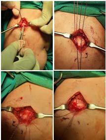
Emergency Obstetric Hysterectomy (EOH) is defined as extirpation of the uterus either at the time of cesarean section or following vaginal delivery or within the puerperium period. This retrospective study was carried out at tertiary health care center in Western India from January, 2019 to February, 2022. Obstetric hysterectomy is still a life-saving surgery in modern obstetrics.The maternal outcome, in terms of reduction in mortality and prevention of postoperative complications, greatly depends on the timely decision, surgical skill and speed of performing obstetric hysterectomy.
Emergency Obstetric Hysterectomy is defined as extirpation of the uterus either at the time of cesarean section or following vaginal delivery, or within the puerperium period. Obstetric emergencies are the most common cause of maternal mortality Worldwide, of which obstetric hemorrhage is the leading contributor 1 Medical methods and conservative measures have all been in practice to manage obstetric hemorrhage effectively 2 With the conservative methods, need for emergency Obstetric Hysterectomy (OH) has reduced to some extent, but still is the last resort to save maternal life in case of massive obstetric hemorrhage3 In a rapidly developing situation, striking a balance between spending excessive time on alternative techniques, leading to further delay and hemorrhage and moving to the definitive lifesaving hysterectomy becomes crucial.
In the developing World, preventable factors such as uterine atony or uterine rupture are the most common indications for Obstetric Hysterectomy; while conditions like postpartum hemorrhage, placenta accreta and placenta previa, apart from uterine rupture have been majorly responsible in our country4,5 Offlate, placenta accreta has been observed to have become the leading indication for emergency peripartum
Department of Obstetrics and Gaynecology, Sardar Vallabhbhai Patel Institute of Medical Sciences and Research, Ahmedabad, Gujarat 380006
1MS (Obstat & Gynaecol), Assistant Professor 2MBBS, Resident Doctor and Corresponding Author 3MD, DGO, Associate Professor, 4MD, DGO, Professor 5MBBS, Resident Doctor
Received on : 10/01/2023
Accepted on : 08/03/2024
Editor's Comment : We should reduce primary cesarean section rate to avoid its devastating complications in future pregnancy and ultimately reduces the need of Obstetric Hysterectomy.
hysterectomy, especially in developed World6,7. This is due to the rising incidence of placenta previa or accreta associated with the increasing number of women with previous cesarean section8
AIMS AND OBJECTIVE
The present study is aimed to evaluate the incidence, indications, postoperative complications, total transfusion of blood products and maternal and fetal outcome in the cases managed by Emergency Obstetric Hysterectomy at a Tertiary Care Hospital.
MATERIALS AND METHODS
This retrospective, record based study was carried out in the department of Obstetrics and Gynecology at Tertiary Health Care Hospital in Western India from January, 2019 to February, 2022 . The study population consisted of 30 patients who underwent obstetric hysterectomy at the study center during the mentioned period.
Inclusion Criteria :
(1) All women admitted in the labour room who underwent Obstetric Hysterectomy during the study period.
(2) All women who underwent Hysterectomy for any indication during pregnancy (including those done for complications of extra uterine pregnancies or molar pregnancies or termination of pregnancy such as perforation and sepsis), labour or puerperium .
Exclusion Criteria :
(1)The women who were operated for Obstetric Hysterectomy outside and were sent to our hospital for further management.
The data was obtained by reviewing the labour room register, operation room register for emergency and elective cases, case records, referral slips and mortality register. The records of all the patients who had undergone Obstetric Hysterectomy were analyzed in detail. The clinical parameters studied were maternal age, parity, whether she was a registered case or referred case, obstetric history, route of termination of pregnancy, methods of induction of labour, indication of Obstetric Hysterectomy, type of Obstetric Hysterectomy, postoperative complications, maternal morbidity and mortality and perinatal outcome (Tables 1-6).
Table 1 — Demographic characteristics
Characteristic
(A) Age
<20 years2 (6.66%)
20-29 years 4 (13.33%)
30-35 years14 (46.66%)
>35 years9 (30%)
(B) Parity
11 (3.33%)
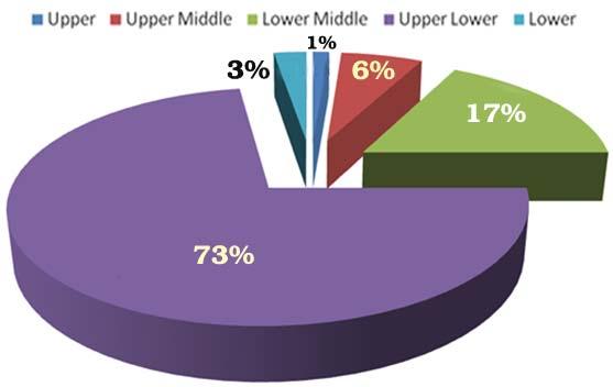
2-522 (73.33%)
>57 (23.33%)
(C) Mode of delivery
Vaginal delivery6 (20%)
Emergency cesarean section 17 (56.66%)
Elective cesarean section7 (23.33%)
(D) Place of residence
Urban19
Rural11
During the three-year study period a total of 15,840 deliveries were performed, of which 30 were EOH yielding a prevalence of 1.8 per 1,000 deliveries. The mean age was 30±5.2 years. Most women had parity between 2 and 5(73.33%). The Caesarean section was the main route of delivery (80%).
Table 2 — Various Indications for Emergency Obstetric Hysterectomy
Indication Number Percentage
Atonic PPH1240
Uterine rupture1 3.33
Morbidly adherent placenta930
Placenta previa516.66
Traumatic PPH310 Total30100
The most common indication for Obstetric Hysterectomy (OH) in this present study was atonic postpartum hemorrhage, which accounted for 12 (40%) cases, followed by morbidly adherent placenta (30%), in placenta previa 5 (16.66%) cases ,uterine rupture in 1 (3.33%) cases, with 3 cases of traumatic postpartum hemorrhage (PPH) completing the numbers.
Majority of the patients required postoperative intensive care. The most common complication was DIC (56.66%) followed by bladder injury (36.66%). Five patients had sepsis.Three patients had acute kidney injury of which one underwent dialysis and recovered and five patients had paralytic ileus. Two patients had blood transfusion reactions. One patient had acute liver injury presenting with jaundice. One patient had post op wound infection and underwent wound resuturing.
DISCUSSION
Hysterectomy is usually used as a last resort to save the life of the mother when all other means fail. The decision to perform Emergency Obstetrical Hysterectomy in the cases under study was easier in multiparous women, unlike primiparous women, where this difficult decision is made to save a life. Though the maternal mortality is reduced thereby, the reproductive capacity of the woman is compromised.
When one is forced to decide upon hysterectomy it is wise to perform it in time before the patient’s condition deteriorates further. Knowledge of this operation and surgical skill saves lives in catastrophic events like morbidly adherent placenta or uterine rupture or intractable PPH. Majority of patients who underwent hysterectomy were in the 30-35 years age group and were multiparous.
In our study the most common indications were Atonic postpartum hemorrhage and placenta accreta (Morbidly adherent placenta), Atonic postpartum hemorrhage, which accounted for 12(40%) cases, which is compared to study of Kapadiya SN, et al in which most common indication of EOH was atonic postpartum hemorrhage (37%). It has been observed that rates of EOH due to uterine atony have decreased with increasing use of medical management, cho’s and B-Lynch sutures. In this study, all medical and surgical methods were used prior to hysterectomy. Morbidly adherent placenta was the second leading cause of Obstetric Hysterectomy in our patient population, with 9(30%) patients undergoing EOH for this reason. A few ones reported morbidly adherent placenta as one of the top 2 indications, depicting the rising trend of cesarean sections leading to abnormal
Table 4 — Blood Transfusion
Indication NumberPacked Fresh frozen Platelets Cryo cell unitsplasma unit units
Atonic PPH1244561630
Uterine rupture135--
Morbidly adherent placenta932287
Placenta previa51963-
Traumatic PPH31015-Total301081102630
Table 5 — Fetal outcome
Fetal outcomeNumber of patients (n=30)Percentage (%)
% Intrauterine fetal demise2 6.66%
Neonatal death 516.18%
Among the 23 live babies 2 intrauterine demise and 5 neonatal deaths were seen.
Table 6 — Relation between delivery- hysterectomy interval and mortality
<4 Hours>4 HoursNo of Patients Mortality268 Survived16622 Total181230
Table shows that if after 4 hours of postpartum period a patient is operated on, then there is an increase in case of mortality as compared to cases operated before 4 hours of postpartum.
placenta getting morbidly adherent, leading to hysterectomy10,11,14 It was observed in 9 out of 30 patients (30%) in the present study, with previous cesarean section being associated with two third of them.Rapidly increasing incidence of cesarean section was one major contributing risk factor in the present study, responsible in 36.4% of the patients; a finding in-line with available evidence9-14.
In this study most common major complication was DIC (56.66%) followed by bladder injury (36.66%) which is compared Kapadiya SN, et al study, in which DIC (48%) and Bladder injury (31%). In this study DIC is the major complication due to severe pre-eclampsia Jaundice and hemorrhage and bladder injuries are because of most cases were of previous cesarean section with adherent placenta at scar site. Bladder is the nearest organ susceptible to injury during hysterectomy.
As our hospital is a Tertiary Care Center so referred patients from other hospitals were in poor conditions such as DIC or shock or hemorrhage, so even after quick assessment and immediate medical and surgical management such as Emergency Obstetrics Hysterectomy, patients condition is difficult to revert back as they were in state of irreversible condition.
The present study confirms the previous observations that Emergency Obstetrical
Hysterectomies are associated with high maternal morbidity and mortality. So we have studied correlation between deliveryhysterectomy Interval associated with Mortality, in which maternal mortality is higher in patients who were operated after 4 hours as compared to operated before 4 hours. morbidity and mortality were due to the condition for which hysterectomy was done and not due to the operative procedure. The majority of complications observed were DIC, Sepsis, Fever, postoperative ICU care, acute kidney injury, wound infection.
The rate of survival is attributed to meticulous technique, good anesthesia and liberal blood transfusion and good intensive care support despite the poor conditions necessitating hysterectomy.
CONCLUSION
Obstetric Hysterectomy is still a life-saving surgery in modern obstetrics. The maternal outcome, in terms of reduction in mortality and prevention of postoperative complications, greatly depends on the timely decision, surgical skill and speed of performing Obstetric Hysterectomy5,15
However, to reduced maternal complications proper monitoring during antenatal period,essential care during labour, strict observation in immediate postpartum period, early recognition of complications, quick actions and timely referral is needed. Medical management in cases of uterine atony, easy availability of blood products for resuscitation of mother plays a crucial role to reduce maternal morbidity and mortality.
By reducing primary cesarean section rate we can avoid its devastating complications in future pregnancy like rupture of uterus and morbidly adherent placenta and ultimately reduces the need of Obstetric Hysterectomy. Thus, this will reduce maternal morbidity and mortality in the long run.
Acknowledgment : Nil.
Source of Support : Nil.
Conflict of Interest : There is no conflict of interest.
Financial disclosure : None.
REFERENCES
1Say L, Chou D, Gemmill A, Tunçalp Ö, Moller AB, Daniels J, et al — Global causes of maternal death: a WHO systematic analysis. The Lancet Global Health 2014; 2(6): e323-33
2Miller S, Lester F, Hensleigh P — CEU: prevention and treatment of postpartum hemorrhage: new advances for low resource settings. J Midwifery Women’s Health 2004; 49(4): 283-92.
Vol 122, No 07, July 2024Journal of
3Najam R, Bansal P, Sharma R, Agarwal D — Emergency obstetric hysterectomy: a retrospective study at a tertiary care hospital. J Clin Diagnostic Res 2010; 4: 2864-68.
4Zeteroglu S, Ustun Y, Engin-Ustun Y, Sahin G, Kamacý M — Peripartum hysterectomy in a teaching hospital in the eastern region of Turkey. EuJObstGynecolReproBiol 2005; 120(1): 57-62.
5Chawla J, Arora CD, Paul M, Ajmani SN — Emergency obstetric hysterectomy: a retrospective study from a teaching hospital in North India over eight years. Oman Med J 2015; 30(3): 181.
7Zamzami TY — Indication of emergency peripartum hysterectomy: review of 17 cases. Arch Gynecol Obstet 2003; 268(3): 131-5.
8Yaegashi N, Chiba-Sekii A, Okamura K — Emergency postpartum hysterectomy in women with placenta previa and prior cesarean section. Int J Gynecol Obstet 2000; 68(1): 49-52.
9Anita K, Kavita W — Emergency obstetric hysterectomy. J Obstet Gynaecol India 2005; 55(2): 132-4.
10Shirodker SD, Pandey A, Yadav S — Emergency obstetric hysterectomy: review at a tertiary care hospital. Int J Reprod Contracept Obstet Gynecol 2016; 5: 3811-4.
11Richa S, Arun N — Emergency obstetric hysterectomy-a retrospective study of 51 cases over a period of 5 years. J Obstet Gynaecol India 2005; 55(5): 428-30.
12Archana K, Bala SP — A Clinical Review of emergency obstetric hysterectomy. J Obstet Gynaecol India 2009; 59: 427-31.
13Koranne PS, Wahane A, Raut D, Bagdiya NJ, Nalat SA, Dhakne MG — An emergency obstetric hysterectomy in modern era: a conventional surgery still saves lives. Int J Scientific Study 2015; 3(1): 72-7.
14Parveen M, Manjeet K, Anju G — Peripartum hysterectomy-a five year study. J Obstet Gynaecol India 2008; 58(6): 504-6.
15Amarin Z — Subtotal Abdominal Hysterectomy. In Alkatout I, Mettler L, eds Springer, Cham. Hysterectomy: A Comprehensive Surgical Approach. 1st ed. Switzerland AG: Springer Nature; 2018: 1093-9.
Ifyouwanttosendyourqueriesandreceivethe responseonanysubjectfromJIMA,pleaseuse the E-mail or Mobile facility. Know
Website:https://onlinejima.com www.ejima.in
For Reception:Mobile : +919477493033
For Editorial:jima1930@rediffmail.com
Mobile : +919477493027
For Circulation:jimacir@gmail.com
Mobile : +919477493037
For Marketing: jimamkt@gmail.com
Mobile : +919477493036
For Accounts: journalaccts@gmail.com
Mobile : +919432211112
For Guideline:https://onlinejima.com
Original Article
Association between BODE Index and Visceral Fat among COPD Patients Attending a Tertiary Care Hospital — A Cross Sectional Study
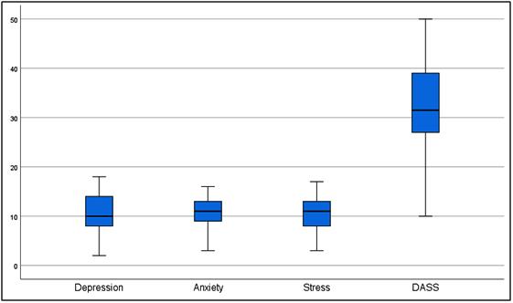
Background : COPD is considered as the 2nd most common cause of death in India. “Body-Mass Index, Airflow Obstruction, Dyspnea and Exercise Capacity Index” (BODE index) predicts 4-year survival rate, making it a better tool for advanced study. So far, the degree of visceral fat accumulation in COPD patients & its effect on their survival rate has not been directly studied in Indian population.
Aims and Objective : To measure the visceral adiposity, estimate the BODE index in COPD Patients & assess the relation between Visceral adiposity & BODE Index in them.
Material and Method : 78 COPD patients were recruited. The BMI, pulmonary function, dyspnea score and the 6minute Walk Tests were done. Visceral fat was measured by Ultrasonography. Patients were divided into 2 groups based on the presence or absence of visceral obesity. BODE index and Approximate Survival rate were compared among them.
Results : The prevalence of visceral obesity in COPD patients was 45.37%. Average BODE index Score was 4.32±1.78 and it was higher in viscerally obese patients.
Conclusion : BODE index worsens with the presence of visceral adiposity. Decreasing visceral fat will improve BODE index & survival rate and decrease cardiovascular risks. Counselling COPD patients in this regard can prevent the progression of COPD.
Key words :COPD, Visceral Adiposity, BODE Index.
According to Global Initiative for Chronic Obstructive Lung Disease (GOLD), Chronic Obstructive Pulmonary Disease (COPD) is a common, preventable and treatable disease that is characterized by persistent respiratory symptoms such as dyspnea, cough and or sputum production. There is presence of airflow limitation that is due to airway and/or alveolar abnormalities usually caused by significant exposure to noxious particles or gases1
Obesity is a chronic disease which is prevalent in developed and developing countries like India and in all strata of society2. As the standards of living are continuing to rise, weight gain and obesity are posing a growing threat to health due to the lifestyle changes. Many theories have been put forward as to how the abdominal fat can affect the lung functions. Among
1MBBS, MD, Resident Physician - PGY1 Department of Internal Medicine, University of Texas Rio Grande Valley / Valley Baptist Medical Center, 2101 Pease St, Harlingen, Texas - 78550, USA
2MBBS, MD, Professor and Head, Department of Physiology, St Peters Medical College Hospital and Research Institute, Krishnagiri, Tamilnadu 635130 and Corresponding Author
3MBBS, MD, Associate Professor, Department of Pulmonary Medicine, MVJ Medical College, Bengaluru, Karnataka 562114
4MBBS, MD, Professor, Department of Radiodiagnosis, Vydehi Institute of Medical Sciences & Research Centre, #82, Nallurahalli, Whitefield, Bangalore 560066
5MBBS,Tutor, Department of Physiology, Chitradurga Institute of Medical Sciences & Research Centre, Karnataka 577501
Received on : 02/09/2022
Accepted on : 09/04/2024
[J Indian Med Assoc 2024; 122(7): 21-7]
Editor's Comment :
Obesity is considered as the mother of all diseases. A healthy weight is crucial in preventing spectrum of diseases, from coronary artery disease to cancer.
Even though our study mainly focuses on the importance of reducing obesity in COPD patients, we want to reinforce the fact that obesity is the cornerstone in preventing many of the diseases plaguing the world today.
This obesity epidemic as we call it, is an enormous burden on the healthcare system and hence should be prioritized, prevented and treated, thereby closing the gateway to all chronic diseases that ensue.
At the community level the policymakers can organize campaigns in localities and schools to create awareness about the ill effects of obesity among parents and children alike.
As they say Education begins at home, we should address this issue at the grass root level by enlightening the parents who in turn can lead their children into a healthier future.
them most important are :
(1)Visceral Adipose Tissue (VAT) is considered to be more metabolically active than Subcutaneous Adipose Tissue (SAT). VAT secretes more inflammatory cytokines, IL-6 and CRP. Elevated CRP and IL-6 is found to be associated with chronic inflammatory airway diseases.
(2)Activated macrophages in adipose tissues are known to cause low grade chronic inflammation;
(3)Adipose tissue in the abdominal cavity compresses the thoracic cage, diaphragm and lungs.
122, No 07, July 2024Journal
The consequences are a decrease in diaphragm displacement, a decrease in lung and chest wall compliance and an increase in elastic recoil, resulting in decrease in lung volumes and an overload of inspiratory muscles3.
COPD has been considered as the second most common cause of death in the list of non-communicable diseases in India4. By 2030, COPD is predicted to be the 3rd cause of death in India. Our country ranks 5th in the list of the most polluted countries in the World. It is not only the men who travel in a polluted environment or smoke that are affected; the adult women and young children are also at a high risk as they are exposed to solid fuels due to their household roles4
Therefore prevention, diagnosis and early treatment play a very crucial role in preventing the mortality in an easily preventable non-communicable disease like COPD. The risk of death in patients with COPD is often graded with the use of a single physiological variable, the forced expiratory volume in first second (FEV1). However, other risk factors, such as a short distance walked in a fixed time, a high degree of functional breathlessness and a low Body-Mass Index (BMI), are also associated with an increased risk of death. “Body-Mass Index, Airflow Obstruction, Dyspnea and Exercise Capacity Index” (BODE index) is a multidimensional grading system, which integrates body mass index, airflow limitation (forced expiratory volume in one second), dyspnea and 6-min walk distance, predicts 4-year survival rate in COPD patients5. In hindsight, BODE Index includes all the effective parameters for accurate mortality prediction, thereby making it a better match for a more informative & advanced study. So, we considered taking the BODE Index in our study.
Previous studies that have suggested links between COPD, cardiovascular diseases and abdominal obesity have used indirect or surrogate markers for abdominal obesity6. So far, the degree of visceral fat accumulation in patients with COPD & its effect on their survival rate has not been directly studied in Indian population. So, the present study aimed at assessing the relation between BODE Index, its individual parameters & visceral adiposity in COPD patients.
MATERIAL AND METHOD
Type of Study : It was a hospital based cross sectional study.
Duration of Study : The data collection was done over a period of 2 months. Ethical clearance was taken from Institutional Ethical Committee.
Stable COPD patients (without acute exacerbation) who were attending the OPD or admitted in the wards
in the Departments of General Medicine & TB and Chest were recruited in the study. The study protocol was explained to the patients in their vernacular language & the patients who agreed to be a part of the study and gave the written informed consent for the same were included in the study.
Sample Size :
(1)The sample size was obtained by purposive sampling method.
(2)The minimum sample size was set to 78 participants with study prevalence of COPD of 5.1%7 and 5% margin of error.
(3)n = 4pq/d2 n = 4x5.1x94.9/25 n =77.43 ~ 78 (Round off to next Whole No)
Patients who fulfilled the following inclusion & exclusion criteria were included in the study.
Inclusion Criteria :
(1)Male or female COPD patients between 30 to 65 years of age.
(2)Patients with Stage 2 to Stage 4 COPD according to GOLD classification.
(3)A baseline (post bronchodilator) FEV1<80% of predicted normal and a baseline (post bronchodilator) FEV1/FVC ratio <0.7.
(4)A signed and informed consent was obtained prior to participation.
Exclusion Criteria :
(1)Patients who were hypertensive or diabetic.
(2)Patients who were morbidly obese.
(3)Patients with Ischemic heart disease.
(4)Patients who had musculoskeletal disorders which can limit their walking ability.
(5)Patients who had undergone previous lung surgeries.
(6)Patients who had any other lung disorder other than COPD.
Tools for Data Collection :
(1)Socio-demographic details
Age, sex, occupation, residence, smoking and Socio-economic status were recorded.
(2)Physiological parameters
Height (cms), weight (Kgs) and Resting Pulse rate were recorded.
(3)Visceral fat and subcutaneous fat:
(a)Ultrasonography (USG) has been shown to be an alternative, noninvasive, reliable method to estimate the two fat compartments8.
(b)The two fat compartments were estimated by ultrasound method in the radiology department.
122, No 07, July 2024Journal
Ultrasound was done using SIEMENS ACUSON 300 with multiple frequency (2-5 MHz) convex probe for measuring visceral fat and linear probe (7-12 MHz) for measuring subcutaneous fat.
(c)Visceral and Subcutaneous fat was measured midway between the umbilicus and xiphisternum. Visceral fat was defined as the depth from the peritoneum to the lumbar spine and subcutaneous fat defined as the depth from the skin to the abdominal muscles9. The cut off value of Visceral Fat Thickness (VFT) of 9 cm- in men and; 6.9 cm in women was used to define visceral obesity9
(4)BODE Index – Body Mass Index, Airway Obstruction, Dyspnea, and Exercise.
(a)BodyMassIndex(BMI)—
Patient’s BMI was calculated by using formula:kg/m2. We classified the BMI of the patients according to the scale given by National Institutes of Health; BMI was classified as10 :
d)Exercise—6-minuteWalkDistance
The 6-minute Walk Distance (6MWD) has been proven useful in assessing the functional status of patients with COPD because it is easy to perform, inexpensive and amenable to standardization5. Most of the day-to-day chores are performed at sub maximal levels of exertion; therefore, 6MWD would better reflect the functional exercise level for daily physical activities12.
Patients were instructed about the procedure. Necessary precautions were taken to attend to any emergencies and equipment like pulse oximeter, oxygen cylinder, emergency drugs, etc, were kept within a hand’s reach. The stop watch was set for 6 minutes. The patient was asked to walk for 6 minutes on a flat, firm surface in an empty corridor of 30 m distance. If the patient felt breathless/ tired at any point of time during the walk, he/ she was allowed to take rest without stopping the clock and encouraged to continue to finish 6-minute walk. The total distance walked in 6 minutes was noted and the points were given based on the distance covered1
Obesity:Grade130-34.9
Grade235-39.9
Extremely/Morbidlyobese:Grade3 Obesity>40
(b)AirwayObstruction—
Pulmonary Function Tests was performed using an RMS Medspiror. Post bronchodilator obstruction was assessed by means of FEV1% predicted and FEV1/ FVC values.
FEV1% predicted: normal value of FVC is above 80% predicted.
FEV1/FVC : The ratio of FEV1/FVC is normally between 0.7 and 0.8. Values below 0.7 are a marker of airway obstruction11
(c)Dyspnea—
Interpretation of breathlessness was done using modified British Medical Research Council (mMRC) Questionnaire. This questionnaire is considered to be adequate and selfexplanatory. mMRC relates well to other measures of health status and predicts future mortality risk2
Dyspnea was graded based on the modified MRC dyspnea scale2 : —
BODE index were calculated as per the following Tables5
Variables Points on BODE Index 0123
FEV1 (% of predicted) >6550-6436-49 <35 Distance walked in 6 min (m) >350250-349150-249 <149 MMRC dyspnea scale0-1234 Body-mass index>21 <21
BODE Index Scoring4 Year survival rate
0-2 points80%
3-4 points67%
5-6 points57% 7-10 points18%
Grade 0No dyspneaI only get breathless with strenuous exercise
Grade 1 SlightI get short of breath when hurrying on dyspnea level ground or walking up a slight hill
Grade 2ModerateOn level ground, I walk slower than dyspnea people of the same age because of breathlessness or I have to stop for breath when walking at my own pace on the level
Grade 3 SevereI stop for breath after walking about dyspnea 100 yards or after a few minutes on level ground
Grade 4Very severeI am too breathless to leave the house dyspneaor I am breathless when dressing
Approximate 4-year survival interpretation was predicted as under (based on BODE Index Scoring)5: (5)Protocol for Analysis : COPD patients were divided into 2 groups based on the presence or absence of visceral obesity (Visceral Fat thickness >9 cm in men; >6.9 cm in women). BODE index was compared among the patients with visceral obesity & patients without visceral obesity. Approximate Survival rate were compared among the 2 groups. (6) Statistical Analysis : Data was expressed in terms
of actual numbers, mean ± SD, frequency and percentage. Statistical analysis was done by using SPSS software version 20. Chi-square test was used to compare the parameters between the 2 groups and P value of < 0.05 was taken as statistically significant. Descriptive statistics and Pearson’s correlation were used for comparison between the factors.
OBSERVATION
A total of 78 COPD patients were recruited in the present study based on the inclusion & exclusion criteria. All of these patients completed the study protocol. Out of a total of 78 patients recruited, 55 of them were male (70.5%) and 23 (29.5%) were females. The mean age & BODE Index among the COPD patients was calculated. The results are displayed in the Table 1.
Table 1 — Subject’s mean age & BODE index parameters
Variables Mean±Standard Deviation
Age (years)54.01±15.12
BMI (Kg/m2)24.07±4.69
Obstruction (FEV1% pred) 46.38±15.61
Dyspnea (mMRC scale)1.31±0.71
Exercise -6MWD (m)194.99±62.59
Total points (BODE Index)4.32±1.78
(BMI - Body Mass Index; FEV1 – Forced Expiratory Volume in 1st second; 6MWD - 6-Minute Walk Distance.)
All COPD patients were subjected to ultrasound examination for measuring the amount of visceral fat as described above. We then divided the patients (n=78) into two groups, based on the visceral fat cut off values as COPD Patients with visceral adiposity and COPD patients without visceral adiposity. It was noticed that out of all the COPD Patients (n=78), 33 patients had Visceral Adiposity and 45 Patients did not have Visceral Adiposity. We further checked the BODE Index among the two groups. It portrayed that the BODE Index was higher in the patients with Visceral Adiposity concluding that their 4-year survival rate was lesser as depicted in the Table 2.
Table 2 — Comparison of BODE Index among viscerally Obese & Non-obese COPD patients
VisceralNo of Percentage BODE IndexP value
Adiposity Patients (Mean±Standard deviation)
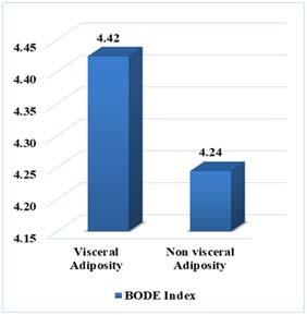
YES3342.37%4.42±1.80 0.73 NO4557.7% 4.24±0.10
BODE Index Parameters & Visceral Fat :
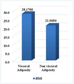
It was observed that the BMI was significantly greater (p=0.000) in patients with visceral adiposity when compared to patients without Visceral Adiposity (Fig 1).
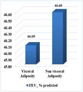
Patients with visceral adiposity did have more obstruction when compared to patients without visceral adiposity. But the difference was not statistically significant as p=0.888 (Fig 2).
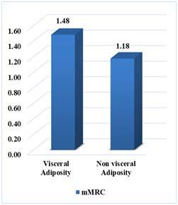
The mean Dyspnea score among viscerally obese patients was significantly more than patients without visceral adiposity (p=0.049). The mean Dyspnea score in patients with visceral obesity was 1.48±0.7 as compared to the score of 1.18±0.06 in viscerally nonobese patients (Fig 3).
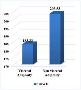
All COPD patients completed the 6-minute walk test, though some took a break while the clock was still on & later walked to complete 6 minutes. The total distance walked by patients with visceral adiposity (183±67.6m) was lesser when compared to patients without visceral adiposity (203.53±58.9m). But it was not significant as p=0.160 (Fig 4).
It was observed that patients with visceral adiposity had a higher BODE Index when compared to patients
Fig 1 — Relation between BMI & Visceral fat
Fig 2 — mMRC Dyspnea score in COPD Patients
without visceral adiposity which was not significant as p=0.382 (Fig 5).
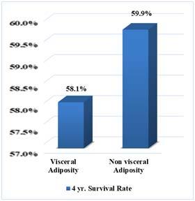
Higher BODE index reflects poor survival rate. So, the 4 Year survival rate in patients with visceral adiposity was lesser than that of patients without visceral adiposity, though was not statistically significant (p=0.706) (Fig 6). Figures showing the comparison of BODE Index individual parameters among the two groups.
Correlation of BMI, Obstruction, Dyspnea and 6min Walk Test with Visceral Adiposity : We tried to correlate BMI, Obstruction, Dyspnea and Exercise with visceral adiposity to know whether any consistent relationship existed using Pearson’s correlation.
There was strong correlation of BMI with Visceral Adiposity. There was intermediate correlation between dyspnea score & Visceral adiposity and other parameters such as Obstruction and Exercise showed weak correlation with Visceral Adiposity as in Table3
DISCUSSION
In our study the prevalence of visceral adiposity among COPD patients was 42.3%. This is in line with the findings of Furutate, et al where the prevalence of visceral adiposity among COPD patients was found to be 52.5% by CT measurement as compared to 38.7% in the control group6. Earlier studies by Steuten, et al reported only 18% prevalence of obesity which was based on the BMI13
Fig 3 — FEV1 % predicted in COPD Patients
Fig 4 — 6-minute Walk Distance in COPD Patients
Fig 6 — 4 year Survival Rate among COPD patients
Fig 5 — Comparison of BODE Index in COPD patients
122, No 07, July 2024Journal
Table 3 — Correlation of BMI, Obstruction, Dyspnea and 6-min walk Test with visceral adiposity
Increase in the visceral fat in COPD may be related to physical inactivity which leads onto excessive fat accumulation in them. Along with this COPD patients are treated with glucocorticoids during exacerbations; this systemic corticosteroid therapy may cause glucocorticoids – mediated redistribution of stored energy & stimulatory effect on food intake leading to visceral obesity6
The BODE index which predicts the 4-year survival rate is also an important prognostic predictor of COPD. BODE Index in our study was found to be less in patients with visceral obesity as compared to nonobese individuals. These findings suggest that Visceral obesity adds on to the burden of morbidity & mortality in COPD patients14
In our study, the mean Body Mass Index was more in Viscerally Obese patients, this may reflect coexistence of general obesity with visceral obesity in these individuals.
Further analysis revealed that in a small fraction of patient’s visceral obesity was seen though their BMI was less than normal. These were the patients in whom the obstruction was more severe (GOLD stage 3 &4). Similar findings were found by Furutate, et al, wherein the prevalence of non-obese subjects with increased VFA was greater in the patients with more obstruction than in those with lesser airflow limitation according to GOLD stages. In these patients there is muscle loss leading on to lesser BMI6
ECLIPSE, a cohort study reported that there was increased Visceral Adipose Tissue and fat accumulation in the muscle tissue in COPD patients with severe airflow limitation (mean FEV1, 40.7% predicted). In this study, muscle fat accumulations were measured by muscle tissue attenuation on CT scan of the thorax among smoking and nonsmoking COPD patients15.
Though FEV 1% predicted was not significantly different between the 2 groups, our study revealed that FEV1% predicted had a negative correlation with the visceral fat.
This decrease in lung function can be attributed to the systemic inflammation triggered by adipose tissue.
Visceral fat tissue is known to be more metabolically active than subcutaneous fat. This secretes or synthesizes inflammatory cytokines, such as tumor necrotic factor –α and interleukins- 6 which are known to be associated with chronic inflammatory airway disease as discussed earlier3.
Dyspnea Scores & Visceral Fat :
Perceived breathlessness as graded by mMRC scale was significantly more severe among viscerally obese patients than the other group. Severity of dyspnea was more in Viscerally obese individuals and it positively correlated with the mMRC scale.
Exercising capacity as estimated by 6-minute Walk Test was less among the viscerally obese patients as compared to others. The total distance covered by viscerally obese patients was less. Earlier study done by Serres, et al showed that physical inactivity is a consequence of the so- called dyspnea spiral, in which COPD patients tend to adopt a sedentary life style to avoid dyspnea16
Further analysis did not show a significant correlation between 6MWD & visceral obesity. As we had ruled out other cardiorespiratory conditions which could further limit the amount of exercise may have been the reason. And also, the lung functions between the groups were not significantly different, there was not much difference in the exercise capacity also. Similar findings were observed in a Japanese study, which was explained by the fact that though 6MWD could be indicative of exercise capacity, it does not necessarily reflect the total amount of physical activity in day-to-day activities6. This could further be explored by using the physical activity monitors in future studies.
The present study has various strengths. First this study included participants in whom the abdominal adipose tissue was measured directly with the help of ultrasound unlike previous studies that used BMI or Waist circumference as surrogate markers of abdominal obesity. We conducted tests of all the parameters that affects a COPD patient with Visceral Adiposity which makes it a reliable and advanced study.
Limitations of the Study
:
There are few limitations in our study. Firstly, as this study was done on a relatively small number of patients due to restriction of time, increasing the sample size would yield refined results. Second, in
122, No 07, July 2024Journal of
our present study we failed to document the corticosteroid intake in our patients, as this also could affect the visceral fat deposition.
CONCLUSION
The National Health Policy of India 2017 recommends that premature mortality from noncommunicable diseases, including chronic respiratory diseases, should be reduced by 25% by 20256. As visceral fat has been proved to deteriorate the survival rate in COPD patients, steps should be taken to decrease the visceral fat, along with the standard pharmacological intervention, thereby decreasing the mortality in COPD patients.
Visceral obesity can be decreased by making certain lifestyle changes :
Inculcating a fiber rich diet with less of fats and carbohydrate.
Exercising on a regular basis within the capacity of the patient which depends on the stage of COPD.
The results of this study can be used by health care professionals to educate their patients regarding the importance of decreasing the visceral fat, which can improve their lung functions and prevent the progression the COPD.
REFERENCES
1Global Initiative for Chronic Obstructive Lung Disease Global Initiative for Chronic Obstructive Lung: Pocket Guide to COPD Diagnosis, Management and Prevention, 2017 Report.
2Chowdhury SG, Mandal O, Datta A, Chakraborti D — Effect of Central Adiposity on Lung Function Tests in Young Adults. IOSR Journal of Dental and Medical Sciences 2015; 14(8): 24-8.
3Choe EK, Kang HY, Lee Y — The longitudinal association between changes in lung function and changes in abdominal visceral obesity in Korean non-smokers. PLOS ONE 2018; 6-7.
4ICMR-PHFI-IHME (2017) India: Health of the Nation States.
5Celli BR, Cote CG, Marin JM — The Body-Mass Index, Airflow Obstruction, Dyspnea, and Exercise Capacity Index in Chronic Obstructive Pulmonary Disease. The New England Journal of Medicine. March 4, 2004: 1006
6Furutate R, Ishii T, Wakabayashi R — Excessive visceral fat accumulation in advanced COPD. International Journal of COPD 2011; 6: 423.
7Rajkumar P — A cross-sectional study on prevalence of chronic obstructive pulmonary disease (COPD) in India: rationale and methods. BMJ Open 2017; 7: e015211.
8Hirooka M, Kumagi T, Kurose K, Nakanishi S, Michitaka K, Matsuura B, et al —A Technique for the measurement of visceral fat by ultrasonography. Comparison of measurements by ultrasonography and computed tomography. Intern Med 2005; 44: 794-9.
9Premanath M, Basavanagowdappa H, Mahesh M — Correlation of abdominal adiposity with components of metabolic syndrome, anthropometric parameters and Insulin resistance, in obese and non-obese, diabetics and nondiabetics: A cross sectional observational study. (Mysore Visceral Adiposity in Diabetes). Indian Journal of Endocrinology and Metabolism 2014; 18(5): 676-82.
10https://www.nhlbi.nih.gov/health/educational/losewt/BMI/ bmi_dis.htm. (Losing Weight, Body Mass Iindex (nih.gov)).
11Spirometry for Health Care Providers Global Initiative for Chronic Obstructive Lung Disease (GOLD).
12American Thoracic Society: Guidelines for the Six-Minute Walk Test.
13Steuten LM, Creutzberg EC, Vrijhoef HJ — COPD as a multicomponent disease: inventory of dyspnea, underweight, obesity and fat free mass depletion in primary care. Prim Care Respir J 2006; 15: 84-91.
14Bartolome R Celli — The Bode Mass Index, Airflow Obstruction, Dyspnea and Exercise Capacity Index In COPD. N Eng J Med, Arch 2004; 350(10): 1005-12.
15Martin M, Almeras N, Despres JP, Coxson HO, Washko GR, Vivodtzev I, et al — Ectopic fat accumulation in patients with COPD: an ECLIPSE sub study. Int J Chron Obstruct Pulmon Dis 2017; 12: 451-60.
16Serres I, Gautier V, Varray A, Préfaut C — Impaired skeletal muscle endurance related to physical inactivity and altered lung function in COPD patients. Chest 1998; 113(4): 900-5.
Original Article
Screening for Congenital Hypothyroidism — Umbilical Cord Blood TSH a Useful Tool : A Single Centre Eight Year Experience
Ravi Bhatia1, Gunjan Bhatia2, Dinesh Rajwaniya3, Priyanka Mishra4, Jobanjeet Kaur4
Background : Congenital hypothyroidism remains the most common preventable cause of mental retardation in pediatric age group. Screening for Congenital Hypothyroidism remains an effective tool to prevent mental retardation among the general population. Umbilical Cord blood TSH is an easily available reliable tool for screening for Congenital Hypothyroidism.
Aims and Objectives : The aim of the study was to find the normative Cord Blood TSH value for the study group and to assess the utility of Cord Blood TSH as a screening tool for Congenital Hypothyroidism.
Design : Cross Sectional study.
Material and Methods : CB TSH levels were measured in 8720 neonates over a study period of eight years. All neonates who had a cord blood TSH level greater than 20 mIU/L were called back on day 7 of life for a repeat thyroid profile.
Results : Cord blood samples of 8848 neonates were sent for analysis, 128 samples were hemolysed hence only 8720 were analysed. A total of 8720 neonates formed the study group. Male to Female ratio was 4720:4000 ie, 1.18:1. The birth weight of the study group ranged between 0.9 kg to 4.2 kg. The average birth weight was 2.92 kg. The study group was divided into two cohorts, cohort 1 comprised of babies who were term gestation (6366 babies, 73%), cohort 2 comprised of babies who were born between after 28 weeks of gestation but before 37 weeks of gestation (2354 babies, 27%). The mean TSH value of the entire study group was 7.34 mIU/ml. majority of neonates (93%) had a cord blood TSH level less than 10 mIU/L. In 130 neonates had a cord blood TSH value greater than 20 and were recalled for a repeat testing on day 7 of life. Out of the 130 neonates recalled for repeat testing (recall rate 1.48%), only 104 neonates turned up, 26 were lost to follow-up. Out of the 104 neonates which turned up for repeat testing, 4 eventually turned to be hypothyroid on repeat testing giving us an incidence of 1 in 2180. TSH values corresponding to the 3rd, 10th, 25 th, 50th, 75th, 90th, 95th and 97th percentile were 2.2, 3.1, 3.9, 6.1, 7.1, 10.67, 13.8, 23 respectively. 98% of our study group had a cord blood TSH value less than 20, so a cord blood TSH of greater than 20 mIU/ml can safely be used for screening for congenital hypothyroidism.
Conclusion : A cut off of Cord Blood TSH value >20 mIU/ml can be used as a screening tool for Congenital Hypothyroidism.
Key words :Congenital Hypothyroidsm, Cord Blood TSH.
Congenital Hypothyroidism remains the commonest cause of preventable mental retardation in the country. In the absence of a nationwide screening program for Congenital Hypothyroidism various studies across pan India have reported the incidence of Congenital Hypothyroidism to range from as low as 1: 3400 to as high as 1: 5001-3. Since clinical features of Congenital Hypothyroidism are quite non specific a high index of suspicion is required for making a diagnosis of Congenital Hypothyroidism. Amongst the
Department of Pediatrics, Pacific Medical College and Hospital, Udaipur, Rajasthan 313001
1DNB (Paediatrics), Professor and Head and Corresponding
Author
2MD (Pathology), Assistant Professor, Department of Pathology, RNT Medical College, Udaipur, Rajasthan 313001
3MD (Paediatrics), Professor
4MBBS, Resident
Received on : 24/10/2022
Accepted on : 08/12/2023
[J Indian Med Assoc 2024; 122(7): 28-31]
Editor's Comment :
Congenital Hypothyroidism remains one of the most common preventable causes of mental retardation among children. Umbilical cord blood TSH level offers an easily available alternative for screening for Congenital Hypothyroidism.
various clinical features prolongation of neonatal jaundice is a key feature amongst patients who have been diagnosed with Congenital Hypothyroidism. Since the clinical features are quite non specific screening for Congenital Hypothyroidism remains imperative and is of paramount importance. Screening for Congenital Hypothyroidism remains the most cost effective method to prevent mental retardation amongst children.
Ideally screening for Congenital Hypothyroidism should be done after 72 hours of life since by then the TSH surge is over but in developing countries wherein the health resources are limited many babies get discharged early and are deprived of screening for
122, No 07, July 2024Journal
Congenital Hypothyroidism. In such scenarios Umbilical Cord Blood comes as an easily available alternative for screening for Congenital Hypothyroidism. Various Asian nations have used Umbilical Cord Blood TSH levels as a screening tool for Congenital Hypothyroidism4-6. There are a few studies from across India where researchers have studied Cord Blood TSH levels as a screening tool for Congenital Hypothyroidism7-14. We started this research project way back in 2014 and are using Cord Blood TSH levels as a screening tool for Congenital Hypothyroidism.
AIMS AND OBJECTIVES
(1)To find the normative values of Cord blood TSH of the study group.
(2)To use Cord Blood TSH level as a screening tool for Congenital Hypothyroidism
MATERIAL AND METHODS
This cross sectional study was carried from January, 2014 to January, 2022 in a private medical college in Rajasthan. Prior approval from the Institutional Ethics Committee was sought before starting the study.
Inclusion Criterion :
All neonates whose gestational age was greater than 28 weeks who were born during the study period formed the study group.
Exclusion Criteria :
(a)All neonates whose gestational age was less than 28 weeks.
(b) All neonates who required resuscitation at birth.
(c)Neonates who were admitted to NICU immediately after birth.
(d)Neonates with major congenital malformations.
(e)Neonates whose mothers were on medications for thyroid disorders
Detailed antenatal history, parity, medical history of mother, birth weight of baby , sex etc were recorded on a pre designed Performa. The umbilical cord was clamped using three clamp technique, one close to the baby and two near the placental end after cessations of pulsations. 5 ml of blood was collected at the time of delivery and was subjected to TSH estimation by chemiluminescence immunoassay method(kit supplied by Roche E411). Cord Blood TSH samples of 8848 neonates were sent for estimation, 128 samples were found hemolysed hence 8720 samples were analysed.
All those neonates whose Cord Blood TSH value was greater than 20 mIU/ml were called back on day 7 of life for a full thyroid profile which meant TSH, T3, T4, fT3 and fT4. If the venous TSH sample value was
greater than 20 mIU/ml it was considered confirmatory for Congenital Hypothyroidism. All the data collected was entered on an excel worksheet and analyzed using SSPS software for windows version (IBM, India).
RESULTS
A total of 8720 neonates formed the study group. Male to Female ratio was 4720:4000 ie, 1.18:1 (Table 1).
Table 1 — Gender wise distribution of the Study group
Gendern (8720)
Male 4720 (54.12%)
Female 4000 (45.88%)
The birth weight of the study group ranged between 0.9 kg to 4.2 kg. The average birth weight was 2.92 kg. Table 2 depicts the weight wise distribution of the study cohort.
Table 2 — Birth weight wise distribution of the study group (n=8720)
Birth Weight in KgNumber of Neonates
< 1118(1.3%)
1.0-1.499 228(2.69%) 1.5-1.99 668(7.66%)
2.0-2.49 906(10.38%) 2.5-2.99 4820(55.27%)
31980(22.70%)
The study group was divided into two cohorts, cohort 1 comprised of babies who were term gestation (6366 babies, 73%), cohort 2 comprised of babies who were born between after 28 weeks of gestation but before 37 weeks of gestation( 2354 babies, 27%). The mean TSH value of the entire study group was 7.34 mIU/ml. majority of neonates (93%) had a cord blood TSH level less than 10 mIU/L. The Distribution of study group according to Cord Blood TSH values is given in Table 3.
Table 3 — Distribution of Cord Blood TSH values in the study group
TSH value(mIU/ml)n= 8720(%)
Below 108110(93%)
11-20480(5.51%)
21-50 124(1.42%)
>5006(0.07%)
In 130 neonates had a cord blood TSH value greater than 20 and were recalled for a repeat testing on day 7 of life. Out of the 130 neonates recalled for repeat testing (recall rate 1.48%), only 104 neonates turned up, 26 were lost to follow up. Out of the 104 neonates which turned up for repeat testing, 4 eventually turned to be hypothyroid on repeat testing giving us an incidence of 1 in 2180. TSH values corresponding to
Vol 122, No 07, July 2024Journal
the 3 rd , 10 th , 25 th , 50 th , 75 th , 90 th , 95 th and 97 th percentile were 2.2, 3.1,3.9, 6.1, 7.1, 10.67, 13.8, 23 respectively. 98% of our study group had a cord blood TSH value less than 20, so a cord blood TSH of greater than 20 mIU/ml can safely be used for screening for congenital hypothyroidism.
DISCUSSION
Since Congenital Hypothyroidism is the most common preventable cause of mental retardation among children screening for Congenital Hypothyroidism remains of paramount importance. Furthermore, since the symptoms of Congenital Hypothyroidism are quite non specific in nature the only way to pick up cases early is by screening neonates for Congenital Hypothyroidism. Unfortunately in the absence of a nationwide screening program for Congenital Hypothyroidism studies from across the country are few. Lack of awareness, over reliance on a venous sample, cost issue have all lead to screening for Congenital Hypothyroidism not being implemented on a nationwide basis. Many neonatologists are skeptical of using cord blood TSH as a screening tool for Congenital Hypothyroidism since Cord Blood TSH values are influenced by various maternal and perinatal factors but researchers across the World especially from South East Asia have demonstrated that Cord Blood TSH estimation is more practical and cost effective screening tool. In our study only 7% of the study group had a Cord Blood TSH value greater than 10 mIU/ml which is quite similar to that reported by other authors. The mean Cord blood TSH value of our study group was 7.34 mIU/ml which is quite similar to what reported by Manglik, et al; Bhatia, et al. Our recall rate in the study was 1.48% whilch is lower to what reported by Wu, et al which had a larger cohort of 11000 neonates. Manglik et.al reported a recall rate of 1.833% but their cohort included only term neonates. Zion, et al in their study had a recall rate as high as 10% but their sample size was quite small only 73 neonates. Another study from our state had a high recall rate of 5.57% which is quite high as compared to other studies from across the country.
There is no universally accepted cut off for TSH levels as far as screening is concerned. Majority of researchers who have studied Cord Blood TSH as a screening tool have used the cut off level of 20. Had we used a cut off of 30, our recall rate would have fallen to 1.2% and a higher cut off of 40 would have seen a recall rate fall to 0.8%. Annually 20 million babies are born in our country every year, a recall rate of 1.48% would mean 2.96 lacs babies being called
back for repeat testing. It would put a huge burden on our health care systems. The best option would be to have large scale nationwide studies so that a consensus on a cord blood TSH cut off value be derived for uniform testing.
Four babies in our cohort of 8720 neonates turned out to hypothyroid giving us an incidence of 1 in 2180. From our own centre we had earlier reported an incidence of 1 in 1824 but that study included only term neonates. Another study from our state has reported an incidence of 1 in 1370. The incidence of Congenital hypothyroidism from across India from various studies is as varied as 1 in 248 to 1 in 170015-16. Geographic factors, sample size and cut off used for screening are known to influence the incidence of the disease.
Our figures have shown a comparable trend with the normative values for Cord Blood TSH as reported by other researchers across the World.
One big drawback of our study in spite of it being a eight year long research project is the relatively small sample size as it being a private medical college the number of deliveries are less.
We require large population based multi centric studies to gauge and calculate the incidence of Congenital Hypothyroidism in the country.
CONCLUSION
When compared to data from Western World the incidence of Congenital Hypothyroidism seems to be high probably due to the methodology used. Delay in diagnosis can lead to permanent intellectual impairment. With babies being discharged early venous sample or a heel prick testing doesn’t seem to be a practical option in our country, hence the use of Umbilical Cord Blood TSH is practical and easily available option for screening for Congenital Hypothyroidism in our country. Various studies including ours have reiterated the fact that a cut off of Cord Blood TSH value >20 mIU/ml can be used safely for screening of Congenital Hypothyroidism. Still we need large scale multi centric population based studies to further establish normative values for Cord blood TSH values.
REFERENCES
1Sanghvi U, Diwakar KK — Universal newborn screening for congenital hypothyroidism. Indian Pediatr 2008; 45: 331-2.
2Singh Ra, Devi KG, Devi Kl, Bainik U — Newborn screening for Congenital hypothyroidism in Manipur by measurement of umbilical cord thyroid stimulating hormone: A hospital based study. Med Soc 2013; 27: 127-30.
3Kaur G, Srivastav J, Jain S, Chawla D, Chavan BS, Atwal R, etal—Preliminary report on neonatal screening for Congenital
Vol 122, No 07, July 2024Journal of the Indian Medical Association
hypothyroidism, congenital adrenal hyperplasia and glucose -6- phosphate dehydrogenase deficiency : A Chandigarh experience. Indian J Pediatr 2010; 77: 969-73.
4Wu LL, Sazali BB, Adeeb N, Khalid BA — Congenital hypothyroid screening using Cord blood TSH. SingaporeMed J 1999; 40: 23-6.
5Mekennon Y, Gizachew WH, Chamiso B, Raue F — Thyroid stimulating hormones in cord blood in neonates. Ethiop Health Dev 2003; 17: 125-30.
6Ordoookhani A, Miriman P, Najafi R, Hedayati M, Azizi F — Congenital Hypothyroidism in Iran. Indian J Pediatr 2003; 70: 625-8.
7Chaudhary M, Soni JP, Goyal VK, Sharma P, Makwana M, Jora R, et al — Incidence of congenital hypothyroidism in western Rajasthan using cord blood thyroid – stimulating hormones as a screening tool : A cross – sectional hospital based study. Indian J Endocrinol Metab 2018; 22: 417-20.
8Bhatia R, Rajwaniya D — Congenital hypothyroidism screening in term neonates using umbilical cord blood TSH values. Indian J Endocrinol Metab 2018; 22: 277-9.
9Bhatia R, Rajwaniya D — Cord blood thyroid- stimulating hormone as a screening tool for Congenital Hypothyroidism: A single centre 5 year- experience. Thyroid Res Pract 2019; 16: 76-9.
10Manglik AK, Chatterjee N, Ghosh G — Umbilical cord blood TSH levels in term neonates: A screening tool for congenital hypothyroidism. Indian Pediatr 2005; 42: 1029-32.
11Paul PG, Rebekah G, Korula S, Kumar M, Bondu JD, Palany R et al — Optimizing cord blood thyroid stimulating hormone for screening of congenital hypothyroidism – Experience from screening 164,000 newborns in a tertiary hospital in India. Indian J Endocr Metab 2021; 25: 348-53.
12Raichurkar A, Jahagirdar R, Padwal M, Pore M — Establishing Umbilical Cord thyroid stimulating Hormones in neonates at a tertiary care teaching hospital for Screening Congenital Hypothyroidism. Indian J Medical Bio 2021; 25: 121-24.
13Soni L, Rani N — Screening of Congenital Hypothyroidism by Cord Blood T4-TSH- A hospital based pilot study. GJRA 2019; 8: 32-35
It is well known that for detection of COVID-19, Rapid Antigen Test (RAT) is less sensitive than RT-PCR test. Here, we have compared both the tests when done in same patients, at same time, in a series of 352 patients from our lab. Symptoms of patients were also taken into consideration. This data is quite relevant as Government statistics include results of RAT and RT-PCR combined to determine daily and weekly positivity rates. The decision of Government restrictions to be imposed by civic authorities also depends on the positivity rate. RAT testing numbers are increasing for their low cost, less time to report and ease of doing the test. However, considering its low sensitivity, it portrays a false picture of low prevalence.
[J Indian Med Assoc 2024; 122(7): 32-6]
Key words :SARS-CoV-2, COVID-19, Real-time PCR, Rapid Antigen Test (RAT).
Asequence of numerous mysterious viral pneumonia cases of strange cause emerged in Wuhan, Hubei, China, in December, 2019. It was later on recognized as Corona Virus Disease 2019 (COVID19) caused by Severe Acute Respiratory Syndrome Coronavirus-2 (SARS-CoV-2)1. All countries world over have been battling various waves of COVID-19 since it was declared as virus with pandemic potential by WHO in January, 20202
Corona viruses are enveloped viruses with a positivesense single stranded RNA and a nucleocapsid of helical symmetry with characteristic club shaped spikes on the surface3. They are highly diverse due to constant mutations and recombinations. Corona virus belongs to Coronaviridae family and Orthocoronavirinae subfamily. There are about 40 different varieties of corona viruses distributed mainly into 4 genera namely alpha, beta, gamma and delta. SARS-CoV-2 belongs to β (beta) corona virus, subgenus Sarbeco virus, 150200 nm in diameter with a genome size of about 30 kb. Corona viruses cause mild to moderate illness and majority of the infected patients recover without any hospitalization4,5. The possible modes of transmission
1MD, MS, Director, Department of Pathology and Corresponding Author
2 MSc, Laboratory Technician, Department of Clinical Biochemistry
3PhD, Consultant, Department of Clinical Biochemistry
4MD, Consultant, Department of Medical Microbiology and Molecular Biology
5M Tech, Data Analyst, Department of Molecular Biology
Received on : 09/08/2021
Accepted on : 07/02/2024
Editor's Comment :
Rapid Antigen Tests (RAT) for COVID-19 detection are not very sensitive. However, positive tests help in early detection of COVID-19 till the confirmation from RT-PCR is awaited. RAT positive tests also help in segregation and isolation of the patients from epidemiological point of view. However, they should not be considered for determination of positivity rates in a geographical area.
for SARS-CoV-2 include droplet, airborne, contact, fomites, faecal-oral, blood borne, mother-to child and animal-to-human transmission6,7. Frequently observed symptoms are dry cough, fever, headache, body ache, and less frequent symptoms are conjunctivitis, diarrhoea, loss of smell, skin rash, or discolouration of fingers or toes. The severe symptoms are breathlessness, chest discomfort and loss of speech or movement8
The novel Corona Virus Disease (COVID-19) pandemic has affected all the countries without any discrimination. Till August 5, 2021, 202,573,486 COVID-19 cases were detected. This includes 4,294,265 deaths. The total cases in India were 31,895,38510. In India, first case of COVID-19 infection was reported in Kerala on January 27, 2020. A 20 year old, female patient, presented to the Emergency Department in General Hospital, Thrissur, Kerala, with a one-day history of dry cough and sore throat. In Maharashtra state, first case was confirmed on March 9, 2020. Now as of August 5, 2021, the total cases in Maharashtra are 63,41,759. The first positive case in Nagpur was detected on March 12, 2020 and now as of August 5, 2021 the total positive cases of COVID19 in Nagpur are 4,93,04511
All the countries are fighting against COVID-19 with all the available resources. The important pillars in any country’s strategy to tackle COVID-19 are trace, test, isolate and treat9. As the symptoms are very much similar to other endemic viral illnesses, it is quite prudent to confirm diagnosis of COVID-19 by various modalities available. The diagnostic modalities most commonly employed for detection of COVID-19 are molecular and serological methods for direct confirmation; and radiological and other laboratory blood parameters for indirect evidences.
Most commonly used Nucleic Acid Amplification Test (NAAT) for detection of COVID-19 is Reverse Transcription Polymerase Chain Reaction (RT-PCR). RT-PCR has very high sensitivity and specificity and becomes positive within few days of exposure. It is considered as a gold standard during this pandemic. All other diagnostic modalities are compared with the results of RT-PCR only. In a multiplex PCR, at least 1 screening gene (E) and 2 specific genes (N/RdRp/ORF) are included. S gene though specific, is not included in many kits as this gene is likely to undergo many mutations owing to lack of good proof reading mechanism in the RNA viruses12
Rapid Antigen Tests (RAT) are rapid immunoassays to sense the presence of a particular viral antigen, which implies present viral infection. These point-ofcare Rapid Antigen Tests have specific viral antigens such as the Nucleocapsid (N) protein and Spike (S) protein13,14
MATERIALS AND METHODS
Study Design and Participants :
This is a retrospective study to compare the results of RT-PCR and RAT. The study compared the two tests that were conducted at Dhruv Labs between November 1, 2020 and May 31, 2021. Dhruv Molecular Lab is a lab of repute in Central India. It is not attached to any single hospital and gets referral from various hospitals in Central India. It is NABL accredited. It is approved by Government of India and ICMR for COVID testing. The lab participates in proficiency testing/ external quality assurance programs for COVID-19 conducted by ICMR and AIIMS, Nagpur. As of August 5, 2021 the lab has conducted 159,356 RT-PCR tests and 2,608 RAT tests.
Sample Collection :
Sample collection and analysis were done as per standard guidelines recommended by ICMR. Nasopharyngeal and Oropharyngeal swabs were collected at our sample collection facility with due care. Samples were also collected from hospitals or homes
by personnel wearing all protective gear. Cotton swabs were not used for sample collection. Dacron or nylon swabs were used. The swabs were immersed in Viral Transport Medium (VTM)/ Viral Lysis Medium (VLM) and the tube was capped.
For Rapid Antigen Test — After taking the sample, nasopharyngeal swab was immediately transferred to the pre-labelled extraction buffer and transferred to the lab in cold chain for testing.
For RT-PCR — After taking sample, both nasopharyngeal and oropharyngeal swabs were immersed in pre-labelled Viral Transport Medium (VTM) or Viral Lysis Medium (VLM). The tubes were then put in zipper polythene bags and transported immediately in cold chain to the testing centre. Samples were processed by qualified personnel. Patient data was gathered as per ICMR guidelines in a printed requisition form. This compulsorily included address and phone number. Aadhar card was not mandatory. Total 150,209 RT-PCR samples were processed from May, 2020 to June, 2021, out of which 90,769 were negative and 58,200 were positive.
COVID-19 Antigen Lateral Flow Test :
Throughout the study period, Antigen testing was done by using PathoCatch Ag lateral flow test kit from Mylab Discovery Solutions. It is based on Immunochromatoghaphy principle. Nitrocellulose membrane used in this device is coated with control specific antibodies on Control line (C) and SARS-CoV-2 specific monoclonal antibodies on Test line (T). Colloidal gold conjugate pad consist of control solution specific antibodies and SARS-CoV-2 specific monoclonal antibodies conjugate with colloidal gold nano particles. When sample (specimen and lysis buffer mixture) is added on the sample port of test device, the sample migrates along with the colloidal gold nanoparticles. If sample contains detectable level of COVID-19 antigen, it reacts with the conjugated monoclonal antibodies in colloidal gold particles to form Ag-Ab complex. This complex then migrates on the membrane and reacts with coated SARS-CoV-2 monoclonal antibodies on the test line to form a test band (coloured line on test side)15
Antigen test was performed within 30 min of collection and results noted at 20 min after loading the swab samples along with buffer. Results were interpreted based on internal control line C and Test line T. If only internal control line C was seen, it was interpreted as negative for COVID-19 antigen. If both the internal control Line C and the Test line appeared, it was interpreted as positive for COVID-19 antigen. If the internal control Line C is not observed, the test
was invalid regardless of whether there was Test line. Then the test was repeated again with fresh card16. A total of 2593 sample were received from November 1, 2020 to June 2, 2021 for Antigen testing for COVID19. Out of which 197 were found positive and 2396 were found negative.
Extraction of Ribonucleic Acid (RNA) from SARS CoV-2 virus :
The SARS-CoV-2 Ribonucleic Acid (RNA) was extracted by various methods, including manual spin column extraction and automated extraction. Automated extraction reduces the extraction time, optimizes the yield of RNA, increases the quantity and quality of isolated RNA and reduces probability of cross contamination. The extracted RNAs were stored at 2 to 8°C until amplification17
Real-Time PCR for Detection of SARS CoV-2 :
Multiplex RT-PCR (3/4 channel) were done to detect SARS-CoV-2 by targeting E, N and RdRp genes. RTPCR was done on Quant Studio 5, from Thermo Fisher Scientific company by following manufacturer’s instructions regarding choice of the dyes and PCR conditions. The kits used were either from Mylab, HiMedia or Genetix Biotech. For HiMedia, the dye selection was as follows: N gene - FAM; E geneRox; RdRp gene - Cy5; RPPH1 gene -VIC.
Temperature conditions for HiMedia were as follows: 50°C for 15 min; 95°C for 180 sec; 95°C for 15 sec, 58°C for 30 sec (40 cycles). Acquisition was done at last cycle.
For Genetix Biotech, the dye selection was as follows: RNaseP gene - VIC; E gene - FAM; RdRpCy5. Temperature conditions for Genetix Biotech kits were as follows: 50°C for 15 min; 95°C for 3 min; 95°C for 10 sec; 60°C for 30 sec (45 cycles). Acquisition was done at last cycle.
The cycle threshold (Ct) of 35 was kept to determine positive and negative samples as per ICMR guidelines.
The lab reports of RT-PCR included Ct values. The lab believes that it is not of much relevance in depicting the viral load. Ct values are dependent on how the sample has been taken, transported and stored. Thus, it depends more on pre-analytical variables18-20. All the RT-PCR and RAT test results were submitted to the health department of Nagpur Municipal Corporation and Indian council of Medical Research (ICMR) health portal as per the guidelines proposed by Indian Government for national surveillance.
RESULTS
We first identified 352 patients where both COVID19 RAT and RT-PCR tests were done. One hundred
and twenty two (34.6%) patients who voluntarily discussed their COVID-19 symptoms were further identified. Out of those who reported their symptoms, around 96 (78.7%) patients had reported common symptoms ie, respiratory infections, such as fever, cough, body ache, chest tightness and dyspnoea. Whereas, 24 (19.7%) patients had reported less common symptoms like loss of smell or taste, chills, chest pain, gastrointestinal symptoms, arthritis, anorexia, allergy-like symptoms, insomnia, ear and eye related symptoms, skin problems, dry skin, rash, and itching. Moreover, 2 (1.6%) patients have reported rare symptom such as, loss of appetite and hiccups.
While interpreting data, we noticed that 83 patients were RAT Positive; PCR Positive (ie, True Positives); while 58 patients were RAT Negative; PCR Positive (ie, False Negatives) (Table 2).
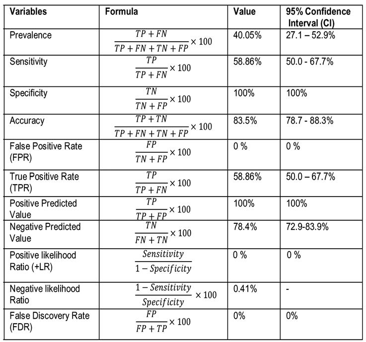
The demographics (gender distribution and prevalence) amongst positive patients are mentioned in Table 2. We have also noticed that the average Ct value of RAT+PCR+ was 19 (CI 16-21); whereas in patients that were RAT-PCR+ was 24 (CI 22-29) (Table 3). The statistical assessment and diagnostic accuracy of RAT is elaborated in Table 4.
DISCUSSION
RAT is an immunoassay that detects the presence of specific proteins on the outer portion of the virus, such as the spike protein which implies recent viral infection. Rapid Antigen Tests (RAT) are comparatively
Table 1 — Interpretation of RT-PCR and RAT for COVID-19 RT-PCRRAT Interpretation
NegativeNegativeNegative
NegativePositivePositive
PositiveNegativePositive
PositivePositivePositive
Positive Inconclusive Positive
Negative Inconclusive Negative
Inconclusive PositivePositive
Inconclusive NegativeRepeat RT-PCR after 3 days
Table 2 — Table showing RAT and RT-PCR test results RT-PCR (Positive)RT-PCR (Negative)Total
RAT (Positive)83 (True Positive)00 (False Positive)83 RAT (Negative)58 (False Negative)211 (True Negative)269 Total141211352
Table 3 — Demographics and Ct values of Positive samples VariablesNValue
Age in years - Mean ±SD35241±17.7
Male - No. (%)352209 (59.37%)
Female - No. (%)352143 (40.62%)
Prevalence - No. (%) 352141 (40.05%)
Ct Value of RAT + and RT-PCR + Cases - Median (IQR)5819 (16-21)
Ct value of RT-PCR Positive cases8324 (22-29)
low-cost, and mainly can be used at the point of care. Most of the currently authorized tests return results in roughly 15-30 minutes. The real-time reverse Transcription Polymerase Chain Reaction (RT-PCR) and other Nucleic Acid Amplification Tests (NAATs) are more sensitive as compared to the antigen test for detecting the presence of viral nucleic acid. On the other hand, RT-PCR can remain positive for weeks to months after early infection and can identify levels of viral nucleic acid even when virus cannot be cultured. Information about antigen tests interpretation is mentioned in Table 1.
Whole Generation Sequencing (WGS) is done for highly sensitive and specific results. It provides complete information and can identify novel strains and mutations as well.
The most common clinical symptoms of COVID19 infections are mild respiratory symptoms like sore throat, dry cough, fever sometimes gastrointestinal symptoms like loose motions may be the presenting complaint. Serious clinical symptoms like pneumonia, stroke, cardiac complications are highly atypical. The present study also shows a similar picture.
Though laboratory and radiological parameters aid in diagnosis and management of COVID-19, molecular detection of SARS CoV-2 remains gold standard. There are other supportive and prognostic modalities which help in the management of COVID-19. Imaging modality, CT thorax is the most useful in detecting the extent of lung involvement. The laboratory investigations that help in management include Complete Blood Counts, blood sugar levels, d-dimer, C-reactive Protein (CRP), ferritin, Interleulin-6, LFT and KFT.
The Maharashtra State Government had included reports of both RAT and RTPCR to determine the daily and weekly positivity rate. In May and June, 2021, the dependence upon RAT had increased drastically. While the ratio used to be 70:30 between RT-PCR and RAT, only 41% tests conducted in June, 2021 were done using RT PCR. After a recent order to exclude RAT for determining positivity rate in Maharashtra state, the daily and weekly positivity rate (period June, 19 to 25) jumped from 5.2% to 8.7%. The positivity rate determines the level of restrictions to be imposed in various cities. Twelve districts, including Kolhapur, Sangli, Palghar, Sindhudurg, Pune and Satara have a positivity rate of over 10% that puts them in stringent level 4 restrictions.
Districts of Buldhana, Gadchiroli, Sangli, Osmanabad and Palghar where RAT number is more, reported a positivity rate of less than 20% during the peak months. The rates of other districts have also been revised. The rate for Mumbai has been revised from 3.7 to 4.7% now. On June, 25, Mumbai city performed 17,200 RAT and 16,600 RT-PCR tests. The positivity rate of RAT was close to 1% and that of RTPCR was almost 4%. The overall positivity rate was close to 2%. Although RAT delivers quick results, they are not very accurate and many times give false negative results. This is because of low sensitivity of
122, No 07, July 2024Journal
RAT. Experts do not recommend use of RAT. Random RAT screening of general population gives a false belief of decreased positivity rate and has implications on restrictions to be enforced (TOI, June 27, 2021).
CONCLUSION
The results of RAT tests should be interpreted considering the symptoms of the patients. It should be done only in acute cases. Whenever done, RT-PCR tests should be done in parallel. We also believe that RAT should be excluded while deciding the daily and weekly positivity rates as these rates determine the level of restrictions to be imposed in a particular district, city or town.
REFERENCES
1She J, Jiang J, Ye L, Hu L, Bai C, Song Y — Novel coronavirus of pneumonia in Wuhan, China: emerging attack and management strategies. Clin Transl Med 2020; 9: e19.
2Ghosh A, Nundy S, Mallick T — How India is dealing with COVID-19 pandemic, Sensors International, 2020; 1: 100021.
3Malik YA — Properties of Coronavirus and SARS-CoV-2. Malays J Pathol 2020; 42(1): 3-11.
4Li H, Liu SM, Yu XH, Tang SL, Tang CK — Coronavirus disease 2019 (COVID-19): current status and future perspectives. Int J Antimicrob Agents 2020; 55(5): 105951.
5Singh A, Mehra M, Gulyani S — A modified variable-order fractional SIR model to predict the spread of COVID-19 in India. Math Meth Appl Sci 2021; 1-15.
6Girolamo P — Assessment of the potential role of atmospheric particulate pollution and airborne transmission in intensifying the first wave pandemic impact of SARS-CoV-2/COVID-19 in Northern Italy. Bull of Atmos Sci & Technol 2020; 1: 515-50.
7Karia R, Gupta I, Khandait H, Yadav A, Yadav A — COVID-19 and its Modes of Transmission. SN Compr Clin Med 2020; 1: 1-4.
8Robson F, Khan K, Le T, Paris C, Demirbag S, Barfuss P, et al Coronavirus RNA Proofreading: Molecular Basis and Therapeutic Targeting. Mol Cell 2020; 79(5): 710-27.
9La Marca A, Capuzzo M, Paglia T, Roli L, Trenti T, Nelson SM — Testing for SARS-CoV-2 (COVID-19): a systematic review and clinical guide to molecular and serological in-vitro diagnostic assays. Reprod Biomed Online 2020; 41(3): 483499.
10Available from: https://covid19.who.int/ [Last access on August 5, 2021]
11Available from: https://www.covid19india.org [Last access on August 5, 2021]
12Mustafa Hellou M, Górska A, Mazzaferri F, Cremonini E, Gentilotti E, De Nardo P, et al — Nucleic acid amplification tests on respiratory samples for the diagnosis of coronavirus infections: a systematic review and meta-analysis. Clin Microbiol Infect 2021; 27(3): 341-351.
13Osterman A, Baldauf HM, Eletreby M, Wettengel JM, Afridi SQ, Fuchs T, et al — Evaluation of two rapid antigen tests to detect SARS-CoV-2 in a hospital setting. Med Microbiol Immunol 2021; 210(1): 65-72.
14Enjuanes L, Gorbalenya AE, de Groot RJ, Cowley JA, Ziebuhr J, Snijder EJ — Nidovirales Encyclopedia of Virology 2008; 419-30.
15Brian D, Baric R — Coronavirus genome structure and replication. Curr Top Microbiol Immunol 2005; 287: 1-30.
16Tanna J, Singha B, Nayak A, Husain A, Raje D, Desai S, et al Incidence and Epidemiological study of COVID-19 in Nagpur urban region (India) using Molecular testing. https://doi.org/ 10.1101/2021.05.11.21256719
17Mukherjee TK, Malik P, Maitra R, Hoidal JR — Ravaging SARSCoV-2: rudimentary diagnosis and puzzling immunological responses. Curr Med Res Opin 2021; 37(2): 207-217.
18Carter L, Garner L, Smoot J, Li Y, Zhou Q, Saveson C, et al — Assay Techniques and Test Development for COVID-19 Diagnosis. ACS Central Science 2020; 6(5): 91-605.
19Ambrosi C, Prezioso C, Checconi P, Scribano D, Sarshar M, Capannari M, et al — SARS-CoV-2: Comparative analysis of different RNA extraction methods. J Virol Methods 2021; 287: 114008.
20Hou H, Wang T, Zhang B, Luo Y, Mao L, Wang F, et al — Detection of IgM and IgG antibodies in patients with coronavirus disease 2019. Clin Transl Immunology 2020; 9(5): e01136.
Original Article
Assessment of Time To Positivity (TTP) and Loading Delay Influencing the Positivity of Blood Culture by an Automated System
Naimika Patel1, Riddhi Patel2, Rupal Patel3
Background : The presence of living micro-organisms in blood is known as bloodstream infection. It can be leading to sepsis, a critical condition associated with mortality ranging from 14% to 34%. Blood cultures are still considered the gold standard test for the detection of bacteremia and fungemia. The bacterial load in blood culture is assessed by the parameter of time to positivity in an automated system.
Materials and Methods : It was a prospective observational study conducted 3 months after IEC approval. A minimum of one & maximum of three samples were collected from each patient. After receiving them at the microbiology laboratory all bottles were loaded into the BacT/ALERT machine. All the signaled positive bottles were studied for a time to detection and factors influencing it and the second objective was to observe the effect of loading delay on isolation rate and time to detection. The following parameters were prospectively extracted from BacT/ALERT systems™ software the cell number, loading time, signal positive time and unloading time (hour, minute).
Observations : A total of 761 blood culture bottles were received during the study period. Maximum bottles were received from male as compared to female patients. Maximum blood cultures were received from the 0-10 years of age group followed by from 21-30 years and from 51-60 years. The mean TTD for all the isolates was 22.71 hours. 81% of true pathogens were detected within 24 hours and 98% of true pathogens were detected within 72 hours. We observed that inadequate blood volume took longer TTP for GNB & Yeast isolates than adequate volume bottles. The true pathogen positivity rate decreases in case of loading delay. The mean unloading time during routine & emergency hours was 0.6 hours & 1.6 hours.
Conclusion : As positive blood cultures are critical alerts; every step should be taken to decrease the loading & unloading delay of blood culture bottles for the final reduction of turn-around time and timely intimation of positive blood culture results to clinicians.
[J Indian Med Assoc 2024; 122(7): 37-42]
Key words :Time To Positivity / Detection, Loading Delay, Unloading Delay, BacT / ALERT.
The presence of living micro-organisms in the blood is known as a blood stream infection. It can be leading to sepsis, a critical condition associated with mortality ranging from 14% to 34%. Blood cultures are still considered the gold standard test for the detection of bacteremia and fungemia. Positive blood culture results can help clinicians to early diagnosis and therapy against the specific organism/s and provide prognostic value. Today many laboratories use automated blood culture systems for microbial detection methods. Rapid detection and early reports of blood culture are crucial for treating sepsis patients and for reducing hospital stays1,2
Time To Positivity (TTP) is defined as the time required from the beginning of culture incubation to
Department of Microbiology, Pramukhswami Medical CollegeBhaikaka University, Anand, Gujarat 388325
1MD, Assistant Professor and Corresponding Author
2MSc, Student
3MD, Professor
Received on : 20/07/2023
Accepted on : 23/07/2023
Editor's Comment :
Positive blood cultures are critical alerts. Thus, every step is important to follow the manufacturer guideline and laboratory procedure for the final reduction of turn-around time.
Timely intimation of positive blood culture results to clinicians is helpful for patient managment.
the detection of bacterial growth by an automated system. The bacterial load in blood culture is assessed by the parameter of TTP in an automated system. It has been proposed as a diagnostic and prognostic tool and is an independent predictor of fatal outcomes. Time to positivity of blood culture bottles can be influenced by various factors eg, the concentration of a pathogen in the primary sample, collection time, the volume of blood, delay in transfer of sample and culture processing and level of contaminants (coagulasenegative staphylococci)3,4
At our hospital, the current method for processing blood culture is BacT/ALERT 3D Microbial Detection System (Biomerieux, France). This study aimed (i) to
122, No 07, July 2024Journal of the Indian Medical Association
determine TTP for positive blood culture bottles & to study factors influencing TTP and (ii) to observe the effect of loading delay on isolation rate and TTP.
MATERIAL AND METHODS
Source of Data :
The study was conducted at the Microbiology laboratory, a NABL accredited laboratory of Shree Krishna Hospital, Karamsad. The study was conducted after the approval of Institutional Ethics Committee.
Methodology :
Study design : Prospective Observational study.
Duration of study : 1st May, 2019 to 31st July, 2019.
Inclusion Criteria : Blood culture requests received during the study duration from all the Indoor & Outdoor patients of all age groups were included in the study.
Exclusion Criteria : Nil.
BacT/ALERT® FA plus and BacT/ALERT® PF plus culture bottles were used for adult and pediatric patients, respectively. After collection at the respective area, all blood culture bottles were transferred to the Microbiology laboratory, a part of the Central Diagnostic Laboratory (CDL), NABL accredited laboratory. All bottles were loaded into the BacT/ALERT ® 3D Microbial Detection System as per the standard protocol. Culture bottles were incubated for 5 days/ until signaled positive for growth. Signal-positive bottles were further processed for the identification of microorganisms and susceptibility testing using the Vitek 2 Compact system.
The Following Parameters for Blood Culture Bottles were Studied :
(1)Loading delay : To observe the effect of rapid loading of bottles into BacT/ALERT, the time interval between various stations was calculated & studied as per the following : (1) collection time to pneumatic receiving time, (2) pneumatic receiving time to Microbiology laboratory receiving time, (3) Microbiology receiving time to loading time into BacT/ALERT machine. The overall time taken from collection to loading into BacT/ALERT was calculated. The effect of loading delay on positivity rate and TTP for true pathogens was assessed.
(2)For TTP calculation, the time interval between loading of bottles and positive signal in BacT/ALERT was calculated & studied. The day of receipt of blood
culture in the laboratory was defined as day 0. TTP for different organisms was calculated. Association of TTP with blood volume was compared.
(3)Contaminant : The contaminant was defined as the growth of skin flora from a positive blood culture bottle (eg, Bacillus spp, Corynebacterium spp, Propionibacterium spp and Micrococcus spp) Coagulase-negative staphylococci grown from single/ multiple blood culture was clinically correlated to decide whether it was a true pathogen or a skin contaminant. Antimicrobial susceptibility results of Coagulase-negative staphylococci which were identified as contaminants were not reported. All other Grampositive Cocci (GPC), Gram-negative Bacilli (GNB)/ Coccobacilli (GNCB) & Yeast isolates were considered true pathogens.
(4)Time intervals between (i) loading to signal positive and (ii) signal positive to unloading of bottles during routine (from 9:00 am - 5:00 pm) & emergency (from 5:00 pm - 9:00 am) hours were compared.
Patient Details and Analysis of DATA :
For each blood culture bottle, the following parameters were prospectively extracted from the laboratory information System database: demographic characteristics of the patient (age, sex), collection time, a pneumatic station receiving time, microbiology laboratory receiving time and microbiological result. The following parameters were prospectively extracted from BacT/ALERT system’s software: the cell number, loading time, signal positive time and unloading time.All the data were entered and analyzed in Microsoft Excel 2010.
RESULTS
During the study period, a total of 761 blood culture bottles from 604 patients were received for blood culture at the Microbiology laboratory. Maximum bottles were received from 354 (59%) males compared to 250 (41%) females. A total of 182 (30%) blood cultures were received from the 0-10 years of age group followed by 78 (13%)from 21-30 years and 75(12%) from 51-60 years. Out of 761 blood cultures,97 (13%) were identified as True pathogens & 139 (18%) were identified as Contaminants (CONS and Bacillusspp). However, 525(69%) blood cultures were negative.
As shown in Table 1, the mean TTD for all the isolates was 22.71 hours. 98% of all isolates were detected by day 2. A total of 73% (173/236) of GPC, GNB and Contaminants were detected within 24 hours and the other 19% (44/236) including Yeast were
Table 1 — Comparison of TTP in GPC, Yeast, GNB & Contaminants (N=236)
OrganismNumbers TTP-No of blood cultures signaled positive in ofBacT/ALERT on the day(hours) isolates Mean Time 0(24)1(48)2(72)3(96) 4(120) (hours)
detected within 48 hours. In 81% of true pathogens were detected within 24 hours and 98% of true pathogens were detected within 72 hours. 68% of contaminants were detected in 24 hours.
The mean TTP for GPC & Yeast isolates is shown in Table 2. Out of 24 GPC isolates, 88% were detected within 24 hours, 96% were detected within 48 hours and 100% were detected within 72 hours. 72% of Staphylococcusaureus isolates and all other GPCs were signaled positive within 24 hours (by day 0).
The mean TTP for Gram-negative bacilli is shown in Table 3. Out of 69 GNB isolates, 84%, 96%, 99% & 100% were detected within 24 hours, 48 hours, 72 hours and 96 hours respectively. None were detected on day 4 (on the 5th day).
As shown in Table 4, the mean TTD for Contaminants was 25.13 hours. 68% (94/ 1of 39) Contaminants were detected on day 0, and 23% (32/139) were detected on day 1.
The relation between the volumes of blood with TTP is shown in Table 5. Overall TTD from bottles with adequate volume
and inadequate volume was 18.45 hours & 19.71 hours respectively.
Table 6 represents the time taken at various stations before loading blood culture bottles into BacT/ALERT machine. Table 7 shows the effect of loading delay on positivity rate and TTP for true pathogens. The mean time from loading to signal positive was 23.32 & 23.39 hours during routine & emergency hours. However, the mean unloading delay during routine & emergency hours was 36.74 & 98.8 minutes respectively. During routine &
OrganismNumbers TTP-No of blood cultures signaled positive in ofBacT/ALERT on the day(hours)
Table 3 — TTP for Gram-negative bacilli (N=69)
OrganismNumbers TTP-No of blood cultures signaled positive in ofBacT/ALERT on the day(hours)
Escherichia coli
Klebsiella pneumoniae 1810.9218____
Salmonella typhi
Salmonella paratyphi A
Acinetobacter spp
Pseudomonas spp
Citrobacter sedalkii
Enterobacter cloacae
Shigella sonnei
Aeromonas hydrophilia
Brucella melitensis
Burkholderia cepacia
Ralstonia
Stenotrophomonas maltophilia
Sphingomonaspaucimobilis
Table 4 — TTP for Contaminants (N=139)
OrganismNumbers TTP-No of blood cultures signaled positive in ofBacT/ALERT on the day(hours) isolates Mean Time 0(24)1(48)2(72)3(96) 4(120) (hours)
Coagulase Negative 9725.46525511
Staphylococci (CONS)
Bacillus spp 4224.129751_ 13925.1394321021
Table 5 — Comparison of blood volume with TTP for true Pathogens (N=97)
Burkholderia pseudomallaei. They also mention that a short TTP reflects a surrogate marker of bacterial concentration in blood and it suggests severe bacteremia. It can affect clinical outcomes such as an increase in mortality rates, increased length of stays and hospitalization costs3.
A continuous monitoring system detects all positive signals in a relatively short period, the majority within 24 hours6,7 In our study, the mean TTP for all the isolates (true pathogens & contaminants) was 22.71 hours. Lambregts, et al found that the median TTP was 15.7 hours which is similar to our findings. The authors also found that neutropenia was a predictor for short TTP and antibiotic pre-treatment was a predictor for prolonged TTP8
Table 6 — Time taken at different stations before loading blood culture bottles (N=761) Meantime (minutes)
Table 7 — Comparison of loading delay (collection to loading duration) with TTP for true Pathogens (N=97)
emergency hours, the maximum time taken to unload the bottles was 249 & 518 minutes (4.15 & 8.6 hours) respectively.
DISCUSSION
Time to positivity is a parameter provided by the automated blood culture system. Time is calculated from the incubation of bottles until a positive signal is detected2,5. Many published studies mention that TTP of blood cultures is a prognostic factor in the cases of bacteremia caused by Gram-positive and Gramnegative micro-organisms such as S aureus, S pneumoniae, E coli, Klebsiella pneumoniae and
Previous reports have also shown that clinically significant organism was generally detected within 3 days of incubation5,7,9,10 In our study, 81% of true pathogens were detected within 24 hours and 98% of true pathogens were detected within 72 hours. The Mean TTP for GPC, GNB & Yeast was 16.29 hours, 17.52 hours and 66.98 hours respectively. Out of 24 GPC isolates, 88% were detected within 24 hours. A total of 72% of Staphylococcus aureus isolates and all other GPCs were signaled positive within 24 hours. Out of 69 GNB isolates, 84% were detected within 24 hours. All (100%) isolates of Candida albicans were detected within 48 hours. Kurlat, et al reported that all isolates of group B streptococcus, Escherichia coli, Klebsiella species and Staphylococcus aureus were identified by day 210. In our study, 100% GPC & GNB isolates were detected within 72 hours and 96 hours respectively. Similar to our findings, TTD for fungal isolates varied between 48 hours – 120 hours10 Pan F, et al found that the average TTP of all positive blood cultures was 30.97 and the TTP of Gramnegative strains was significantly shorter than that of Gram-positive strains and fungi11.
In our study,the mean TTP for Contaminants was 25.13 hours. Amongst them, 68% of Contaminants were detected within 24 hours whereas 32% of Contaminants were detected after 24 hours. A study conducted by Kennedy found that those generally regarded as Contaminants (CoNS & Bacillus spp) were detected much later than other isolates7 Although
lower inoculum is associated with longer TTP for Contaminants, there is no consensus on whether the TTP is predictive of contamination versus true infection. This has been emphasized that those isolates that were considered clinically relevant were detected earlier than those regarded as contaminates.
TTP can be influenced by various factors. During the study, we tried to assess the effect of blood volume on TTP. We observed that inadequate blood volume took longer TTP for GNB & Yeast isolates than adequate volume bottles. TTP was not affected by blood volume in GPC isolates. Overall, TTP from bottles with inadequate volume was higher than from bottles with adequate volume indicating the significance of blood volume on the TTP.
The second important parameter that affects TTP is the loading delay of the bottles into the instrument. Published guidelines recommend that the interval between the collection of blood and the entry of the bottles into an automated blood culture system should not be longer than 2 or 4 hours; also, manufacturer instructions indicate that inoculated vials should be transported to the laboratory as quickly as possible2. Many studies observed that in half of the investigated laboratories, bottles were not immediately incubated during nightshifts. Studies show that a delay in processing can impact time to positivity, presumptively due to an extended lag phase resulting from storage at suboptimal temperature8. In our study, most of the bottles were loaded into the instrument within six hours. With the increase in loading delay, the true pathogen positivity rate decreases in the present study. However, when loading delay was compared with TTD, the mean TTD of GPC & GNB isolates was not affected. Different patient populations might be the reason for such variations. Moreover, contrary to the Italian study where the laboratory in the university hospital is closed on Sundays and holidays, a central diagnostic laboratory at our place operates on weekends and holidays12 Janapatla, et al conclude that an overnight delay of 15 hours of bottles leads to an increase in the detection time of the pathogen from 25.9 hours to 40 hours, which might influence clinical therapy13. In contrast, Panday, et al in their study showed that preanalytical time for the three pre-defined cut-off values did not influence blood culture yield12
Time to removal is defined as the time taken from the positive signal in system until the bottle is removed for subsequent sub-culturing for isolation of the organism2 In our study, the mean unloading time
41
during routine & emergency hours was 36.74 (0.6 hours) minutes & 98.8 (1.65 hours) minutes respectively. The study conducted by Emeraud, et al conclude that the mean unloading time for positive bottles is 5.8 hours which is explained by the fact that the positive bottles are not unloaded during the night duty period this can result in obtaining false negative results14 In our study, maximum time taken to unload the bottles was 249 & 518 minutes (4.15 & 8.6 hours) during routine & emergency hours respectively. At our place, diagnostic laboratories were open for 24 hours throughout the year. Blood culture bottles were processed during routine and emergency hours by trained technicians. However, the number of laboratory technicians during routine and emergency hours differ (less number during emergency) which might be responsible for delays in loading & unloading bottles. The reasons for unloading delay during working hours must be identified and necessary actions should be taken. Such a delay in unloading the positive bottles can affect the total time required to identify the organism and inform the result of the clinicians. Positive blood cultures are critical alerts and must be processed immediately to decrease the turn-around time which can save the life of the patients. Our data and results refer to typical operative conditions and describe what was actually occurring in the laboratory. The findings of the study require attention by the laboratory staff and Head of the department for future interventions to improve the quality of blood culture results.
Limitation of study : The study was conducted at a Tertiary Care Teaching Hospital with a smaller number of blood samples (761) & patients (604) within a short period of time and the findings may not be generalizable to all hospitals. Parameters including timing of venepunctures, skin antisepsis, antibiotic treatment prior to sampling, patient comorbidities and manual measurement of blood volume in bottles (as per manufacturer’s instructions) might have affected the results.
CONCLUSION
As positive blood cultures are critical alerts; every step should be taken to decrease the loading & unloading delay of blood culture bottles for the final reduction of turn-around time and timely intimation of positive blood culture results to clinicians. For analytic issues of loading and unloading delays at the laboratory, all the concerned staff should be informed about the importance of the blood cultures and the effect of such delays on patient outcomes.
Vol 122, No 07, July 2024Journal of the Indian Medical Association
REFERENCES
1Byun JH, Kim SW, Koh EH, Kim S, Kim SC — Comprehensive Analysis of Blood Cultures Using BacT/Alert 3D FA Plus and FN Plus Media. Laboratory Medicine Online 2016; 6(3): 17682.
2Dreyer A W — Blood culture systems: From patient to Result. In: Azevedo L, editor. Sepsis. An ongoing and significant challenge. Rijeka: InTech; 2012. Ch-15.
3Tang PC, Lee CC, Li CW, Li MC, Ko WC, Lee NY — Time-topositivity of blood culture: an independent prognostic factor of monomicrobial Pseudomonas aeruginosa bacteremia. Journal of Microbiology, Immunology and Infection 2017; 50(4): 486-93.
4Ning Y, Hu R, Yao G, Bo S — Time to positivity of blood culture and its prognostic value in bloodstream infection. European Journal of Clinical Microbiology & Infectious Diseases 2016; 35(4): 619-24.
5Kumar Y, Qunibi M, Neal TJ, Yoxall CW — Time to positivity of neonatal blood cultures. Archives of Disease in ChildhoodFetal and Neonatal Edition 2001; 85(3): F182-6.
6Lamy B, Dargere S, Arendrup MC, Parienti J-J and Tattevin P — How to Optimize the Use of Blood Cultures for the Diagnosis of Bloodstream Infection? A Stat-of-the Art Front Microbiol 2016; 7: 1-13
7Kennedy GT, Barr JG, Goldsmith C — Detection of bacteremia by the continuously monitoring BacT/Alert System. J Clin Pathol 1995; 48: 912-4.
8Lambregts MMC, Bernards AT, van der Beek MT, Visser LG, de Boer MG — Time to positivity of blood cultures supports early re-evaluation of empiric broad-spectrum antimicrobial therapy. PLoS ONE 2019; 14(1): e0208819. https://doi.org/ 10.1371/journal.pone.0208819
9Hardy DJ, Hulbert BB, Migneault PC — Time to Detection of Positive BacT/Alert Blood Cultures and Lack of Need for Routine Subcultures of 5- to 7- Day Negative Cultures. J Clin Microbiol 1992; 30: 2743-5.
10Kurlat I, Stoll BJ, McGowan JE Jr — Time to positivity for detection of bacteremia in neonates. J Clin Microbiol 1989; 27(5): 1068-71. doi: 10.1128/jcm.27.5.1068-1071.1989. PMID: 2745679; PMCID: PMC267484.
11Pan F, Zhao W, Zhang H — Value of time to positivity of blood culture in children with bloodstream infections. Canadian Journal of Infectious Diseases and Medical Microbiology 2019; 2019. doi: 10.1155/2019/5975837.
12Nannan Panday RS, Wang S, van de Ven PM, Hekker TAM, Alam N, Nanayakkara PWB — Evaluation of blood culture epidemiology and efficiency in a large Europenteching hospital. PLoS ONE; 14(3): 1-7.
13Janapatla RP, Yan JJ, Chien ML, Chen HM, Wu HM, Wu JJ— Effect of overnight storage of blood culture bottles on bacterial detection time in the BACTEC 9240 blood culture system. J Microbiol Immunol Infect 2010; 43(2): 126-32. doi: 10.1016/ S1684-1182(10)60020-5. PMID: 20457429.
14Emeraud C, Yilmaz S, Fortineau N, Cuzon G, Dortet L — Quality indicators for blood culture: 1 year of monitoring with BacT/Alert Virtuo at a French hospital. Journal of Medical Microbiology 2021; 70(3). 001300. doi: 10.1099/jmm.0.001300
Original Article
A Study on Non-motor Manifestations in Young Onset Parkinsons Disease (YOPD) in Eastern Indian Population
Praveen Kumar Yadav1, Samsudheen K K2
Background : Non-motor Symptoms (NMS) are extremely common in Young Onset Parkinson Disease (YOPD) with high incidence of neurobehavioral and autonomic dysfunction which should be given greater emphasis as it will affect the Quality of Life.
Materials and Methods : This is a cross-sectional study in which all consecutive patients of age less than 50 years satisfying the UPKRDS Diagnostic criteria for Idiopathic Parkinson’s Disease (IPD) attending the Neurology Outpatient services at a super speciality clinic during study period July 1, 2021 to April 30, 2022 were included in the sample. All secondary causes of parkinsonism like drug induced, multi-infarct state and normal pressure hydrocephalus were excluded. All patients were examined, and demographic data and non-motor symptoms were documented using the Non-motor Scale (NMSS) of International Parkinson’s and movement disorder society and modified Hoehn & Yahr staging used for staging.
Results : A total 32 patients were diagnosed with YOPD during the study period. Out of which 19 (59.37%) were Males and 13 (40.32 %) were Females. Majority of the patients were in the age group of 40-50 years (84%) and onset of illness in 1 month to 5 years (78.17%) with 37% each of patients were in the H & Y Stages I-1.5 and 1.5-2. Common Non-motor Symptoms (NMS) observed were anxiety (71.87%), memory loss (59.37%) and depression (56.25%). Most of the patients had one or more autonomic symptoms and sleep disturbances.
Conclusion : Non-motor Symptoms were present in all patients (100%) with anxiety, memory loss, depression and constipation are being the commonest of the NMS.
[J Indian Med Assoc 2024; 122(7): 43-6]
Key words :Non-motor Manifesations, Young Onset Parkinsons Disease (YOPD).
Parkinson’s Disease (PD) is a neurodegenerative disorder characterized by bradykinesia, rest tremor, rigidity and postural instability along with variety of Non-motor Symptoms (NMS)1. It is usually considered as age related disease with mean age in early to mid 60s, but it can occur in early life also2 Based on age of onset, PD can be divided into early and late [Late Onset PD (LOPD)]. Early Onset PD (EOPD) further subdivided into Juvenile Onset PD (JOPD) (before 21 years) and Young Onset PD (YOPD) (between 21-40 years of age) while onset after 60 years defines LOPD3-5 . Due to lack of consensus, the maximal age for YOPD has varied from 40 to 553,6,7 and minimal age for LOPD has varied from 50 to 703,8,9.
The prevalence of PD in the Western World has been reported to range from 130 to 200 per 100,000 in community-based studies but reported as high as
1MBBS, MD, DM, MRCP, FRCP, FEBN, FIAMS, FIACM, FIMSA, MNAMS, Senior Consultant Neurologist and Director, Department of Neurology, Aarogyam Neuro Clinic, Durgapur, West Bengal 713214 and Corresponding Author
2DNB (Medicine), Resident, Department of General Medicine, Durgapur Steel Plant Main hospital, Durgapur, West Bengal 713205
Received on : 03/09/2023
Accepted on : 23/09/2023
Editor's Comment :
Non-motor manifestations are very commonly seen in YOPD. Depression, anxiety and constipation are the commonest to be seen in this study.
2000/100,000 in individuals over 80 years of age10-13 and YOPD represents 5-7% of this. As per WHO (14 June, 2022) prevalence of Parkinson disease has doubled in the past 25 years with global estimates in 2019 showing over 8.5 million individuals living with PD, 5.8 million disability-adjusted life years and 329,000 deaths.
YOPD and LOPD are similar in both clinical and pathological features, except for higher rate of treatment-related dyskinesias 14 , slow onset of progression and impact on Quality of Life (QOL) especially due to NMS14-18. YOPD patients facing occupational and life style challenges than LOPD not only due to motor symptoms but also due to NMS like depression, sexual dysfunction, marital conflict, loss of occupation and future uncertainty19-21. It also leads to behavioural disorder like addiction, impulse control disorders like gambling, compulsive shopping and sexual addiction.
MATERIALS AND METHODS
This is a cross sectional study in which all Consecutive patients less than fifty years satisfying the UPKRDS Diagnostic criteria for Idiopathic Parkinson’s Disease (IPD) attending the Neurology Outpatient services at a Super Speciality Neurology Clinic, Durgapur during study period July 1, 2021 to April 30, 2022 were included in the sample. All patients were examined and demographic data and Non-motor Symptoms were documented using the Non-motor Scale (NMSS) of International Parkinson’s and movement disorder society and modified Hoehn & Yahr staging used for staging.Local Ethical committee clearance was taken by Asansol Durgapur Ethics Committee before starting the study, Letter no -(15/ 21). Exclusion criteria- All secondary causes of parkinsonism like drug related, multi-infarct state and normal pressure hydrocephalus were excluded.
RESULTS
A total 32 patients were diagnosed with YOPD and satisfied the inclusion criteria of study. Out of which 19 (59.37%) were Males and 13 (40.32%) were Females. In 27 patients were within 40-50 years (84%) and remaining 5 patients were within 30-39 years (16%). The Hoehn & Yahr (H& Y) stages were 1-1.5 (37 %), 2(37%) and least common H & Y stage was 3-5 (25%) (Table 1). Onset of illness was most commonly between 1 month - 5 years (78.17%) followed by 6 years -10 years (21.87%) and 11years - 15 years (6.25%) in descending order (Fig 1).
Non-motor Symptoms (Table 2) seen in decending order were Memory loss (59.37%), constipation (46.87%), Sexual dysfunction (40.62%), Anxiety
(71.87%), Depression (56.25%), Nocturia (43.75%), Urinary Frequency (28.12%), Insomnia (43.75%), Urinary Urgency (15.62%), Day Time Sleepiness (13.25%), Loss of Taste and Smell (28.12%), Excess Sweating (31.25%), Frozen Shoulder (31.25%), Restless Leg Syndrome (12.05%), Dizziness (9.37%), Panic Attack (18.75%), Fainting (15.05%), RBD (9.37%) and Hypersalivation (3.12%).
DISCUSSION
YOPD is almost similar to LOPD in clinical picture except in slow disease progression 22 , less falls, freezing 3 and increased treatment related motor complications14. In this study, it is only focussed on clinical profile of Non-motor Symptoms of YOPD regarding their relation to stage and duration of disease. Out of 32 patients included in this study majority are coming under the age group of 40-50 years (84%) followed by 30 -39 years. Onset of illness in majority of patients are 1 month to 5 years (78.17%) followed by 6 years -10 years (21.87 %) and 11years-15 years (6.25 %) with equal distribution in both 1-1.5 and 2 H&Y staging (37% each) and remaining 26% patients are in 3 -5 stage. NMS included in this study consist of autonomic dysfunction, sleep disorders, psychological and behavioural disorders, sensory and cognitive symptoms and all patients were shown one or more NMS.
The most common NMS is Anxiety (71.87%) followed by Memory loss (59.37%), Depression (56.25%) and Constipation (46.87%) compared to a study conducted in Indian populations which shows Depression (45.6%), Anxiety (45.4%) and Apathy Table 2 — NMS in descending order of prevalence in study population
(30.5%)23. A similar study in USA shows similar rate of Depression (48.3%) 24 . YOPD usually defines between 21-40 years but there is lack of consensus regarding the upper limit which varies from 40 to 55 years in different studies. We considered the upper limit of age as 50 years. Cognitive decline is less in YOPD compared to LOPD25,26, even though it is more dependent on age. In this study Memory loss was the 3rd most common NMS which is probably due to the increased age of patient (84% is between 40-50 years) . Genetic study was not included in this study even though genetic predisposition is well recognised with age of onset-younger the age higher the genetic association27. Family history is reported in 20% of YOPD patients compared to 6.9% of LOPD patients, and the age-specific risk of PD is 7.8-fold higher in the relatives of patients with YOPD compared to 2.9-fold among the relatives of patients with LOPD7,28. Many genes are considered causing PD which mainly include SNCA, LRRK2, GBA. Duplication of PARK 1 gene is associated with NMS like severe psychiatric features. In 1/3rd of the study population frozen shoulder was seen and panic attack was documented in 18.75% of the study population. In several studies restless leg syndrome had a higher rate5,9,18,29-33 compared to 12.05% in our study. Other common manifestations were constipation, urinary urgency, frequency, sexual dysfunction, hyper salivation, excessive sweating, fainting and loss of smell and taste.Sleep disturbances like REM Behavioural Disorders, excessive day time sleeping and insomnia were seen commonly in the study population. NMS in YOPD has to be diagnosed early and treated because it affects the peak and productive years of life which leads to loss of employment, social and family conflicts, loss of selfesteems and various addictive behavioural problems. There is a diagnostic challenge in YOPD mainly due to rarity and age of onset because PD is mainly considered as a disease of elderly and this leads to multiple neurologist visit, multiple investigations and delay in diagnosis and treatment19,6.
Limitations: Small sample size, cross sectional study, genetic study is not done in the patients
CONCLUSION
Non-motor Symptoms were seen in all the patients with Young Onset Parkinsons Disease (YOPD) in our study. Anxiety, Memory Loss, Depression and Constipation are the commonest non motor manifestations.
Financial support : Nil.
Conflict of interest : There is no conflict of interest.
REFERENCES
1Obeso JA — Past, present, and future of Parkinson’s disease: A special essay on the 200th Anniversary of the Shaking Palsy. Movement Disorders 2017; 32(9): 1264-310.
3Schrag A, Schott JM — Epidemiological, clinical, and genetic characteristics of early-onset parkinsonism. Lancet Neurol 2006; 5: 355-63.
4Morales-Briceno H, Mohammad SS, Post B, Fois AF, Dale RC, Tchan M, Fung VSC — Clinical and neuroimag- ing phenotypes of genetic parkinsonism from infancy to adolescence. Brain 2020; 143: 751-70.
6Rana AQ, Siddiqui I, Yousuf M — Challenges in diagnosis of young onset Parkinson’s disease, J Neurol Sci 2012; 323: 113-6.
7Mehanna R, Moore S, Hou JG, Sarwar AI, Lai EC — Comparing clinical features of young onset, middle onset and late onset Parkinson’s disease. Park Relat Disord 2014; 20: 530-4.
8Wickremaratchi MM, Perera D, O’Loghlen C — Prevalence and age of onset of Parkinson’s disease in Cardiff: a community based cross sectional study and meta-analysis. J Neurol Neurosurg Psychiatry 2009; 80: 805-7.
9Wickremaratchi MM, Ben-Shlomo Y, Morris HR — The effect of onset age on the clinical features of Parkinson’s disease. Eur J Neurol 2009; 164: 450-6.
10Schrag A, Ben-Shlomo Y, Quinn N — Cross sectional prevalence survey of idio- pathic Parkinson’s disease and parkinsonism in London. BMJ 2000; 321: 21-2.
11Dorsey ER, Bloem BR — The Parkinson pandemic-A call to action. JAMA Neurol 2018; 75: 9-10.
12Pringsheim T, Jette N, Frolkis A, Steeves TD — The prevalence of Parkinson’s ( disease: a systematic review and metaanalysis. Mov Disord 2014; 29: 1583-90.
13Wanneveich M, Moisan F, Jacqmin-Gadda H, Elbaz A, Joly P — Projections of (prevalence, lifetime risk, and life expectancy of Parkinson’s disease (2010-2030) in France. Mov Disord 2018; 33: 1449-55.
14Schrag A, Hovris A, Morley D, Quinn N, Jahanshahi M — Young- versus older-onset Parkinson’s disease: impact of disease and psychosocial consequences. Mov Disord 2003; 18: 1250-6.
15Kupsch A, Oertel WB — Selegiline, pregnancy, and Parkinson’s disease. Mov Disord 1998; 13: 175-6.
16Schrag A, Banks P — Time of loss of employment in Parkinson’s disease. Mov Disord 2006; 21: 183943.
17Murphy R, Tubridy N, Kevelighan H, O’Riordan S — Parkinson’s disease, How is employment affected? Ir J Med Sc 2013; 182: 4159.
18Knipe MD, Wickremaratchi MM, Wyatt-Haines E, Morris HR, Ben-Shlomo Y — Quality of life in young- compared with late-onset Parkinson’s disease. Mov Disord 2011; 26: 20118.
Vol 122, No 07, July 2024Journal of
19Schrag D, Morley N, Quinn M — Jahanshahi, Impact of Parkinson’s disease on patients’ adolescent and adult children, Park Relat Disord 2004; 10: 391-7.
20Carter, Julie H — Living with a person who has Parkinson’s disease: the spouse’s perspective by stage of disease.” Movement disorders: official journal of the Movement Disorder Society 1988; 13(1): 20-8.
21Ravenek M, Rudman DL, Jenkins ME, Spaulding S — Understanding uncertainty in young-onset Parkinson disease. Chronic Illness 2017; 13: 288-98.
22Alves G, Wentzel-Larsen T, Aarsland D, Larsen JP — Progression of motor impair- ment and disability in Parkinson disease: a population-based study. Neurology 2005; 65: 143641.
23Kukkle P, Goyal V, Geetha T, Mridula K, Kumar H, Borgohain R, et al — Clinical Study of 668 Indian Subjects with Juvenile, Young, and Early Onset Parkinson’s Disease. Canadian Journal of Neurological Sciences / Journal Canadien Des Sciences Neurologiques 2022; 49(1): 93-101. doi:10.1017/ cjn.2021.40
24Mehanna R, Moore S, Hou JG, Sarwar AI, Lai EC — Comparing clinical features of young onset, middle onset and late onset Parkinson’s disease. Parkinsonism Relat Disord 2014; 20: 530-4.
25Schrag A, Ben-ShlomoY, Brown R, Marsden CD, Quinn N — Young-onsetParkinson’s disease revisited—clinical features, natural history, and mortality. Mov Disord 1998; 13: 885-94.
26Chaudhary S, Joshi D, Pathak A, Mishra VN, Chaurasia RN, Gupta G — Comparison of cognitive profile in young- and late-onset Parkinson’s disease pa- tients Ann Indian Acad Neurol 2018; 21: 130-2.
27Deng H, Wang P, Jankovic J — The genetics of Parkinson disease. Ageing Res Rev 2018; 42: 72-85.
28Payami H, Zareparsi S, James D, Nutt J — Familial aggregation of Parkinson disease: a comparative study of early-onset and late-onset disease. Arch Neurol 2002; 59: 848-50.
29Calne SM, Lidstone SC, Kumar A — Psychosocial issues in young-onset Parkinson’s disease: current research and challenges. Park Relat Disord 2008; 142: 143-50.
30Spica V, Pekmezovic T, Svetel M, Kostic VS — Prevalence of non-motor symptoms in young-onset versus late-onset Parkinson’s disease. J Neurol 2013; 260: 131-7.
31Cilia ER Cereda, Klersy C, Canesi M, Zecchinelli AL, Mariani CB, Tesei S, et al — Parkinson’s disease beyond 20 years, J Neurol Neurosurg Psychiatry 2015; 86: 849-55.
32Kostic KS, Filipovic SR, Lecic D, Momcilovi D, Sokic D, Sternic N — Effect of age at onset on frequency of depression in Parkinson’s disease. J Neurol Neurosurg Psychiatry 1994; 57: 1265-7.
33Dissanayaka NN, Sellbach A, Matheson S, O’Sullivan JD, Silburn PA, Byrne GJ, et al —Anxiety disorders in Parkinson’s disease: pre- valence and risk factors. Mov Disord 2010; 25: 838-45. 46
Original Article
Psychological Distress and Health-related Quality of Life in Lung Cancer Patients
Arun Kumar1, Ajay Kumar Verma2, Jyoti Bajpai3, Anuj Kumar Pandey4, Surya Kant5, Sujita Kumar Kar6, S K Verma2, R A S Kushwaha2, Santosh Kumar2, Rajiv Garg2, Anand Srivastava2, Darshan Kumar Bajaj2, Ankit Kumar3, Pranjal Tripathi7
Background : The symptom burden of lung cancer is higher than that of other malignancies, but less is known about the incidence of psychological distress and how it can be connected to the physical symptom load in lung cancer patients. We evaluated the impact of physical illness on psychological distress among stable patients with lung cancer.
Materials and Methods : The respiratory medicine department of a tertiary health care centre recruited 90 patients with stages I to IV lung cancer for a study. The study used the DASS-21 scoring tool to evaluate psychological distress. Regression analyses were conducted to determine the relationship between distress and symptoms, type of cancer and Tumor, Node and Metastasis staging.
Results : Out of all the patients enrolled, 26.67% had moderate depression according to the DASS-21 scale. On the Likert scale, 55.56% of participants reported moderate anxiety and 9% had clinically significant severe anxiety. Mild depression was present in 34.44% of patients and moderate depression in 26.67%. Only 12% of enrolled participants showed signs of stress. Our analysis indicated that males were more likely to report symptoms of depression, anxiety and stress. Age also affected depression, anxiety and stress noted in our study participants. In addition, smoking, cancer stage and clinical symptoms showed significant associations with all DASS-21 subscales.
Conclusion : This study showed that patients with lung cancer had a high proportion of depression and anxiety. It has been suggested that the psychological well-being of lung cancer patients should be given greater consideration to improve their overall management.
[J Indian Med Assoc 2024; 122(7): 47-51]
Key words :Psychological Distress, Health-related Quality of Life, Lung Cancer.
LEditor's Comment :
Lung cancer patients may suffer from depression and anxiety. In this study, more than half experienced moderate anxiety, and more than a quarter had moderate depression. The primary factors related to psychological distress in patients with lung cancer are age, smoking history, cancer stage, and the presence of physical symptoms. The management of lung cancer should prioritize psychological distress due to its high prevalence and detrimental effects. Early identification and intervention in moderate and severe cases can improve patients’ overall well-being and potentially improve treatment outcomes.
Department of Respiratory Medicine, King George’s Medical University, Lucknow, Uttar Pradesh 226003, India
1MD, Resident Doctor
2MD, Professor
3MD, Assistant Professor
4PhD, Research Scholar
5MD, Professor and Corresponding author
6MD, Professor, Department of Psychiatry
7MD, Junior Resident, Department of Psychiatry, Andhra Medical College, Vishakhapatnam, Andhra Pradesh 530002, India
Received on : 16/12/2023
ung cancer is the most common cancer and the leading cause of cancer-related deaths Worldwide. As per the latest report of GLOBOCAN, 1.83 million people have lung cancer, accounting for 12.9% of all cancers Worldwide 1 . It is a lethal disease characterized by uncontrolled malignant proliferation of the epithelial cell lining of the lower respiratory tract. In India, lung cancer accounts for 5.9% of all cancers2 Being diagnosed with cancer and undergoing anticancer treatment can result in physical symptoms such as exhaustion, shortness of breath and various psychological issues. These issues may persist throughout the cancer journey. They can affect anywhere from 20% to 58% of patients. Three studies have shown that lung cancer patients experience the highest levels of psychological distress, ranging from 17.0% to 73.0%4. As per the definition given out by the National Comprehensive Cancer Network (NCCN) guidelines, distress refers to a multifactorial and unpleasant emotional experience involving psychological, social, spiritual and physical changes. Clinically significant psychological distress is linked to a number of adverse outcomes, including discontinuing anti-cancer therapy, prolonged hospital stay, higher suicidal risk, poor Quality of Life and higher
Accepted on : 29/01/2024
Vol 122, No 07, July 2024Journal of the Indian Medical Association
risk of mortality. Moreover, a previous study also evidenced that psychological distress can accelerate the growth of tumour cells. Emphasizing the significance of diagnosing and treating emotional distress and mood disorders, healthcare professionals Worldwide recognize the sixth vital sign when treating cancer patients.
To improve clinical care and nursing in the future, the present study was proposed to evaluate the impact of physical illness on psychological distress among stable patients with lung cancer.
MATERIALS AND METHODS
Study Design and Setting :
A cross-sectional questionnaire-based study was conducted in Kasturba Chest Hospital, Department of Respiratory Medicine, King George’s Medical University, Lucknow, UP. This study was approved by the Institutional Ethics Committee (Ref. Code: XIII PGTSC-IIA/P4). A written informed consent was obtained from all the participants prior to enrolment. Data was compiled in pre-formed study-questionnaire.
All the patients coming to OPD and IPD who were histopathologically diagnosed with lung cancer of age greater than 18 years were enrolled in the study from February, 2021 to January, 2022. Exclusion criteria were patients who didn’t give consent, patients with pulmonary metastasis of extra-pulmonary malignancy and sputum positive for pulmonary tuberculosis.
Measures :
The DASS-21 questionnaire was chosen to measure depression, anxiety, and stress simultaneously and does not take into account cultural background and we chose the validated short version to save time. The domain of depression evaluates low self-esteem, low positivity and hopelessness. The anxiety domain assesses subjective feelings of fear, physiological hyperarousal and autonomic arousal and the stress domain considers negativity, agitation and tension. This is in accordance with the tripartite theory, which suggests that anhedonia (loss of pleasure), or lack of positivity, is an integral factor differentiating depression from anxiety. This instrument aims to assess and discriminate symptoms of anxiety and depression, based on the Tripartite Model, which groups the symptoms of anxiety and depression into three factors5 : (a) the presence of negative affect (depressed mood, insomnia, irritability), (b) specific factors of depression (anhedonia, absence of positive affect) (c) specific symptoms of anxiety (somatic tension and hyperactivity).
Consisting of 21 items, each factor groups seven items answered using a four-point Likert-type scale, indicating the severity and frequency of symptoms experienced in the previous seven days. Responses were measured on a Likert scale ranging from 0 to 3: did not apply to me at all. applied to me to some degree, or some of the time.
applied to me to a considerable degree or a good part of the time. applied to me very much or most of the time.
Interpretation of Depression, Anxiety and Stress Scale Questionnaire :
DASS-21, a shorter version of 42 item questionnaire, is a validated tool for evaluating psychological burden and assessing the psychological state in a wide range of diseases. There were 21 questions evaluating depression, anxiety and stress components in a clinical setting. As represented in Table 3, scores of 0-9, 10-13, 14-20 and 21-27 were considered normal, mild, moderate and severe, respectively. A score above 27 was considered as having an extremely severe condition of depression.
Data Analysis
:
The measurement variables were presented as mean standard deviation, whereas the enumeration data, such as gender, age, smoking, etc, were expressed by frequency (cases). To look at the respondents’ demographics and other chosen variables, an analysis of descriptive statistics was done. To compare the variations among subgroups, the T-test, One-way Analysis of Variance (ANOVA), and Kruskal-Wallis test were employed. By modifying the important variables in a univariate analysis at P<0.10, multiple linear regression was utilised to investigate the psychological impact and probable contributors. The significance threshold was established at P<0.05.
RESULTS
Patient Characteristics
:
In 90 patients, who consented to complete the DASS-21 questionnaire, were enrolled in the study. Patient demographics and baseline characteristics are shown in Table 1. The patients enrolled in the study ranged from 38 to 76 years. The mean age of subjects was 54.14 ± 9.4 years. The majority of patients were male (80%), with only 20% females. 37.78% of the patients were educated at the primary level, 18.89% at the secondary level and 28.89% at the tertiary level, respectively, 14.44% were uneducated. Of the 90
participants, most respondents (62.22%) were employed and 37.7 were unemployed.
Likewise, 56.67% were living in urban areas, while 43.33% were rural residents. 13.33% of the patients were current smokers and 52.22% were ex-smokers. 34.44% of the participants were non-smokers too. The average time since first lung cancer diagnosis in this patient cohort was 232 days (Table 1).
The duration of smoking among smokers and exsmokers ranged from 0 to 15 years. Most of the participants (41%) had a smoking history of more than ten years. While 30% of the smokers had a history of 0-5 years, followed by 29% with 5-10 years of duration of smoking, respectively.
The clinical presentation of patients is shown in Table 2. The most common clinical finding observed was hemoptysis (65.56%), followed by breathlessness (52.22%) and chest pain (46.67%). The less common clinical finding among lung cancer patients in our study was cough which was presented by 42.22% of the participants.
In 86.67% of the participants had NSCLC, while SCLC (13.33%) was less prevalent among our study group. In 71.11% (n = 64) of the study participants
were in Stage III/IV at the time of questionnaire administration. The remaining 13% of the stage II patients were diagnosed at earlier stages and had progressed to Stage III.
Prevalence of Psychological Distress in Study Participants :
Fig 1 represents the frequency distribution of different scores of DASS-21 parameters. Depression scale scores ranged from 2 to 18, while anxiety scale scores ranged from 3 to 16 and stress scores from 3 to 17, respectively, for all the lung cancer patients enrolled in the study. As regards the psychological distress of our sample, we observed that the mean score for depression was 10.73 ± 3.5, for anxiety was 10.64 ± 3.02 and for stress was 10.71 ± 3.16. Most patients had mild depression (average 10.73) and moderate anxiety (average 10.64).
Table 3 is a representation of the distribution of patients in psychological distress categories. Anxiety was very common among all the lung cancer patients enrolled in our study. 34.44% of the patients had mild depression, while 26.67% had moderate depression. 55.56% of patients were having a moderate level of anxiety at the time of enrolment and 10% were suffering from severe anxiety. Stress was observed in only 12% of all the enrolled participants.
Regression Analysis :
For regression analysis, all socio-demographic variables from Table 1 were included, except educational background, residential area and family income, as these variables had a high percentage of missing data. Results of univariate and multivariate regression analysis for depression are presented in
Table 3 — Patients Distribution on Psychological Distress Categories
Table 1 — Socio-demographic characteristics of patients (n=90)
Table 2 — Clinical characteristics of patients (n=90)
Fig 1 — Prevalence of psychological distress in study participants
Table 4. The multivariate analysis indicated that Males were more likely to report symptoms of depression, anxiety and stress (95% CI: 2.09-13.08; 95% CI: 1.23–6.89; 1.19-3.92 respectively), Similarly, age-associated depression, anxiety and stress were also noted in our study participants (95% CI: 0.080.88; 95% CI: 0.05-0.16; 95% CI:0.06-0.19). Furthermore, smoking, stage of cancer and clinical symptoms were also significantly associated with all DASS-21 subscales.
Table 4 — Multiple linear regression analysis of influence factors of anxiety,depression and stress symptoms
Variable Depression AnxietyStress B (95% CI)PB (95% CI)PB (95% CI)p
TNM stagingII -0.27-0.070.5620.06-0.850.0211.03-3.980.041 III / IV0.21-0.660.0010.21-0.890.0041.02-1.070.001
DISCUSSION
Lung cancer is a major public health problem in both developed and developing countries. The diagnosis of cancer or its treatment may have a negative impact on essential areas of a patient’s well-being in lung cancer. Therefore, one of the main goals of palliative care is to alleviate uncomfortable symptoms across a variety of domains. However, reports of palliative clinical trials in the field of cancer are frequently restricted to the effect of treatment on survival, toxicity, or physical symptoms rather than psychological effects6. In the present study, we explored the level of psychological distress with histologically proven lung cancer patients and further explored the related factors of depression and anxiety. The patients with lung cancer coming to OPD and IPDs of Department of Respiratory Medicine in the year 2021-2022 were recruited. In our study, the mean age of the participants was 54.14 ± 9.4. In our study,the majority of patients (80%) were males, followed by females, who were 20%. This is in accordance with the American Cancer Society, 20167
The majority of the patients were Ex-smokers (52.22%), 13.33% were smokers and 34.44% of patients were non-smokers. This is in contrast to the study by Rittberg, et al 20208. In our study; we found that hemoptysis was very common in nearly 65.56%, followed by respiratory distress (60%). This is in accordance with the work done by Zhang, et al 20199
Regarding the clinical presentation of the patients, 86.67% suffered from NSCLC and 13.33% from SCLC, which is again in line with the previous study conducted by Prapa, et al 2021 10. It has been shown that psychological distress is linked to a number of poor
clinical outcomes, including the discontinuation of cancer therapy, low Quality of Life and increased morbidity and death11. It is imperative to clarify the potential mechanisms of the development and progress of psychological distress among patients with lung cancer to develop a more effective intervention protocol. The characteristics of the patients related to depression and anxiety were investigated. It was found that 26.67% of the total enrolled patients had moderated depression as per the DASS-21 scale. Our results were inconsistent with previous findings by Tian, et al 202112. Although some studies reported a higher detection rate13-14 The proportion of anxiety in the patients with lung cancer in our study was relatively high, ie, 55.56% of the participants had moderate anxiety on a Likert scale, followed by 9% suffering from severe anxiety. Compared with the previous investigations, the proportion of anxiety reported in our study was higher15. This might be related to the fact that patients treated in tertiary hospitals were generally diagnosed in the past. Research showed that anxiety was the main psychological disorder that gradually increased as time passed, with further investigations16. This study also showed gender, age and smoking were related factors for depression and anxiety. The percentages for the prevalence of psychological distress vary among many studies. For instance, in a recent multi-centre study involving 561 patients, the prevalence of depression and anxiety was 19%17. In another crosssectional study, these percentages increased to 88% for depression and 71.5% for anxiety. This variance can easily be explained by considering the differences in data collection methods, tools and settings. Additionally, several factors should be considered when interpreting data; for example, self-reported diagnosis
No 07, July 2024Journal
lacks credibility and cultural differences can cause differences in stress responses.
This study has some potential limitations that should be further interpreted. First,the sample size was relatively small; Quality of Life should also be assessed simultaneously because it could help doctors in daily practice as they closely monitor patients. Also, Quality of Life is affected by the patient’s perception of illness and treatment and it is altered by factors such as harm, stress, perceptions and social opportunities.
CONCLUSION
Our data shows that the patients with lung cancer had a high proportion of depression and anxiety. Conclusions indicated that the psychological disorders of lung cancer should be paid more attention to for better management of the patients. In fact, in patients with mild symptoms of distress, psychological therapy and those with moderate to severe distress should be given psychological as well as pharmacological therapy for better management and adherence to treatment in lung cancer patients.
Conflicts of Interest : None
REFERENCES
1Thronicke A, von Trott P, Kröz M, Grah C, Matthes B, Schad F — Health-Related Quality of Life in Patients with Lung Cancer Applying Integrative Oncology Concepts in a Certified Cancer Centre. Evid Based Complement Alternat Med 2020; 2020: 5917382. doi:10.1155/2020/5917382. PMID: 32454866.
2Bray F, Ferlay J, Soerjomataram I, Siegel RL, Torre LA, Jemal A — Globalcancer statistics 2018: GLOBOCAN estimates of incidence and mortalityworldwide for 36 cancers in 185 countries. CA Cancer J Clin 2018; 68(6): 394-424. doi: 10.3322/caac.21492. Epub 2018 Sep 12. Erratumin: CA Cancer J Clin. 2020;70(4):313. PMID: 30207593.
3Sun H, Thapa S, Wang B, Fu X, Yu S — A Systematic Review and Meta-analysis of the Distress Thermometer for Screening Distress in AsianPatients with Cancer. J Clin Psychol Med Settings 2021; 28(2): 212-20.doi: 10.1007/s10880-02009705-9. PMID: 32040797.
4Riba MB, Donovan KA, Andersen B, Braun I, Breitbart WS, Brewer BW, et al — Distress Management, Version 3.2019, NCCN Clinical PracticeGuidelines in Oncology. J Natl Compr Canc Netw 2019; 17(10): 1229-49. doi: 10.6004/ jnccn.2019.0048. PMID: 31590149.
5Hopwood P, Stephens RJ — Depression in patients with lung cancer:prevalence and risk factors derived from quality-oflife data. J Clin Oncol 2000; 18(4): 893-903. doi: 10.1200/ JCO.2000.18.4.893. PMID: 10673533.
6Brown JS, Eraut D, Trask C, Davison AG — Age and the treatment of lungcancer. Thorax 1996; 51(6): 564-8. doi: 10.1136/thx.51.6.564. PMID:8693434; PMCID: PMC1090483.
7American Cancer Society. Cancer Facts and Figures 2016. Assessed on Jan 01, 2023.
8Rittberg R, Green S, Aquin T, Bucher O, Banerji S, Dawe DE — Effect ofhospitalization during first chemotherapy and performance status onsmall-cell lung cancer outcomes. Clin Lung Cancer 2020; 21(5): e388-e404.
9Zhang F, Wang J, Hong L, Xu Y, Jiang D, SWeiShow S — Risk factors ofpsychological distress and quality of life in lung cancer patients. Journal of Clinical Oncology 2019; 37(15_suppl):
10Riba MB, Donovan KA, Andersen B, Braun I, Breitbart WS, Brewer BW — Distress Management, Version 3.2019, NCCN ClinicalPractice Guidelines in Oncology. J Natl Compr Canc Netw 2019; 17: 1229-49. 10.6004/jnccn.2019.0048.
11Prapa P, Papathanasiou IV, Bakalis V, Malli F, Papagiannis D, Fradelos EC — Quality of Life and Psychological Distress of Lung Cancer Patients 46Undergoing Chemotherapy. World J Oncol 2021; 12(2-3): 61-66. doi:10.14740/wjon1371.
12Lei H, Tian X, Jin YF, Tang L, Chen WQ, Jiménez-Herrera MF — The chain mediating role of social support and stigma in the relationshipbetween mindfulness and psychological distress among Chinese lungcancer patients. Support Care Cancer 2021; 29: 6761-70.
13Tian X, Jin YF, Chen H, Tang L, Jimenez-Herrera MF — Relationshipsamong Social Support, Coping Style, Perceived Stress, and PsychologicalDistress in Chinese Lung Cancer Patients. Asia-Pacific J Oncol Nurs 2021; 8: 172-9. 10.4103/ apjon.apjon_59_20
14Carlson LE, Angen M, Cullum J, Goodey E, Koopmans J, Lamont L, et al — High levels of untreated distress and fatigue in cancer patients. Brit J Cancer 2004; 90: 2297-304. 10.1038/ sj.bjc.6601887
15Kumar K, Kumar S, Mehrotra D, Tiwari SC, Kumar V, Khandpur S, et al — Prospective evaluation of psychological burden in patientswith oral cancer. Br J Oral Maxillofac Surg 2018; 56(10): 918-24. doi:10.1016/j.bjoms.2018.09.004. Epub 2018 Nov 7. PMID: 30413268.
16Buchanan D, Milroy R, Baker L, Thompson AM, Levack PA — Perceptionsof anxiety in lung cancer patients and their support network. Support Care Cancer 2010; 18(1): 29-36. doi: 10.1007/s00520-009-0626-2. Epub 2009Apr 7. PMID: 19350285.
17Eichler M, Hechtner M, Wehler B, Buhl R, Stratmann J, Sebastian M, et al — Psychological distress in lung cancer survivors atleast 1 year after diagnosis-Results of a German multicenter cross-sectional study. Psychooncology 2018; 27(8): 2002-8.
18Diaz-Frutos D, Baca-Garcia E, García-Foncillas J, LópezCastroman J — Predictors of psychological distress in advanced cancer patients under palliative treatments. Eur J Cancer Care 2016; 25(4): 608-15.
Original Article
Functional Outcome
of Patients Operated by Mini Open Rotator
Cuff
Repair in Tertiary Care Hospital of Ahmedabad City
Janak Mistry1, Tejas R Patel2, Parth M Patel3
Background : Rotator Cuff Tear is likely due to trauma and age related degeneration. The incidence of rotator cuff tears increases with age. Symptomatic large full thickness tear can be progress, so needed early intervention. In management of rotator cuff pros and cons of operative and non-operative treatment should be consider along with patient age and occupation of patient.
Aims and Objectives : (1) To evaluate the outcome of rotator cuff repair by mini open technique. (2) To assess post operative pain in rotator cuff repair patients.
Materials and Methods : In this prospective study, 30 patient treated with mini open rotator cuff repair between March, 2021 to March, 2023 are considered. Outcome of this patient was evaluated with oxford shoulder score.
Results : 30 patient with operated for rotator cuff tear with mini open rotator cuff repair between age 18 to 65 were studied. Most common cause was degenerative (43.4%), by chronic impingement (33.3%) and by Traumatic (23.3%). Optimum range of movement was achieved in these patient in mean time to 1.5 months with minimal residual pain or discomfort. 86.7% patients had 40-48 Oxford shoulder score. 56.7% patients had no difficulties postoperatively in follow-up.
Conclusion : Result of this study indicate that rotator cuff tear repair with mini open technique shows good outcome in term of range of movement at shoulder joint and minimal or no postoperative pain with minimal postoperative complication.
[J Indian Med Assoc 2024; 122(7): 52-4]
Key words :Rotator Cuff Repair, Mini Open Technique, Oxford Shoulder Score, Outcome.
The rotator cuff is formed by 4 muscles of the shoulder which maintains the humeral head in glenoid cavity and maintains the glenohumeral joint. The rotator cuff includes the subscapularis, supraspinatus, infraspinatus and teres minor muscles1-3.
Glenohumeral joint is a ball and socket type of joint in which a large humeral head fits in a smaller glenoid. This makes the joint highly mobile but also makes the joint unstable. The labrum around the joint, capsule and the glenohumeral ligament along with the rotator cuff muscles makes the joint more stable. The labrum increases the depth of the socket by 50% around the humeral head and increases its stability. The tendinous insertions of rotator cuff muscle, the articular capsule, the coracohumeral ligament and glenohumeral ligament complex blends into a confluent sheet before its insertion over the humeral tuberosities. The tendons of the infraspinatus and supraspinatus muscles fuse
Department of Orthopaedics, GCS Medical College, Hospital and Research Centre, Ahmedabad, Gujarat 380025
1MS, Assistant Professor and Corresponding Author
2MBBS, 2nd Year Resident
3MBBS, 1st Year Resident
Received on : 22/10/2023
Accepted on : 08/12/2023
Editor's Comment :
The results of mini open rotator cuff repair are equivalent to that of arthroscopic rotaor cuff repair in terms of function outcome and recovery.
Mini open rotator cuff repair can be performed at a low cost operation theatre setup as compared to arthroscopic rotator cuff repair.
Arthroscopic rotator cuff repair has a longer learning curve in comparison with mini open rotator cuff repair.
approximately 15mm proximal to their insertion and cannot be easily separated by blunt dissection. The infraspinatus and teres minor fuse near their musculotendinous junction. The supraspinatus and subscapularis tendons join as a sheath that surrounds the biceps tendon at the entrance of the bicipital groove. The roof of this sheath consists of portion of the supraspinatus tendon and a sheet of subscapularis tendon forms the floor. The rotator cuff act simultaneously and synergistically.
Rotator cuff can be repaired either by mini open surgery or arthroscopically. For the purpose of this study, 30 patients were operated by mini open technique and the torn tendon was fixed witha 2.8mm suture anchor. The purpose of this study is to determine functional outcome of the patient treated with mini open rotator cuff repair technique.
MATERIALS AND METHODS
The 30 patients who were part of this study were primary evaluated clinically following which an MRI of the affected shoulder was done to determine the tear and the retraction (Figs 1 & 2).
MiniOpenRotatorCuffRepair:
A small skin incision of size 3-4 cm from anterolateral edge of the acromion is made and dissection is done up to the raphe between the anterior and middle deltoid. The raphe is split and subacromial bursectomy is done. Care must be taken to protect the biceps tendon. The torn tendon of supraspinatus is identified. In case the tendon is retracted to the level of coracoid process, the tendon is pulled out closer to the insertion site at the greater tubercle by mobilizing the tendon. A suture anchor is gently hammered at the outer edge of the insertion site and the suture is passed 5mm medial to free end of the tendon. Suture anchor is tied down on top of the tendon with four or five knots to prevent impingment of suture materials. The suture is secured by single row repair. Wash with normal saline given. Overlying deltoid muscle is repaired. Further closure is done in layers4,5
Inclusion Criteria :
•Age between 18 to 65 years with restricted range of movement
•Full passive range of movement at shoulder joint
•Fresh injury (retraction not beyond coracoid process)
Exclusion Criteria :
•Associated bony injury
•Retraction of rotator cuff beyond coracoid process
•Patient with overlying skin disease
•Stiff shoulder
Follow-up of all patients was done at regular predecided intervals of 6 weeks, 3 months, 6 months and 12 months.
RESULT
30 patients operated for rotator cuff tear with mini open rotator cuff repair between the ages of 18 and 65 were studied. Most common cause was traumatic (17 patients) followed by chronic impingement (10 patients) and degeneration (3 patients) (Tables 1 & 2).
The average duration of operation was 86 minutes. 8 patients were found to have retraction greater than suggested in MRI and so the duration in these patients was more. Postoperative stay were uneventful for these patients. Patients were advised to keep the shoulder in abduction for 1 week. Arm sling was given for 4
weeks. Range of movement physiotherapy such as gentle assisted forward flexion, extension and pendulum movement were started on the first postoperative day. Patients were allowed full active range of movements at 3 months postoperatively. All patients were encouraged to increase their range of movement gradually. Out of 30 patients, 1 patient encountered failure of repair which needed revision surgery. Surgical site infection was seen in 2 patients, which was managed by regular dressing and antibiotics. Optimum range of movement was achieved in these patients in an average time of 3.5 months
Fig 2 — 6 weeks Postoperative Clinical Photos
Fig 1 — Intra-operative pictures
Table 1 — Cofield classification of rotator cuff tear based on size of tear6
Small<1 cm
Medium 1-3 cm
Large 3-5 cm
Massive> 5 cm
Table 2 — Cause of tear
Mode of rotatorNumber of cuff tear patients
Traumatic17
Chronic impingement10 degenerative3
with minimal residual pain or discomfort.
Patients were evaluated at 3 weeks, 6 weeks, 6 months and 12 months interval. The functional outcome of these patients was assessed on the basis of DASH score. Most patient had a good functional outcome (Table 3).
DISCUSSION
In this study, patients between the age of 18 and 65 years were selected, mean age of the patients was 45 years. Among these patients, the most common cause was traumatic followed by chronic impingement and least common case was degenerative changes. Most patients had a good outcome with minimal postoperative pain and good range of motion except 1 patient, who had failure and underwent revision surgery. Two other patients presented with residual pain and no improvement in range of movement due to inadequate physiotherapy. Overall, 23 patients as had improvement in range of movement and minimal to no pain at shoulder.
CONCLUSION
Result of this study indicates that rotator cuff tear repaired with mini open technique shows good outcome in term of range of movement at shoulder joint and minimal or no postoperative pain with minimal postoperative complication. Early postoperative physiotherapy may give good result. Mini open rotator
Table 3 — Outcome of the patients based on the Disabilities of Arm, Shoulder and Hand (DASH) questionnaire7
Score Number of patients 0 (no difficulties)11 1-25 (mild difficulties)12 26-50 (moderate difficulties)5 51-75 (sever difficulties)2 76-100 (unable)0
cuff repair still stands as an effective treatment for rotator cuff tear. However, studies with a greater number of patients and a better design are needed to validate the outcome.
REFERANCES
1Varacallo M, El Bitar Y, Mair SD — Rotator Cuff Syndrome. 2022 Sep 4. In: StatPearls [Internet]. Treasure Island (FL): StatPearls Publishing; 2023 Jan–. PMID: 30285401.
2Varacallo M, El Bitar Y, Mair SD — Rotator Cuff Tendonitis. 2023 Aug 4. In: StatPearls [Internet]. Treasure Island (FL): StatPearls Publishing; 2023 Jan–. PMID: 30335303.
3Cowan PT, Mudreac A, Varacallo M — Anatomy, Back, Scapula. 2023 Aug 8. In: StatPearls [Internet]. Treasure Island (FL): StatPearls Publishing; 2023 Jan–. PMID: 30285370.
4Azar, Frederick M, James H Beaty — Campbell’s Operative Orthopaedics, 4-Volume Set. 14th ed., vol. 3, Elsevier, 2020.
5Cho CH, Song KS, Min BW, Jung GH, Lee YK, Sin HK — Anterolateral approach for mini-open rotator cuff repair. Int Orthop 2012; 36(1): 95-100. doi: 10.1007/s00264-011-13058. Epub 2011 Jun 30. PMID: 21717201; PMCID: PMC3251680.
7Hudak PL, Amadio PC, Bombardier C — Development of an upper extremity outcome measure: the DASH (disabilities of the arm, shoulder and hand) [corrected]. The Upper Extremity Collaborative Group (UECG). Am J Ind Med 1996; 29(6): 6028.doi: 10.1002/(SICI)1097-0274(199606)29:6<602::AIDAJIM4>3.0.CO;2-L.
Original Article
Study of Assessment of Change in Lipid Profile Pattern in Patients on Hemodialysis
Zahoorunnisa Deba1, Triveni A Jambale2, Pavan S Kalasker3, Syed Abdul Jaweed4
Background : Chronic Kidney Disease (CKD) is a debilitating condition associated with high Cardio Vascular Disease (CVD) morbidity and mortality. CVD is the leading cause of death in Hemodialysis (HD) patients accounting for almost 50% of death. One of the main risk factor for cardiovascular events is Dyslipidemia.
Aims and Objectives : Aim of the study is toevaluate the pattern of development of Dyslipidemia in patients on Hemodialysis (HD).
Materials and Methods : A case control study was done from April, 2022 to September, 2022. In 30 patients who were on hemodialysis and age and sex matched 30 healthy controls were included in the study. In all the subjects the serum levels of total cholesterol (CHOD-PAP), LDL cholesterol (Friedwald formula), HDL-cholesterol (CHOD-PAP) and triglycerides (GPO-PAP) were estimated. BMI was calculated in all the subjects as per WHO guidelines.
Results : The study showed there is significant increase in TG and significant decrease in TC, LDL-C, HDL-C and BMI in patients on HD compared to controls.
Conclusion : Patients on HD have significant decrease in TC, LDL-C, HDL-C and BMI depicting malnutrition leading to inflammation accelerated atherosclerosis process and cardiovascular complications.
Key words :Cardiovascular Disease, Dyslipidemia, Hemodialysis.
Chronic Kidney Disease (CKD) is kidney damage defined as reduction of renal function and GFR of less than 60 ml/min/1.73m2 for more than 3 months1. It is estimated that in India 100,000 new patients of ESRD enter renal replacement program annually and an alarming number of about 8 million people are suffering from CKD2,3. Chronic Kidney Disease is a debilitating condition associated with high Cardio Vascular Disease (CVD) morbidity and mortality4,5 CVD is the leading cause of death in Hemodialysis (HD) patients accounting for almost 50% of death4. One of the main risk factor for cardiovascular events is Dyslipidemia. Dyslipidemia has been established as a well known traditional risk factor for CVD in patients on maintenance HD. CKD is known to cause an increase in triglycerides and a decrease in HDL that mimic the lipid abnormalities of the metabolic syndrome, which accelerate the progression of CKD and increase the risk for CVD mortality6. The Kidney Dialysis Outcome Quality Initiative (K/DQQI) guidelines
Department of Biochemistry, Bidar Institute of Medical Sciences, Karnataka 585401
1MBBS, MD, Assistant Professor and Corresponding Author
2MBBS, MD, Associate Professor, Department of Biochemistry, Gadag Institute of Medical Sciences, Karnataka 582103
3MBBS, MD, Assistant Professor, Department of Community Medicine, Gulbarga Institute of Medical Sciences, Karnataka 585101
4MBBS, MD, Professor & Head
Received on : 12/05/2023
Accepted on : 25/06/2023
[J Indian Med Assoc 2024; 122(7): 55-7]
Editor's Comment :
Education on dietary modification in patients on HD and implementation of excercise in dialysis centres can prevent the malnutrition and complications associated with malnutrition inflammation leading to atherosclerosis which leads to CVD.
state that patients on MHD with fasting Triglycerides (TG) >5.65 mmol/L, Low Density Lipoprotein (LDL) >2.59 mmol/L and non HDL cholesterol >3.36 mmol/ L, should be considered for treatment to reduce the cardiovascular complications in these patients5,6 Keeping in view the mortality associated with CVD in patients on hemodialysis and the association of cholesterol levels with CVD in HD patients, we planned to study the lipid profile of patients on hemodialysis in comparison with healthy controls. This study was done to know the type of lipid dysfunction in our HD patients to adopt appropriate measures to decrease CVD mortality in them6
MATERIALS AND METHODS
A case control study was carried out for a period of 6 months from April, 2022 to September, 2022 in Department of Biochemistry, Bidar Institute of Medical Sciences Bidar. After obtaining approval from Institutional Ethical Committee 60 subjects in the age group of 30-45 years were selected from Department of Medicine (Nephrology) Bidar Institute of Medical Sciences and Hospital. Samples were collected from
122, No 07, July 2024Journal
all the subjects after obtaining an informed consent. Clinical history and physical examination of each subject was carried out. The height and weight of all individuals were measured. Body Mass Index (BMI) was calculated in kg/m2.
Inclusion Criteria :
60 subjects within the age group of 30-45 years of which
•30 patients with end stage renal disease on HD were taken as cases.
•30 normal people were taken as controls.
Exclusion Criteria :
Individuals having Hypertension, Diabetes Mellitus, Ischemic Heart Disease, Nephrotic Syndrome, Hypothyroidism, Chronic Liver Disease and patients taking lipid-lowering medications were excluded.
Sample Collection :
After 12 hours of overnight fasting about 5 ml of venous blood was drawn from the subjects including both cases and controls under aseptic precautions in a sterile plain bulb and allowed to clot. The serum was separated by centrifugation and used for estimation of levels of TC by CHOD-PAP method7, HDL-C by phosphotungstic acid method7, TG by GPO- Trider end point method7, with the commercially available kit method for ERBA CHEM-5 v2 plus semiautoanalyser7
Serum LDL-C level was calculated from the measured parameters by Friedwald formula7.
RESULTS
Results were expressed as Mean ± SD. For all the tests, the probability value (p-value) of less than 0.05 was considered statistically significant and <0.001 as highly significant. Decrease in BMI was seen in cases with mean value of 17.22±1.66 and 21.35±1.5 in controls which was highly significant (Table 1). A significant elevation in serum triglycerides (p<0.001) and decreased in serum levels of TC, LDL-C and HDL-C was seen in cases as compared to controls (p<0.001). The mean serum TG is 172.5±9.17 and 138.6±8.6 in cases as compared to controls. The mean serum TC is 142.5±5.5 and 152.33±4.36 in cases as compared to controls.The mean serum HDL-C is 34.513±2.09 and 39.17±1.88 in cases as compared to controls. The mean serum LDL-C is 84.4±3.6 and 119.53±6.2 in cases as compared to controls (Table 2).
Table 2 — Comparison of serum Total, HDL, LDL-cholesterol and Triglycerides (TG) in controls and cases
Parameter
DISCUSSION
Dyslipidaemia is highly prevalent in patients on HD, with predominance of the atherogenic triad, ie, hypertriglyceridemia, elevated VLDL and reduced HDL 6,8 . This accelerates the progression of atherosclerosis and increase the risk for Cardiovascular mortality. Patients with CKD are in the highest risk category, ie, a Coronary Heart Disease (CHD) risk equivalent, for risk factor management of CVD.The incidence of Cardio Vascular Disease (CVD) is high in patients on haemodialysis6. The present study showed hypertriglyceridemia, reduced TC, LDLC and HDL-C and significantly lower BMI in cases as compared to controls. These findings are in consistent with the CHOICE study and study done by Pennel P, et al6,9,10. Hypocholesterolemia is common in chronic dialysis population, however the mechanisms are not well delinated. Hypocholesterolemia in CKD patients on HD is true even after adjusting for confounding factors like age and BMI. Cytokinemia, which may be related to impaired removal of substances or to exposure of dialysis patients to an occult chronic inflammation, can explain the cause. It is also an indicator of declining health or dietary, metabolic and other clinical abnormalities similar to those associated with old age. Low level of serum cholesterol may indicate the existence of malnutrition which is explained by decreased BMI in cases as compared to controls11,12 Dialysis patients often have altered lipid and lipoprotein profile; a condition known as uremic dyslipidemia11 Uremic dyslipidemia has an abnormal apolipoprotein profile and composition. It is characterized by reduced concentrations of apo A-containing lipoproteins in Highdensity Lipoprotein (HDL) and increased concentrations of intact or partially metabolized triglyceride-rich apo B-containing lipoproteins in VeryLow-density Lipoprotein (VLDL), Intermediate-density Lipoprotein (IDL) and LDL.513. Hypertriglyceridemia is caused by increased production of apo B protein and a marked decrease in the metabolism of VLDL, primarily as a result of decreased endothelial cell debilitation of VLDL.The lipoprotein abnormalities in HD patients are thought to be a significant factor in increased atherosclerosis. Serum total cholesterol and particularly LDL-cholesterol is known to be correlated
Table 1 — Showing the age and BMI differences between controls and cases
Vol 122, No 07, July 2024Journal of the Indian Medical Association
with increased cardiovascular mortality in the general population. A similar correlation has also been reported in dialysis patients13. However, it is today generally agreed that in the HD patient group, a low LDL cholesterol level is correlated with malnutrition and increased mortality. In this setting, there is an inverse relationship between mortality and the cholesterol concentration5,6. This pattern of reverse epidemiology, ie, hypercholesterolemia associated with decreased mortality and low cholesterol concentration in HD patients associated with increased CVD mortality has been associated with malnutrition inflammation atherosclerosis complex6,14. Malnutrition may lead to inflammation and vice versa15,16. Malnourished dialysis patients have hypocholesterolemia; deficient of antioxidants and are predisposed to infection that may decrease the ability to remove circulating endotoxins6. Uremia and renal replacement therapies result in markedly enhanced oxidative stress, the production of complement fragments and cytokines, increased adhesion molecules in endothelial cells and other proinflammatory factors. These factors may provide the proper milieu for the development of accelerated atherosclerosis6 . Survival among HD patients is enhanced in over weight individuals. Every one unit increase in BMI is associated with reduction of 30% in relative risk of dying6
CONCLUSION
Hypocholesterolemia and decreased HDL-C along with low BMI are prevalent in our MHD patients. This may increase mortality in these patients through malnutrition inflammation atherosclerosis process leading to CVD complications. Dietary education of HD patients, improvement in dialysis practices and inclusion of exercise programs in dialysis centres is likely to improve CVD in HD patients.
ACKNOWLEDGMENTS
We would like to thank all the Subjects, Staff, Postgraduates and Technical staff of our Department for their co-operation.
REFRENCES
1National kidney foundation, K/DOQI clinical practise guidelines for chronic kidney disease: Evolution,classification and stratification. American J Kidney Dis 2002; 39: s46-s75.
2Agarwal SK, Dash SC, Irshad M — Prevalence of chronic renal failure in adults in Delhi,India. Nephrol dial transplant 2005; 20: 1638-42.
3Pragna R, Reddy G C, Kanagasabapathy A — Malnutritioninflammation-atherosclerosis syndrome in chronic kidney disease. Indian Journal of Clinical Biochemistry 2008; 23(3): 209-17.
4Toshinori U, Shigehiro D, Ayumu N — The Serum Lipids Levels may be underestimated in Patients on Hemodialysis. Intern Med 2015; 54: 887-94.
5Maheshwari N, Ansari Rafique M, Laghari Shahzad M — Pattern of lipid profile in patients on maintenance hemodialysis. Saudi J Kidney Dis transpl 2010; 21(3): 565-70.
6Ashfaq A, Abdul H, Ahmad Dilshad JK — Assessment of lipid dysfunction in patients on maintenance haemodialysis. JAyub Med Coll Abbottabad 2007; 19(4)
7Nader R, Warnick G — Lipids, lipoproteins, apolipoproteins and other cardiovascular risk factors. In: Brutis C, Ashwood E, Bruns D (Edts).4thed. New Delhi: Teitz Textbook of clinical chemistry and Molecular diagnostics, Elsevier 2006; 916-52.
8Abumwaisqasemjamal, idrisfadulomer, Lipid profiles of hemodialysis patients in the jenin district of palestine. Ibnosina journal of medicine and biomedical sciences (2014).
9Longenecker JC, Coresh J, Levey AS — Traditional cardiovascular disease risk factors in dialysis patients compared with the generl population: The CHOICE study. J Am Soc Nephrol 2002; 13(7): 1918-27.
10Pennell P, Leclrcq B, Delahunty MI Walters BA — The utility of non-HDL I managing dyslipidemia of stage 5 chronic kidney diseases. Clin Nephrol 2006; 66(5): 336-47.
11Iseki K, Yamazato M, Tozawa M, Takishita S — Hypocholesterolemia is a significant predictor of death in cohort of chronic hemodialysis patients. Kidney International 2002; 61: 1887-93.
12Czlowiekazywieniazaklad, zdrowiunaukiwydzial, Malnutrition, inflammation, atherosclerosis in hemodialysis patients. Roczn Pzh 2007; 58: nr 1, 8-88.
13Ulusoy S, Özkan G — Lipid Abnormalities in Hemodialysis Patients. Karadeniz Technical University, School of Medicine, Department of Nephrology. Turkey. http://dx.doi.org/10.5 772/ 52925.
14Stenvinkel P, Heimburger O, Lindholm B — Are there two types of malnutrition in chronic renal failure? Evidence for relationships between malnutrition, inflammation and atherosclerosis (MIA) syndrome. Nephrol Dial Transplant 2000; 15: 953-60.
15Liu Y, Coresh J, Eustace JA — asoociation between cholesterol level and mortality in dialysis patients: role of inflammation and malnutrition. JAMA 2004; 291: 451-9.
Assessing the Diagnostic Efficacy of Multi-detector CT Angiography versus Digital Subtraction Angiography in Coronary Artery Disease : A Tertiary Care Centre Study
Background : Coronary Artery Disease (CAD) represents a significant global health challenge, leading to high mortality rates. Traditional diagnostic approaches, primarily invasive, have limitations, necessitating the exploration of alternative, non-invasive diagnostic modalities like MDCT angiography.
Aims and Objectives : The study aims to evaluate the diagnostic performance of MDCT angiography in assessing CAD, comparing its effectiveness with traditional catheter angiography and examining its potential as a primary diagnostic tool.
Design : This research is a diagnostic accuracy study, employing a prospective cohort design to assess the efficacy of MDCT angiography.
Setting : The study was conducted in the Department of Radiodiagnosis at a tertiary care center, ensuring access to advanced diagnostic technology and a diverse patient population.
Materials and Methods : A cohort of 30 patients presenting with symptoms indicative of CAD underwent MDCT angiography followed by catheter angiography. The study utilized a 128-slice Philips CT machine for imaging and images were analyzed for stenosis level, plaque characterization, and other findings.
Main Outcome Measures : The primary outcome measures included the sensitivity, specificity, positive predictive value (PPV) and Negative Predictive Value (NPV) of MDCT angiography in diagnosing CAD, alongside a comparison of diagnostic accuracy between MDCT and traditional angiography.
Sample Size : The study sample consisted of 30 patients, selected based on presenting symptoms and preliminary tests suggestive of CAD.
Results : MDCT angiography demonstrated high diagnostic accuracy, with overall sensitivity and specificity rates comparable to catheter angiography. It was particularly effective in identifying the degree of stenosis, with accuracy rates exceeding 96% for various stenosis levels. Calcium scoring provided additional prognostic information, correlating with the severity of CAD.
Conclusions : MDCT angiography is a reliable, non-invasive diagnostic tool for CAD, offering comparable accuracy to catheter angiography. Its ability to assess stenosis level, along with additional insights into plaque composition and coronary anatomy, supports its use as an alternative diagnostic approach.
Coronary Artery Disease (CAD) poses a significant Worldwide health threat, characterized by compromised blood flow through coronary arteries due to atheromas1. Its impact is profound, constituting a leading cause of mortality worldwide, with over 75% of deaths occurring in developing nations. In 2016, CAD-
Department of Radiology, Saveetha Medical College and Hospital, Saveetha Institute of Medical and Technical Sciences (SIMATS), Saveetha University, Chennai, Tamil Nadu 602105
1MBBS, MD, Assistant Professor
2MBBS, MD, Consultant Radiologist
3MBBS, Postgraduate Trainee and Corresponding Author
4MBBS, Postgraduate Trainee
5MBBS, MD, Professor and Head
Received on : 03/03/2024
Accepted on : 03/04/2024
Editor's Comment :
This study demonstrates that MDCT angiography is highly accurate for diagnosing Coronary Artery Disease (CAD), comparable to traditional catheter angiography. It offers detailed insights into stenosis severity, plaque characteristics and coronary anatomy, highlighting its potential as a primary diagnostic tool in CAD assessment.
related diseases claimed 17.5 million lives globally, reflecting a stark contrast between developed and developing countries due to epidemiological transitions. India mirrors this trend, grappling with a rising cardiovascular disease burden, notably CAD, with an estimated 54.5 million cases2. The urgency for effective diagnostic and therapeutic strategies is evident.
122, No 07, July 2024Journal
Traditionally, invasive techniques like catheter coronary angiography were the gold standard for CAD diagnosis but offered limited insights3. The advent of non-invasive imaging, notably multi-detector Computed Tomography (CT) angiography, revolutionized CAD diagnosis. With high-resolution imaging and minimal radiation exposure, CT angiography provides precise anatomical detail and plaque characterization.
The evolution of CT technology, from single-row to multi-row detectors, has enhanced diagnostic accuracy4. This shift underscores a transformative era in CAD diagnosis, with CT angiography offering unprecedented clarity in visualizing coronary plaques and assessing calcification.
This research aims to measure the diagnostic performance of MDCT angiography in evaluating CAD in a tertiary care centre. It aims to prove that it can be utilized as an alternative diagnostic modality in the evaluation of CAD.
This discourse traces the evolution of CAD diagnosis, emphasizing the transition from invasive to non-invasive modalities. Through historical milestones and technological advancements, it highlights the journey towards improved patient outcomes and cardiovascular health. Ultimately, the narrative underscores the pivotal role of CT angiography in reshaping CAD diagnosis and management paradigms, promising a future of enhanced precision and clinical efficacy.
MATERIALS AND METHODS
The study was commenced after receiving approval from the Institutional Ethical Committee. A cohort of 30 patients presenting with chest pain symptoms was enrolled for CT coronary angiography prior to undergoing a Catheter angiogram.
All patients exhibiting clinical, ECG, or preliminary imaging indicating CAD have been involved in the research. Patients with Chronic Kidney Disease, pregnant women, those having a history of contrast allergy, and individuals unwilling to consent to the study were excluded from the study.
The study involved both male and female patients across various age groups referred to the Department of Radiodiagnosis at our hospital. Prior radiological and blood investigation reports were collected and informed consent was attained from each patient after explaining the investigation procedure.
Using a 128-slice Philips CT machine, patients were positioned supine on the table and ECG leads were connected to monitor heart rate. Prospective ECG gating was employed to ensure a desirable heart rate of 60-65 bpm before initiating the study. The procedure
included obtaining the calcium score and placing the tracker in the descending aorta. A non-ionic contrast of approximately 80 ml was injected at 6ml/sec, followed by a saline rush of 50ml at 5ml/sec via a doublebarrel injector.
Images were saved and reconstructed, and then subjected to post-processing using Maximum Intensity Projection (MIP), Volume Rendering Technique (VRT), and Multiplanar Reconstruction (MPR). Subsequently, images were meticulously assessed on the console, and the level and percentage of stenosis were noted. Additionally, other cardiac along non-cardiac findings were documented.
The gold standard technique for confirmation was invasive catheter angiography, performed by cardiologists in the cardiac catheterization lab under C-arm guidance5. A comparative analysis of images from both studies was conducted and charts were prepared accordingly.
A 57 y/o male presented with chest pain clinically diagnosed as recent Non-ST elevated MI and underwent a CT angiogram before a conventional coronary angiogram. It showed LAD narrowing, while VR and cMPR images display diffuse LAD narrowing (Fig 1). The MIP and VR images also illustrated the diseased LAD (Fig 2). A coronary angiogram revealed proximal LAD showing a diffuse eccentric lesion with 80-90% stenosis at its narrowest part (Fig 3). Subsequently the patient underwent stenting.
Calculation of positive predictive value (PPV), sensitivity, specificity, and Negative Predictive Value (NPV) was performed based on patients diagnosed with CAD.
RESULT
The research encompassed 30 patients presenting with the symptoms suggestive of CAD. The age distribution revealed a predominant involvement in the 51-60 years age group, comprising 36% of the cases, followed by the 41-50 years age group at 23.33% (Fig 4). Males were more frequently affected than females, constituting 63.33% of the patient cohort (Table 1).
Anatomical variants observed in the study included myocardial bridging (10%), ramus intermedius (6.67%), and anomalous origin of the left primary coronary artery (3.33%) (Fig 5). Coronary dominance analysis revealed that 80% of cases exhibited right dominance, followed by left dominance and co-dominance (Fig 6).
Agatston calcium scoring demonstrated that 70% of patients had scores ranging between 100 and 400, indicating a significant calcification burden in the studied population (Table 2). There exists a progressive increase in calcium stores with advancing age,
indicating a correlation between age and coronary artery calcification (Fig 7).
Assessment of coronary artery segments revealed the middle, proximal and distal segments of the LAD (left anterior descending) artery to be the most commonly involved (70%) followed by similar right coronary artery segments (37%) (Fig 8).
The overall accuracy of CT coronary angiogram in
years526.32218.18
years 842.11327.27 61-70 years315.79436.36 71-80 years15.2619.09 >80 years1 5.2600
assessing stenosis of <50%, 50-70%, and >70% were 96.55, 96.58% and 98.28% respectively (Fig 9). For mild 50-70% (p=0.316) and severe (p=0.85) stenosis, the demonstrative accuracy of traditional coronary angiography and 128-slice Computed Tomography was equivalent, with no significant variations among the 2 techniques (p >0.05).
Evaluation of the level of stenosis demonstrated high sensitivity and specificity of CT coronary angiogram in detecting stenosis levels, with sensitivity ranging from 92% to 98.25% and specificity ranging from 95% to 100% across different stenosis levels. Positive Predictive Values (PPV) ranged from 94.92% to 100%, while Negative Predictive Values (NPV) ranging from 97.8% to 100%. Total accuracy ranged from 96.55% to 98.28%, indicating the robust diagnostic performance of CT coronary angiogram in detecting and characterizing coronary artery stenosis when in comparison to catheter angiography, the gold standard (Table 3).
In summary, the study underscores the efficacy of CT coronary angiography in diagnosing CAD and assessing coronary artery stenosis levels with high
Fig 2 — (A) MIP and (B) VR images showing the diseased LAD
Fig 3 — (A,B) LAD – Type III, Proximal LAD shows diffuse eccentric lesion with 80 – 90% stenosis
Table 1 — Showing illustrates the distribution of age groups categorized by gender among patients
Table 2 — Agatston calcium scoring, 70% patients have score of 100-400
Agatston CalciumN%
accuracy and reliability, offering valuable insights into patient management and treatment planning.
DISCUSSION
Our study focused on evaluating the diagnostic performance of 128-slice MDCT in assessing CAD among 30 patients presenting with cardiac complaints. Our findings provide valuable insights into the efficacy of MDCT in diagnosing CAD, assessing stenosis grading and identifying anatomical variants.
Age and gender distribution within our study cohort revealed a predominant representation of patients aged 60 to 70 years, with males exhibiting a higher prevalence, particularly in the 50-60 years age group. These demographic trends reflect the well-established
association between advancing age, male gender and increased susceptibility to CAD, emphasizing the need for targeted screening and diagnostic interventions in at-risk populations.
Table 3 — Catheter angiogram findings are taken as control and the diagnostic performance of CT coron
Assessment of stenosis grading using MDCT demonstrated high sensitivity and specificity across different severity categories, with notable values for stenosis <50% (92%), 50-70% (98.25%) and >70% (94.12%). These findings corroborate previous research by Doris, et al and Antonio Moscariello, et al highlighting the consistent diagnostic accuracy of MDCT in CAD assessment6,7
False Positive 2(1.72%)3(2.56%) 0(0%)
False Negative 2(1.72%)1(0.85%)2(1.72%)
True Negative 89(76.72%)57(48.72%)82(70.69%)
Sensitivity92% (73.97%98.25% (90.61%94.12% (80.32% (95% C.I)to 99.02%) to 99.96%)to 99.28%)
Moreover, our study revealed an overall accuracy of 128-slice MDCT in assessing stenosis (<50%, 50-70%, and >70%) ranging from 96.55% to 98.28%. Importantly, comparative analysis with conventional coronary angiography demonstrated comparable diagnostic precision, with no significant differences observed for moderate (50-70%) and severe stenosis. However precision of 128-slice MDCT was better than
Fig 4 — Graphic representation of age group involvement, 36% patients are from 51-60 years age
Fig 5 — Graphical representation of anatomical variants found in the study
Fig 6 — Graphical representation of coronary dominance
Age and calcium scoring comparison according to age in a graphical representation cohort, albeit at lower frequencies compared to prior literature likely due to genetic and geographic disturbances 13. These variations underscore the diverse anatomical manifestations of CAD and highlight the importance of individualized patient evaluation and management strategies14
that of 64-slice MDCT as proved in Bayar SN, et al8
As per Miller, et al (2008)’s research, patients who underwent coronary calcium scoring and CT angiography before undergoing conventional invasive coronary angiography demonstrated that coronary CTA had a diagnostic accuracy of 85% at ruling out or detecting coronary stenoses, 90% at specificity and 91 percent at positive predictive negative predictive values for coronary CTA9
Gorenoi, Schonermark Hagen, et al (2012) says CT coronary angiography had a greater sensitivity in comparison to invasive coronary angiography (80% versus 67%), and the specificity of coronary CTA was 67%, compared to 75% in invasive coronary angiography10.
Notably, MDCT tends to overestimate moderate (5070%) stenosis, particularly in regions with heavily calcified plaques. This emphasizes the importance of comprehensive interpretation and integration of MDCT findings with clinical and angiographic assessments to optimize diagnostic accuracy and guide therapeutic decision-making11,12.
Anatomical variants such as myocardial bridging and ramus intermedius were observed in our study
Assessment of plaque characteristics using MDCT enabled the differentiation of calcified, non-calcified, and mixed plaques, providing valuable insights into plaque vulnerability and potential prognostic implications15. Additionally, the evaluation of coronary artery dominance and the presence of stents facilitated comprehensive anatomical assessment and therapeutic planning.
Our study also investigated the prognostic value of MDCT through calcium scoring, revealing predominant calcium scores between 100 and 400, particularly among patients aged above 60 years. These findings underscore the prognostic importance of coronary artery calcification burden in CAD risk stratification and management16
Importantly, MDCT demonstrated favorable radiation measurements, albeit higher than conventional angiography, with no observed contrastrelated complications in our study cohort. This highlights the safety and feasibility of MDCT as a noninvasive imaging modality for CAD assessment17
In summary, our study underscores the diagnostic efficacy and prognostic value of 128-slice MDCT in CAD assessment, with high sensitivity, specificity, and overall accuracy in detecting coronary vessel stenosis. Despite some limitations, including overestimation of moderate stenosis and variations in anatomical findings, MDCT remains a valuable tool in CAD diagnosis and management.
CONCLUSION
MDCT coronary angiogram is an invasive, cheaper, faster modality that can be utilized as an alternative
Fig 8 — Among all segments of coronary arteries proximal and mid segments are commonly involved
Fig 9 — Graphical representation of the predictability of CT angiogram
Fig 7 —
Vol 122, No 07, July 2024Journal of the Indian Medical Association
diagnostic modality to evaluate the degree of stenosis in a CAD patient. The overall accuracy of CT angiogram in finding true negative cases (no plaque/narrowing) is 100%. The CT coronary angiogram diagnostic performance in detecting the percentage of stenosis in coronary arteries was found to be comparable with a catheter angiogram. In addition, we also found that a CT angiogram provides more information about calcium load, anatomical variants, nature of the plaque, and non-coronary findings like chamber anatomy.
Limitations of the study : Smaller study population and a shorter duration of follow-up of cases were the two limitations of the study.
Acknowledgment : The authors acknowledge the patient as a great source of learning.
Ethics approval : Not applicable.
Declaration of consent : The authors at test that they have all the necessary permissions in place to publish this case study and any related photos.
Competing interests : The authors claim they have no competing interests, either financial or nonfinancial.
Financial support and sponsorship : The authors state that sponsorship was secured for this study and that they do not receive any funding.
Declaration of generative AI and AI-assisted technologies in the writing process : During the preparation of this work, Open AI was used in order to improve language and readability. After using this tool/ service, the author(s) reviewed and edited the content as needed and take full responsibility for the content of the publication.
Conflict of Interest : The authors declare no conflict of interest.
REFERENCES
1Calvet D, TouzeìE, Varenne O, Sablayrolles JL, Weber S, Mas JL — Prevalence of Asymptomatic Coronary Artery Disease in Ischemic Stroke Patients. Circulation 2010; 121(14): 16239.
2Prabhakaran D, Jeemon P, Sharma M, Roth GA, Johnson C, Harikrishnan S, et al — The changing patterns of cardiovascular diseases and their risk factors in the states of India: the Global Burden of Disease Study 1990–2016. The Lancet Global Health [Internet] 2018; 6(12): e1339–51.
3Lim MJ, White CJ — Coronary Angiography Is the Gold Standard for Patients with Significant Left Ventricular Dysfunction. Progress in Cardiovascular Diseases 2013; 55(5): 504-8.
4Goldman LW — Principles of CT and CT Technology. Journal of Nuclear Medicine Technology 2007; 35(3): 115-28.
5Naidu SS, Abbott JD, Bagai J, Blankenship J, Garcia S, Iqbal SN, et al — SCAI expert consensus update on best practices in the cardiac catheterization laboratory. Catheterization and Cardiovascular Interventions 2021; 98(2): 255-76.
6Doris M, Newby DE — Coronary CT Angiography as a Diagnostic and Prognostic Tool: Perspectives from the SCOTHEART Trial. Current Cardiology Reports 2016; 18(2)
7Moscariello A, Vliegenthart R, Schoepf UJ, Nance JW, Zwerner PL, Meyer M, et al — Coronary CT Angiography versus Conventional Cardiac Angiography for Therapeutic Decision Making in Patients with High Likelihood of Coronary Artery Disease. Radiology 2012; 265(2): 385-92.
8Bayar SN, Feng QY — Comparison of Diagnostic Performance of Multi Detector CT Angiography with Conventional Coronary Angiography for Assessment of Coronary Artery Disease. Journal of Pain & Relief 2017; 6(5)
9Arbab-Zadeh A, Miller JM, Rochitte CE, Dewey M, Niinuma H, Gottlieb I, et al — Diagnostic Accuracy of Computed Tomography Coronary Angiography According to Pre-Test Probability of Coronary Artery Disease and Severity of Coronary Arterial Calcification. Journal of the American College of Cardiology [Internet] 2012 [cited 2019 Dec 1]; 59(4): 379-87.
10Gorenoi V, Schönermark MP, Hagen A — CT coronary angiography vs. invasive coronary angiography in CHD. GMS Health Technology Assessment [Internet] 2012; 8.
11Tsai I-Chen, Chen MC, Lee WL, Lin PC, Tsai I-Tzun, Liao WC, et al — Comprehensive Evaluation of Patients with Suspected Renal Hypertension Using MDCT: From Protocol to Interpretation. American Journal of Roentgenology 2009; 192(5): W245-54.
12Patwardhan AJ, Helman E, Clark M, Church D, Boyle S, Pratt M, et al — Accuracy and content-enhanced exome and transcriptome sequencing to guide therapeutic decision making in cancer treatment. Journal of Clinical Oncology 2014; 32(15_suppl): e22107–7.
13Ali SM — Angiographical study of ramus intermedius coronary artery in Basrah. The Medical journal of Basrah University 2017; 35(2): 91-6.
14Samaras AD, Moustakidis S, Apostolopoulos ID, Papandrianos N, Papageorgiou E — Classification models for assessing coronary artery disease instances using clinical and biometric data: an explainable man-in-the-loop approach. Scientific Reports 2023; 13(1)
15Williams MC, Earls JP, Hecht H — Quantitative assessment of atherosclerotic plaque, recent progress and current limitations. JournalofCardiovascularComputedTomography 2022; 16(2): 124-37.
16Liu W, Zhang Y, Yu CM, Ji QW, Cai M, Zhao YX, et al — Current understanding of coronary artery calcification. Journal of geriatric cardiology : JGC [Internet] 2015; 12(6): 668-75.
17Arsava EM, Topcuoglu MA, Ay I, Ozdemir AO, Gungor IL, Togay Isikay C, et al — Assessment of safety and feasibility of non-invasive vagus nerve stimulation for treatment of acute stroke. Brain Stimulation [Internet] 2022; 15(6): 1467-74.
Original Article
Prospective Study for Anterior Bridge Plate (ABP) for Humerus Shaft Fracture with Combination Screw : Innovative SPV Technique
Siddharth D Patel1, Ankit Kumar A Desai2, Amit A Patel3
Background : The traditional conservative treatment for a shaft of humerus fracture is either immobilization or open reduction internal fixation with plate osteosynthesis. A novel method of internal fixation for humerus fractures is anterior bridge plating. The present study was conducted to find out the effect of Anterior Bridge Plate with combination screw for humerus shaft fracture patients.
Material and Methods : The present prospective study was conducted among 20 patients of humerus shaft fracture at Department of Orthopedics for a period of one year. Using the UCLA score for the shoulder and the MEPI score for the elbow, the clinical and functional results of the procedure were evaluated at each follow-up period from the time of discharge on postoperative day 13 until the 6 months postoperative.
Results : Maximum patients were in the age group of 29 to 38 years (50%) and least were in the age group of above 58 years (5%). Male patients (65%) were more as compared to female patients (35%). Fracture union was observed in the majority of the patients (60%) at 9-12 weeks postoperative. At 6 months almost all (95%) patients had an excellent MEPI score, while only 5% patient had a good score. There was no significant difference in MEPI scores over time with p>0.05. The UCLA score at 6 months was excellent or good in almost all 95% patients while only one patient had a fair score. There was no significant difference in UCLA scores over time (p>0.05).
Conclusion : Anterior bridge plating with combination screw produces high rates of union, excellent functional recovery and minimal biological disruption.
[J Indian Med Assoc 2024; 122(7): 64-9]
Key words : Anterior Bridge Plate, Fracture, Humerus, Orthopedics, Shaft, Sidhdharath Patel Vapi (SPV).
Twenty percent of all humeral fractures and 1-3 percent of all adult fractures are humeral shaft fractures1-3 The annual incidence of these fractures is from 13.5 to 15.5, per 1,00,000 persons4,5. In addition to promoting strong bone repair, the treatment aims to promptly restore limb function and complete range of motion. Even while non-operative care is still the go-to approach for treating isolated humeral shaft fractures, there are drawbacks to this strategy, including nonunion and shoulder disability6-9. Additionally, 12.6% of patients treated with this approach have consolidation, meaning that more than 10° of displacement has occurred, and 14% of patients have restricted range of motion10
Intermedullary nailing and plate fxation are examples of operational management. Compared to nail fixation, which has been linked to increased rates of shoulder dysfunction and reoperation and is typically recommended for pathologic or highly comminuted
1MBBS, D Ortho, DNB, Consultant Surgeon, Department of Orthopaedics, Ashirvad Hospital, Vapi, Gujarat 396191
2MBBS, D Ortho, DNB, Consultant Surgeon, Department of Orthopaedics, Haria L G Rotary Hospital, Vapi, Chharwada, Gujarat 396195 and Corresponding Author
3MBBS, MS (Ortho), Consultant Surgeon, Department of Orthopaedics, Unity Hospital, Surat, Gujarat 395010
Received on : 12/06/2024
Accepted on : 02/07/2024
Editor's Comment :
Minimal Invasive Technique for Fracture Fixation with Maintaining Biology is key to achieve the union at fracture site.
Particularly in the humerus Anterior Bridge Plating (ABP) with Combination Screw.
fractures, plate fixation yields better outcomes, such as a high rate of union, good functional scores and low complication rates11
Although the conventional open posterior plating method is safe for the rotator cuff, there have been concerns raised about the direct handling of the radial nerve, poor cosmetic scarring and biological disruption of soft tissue12,13. The newest procedure on this list is Anterior Bridge Plating (ABP), which makes use of the minimally invasive technique known as Minimally Invasive Percutaneous Plate Osteosynthesis (MIPPO). Many papers on this subject in a range of case studies are found by conducting a recent literature search14-16
The ABP is minimally invasive, cosmetically pleasing, and requires little manipulation of important structures since it combines the best aspects of intermedullary and posterior plate fixation methods17-19
The present study was conducted to find out the effect of ABP with combination screw for humerus shaft fracture patients.
MATERIAL AND METHODS
The present prospective study was conducted among patients of humerus shaft fracture at Department of Orthopedics for a period of one year. Ethical approval was taken from institutional ethical committee before commencement of study. Patients were asked to sign an informed consent form after explaining them the complete procedure.
Total 20 cases were selected for the study on the basis of inclusion and exclusion criteria. Skeletally mature patients with closed fractures as well as Gustillo Anderson type I open fractures of the humeral shaft were included. Excluded patients were with pathological fractures, as well as cases with intraarticular extension of the fracture, associated fracture of the same limb radius, ulna, or clavicle or associated neurovascular injury.
Technique in Brief :
Positioning and Anasthesia :
•General / Regional Anasthesia.
•Supine position with the arm in 60 - 90°abduction and the forearm in full supination. The arm is rested on radiolucent side-table.
Anterior Bridge Plate Approach :
•The proximal incision, the interval between the lateral border of the proximal part of the biceps and the medial border of the deltoid are palpated.
Distal Incision :
•The 3-cm distal incision is made along the lateral border of the biceps approximately 3 cm proximal to the flexion crease of the elbow.
•The brachialis is split longitudinally along its midline to reach the anterior surface of the distal humerus. The musculocutaneous nerve is retracted along with the medial half of the brachialis using the right angle retractor. The lateral half of the brachialis which serves as a cushion to protect the radial nerve is retracted with the retractor.
•A subbrachial tunnel is created by passing the tunneling instrument deep to the brachialis from the distal to the proximal incision.
To Avoid Nerve Injury :
•In the proximal and distal incisions, Do Not Use Hohmann Retractors instead use right angle retractor. To avoid to catch the radial nerve on the medial of the proximal incision and on the lateral of the distal incision.
•The musculocutaneous nerve has to be identified before splitting the brachialis to ensure that the nerve will be retracted with the medial half of the brachialis.
Unique Method to Reduce and Maintain Reduction for Ease of Fixation :
Once the submuscular tunnel is made, usaully a 10, 11 or 12 hole locking combination hole plate is inseted, one assistant reduces the fracture by longitudinal traction in approprirate rotation, the fracture reduction is checked on C-Arm in AP view for acceptable bony contact and rotaion, next two locking drill sleeve is applied to the plate, one distally and other proximally, with the plate in center postion over distal humerus shaft, a 3.2 mm drill is drilled through the drill sleeve and left insitu, now the again the fracture reduction is checked in ap plane and if we require more compression at fracture site, holding the proximal drill sleeve traction is given in proximal direction to achieve bony contact at the fracture site and than another 3.2 mm drill is inserted through the locking sleeve and left insitu, now our reduction is maintained in ap plane because of the drill bit left insitu on either end of the plate.
Next the hole next to the locking sleeve (in situ) is predrilled with 3.2 drill beath, both the cortical screw on either end are now inserted simulataneously, on insertion of the screw the fracture is visualised in lateral plane via C-Arm which will indirectly reduce the fracture in lateral plane.
Both the cortical screw are tightened, now the 3.2 drill left in the locking drill sleeve are removed after checking the fracture reduction in ap and lateral plane, they are now redrilled with locking screw drill bit (4.0mm) on either end and appropriate length locking screw are inserted with tourque controlled screw driver. if possible additional one locking screw are inserted one either end, giving us a final construct of one cortical and two locking screw on either end of the fracture.
Anteroposterior and lateral images of the humerus were taken on the first postoperative day, and then again at one, three, and six month intervals until the fracture union. Using the UCLA score for the shoulder and the MEPI score for the elbow, the clinical and functional results of the procedure were evaluated at each follow-up period from the time of discharge on postoperative day 13 until the 6 months post-op. Furthermore, the length of surgery and radiation exposure were documented.
Numerical variables are expressed as Mean±standard Deviation (SD), while categorical variables are expressed as counts and percentages. Using the chi-square, student t, and Fisher’s tests, associations between study groups were evaluated. P values below 0.05 were regarded as significant.
RESULTS
Maximum patients were in the age group of 29 to 38 years (50%) and least were in the age group of above 58 years (5%). Male patients (65%) were more
as compared to female patients (35%) as shown in Table 1.
Table 1 — Distribution patients according to Age and Gender VariableN (%)
Age18-284 (20)
29-3810 (50)
39-483 (15)
49-582 (10)
Above 58 years1 (5)
Gender Male13 (65)
Female7 (35)
•The average week of union was 12.20 weeks. The MEPI score was classified as follows: A score of >90 was graded as excellent, 75-89 as good, 60-74 as fair and <60 as poor. At 6 months almost all (95%) patients had an excellent MEPI score, while only 5% patient had a good score. There was no significant difference in MEPI scores over time with p >0.05 as shown in Table 2.
Table 2 — Postoperative MEPI score at discharge and follow-up MEPI score DischargeOneThreeSixP value N (%)monthmonthmonth N (%)N (%) N (%)
Excellent16 (80)17 (85) 18 (90)19 (95) >0.05
Good3 (15)3 (15)2 (10)1 (5) Fair1 (5)000 Poor0000
The UCLA score was classified as follows : >27 points was graded as excellent to good and <27 as fair to poor. At discharge, 80% patients had an excellent to good score while the remaining 20% patients had a fair score. The UCLA score at 6 months was excellent or good in almost all 95% patients while only one patient had a fair score. There was no significant difference in UCLA scores over time (p>0.05) as shown in Table 3.
Table 3 — Postoperative score at discharge and follow-up UCLA score DischargeOne ThreeSixP value N (%)monthmonthmonth N (%)N (%) N (%)
Excellent to Good16 (80)17 (85)18 (90)19 (95) >0.05
Fair to Poor4 (20)3 (15)2 (10)1 (5)
Mean duration of radiation exposure was 175.7±40.2 seconds while mean duration of surgery was 126±18.32 minutes as shown in Table 4.
Table 4 — Mean duration of surgery and radiation exposure
Variable Mean±SD
Duration of surgery (minute)126±18.32
Duration of radiation exposure (seconds)175.7±40.2
DISCUSSION
The goal of soft tissue preservation and the requirement for complete anatomical reduction present significant challenges for surgical treatment. Traditional intramedullary nailing is less intrusive, however it might cause shoulder impingement due to rotator cuff injury. This can happen because of injury to the rotator cuff in its crucial zone of hypovascularity, which can lead to tendon tears, or because of subacromial impedement by a protruding nail or scar tissue. There is a biological cost associated with exact reduction and completely stable fixation in terms of soft tissue loss. Numerous research have looked into biological fixation as a potential solution to this stable mechanical fixation problem and have shown the later to be preferable20,21 This has resulted in improvements to biological fixation methods, such as the creation of stabilizing mechanisms22, 23 The most recent addition to this list is anterior bridge plating, which makes use of the minimally invasive procedure24 Furthermore, relative and elastic stability provided by anterior bridge plating is preferable to the absolute rigidity provided by open reduction and internal fixation utilizing the posterior technique. This is due to the fact that primary healing occurs in the later scenario while secondary healing and callus formation-which is stronger-occur in the former25,26 In addition, by spreading the tension over a greater surface area, a lengthy plate is used in anterior bridge plating to reduce stress per unit area27. Consequently, compared to the shorter plate, the plate positioned on the anterior tensile surface is able to tolerate higher rotational and bending forces.
The present study was conducted for a period of one year among 20 patients who visited to the Department of Orthopedics with humerus shaft fracture and treated with anterior bridge plate fixation with combination screw. It was found that the anterior bridge plating was associated with favorable radiological, clinical and functional outcomes. Sixty percent of the fractures in our study were united in nine to twelve weeks. Research by Mahajan, et al26 Sharma, et al27, Vegad, et al28 and Ibrahim, et al29 revealed similar findings.
16 patients (80%) had excellent MEPI scores at the time of discharge, compared to 3 patients (15%) and 1 patient (5%) who had good and fair scores, respectively. Only 1 (5%) patient had a good score at the 6-month follow-up, compared to 19 (95%) patients who had exceptional scores. According to the chisquare test, there was no discernible variation in the MEPI score (p>0.05). This is similar to research conducted by Mahajan, et al and Sharma, et al26,27
122, No 07, July 2024Journal
At the time of discharge, 4 patients (20%) had a fair UCL score, while 16 patients (80%) had an excellent to good score. Only 1 (5%) patient received a fair grade at the 6-month follow-up, compared to 19 (95%) patients who had excellent to good scores. The UCLA score did not differ significantly (p>0.05). The results of Vegad, et al and Ibrahim, et al28,29 are comparable to this.
No complication was seen in our study. Lack of comparison group and short duration of follow up were main limitations of this study.
CONCLUSION
We discovered that good radiological, clinical, and functional results were linked to the Anterior Bridge Plating approach for humeral shaft fractures. For Midshaft Humerus Fractures, Anterior Bridge Plating is a safe and efficient treatment option that produces superior cosmesis, high union rates and great functional recovery with little biological disruption.
REFERENCES
1Emmett JE, Breck LW — A review and analysis of 11,000 fractures seen in a private practice of orthopaedic surgery, 1937-1956. JBJS 1958; 40(5): 1169-75.
2Schemitsch EH, Bhandari M, Talbot M — Fractures of the humeral shaft. In: Skeletal Trauma: Basic Science, Management and Reconstruction, 4th ed. Philadelphia, PA: Saunders 2008; 2: 1593-4.
3Rose SH, Melton JL, Morrey BF, Ilstrup DM, Riggs BL — Epidemiologic features of humeral fractures. Clini Orthop Rel Res 1982; 168: 24-30.
4Brinker MR, O’Connor DP — The incidence of fractures and dislocations referred for orthopaedic services in a capitated population. JBJS 2004; 86:A(2): 290-7.
5Ekholm R, Adami J, Tidermark J, Hansson K, Törnkvist H, Ponzer S — Fractures of the shaft of the humerus: an epidemiological study of 401 fractures. JBJS Br 2006; 88(11): 1469-73.
6Sarmiento A, Kinman PB, Galvin EG, Schmitt RH, Phillips JG — Functional bracing of fractures of the shaft of the humerus. JBJS 1077; 59: 596-601.
7Balfour GW, Marrero CE — Fracture brace for the treatment of humerus shaft fractures caused by gunshot wound. Orthop Clin N Am 1995; 26(1): 55-63.
8Rosenberg N, Soudry M — Shoulder impairment following treatment of diaphysial fractures of humerus by functional brace. Arch Orthop Trauma Surg 2006; 126(7): 437-40.
9Denard A Jr, Richards JE, Obremskey WT, Tucker MC, Floyd M, Herzog GA — Outcome of nonoperative vs operative treatment of humeral shaft fractures: a retrospective study of 213 patients. Orthopaedics 2010; 11(8): 33.
10Wallny T, Westermann K, Sagebiel C, Reimer M, Wagne UA — Functional treatment of humeral shaft fractures: indications and results. J Orthop Trauma 1997; 11(4): 283-7.
11Attum B, Obremskey W — Treatment of humeral shaft fractures: a critical analysis review. JBJS Rev 2015; 3(9)
12Kim JW, Oh CW, Byun YS, Kim JJ, Park KC — A prospective
randomized study of operative treatment for noncomminuted humeral shaft fractures: conventional open plating versus minimal invasive plate osteosynthesis. JOrthopTrauma 2015; 29(4): 189-94.
13Bhandari M, Devereaux PJ, McKee MD, Schemitsch EH — Compression plating versus intramedullary nailing of humeral shaft fractures: a meta-analysis. Acta Orthop 2006; 77(2): 279-84.
15Concha JM, Sandoval A, Streubel PN — Minimally invasive plate osteosynthesis for humeral shaft fractures: are results reproducible? Int Orthop 2010; 34(8): 1297-305
16Shetty MS, Kumar MA, Sujay K, Kini AR, Kanthi KG — Minimally invasive plate osteosynthesis for humerus diaphyseal fractures. Indian J Orthop 2011; 45(6): 520-6.
17Ziran BH, Belangero W, Livani B, Pesantez R — Percutaneous plating of the humerus with locked plating: technique and case report. J Trauma 2007; 63(1): 205-10.
18Zhiquan A, Bingfang Z, Yeming W, Chi Z, Peiyan H — Minimally invasive plating osteosynthesis (MIPO) of middle and distal third humeral shaft fractures. J Orthop Trauma 2007; 21(9): 628-33.
19Matsunaga FT, Tamaoki MJ, Matsumoto MH, dos Santos JB, Faloppa F, Belloti JC — Treatment of the humeral shaft fractures: minimally invasive osteosynthesis with bridge plate versus conservative treatment with functional brace: study protocol for a randomised controlled trial. Trials 2013; 14: 246.
20Iwegbu G — Principles and Management of Acute Orthopaedic Trauma. 3rd ed. Bloomington: Author House. 2015.
21Baumgaertel F, Buhl M, Rahn BA — Fracture healing in biological plate osteosynthesis. Injury 1998; 29(3): 3-6.
26Mahajan AS, Kim YG, Kim JH, D’sa P, Lakhani A, Ok HS — Is anterior bridge plating for mid-shaft humeral fractures a suitable option for patients predominantly involved in overhead activities? A functional outcome study in athletes and manual laborers. Clin Orthop Surg 2016; 8(4): 358-66.
27Uhthoff HK, Poitras P, Backman DS — Internal plate fixation of fractures: short history and recent developments. J Orthop Sci 2006; 11(2): 118-26.
28Sharma J, Jain A, Jain PG, Upadhyaya P — Anterior bridge plating with mini incision MIPO technique for humerus diaphyseal fractures. Indian J Orthop Surg 2015; 1: 171-5.
29Vegad T, Suthar D — Follow-up assessment of patients with humeral bridge plate technique with two year period. Int J Res Orthop 2017; 3: 867-70.
30Ibrahim M, Rathod VH — A clinical study of minimal invasive anterior bridge plating for humerus shaft fractures. Int J Orthop Sci 2018; 4: 1-5.
Original Article
Assessment of the Level of Knowledge and Skills Pertaining to Selfmanagement among Adult Patients of Bronchial Asthma and COPD
Background : A large proportion of patients prescribed inhaled medications do not use their inhalers correctly. Hence in this backdrop, our study was done to assess the level of knowledge about the condition as well as skills for using inhaler devices.
Materials and Methods : It was a cross-sectional study with consecutive 80 eligible patients diagnosed either with asthma or COPD, were recruited after taking informed consent and their skill and inhaler use technique was captured in data collection form. Knowledge about the disease the patient was suffering was assessed using prestructured questionnaire and expressed as percentage of patients of asthma or COPD, answering the questions positive or negative. Skills pertaining to self-management were checked using structured checklist (taking reference from previous studies and guidelines and modified) and asking the patient to demonstrate the technique of using inhalational medication. The total score obtained by summing up scores for different steps were named as Device Appropriateness Index (DAI).
Results : Certain steps in proper device technique were commonly omitted. Among MDI users, the steps least often optimally completed were exhalation to residual volume prior to putting the inhaler in the mouth (46.16%), shaking the device before use (41.03%) and holding the breath for 5-10s after removal of the inhaler (33.33%). Among DPI users, the steps least often completed were exhalation to residual volume prior to putting the inhaler in the mouth (44.44%), to inhale forcefully and deeply from the inhaler (37.04%) and holding the breath for 5-10s after removal of the inhaler (29.63%).
Conclusion : The inappropriate use of inhalers and knowledge about their own conditions can lead to suboptimal adherence, poor clinical control, dissatisfaction and consequently high economic burden in patients suffering from asthma and COPD. The misuse of inhalers has been an important roadblock to both patients and clinicians for years.
[J Indian Med Assoc 2024; 122(7): 70-5]
Key words : Self-management, Knowledge, Skills, Bronchial Asthma, COPD.
Bronchial Asthma and COPD are common multicomponent diseases that impose an enormous burden on the patient, medical professionals as well as society at large in terms of morbidity, mortality, healthcare resource utilization and cost. Chronic
1MBBS, MD, Associate Professor, Department of Pharmacology, Santiniketan Medical College, Bolpur, West Bengal 731204
2MBBS, MD, Associate Professor, Department of General Medicine, ICARE Institute of Medical Science and Research, Haldia, West Bengal 721645
3 MBBS, MD, Associate Professor, Department of Anesthesiology, Santiniketan Medical College, Bolpur, West Bengal 731204
4 MBBS, MD, DM, Assistant Professor, Department of Pharmacology, JMN Medical College, Chakdaha, West Bengal 741222
5MD, DNB, MNAMS, DM, Professor & Head, Department of Pharmacology, ICARE Institute of Medical Sciences and Research, Haldia, West Bengal 721645 and Corresponding Author
6MBBS, MD, DM, Professor and Principal, Department of Pharmacology, Jagannath Gupta Institute of Medical Sciences and Hospital, Kolkata, West Bengal 700137
Received on : 13/05/2024
Accepted on : 02/07/2024
Editor's Comment :
There are often inappropriate skills in usage of various inhaler devices as well as suboptimal patient knowledge about the conditions- asthma or COPD, which should be checked by Clinicians in each visit. This may need educational intervention to patients, which may improve adherence to medication and optimal treatment outcome for these chronic conditions.
diseases are the main cause of death and disability worldwide and as the population ages,prevalence of chronic conditions will increase 1 Asthma affects people of all ages whereas COPD mainly affects older people. These are chronic diseases and therefore treatment is likely to continue over many years. A World Health Organization (WHO) report suggests that 50% of patients from developed countries with chronic disease do not use their medications as recommended2 While a variety of effective treatment options exist for patients with asthma and COPD, longterm adherence to medications tends to be suboptimal, with the reported rates of non-adherence
Vol 122, No 07, July 2024Journal
ranging from 30% to 70%3. This has adverse effects on disease control and treatment costs. The reasons behind non-adherence revolve around patient knowledge or education, inhaler device convenience and satisfaction, age, adverse effects and medication costs. Although patients may adhere to the dosing schedule, they may use the inhaler improperly. Patient technique is a process that encompasses an individual’s previous experiences, education, abilities and the teaching received on the specific device. These factors may interact to various degrees with the different types of inhaler devices to influence eventual technique and adherence. A large proportion of patients prescribed inhaled medications do not use their inhalers correctly. Overall, up to 90% of patients show incorrect technique in clinical studies with either standard pressurised Metered Dose Inhalers (MDIs)4,5 or Dry-powder Inhalers (DPIs)6. Hence in this backdrop, our study was done to assess the level of knowledge about the condition as well as skills for using inhaler devices.
MATERIALS AND METHODS
Study Setting and Population :
The study was conducted at Medication Reconciliation Clinic (MRC) under Department of Clinical and Experimental Pharmacology at School of Tropical Medicine, Kolkata. The subjects were drawn from among the adult patients diagnosed with asthma and COPD who were referred to the MRC from two neighbouring public hospital OPDs, (1) Medical College, Kolkata, (2) Kolkata Municipal Corporation
OPD Dispensary under Borough V at Baithakkhana.
Screen-eligible adult patients (18-65 years) of both sexes, willing to participate in the study, were considered for enrolment. Pre-set inclusion-exclusion criteria were used to screen the study participants.
Inclusion Criteria :
•Adult patients (18-65 years) diagnosed as suffering from asthma or COPD
•Patients of either gender
•Ambulatory patients, on treatment for at least 6 months
•Patients on inhalational medication
•Willingness to participate
Exclusion Criteria :
•Pregnant and lactating females
•Suffering from any serious disease such as unstable coronary heart disease, heart failure, advanced kidney or liver failure
•Age group <18 years or >65 years
•Audio visually impaired patients
Study Design :
It was a cross-sectional study with consecutive 80 eligible patients diagnosed either with asthma or COPD, according to inclusion-exclusion criteria, were recruited after taking informed consent and their skill and inhaler use technique were captured in data collection form. Knowledge about the disease the patient was suffering was assessed using prestructured questionnaire and expressed as percentage of patients of asthma or COPD, answering the questions positive or negative.
Skills pertaining to self-management were checked using structured checklist (taking reference from previous studies and guidelines7,8 and modified) and asking the patient to demonstrate the technique of using inhalational medication. Each step was given 1 score for patient performing the step correctly and 0 score for incorrect technique. General pre-requisites were common for all patients and other steps were specific for patients using that specific inhaler device. The total score obtained by summing up scores for different steps were named as Device Appropriateness Index (DAI). Thus, maximum score for the index was 14 for each patient [6 (General Prerequisites) + 8 (Specific steps as per inhaler device)] and minimum score is 0. Thus, the following is calculated: Device appropriateness Index scores of the patients.
RESULTS
Out of 80 patients, 55 were asthma patients and 25 were of COPD. It was seen that, in the Intervention group, there were 28 asthma patients and 11 COPD patients while in Non-intervention group, there were 27 asthma patients and 14 COPD patients (Table 1). Most of the patients with asthma or COPD were male and means age of asthma patients (42.86 ± 14.3 years) was lower than that of COPD (51.12 ± 8.6 years).
Among patients with asthma or COPD, 49 patients used Metered Dose Inhaler (MDI), 24 Dry Powder Inhaler (DPI) and rest 17 Metered Dose Inhaler with Spacer (MDI+S) according to their physicians’ advice. Socio-economic status of the patients was calculated using Modified Kuppuswamy’s socio-economic status scale, from their education, occupation and income. According to the type of inhaler use, majority of patients (49%) used pressurised Metered Dose Inhaler (MDI) than Dry Powder Inhaler (DPI) (30%) and Metered Dose Inhaler with Spacer (MDI+S) (21%) (Fig 1).
As expected, the patients under study predominantly (73%) belonged to upper lower Socioeconomic status according to modified Kuppuswamy’s Socio-economic status scale (Fig 2). Patients’
Table 1 — Descriptive statistics for numerical demographic parameters in the study population Characteristics
Table 2 — Knowledge about the disease the patient was suffering
Knowledge about % of patients with optimum awareness
Treatment duration (years)
Mean±SD5.55±5.4611.19±7.81
Mean±SD3.75±3.57.7±4.64
knowledge about the disease they were suffering (asthma or COPD) was assessed using structured questionnaire, which is depicted in the following tables (Tables 2 & 3).
Assessment of Skills for Using Inhaler :
Certain steps in proper device technique were commonly omitted. Among MDI users, the steps least often optimally completed were exhalation to residual volume prior to putting the inhaler in the mouth (46.16%), shaking the device before use (41.03%) and
Idea
Inhalation
Table 3 — Counselling gap about use of inhalers
Question items% of patients in whom the given item was complied
Asthma COPD (n=55)(n=25)
Care giver demonstrated during prescribing encounter how to use the inhaled medication
Care giver observed the patient using inhaled medication 78.276
Care giver re-evaluated the inhaled medication (and device) usage technique by the patient at every medical visit4044
holding the breath for 5-10s after removal of the inhaler (33.33%). Among DPI users, the steps least often completed were exhalation to residual volume prior to putting the inhaler in the mouth (44.44%), to inhale forcefully and deeply from the inhaler (37.04%) and holding the breath for 5-10s after removal of the inhaler (29.63%). Among MDI with Spacer users, the skills were suboptimal in steps like shaking the inhaler before use (44.45%), to breath out gently before pressing inhaler (42.86%) and in case of general pre-requisites for inhaler use, cleanliness of the device were not satisfactory in 40% patients, about 33% forgot to wipe saliva off mouthpiece and 31% to rinse out mouth after their inhaler use (Figs 3-6).
Mean of Device Appropriateness Index: 10.9 (Optimal score: 14)
DISCUSSION
The most decisive factor with regards to the efficiency of management is that the patient should use the inhaler in appropriate manner with optimal knowledge about the condition, he or she is suffering, so as to optimize the therapeutic response. Poor inhaler technique is associated with a reduced asthma control, deteriorating COPD outcomes and wastage of economic resources9 The use of inhaler devices can be challenging and this may involve critical errors
Fig 1 — Pie chart showing usage of different inhalation devices by the patients
Fig 2 — Pie chart showing the distribution of patients in different Socio-economic strata according to the Modified Kuppuswamy’s Socio-economic status scale
in handling, thus, reducing significantly the targeted delivery of drugs into the lungs, consequently increasing the risk of potential adverse effects10
Most of the patients with asthma or COPD were male and means age of asthma patients (42.86± 14.3 years) was lower than that of COPD (51.12 ± 8.6 years). As expected, the patients under study predominantly (73%) belonged to upper lower Socioeconomic status according to modified Kuppuswamy’s Socio-economic status scale. This was no surprise as the study setting was Government Hospital. Regarding the occupation, most of the study participants were either running small business or manual worker, thus reflecting their poor economic status.
According to the type of inhaler use, majority of patients (49%) used pressurised Metered Dose Inhaler (MDI) than Dry Powder Inhaler (DPI) (30%) and Metered Dose Inhaler with Spacer (MDI+S) (21%).
None of the subjects were provided with any educational material about the disease process in their routine care settings. Knowledge regarding the disease they were suffering was less evident in patients suffering from COPD than asthma. Majority (about 88%) of patients reported that they had been given demonstration of inhalation technique for use of their medication by physician or another health care professional at the time of prescribing but only about
76-78% patients were actually observed to take inhalational medication ever in front of prescribing physicians. Findings were worse (only 40-44%) for both asthma and COPD patients regarding re-evaluation of their inhalation technique at every medical visit. Certain steps in proper device technique were commonly omitted. Among MDI users, the steps least often optimally completed were exhalation to residual volume prior to putting the inhaler in the mouth (46.16%), shaking the device before use (41.03%) and holding the breath for 5-10s after removal of the inhaler (33.33%). Among DPI users, the steps least often completed were exhalation to residual volume prior to putting the inhaler in the mouth (44.44%), to inhale forcefully and deeply from the inhaler (37.04%) and holding the breath for 5–10 s after removal of the inhaler (29.63%). Among MDI with Spacer users, the skills were suboptimal in steps like shaking the inhaler before use (44.45%), to breath out gently before pressing inhaler (42.86%) and in case of general pre-requisites for inhaler use, cleanliness of the device were not satisfactory in 40% patients, about 33% forgot to wipe saliva off mouthpiece and 31% to rinse out mouth after their inhaler use.
Fig 3 — Appropriateness in steps of MDI use
Fig 5 — Appropriateness in steps of MDI with Spacer use
Fig 6 — Appropriateness in general pre-requisites of inhaler use
Fig 4 — Appropriateness in steps of DPI use
122, No 07, July 2024Journal
Our study supported various studies documenting problems patients have using aerosol devices and thus, common patient errors, particularly with MDIs11-14 Despite advancements in technology, which have permitted the introduction of more user-friendly devices, our study has shown that inhaler mishandling remains a serious issue for currently available inhalers. Unfortunately, in real life, as our study has confirmed, many patients did not receive any inhaler education. We found that the rate of critical errors for DPIs was not lower than that of MDIs. This result has to be evaluated cautiously, as our survey was not designed to compare different devices and our choice of critical errors was not balanced between inhalers.
In a study done at Brazil15 , it was shown that although the majority of the patients claimed to know how to use inhalation devices, the fact that 94.2% committed at least one error shows that their technique was inappropriate and reveals a discrepancy between understanding and practice. In our study, about 89% asthma and 92% COPD patients demonstrated suboptimal skill in at least one step of their inhaler use. About 40% patients were not aware of the fact that inhaler technique was important for optimum benefit. In a recent study investigating 300 patients, 70.2% used their inhaler drugs incorrectly and the rate of misuse among MDI users was significantly higher than those using DPIs (77.6% versus 64%; p=0.002)16
When these steps were scored and Device Appropriateness Index (DAI) was calculated, our study showed significant gap between mean score and optimal score, whereas optimal score for each patient is must for appropriate use of inhalers. In a study by Vitacca, et al17, in outpatient settings, mistakes using inhaler devices has been found in a range of 6 to 71%, with 40% of patients presenting at least one vital mistake and patients’ mean level of knowledge and skills were 73% and 58%, respectively with significant difference in level of skills (p=0.046) among device families, DPIs resulting worst and MDIs best. In another study by Ahn JH, et al18, 43.2% of COPD patients (133/308) showed at least one critical while handling inhaler device. In the study by Pothirat C, et al19 74.8% of 103 COPD patients, who were investigated, performed at least one step incorrectly, where low education level was the single most important factor related to incorrect technique. All these studies corroborate our findings.
As asthma and COPD are chronic conditions, the therapeutic adherence of inhaler and their routine use with the correct techniques are the cornerstones in
the optimal management, which is lacking in many patients, as evidenced in our study. Correct inhaler technique maintenance is related to patient’s psychosocial factors, like motivation and personality traits intrinsically linked to overall medication taking and there is substantial evidence that inhaler technique and health behaviours such as poor adherence coexist20. It is imperative to understand and quantify device-use errors so that patient interventions can be effectively introduced and new devices designed to avoid common errors in usage of inhalers and to complete appropriate delivery of inhaled medication. Further, it is important to recognize these particular factors and errors which are complex, but yet evidence suggests that these challenges can be addressed21
CONCLUSION
The inappropriate use of inhalers and knowledge about their own conditions can lead to suboptimal adherence, poor clinical control, dissatisfaction and consequently high economic burden in patients suffering from asthma and COPD. The misuse of inhalers has been an important roadblock to both patients and clinicians for years. Therefore, we investigated the inhaler misuse and it was clear that there were many areas where the inhaler usage technique as well as patients’ knowledge and perception about the disease or its management strategies, that needed more proactive intervention and co-ordination. Strong educational initiatives involving regular inhaler-specific training of patients and caregivers in the correct inhaler preparation and use enable patients to respond optimally to the prescribed therapeutic regimens and this is an essential component in the process towards achieving reliable and repeatable medication delivery.Improving efforts by the physicians to increase health literacy about the illness and the management options and algorithms along with the inclusion of counselling and monitoring reinforcement may be beneficial in improving adherence to therapy in patients with asthma and COPD who find every day a challenge to adhere to their therapeutic regimen.
Source of Funding : This research did not receive any specific grant from funding agencies in the public, commercial, or not-for-profit sectors.
Conflict of Interest : The authors have no conflict of interest to declare.
Ethical Approval : This study was approved by the Clinical Research Ethics Committee of Calcutta School of Tropical Medicine-CREC-STM Ref: 15/2013 dated 09.02.2013.
Vol 122, No 07, July 2024Journal of
REFERENCES
1World Health Organization. Chronic diseases and their common risk factors. Geneva: World Health Organization, 2009.
2World Health Organization. Adherence to long-term therapies: evidence for action. Geneva: World Health Organization, 2003
3Bender B, Milgrom H, Rand C — Non-adherence in asthmatic patients: is there a solution to the problem? Ann Allergy Asthma Immunol 1997; 79: 177-85.
4Plaza V, Sanchis J — Medical personnel and patient skill in the use of metered dose inhalers: a multicentric study. CESEA Group. Respiration 1998; 65: 195-8.
5De Oliveira MA, Bruno VF, Ballini LS, BritoJardim JR, Fernandes AL — Evaluation of an educational program for asthma control in adults. J Asthma 1997; 34: 395-403.
6Lavorini F, Magnan A, Dubus JC — Effect of incorrect use of dry powder inhalers on management of patients with asthma and COPD. Respir Med 2008; 102: 593-604.
7National Asthma Council Australia.Inhaler technique in adults with asthma or COPD., 2008.
8Hämmerlein A, Müller U, Schulz M — Pharmacist-led intervention study to improve inhalation technique in asthma and COPD patients. J Eval Clin Pract 2011; 17: 61-70.
9Lavorini F — Inhaled drug delivery in the hands of the patient. J Aerosol Med Pulm Drug Deliv 2014; 27(6): 414-8.
10Capstick T.G., Clifton IJ — Inhaler technique and training in people with chronic obstructive pulmonary disease and asthma. Expert Rev Respir Med 2012; 6(1): 91-101.
11Orehek J, Gayard P, Grimaud C, Charpin J — Patient error in use of bronchodilator metered aerosols. Br Med J 1976; 1(6001): 76.
12McFadden ER Jr — Improper patient techniques with metered dose inhalers: clinical consequences and solutions to misuse. J Allergy ClinImmunol 1995; 96(2): 278-83.
13Guidry GG, Brown WD, Stogner SW, George RB — Incorrect use of metered dose inhalers by medical personnel. Chest 1992; 101: 31-3.
14Jones JS, Holstege CP, Riekse R, White L, Bergquist T — Metered dose inhalers. Do emergency health care providers know what to teach? Ann Emerg Med 1995; 26: 308-11.
15Souza ML, Meneghini AC, Ferraz E, Vianna EO, Borges MC — Knowledge of and technique for using inhalation devices among asthma patients and COPD patients. J Bras Pneumol 2009; 35: 824-83.
16Çakmaklý S, Özdemir A, Fýrat H, Aypak C — An evaluation of the use of inhalers in asthma and chronic obstructive pulmonary disease. J TaibahUniv Med Sci 2023; 18(4): 8607.
17Vitacca M, Paneroni M, Fracassi M, Mandora E, Cerqui L, Benedetti G, et al — Inhaler technique knowledge and skills before and after an educational program in obstructive respiratory disease patients: A real-life pilot study. Pulmonology 2023; 29(2): 130-7.
18Ahn JH, Chung JH, Shin KC, et al — The effects of repeated inhaler device handling education in COPD patients: a prospective cohort study. Sci Rep 2020; 10: 19676.
19Pothirat C, Chaiwong W, Phetsuk N, Pisalthanapuna S, Chetsadaphan N, Choomuang W — Evaluating inhaler use technique in COPD patients. Int J Chron Obstruct Pulmon Dis 2015; 10(1): 1291-8.
20Bosnic-Anticevich SZ, Cvetkovski B, Azzi EA — Identifying Critical Errors: Addressing Inhaler Technique in the Context of Asthma Management. Pulm Ther 2018; 4: 1-12.
21Miravitlles M, Soler-Cataluña JJ, Alcázar B, Viejo JL, GarcíaRío F — Factors affecting the selection of an inhaler device for COPD and the ideal device for different patient profiles. Results of EPOCA Delphi consensus. Pulm Pharmacol Ther 2018; 48: 97-103.
Review Article
The Utility of MatriDerm (Skin Substitute) with Autologous Thin Split Skin Graft to Cover Deperiosted Exposed Tibia 6x4 cm Wound in Diabetic Old Man
Sudhir Singh1
It has been found that MatriDerm (Skin Substitute) can successfully cover the deperiosted exposed bone if breadth is less than 3cm irrespective of length as vascularization is attained from adjacent vascular bed and migration of newer vascular tissue is at the tune of 15mm from one edge as reported in one study. My case was exposed left tibial bone of about 6x4 cm devoid of periosteum in a diabetic 87 years old man who sustained scald burn with this complication. It was successfully covered with MatriDerm with thin split skin graft in single stage and thus avoided local flap or microsurgical free flap surgery. This proves that MatriDerm can vascularise over the small exposed bone even if it is devoid of periosteum. Another inference is MatriDerm is able to cover beyond 3cm wound breath inspite of loss of periosteum and is first time to be reported in literature with long followup. So no drilling of bone required to sprout granulation tissue from bone marrow to create granulation vascularised bed for second stage skin grafting. It thus avoids local flap or microsurgical free flap surgery for small exposed deperiosted bone.
[J Indian Med Assoc 2024; 122(7): 76-8]
Key words :Deperiosted Exposed Bone, MatriDerm the Skin Substitute, Diabetic Wound, Post Scald Burn Wound, Split Skin Graft, Single Stage.
We are reporting the case of simple procedure to cover small exposed bone without periosteum with MatriDerm the skin substitute with split skin graft as single stage. Only single study has come where only 2 cm breadth was successfully covered in 2 out of 6 cases with this6. However we are reporting a case of 4 cm breadth exposed tibia bone covered with this successfully that too in 87 years old highly diabetic male patient with 11 months follow-up. This may avoid use of microsurgical free flap or local flaps and aesthetically more pleasing for small exposed deperiosted bone.
History of the Wound :
Eighty seven years old Mr Indrasan Diwedi, Male retired railway employee of Bihar, India with irregularly controlled diabetes on oral antidiabetic treatment for last 10 years had four weeks old infected wound over the left tibia (shin) which was debrided by another doctor outside who had exposed the tibial bone and there was loss of periosteum. Infact two more wounds had also occurred over the either side of left ankle which we later grafted with skin at same time when dealing with exposed bone. The incident was due to patient slipped in bathroom and swelling occurred in the lower half of left leg and hot fomentation created scald burn
1MBBS, MS, MCh (Plastic Surgery), Hon IMA Professor, Department of Plastic Surgery, Getwell Hospital, Varanasi, Uttar Pradesh 221005 and Corresponding Author
Received on : 23/09/2023
Accepted on : 08/12/2023
Editor's Comment :
MatriDerm, the collagen scaffold, can effectively vascularize over small exposed bone areas, supporting graft uptake and healing without periosteum.
The positive outcome over an 11-month follow-up indicates MatriDerm as a viable, aesthetically pleasing option for similar complex wounds, eliminating the need for more invasive procedures like local or microsurgical free flaps.
For the first time, successfully covered a 4 cm breadth defect, irrespective of length.
which ultimately got infected with infected serosanguinous discharge exudation within a week (Fig 1). Patient is diabetic. The patient was treated by local doctor for a week who did debridement exposing the bone and later referred to higher centre where frequent dressing were done and pseudomonas infection was controlled. After total 4 weeks patient referred to us for wound cover.
Fig 1 — Pre-operative infected wound - bone deep on shin area
Proper diabetic control with regular insulin and sensitive antibiotic care were done. Patient was looking frail and weak.
Management and Outcome :
VAC at -125 mercury pressure was applied after debridement on all three wounds for one week. Fig 2 is the status of wound over exposed tibia bone devoid of periosteum with some granulation tissue. This was covered with single stage 37x52 mm MatriDerm 1 mm thickness and very thin split skin graft (Figs 3-6). We also covered the either side of left ankle region soft tissue granulating wound with split skin graft of medium size only. The dressing was done after seven days and 100 percent graft uptake was there (Fig 7). Proper diabetic control and sensitive antibiotics were continued the wound healed completely after three weeks and dressing repeated at 5 days interval. However patient once had electrolye disbalance due to decrease serum sodium which was managed promptly. The followup at 9 weeks was good (Fig 8).
DISCUSSION
This was exposed left tibial bone of about 6x4 cm devoid of periosteum was successfully covered with MatriDerm and Split skin graft in single stage
successfully and thus avoided local flap or microsurgical free flap surgery. This patient was highly uncontrolled diabetes with decreased sensation so had scald burn complicating to infected wound. The followup at 3 months was without any complication (Fig 9). Here we prove that MatriDerm can vascularise over the small exposed bone even if it is devoid of periosteum. It is natural to assume that MatriDerm can cover exposed bare bone area of at least 15 mm on either side as nutrient diffusion from near by vascularised bed of granulation tissue but we have gone beyond it inspite of loss of periosteum and no drilling of bone done to sprout granulation tissue from bone marrow. In the past skin substitute MatriDerm used to cover successfully in extensive scalp bare bone where outer table removed or in second stage after getting good granulation tissue by multiple drilling of outer cortex. Beside that over exposed bone of extremity where periosteum is present or as second stage after getting granulation tissue by multiple drilling of bone followed by VAC application1-5. Until now few cases were done on extensive deperiosted tibia to extent of 12 cm in length but breadth not more than 2 cm so MatriDerm was successful in a sense that can easily form the bridge for successful neodermis vascularization6. Infact in that only two cases out of six were successful. But here small area beyond 3 cm in either dimension we were able to cover the bare devoid of periosteum tibia bone in single stage successfully without help of drilling
Fig 3 — 37 X 52 X 1 mm sizematriderm to cover the bone
Fig 4 — MatriDerm applied
Fig 5 — MatriDerm and SSG over bone exposed shin area
Fig 2 — Exposed bare bone devoid of periosteum after debridement and VAC application
Fig 6 — Wound covered
Fig 7 — 1st dressing after 7 days - matriderm + skin graft on bone area
the bone and this has been reported by me of initial result as Letter to Editor without long term follow-up for first time in any literature till date4. It is but natural that essentially skin substitute should at least go beyond bare bone by 1cm to cover vascular area at the edges to get vascularized as the former behave as vascularising bridge to support thin autoskin split graft applied over it as a single stage. Experimental studies7 showed 2 cm oxygen transmission potential which is clinically approved by our this case of 4 cm width defect but It needs further elaborate study by doing more cases like this.
loss of periosteum and is first time to be reported in literature. So no drilling of bone required to sprout granulation tissue from bone marrow to create granulation vascularised bed for second stage skin grafting. It thus avoids local flap or microsurgical free flap surgery for small exposed deperiosted bone.The combination of MatriDerm and skin grafting was found to be more effective than skin grafting alone in the treatment of post-traumatic severe and chronic wounds with bone exposure, resulting in shorter healing time, reduced wound contraction, and improved elasticity and quality of scar tissue. It has been found to enable effective healing and improve elasticity in these difficultto-heal wounds.
MatriDerm® is recommended for avascular-based wounds.
Fig 10 — 11 months follow-up
Long Term Result : At the 11th-month follow-up, the patient showed significant improvements, with excellent pliability, elasticity, and movements of the skin over the bone (Figs 10-12). There was also no tension or stretching observed over the healed skin during leg movements. Even subjective assessment showed an excellent outcome with free pinching of the skin and marked wrinkles indicative of good graft take. It’s also noted that the sensation returned, being similar to the surrounding skin.
CONCLUSION
This proves that MatriDerm can vascularise over the small exposed bone even if it is devoid of periosteum. Oxygen transmission from the periphery to the center of wounds is crucial.
MatriDerm® exhibited the best oxygen transmission with a 2cm oxygen transmission length7
Further studies and improvements are needed in the experimental model. Another inference is MatriDerm is able to cover beyond 3cm wound breath inspite of
Fig 11 — 11 Months Postoperative wrinkles can be seen after pressing the skin
REFERENCES
Fig 12 — 11 Months Postoperative - skin can be pinched with forcep
1Hones KM, Hones JA, Satteson ES, Chim H — Treatment of complex extremity wounds with MatriDerm: first clinical experience in the US. Journal of Wound Care 2023; 32(3): 167-71.
2Ryssel H, Radu CA, Germann G — Single-stage Matriderm® and skin grafting as an alternative reconstruction in high-voltage injuries. International Wound Journal 2010; 7(5): 385-92.
3Enoch S, Kamolz LP — Indications for the use of MatriDerm® in the treatment of complex wounds. 2010.
4Singh S —: Use of MatriDerm® (skin substitute) and split skin graft in one stage in wound healing of exposed tibia without periosteum in two patients; Letter to Editor. Indian Journal of Clinical and Experimental Dermatology 2023; 9(2): 116-9.
5Santi GD, Greca CA, Bruno A, Palombo M — The Use of Dermal Regeneration Template (Matriderm® 1 mm) for Reconstruction of a Large Full-Thickness Scalp and Calvaria Exposure. Journal of Burn Care & Research 2016; 37(5):
7Yastý AC, Colak B, Özcan F, Kismet K — Oxygen transmission rates of skin substitutes and graft survival. 01 May 2023Burns.
Fig 8 — After 2 months and 1 week follow-up
Fig 9 — After 3 months follow-up
Case Report
Müllerian Duct Fusion Anomaly Associated with Unilateral Renal Agenesis
Vishwaprem Raj D R1, Naveen D2, Mallikarjunappa B3, Akshay Gupta4
The purpose of this study was to check for the associated anomalies present in a suspected case of Müllerian duct fusion anomalies. Almost one-third of women with a renal anomaly will have Müllerian duct anomalies. MRI plays an important role in evaluating abnormal uterine morphology with other association like unilateral renal agenesis and vertebral anomaly. In this case report we present two cases of rare Müllerian fusion anomaly associated with unilateral renal agenesis. [J Indian Med Assoc 2024; 122(7): 79-80]
Key words :Müllerian Duct anomaly.
Congenital uterine anomaly occurs due to abnormal formation, fusion or reabsorption of the Müllerian ducts during fetal life. The process may be partial or total and affect one or multiple parts of the female urogenital tract1. Müllerian duct anomalies is a rare developmental anomalies which occur in about 7% of the general population and in almost one-third of women with a renal anomaly 2
CASE 1
A 19-year-old female unmarried patient referred to Department for ultrasound pelvis and MRI pelvis due to menstrual cramps and progressive pain in the hypogastric region. She had started menstruating for 8 months. Menstruation is irregular with associated mild pain during menstruation. Abdominopelvic ultrasound scan revealed uterus didelphys with right hematocolpus measuring ~38mm × 28mm × 51mm, obstructed right hemivagina. The right kidney was not visualized. MRI (Fig 1) revealed two uterine horns with two external os opening into two separate vagina. Marked distention of right hemivagina with fluid levels due to transverse band at lower third right hemivagina and right renal agenesis were noted. Patient underwent surgical management for obstructed hemivagina and the pre-operative findings confirmed the imaging findings of HerlynWerner-Wunderlich Syndrome (HWWS).
CASE 2
Editor's Comment :
This study aimed to identify anomalies associated with Müllerian duct fusion anomalies. About one-third of women with renal anomalies also have Müllerian duct anomalies. MRI is essential for evaluating abnormal uterine morphology and its associations, such as unilateral renal agenesis and vertebral anomalies. All women with Müllerian duct fusion anomalies should be screened for renal agenesis. Radiological investigations, including ultrasound and MRI, are crucial for assessing uterine and obstructive uterovaginal anomalies.
1
T2
Department of Radiodiagnosis, Sapthagiri Institute of Medical Sciences and Research Centre, Bengaluru, Karnataka 560090
1MBBS, MD (Radiodiagnosis), Associate Professor and Corresponding Author
2DMRD, DNB, Associate Professor, Department of Radiology
3MBBS, MD (Radiodiagnosis), Professor and Head
4MBBS, Postgraduate Trainee
Received on : 06/07/2023
Accepted on : 28/11/2023
images showing two separate cornua of the uterus. (b) T2 coronal images showing two separate vagina, with heterogenous collection in the right vaginal vault. (c) T2 coronal images showing right vaginal heterogenous collection with a septum at lower aspect, left vaginal vault is patent. (d) T2 coronal images showing absent right kidney with hypertrophy of left kidney.
A 25-year-old female married patient referred to Department for MRI pelvis due to menstrual cramps, progressive pain in the hypogastric region and right iliac fossa. Physical examination showed tenderness in right iliac fossa and a palpable mass. Pelvic ultrasound scan report showed complex right adnexal cyst with bicornuate uterine morphology. Patient was referred to MRI for better characterization of adnexal pathology. MRI (Fig 2) revealed two uterine horns with single cervix and single vagina. Marked distention of right cornua of uterus and the opening of right cornua into the cervix is not clearly visualized. There is a right ovarian hemorrhagic cyst with scoliosis of lumbar spine with hemi L2 vertebra with absent right kidney. Final diagnosis is Bicornuate unicollis uterine morphology with obstructed right cornua associated with hematometra. Right renal agenesis with L2 hemivertebra and right ovarian hemorrhagic cyst. The
Fig
— (a)
coronal
122, No 07, July 2024Journal
patient was advised surgical management for cystic lesion but the patient opted for conservative treatment and we could not do follow up of the patient.
DISCUSSION
Müllerian duct normally fuses between the 6th and 11th weeks of gestation to form the uterus, fallopian tubes, cervix and proximal two-thirds of the vagina.
Any Interruption of the Müllerian duct during the fusion process gives rise to uterine didelphys and bicornuate uterus subtypes of Müllerian ductal anomalies3
There is a close embryologic relationship between the development of the urinary and reproductive organs, hence Müllerian ductal malformation are associated with urinary tract anomaly. Renal anomalies occur most frequently in patient with unicornuate (29.5%) and didelphic uterus (29.1%). Lower rates of abnormal renal tract (11.7-15.2%) were noted in women with uterine agenesis, septate or bicornuate uterus. Congenital absence of one kidney has been the most common urologic anomaly associated with obstructive uterovaginal anomalies4
According to the ASRM 2016 guidelines (American Society for Reproductive Medicine Classification System), the bicornutate uterus is defined as an external fundal indentation of greater than 1 cm and the uterine didelphys is defined as two separate uterine bodies with duplication of the cervix2
Bicornuate uterus accounts for approximately 10% of MDA and it occurs due to incomplete or partial fusion of the Müllerian ducts. The duplicated endometrial cavity may be associated with cervix duplication (bicornuate bicollis) or be without cervix duplication (bicornuate unicollis).
Uterus didelphys results from complete failure of Müllerian duct fusion. Each duct develops fully with duplication of the uterine horns, cervix and proximal vagina. Transverse hemivaginal septum may be seen in proximal duplicated vagina which results in ipsilateral obstruction and hematometrocolpos3.
Herlyn-Werner-Wunderlich Syndrome (HWWS) is a rare Müllerian duct anomalies with mesonephric duct anomalies with the triad of uterus didelphys, obstructed hemivagina and ipsilateral renal agenesis. The incidence of didelphys uterus, related to HWW, is approximately 1/ 2,000 to 1/28,000 and it is accompanied by unilateral renal agenesis in 43% of cases4
Patients may be asymptomatic until menarche but may present with progressive pain due to hydrometrocolpos and hemivaginal obstruction within the first year of menstruation. The most common clinical presentation is abdominal pain, cyclical dysmenorrhoea at puberty and abdominal mass secondary to hematocolpos and normal menstrual periods5,6 This condition can be managed by full resection of the vaginal septum to relieve the obstruction and also helps to prevent the development of further complications, and restore functionality of genital system. Laparoscopic hemihysterectomy may be a effective alternative treatment for patients with a didelphic uterus with a hypoplastic cervix. Ipsilateral hysterectomy is recommended in cases
2 — (a) T2 coronal images showing absent right kidney and L2 hemivertebra. (b) T2 axial images showing two separate uterine cornua with collection in right uterine cornu. (c) T2 coronal images showing two separate uterine cornua with collection in right uterine cornua and a right ovarian hemorrhagic cyst. (d) T2 fat suppression oblique coronal showing single cervix with two separate uterine cornua.
of cervical atresia, because resection of the septum would not relieve obstructed symptoms7
CONCLUSION
All women with Müllerian duct fusion anomalies should be checked for presence or absence of renal agenesis. Radiological investigation including Ultrasound and MRI plays an important role in assessing the type of uterine and obstructive uterovaginal anomalies.
REFERENCES
1Heinonen KP — Renal tract malformations associated with Müllerian duct anomalies. Clinical Obstetrics, Gynecology and Reproductive Medicine 2018; 4(1)
2Sugi MD, Penna R, Jha P, Pôder L, Behr SC, Courtier J, etal— Müllerian duct anomalies: Role in fertility and pregnancy. Radio Graphics 2021; 41(6): 1857-75.
3Behr SC, Courtier JL, Qayyum A — Imaging of müllerian duct anomalies. Radio Graphics 2012; 32(6)
4Del Vescovo R, Battisti S, Di Paola V, Piccolo CL, Cazzato RL, Sansoni I, et al — Herlyn-Werner-Wunderlich Syndrome: MRI findings, Radiological Guide (two cases and literature review), and differential diagnosis. BMC Medical Imaging 2012; 12(1)
6Fascilla FD, Olivieri C, Cannone R, Palma D De, Manosperta F, Costantino AS, et al — In-office Hysteroscopic Treatment of Herlyn-Werner-Wunderlich Syndrome: A Case Series. J Minim Invasive Gynecol 2020; 27: 1640-5, 10.1016/ j.jmig.2020.04.013.
7Lee C-L, Wang C-J, Swei L-D, Yen C-F, Soong YK — Laparoscopic hemi-hysterectomy in treatment of a didelphic uterus with a hypoplastic cervix and obstructed hemivagina: Case report. HumanReproduction 1999; 14: 1741-3, 10.1093/ humrep/14.7.1741
Fig
Case Report
Infiltrative Cardiomyopathy Causing Heart Failure with Compromised Ejection Fraction (HFrEF) : An entity to look out for
Henry Owuor1, Anthony Gikonyo2, Premanand Menon Ponoth3, Dan Gikonyo4
Infiltrative cardiomyopathy is characterized by deposition of abnormal substances within the heart tissue resulting in diastolic dysfunction and less commonly systolic dysfunction late stage of the disease. The more common types of infiltrative cardiomyopathy are cardiac amyloidosis, sarcoidosis and hemochromatosis. We present the case of a 73 year old male with dyspnea on exertion, orthopnea, paroxysmal nocturnal dyspnea and fainting episodes. Electrocardiogram (ECG) showed low-voltage QRS complexes, Right Bundle Branch Block (RBBB) with associated Left Anterior Fascicular Block (LAFB) and on echocardiogram demonstrated reduced systolic function. The Cardiac MRI demonstrated restrictive cardiomyopathy with both systolic and diastolic dysfunction concluding that there is infiltrative cardiomyopathy due to either sarcoidosis or amyloidosis. Caution must be exercised while using the guideline medical therapeutic drugs that form the pillar of comprehensive heart failure therapy as they have many untoward side effects.
Infiltrative cardiomyopathy is characterized by deposition of abnormal substances within the heart tissue resulting in diastolic dysfunction and less commonly systolic dysfunction late in the course of the disease1 The more common types of infiltrative cardiomyopathy are cardiac amyloidosis, sarcoidosis and hemochromatosis.
Amyloidosis describes the multisystem deposition of insoluble fibrillary proteins known as amyloid fibrils. Cardiac involvement is common and is a major cause of morbidity and mortality in patients with amyloidosis 2 Cardiac involvement occurs in about 50% of Amyloid Light Chain (AL) cases, the most aggressive form resulting from an underlying plasma cell dyscrasia and has poor prognosis3. Hereditary Transthyretin-derived (ATTR) is an autosomal dominant condition classically manifests in the sixth decade of life, causes neuropathy and cardiomyopathy and is frequently misdiagnosed as hypertensive cardiomyopathy 4,5. The heart is rarely involved in amyloidosis stemming from chronic inflammation such as secondary (AA) amyloidosis 6 . Senile Systemic Amyloidosis (SSA) is similar to ATTR in its course7
Cardiac deposition of amyloid fibrils affects cardiac contractility, conduction and coronary blood flow resulting
The Karen Hospital, Nairobi, Kenya
1MbChb, M Med, Cardiology Fellow, Department of Cardiology and Corresponding Author
2MRCP, FACC, Interventional Cardiologist
3MS, Mch, FACS, FACC, FICS, FIACS, FACP, Professor & Chief, Department of Cardiothoracic & Vascular Surgery
4MbChb, M Med, FACC, Senior Cardiologist
Received on : 05/07/2023
Accepted on : 23/07/2023
Editor's Comment :
When we deal with infiltrative cardiomyopathy, possibility of amyloid ethiology should be there at the back of our mind. Low-voltage QRS (all limb leads <5 mm in height) in conjunction with other abnormalities of conduction is pathognomonic of cardiac amyloid. Even though gold standard for diagnosis of cardiac amyloidosis is endo myocardial biopsy., ECG, echo and CMR in conjunction with tissues from other organs like abdominal fat pad (fine needle aspirate), rectum or kidney staining positive for amyloid will confirm the diagnosis.
in progressive biventricular diastolic dysfunction with or without systolic dysfunction, heart block, ventricular arrhythmias and myocardial ischemia 1 . The main manifestation of amyloid cardiomyopathy is clinical heart failure8. Syncope and Presyncope, high-grade conduction disease and thromboembolism from atrial fibrillation 9-11 are also known.
CASE REPORT
This case report of a 73-year-old retired security officer who presented to the cardiac clinic with dyspnea on exertion, orthopnea, paroxysmal nocturnal dyspnea, abdominal swelling and pain, dizziness, fainting episodes, low BPs and soft tissue injury to the left leg following a fall. He had been on Torsemide 10mg OD, losartan 50mg OD, Bisoprolol 5mg OD, Spironolactone 25mg OD and Ivabradine 7.5mg od without improvement. He neither smoked nor consumed alcohol. He has slightly lower blood pressure (95/58mmHg) with a bradycardia of 53bpm. He had a regular pulse with elevated internal jugular venous pressures, loud heart sounds with pan systolic murmurs loudest at the apex, tender
No
July
hepatomegaly and a grade 3 edema. His labs showed elevated creatinine of 237µmol/l (60-130), uric acid 728 mmol/l (202-416), mild hypochromic microcytic anemia of 11.5g/dl [Mean Corpuscular Volume (MCV)=91, Mean Corpuscular Hemoglobin (MCH)=25, Mean Corpuscular Hemoglobin Concentration (MCHC)=27]. HbA1c and TSH were normal. Left foot X-rays had shown weber B1 lateral malleolar fracture with a non-displaced fracture of the base of the proximal phalanx of the first toe. Losartan and bisoprolol doses were reduced due to low BP.
The patient had some improvement though when he presented to the clinic three months later. He had dyspnea NYHA class 2 with bilateral pedal edema and a lack of coordination. There was no orthopnea or Paroxysmal Nocturnal Dyspnea (PND). He had a normal coronary angiogram. An ultrasound showed acute focal small bowel inflammatory disease; CT Abdomen (CTA) with IV and oral enhancement be undertaken for correlation. He also had mild bilateral scrotal hydrocele. His creatinine improved to an average of 180µmol/l and his hemoglobin had gone up to 13.2g/dl. He was put on Sacubitril&Valsartan 100mg PO OD, Bisoprolol 10mg PO OD, Eplerenone 25mg PO OD and Torsemide 20 mg PO OD. Phosphorus and calcium levels were normal, H pylori was negative.
Two months later, his hemoglobin had gone to 14.3g/ dl, but the creatinine had gone up to 223µmol/l. The Electrocardiograms (ECG) had consistent features of lowvoltage QRS complexes, Right Bundle Branch Block (RBBB) with associated Left Anterior Fascicular Block (LAFB); echocardiogram had consistently shown reduced systolic function with a Left Ventricular Ejection Fraction (LVEF) of 31%. Cardiac MRI (CMR) showed a patient in heart failure with an ejection fraction of 24% features of systolic and diastolic dysfunction and features of a restrictive cardiomyopathy. Provisional diagnosis of cardiac sarcoidosis / cardiac amyloidosis (ATTR-amyloidosis) were considered (Figs 1&2).
DISCUSSION
Clinical features of heart failure is the main manifestation of amyloid cardiomyopathy.
Presyncope, syncope and conduction disease manifesting as RBBB and LAFB are also associated features. Extra-cardiac
manifestation Chronic Kidney Disease (CKD), neuropathy, hypothyroidism and possible gastrointestinal disease manifesting with iron deficiency anemia possibly from gastrointestinal bleeding are also known to occur.
Low-voltage QRS (all limb leads <5 mm in height) is pathognomonic of cardiac amyloid12. This tends to occur with other abnormalities of conduction like 1st, 2nd, or 3rd degree Atrioventricular (AV) block and nonspecific intraventricular conduction delay. Atrial fibrillation, atrial flutter and ventricular arrhythmias may occur rarely12. The low voltage QRS complexes and the conduction abnormalities were a strong hint to the underlying pathology causing the patients symptoms.
On echocardiogram,biventricular wall hypertrophy, normal or reduced LV size, preserved ejection fraction, bi-atrial enlargement and progressively worsening diastolic dysfunction toward a restrictive pattern are the hallmarks of amyloid cardiomyopathy13. This is also good for monitoring disease progression1. Pericardial effusion and thickening of valves and papillary muscles are commonly found in echo. Reduced cardiac output points to a disease in its late stage. Low-voltage QRS on ECG combined with LVH on echo is very suggestive of cardiac amyloid 12 .
Global transmural or subendocardial Late Gadolinium Enhancement (LGE) is the classical finding on CMR 14 The CMR of our patient indicated transmural LGE involving the basal LV and extending into the RV with areas of subendocardial sparing at the RV free wall; subepicardial and mid wall LGE in the mid cavity LV with sparing of segment 7 (anterior); and subepicardial in mid wall LGE at the apex with sparing of segment 13
(anterior), thus making a strong case for cardiac amyloidosis.
The gold standard for diagnosis of cardiac amyloidosis is endomyocardial biopsy. However, features suggestive of cardiac amyloid on ECG, echo and CMRI in conjunction with tissues from other organs like abdominal fat pad (fine needle aspirate), rectum or kidney staining positive for amyloid affirms the diagnosis of cardiac amyloid 1 . The suspicion of AL amyloidosis warrants additional investigations: a bone marrow biopsy to evaluate the underlying plasma cell dyscrasia and serum and/or urine protein electrophoresis1
The major differential diagnosis of cardiac amyloidosis is cardiac sarcoidosis, a condition in which noncaseating granulomas, a pathological feature of sarcoidosis, form in the cardiac tissue leading to progressive heart failure and Sudden Cardiac Death (SCD). Cardiac involvement only occurs in 5% of people with sarcoidosis, whose peak incidence is in the ages between 25-45 years, marking a major difference from cardiac amyloidosis demographics15. Tissue edema in the early stages cause myocardium thickening and diastolic dysfunction whereas in the latter phase of fibrosis the ventricles dilate, hypokinesia ensue and systolic dysfunction predominates. It can also result in
conduction system abnormalities including reentrant arrhythmias 16
Patients with cardiac sarcoidosis usually present with asymptomatic electrocardiographic findings, heart failure and Sudden Cardiac Death (SCD) from complete heart block or ventricular tachyarrhythmia’s, manifesting as palpitations or syncope1. CMR and Positron Emission Tomography (PET) scan are techniques of choice in cardiac sarcoidosis workup in addition to ECG and echo 17 Pacemakers and Implantable Cardioverterdefibrillators (ICD) to prevent SCD, with steroids and/or immuno-suppressive agents is the standard of care17
Similar infiltrative cardiomyopathies are hemo-chromatosis and iron overload cardiomyopathy, Fabry disease, Danon disease and Friedreich’s ataxia1
Cardiac amyloidosis treatment is twopronged: managing heart failure and targeted therapy for underlying protein disorder. Judicious use of diuretics and avoidance of betablockers and ACEinhibitors to avert profound hypotension due to over-diuresis and autonomic neuropathy is advised 18,19 . Maintenance of adequate filling pressures and the heart rate are vital because of the restrictive pathophysiology. If not careful, the medication may lead hypotension and progressive worsening of renal function. The medication has to be titrated for achieving optimal outcomes in these patients. Chemotherapy, and rarely cardiac transplant is indicated for AL amyloidosis 20 . In ATTR liver transplantation is potentially curative if performed before cardiac involvement21. Pharmacotherapies designed to reduce, stabilize or silence the autosomal dominant amyloidosis activity (ATTR) are under active investigation and include Non-steroidal Anti-inflammatory Drugs (NSAIDs) Diflunisal and its non-NSAID analog Tafamidis22. Our patient should volunteer for enrolment in these ongoing studies to further elucidate the importance of these novel drugs in cardiac amyloidosis more so to people of African descent.
CONCLUSION
Cardiac amyloidosis is challenging to diagnose and to treat. Clinical features in addition to concentric LVH on echocardiography in the presence of low voltage QRSs in the limb leads on ECG are highly suggestive. CMR is quite important in increasing the probability of amyloidosis as well as ruling out other differential diagnosis even in the elderly persons. Caution must be exercised while using the guideline medical therapeutic drugs that form the pillar of comprehensive heart failure therapy as they have many untoward side effects.
Fig 2 — Cardiac MRI illustrating late gandolium enhancement involving left and right ventricle
Vol 122, No 07, July 2024Journal of the Indian Medical Association
Advanced cases of cardiac amyloidosis, cardiac transplantation is an indication.Patient stable on follow up and awaiting further evaluation to confirm diagnosis.
REFERENCES
1Bejar D, Colombo PC, Latif F, Yuzefpolskaya M — Infiltrative Cardiomyopathies. Clin Med Insights Cardiol 2015; 9(Suppl 2): 29-38. Published 2015 Jul 8. doi:10.4137/CMC.S19706.
2Seward JB, Casaclang-Verzosa G — Infiltrative cardiovascular diseases cardiomyopathies that look alike. J Am Coll Cardiol 2010; 55(17): 1769-79.
3Dispenzieri A, Gertz MA, Kyle RA — Serum cardiac troponins and N-terminal pro-brain natriuretic peptide: a staging system for primary systemic amyloidosis. J Clin Oncol 2004; 22(18): 3751-7.
5Jacobson DR, Pastore RD, Yaghoubian R — Variant-sequence transthyretin (isoleucine 122) in late-onset cardiac amyloidosis in black Americans. N Engl J Med 1997; 336(7): 466-73.
6Dubrey SW, Cha K, Simms RW, Skinner M, Falk RH — Electrocardiography and Doppler echocardiography in secondary (AA) amyloidosis. Am J Cardiol 1996; 77(4): 3135.
7Ruberg FL, Maurer MS, Judge DP — Prospective evaluation of the morbidity and mortality of wild-type and V122I mutant transthyretin amyloid cardiomyopathy: the Transthyretin Amyloidosis Cardiac Study (TRACS) AmHeartJ 2012; 164(2): 222-8.e1.
9Chamarthi B, Dubrey SW, Cha K, Skinner M, Falk RH — Features and prognosis of exertional syncope in light-chain associated AL cardiac amyloidosis. Am J Cardiol 1997; 80(9): 1242-5.
10Mathew V, Olson LJ, Gertz MA, Hayes DL — Symptomatic conduction system disease in cardiac amyloidosis. Am J Cardiol 1997; 80(11): 1491-2.
11Feng D, Edwards WD, Oh JK — Intracardiac thrombosis and embolism in patients with cardiac amyloidosis. Circulation 2007; 116(21): 2420-6.
12Murtagh B, Hammill SC, Gertz MA, Kyle RA, Tajik AJ, Grogan M — Electrocardiographic findings in primary systemic amyloidosis and biopsy-proven cardiac involvement. Am J Cardiol 2005; 95(4): 535-7.
13Tsang W, Lang RM — Echocardiographic evaluation of cardiac amyloid. CurrCardiol Rep 2010; 12(3): 272-6.
14Syed IS, Glockner JF, Feng D — Role of cardiac magnetic resonance imaging in the detection of cardiac amyloidosis. JACC Cardiovasc Imaging 2010; 3(2): 155-64.
15Silverman KJ, Hutchins GM, Bulkley BH — Cardiac sarcoid: a clinicopathologic study of 84 unselected patients with systemic sarcoidosis. Circulation 1978; 58(6): 1204-11.
16Birnie DH, Sauer WH, Bogun F — HRS expert consensus statement on the diagnosis and management of arrhythmias associated with cardiac sarcoidosis. Heart Rhythm 2014; 11(7): 1304-23.
17Youssef G, Beanlands RSB, Birnie DH, Nery PB — Cardiac sarcoidosis: applications of imaging in diagnosis and directing treatment. Heart 2011; 97(24): 2078-87.
18Banypersad SM, Moon JC, Whelan C, Hawkins PN, Wechalekar AD — Updates in cardiac amyloidosis: a review. J Am Heart Assoc 2012; 1(2): e000364.
19Gertz MA, Dispenzieri A, Sher T — Pathophysiology and treatment of cardiac amyloidosis. NatRevCardiol 2015; 12(2): 91-102.
20Merlini G, Seldin DC, Gertz MA — Amyloidosis: pathogenesis and new therapeutic options. J Clin Oncol 2011; 29(14): 1924-33.
21Dubrey SW, Davidoff R, Skinner M, Bergethon P, Lewis D, Falk RH — Progression of ventricular wall thickening after liver transplantation for familial amyloidosis. Transplantation 1997; 64(1): 74-80.
22Berk JL, Suhr OB, Obici L — Diflunisal Trial Consortium. Repurposing diflunisal for familial amyloid polyneuropathy: a randomized clinical trial. JAMA 2013; 310(24): 2658-67.
Letter to the Editor
[The Editor is not responsible for the views expressed by the correspondents]
Impending Adrenal Crisis in Exogenous Cushing Following Cam Use
SIR, — Exogenous administration of glucocorticoids is one of the common causes of Cushing syndrome1. The term Cushing’s syndrome is used to describe all causes of Glucocorticoid excess whereas the term Cushing’s disease is particularly reserved for pituitary dependent Cushing’s syndrome3. In many scenarios, glucocorticoid has been found as an adulterant in various complementary and alternative medicines. Thus in practice it is very much essential for clinicians to enquire about history of CAM use as a part of comprehensive patient care. We report a case of 48year old female presented with facial puffiness after use of CAM.
A 48 years old female (Fig 1), with puffiness of face, presented to our OPD with extreme weakness. Her weight is 69 kgs with a BMI of 30.7kg/m2. We noticed her face to be hirsute (Ferriman-Gallwey Score >8) along with easy bruisability. She reported frequent panic symptoms which prompted her to take some unidentified CAM for around last 5-6 years. Further, when we saw her, she appeared extremely weak with coarse tremors and a BP of 100/ 60mmhg. Her family members noted that she has increased appetite which was associated with weight gain of 10kgs in last 1year. Examination did not reveal buffalo hump or purple striae. Her cortisol after withdrawing CAM for around 1 week was 0.7 (4.3-22.4) and ACTH was 5.52 (10-46). With this, we arrived at a conclusion that, this lady was suffering from Exogenous Cushing subsequent to surreptious steroid exposure in the form of unidentified CAM.
Chronic exposure to excess glucocorticoids leads to development of Cushing’s Syndrome1. Most of times, the cause of Cushing’s syndrome was iatrogenic, from administration of exogenous glucocorticoids. There are also other causes of Cushing’s syndrome including excess ACTH production usually by pituitary adenoma (Cushing’s disease) or by ectopic ACTH secretion like lung carcinoma (small cell). It can also result from excess glucocorticoid secretion by adrenal mass like adenoma or carcinoma1,2 There were several reported cases of Cushing’s syndrome caused by traditional Chinese medicine use in the treatment of gout4 and most of the Malaysians5 use CAM for treatment of chronic diseases. Among Indian traditional medicine practioners of homeopathy, Ayurveda, siddha, there is rampant usage of unknown component in their medicines.We want to enlighten all physicians that CAM may contain steroids and we must be alive to this condition when patients present to us with so called moon-facies.
Table 1 — Lab Parameters
Lab ParametersValue Lab ParametersValue
HB10.4 Urea53.5
TLC14200 Creatinine 0.61
Platelet 2.36FSH46.75(25.8-134.8)
HBA1C5.9LH38.23 (7.7-58.5) FBS145ACTH 5.52(10-46)
PPBS171 Prolactin 2.36(4.79-23.3)
TSH1.78 Serum 8am
FT41.130 Cortisol 0.7(4.3-22.4)
REFERENCES
1Orth DN — Cushing Syndrome. N Engl J Med 1995; 332(12): 791-803.
2Boscaro M, Barzon L, Fallo F — Cushing’s syndrome. Lancet 2001; 357(9258): 783-91.
4Edwards CJ, Lian TY, Chng HH — Cushing’s syndrome caused by treatment of gout with traditional Chinese medicine. QJM 2002; 95(10): 705.
5Mahmud SZ, S Mohamed AF, Tahir A — Pattern of traditional and complimentary medicine utilization by Malaysian Public. 2005 Nov 22-23; 21st Annual Seminar of Natural Products Society.
Department of General Medicine, Mainak Mandal1 KPC Medical College & Hospital, Abhishek Chanda1 Kolkata 700032 Nirmalya Roy2 1MBBS, Postgraduate Resident 2MD, Professor
Fig 1 — Patient Showing Features of Facial Puffiness