























Online Learning — Boon or Bane
— Nandini Chatterjee MD, FRCP (Glasgow), FICP Professor, Department of Medicine, IPGME&R and SSKM Hospital, Kolkata 700020 and Hony Editor, JIMA
Necessity is the mother of invention. The COVID-19 pandemic has presented us with a priceless innovation which has revolutionized the ways of learning. The virtual platform has made the world a small place During lockdowns, the students had no other option but to log in to their computers for online learning. All around the world, educational institutions are taking resort to online learning platforms to disseminate knowledge.
Attractive audiovisual presentation
Virtual teaching learning allows teachers to deliver lessons in innovative ways that make life easier for the students. Various tools, such as videos, animations, podcasts, voting pads and masterful use of artificial intelligence take teaching beyond traditional text books to a higher level of comprehension.
Wherever you are …
Students are able to attend classes from any location on the globe. This has enabled teachers to reach out to an extensive network of learners, from one corner of the world to another. Moreover, online lectures can be recorded, archived and shared, to be accessed or downloaded in a convenient time .Videos of classes may be repeatedly watched at leisure, though the effectiveness of such an exercise is doubtful.
Less pinch on your pockets..
Online education is more cost effective as compared to physical learning. The expenses involved in transportation, student meals and study materials/books are curtailed. A vast expanse of information is stored online easily available at the click of a button, most often free of cost.
However, the immense potential of virtual learning is riddled with a number of impediments. There are also certain inherent disadvantages when compared to one to one traditional teaching.
Distraction
One of the biggest challenges of online learning is to maintain one’s concentration and focus on the screen. This happens more frequently if the class is prolonged beyond twenty minutes. Distractions towards social media or other sites or even to
Vol 121, No 8, August 2023Journal of the Indian Medical Association 13
other leisurely activities if one is at home, is a practical problem. It is the onus of the teachers to keep their online classes concise, interesting and interactive to help students stay focused on the lesson.
Technology Hurdles
Internet connectivity is a central issue in virtual learning. A consistent internet connection is mandatory for a continuous, smooth viewing experience and also for sustaining interest. However, students in remote areas may suffer from poor connectivity or slow internet speeds.
The Human Touch
The main drawback of online classes are- minimal physical interactions between students and teachers. The students may feel isolated and less motivated. A peer group is very important for overall development of healthy competition and boosts the level of performance. Even the teachers feel the absence of direct interactions and receptivity of the students. Whether the students are comprehending the topic in question, what is their general mood or reaction to the class, is also very important for the teacher. This is impossible to gauge in an online class.
One way out is to maintain various forms of communication between the students, peers, and teachers like online messages, emails and video conferencing that will allow for face-to-face interaction and counteracts the feeling of isolation. Discussions and asking for timely feedback about classes help in addressing the expectations and requirements of the students.
Teacher needs to learn
One prerequisite for the teachers, is to have a basic understanding of digital forms of instruction. Also the necessary resources and tools to conduct online classes needs to be at their disposal. Training of teachers are important so that they are conversant with the technology and logistics of virtual teaching. Also there might be hidden costs, like specific software, constant electricity, and high-bandwidth internet and other computer applications to support the online classes. The technology used may may be difficult to grasp for the teacher and time-consuming.
Digital literacy and awareness among both the teacher and students regarding online communication etiquette and responsibilities is the need of the hour. Is health at stake?
Another concern is the health hazardpotential of sedentary habits and continuous screen gazing. Bad posture and its related problems, eye strain, headaches, obesity, anxiety and depression all are the consequences of prolonged in screen time in online learning.
To conclude, online learning has its share of advantages as well as pitfalls. It is capable of imparting an ocean of knowledge to anyone who cares to delve into it.
However, true wisdom comes from experience, specially in the medical terrains.
It is the “Parampara ” that needs to be handed down from teacher to student in face to face discussions and dialogue.
The greatest teachers of medicine are the patients, and —
They need to be heard with empathy and compassion,
They need to be examined with our own hands and They need to be reassured by us in person.
Thus virtual teaching should be complementary to classical forms of instruction and guidance but it can not and should not totally replace it unless we are faced with yet another catastrophe like the COVID Pandemic .
FURTHER READING
1Davis, Nicole L, Mimi Gough, Lorraine L. Taylor — Online teaching: Advantages, obstacles and tools for getting it right. Journal of Teaching in Travel & Tourism 19.3 (2019): 256263.
2Mukhtar, Khadijah, et al — “Advantages, Limitations and Recommendations for online learning during COVID-19 pandemic era.” Pakistan journal of medical sciences 36.COVID19-S4 (2020): S27.
3Jayara, Stuty. “The advantages and disadvantages of online teaching in medical education.” Journal of Medical Evidence
1.2 (2020): 144.
Vol 121, No 8, August 2023Journal of the Indian Medical Association 14
Original Article
Causality Assessment of Different Set of Variables using Time Series Data Analysis — A Prospective Cohort Study
Mamtarani Verma1, J K Kosambiya2, Divakar Balusamy3, Sachin Mehta4
Background : As per Targeted Intervention guidelines FSWs have to attend regular medical check-ups and counselling sessions quarterly a year. Go for RPR testing for syphilis and ICTC testing for HIV at least twice a year. STIs detection and treatment is done through Drop-In Centre (DIC) Clinics or Urban Health Centres.
Aims and Objectives :To assess Long-run causality between attendance at counselling sessions and regular medical check-ups. Causality between other pair of variables was also assessed.
Materials and Methods : Study design - Prospective cohort with retrospective comparison. Mix of primary and secondary data analysis was carried. Data was collected prospectively for 2013-14 and it was compared with programme data collected retrospectively for rest of the years. Study setting - Two different drop in centre clinics. Data entry and analysis was done using SPSS software and EViews software. Long-run causality assessment between attendance at counselling sessions and ICTC testing, attendance at counselling sessions and RPR testing etc. was done using time series analysis technique. First, Augmented Dickey - Fuller unit root test was done to check stationarity of data ie, whether data have equal mean and variance over a period of time), then Johansen cointegration technique was applied to check integration at same level of stationarity. Then final step of causality assessment was applied.
Results : Correlation between counselling sessions and regular medical check-ups among FSWs each year from 2009 to 2014 was statistically significant with p value <0.05. Correlation between counselling sessions and RPR testing for syphilis among them each year from 2009 to 2014 was statistically significant with p value <0.05.
Conclusion : Our study re-establishes the cause and effect relationship between regular medical check-ups and attendance at counselling sessions, RPR testing and ICTC testing, STD positivity, etcusing different steps of time series analysis. [J Indian Med Assoc 2023; 121(8): 15-21]
Key words :Causality assessment, Targeted Interventions, Time Series Data Analysis, Drop-In Centre (DIC) Clinics.
Targeted interventions among Female Sex Workers bring awareness about health implications of unsafe sex and HIV/AIDS issues. The TIs reduce sex workers vulnerability to STIs and HIV/AIDS, through promotion of STI services, condom use, Behaviour Change Communication (BCC) through peer and outreach, building enabling environment, ownership building in the community and linking prevention to HIV related care and providing continuum of services.1
All TIs are rights based; they empower the high risk groups. NGOs/CBOs engaged in TIs are networked and linked to general healthcare facilities to ensure that HRGs access them without stigma or discrimination; they are also linked to community care
Department of Community Medicine, Government Medical College, Surat, Gujarat 395001
1MD (PSM), Assistant Professor and Corresponding Author
2MD (PSM), Professor and Head
3MD (Pharmacology), Assistant Professor, Department of Pharmacology
4B Com, Assistant Professor, Department of Commerce, Government Arts and Commerce College, Ahwa, Gujarat 394710
Received on : 14/05/2022
Accepted on : 14/07/2023
Editor's Comment :
Based on different steps of time series analysis cause and effect relationship between regular medical check-ups and attendance at counselling sessions, RPR testing and ICTC testing, STD positivity is observed.
centres, counselling and testing centres and ART centres. The prevention strategies are thus linked to care and treatment and empower the HRGs against stigma and discrimination1
As per Targeted Intervention guidelines FSWs have to attend regular medical check-ups and counselling sessions quarterly a year. Go for RPR testing for syphilis and ICTC testing for HIV at least twice a year. STIs detection and treatment is done through Drop-In Centre (DIC) Clinics or Urban Health Centres. Presumptive treatment is given to those who are detected with new STDs and coming late to Drop-In Centre clinic after 6 months instead of coming regularly.
The targeted intervention project is run by our institute since 1997 and above mentioned services (regular medical check-ups, counselling sessions, RPR testing, ICTC testing, presumptive treatment) are
Vol 121, No 8, August 2023Journal of the Indian Medical Association 15
provided to enrolled beneficiaries. Moreover, other services like Information Education Communication and free condom distribution and other social welfare programmes are also being done. Service provision under this project is maintained through various records.
In our study we want to prove the long term association and causality (one to one cause and effect relationship/ one event leading to happening of other event). If we want to decrease the RPR positivity for syphilis and ICTC positivity for HIV and STDs other thansyphilisandHIV”regular medical check-ups” and “attendance at counselling sessions” is very important. We are trying to identify the interrelationships among these set of variables, whether RPR positivity is leading towards ICTC positivity? We want to see is there any long run association between regular medical checkups and attendance at counselling sessions, association between STD positivity and RPR positivity, HIV testing and HIV positivity, RPR positivity and HIV positivity etc ?
We have to tried to prove this thing and similar other long term associations with the help of causality assessment using econometric technique, time series data analysis.
AIM AND OBJECTIVES
To assess Long-run causality between attendance at counselling sessions and regular medical checkups.
To assess Long-run causality between attendance at counselling sessions and RPR testing.
To assess Long-run causality between attendance at counselling sessions and ICTC testing.
To assess Long-run causality between STD positivity and RPR positivity.
To assess Long-run causality between STD positivity and HIV positivity.
To assess Long-run causality between RPR positivity and HIV positivity.
To assess Long-run causality between ICTC testing and RPR testing.
To assess Long-run causality between ICTC positivity and ICTC testing.
MATERIALS AND METHODS
Study design-Prospective cohort with retrospective comparison. This was a Prospective cohort study with comparison to existing programme data. Mix of primary and secondary data analysis was carried out among 2193 Female Sex Workers (FSWs) over a period of 5 years from 2009 to 2014. Data was collected prospectively for 2013-14 and it was compared with programme data collected retrospectively from 2009
to 2012. The selected FSWs were beneficiaries of project implemented by Community Medicine Department, Government Medical College under State AIDS Control Society. Permission from State AIDS Control Society and Institutional Ethical Committee was obtained.This data was validated & a tracking sheet was generated from 2009 onwards for 5 years. Study setting- Study was done at two different DIC NGOs/clinics and project field area of Surat City. Study tool and process of data collection-Study tool was designed with the help of records maintained by two Drop in Centre clinics as per National AIDS Control Organization (NACO) guidelines. Thus pre-tested semi-structured questionnaire was used for data collection of both clinics. Tracking sheets are maintained at DIC clinics to track each FSW. Tracking sheet comprised information on socio-demographic profile (age, marital status, education, employment status, alcohol consumption), Regular Medical Checkups (RMC) (quarter wise and year wise), STDs, syndrome wise STDs, Presumptive Treatment (PT), Rapid Plasma Reagin (RPR testing for syphilis and its positivity), Integrated Counselling and Testing (ICTC and its status) and attendance at counselling sessions (quarter wise and year wise) etc. Data collection for each variable was done for each year from 2009 onwards till 2014. Data entry from different records, data cleaning and merging different variables of both clinics was done with the help of excel software. Information was collected separately for both clinics and later compiled into a single sheet. Combined sheet was used for further analysis. Validation of data was done during field visits and at DIC clinics.
Causality assessment using time series data analysis- This study investigates the Long-run causality between attendance at Counselling sessions and Regular Medical Check-ups (RMCs), attendance at counselling sessions and RPR testing, attendance at counselling sessions and ICTC testing, STD positivity and RPR positivity, STD positivity and HIV positivity, RPR positivity and HIV positivity, ICTC testing and RPR testing, ICTC positivity and ICTC testing. It was done using time series analysis technique.
The following time series econometric techniques were applied, First, Augmented Dickey-Fuller (ADF) unit root test was done to check stationarity of data ie, whether data have equal mean and variance over a period of time, then Johansen co-integration technique was applied to check integration at same level of stationarity ie, whether variables move parallel or not2-4
First step : The unit-root test was applied to identify whether a variable is stationary or not. The test also helps in finding the order of integration (at level or at
Vol 121, No 8, August 2023Journal
of the Indian Medical Association
16
first difference) at which the variables become stationary. These types of tests are necessary to avoid spurious correlation between variables. Testing for the presence of unit root in the variables is the primary task before attempting cointegration. The augmented Dickey-Fuller unit root has been applied to test stationarity of data both at levels and at their first difference in our study. Variables which were tested for stationarity were Regular Medical Check-ups (RMCs), attendance at Counselling sessions, Presumptive Treatment (PT), STD Positivity (STD +ve), RPR Testing for syphilis (RPR), RPR Positivity (RPR +ve), ICTC Testing for HIV and ICTC positivity for HIV. Variables which are not integrated at order one, I(1) were not considered for further analysis.
Second step : Johansen Test for co-integration between different variables (counselling and regular Medical check-ups, counselling and RPR testing for syphilis, counselling and ICTC testing for HIV, RPR testing for syphilis and ICTC testing for HIV, STD positivity and RPR positivity for syphilis, STD positivity and ICTC positivity, RPR positivity for syphilis and ICTC positivity for HIV were applied. At this step the pair of variables which showed the integrated relationship were identified and considered for next step of analysis.
Third step : The pairs of variables which showed the one Co-integrating relationship in second step of analysis were considered for final step of analysis ie, - Long run causality test based on Vector Error Correction Model (VECM). On the basis of this final step test uni-directional and bi-directional causality between pair of variables was established.
Statistical Analysis : Data entry and analysis was done using SPSS software version 22 and E Views software 7.0 version.
RESULTS
to 2014 was also statistically significant with p value <0.05.
In Table 1, Augmented Dickey-Fuller unit root (ADF test) suggests that all series are integrated of order one, I(1) at their levels except for presumptive treatment (PT). It means all variables have equal mean and variance at the same level except presumptive treatment. For further analysis, series whose order of integration is the same are only retained for empirical analysis. Therefore, PresumptiveTreatment(PT)has not been considered for further analysis
Table 2 to Table 8 expresses the results of the cointegration test using “Johansen test for co-integration (Trace Test). Pair of variables which were tested forcointegration at this stage were counselling and regular Medical check-ups, counselling and RPR testing for syphilis, counselling and ICTC testing for HIV, RPR testing for syphilis and ICTC testing for HIV, STD positivity and RPR positivity for syphilis, STD positivity and ICTC positivity, RPR positivity for syphilis and ICTC positivity for HIV.
Null hypothesis - there is no co-integration/no cointegrated equation among the variables.
Alternate Hypothesis - there is co-integration among the variables.
If the value of The Trace-Statistic and maximum Eigen value test statistics comes out to be greater than the critical values 5% levels. We reject the null hypothesis of no co-integration among the variables.
Table 1 — Augmented Dickey Fuller (ADF) unit root test for ordinal variables
First
Results were obtained from analysis of 2193, Female Sex Workers obtaining services from the DIC and Municipal Corporation linked health centres. Mean age of FSWs was 32.48±4.67 years.
Correlation between counselling sessions and regular medical check-ups among FSWs each year from 2009 to 2014 was statistically significant with p value <0.05. Correlation between counselling sessions and RPR testing for syphilis among them each year from 2009 to 2014 was statistically significant with p value <0.05. Year wise correlation between counselling sessions and ICTC testing for HIV among them from 2009
Table 2 — Johansen Test for co-integration between counselling and Regular Medical Check-ups
Johansen Test for Co-integration
Vol 121, No 8, August 2023Journal of the
Medical
Indian
Association
VariablesAt LevelAt
DifferenceConADFSignADF Sign.clusion Regular Medical Check-ups (RMCs) -1.280.61-1.960.04I (1) Counselling -1.280.61-1.960.04I (1) Presumptive Treatment (PT)-19.82 0.00-3.34 0.03I (0)* STD Positive (STD +ve) -0.720.48-2.250.02I (1) RPR Testing for syphilis (RPR) -1.310.60 -13.66 0.00I (1) RPR Positive (RPR +ve) -4.540.00-6.790.00I (1)* ICTC Testing -1.230.64 -13.92 0.00I (1) ICTC positive -4.420.00-9.070.00I (1)* *R square value is
model
high for this
(Trace Test) Hypothesized Trace
CriticalProb.Conclusion No of CE(s) Statistic Value None (r = 0)13.6315.490.09No Co integrating At most 1 (r > 0) 0.183.840.67 Relationship Johansen Test for Co-integration (Maximum Eigen value Test) Hypothesized Max-Eigen 0.05 CriticalProb.Conclusion No of CE(s)Statistic Value None (r = 0)13.4514.260.06No Co-integrating At most 1 (r + 1) 0.183.840.67 Relationship Source : Estimated by researcher 17
0.05
We reject the null hypothesis of no cointegration only if both the trace-statistic and maximum Eigen values are greater than the critical values 5% levels and if both the tracestatistic/maximum Eigen values are lesser than the critical values 5% levels we accept the null hypothesis of no-difference.
From Tables 2-8 on the basis of trace-statistic values and maximum Eigen values test statistics results have been expressed.
There is no long run relationship between attendance for counselling sessions at DIC clinics and undergoing Regular Medical Check-ups (RMCs)(Table 2).
There is long run relationship between attendance for counselling sessions at DIC clinics and undergoing RPR testing for Syphilis (Table 3).
There is long run relationship between attendance for counselling sessions at DIC clinics and undergoing ICTC testing for HIV (Table 4).
There is long run relationship between RPR testing for Syphilis among FSWs and their ICTC testing for HIV (Table 5).
There is long run relationship between STD positivity observed among FSWs and their RPR positive status for Syphilis (Table 6).
There is long run relationship between STD positivity observed among FSWs and their ICTC positivity for HIV (Table 7).
There is long run relationship between RPR positivity for Syphilis among FSWs and their ICTC positivity for HIV (Table 8).
The results of the final step in establishing cause and effect relationship between tested variables (Table 9). If the error correction term for co-integrating equation between tested variables is negative and significant we can say there is unidirectional/bi-directional causal relationship between two variables. On the basis of this final step of testing unidirectional/bi-directional causal relationship is established between following variables and results are shown in Table 9.
The result showed that the error correction term for co-integrating equation with RPR testing as the dependent variable is negative and significant and error correction term for co-integrating equation with counselling as the dependent variable is nonnegative and significant. It means that there is long run unidirectional causal relationship running from counselling to RPR testing.
Similarly, there is long run unidirectional causal relationship running from counselling to ICTC
Johansen Test for Co-integration (Trace Test)
Hypothesized Trace 0.05
No of CE(s) Statistic Value
Johansen Test for Co-integration (Maximum Eigen value Test)
Hypothesized Max-Eigen 0.05 CriticalProb.Conclusion
No of CE(s)Statistic Value
None (r = 0)41.8514.260.00 One Co-integrating
At most 1 (r + 1) 4.283.840.03
Source : Estimated by researcher
Relationship
Table 4 — Johansen Test for co-integration between counselling and ICTC testing for HIV
Johansen Test for Co-integration (Trace Test)
Hypothesized Trace 0.05 CriticalProb.Conclusion
No of CE(s) Statistic Value None
Table 5 — Johansen Test for co-integration between RPR testing for syphilis and ICTC testing for HIV
Johansen Test for Co-integration (Trace Test)
Hypothesized Trace 0.05
Co-integrating
At most 1 (r + 1) 9.853.840.00
Source : Estimated by researcher
Table 6 — Johansen Test for co-integration between STD positivity and RPR positivity for Syphilis
Johansen Test for Co-integration (Trace Test)
Hypothesized Trace 0.05
No of CE(s) Statistic Value
None (r = 0)36.8315.490.00
At most 1 (r > 0) 13.073.840.03
Co integrating
Johansen Test for Co-integration (Maximum Eigen value Test)
Hypothesized Max-Eigen 0.05 CriticalProb.Conclusion
No of CE(s)Statistic Value
None (r = 0)23.7514.260.00
At most 1 (r + 1) 13.073.840.00
Source : Estimated by researcher
Co-integrating
Vol 121, No 8, August 2023Journal of
the Indian Medical Association
Table 3 — Johansen Test for co-integration between counselling and RPR testing for syphilis
CriticalProb.Conclusion
One
None (r = 0)46.1415.490.00
Co integrating
At most 1 (r > 0) 4.283.840.03 Relationship
One Co integrating
4.123.840.04 Relationship
(r = 0)46.6015.490.00
At most 1 (r > 0)
Johansen Test for Co-integration (Maximum Eigen value Test)
CriticalProb.Conclusion No
CE(s)Statistic Value
(r = 0)42.4714.260.00 One Co-integrating At most 1 (r + 1) 4.123.840.04 Relationship
Hypothesized Max-Eigen 0.05
of
None
Source : Estimated by researcher
CriticalProb.Conclusion No
None (r
0)33.0715.490.00 One Co
At
9.853.840.00 Relationship
of CE(s) Statistic Value
=
integrating
most 1 (r > 0)
Johansen Test for Co-integration (Maximum Eigen value Test)
CriticalProb.Conclusion No
Value None (r = 0)23.2214.260.00 One
Hypothesized Max-Eigen 0.05
of CE(s)Statistic
Relationship
CriticalProb.Conclusion
One
Relationship
One
Relationship
18
Source
Johansen Test for Co-integration (Trace Test)
guidelines high risk group population enrolled under this project have to undergo these tests as per guidelines. We want to improve their testing as much as possible. Usually targets are set to achieve maximum coverage of beneficiaries for undergoing these screening tests so that we can identify health problems at the early stage through screening tests and minimize its further transmission and ensure timely completion of treatment.
Johansen Test for Co-integration (Trace Test)
Wairiki WMV, et al in a systematic review (2012) reported several successful behavioural interventions including interventions to reduce HIV/ STI incidence and prevalence, change behaviour, promote condom use, improve condom availability, and increase sexual health knowledge5
Johansen Test for Co-integration (Maximum Eigen value Test)
Hypothesized Max-Eigen 0.05 CriticalProb.Conclusion No of CE(s)Statistic Value
None (r = 0)19.1514.260.00 One Co-integrating
At most 1 (r + 1) 3.733.840.05
Source : Estimated by researcher
testing, long run unidirectional causal relationship running from RPR testing to ICTC testing, long run bidirectional relationship running between STD positivity and RPR positivity, long run unidirectional causal relationship running from ICTC positivity to STD positivity (Table 9).
DISCUSSION
Importance of Regular Medical Check-ups, attendance at counselling sessions, RPR testing for syphilis and ICTC testing for HIV positivity is established in literature. Under targeted intervention
Meda N, et al in Senegal (1999) reported that regular screening of sex workers regardless of symptoms, regular screening and treatment with high quality diagnostics, where available, can be highly cost-effective, given sex worker’s high rates of curable STIs. Clinical services for sex workers that include regular screening coupled with prevention messages have reported increase in condom use and reductions in STI and HIV prevalence. Regular screening and treatment services for sex workers in Senegal have been credited with contributing to low and stable HIV seroprevalence in Senegal (where sex work is legal)6.
Services like regular medical check-ups, presumptive treatment, detection of STDs and its syndromic management were provided at DIC clinic while RPR testing and ICTC testing were done at Municipal Corporation linked Health Centres. Peer educators and outreach workers accompanied the sex workers for testing at health centres and reports were collected by outreach workers on given date. If confirmed positive, sex workers were given treatment
Vol 121, No 8, August 2023Journal of the
Indian Medical Association
Table 7 — Johansen Test for co-integration between STD positivity and ICTC positivity
HypothesizedTrace 0.05 CriticalProb.Conclusion No of CE(s) Statistic Value None (r = 0)29.3715.490.00 One Co integrating At most 1 (r > 0) 2.363.840.12 Relationship Johansen Test
Co-integration (Maximum Eigen value Test) Hypothesized Max-Eigen 0.05 CriticalProb.Conclusion No of CE(s)Statistic Value None (r = 0)27.0014.260.00 One Co-integrating
for
Relationship
At most 1 (r + 1) 2.363.840.12
: Estimated by researcher
Table 8 — Johansen Test for co-integration between RPR positivity for Syphilis and ICTC positivity for HIV
CriticalProb.Conclusion No
Statistic Value
0)22.8815.490.00 One Co integrating
HypothesizedTrace 0.05
of CE(s)
None (r =
At most 1 (r > 0) 3.723.840.05 Relationship
Relationship
Direction of CausalityDirection of CausalityECMt-1 T-Statistic P-Value Result Causality between RPRCausality from Counselling to RPR testing-2.48 -3.230.00 Uni directional testing and CounsellingCausality from RPR testing to Counselling 1.022.530.02 Causality Causality between ICTCCausality from counselling to ICTC testing-2.60 -3.480.00 Uni directional testing and counsellingCausality from ICTC testing to counselling 1.022.530.02 Causality Causality between ICTCCausality from RPR testing to ICTC testing-20.76 -6.200.00 Uni directional testing and RPR testingCausality from ICTC testing to RPR testing 5.271.140.27 Causality Causality between STDCausality from STD +ve to RPR +ve-0.06 -3.410.00 Bi- directional +ve and RPR +veCausality from RPR +ve to STD +ve-1.82 -5.450.00 Causality Causality between ICTCCausality from STD +ve to ICTC +ve 0.723.050.00 Uni directional +ve and STD +veCausality from ICTC +ve to STD +ve-3.06 -8.550.00 Causality Source: Estimated by researcher 19
Table 9 — Long run causality test based on VECM: Counselling and RPR testing counselling & ICTC testing, ICTC & RPR testing, STD positive & RPR positive, STD positive & ICTC positive.
for STDs, syphilis or HIV at DIC clinic or were accompanied for appropriate treatment at skin clinic or ART centre.
In a systematic review done by Richard Steen (2012), it was concluded that periodic presumptive treatment can reduce prevalence of gonorrhoea, chlamydia and ulcerative STIs among sex workers populations where prevalence is high. Sustained STI reductions can be achieved when periodic presumptive treatment is implemented together with peer interventions and condom promotion. Additional benefits may include impact on STI and HIV transmission at population level7.
Presumptive treatment was given to Female Sex Workers who were newly enrolled with the TI project for getting services. FSWs who have not visited the DIC clinic within 6 months duration due to any reason were also provided with presumptive treatment. In a study done by Richard Steen (2003) Targeted presumptive periodic treatments attempt to bypass the need for treatment seeking, since STIs are frequently asymptomatic, with the aim to reduce incidence by reducing the pool of infected individuals in populations with high STI prevalence or incidence7
Assessment ofrate of adherence to targeted screening program for prevention of STIs was done using parameters like —
(1) How many FSWs have received regular medical check-ups (RMCs)?
(2) Presumptive treatment given to how many sex workers?
(3) How many have completed course of syndromic management for STDs?
(4) How many underwent RPR testing for syphilis?
(5) How many underwent ICTC testing for HIV?
Looking to the importance of on-going TIs each and every component is very much essential for effective running of the TI project. As per TI guidelines, FSWs have to undergo regular medical check-up quarterly each year. In this study, quarter wise proportion of Regular Medical Check-up testing was calculated. What we have done in our study is, we have tried to identify the long term causality among different set of variables, and how different variables are associated with each other. Whether RMC can be helpful in improving attendance at counselling sessions or vice versa or both run parallel (bi-directional causality)? We have tried to answer these types of questions using time series data analysis as this project is following the FSWs since long time and record maintenance is also being done. Data was collected retrospectively from 2009 to 2012 while for 2013-2014 it was collected prospectively.
Data of variables like Regular Medical Check-ups,
presumptive treatment, RPR testing (for syphilis), ICTC testing (for HIV), attendance at counselling sessions, STDs (sexually transmitted diseases), RPR positivity for syphilis and ICTC positivity for HIV was collected quarter wise each year from 2009 onwards till 2014. Upon analysis, it was found that there was a long run unidirectional causal relationship running from attendanceatcounsellingsessionstoRPRtesting. It emphasizes that those who were going for attending counselling sessions were also undergoing RPR testing for syphilis. If we want to improve RPR testing, sex workers must be motivated for attending counselling sessions also. There was a long run unidirectional causal relationship running from attendance at counselling sessions to ICTC testing. Similarly if we want to improve ICTC testing for HIV, sex workers must be motivated for attending counselling sessions. If we want to improve the RPR testing for syphilis and ICTC testing for HIV attendance at counselling sessions is very much important (Each FSW has to attend counselling sessions at the DIC Clinic. There is a separate post of counsellor at the clinic.Theyhavetoattendminimumfourcounselling sessions each year (quarterly)). Our study is proving this relationship as one-to-one cause and effect relationship. There are very few studies in medical literature which can prove these associations between different set of variables on the basis of cause and effect relationship. Overs Cin WHO tool kit (2005) gives the importance of attendance at counselling sessions, Effective STD control in commercial sex is feasible and has been achieved in diverse settings. When STD interventions are implemented with the active involvement of sex workers themselves, chances for success are greater and additional benefits accrue— increased sexual health knowledge and skills acquired by sex workers lead to greater diffusion of prevention information through often hard-to-reach transmission networks. Rather than remaining passive recipients of services, sex workers can become ‘‘part of the solution’’8.Causal relationship is also in agreement with correlation findings of our study results; correlation between attendance at counselling sessions and RPR testing for syphilis among FSWs was positive and statistically significant (p<0.05) each year from 200910 to 2013-14. Correlation between attendance at counselling sessions and ICTC testing for HIV among FSWs was also positive and statistically significant (p<0.05) each year from 2009-10 to 2013-14.
According to WHO, greater than or equal to 5% sex workers were infected with Syphilis in 18 of 31 countries in 20179. As per policy of targeted intervention programme, each FSW has to undergo at least two
Vol 121, No 8, August 2023Journal of
the Indian Medical Association
20
times RPR testing for syphilis each year. RPR testing is very much important for detection of active syphilis and its timely treatment.
Lucy M in a study in rural Uganda (2002) establishes that HIV transmission has been shown to be strongly associated with repeated Sexually Transmitted Infections (STIs) and sexual behaviour10
As per the NACO guidelines, all core HRGs should be tested for HIV once every six months. These include interventions to change behaviour, promote the use of condoms, improve condom availability, introduce voluntary HIV counselling and testing, educate about sexual health and the effective management of STDs11.
There was a long run unidirectional causal relationship running from RPRtestingtoICTCtesting; Those who are going for RPR testing are also availing ICTC testing for HIV. In TI project, also as per guidelines, sex workers should undergo RPR testing and ICTC testing simultaneously twice a year. Somehow those who are undergoing ICTC testing each and every one is not availing RPR testing these reasons need to be explored, according to TI programme both should be done simultaneously.
There was strong longrunbi-directionalrelationship running between STD positivity and RPR positivity; means STD positivity and RPR positivity are interrelated to each other as evident in literature also, those having STDs are at risk of getting syphilis and vice versa.
There was longrununidirectionalcausalrelationship running from HIV positivity to STD positivity; those who were HIV positive were at risk of getting other STDs this is also in correspondence with available literature. In Mahapatra B, et al (2013), Karnataka study among FSWs, HIV prevalence was positively and significantly related to Syphilis prevalence. For example, 21% of clients with Syphilis were also HIV positive, compared to 5% of clients who were not infected with Syphilis (p<.0001)12
CONCLUSION
Importance of targeted interventions in any TI project cannot be ignored. Screening tests like Regular Medical Check-ups (RMCs), attendance at counselling sessions, RPR testing for syphilis, ICTC testing for HIV, positivity for sexually transmitted diseases (STDs), RPR positivity for syphilis and ICTC positivity for HIV are interrelated to each other.
Our study re-establishes the cause and effect relationship between regular medical check-ups and attendance at counselling sessions, RPR testing and ICTC testing, STD positivity, RPR positivity and HIV positivity using different steps of time series analysis. Unidirectional and bi-directional causal relationship
between pair of variables has been identified. Results of time series data analysis are also in match with positive correlation observed between regular medical check-ups and counselling sessions, RPR testing and counselling sessions and ICTC testing and counselling sessions.
ACKNOWLEDGEMENT
We would like to acknowledge SACS for data access and approval. We are thankful to Institutional Ethical Committee for providing permission to carry out this study. We are thankful to staff of DIC clinics for providing necessary support and co-operation at the time of data collection. We are thankful to study participants.
REFERENCES
1National AIDS Control Organization, Ministry of Health and Family Welfare,Government of India.Targeted Interventions for High Risk Groups. [Internet]. [cited 2018 Oct 7]. Available from: http://www.naco.gov.in/tis-high-risk-groups
2Granger CWJ — Investigating causal relations by econometric models and cross-spectral methods. Econometrica 1969; 37(3): 424-38.
3David A D, Wayne A F — Distribution of the estimators for autoregressive time series with a unit root. Journal of the American Statistical Association 1979; 74(366): 427-31.
4Johansen Søren. Statistical analysis of cointegration vectors. Journal of Economic Dynamics and Control. 1988; 12(2-3): 231-54.
5Windy MVW, Erika O, Rintaro M, Ai K, Narumi H, Kenji S — Behavioral interventions to reduce thetransmission of HIV infection among sex workers and their clients in low and middle income countries. Cochrane Database of Systematic Reviews 2012; (2).
6Nicolas M, Ibra N, M’Boup Souleymane — Low and stable HIV infection rates in Senegal: Natural course of the epidemic or evidence for success of prevention? AIDA 1999; 13(11): 1397-405.
7Richard S, Gina D — Sexually transmitted infection control with sex workers: regular screening and presumptive treatment augment efforts to reduce risk and vulnerability. Reproductive Health Matters 2003; 11(22): 74-90.
8Overs C — Sex workers/ : part of the solution: an analysis of HIV prevention programming to prevent HIV transmission during commercial sex in developing countries. WHO tool kit. 2005; 6.
9World Health Organization. Sexually transmitted infections global health observatory data. Geneva. 2018 [Internet]. [cited 2018 Oct 8]. Available from: http://www.who.int/gho/sti/en/
10Lucy MC, Anatoli K, Mary P — Independent effects of reported sexually transmitted infections and sexual behavior on HIV-1 prevalence among adult women, men, and teenagers in rural Uganda 2002; 29: 174-80. Journal of Acquired Immune Deficiency SyndromesA. 2002; 29(2): 174–80.
11National AIDS Control Organization, Ministry of Health and Family Welfare,Government of India.Targeted Interventions for High Risk Groups. [Internet]. [cited 2018 Oct 7]. Available from: http://www.naco.gov.in/tis-high-risk-groups
12Bidhubhusan M, Catherine ML, Sanjay Kumar M — Factors associated with risky sexualpractices among female sex workers in Karnataka, India. PLoS ONE 2013; 8(4).
Vol 121, No 8, August 2023Journal of
the Indian Medical Association
21
Original Article
A Clinical Study of Dyselectrolytemia in Patients with Cirrhosis of Liver and its Association with Severity of Disease and Development of Complications
Bikash Narayan Choudhury1, Bhaskar Jyoti Baruah2, Nikhil Gandhi3, Mallika Bhattacharyya4, Utpal Jyoti Deka2, Jayanta Nanda5, Pallab Medhi6, Antara Sen6, Preeti Sarma7, Biswajit Kalita7, Suranjana Jonak Hazarika8, Juchidananda Bhuyan9
Background : Dyselectrolytemia is a commonly observed entity in patients with cirrhosis particularly at advanced stage. This occurs due to primary disease per se, side effects of the drugs as well as dietary restrictions.
Aims and Objects : To study the prevalence of dyselectrolytemia in cirrhotics and its correlation with disease severity and development of complications like HE and HRS.
Materials and Methods : A prospective observational study was conducted on 150 patients with liver cirrhosis. Routine investigations including electrolytes were obtained at admission and compared with Child Pugh and Model for End Stage Liver Disease (MELD) score. Values of these electrolytes were also compared with patients who had complications like HE and HRS with the ones who did not.
Results : Out of 150 cirrhotics, dyselectrolytemia was observed in 113 patients (75.30%). The mean sodium, potassium and magnesium level in the study was 128.40±2.50 meq/l, 3.29±0.48 meq/l and 1.55±0.28 meq/l respectively. The most common abnormality was hyponatremia (69.3%) followed by hypokalemia (54.7%), hypomagnesaemia (53.3%), hyperkalemia (2%) and hypernatremia (1.3%). Compared to Child- Pugh class- A (20%), dyselectrolytemia was considerably greater in class B (76.1%) and C (90.2%). Similarly, the mean electrolyte levels were higher in those with MELD score <14 as compared to patients with MELD score >21. This decrease in the electrolyte values with the increasing Child-Turcotte-Pugh (CTP) and MELD score was statistically significant (p <0.0001). A strong association was observed between the low magnesium level and HE (95% CI: - 1.30 - 12.14; OR 4.0; p<0.05). On the other hand, hyponatremia was observed to be strongly associated with HRS [95% CI: - 3.1860.89; OR 13.935; p<0.05].
Conclusion : Dyselectrolytemia is highly prevalent in cirrhotic patients. Their values may reflect the level of liver dysfunction as evidenced by their strong correlation with disease severity scores such as Child-Pugh and MELD score and association with complications like HE and HRS.
[J Indian Med Assoc 2023; 121(8): 22-6]
Key words :Cirrhosis, Dyselectrolytemia, MELD Score, CTP Score, Hepatic Encephalopathy, Hepatorenal Syndrome.
Cirrhosis is a chronic entity characterized by diffuse hepatic fibrosis with replacement of normal liver parenchyma by regenerative nodules. It is one of the leading causes of global disease burden with 1.6% and 2.1% of disability-adjusted life years and years of life lost respectively and kills more than a million people annually1. High mortality and morbidity in
Department of Gastroenterology, Guwahati Medical College and Hospital, Guwahati, Assam 781032
1DM, Professor & Head
2DM, Assistant Professor
3DM, Senior Resident (2nd year) and Corresponding Author
4PhD, MD, Associate Professor
5DM, Senior Resident (2nd year)
6DM, Senior Resident (1st year)
7MD, Registrar
8MDS, Senior Research Fellow
9MSc, Junior Research Fellow
Received on : 16/11/2022
Accepted on : 08/12/2022
Editor's Comment :
Dyselectrolytemia is common in cirrhosis particularly in the advanced stage.
Hyponatremia is the most common electrolyte abnormality observed in cirrhotics.
The mean value of sodium, potassium and magnesium decreases proportionately with the increasing CTP and MELD score.
Their values (sodium, potassium and magnesium) can help us determine the severity of disease and predict the development of complications like HRS and HE.
cirrhotic patients is likely due to progressive liver failure and development of various complications such as tense ascites, variceal bleed, hepatic encephalopathy, hepato-renal syndrome and hepatocellular carcinoma. This leads to frequent hospitalizations and imposes a heavy financial and administrative load on both the patient and the healthcare system2
Vol 121, No 8, August 2023Journal of the Indian Medical Association 22
Electrolyte disturbances are commonly documented in cirrhotic patients particularly at advanced stages. This occurs due to disease process per se, side effects of medications like diuretics as well as dietary restrictions imposed on them. Studies have suggested that altered electrolyte values acts as independent predictors of disease severity and poor prognosis in cirrhotic patients. Out of various electrolyte disturbances in hospitalized patients, hyponatremia has been found to be the most common electrolyte abnormality accounting for 57% 3 Hyponatremia has been linked to more severe sequelae, as difficult-to-control ascites, post-transplant problems like neurological abnormalities, renal failure, infectious complications and even mortality4
In unstable cirrhotic individuals, the serum potassium level varies greatly, with hypokalemia (20%) being more common than hyperkalemia (12%)5 Important causes of hypokalemia include decreased intake, usage of loop diuretics, laxative administration causing prolonged diarrhea and infections.
Likewise, prevalence of magnesium deficiency is about 10% in hospitalized cirrhotic patients6. Higher magnesium intake has been linked to lower risk of liver disease-related mortality, notably in alcoholics and people with hepatic steatosis7. Also, studies have shown that serum magnesium levels tend to decrease significantly with severity of liver cirrhosis8
Limited research work has been done to highlight electrolyte imbalance in cirrhotic patients particularly, potassium and magnesium. Hence, this study was performed to determine the prevalence of dyselectrolytemia in cirrhotic patients and to stratify it according to disease severity scores like Model for End Stage Liver Disease (MELD) and Child Pugh score. Further, this study also tried to find any association between these electrolyte abnormalities and complications of liver cirrhosis like Hepatic Encephalopathy (HE) and Hepatorenal Syndrome (HRS).
MATERIALS AND METHODS
This study was carried out as a cross-sectional, observational study conducted on 150 patients with diagnosis of liver cirrhosis, admitted in the Department of Gastroenterology at Gauhati Medical College and Hospital, Guwahati. The data collection period spanned from August, 2021 to September, 2022. The study included patients of age 18 years and above who had liver cirrhosis of any etiology and expressed their willingness to participate in the study. The study excluded individuals who had heart failure, Chronic
kidney disease, malignancies, were taking medications like Selective Serotonin Reuptake Inhibitors (SSRI) and Tricyclic Antidepressants (TCA), anti-hypertensive drugs such as AngiotensinConverting Enzyme (ACE) and Angiotensin Receptor Blockers (ARB) and supplements of sodium, potassium and calcium, and those who refused to give written consent. The study participants underwent a comprehensive evaluation consisting of meticulous history, thorough clinical examination and various investigations. These tests included a complete blood count, liver function test, renal function test, serum electrolytes (sodium, potassium and magnesium), ascitic fluid analysis, ultrasonography, upper gastrointestinal endoscopy and disease severity assessment using the Child Pugh Score and MELD score. Approval of the study was granted by our Institutional Ethical Committee. Each patient provided informed written consent. Serum electrolytes (sodium, potassium and magnesium) obtained at the time of admission was compared with Child Pugh Score and MELD scores. Values of these electrolytes at admission were also compared between patients who had complications like hepatic encephalopathy and hepatorenal syndrome with the ones who did not.
Data analysis was carried out using Statistical Package for Social Sciences (SPSS) version 20.0. Categorical variables were calculated as frequency and percentages. Comparative analysis using chi square test was done to look for association between individual electrolyte abnormality and complications like HE and HRS. The level of statistical significance was set at 0.05.
RESULTS
Out of 150 patients, majority were males (80.6%) with the mean age of study population being 49.2±11.8 years. Alcohol (70.7%) was the most common etiology of liver cirrhosis followed by NASH (9.3%), hepatitis B (8%) and cryptogenic (6.7%). 90% of our patients presented with ascites, 65.30% with jaundice, and 45.3% with gastrointestinal bleed. Spontaneous Bacterial Peritonitis (SBP), Hepatic Encephalopathy (HE) and Hepatorenal Syndrome (HRS) was present in 22.60%, 24% and 37.30% respectively.
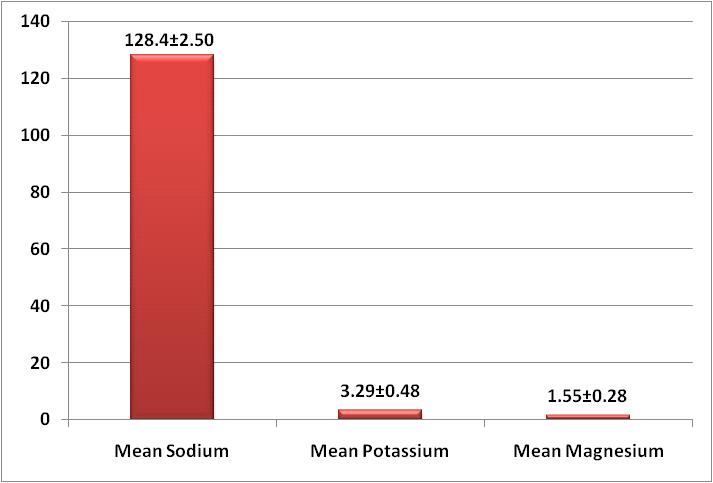
Most of our patients belonged to Child Pugh B (56%) followed by Child Pugh C (34%). 80% of our patients had MELD score greater than 14. The mean CTP and MELD scores of the participants were 9.36±2.38 and 19.72±7.02 respectively as depicted in Table 1. The mean serum sodium, potassium and magnesium values in meq/l were 128.40±2.50,
Vol 121, No 8, August 2023Journal of
the Indian Medical Association
23
3.29±0.48 and 1.55±0.28 respectively (Fig 1).
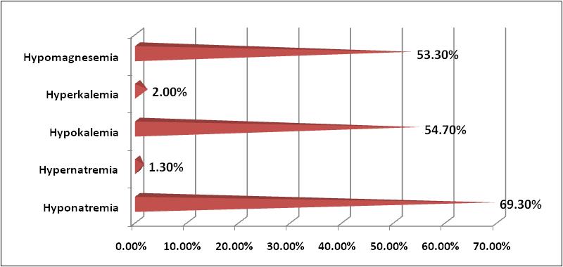
Dyselectrolytemia was observed in 113 (75.3%) patients of our study group. The most common electrolyte abnormality was hyponatremia (69.3%) followed by hypokalemia (54.67%), hypomagnesemia (53.3%), hyperkalemia (2%) and hypernatremia (1.3%) as shown in Fig 2.
When stratified according to CTP scores, 46 patients out of 51 (90.2%) in CTP- C class had dyselectrolytemia whereas 76.1% of patients in CTP-B class and 20% of patients (3 out of 15) in CTP-A class had electrolyte imbalance. Patients belonging to CTPA group had the highest mean serum sodium level (133.8±1.70 meq/l) while those in CTP-C group had the lowest mean sodium levels (121.85±2.40 meq/l). The mean serum potassium level was 3.78±0.46 meq/l in CTP-A group and 2.82±0.51 meq/l in patients belonging to CTP-C group. Likewise, the mean serum magnesium level was highest in CTP-A (1.9±0.35 meq/l) and lowest in CTP-C (1.25 ±0.27 meq/l). This decrease in electrolyte values with the increasing CTP score was statistically significant for all the electrolytes (P< 0.0001) as shown in Table 2. Similarly, electrolyte disturbance was
seen in 92.3%, 86.7% and 20% in patients belonging to MELD score group of >21, 15-21 and <14 respectively. The mean serum sodium, potassium and magnesium level was highest in those with MELD score <14 and the least in patients with MELD score >21. This decrease in electrolyte values with the increasing MELD score was statistically significant for all the electrolytes (P<0.0001) as shown in Table 3.
Vol 121, No 8, August 2023Journal of the Indian Medical Association
CharacteristicsNo of patients (N=150) Mean age (years)49.2±11.8 Gender Male 121 (80.6%) Female29 (19.3%) Etiology of cirrhosis Alcohol106 (70.7%) NASH14 (9.3%) Hepatitis B12 (8.0%) Hepatitis C 05 (3.3%) Autoimmune03 (2.0%) Cryptogenic10 (6.7%) Child-Pugh scoreA (%)15 (10.0%) B (%)84 (56.0%) C (%)51 (34.0%) MELD score <14 (%)30 (20.0%) 15–21 (%)68 (45.3%) >21 (%)52 (34.7%) Mean CTP score09.36±2.38 Mean MELD score19.72±7.02 Presenting features Ascites 135 (90.0%) Jaundice98 (65.3%) GI bleed68 (45.3%) SBP34 (22.6%) HE36 (24.0%) HRS56 (37.3%)
Table 1 — Baseline characteristics of study population
Fig 1 — Mean electrolyte levels in the study population
Electrolytes observed CTP-ACTP-B CTP-Cp value Mean sodium value 133.8±1.70129.57±3.42121.85±2.40 Mean potassium value3.78±0.463.28±0.47 2.82±0.51 <0.0001*** Mean magnesium value1.90±0.351.51±0.241.25±0.27
Electrolytes observed MELD <14MELD 15-21 MELD >21p value Mean sodium value 132.87±1.80128.80±3.50123.55±2.62 Mean potassium value3.86±0.50 3.14±0.412.88±0.53 <0.0001*** Mean magnesium value1.88±0.311.58±0.271.20±0.28
Table 2 — Distribution of electrolyte levels according to CTP scores
Table
3 — Distribution
of
electrolyte levels according to MELD score
24
Fig 2 — Different electrolyte abnormalities in the study population
Out of 36 patients who had hepatic encephalopathy at admission, hypomagnesemia was observed in 32 patients (95% CI: - 1.30 - 12.14; OR 4.0; p<0.05) indicating a strong association between the two (Table 4). However, for hyponatremia [95% CI: - 0.78 to 4.53] and hypokalemia [95% CI: - 0.48 to 2.54] no statistical significance was seen (p >0.05). Hyperkalemia and hypernatremia was not seen in patients who had HE.
On the other hand, hyponatremia was observed in 54 out of 56 the patients who had HRS [95% CI: - 3.18 - 60.89; OR 13.935; p<0.05] and statistical analysis indicated a strong association between the two (Table 5). For hypokalemia [95% CI: - 0.29 - 1.12] and hypomagnesemia [95% CI: - 0.33 -1.28] no statistical significance was determined. Hyperkalemia and hypernatremia was not seen in patients who had HRS.
DISCUSSION
In our study group, the most common etiology of cirrhosis was alcohol (70.6%) with ascites being the most common clinical presentation (90%). This is similar to findings observed by Singh, et al9 and Almani, et al10
In the present study, prevalence of dyselectrolytemia was 75.3% such that hyponatremia was observed in 69.3%, hypokalemia in 54.67%, hypomagnesemia in 53.3%, hyperkalemia in 2% and hypernatremia in 1.3%. The prevalence of hyponatremia in previous studies performed by Singh, et al9, Angeli, et al11 and Borroni, et al12 was observed to be 47%, 21.6% and 14.9% respectively whereas the prevalence of hypokalemia in the prior studies carried out by Devrajani, et al13 Kashyap, et al14 and
Mumtaz, et al15 was found to be 33%, 15%, and 6.4% respectively.
The mean serum sodium, potassium and magnesium values in meq/l were 128.40±2.50, 3.29±0.48 and 1.55±0.28 respectively which were lower than the normal accepted range. Most common reason for hyponatremia in Cirrhosis is mainly due to impairment of solute free water excretion which is due to the increased Anti Diuretic Hormone (ADH) secretion and decreased effective arterial volume16
Magnesium deficiency in Cirrhosis is a burning research topic with few available literatures. It may result from low nutrient uptake, decreased absorption due to intestinal mucosal edema secondary to portal hypertension, greater urinary secretion due to diuretics, low serum albumin concentration, and/ or hormone inactivation17.
The mean serum sodium, potassium and magnesium levels were highest among the patients in CTP-A group (133.8±1.70, 3.78±0.46, 1.9±0.35 meq/ l,) and lowest in CTP-C group (121.85±2.40, 2.82±0.51, 1.25 ±0.27 meq/l) and this decrease in the electrolyte values with the increasing CTP score was statistically significant (p <0.0001). The results were in accordance with the studies done by Quereshi, et al18 and Safarikish B, et al19
The declining values of serum electrolytes with increasing MELD score was also observed in our study. The mean serum sodium, potassium and magnesium levels were higher (132.87±1.80, 3.86±0.50, 1.88±0.31 meq/l ) in those with MELD score <14 as compared to patients with MELD score >21. This decrease in the electrolyte values with the increasing MELD score was statistically significant (p<0.0001). Kim, et al20 identified a significant interaction between the serum sodium concentration and the MELD score, demonstrating that the serum sodium concentration was greater in patients with a lower MELD score. In study by Safarikish, et al19, it was observed that patients with hypomagnesemia had higher MELD score (16.5±5.8) than those with normal magnesium values (14.3 ± 5.4).
Our study found significant association between hypomagnesemia and HE (95% CI: - 1.30 - 12.14; OR 4.0; p<0.05). This is in accordance to the work done by Lopes, et al21 who highlighted that lower levels of serum magnesium were associated with a greater risk of HE in both liver graft donors and recipients during the first few days after surgery. According to Nangliya, et al22 , serum magnesium levels significantly decreased as liver cirrhosis progressed, which is consistent with our findings.
Vol 121, No 8, August 2023Journal
of the Indian Medical Association
Electrolyte HE+Chi-square ORP value abnormality (N=36)(95% CI) Hyponatremia 28(19%)1.5321.892 0.2158 (0.78 to 4.53) Hypokalemia 26(17%) 0.00063 1.105 0.9799 (0.48 to 2.54) Hypomagnesemia 32(21%)5.6454.0 0.0175 (1.30 to 12.14)
Table 4 — Correlation between different electrolyte abnormalities and hepatic encephalopathy (HE)
Electrolyte HRS+ Chi-square ORP value abnormality (N=56)(95% CI) Hyponatremia54 (36%)16.891 13.935<0.0001 (3.18-60.89) Hypokalemia27 (18%) 2.080 0.57790.1493 (0.29-1.12) Hypomagnesemia32 (21%)1.114 0.66000.2911 (0.33-1.28) 25
Table 5 — Correlation between different electrolyte abnormalities and hepatorenal syndrome (HRS)
Hyponatremia was observed to be significantly associated with Hepatorenal Syndrome (HRS), [95% CI: - 3.18 - 60.89; OR 13.935; p<0.05]. This is in accordance to the previous study conducted by; Shaikh, et al23 who found that the frequency of hepato renal syndrome was strongly associated with decreased serum sodium concentration, suggesting more severe circulatory dysfunction in cirrhotics with hyponatremia than those without it.
In our study, hypokalemia was not found to be significantly associated with both HE and HRS which is in contrast to the findings observed by Kaplan, et al24 where, hypokalemia was also stated to be an important prognostic factor in cirrhotic patients. Both hypernatremia and hyperkalemia was not seen in patients who had HE or HRS. Our study had few limitations such that potential causes of dyselectrolytemia (diuretics, infections or hypovolemia) were not assessed, sample size was small and follow up of the cases couldn’t be done. Nevertheless, our study clearly highlights that electrolyte abnormalities are quite common in patients with cirrhosis of liver and warrants special attention during the treatment process. Their values can help us determine the severity of disease and predict the development of complications as evidenced by their significant association with complications like HE and HRS.
Credit authorship contribution statement : All authors contributed equally in carrying out the study, writing and drafting of the manuscript.
Conflicts of interest : None
Source of funding : None
REFERENCES
1Asrani S K — Burden of liver diseases in the world. Journal of Hepatology 2019; 70: 151-71.
2Ge PS, Runyon BA — Treatment of patients with cirrhosis. N Engl J Med 2016; 375: 767-77.
3Hassan ZBA, Kadum J — Electrolytes disturbance in liver cirrhosis Graduation thesis, Al-Nahrain College of Medicine, Iraq 2019 Available from: http s://www.colmedalnahrain.edu.iq/upload/res/884.pdf.
4Gaglio P — Hyponatremia in Cirrhosis and End-Stage Liver Disease: Treatment with the Vasopressin V2-Receptor Antagonist Tolvaptan. Dig Dis Sci 2012; 57(11): 2774-85.
5Artz SA, Paes IC, Faloon WW — Hypokalemia induced hepatic coma in cirrhosis: Occurrence despite neomycin therapy. Gastroenterology 1966; 51: 1046 53.
6Swaminathan R — Magnesium metabolism and its disorders. ClinBiochemRev2003; 24: 47-66. PMID 18568054.
7Wu L — Magnesium intake and mortality due to liver diseases: Results from the third national health and examination survey cohort. Sci Rep 2017;7:17913.
8Veena G, James R — Prevalence of hypomagnesemia in cirrhosis of liver and its association with severity of the disease. Asian J Pharm Clin Res ; 15(8): 92-5.
9Singh Y — Study of electrolyte disturbance in chronic liver disease patients attending a hospital in Kumaon region. Journal of Family Medicine and Primary Care 2022; 11(8): 4479-82.
10Almani SA — Cirrhosis of liver: Etiological factors, complications and prognosis. JLUMHS 2008; 2: 61-6.
11Angeli P — Hyponatremia in cirrhosis: Results of a patient population survey Hepatology 2006; 44: 1535-1542.
12Borroni G — Clinical relevance of hyponatremia for the hospital outcome of cirrhotic patients. Dig Liver Dis 2000; 32: 605-10.
13Devrajani BR — Precipitating factors of hepatic encephalopathy at a tertiary care hospital Jamshoro, Hyderabad JPMA. J Pak Med Assoc 2009; 59: 683.
14Kashyap C, Borkotoki S, Dutta RK — Study of serum sodium and potassium level in patients with alcoholic liver disease attending Jorhat Medical College Hospital—A hospital-based study. Int J Health Sci Res 2016; 6: 113-6.
15Mumtaz K — Precipitating factors and the outcome of hepatic encephalopathy in liver cirrhosis. J Coll Physicians Surg Pak 2010; 20: 514-8.
16Kim J H, Lee J S, Lee S H — The Association between the Serum Sodium Level and the Severity of Complications in Liver Cirrhosis. Korean J Intern Med 2009; 24(2): 106-112.
17Liu M, Yang H, Mao Y— Magnesium and liver disease. Ann Transl Med 2019; 7(20): 578.
18Qureshi Muhammad. Correlation of Hyponatremia with Hepatic Encephalopathy and Severity of Liver Disease. Journal of the College of Physicians and Surgeons Pakistan; 24: 135137.
19Safarikish B — The Relationship Between Plasma Magnesium Concentration and Hepatic Encephalopathy in Liver Cirrhosis Patients: A Preliminary Result of a Referral Center in Iran. Middle East J Rehabil Health Stud 2022; 9(2): e122978.
20Kim WR — Hyponatremia and mortality among patients on the liver-transplant waiting list. N Engl J Med 2008; 359: 101826.
21Lopes PJ — Correlation between serum magnesium levels and hepatic encephalopathy in immediate post liver transplantation period. Magnes Res 2013; 45(3): 1122-5.
22Nangliya V — Study of trace elements in liver cirrhosis patients and their role in prognosis of disease. Biol Trace Elem Res 2015; 165(1): 35-40.
23Shaikh S — Frequency of hyponatraemia and its influence on liver cirrhosis-related complications. J Pak Med Assoc 2010; 60(2): 116-20.
24Kaplan M — Prognostic Utility of Hypokalemia in Cirrhotic Patients. Acta Gastro-Enterologica Belgica 2018: 398-403.
Vol 121, No 8, August 2023Journal
of the Indian Medical Association
26
Original Article
A Study on the Evaluation of Swallowing in Post-CVA Patients
Anton Dev X1, Gauranga Prosad Mondal2, Somnath Saha3
Aims and Objectives : (1) To find the incidence of Swallowing difficulty in Cerebrovascular Accident (CVA) patients. (2) Evaluation of degree of dysphagia in post stroke patients by the use of Functional Endoscopic Evaluation of Swallowing (FEES). (3) Assess for any possible relationship between swallowing defects and region of brain involved.
Materials and Methods : The present study is an observational study done over a 1 year period in a Tertiary Care set-up. Mann Assessment of Swallowing Ability (MASA) test and Functional Endoscopic Evaluation of Swallowing (FEES) were used to assess swallowing function. The findings from FEES were graded using the Penetration –Aspiration Scale (PAS). We had used radiologic methods to determine the site of lesion and Arterial territory involved.
Results : In the present study, MASA scores were normal in 50% and Dysphagia was Mild in 16.0%, moderate in 6.0% and Severe in 28.0%. Based on the PAS interpretation, no entry was present in 20%. Aspiration was seen in 24% and penetration was seen in 56%. Patient age and MASA grade had a significant association. The MASA values and PAS scores were found to be inversely correlated. Swallowing abnormalities were detected in Stroke of the following regions : Basal Ganglia, Thalamus, Pons & Lateral Medulla. MCA territory infarcts were commonly found to cause Dysphagia.
Conclusion : In the present study, 50 %of the total stroke patients had dysphagia, as determined by the MASA test. FEES detected Dysphagia in 80 % of the patients. A strong inverse correlation was present between the MASA test score and the PAS score, with correlation coefficient as -0.935. Stroke of certain specific brain regions and arterial territories were more commonly found to cause Swallowing difficulties.
[J Indian Med Assoc 2023; 121(8): 27-31]
Key words :Post-CVA, Cerebrovascular Accident, Swallowing Dysfunction, Dysphagia, MASA test, FEES, Arterial Territory.
Global occurrence of stroke is high with numbers of upto 15 million1 of which more than half will have swallowing problems, out of which half will be symptomatic2. The cases of Stroke account for a significant volume of Disability and Morbidity, the world over3. A large number of stroke patients recover normal swallow spontaneously. Despite this, a significant number (upto half) of the patients may have dysphagia at 6 months4,5. Persistent dysphagia has been found to independently predict overall poor outcome6. Dysphagia leading to aspiration has also been seen to have significance for occurrence of pneumonia following stroke7. Confirmed aspiration confers a higher risk3,8
For Hospitalised Stroke patients, the relative risk of death due to pneumonia : 5.77. Early studies had included referral patients with diagnosed Dysphagia. This inadvertently caused a false rise in the detected incidence of Aspiration9,10. There have been no current studies based on an unselected population5,11
Department of Otorhinolaryngology, Calcutta National Medical College & Hospital, Kolkata 700014
1MS, Senior Resident, At present : Senior Resident, Department of Otorhinolaryngology, M R Bangur Hospital, Kolkata 700033 and Corresponding Author
2MD (General Medicine), DM (Neurology), DCH, Professor and Head, Department of Neurology
3MS (ENT), Professor and Head
Received on : 02/04/2022
Accepted on : 25/06/2023
Editor's Comment :
Swallowing difficulty is a common occurence in Stroke patients and so a proper assessment tool to detect it, is of paramount importance.
A greater proportion of stroke patients were found to have swallowing pathology when assessed with the Assessment tool Functional Endoscopic Evaluation of Swallowing (FEES) and scored using the Penetration-Aspiration Scale (PAS), as compared to Clinical methods (MASA test). The PAS scores and MASA test values were inversely correlated. Stroke of certain specific brain regions and arterial territories were seen to cause Swallowing disturbances more commonly.
In acute stroke, Dysphagia has been found to have a prevalence of about 28 to 65%5,11-13. Dysphagia is found to improve significantly in the early days after the occurrence of Stroke2,5. A certain small percentage of patients will have longer duration of Dysphagia4.
An observed finding is that, in patients whom the swallow does not recover in the first 10 days, recovery requires a longer period of upto 2-3 months14. Silent aspiration has been found to occur in about 40% of Dysphagic patients15. Patients with aspiration have associated Abnormal pharyngeal sensation with Silent aspiration seen to occur in 8%16
The Presence of Swallowing difficulty following stroke depends on the following factors:
(1)Site of Involvement (eg: Corticobasilarfibres,
Vol 121, No 8, August 2023Journal of the Indian Medical Association
27
Medulla Lateral Medullary Syndrome, Brain Stem Pons )
(2)Size of lesion (Pressure effect )
(3)Sensorium of Patient
(4)Bilaterality (eg, Corticobulbar tract )
Specific Sites known to cause swallowing deficits in relation to their Arterial supply :
(1)Lacunar Infarcts - Mainly Bilateral MCA territory17
(2)Area surrounding Area 43 and 44 ( Frontoparietal operculum ) – Striate branches of MCA17
(3)Cerebellum and Lateral Medulla ( Lateral Medullary Syndrome ) – PICA18,19
(4)Pyramids involvement20
(5)Pons21 and Medulla19 – Perforating branches of Basilar Artery
(6)Corpus Striatum supplied by Recurrent Artery of Heubner22
(7)Thalamus23
Tests for Clinical Assessment of Dysphagia :
(1) 3-oz Water swallow test24
(2) The Standardized Swallowing Assessment25
(3) Acute Stroke Dysphagia Screen26
(4) Yale Swallow Protocol27
(5) Mann’s Assessment of Swallowing Ability (MASA)28
In the present study, we had used the MASA test for Clinical evaluation of Swallowing.
Mann’s Assessment of Swallowing Ability (MASA) test28 :
The MASA test employs a set of swallowing tasks, using which Trained healthcare professionals rate the patient’s performance. The MASA defines risk of Dysphagia and Aspiration. It also provides a severity scale28
The newer technology in Swallowing assessment includes Videofluoroscopy (VFSS) and The Functional Endoscopic Evaluation of Swallowing (FEES). These modalities have surpassed the earlier tools.
In the present study, Dynamic assessment of Swallowing is done using FEES29. Grading of the observed findings , was done using the PenetrationAspiration Scale (PAS)30
Fiberoptic Endoscopic Evaluation of Swallowing (FEES)29:
•Delayed swallow initiation and presence of pharyngeal residue are better evaluated using this modality.
•Swallowing of different food consistencies is evaluated.
In FEES, the following features are observed :
•Transition duration
•Evidence of penetration and aspiration
•Number of swallows to clear the bolus
•Extent of airway closure
MATERIALS AND METHODS
Study design :
Observational study at a Tertiary Centre in West Bengal.
Study population :
Patients admitted in the Neurology IPD.
Sample design :
Inclusion criteria —
All patients of Cerebrovascular Accidents (CVA) including :
•Patients with Ischemic stroke.
•Patients with Hemorrhagic stroke.
•Only Medically Stable patients
Exclusion criteria —
(1) Recurrent stroke.
(2) Hemodynamically unstable patients.
(3) Unconscious patients.
(4) Patient having Dysphagia from before the stroke due to any other cause.
(5) Subarachnoid haemorrhage.
(6) Patients with Severe agitation.
(7) Severe movement disorders (Dyskinesia)
(8) Severe bleeding disorders and/or Recent severe epistaxis (nosebleed).
(9)History of recent trauma to the nasal cavity or surrounding tissue and structures secondary to surgery or injury.
(10) Bilateral obstruction of the nasal passages.
(11) Patients with any other progressive neurological condition such as parkinsonism, schizophrenia.
(12) Mentally impaired patients who cannot provide a proper informed consent & who would be uncooperative for examination.
(13) Patients of Childhood Stroke Syndromes with various underlying genetic defects are excluded as they may skew the inferences, as a result of the differences in their baseline Physiology.
Sample size : 50 cases
Study Tools :
(1) History including any past history of Cerebrovascular Accidents.
(2) Basic Haematology including Complete Blood Count, Platelets, BT/CT, Urea, Creatinine, Serology.
(3) Mann Assessment of Swallowing Ability (MASA) test, used to assess swallow.
(4) Fiberoptic Endoscopic Evaluation of Swallowing (FEES), was performed using the protocol defined by Susan Langmore31
(5) We have used Radiologic imaging methods coupled with expert radiologist opinion to ascertain the
Vol 121, No 8, August 2023Journal
of the Indian Medical Association
28
region of brain involved and the arterial territories affected.
RESULTS
Comparison of Age and gender of the study population.
Mean ages:
•Males : 70.0±7.7 years
•Females : 58.1±11.3 years.
•No significant difference was detected , between the means (Tables 1&2).
Table 3 shows us that the MASA scores were normal in half of the patients. Dysphagia was Mild in 16.0%, moderate in 6.0% and Severe in 28.0% according to MASA test (Fig 2).
The Table 4 categorises the frequencies in the various grades of MASA score in our study.
The Table 5 categorises the PAS interpretations. Aspiration was seen in 24%, No entry was present in 20% and penetration was seen in 56%.
From Table 6, we infer the association that, as the age increased, the Dysphagia severity as determined by MASA grade was also increasing (Statistically significant).
Table 7, Correlates the MASA and PAS scores. The two variables were highly correlated. The correlation coefficient was -– 0.935. That means, when MASA score is increasing, the PAS value is decreasing. The decrease was 87.4%.
Table 8 states the relationship between the Dysphagia severity (determined from PAS score) and Lesion side. We find no significant relationship (P>0.05) (Table 9).
DISCUSSION
In the present study we have evaluated a total of 50 Post -CVA patients in the domain of swallowing. Evaluation was performed using both a clinical test (MASA) and an endoscopic study (FEES).
(1) In the study done by Chojin, et al an abnormal MASA score was seen in 71.9% patients. Out of them, maximum patients were in the severe category, followed by the mild and moderate categories32. In the present study, using MASA test, we have observed a lesser incidence of Dysphagia (50%). Butupon categorization of MASA values, maximum patients were in the severe group, followed by the mild and then moderate groups, which is in accordance with the earlier study by Chojin, et al
(2) Logeman JA, etalobserved that the Clinical tests alone tend to underestimate the incidence of Dysphagia33. In the present study, Fiberoptic endoscopy (FEES) revealed that 80% of the patients had swallowing dysfunction while in the MASA test, only 50% of the patients were found
Vol 121, No 8, August 2023Journal of the Indian Medical Association
Age groupMales FemalesTotal (years)No%No%No% 30-3900.024.024.0 40-4912.048.05 10.0 50-597 14.0816.01530.0 60-699 18.01020.01938.0 70-796 12.036.0918.0 Total2346.02754.050100.0 Mean ±SD 70.0±7.758.1±11.3Mean=59.8±9.9 Range=38-79 years Significance “t” = 1.393, df = 48, P = 0.170
Insult classification Frequency% Hemorrhage16 32.0 Infarct3468.0 Total50100.0
CategoryScore Frequency% Normal 170-2002550.0 Mild 149-169816.0 Moderate 141-14836.0 Severe <1401428.0 Total50100.0
Table 1 — Study subjects Ages
Table
2
— Classification of the cerebrovascular insults (in our study)
Table 3 — MASA Score categories
MASA grade Frequency% Mild9 27.3 Moderate16 48.5 Severe824.2 Total33100.0
Table 4 — Frequency of MASA grades
PAS ScorePAS interpretation FrequencyPercentage 1No entry10 20.0 2,3,4,5 Penetration28 56.0 6,7,8Aspiration1224 Total50100.0
Table 5 — PAS interpretation
Mild Moderate SevereTotal Results < 5003407 50-59932115 χ2 =25.156 60-697372 19df=9 70-7910359 P=0.002 Total17916850
Table 6 — Association between age group with MASA Grade Age groupNil
29
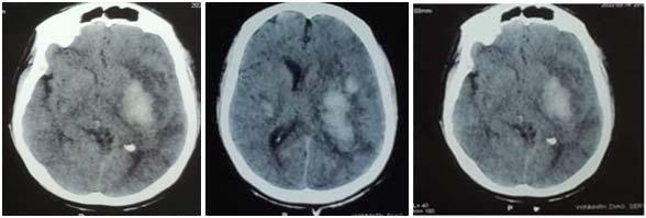
Fig 1 — CT Imaging from a patient of Basal Ganglia hemorrhage in the present study
of the Indian Medical Association
to have abnormality. Therefore our findings are in concordance with the earlier study.
(3) EunKyu
•Pontine infarct & Lateral Medullary Infarct depending on the size and subsite involved caused Swallowing Dysfunction. Similar findings were observed by Chang MC, et al21 and Ayodogdu, et al19 respectively.
Hemorrhage :
•Putamen hemorrhage & Basal Ganglia Hemorrhage were found to cause Swallowing dysfunction. Similar findings were reported by Maeshima S, et al37 and Logeman JA, et al respectively38
•Cerebellar Hemorrhage was found to cause Swallowing Dysfunction. Similar findings were observed by Pe’rie’ S, et al39
•Hemorrhage of PCA territory involving the Parieto-Occipital area also caused Swallow Dysfunction. Similar findings were observed by Dehaghani, et al35
CONCLUSION
Ji, et al observed that the MASA score and the Penetration Aspiration Scale value obtained from FEES were found to be strongly negatively correlated34. A similar finding was observed in the present study.
(4) Dehaghani, et al had observed that Stroke of certain specific Brain regions cause Swallowing dysfunction35. Similar findings were observed in the present study.
(5) The individual regions we had observed to cause swallowing disturbance are as follows : Infarcts :
•MCA territory infarcts & PCA territory infarcts (especially of P1 and P2 region ) caused Swallowing Dysfunction.Similar findings were observed by Paciaroni M, et al17 and Javed K, et al36 respectively.
•ACA territory infarcts caused Dysphagia when associated with MCA territory infarct.
•We had observed Dysphagia in about half the patients in our study. Severity of Dysphagia as obtained from MASA questionnaire was graded and 16% had mild, 6% had moderate and 28% of the stroke patients had severe dysphagia.
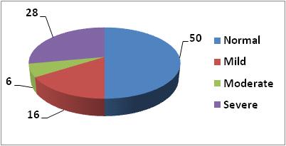
•Using the FEES study we found varying degrees of Swallowing impairment in 80 % of the patients which is significantly higher than the frequency of impairment as determined using the clinical method (50%).
•On attempting to find correlation between the MASA score and PAS value, we found that the two variables were strongly negatively correlated. The correlation coefficient was -– 0.935.
•Lesion side and the Presence of Dysphagia were not significantly associated with each other.
•Stroke of certain specific brain regions and arterial territories were found to cause Dysphagia. Limitations :
Putamen Hemorrhage3
Basal Ganglia Hemorrhage (with sensorium impairment )3
Unilateral Cerebellar Hemorrhage2
Hemorrhage of PCA territory1
Thalamic Hemorrhage1
Unilateral MCA territory infarct12
Large Lacunar Infarct2
Medullary Infarct (Lateral Medulla)1
Unilateral PCA infarct7
Pontine Infarct2
Bilateral PCA infarct1
Unilateral ACA and MCA infarct5
•The various regions of brain we had observed to be related to Swallowing Dysfunction in the present study, were in concordance with that seen in earlier studies. But since the number of cases for each region of the brain were limited, we were unable to draw a Statistical significance.
•In the present study, we had considered the presence or absence of swallowing dysfunction but not the specific attributes of the Swallowing dysfunction associated with each arterial territory and brain region involved .
•We had not given consideration to the etiopathogenesis of the stroke and the impact they would have on the Resultant swallowing dysfunction.
•We did not have any cases of solely ACA
Vol 121, No 8, August 2023Journal
PASB/LLeft
interpretationNo%No%No%No% Aspiration00.0816.048.01224.0 χ2 =2.747 No Entry00.05 10.0510.01020.0 df=4 Penetration12.020 40.0714.02856.0 P=0.601 Total12.03366.01632.050100.0
Table 8 — Association between Interpreted PAS score with Lesion side
RightTotal Results
Number of Cases
Table 9 — Region wise classification of Stroke causing Swallowing Dysfunction as seen in the present study with number of cases in each category
in the present study
Variablesr significancer2 % of r2 Determination MASA X PAS-0.935 P<0.001 0.87487.487.4
Table 7 — Relationship between MASA score and PAS score
30
Fig 2 — Categorisation of MASA test scores
territory infarct, hence we could not determine the standalone significance of ACA territory stroke in causing Dysphagia.
REFERENCES
1Mackay J and Mensah G — Atlas of heart disease and stroke. Geneva, Switzerland: World Health Organization, 2004.
2Martino R, Foley N, Bhogal S, Diamant N, Speechley M, Teasell R — Dysphagia after stroke: incidence, diagnosis, and pulmonary complications. Stroke 2005; 36(12): 2756-63.
3Kumar S, Selim MH, Caplan LR — Medical complications after stroke. Lancet Neurol2010; 9(1): 105-18. doi: 10.1016/S14744422(09)70266-2. PMID: 20083041.
4Smithard DG, O’Neill PA, Parks C, Morris J — Complications and outcome after acute stroke. Does dysphagia matter? Stroke 1996; 27(7): 1200-4.
5Mann G, Hankey GJ, Cameron D — Swallowing disorders following acute stroke: prevalence and diagnostic accuracy. Cerebrovasc Dis 2000; 10(5): 380-6.
6Smithard DG, Smeeton NC, Wolfe CD — Long-term outcome after stroke: does dysphagia matter? AgeAgeing 2007; 36(1): 90-4 .
7Wilson RD — Mortality and cost of pneumonia after stroke for different risk groups. J Stroke Cerebrovasc Dis 2012; 21(1): 61-7.
8Rofes L, Arreola V, Almirall J, Cabré M, Campins L, GarcíaPeris P, et al — Diagnosis and management of oropharyngeal Dysphagia and its nutritional and respiratory complications in the elderly. Gastroenterology Research and Practice 2011, 818979.
9Leder SB, Acton LM, Lisitano HL, Murray JT — Fiberoptic endoscopic evaluation of swallowing (FEES) with and without blue-dyed food. Dysphagia 2005; 20(2): 157-62.
10Schmidt J, Holas M, Halvorson K, Reding M — Videofluoroscopic evidence of aspiration predicts pneumonia and death but not dehydration following stroke. Dysphagia 1994; 9(1): 7-11.
11Smithard DG, O’Neill PA, England RE, Park CL, Wyatt R, Martin DF, et al — The natural history of dysphagia following a stroke. Dysphagia 1997; 12(4): 188-93.
12Ramsey D, Smithard D, Kalra L — Silent aspiration: what do we know? Dysphagia 2005; 20(3): 218-25. doi: 10.1007/ s00455-005-0018-9.
13Falsetti P, Acciai C, Palilla R — Oropharyngeal dysphagia after stroke: incidence, diagnosis, and clinical predictors in patients admitted to a neurorehabilitation unit. Cerebrovasc Dis 2009; 18: 329-35.
14Mann G, Hankey GJ, Cameron D — Swallowing function after stroke: prognosis and prognostic factors at 6 months. Stroke 1999; 30(4): 744-8.
15Leder SB, Cohn SM, Moller BA — Fiberoptic endoscopic documentation of the high incidence of aspiration following extubation in critically ill trauma patients. Dysphagia 1998; 13(4): 208-12.
16Kidd D, Lawson J, Nesbitt R, MacMahon J — The natural history and clinical consequences of aspiration in acute stroke. QJM 1995; 88(6): 409-13.
17Paciaroni M, Mazzotta G, Corea F — Dysphagia following Stroke. European Neurology 2003; 51: 162-7.
18Teasell R, Foley N, Fisher J — The incidence, management, and complications of dysphagia in patients with medullary strokes admitted to a rehabilitation unit. Dysphagia 2002; 17: 115-20.
19Aydogdu I, Ertekin C, Tarlaci S — Dysphagia in Lateral Medullary Infarction (Wallenberg’s Syndrome). Stroke 2001; 32(9): 2081-7.
20Emilia G, Hooman K, Eric CB — Ischemic Stroke of the Pyramidal decussation causing Quadriplegia and Anarthria. Journal of
Stroke & Cerebrovascular Diseases 2012; 21(7): 620.E1620E2.
21Chang MC, Kwak SG, Chun MH — Dysphagia in patients with isolated pontine infarction. Neural Regeneration Research 2018; 13(12): 2156-9.
22Ghaemi H, Sobhani-Rad D, Arabi A — Role of Basal Ganglia in Swallowing Process: A Systematic Review. Iranian Rehabilitation Journal 2016; 14(4): 239-45.
23Maeshima S, Osawa A, Yamane F — Dysphagia following Acute Thalamic Haemorrhge : Clinical correlates and outcomes. European Neurology 2014; 71: 165-72.
24DePippo — Validation of the 3-oz water swallow test for aspiration following stroke. Arch Neurol 1992; 49: 1259-61.
25Jiang JL, Fu SY, Wang WH, Ma YC — Validity and reliability of swallowing screening tools used by nurses for dysphagia: A systematic review. Ci Ji Yi XueZaZhi 2016; 28(2): 41-8.
26Edmiaston J, Connor LT, Loehr L, Nassief A — Validation of a dysphagia screening tool in acute stroke patients. American journal of critical care. 2009.
27Validation of the Yale Swallow Protocol: a prospective doubleblinded videofluoroscopic study. Suiter DMetal. Dysphagia 2014; 29(2): 199-203. doi: 10.1007/s00455-013- 9488-3. Epub 2013 Sep 12.
28Mann G — MASA: The Mann Assessment of Swallowing Ability. Clifton Park, NY: Singular; 2002.
29Gonzaìlez-Fernaìndez M, Ottenstein L, Atanelov L, Christian AB — Dysphagia after Stroke: an Overview. Curr Phys Med Rehabil Rep 2013; 1(3): 187-96.
30John C. Rosenbek, Jo Anne Robbins, Ellen B — Roecker, Jame L. Coyle, Jennifer L. Wood. A penetration-aspiration scale. Dysphagia 1996; 11(2): 93. doi:10.1007/BF00417897
31Langmore SE — Endoscopic evaluation and treatment of swallowing disorders. New York, NY: Thieme, 2001
32Chojin Y, Kato T, Rikihisa M, Omori M, Noguchi S, Akata K, et al — Evaluation of the Mann Assessment of Swallowing Ability in Elderly Patients with Pneumonia.Agingand Disease 2017; 8(4): 420-33. doi:10.14336/AD.2017.0102
33Logemann JA, RoaPauloski B, Rademaker A, Cook B, Graner D, Milianti F, Beery Q, et al — Impact of the diagnostic procedure on outcome measures of swallowing rehabilitation in head and neck cancer patients. Dysphagia 1992; 7(4): 179-86. doi: 10.1007/BF02493468. PMID: 1424831.
34Ji EK, Wang HH, Jung SJ, Lee KB, Kim JS, Hong BY, et al —Is the modified Mann Assessment of Swallowing Ability useful for assessing dysphagia in patients with mild to moderate dementia? J Clin Neurosci [Internet] 2019; 70: 169-72.
35Dehaghani SE, Yadegari F, Asgari A, Chitsaz A, Karami M — Brain regions involved in swallowing: Evidence from stroke patients in a cross-sectional study. Journal of research in medical sciences : the official journal of Isfahan University of Medical Sciences 2016; 21: 45. https://doi.org/10.4103/ 1735-1995.183997
36Javed K, Reddy V, M Das J — Neuroanatomy, Posterior Cerebral Arteries. 2021 Jul 31. In: StatPearls [Internet]. Treasure Island (FL): StatPearls Publishing; 2022 Jan–. PMID: 30860709
37Maeshima S, Okazaki H, Okamoto S, Mizuno S, Asano N, Tsunoda T, et al — Dysphagia Following Putaminal Hemorrhage at a Rehabilitation Hospital. J Stroke Cerebrovasc Dis 2016; 25(2): 389-96.
38Logemann JA, Shanahan T, Rademaker AW, Kahrilas PJ, Lazar R, Halper A — Oropharyngeal swallowing after stroke in the left basal ganglion/internal capsule. Dysphagia 1993; 8(3): 230-4.
39Périé S, Wajeman S, Vivant R, St Guily JL — Swallowing difficulties for cerebellar stroke may recover beyond three years. Am J Otolaryngol 1999; 20(5): 314-7. doi: 10.1016/ s0196-0709(99)90033-9.
Vol 121, No 8, August 2023Journal
of the Indian Medical Association
31
Original Article
Implementation of an Interactive Teaching Session using ‘Snowball Technique’ for Introducing 1st Professional MBBS Students to Clinical Correlation in Biochemistry
Niket Verma1, Kusum Singla2, Bishambar Toora3, Richa Dixit4
Under the new Competency Based Medical Education curriculum in India, two-thirds of the teaching schedules must comprise of interactive teaching sessions. Snowball Discussion is an interactive and collaborative technique which starts with the introduction of a clinical challenge to individual learners and then proceeds by doubling. The current study was conducted with the aim of assessing the perception of learners from 1st professional MBBS towards a recently conducted teaching learning session using Snowball technique in Biochemistry. A Snowball teaching session was conducted on ‘Diabetes Mellitus’. 73 learners attended the session. The facilitators sensitized the learners about the Snowball technique after which the session was conducted step-wise. This was followed by an integrated learning session in which guest speakers shared important points about diabetes mellitus relevant to their subjects. Feedback was obtained from the learners using an online feedback form 46 learners submitted the feedback form. There was a high level of satisfaction among the learners regarding the session. 45 respondents (97.8%) agreed that attending the Snowball session generated their interest in the topic and they were able to better correlate the topic clinically. 44 respondents (95.7%) agreed that after attending the Snowball session they were motivated to read further about the topic and that a Snowball session was better than a traditional lecture. The ‘Snowball’ technique is an interactive teaching learning method which can be utilized for introduction of clinical correlation to undergraduate medical students. Specific topics that are integrated with clinical subjects can be easily adapted to this teaching technique.
[J Indian Med Assoc 2023; 121(8): 32-6]
Key words :Curriculum, Learning, Clinical Reasoning.
“I hear and I forget. I see and I remember. I do and I understand.”
— Confucius
In India, Biochemistry is taught in the first year of MBBS. The traditional pattern of teaching biochemistry includes didactic teacher-oriented lectures followed by small group tutorials and laboratory practical classes1 The increase in the intake of MBBS learners in many medical colleges over the past few years has not been accompanied with a corresponding increase in faculty numbers. Didactic lectures have gained even more acceptability in such a scenario because they allow the sharing of knowledge with a large number of learners in a short period of time with minimum faculty requirements2.
1MBBS, MD, Assistant Professor, Department of General Medicine, AIIMS Bathinda, Punjab 151001
2MD, Associate Professor, Department of Biochemistry, Army College of Medical Sciences, Delhi Cantonment, New Delhi, Delhi 110010 and Corresponding Author
3MD, Professor, Department of Biochemistry, World College of Medical Sciences and Research, Haryana 124103
4PhD, Tutor, Department of Biochemistry, Army College of Medical Sciences, Delhi Cantonment, New Delhi, Delhi 110010
Received on : 25/07/2022
Accepted on : 21/01/2023
Editor's Comment :
The ‘Snowball’ technique is an interactive teaching learning method which can be utilized for introduction of clinical correlation to undergraduate medical students. Specific topics that are integrated with clinical subjects can be easily adapted to this teaching technique.
However, with the introduction of the Competency Based Medical Education (CBME) curriculum in India from August 2019, a paradigm shift is already underway in medical education. Under the new guidelines twothirds of the teaching schedules must comprise of interactive teaching sessions while didactic lectures must be limited to not more than one-third of the schedules3
Snowball Discussion is one such interactive and collaborative teaching-learning technique; also referred to as a Pyramid discussion technique, the Snowball session starts with the introduction of certain information or a problem/clinical case/challenge to the individual learners and then proceeds by doubling. Therefore, students first work individually, then in pairs, then in groups of four, then eight and so on. Ideally, at each step the complexity of the problem should keep increasing, either by introducing more information or by setting new challenges. The end point may vary
Vol 121, No 8, August 2023Journal of the Indian Medical Association
32
from the stage of eight learners or sixteen learners to a large group involving the entire class. If working with multiple small groups, the final stage of the discussion usually involves inviting one or two representatives from each group to present their group’s conclusions in front of the larger group. This may be followed by a large group discussion in which all students can share their knowledge and perceptions about the topic4-7
The current study aims to assess the perception of student learners (from 1st professional MBBS) and faculty facilitators towards a recently conducted interactive teaching learning session using Snowball technique in the Department of Biochemistry.
MATERIALS AND METHODS
The Snowball teaching session was conducted in the Department of Biochemistry. All 100 1 st Professional MBBS students consented to be a part of the study. However only 73 learners actually attended the session.The session was conducted during a three-hour Early Clinical Exposure (ECE) slot from 1030 hours to 1330 hours. A total of 5 facilitators were part of the session, 4 from the Department of Biochemistry and 1 from the Department of General Medicine. The topic selected for the session was ‘Diabetes Mellitus’.1 guest speaker was invited from each of the following Departments – General Medicine, Gynaecology & Obstetrics, and Microbiology. The coordinator of the Medical Education Unit was also invited as an observer.
The session was conducted in 2 adjacent rooms and seating arrangements were made accordingly. 48 learners (selected randomly from the class of 73 learners) were invited to Room 1. Facilitators sensitized the learners about the Snowball teaching methodology, explained the different steps of the technique and informed them that 2 clinical case cards (relating to the same case/patient) will be introduced at specific points in the session.
Step 1 - Card number 1 (containing brief history and chief complaints of a patient with diabetes mellitus) was then distributed to all the 48 learners and they were asked to read the case scenario given in the card individually (without any discussion) and write down the key points. The time allotted to this step was 10 minutes and at the end of time a bell was rung by the facilitators.
Step 2 - Learners were instructed to work in pairs (ie, a group of 2 participants) to discuss the case scenarios amongst themselves and compare the key points they had marked or written down. They were also asked to discuss the presenting signs and symptoms of the patient and the importance of each
sign and/or symptom in the given patient. 10 minutes were allotted to this step and a bell was rung at the end of time.
Step 3 - 2 consecutive pairs were asked to combine to form groups of 4 learners and discuss the preliminary approach to such a patient, including the diagnostic workup. Time allotted to this step was 10 minutes.
Step 4 - Adjacent teams of 4 were asked to combine and discuss in groups of 8 learners. Card Number 2 (containing a list of investigations conducted and sample test reports of the same patient) was distributed to each group (48 learners, 6 groups of 8 learners each, therefore 6 cards were used, 1 card per group). Groups were asked to discuss the sample investigation reports and the interpretation of the reports. They were also asked to suggest any further investigations they would want to get conducted. Time allotted for this step was 15 minutes. At this stage the groups were asked to follow principles of group dynamics (explained during the initial sensitization) and select a group leader, a summarizer and a time keeper. The group leader was asked to ensure that all 8 members in the team got a chance to put forth their views, the summarizer was tasked with noting down and summarizing the discussion.
Step 5 - All 16 learners seated at a table were now asked to form a single team and work together to discuss the approach to this patient. Once again, groups were asked to follow group dynamics and select a group leader, a summarizer and a timekeeper. Time allotted for this step was 15 minutes. The group leader was asked to ensure that all 16 members in the team got a chance to put forth their views, the summarizer was tasked with writing down the key points and summarizing the discussion for presentation in front of the large group.
The 2nd sub-session was being simultaneously conducted in the adjacent demonstration room and all the steps explained above were followed.
Thus,all the steps were completed at the end of approx. 75 minutes (60 minutes for conducting the steps and 15 minutes for sensitization, explaining the steps, clearing any doubts etc). After this the 2 groups from Room 2 were also invited to Room 1and all the 5 groups were now seated in the Room 1. Each group was then asked to present a summary of their approach to the case in front of the large group and each group was allotted 6 minutes for the presentation. The summarizers were asked to first present the differential diagnosis and most probable diagnosis. (this was cross checked with the points written by the summarizer on the paper) All teams had reached the correct diagnosis
Vol 121, No 8, August 2023Journal of
the Indian Medical Association
33
of Diabetes Mellitus; so the summarizers were asked to present the approach in 6 minutes. Then, 4 minutes were allotted for the discussion of one aspect of Diabetes Mellitus by the rest of the team members. This was extempore discussion based on the lectures and tutorials on Diabetes that had been conducted over the past week. Team 1 was asked to discuss the Metabolic and Biochemical changes associated with Diabetes Mellitus, Team 2 discussed Diagnostic Criteria, Team 3 discussed Investigations, Team 4 discussed Acute Complications and Team 5 discussed Chronic Complications of Diabetes Mellitus. All members of the presenting team therefore got a chance to speak and share their views and during this step questions were also asked to the other teams.
After all the 5 groups had finished their presentations, there was an integrated learning session in which guest speakers share some important points about diabetes mellitus relevant to their respective subjects.The guest speaker from Microbiology discussed about the common infections in diabetic patients with special focus on diabetic foot; the guest speaker from General Medicine discussed about acute and chronic complications of diabetes mellitus; the guest speaker from Gynaecology & Obstetrics discussed about Gestational Diabetes Mellitus and its complications. Finally, the facilitators answered any queries from the students regarding the topic after which the HOD of the department summarized the discussion.
At the end of the discussion, the students were administered an online feedback form to be completed by the end of the day (using Google Forms). For the purpose of obtaining feedback, the principal author also conducted individual interviews with the other facilitators.
RESULTS
A total of 46 learners submitted the feedback form. 45 respondents (97.8%) had attended a Snowball session for the first time.
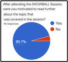
Respondents were asked to rate the Snowball session on a 5-point Likert Scale (1=Highly UNSatisfactory; 2=UN-Satisfactory; 3=Neutral; 4=Satisfactory; 5=Highly Satisfactory). 25 respondents (54.3%) rated the session 5 while 17 respondents (37%) rated the session 4 (Fig 1) thereby signifying a high level of satisfaction among the respondents.
45 respondents (97.8%) agreed that attending the Snowball session generated their interest in the topic being covered (Fig 2), that they were able to gain more knowledge about the topic being covered and were able to better correlate the topic clinically(Fig 3). 44 respondents (95.7%) agreed that after attending the Snowball session they were motivated to read further about the topic (Fig 4).
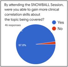
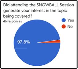
The respondents were then asked what in their opinion was the best part of the Snowball session. The thematic analysis of the responses is presented in Table 1.
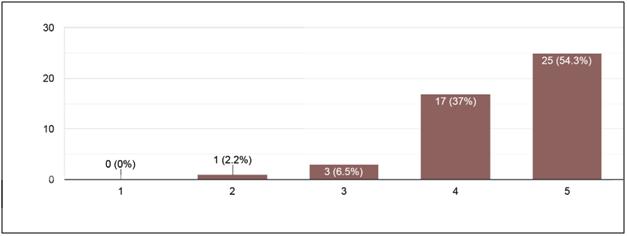
Vol 121, No 8, August 2023Journal
of the Indian Medical Association
Fig 2 — Respondents’ opinion when asked whether attending the Snowball session generated their interest in the topic (n=46)
Fig 1 — Overall rating of the SNOWBALL session (on a scale of 1 to 5; 1=Highly UN-Satisfactory and 5=Highly Satisfactory) (n=46)
Fig 3 — Respondents’ opinion when asked whether they were able to gain clinical correlation skills on the topic after attending the Snowball session
34
Fig 4 — Respondents’ opinion when asked whether attending the Snowball session motivated them to read further on the topic
45 respondents (97.8%) agreed that they would like to attend more Snowball sessions in Biochemistry. The respondents were asked to suggest additional topics which could be taught in a Snowball session. Some of the suggestions received were Amino acid metabolism, Jaundice, Carbohydrate metabolism, Vitamin deficiencies, Nutritional disorders and Lipid metabolism.
Table 1 — Thematic analysis of respondents’ opinion when asked what they thought was the best part of the Snowball session
Theme
Selected Responses
Interactive learning• Participation of all students and time for individual thinking and analysis
• Every individual had a role in it
• Ideas sharing by each group at the end
• The progression of discussion was the best part Improved learning• More clinical knowledge outcomes• Building up concepts
39 respondents (84.8%) agreed that the session started and finished as per schedule. All 46 respondents (100%) agreed that the session was conducted in a safe nonthreatening environment and that they were able to understand the steps of the Snowball session and the information given in the ‘information cards’ used during the session. 45 respondents (97.8%) agreed that the session was conducted without unnecessary interruptions. 44 respondents (95.7%) agreed that sufficient time was allocated for every step of the session
Involvement of multiple• Especially the last part when all the faculty departments gave there points on the applied part of concerned topic that were really helpful
• Where different departments explained the clinical scenarios related to their respective departments”
Innovative / Interesting• It was all very interesting concept• Innovative concept and best method ever
(c) “Provideopportunityforpresentationsbymore number of students”
(d) “It should be done once in every month”
(e) “Goodinitiative.Moretopicsshouldbecovered” The faculty/facilitators expressed satisfaction with the Snowball technique. All of them felt that it was a feasible interactive teaching learning method for introduction of clinical correlation. All facilitators felt that specific topics that are integrated with clinical subjects could be adapted to this teaching technique.
Table 2 — Thematic analysis of respondents’ opinion when asked for the reason they preferred a Snowball session over a traditional lecture (n=46)
Theme
Regarding the faculty facilitators conducting the session, 45 respondents (97.8%) agreed that the facilitators explained the process and steps of Snowball at the beginning of the session. 44 respondents (95.7%) agreed that the facilitators answered all queries and doubts asked by the learners and guided the learners through the different stages of the Snowball session. 43 respondents (93.5%) agreed that the facilitators encouraged all students to participate actively in the snowball session while 41 respondents (89.1%) agreed that the facilitators helped facilitate discussion within the groups.
44 respondents (95.7%) agreed that a Snowball session was better than a traditional lecture. Learners were also asked the reason for their preference and a thematic analysis is presented in Table 2.
Finally, the respondents were asked for suggestions for further improvements in future Snowball sessions. Some of the responses obtained are presented verbatim below –
(a) “Time for each step should be increased by 1-2 minutes”
(b) “Should be conducted more frequently”
Selected Responses
More interactive• Every student got the chance to speak and explore
• We were able to gather views of different
Sub-themes – people on a particular topic
a) equal chance to speak• It helps in understanding other’s point of view
b) understand different viewpoints• Group discussion is quite helpful
c) group discussion• It enabled is to analyse the situation individually and then discuss the possibilities and the solutions in the group
Active participation• In traditional lecture we are more passive learner. But in snowball we participated actively
• Student and teacher both are equally involved
Better understanding of the topic• Snowball helped me understand the topic very well as it was not just one way teaching,
Sub-themes – in the first round I applied the knowledge that
a) discussion promotes learning I had and in the progressing rounds discussed it.
b) better clinical correlation• The discussion part helped me gain more knowledge about the topic
• We are introduced to clinical knowledge teaches us the different point of view to same case. And how to tackle it
More interesting• Traditional lectures are generally boring and everything that is taught is not registered by the students
Self-satisfaction• There is space for discussion, without the fear of getting judged
• Everyone’s involvement is appreciated
Vol 121, No 8, August 2023Journal of the
Indian Medical Association
35
DISCUSSION
Interactive teaching learning goes one step ahead of active teaching learning. Whereas in the active approach, there is interaction between the teacher and the students, in the interactive approach the students interact not only with the teacher but also amongst themselves8. Interactive teaching ensures participation of all students in the learning process, makes learning fun and leads to better learning outcomes9.
While didactic lectures will always be useful in specific circumstances, student preference for interactive and collaborative teaching learning techniques means that supplementing didactic lectures with interactive teaching sessions can be extremely helpful in introducing topics such as clinical correlation10
Edgar Dale theorized many decades ago that people remember only 10% of what they read and 20% of what they hear as opposed to 70% of what they say and write and 90% of what they say and do while performing a task11. Malcolm Knowles propounded that adult learners prefer active learning techniques which are problem centred, have immediate relevance to their vocation or personal life and which respect the learners and their prior knowledge and past experiences.
The Snowball technique is almost always problem centred; learners are encouraged not only to speak, discuss but also to write down and summarize the important points at every step for easy collaboration with their peers;teaching clinical correlation of basic science topics has immediate relevance to the learners as it enables them to associate the knowledge of basic sciences with clinical concepts in later years12 and interactivity and collaboration with their peers allows students to put forth their points of view in a respectful environment. Working in teams with their peers promotes teamwork and inculcates the importance of working as equal members of healthcare teams in the future13. Previous studies have shown that incorporation of clinical correlation in teaching learning sessions leads not only to better learning outcomes but also improves clinical reasoning ability and helps learners in making better diagnostic decisions14-16
CONCLUSION
The ‘Snowball’ technique is a feasible interactive teaching learning method for introduction of clinical correlation to undergraduate medical students. In light of the inherent advantages of interactive teaching and the incorporation of clinical correlation in day-to-day learning, the authors recommend more widespread use of the Snowball technique for introducing clinical correlation in Biochemistry.
REFERENCES
1Bobby Z, Nandeesha H, Sridhar MG, Soundravally R, Setiya S, Sathish Babu M, et al — Identification of mistakes and their correction by small group discussion as a revision exercise at the end of a teaching module in biochemistry. Natl Med J India 2014; 27: 22-3.
2Dipiro JT— Why do we still lecture? Am J Pharm Educ 2009; 73: 137.
3Nierenberg DW — The challenge of “teaching” large groups of learners: strategies to increase active participation and learning. Int J Psychiat Med 1998; 28(1): 115-22. [Internet]. Mciindia.org. 2019 [cited 2 May 2020]. Available from: https:/ /mciindia.org/ActivitiWebClient/open/getDocument?path=/ Documents/Public/Portal/Gazette/GME-06.11.2019.pdf
4Edmunds S, Brown G — Effective small group learning: AMEE Guide No. 48. Medical Teacher 2010; 32(9): 715-26.
5Duraman H, Shahrill M, Morsidi N — Investigating the Effectiveness of Collaborative Learning in Using the Snowballing Effect Technique. Asian Journal of Social Sciences and Humanities [Internet]. 2015 [cited 1 June 2020];4(1):148-155. Available from: http://www.ajssh.leenaluna.co.jp/AJSSHPDFs/Vol.4(1)/AJSSH2015(4.1-17).pdf
6Group Work in the Classroom: Types of Small Groups | Centre for Teaching Excellence [Internet]. Centre for Teaching Excellence. 2012 [cited 10 June 2020]. Available from: https:/ /uwaterloo.ca/centre-for-teaching-excellence/teachingresources/teaching-tips/developing-assignments/groupwork/group-work-classroom-types-small-groups
7Miller G — Snowball Technique – A Teaching Strategy [Internet]. bookunitsteacher.com. 2018 [cited 10 June 2020]. Available from: https://bookunitsteacher.com/wp/?p=5826
8Giorgdze M, Dgebuadze M — Interactive Teaching Methods: Challenges And Perspectives. IJAEDU- International E-Journal of Advances in Education 2017; 544-8.
9Surapaneni K, Tekian A— Concept mapping enhances learning of biochemistry. Medical Education Online [Internet]. 2013 [cited 1 June 2020]; 18(1): 20157. Available from: https:// www.tandfonline.com/doi/full/10.3402/meo.v18i0.20157
10Lodhiya KK, Brahmbhatt KR — Effectiveness of collaborative versus traditional teaching methods in a teaching hospital in Gujarat. Indian J Community Med 2019; 44: 243-6
11Anderson H — Dale’s Cone of Experience [Internet]. Queensu.ca. [cited 1 June 2020]. Available from: https:// www.queensu.ca/teachingandlearning/modules/active/ documents/Dales_Cone_of_Experience_summary.pdf
12Klement B, Paulsen D, Wineski L — Clinical Correlations as a Tool in Basic Science Medical Education. Journal of Medical Education and Curricular Development [Internet]. 2016 [cited 1 June 2020];3:JMECD.S18919. Available from: https:// journals.sagepub.com/doi/full/10.4137/JMECD.S18919
13Lerner S, Magrane D, Friedman E — Teaching Teamwork in Medical Education. Mount Sinai Journal of Medicine: A Journal of Translational and Personalized Medicine 2009; 76(4): 318-29.
14Malau-Aduli B, Lee A, Cooling N, Catchpole M, Jose M, Turner R — Retention of knowledge and perceived relevance of basic sciences in an integrated case-based learning (CBL) curriculum. BMC Medical Education [Internet]. 2013 [cited 4 June 2020]; 13(1). Available from: https://bmcmededuc. biomedcentral.com/articles/10.1186/1472-6920-13-139.
15Jacobson K, Fisher D, Hoffman K, Tsoulas K — Integrated Cases Section: A Course Designed to Promote Clinical Reasoning in Year 2 Medical Students. TeachingandLearning in Medicine 2010; 22(4): 312-6.
16Woods N, Brooks L, Norman G — It all make sense: biomedical knowledge, causal connections and memory in the novice diagnostician. Advances in Health Sciences Education 2007; 12(4): 405-15.
Vol 121, No 8, August 2023Journal
of the Indian Medical Association
36
Original Article
Association between Reverse Osmosis Water and Heart Diseases, Back Pain and Joint Pain
Sangita Patel1, Rahul Khokhariya2, Greenam Tarani3, Jagruti Rathod4, Deya
Ghosh Chatterji5, Jesal Patel6
Background : Reverse Osmosis (RO) system causes demineralization of water which leads to various health problems. Therefore, this study was conducted to assess the association between RO water and heart diseases, back pain and joint pain.
Material and Methods : At urban Vadodara city a cross sectional study was carried out with 2609 participants among 720 households. 60 households from each selected water tank area were included. Interviews were conducted to know about usage of RO water and health problems such as coronary heart disease, joint pain and back pain. 38 water samples were tested for Total Dissolved Solutes, calcium hardness and total hardness.
Results : TDS, Calcium hardness and total hardness were within normal range. There was statistically significant association between RO water usage and joint pain (p value 0.0001) at 95% CI while no association was found between RO water usage and Coronary Heart Disease (CHD) (p value 0.3104) and back pain (p value 0.5230) at 95% CI.
Conclusion : Usage of RO water system is associated with joint pain while there is no association of coronary heart disease and back pain with RO water.
[J Indian Med Assoc 2023; 121(8): 37-40]
Key words :RO Water System, Joint Pain, Back Pain, Coronary Heart Disease, TDS, Calcium Hardness, Total Hardness.
One of the primary goals of the WHO and its member states is that “All people, whatever their stage of development and their social and economic condition, have the right to have access to an adequate supply of safe drinking water1. Typical recommended dietary intake of calcium is 1000mg per day. Hard water is said to be responsible for kidney stone2. Calcium and magnesium are essential minerals and beneficial to human health in several respects. Inadequate intake of calcium has been associated with increased risk of osteoporosis3, colorectal cancer, hypertension, stroke, coronary artery disease, insulin resistance and obesity4. Total body stores are in the order of 1200g with about 99% in bones and teeth. Magnesium is the 2nd most abundant cation in intracellular fluid. It is cofactor for some 350 cellular enzymes, many of which are involved in energy metabolism. It is also involved
Department of Community Medicine, Medical College Baroda, Gujarat 390001
1MD, Associate Professor and Corresponding Author
2MD, Medical Officer
3MD, Senior Resident
4PhD, Associate Ecologist, Department of Ecology, Gujarat Ecology Society, Vadodara, Gujarat 390003
5MD, Senior Resident
6MBBS, Intern, Department of Community Medicine, GCS Medical College Hospital and Research Centre, Ahmadabad, Gujarat 380018
Received on : 09/12/2022
Accepted on : 02/04/2023
Editor's Comment :
Total hardness, calcium hardness and TDS was within normal range in all water tanks in Vadodara Municipal Corporation.
Drinking RO Water was associated with joint pain and not associated with coronary heart disease and back pain.
in protein and nucleic acid synthesis and needed for normal vascular tone and insulin sensitivity. Magnesium deficiency has been implicated in the pathogenesis of hypertension5.
In 92-99% of beneficial calcium and magnesium is removed by Reverse Osmosis (RO) system. Consumption of RO water even for a few months leads to serious side effects6. Common complaints observed in Czech and Slovak populations were cardiovascular disorders, tiredness, weakness, and muscle cramps. This deficiency cannot be compensated by diet1.
AIMS AND OBJECTIVES
To assess TDS, calcium hardness and total hardness of water and to know the association between RO water and heart diseases, back pain, and joint pain.
MATERIALS AND METHODS
A Cross sectional study was carried out at urban Vadodara city after taking permission from Institutional ethical committee. From north, south, east and west
Vol 121, No 8, August 2023Journal of the Indian Medical Association
37
zones, average 3 water tanks were selected randomly. Therefore, water sample from 12 water tanks were tested. Two water samples were collected, one from near tank area and another from outer limit of water supply from household level to check TDS, Calcium hardness and total hardness. Thus, 12 water tanks, 24 household water samples, and 2 from main water bodies of Vadodara district (Ajwa and Mahi River) (38 samples) were tested. Standard operating procedure was followed for water sample collection and transport. The water samples were sent to Gujarat Ecological Society for TDS, Calcium hardness and total hardness testing.
Vadodara city has been assigned zones of north, south, east, and west. Randomly three water tanks were selected from each zone using a computerized generated random number. One hundred and eighty households were taken from each zone and a total of 720 households were included in the study. The expected total population to be covered was 3600, assuming the average family size of 5 persons. However, 2609 participants were available during data collection and consent was taken for participation. First house was randomly selected after which 60 sequential houses were included for the study. If any house was closed than sequential next house was selected. Those families who used bore well water, who lived in slum area and flat/apartment area were excluded from the study. The total study duration was 1 year, and the data was collected from November 2016 to August 2017.
A pre-validated and semi structured questionnaire was used to collect data regarding RO water usage and associated health hazards such as heart diseases, back pain, and joint pain after taking written informed consent. All those having coronary heart disease (Angina pectoris, Myocardial Infarction, and irregularities of heart), Joint pain and back pain due to any cause were included in the study.
The process of data collection did not pose any potential risk or harm to the participants. Data safety and confidentiality was given due consideration by keeping the file containing identity related details password protected. The filled Performa were kept in lock and key accessible only to researchers.
Statistical Analysis : The Data was entered in Microsoft excel using strict check files. Water quality parameters were measured in pre-defined measures. Chi square and Fishers exact test was used for categorical data using Epi info 77
the water tanks were below 600 except for one area (Manjalpur water tank which was 786mg/l), Calcium Hardness in all the water tanks was within normal range. Normal Value total Hardness of water is 500mg/l. Total hardness of water was below 500mg/l in all water tanks.
Out of 2609 participants, 1340 (51.36%) were male and 1269 (48.63%) were female. Three fourth 1841 (70.56%) were between 20-64 years, 311 (11.92%) were between 10-19 years, 167 (6.40%) were above 65 years age, 165 (6.32%) participants were less than 5 years, 125 (4.79%) were between 6-9 years.
Most of the participants were graduates 1074 (41.16%), followed by education up to higher secondary 500 (19.16%), Secondary 404 (15.48%), Primary 371 (14.22%) and one tenth of the participants were illiterate.
259 (35.97%) participants were using RO. 147 (20.41%) participants were using UV (ultraviolet filter) filter for purification of water and small number of people used Jug water for drinking water.
141 (34.72%) household used RO/UV water for less than 5years, 160 (39.40%) household were using RO/ UV water filter for 5 to 10 years and 105 (25.86%) were using RO/UV water filter for more than 10 years.
Table 1 shows that history of coronary heart disease was present in 3.64% RO users and 2.84% in nonRO users. There was no statistically significant association in heart problem between RO users and non-RO users. (p-value=0.3104)
Out of total 2609 participants, 644 (24.68%) had history of back pain out of which 14.85% males and 35.06% females had back pain. There was statistically significant association in back pain between male and female (p-value=0.0001) (Table 2).
According to Table 3, 25.44% of RO users and 24.24% of non-RO users had back pain. There was no statistically significant association in back pain
H/O CoronaryNo H/O CoronaryTotal Heart diseaseHeart disease
RO users35 (3.64%)924 (96.35%)959
Non-RO users47 (2.84%)1603 (97.15%) 1650
Total8225272609
Chi-square value = 1.029 p-value = 0.3104 DF = 1
FemaleTotal
Back pain present 199(14.85%)445(35.06%)644(24.68%)
Back pain absent1141(85.14%)824(64.93%) 1969(75.46%)
Total134012692609
Chi-square value = 142.197 p-value = 0.0001 DF = 1
Vol 121, No 8, August 2023Journal
of the Indian Medical Association
Normal TDS value is <600 mg/l. TDS value in all
RESULTS
Table 1 — Comparison of Coronary Heart Disease between RO users and non-RO users (n=2609)
Table 2 — History of Back pain in study participants (n=2609) Male
38
between RO users and non-RO users (p-value = 0.5230).
Table 4 shows that out of total 2609 participants 11.79% males and 23.79% of females had history of joint pain. There was statistically significant association in joint pain between male and female (pvalue = <0.0001).
Table 5. Shows age wise distribution amongst those with and without joint pain. In less than 9 years, no participant had joint pain. In 10-19 years, 10.32% had history of joint pain and 99.68% had no history. In 2064 years, 19.66% had joint pain while 80.34% had no history and for participants above65 years, 58.08% had joint pain and 41.92% had no history of joint pain.
According to Table 6, 20.33% RO users and 16.06% non-RO users had history of joint pain. There was statistically significant association in joint pain between RO andnon-RO users (p-value=0.0068).
DISCUSSION
There was limited literature available on this topic. Usage of RO system leads to demineralization of water specifically calcium and magnesium which leads to
increase prevalence of coronary heart disease, joint pain and back pain and this deficiency cannot be compensated by diet. Therefore, our discussion is based on demineralized water and CHD, joint pain, and back pain.
TDS, calcium hardness and total hardness of water was similar in both RO and non-RO water so, demineralization in RO water was due to usage of RO system.
In all total 38 samples of water were checked, TDS of water was found to be normal except for one area (Manjalpur water tank which was 786mg/l), TSS ranged from 18-84 mg/l, Calcium Hardness of water ranged from 40-84 mg/l. Total Hardness of water ranged from 104 to 320 mg/l.
The association of drinking water hardness and Coronary Heart Diseases (CVDs) has been studied since more than five decades ago. A study in England and Wales showed that cardiovascular deathrates had a favourable effect in towns with harder water and had an adverse effect in towns with softer water8. Other studies showed that high levels of drinking water hardness can be protective against CVD9
However, a review of ecological studies showed that the results are inconsistent between studies. A metaanalysis revealed a negative association between concentration of magnesium in water and CVD mortality10
Chi-square value=0.408 p-value=0.5230 DF=1
Male FemaleTotal
H/O Joint Pain 158(11.79%)302(23.79%)460(17.63%)
No H/O Joint Pain1182(88.20%)967(76.20%)2149(82.36%)
Total134012692609
Chi-square value = 63.880 p-value = <0.0001 DF = 1
Age group H/O jointH/O no jointTotal in years pain pain <90290 (100%)290 10-191 (0.32%)310 (99.68%)311
20-64 362 (19.66%)1479 (80.34%) 1841 >6597 (58.08%)70 (41.92%)167
46021492609
Moreover, another review showed protective role of magnesium concentration in water against CVD in some case-control studies and one cohort study, but the analytical studies showed little evidence about the association of calcium and magnesium levels in drinking water and CVD risk11
Water hardness may be also associated with CVD risk factors, for instance positive correlations of water magnesium and calcium with blood pressure is documented.
In the Netherlands, another study found no overall association between calcium, magnesium or total hardness and ischemic heart disease or stroke mortality12
H/O Joint PainNo H/O Joint PainTotal
RO users195 (20.33%)764 (79.66%)959
Non-RO users265 (16.06%)1385 (83.93%) 1650
Total46021492609
Chi-square value = 7.334 p-value = 0.0068 DF = 1
Our study showed that history of coronary heart disease was 3.14%. Calcium hardness in our study was found to be normal, which ranged from 40-84 mg/ l. Total hardness ranged from 104-140mg/l. TDS value ranged from 137-786mg/l. All the water samples were found to be moderately hard so we have not studied the relationship between CVD and hardness of water, but we have studied the relationship of heart problem with RO users and Non-RO users but there was no statistically significant association in heart problem
Vol 121, No 8, August 2023Journal of
the Indian Medical Association
H/O Back PainNo H/O Back PainTotal RO user244 (25.44%)715 (74.55%)959 Non-RO users400 (24.24%)1250 (75.75%) 1650 Total64419652609
Table 3 — Comparison of history of Back pain between RO users and non-RO users (n=2609)
Table 4 — History of Joint pain in study participants (n=2609)
Table 5 — Age wise distribution of Joint pain in study participants (n=2609)
Table 6 — Comparison of history of joint pain between RO users and Non-RO users (n=2609)
39
between RO users and Non-RO users. The reason may be multifactorial reasons of CVD like cigarette smoking, obesity, behaviour problem, physical inactivity, diet pattern, family history etc.
The study done by Pallav Sengupta found that GI health is also being benefited from hard water since it provides potentially alleviating effects on the onset of constipation in the 85% cases. A rich union of calcium and magnesium in hard water, in a right combination, helps to combat constipation. The calcium in hard water results in teaming up with excess bile and its resident fats to lather up the soap like insoluble substance, which is emitted from the body during bowel movements. Indeed, many renowned scientists have considered hard water as a boon as it has some fantastic health benefits that seem to encourage longer life expectancy and improved health. Magnesium salt has laxative effect. This provides a rapid evacuation of intestine. The American Gastroenterology Association recommends milk of magnesia for the management of constipation as one of the therapeutic options4.
Statistically significant association was observed in joint pain between RO users and non-RO users. No statistically significant association was observed in back pain between RO users and non-RO users.
Age and sex distribution was similar in RO and non-RO users; therefore, it did not act as a confounder. The generalizability of above study data is applicable to whole Vadodara Municipal Corporation area.
CONCLUSION
RO water system usage is associated with joint pain and not associated with CHD and back pain. Further prospective studies are required to test whether RO water causes health problems like CHD, back pain, and joint paint.
REFERENCES
1Water S — Nutrients in drinking water. Water, Sanitation, and Health Protection and the Human Environment, World Health Organization; 2005. 186.
2Bellizzi V, DeNicola L, Minutolo R, Russo D, Cianciaruso B, Andreucci M, et al — Effects of Water Hardness on Urinary Risk Factors for Kidney Stones in Patients with Idiopathic Nephrolithiasis. Nephron 1999; 81(1): 66-70.
3Power ML, Heaney RP, Kalkwarf HJ, Pitkin RM, Repke JT, Tsang RC, et al — The role of calcium in health and disease. Am J Obstet Gynecol 1999; 181(6): 1560-9.
4Sengupta P — Potential health impacts of hard water. Int J Prev Med 2013; 4(8): 866-75.
5Houston M — The Role of Magnesium in Hypertension and Cardiovascular Disease. The Journal of Clinical Hypertension 2011; 13(11): 843-7.
6Verma KC, Kushwaha AS — Demineralization of drinking water: Is it prudent? Vol. 70, Medical Journal Armed Forces India. Medical Journal Armed Forces India 2014; 377-9.
7Epi InfoTM | CDC [Internet]. [cited 2022 Jul 15]. Available from: https://www.cdc.gov/epiinfo/index.html
8Crawford MargaretD, Gardner MJ, Morris JN. Changes In Water Hardness and Local Death-rates. The Lancet 1971; 298(7720): 327-9.
9Ferrandiz J, Abellan JJ, Gomez-Rubio V, Lopez-Quilez A, Sanmartin P, Abellan C, et al — Spatial analysis of the relationship between mortality from cardiovascular and cerebrovascular disease and drinking water hardness. Environ Health Perspect 2004; 112(9): 1037-44.
10Monarca S, Donato F, Zerbini I, Calderon RL, Craun GF — Review of epidemiological studies on drinking water hardness and cardiovascular diseases. European Journal of Cardiovascular Prevention & Rehabilitation 2006; 13(4): 495506.
11Lake IR, Swift L, Catling LA, Abubakar I, Sabel CE, Hunter PR — Effect of water hardness on cardiovascular mortality: an ecological time series approach. J Public Health (Bangkok) 2010; 32(4): 479-87.
12Leurs LJ, Schouten LJ, Mons MN, Goldbohm RA, van den Brandt PA — Relationship between tap water hardness, magnesium, and calcium concentration and mortality due to ischemic heart disease or stroke in The Netherlands. Environ Health Perspect 2010; 118(3): 414-20.
Disclaimer
The information and opinions presented in the Journal reflect the views of the authors and not of the Journal or its Editorial Board or the Publisher. Publication does not constitute endorsement by the journal.
JIMA assumes no responsibility for the authenticity or reliability of any product, equipment, gadget or any claim by medical establishments/institutions/manufacturers or any training programme in the form of advertisements appearing in JIMA and also does not endorse or give any guarantee to such products or training programme or promote any such thing or claims made so after.
121, No 8, August 2023Journal
Vol
of the Indian Medical Association
40
— HonyEditor
Original Article
Clinical Profile of Drug Induced Parkinsonism in Eastern India
Praveen Kumar Yadav1, Kumud Sahu2
Background : Drug Induced Parkinsonism (DIP) is a reversible condition in most of the cases therefore have to be identified and treat early. In our study we presented the dominant pattern of DIP and the medications causing it. Materials and Methods : This is a retrospective study done on outpatient basis from1st January, 2022 to 30th November, 2022 at a super speciality neuro clinic. In this study all patient, satisfying the criteria of DIP were included. The Inclusion criteria were : (1) All patients satisfying more than equal to two of the four criterias of Parkinsonismbradykinesia, rigidity, tremor and postural instability. (2) Patients had no symptoms of parkinsonism before taking drug. 3. There is direct relation of drug intake and symptom onset, and the Exclusion Criteria were : (1) Other causes of secondary Parkinsonism- NPH, vascular parkinsonism, brain tumour, toxin induced etc by appropriate investigations. (2) All other drug induced motility disorder without the features of parkinsonism. The age, sex and other demographic criterias were studied. The dominant pattern of involvement- tremor, akinetic rigidity and mixed were noted. Results: Out of 32 patients 19(59.38%) were female and 13(40.63%) were male. The most common age group was >60 years (65.63%), followed by 51-60years (15.63%) and <30years and between age 41-50 years with equal percentage of 3.13. The mean age was 62.37years with standard deviation of 13.09 years.
The most common clinical pattern observed in our study was tremor (50%) followed by akinetic rigidity (34.38%) and mixed pattern (15.63%). Orofacial dyskinesia along with bradykinesia, rabbit syndrome, head and jaw tremor along with parkinsonian features were all seen in our study with equal percentage of (6.66%). Atypical Parkinson with features of Parkinson plus syndrome is also seen in 6.66%. The most common drug associated with DIP in our study was pro-kinetic drug levosulpiride (46.88%) followed by flunarizine (25%), valproate and haloperidol (6.25%), sulpitac and itopride (3.13%). The combination drugs causing DIP was also seen in 9.36%.
Conclusion : DIP is a preventable and treatable condition in many cases, therefore safer drugs should be prescribed and have to be identified early, respectively.
[J Indian Med Assoc 2023; 121(8): 41-5]
Key words :Parkinsonism, Tremors Drug Induced Rigidity.
Parkinsonism is a motor syndrome, characterized by tremor, rigidity, bradykinesia and postural instability, which was first described by James Parkinson in his essay entitled “The Shaking Palsy”1 Idiopathic Parkinson Disease (IPD) is the most common cause of Parkinsonism with a prevalence of 3.3% and 4.5% followed by Drug Induced Parkinson (DIP) which is 2.7 and 1.7% in community based and population based survey respectively and also IPD is age related and overall the second most common cause of degenerative disease of central nervous system after Alzheimer Disease (AD)2-4
IPD is pathologically characterized by decrease dopamine level in basal ganglia and alpha-synuclein deposition, causing direct neurotoxicity in cell bodies
1MBBS, MD, DM, MRCP, FRCP, FEBN, FIAMS, FIACM, FIMSA, MNAMS, Senior Consultant Neurologist and Director, Department of Neurology, Aarogyam Neuro Clinic, Durgapur, West Bengal 713214 and Corresponding Author
2DNB, Resident, Department of General Medicine, DSP Main Hospital, Durgapur, West Bengal 713203
Received on : 06/02/2023
Accepted on : 24/03/2023
Editor's Comment :
Drug Induced Parkinsonsim (DIP) is a very important cause of reversible secondary parkinsonism.
Proper drug history should be taken in all patients with recent onset parkinsonism.
Early diagnosis and stopping of offending drug can prevent permanent neurological issues.
and axons, known as Lewy bodies and Lewy neuritis, collectively known as Lewy pathology5
It usually present as asymmetrical form in age more than 60 years and more common in males (1.5 times as compared to females) in contrast to DIP, which is less asymmetrical, common in elderly and in females6
Among all the secondary causes of Parkinsonism like- NPH, vascular parkinsonism, toxin induced parkinsonism, chronic traumatic encephalopathy, brain tumour, DIP is the most common cause. Drug-induced movement disorders were described in 1954 in patients who were treated with chlorpromazine and reserpine7 It is frequently induced by anti-psychotics (Typical>Atypical). Others include anti-emetic and prokinetic - metoclopramide, levosulpiride, calcium channel blocker (Verapamil, Diltiazem, flunarizine,
Vol 121, No 8, August 2023Journal of the
Indian Medical Association
41
cinnarizine), anti arrythmics- amiodarone, mood stabilizers- lithium, anti epileptic-valproate.
The mechanism behind DIP is interference of nigrostriatal pathway by various drugs.The pharmacological effect of anti psychotic is seen by blocking of post synaptic D2 receptor by 60-70% and the features of Parkinsonism is seen when blockade is >75%, identified by PET8. There are some relativity related to this, for example, aripiprazole (atypicalanti psychotic), have D2 receptor occupancy of >95%, rarely cause DIP as it is weak agonistic action on dopamine receptors and interacts with 5-HT1A and 5HT-2A receptors9,10. Some drugs also act on presynaptic receptor like tetrabenazine.
The important risk factor are, age (causes destruction of nigral cells), female gender (estrogen act as a causative hormone), genetic predisposition, drug dosage, potency and route of its administration11-13
Many a times, DIP is missed and misdiagnosed as PD, as they have many similar clinical features. Table 1 summarize the difference between DIP and IPD14 :
Since, treatment of PD and DIP is different, early diagnosis, polypharmacy, and stopping of offending drug is necessary. Although, in 20-25% it is persistent and progressive15,16
The most common drug induced motility disorder is DIP followed by tremors, tardive dyskinesia, tardive dystonia, akathisia and myoclonus.
The (DSM-IV), defines DIP as the presence of resting tremor, muscular rigidity, akinesia, or bradykinesia, developing within a few weeks of starting or raising the dosage of a medication (typically a neuroleptic) or after reducing the dosage of an antiparkinsonian agent17
Many scales have been developed one of which includes Simpson- Angus Scale, which emphasis more on rigidity than on bradykinesia18.
AIMS AND OBJECTIVES
The objective of this study is to find the medications leading to DIP and its clinical pattern of neurological involvement
MATERIALS AND METHODS
This is a cross-sectional study of PD patients at the out patient department of a super speciality neurology clinic in Durgapur, West Bengal. This study was done from 1st January 2022 to 30th November, 2022 considering, the inclusion criteria for DIP. The patients were examined thoroughly by a single neurologist in their initial visit and diagnosed to have DIP. All patients related to this study had signed their informed consent. Ethical Committee clearance was taken from the local Ethics Committee. The identity of the subjects was not revealed during the study period.
Inclusion Criteria :
(1) All patients satisfying more than equal to two of the four criterias of parkinsonism- bradykinesia, rigidity, tremor and postural instability.
(2) Patients had no symptoms of parkinsonism before taking drug.
Onset More often in elderly60s
Symmetry ofOften symmetric Asymmetric clinical features
Lower/upper bodyMore severeBoth involvement in upper part
Tremor VariableVariable
Depression CommonCommon
Dementia May be presentRare at onset before onset of parkinsonism
Response toPoor Good treatment with L DOPA or dopamine agonists
Response to drug Good Poor withdrawal
HVA level in CSF High Low/normal
PET/SPECT imaging Normal uptake ofReduced uptake of pre-synaptic markers, pre-synaptic markers, reduced uptake ofnormal uptake of dopamine receptordopamine receptor ligandsligands
(3) There is direct relation of drug intake and symptom onset.
Exclusion Criteria :
(1) Other causes of secondary parkinsonism- NPH, vascular parkinsonism, brain tumor, toxin induced etc by appropriate investigations.
(2) All other drug induced motility disorder without the features of parkinsonism.
The evaluation of offending medications causing symptoms along with age, sex and other demographic features of population were studied. The parkinsonism pattern including tremor, akinetic rigidity and mixed type was noted and stopped the offending drug and prescribed another drug to prevent primary disease worsening after consulting with respective specialist.
RESULTS
In the study period, there were 32 patients, who satisfied the Inclusion criteria. Out of which,
Vol 121, No 8, August 2023Journal
of the Indian Medical Association
Table 1 — Difference between DIP and IPD DIPIPD
42
19(59.38%) were female and 13(40.63%) were male. The most common age group was >60 years (65.63%), followed by 51-60years (15.63%) and the least common group were <30years and between age 4150 years with equal percentage of 3.13. The mean age was 62.37 years with standard deviation of 13.09 years (Fig 1).
The most common pattern observed in our study was tremor (50%) followed by akinetic rigidity (34.38%) and mixed pattern (15.63%)(Fig 2). Tremor was equally distributed in both males and females while akinetic rigidity and mixed pattern type of Parkinsonism is more common in females. Orofacial dyskinesia along with bradykinesia was seen in (6.66%), rabbit syndrome was seen in (6.66%), head and jaw tremor were seen along with parkinsonian features in (6.66%). Parkinson plus syndrome features was seen in 6.66%.
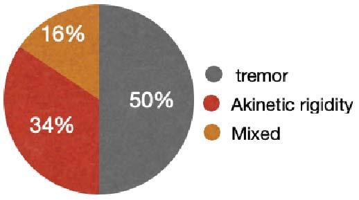
The most common drug associated with DIP in our study was pro-kinetic drug levosulpiride (46.88%) followed by flunarizine (25%), valproate and haloperidol (6.25%), sulpitac and itopride (3.13%). The combination drugs causing DIP was seen in 9.39%.
During the initial visit, patients were diagnosed and stopped the offending drug and trihexyphenidyl was added in to all the patients with a dose from 2mg to 10mg, according to the severity of symptoms.
DISCUSSION
In our study, DIP is more common among age group of >60yrs, correlating with the fact that, age being the most consistent risk factor, as dopamine level decreases with age due to nigral cells degeneration19 Therefore, it is better to avoid polypharmacy and especially the drugs causing motility disorders in elderly. There should be high suspicion and they should be kept under close observation to detect DIP.

We also found that DIP is more common among female gender (59.37%), as oestrogen suppresses the dopamine receptor expression, as per the studies done previously20. Many studies done earlier too showed that the genes involved in GABA receptor signalling in Genome wide screening, leads to Tardive dyskinesia in Schizophrenic patients and DIP21,22. In our study we had 6.25% patients who were schizophrenic, showed tremor predominance parkinsonian features.
It was studied that rigidity and bradykinesia is predominant as compared to tremor and postural instability in DIP23, but we found tremor to be most dominant pattern with 50% in our study then akinetic rigidity (34.37%), and the remaining were mixed (15.62%). Therefore, DIP is easily misdiagnosed as PD. DAT scan by PET/SPECT has been done in past
studies which differentiates between pre-synaptic parkinsonism and post synaptic parkinsonism. It showed decrease uptake in PD and normal uptake in DIP. But sometimes there is decrease uptake in DIP, due to the presence of unmasked PD. Thus, concluded that DIP is multifactorial origin24.
Homovallenic Acid (HVA) level in CSF is another investigation done to differentiate DIP and IPD, which will be increased in DIP and decreased in IPD. The mechanism here is when a patient is treated with neuroleptics for long duration, there is compensatory increase in HVA for short duration, because of increase firing of dopaminergic neurons in response to blockade of dopamine receptor followed by development of tolerance and HVA level come to normal. But failure to come to normal or failure to develop tolerance leads to DIP and tardive dyskinesia25,26
In our study the most common medication leading to DIP was levosulpiride (46.87%). It is a newer emerging pro-kinetic, prescribed by many general physician, which acts on both CNS and gut by selective blocking of pre-synaptic D2 receptor27. Out of the study population, rabbit syndrome was seen in 6.66% population, caused by levosulpiride. 13.33% having mixed predominant features, presented with orofacial dyskinesia along with bradykinesia and head and jaw tremor with equal percentage of 6.66%, more in female gender.
The second common drug in our study was
Vol 121, No 8, August 2023Journal of the Indian Medical Association
Fig 1 — Age distribution in our study
43
Fig 2 — Distribution of DIP pattern in our study
flunarizine (FNZ)(25%), for migraine prophylaxis, which is calcium channel antagonist.
Mitochondrial quality control is essential for normal function of a cell and its defect leads to parkinsonian features. As per the studies, chronic use of FNZ decrease mitochondrial numbers in neurone and astrocytes with or without affecting the nigrostriatal dopaminergic pathway. Also, FNZ inhibits complex 1 and 2 of electron transport chain system. Although the exact mechanism is still unclear28-31
Valproate (6.25%) as an antiepileptic, act by blocking sodium, potassium and calcium channel in neuronal membrane. Therefore, its chronic use can cause physiological inactivation of neurons in basal ganglia and hence cause reversible parkinsonian features32
There were also combination medicines caused Parkinsonian features in 9.36% population eg, Flunarizine and valproate in migraine prophylaxis caused DIP. Therefore, have to be used very cautiously. In a previous study haloperidol was associated with high risk of extra pyramidal system and clozapine was same as placebo[33. The following table summarizes the medicine leading to DIP in our study (Fig 3).
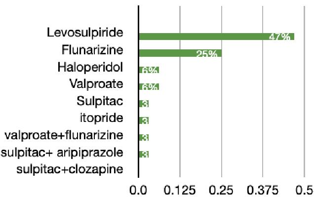
In our study, we did not find any correlation between the medications patients were taking for their comorbidities like anti-hypertensive, oral hypoglycaemic drugs, statins.
The treatment of DIP is to differentiate DIP and IPD, stopping the offending drugs, and change to other safer medication like atypical anti-psychotics in Psychiatric patients like Schizophrenia.
Anticholinergics like trihexyphenidyl, benztropine, amantadine, and levodopa have been tested for their ability to relieve symptoms but has not produced any clear evidence of their effects in DIP patients.[34,35]
Limitations : Limitation of our study was that patient was only seen thoroughly in their first visit, they were not followed up.
CONCLUSION
DIP can present in varied manifestations, so high suspicion along with early diagnosis and treatment is must.
Financial Support : Nil
Conflict of Interest : There is no conflict of interest.
REFERENCES
1Keener AM, Bordelon YM — Parkinsonism. Semin Neurol 2016; 36(4): 330-4.
2Barbosa MT, Caramelli P, Maia DP, Cunningham MC, Guerra HL, Lima-Costa MF, et al — Parkinsonism and Parkinson’s disease in the elderly: a community-based survey in Brazil (the Bambuí study). Mov Disord 2006; 21: 800-8.
3Benito-León J, Bermejo-Pareja F, Rodríguez J, Molina JA, Gabriel R, Morales JM — Neurological Disorders in Central Spain (NEDICES) Study Group. Prevalence of PD and other types of parkinsonism in three elderly populations of central Spain. Mov Disord 2003; 18: 267-74
4Troiano AR, Germiniani FM, Werneck LC — Flunarizine, cinnarizineinducedparkinsonism: A historical and clinical analysis. Parkinsonism Relat Disord 2004; 10: 243-5.
5Pacheco CR, Morales CN, Ramý´rez AE — Extracellular asynuclein alters synaptic transmission in brain neurons by perforating the neuronal plasma membrane. J Neurochem 2015; 132: 731-41.
6Benito-León J, Bermejo-Pareja F, Rodríguez J, Molina JA, Gabriel R, Morales JM — Neurological Disorders in Central Spain (NEDICES) Study Group. Prevalence of PD and other types of parkinsonism in three elderly populations of central Spain. Mov Disord 2003; 18: 267-74.
7Steck H — [Extrapyramidal and diencephalic syndrome in the course oflargactil and serpasil treatments.] [article in French]. Ann Med Psychol (Paris) 1954; 112: 737-44.
8Remington G, Mamo D, Labelle A et al.: A PET study evaluating dopamine D2 receptor occupancy for long-acting injectable risperidone. Am J Psychiatry 2006; 163(3): 396-401.
9Grunder G, Carlsson A, Wong DF — Mechanism of new antipsychotic medications: occupancy is not just antagonism. Arch Gen Psychiatry 2003; 60(10): 974-977.
10Sharma A, Sorrell JH — Aripiprazole-induced parkinsonism. Int Clin Psychopharmacol 2006; 21(2): 127-9.
11Rollema H, Skolnik M, D’Engelbronner J — MPP(+)likeneurotoxicity of a pyridinium metabolitederived from
Vol 121, No 8, August 2023Journal of
the Indian Medical Association
44
Fig 3 — Drugs causing DIP
8, August 2023Journal of the Indian Medical Association
haloperidol: in vivomicrodialysis and in vitro mitochondrialstudies. Pharmacol Exp Ther 1994; 268(1): 380-7.
12Mena MA, Garcia de YebenesMJ,Tabernero C, et al. Effects of calciumantagonists on the dopamine system. Clin Neuropharmacol 1995; 18(5): 410-26.
13Richardson MA, Haugland G, Craig TJ — Neuroleptic use, parkinsonian symptoms, tardive dyskinesia, and associated factors in child and adolescent psychiatric patients. Am J Psychiatry 1991; 148(10): 1322-8.
14Maria A Mena, Justo G de Yébenes — Drug-induced parkinsonism, Expert Opinion on Drug Safety 2006; 56: 75971.
15Llau ME, Nguyen L, Senard JM, Rascol O, Montastruc JL — Drug-induced parkinsonian syndromes: a 10-year experience at a regional center of pharmaco-vigilance. RevNeurol(Paris) 1994; 150(11): 757-62.
16MartiMasso JF, Poza JJ — Drug-induced or aggravated parkinsonism: clinical signs and the changing pattern of implicated drugs. Neurologia 1996; 11(1): 10-5.
17American Psychiatric Association. Diagnostic and Statistical Manual of Mental Disorders. Fifth ed. Arlington, VA: American Psychiatric Association; 2013.
18Simpson GM, Angus JW — A rating scale for extrapyramidal side effects. Acta Psychiatr Scand Suppl 1970; 212: 11-9.
19Jabs BE, Bartsch AJ, Pfuhlmann B — Susceptibility to neuroleptic-induced parkinsonism-age and increased substantia nigra echogenicity as putative risk factors. Eur Psychiatry 2003; 18(4): 177-81.
20Bedard P, Langelier P, Villeneuve A — Oestrogens and extrapyramidal system. Lancet 1977; 2: 1367-8. [PubMed] [Google Scholar]
21Metzer WS, Newton JE, Steele RW, Claybrook M, Paige SR, McMillan DE, et al — HLA antigens in drug-induced parkinsonism. Mov Disord 1989; 4(2): 121-8. PMID: 2567491 DOI: 10.1002/mds.870040203
22Liou YJ, Wang YC, Chen JY, Bai YM, Lin CC, Liao DL, et al — Association analysis of polymorphisms in the N-methyl-Daspartate (NMDA) receptor subunit 2B (GRIN2B) gene and tardive dyskinesia in schizophrenia. Psychiatry Res 2007; 153: 271-5.
23Hardie RJ, Lees AJ — Neuroleptic-induced Parkinson’s syndrome: clinical features and results of treatment with levodopa. J NeurolNeurosurg Psychiatry 1988; 51: 850-4.
24Lorberboym M, Treves TA, Melamed E — [123I]-FP/CIT SPECT imaging fordistinguishing drug-inducedparkinsonism from Parkinson’s disease. Mov Disord 2006; 21(4): 510-4.
25Saito T, Ishizawa H, Tsuchiya F, Ozawa H, Takahata N — Neurochemical findings in the cerebrospinal fluid of schizophrenic patients with tardive dyskinesia and neuroleptic-induced parkinsonism. Jpn J Psychiatry Neurol 1986; 40(2): 189-94.
26Bowers MB Jr, Heninger GR — Cerebrospinal fluid homovanillic acid patterns during neuroleptic treatment. Psychiatry Res 1981; 4(3): 285-90.
27Serra J — Levosulpiride in the management of functional dyspepsia and delayed gastric emptying. Gastroenterol Hepatol 2010; 33: 586-90. [PubMed] [Google Scholar] [Ref list]
28Goldman SJ, Taylor R, Zhang Y, Jin S — Autophagy and the degradation of mitochondria. Mitochondrion 2010; 10: 30915.
29Lin MT, Beal MF — Mitochondrial dysfunction and oxidative stress in neurodegenerative diseases. Nature 2006; 443: 787-95.
30Brucke T, Wober C, Podreka I, Wober-Bingol C, Asenbaum S, Aull S, et al — Harasko-van der Meer, P. Wessely, L. Deecke, D2 Receptor blockade by flunarizine and cinnarizine explains extrapyramidal side Effects. A SPECT study. BloodFlowMetab 1995; 15: 5138.
31Veitch K, Hue L — Flunarizine and cinnarizine inhibit mitochondrial complexes I and II: Possible implication for parkinsonism. Mol Pharmacol 1994; 45: 158-63.
32Ogungbenro K, Aarons L — A physiologically based pharmacokinetic model for valproic acid in adults and children. Eur J Pharm Sci 2014.
33José López-Sendón, Maria A Mena, Justo G de Yébenes — Drug-induced parkinsonism, Expert Opinion on Drug Safety 2013; 12: 4, 487-496. 63: 45-52. 10.1016/j.ejps.2014.06.023
34Hardie RJ, Lees AJ — Neuroleptic-induced Parkinson’s syndrome: clinical features and results of treatment with levodopa. J NeurolNeurosurg Psychiatry 1988; 51: 850-54.
35Fann WE, Lake CR — Amantadine versus trihexyphenidyl in the treatment of neuroleptic-induced parkinsonism. Am J Psychiatry 1976; 133: 940-3.
121, No
Vol
45 Submit Article in JIMA — Online See website : https: // onlinejima.com Any queries : (033) 2237-8092, +919477493027; +919477493033
Original Article
Feasibility of WHO Recommended 3 visit fractional dose Intra-dermal Rabies Vaccine Regimen (IPC) and only Wound Infiltration of RIG in Rabies Post Exposure Prophylaxis to I,84,955 Patients — Evidence from India
Omesh Kumar Bharti1
Background : In 2018, WHO recommended a 3 visit IPC regimen replacing 4 visit TRC regimen after due consultations and the field evidence but many countries are reluctant to follow this new IPC vaccine regimen thinking it to be risky and also practical difficulty of ID vaccine administration.
Method : We at our clinic in DDU Hospital, Shimla had started Pooling Strategy for sharing of Rabies Vaccine vials to optimize costs to patients since 2008. Later, only wound infiltration of eRIG was started since June 2014 owing to non-availability of RIG. Authors got brain samples of local freshly dead animals tested for FAT and found all of them to be positive including that of wildlife apart from getting serum samples of 30 patients for RFFIT after 3 ID injections. All patients were having titers ranging from 4.5 IU/ml- 30 IU/ml on day 14.
Results: Since May 29, 2018 we have followed new WHO guidelines and in four years time, we have vaccinated I84, 955 patients with IPC regimen in state of HP, India with only one confirmed failure of PEP reported due to injury to facial nerve of a girl child near Shimla.
Conclusion: IPC regimen is dose and cost sparing and equally effective to save lives of even those patients bitten by rabid dogs however additional local wound infiltration of RIG is must when indicated in lacerated wounds. Pooling Strategy is the most cost effective way to administer fractional doses of Rabies Vaccine and RIG free of cost to all and to achieve the objective of zero rabies by 2030. [J Indian Med Assoc 2023; 121(8): 46-8]
Key words :IPC regimen, Pooling Strategy, Rabies Prophylaxis, Rabies, Free PEP.
Rabies is a viral zoonotic disease responsible for an estimated 59,000 human deaths and over 3.7 million Disability-Adjusted Life Years (DALYs) lost every year1. Most cases occur in Africa and Asia, with approximately 40% of cases in children aged <15 years. Dogs are the most important reservoir for rabies viruses and dog bites account for >99% of human cases2. Rabies can be prevented if timely prophylaxis is given to the bite victims in the form of rabies vaccine and Rabies Immunoglobulin (RIG) injection into the bite wounds 3.One of the reasons for non-compliance to seek rabies Post Exposure Prophylaxis (PEP) after bite is its high cost. Recently two low cost solutions have been proposed by the new WHO guidelines 2018. Intradermal administration of rabies vaccine on days 0,3 and 7 (IPC Regimen) and only wound infiltration of RIG (Himachal Method). Both these strategies require sharing of vials and for that we at Anti Rabies Clinic & Research Centre (ARCRC) at DDU Hospital Shimla, started first pooling Centre in 2008 so that vials of vaccine and later that of RIG can be shared.
1MBBS, DHM, MAE (Epidemiology), State Epidemiologist and State Master Trainer IDRV, HP, India, State Institute of Health & Family Welfare, Kasumpti 171009 and Corresponding Author
Received on : 08/06/2022
Accepted on : 24/04/2023
Editor's Comment : Pooling Strategy is the most cost effective way to administer fractional doses of rabies vaccine and RIG free of cost to all and to achieve the objective of zero rabies by 2030.
MATERIALS AND METHODS
We started this pooling strategy in 2008, when there was scarcity of Rabies vaccine in Himachal Pradesh in specific and in India in general. We thought of an idea of pooling the patients to optimize the vaccine use4. All nearby health centers in Shimla city were asked to send animal bite patients to a centralized place to DDU Hospital in Shimla, called “Pooling Centre” for vaccination after wound wash and first aid. Each patient was asked to purchase one vial of Rabies vaccine while coming for first dose of vaccine and rest we would give “FREE”. Actually it was not free, we used to divide one vial among four patients by Intradermal (ID) vaccination and keep rest 3 vials of vaccine in the fridge, pull out next vial on subsequent visits and share among four and so on till the completion of 4 visit PEP. Later, author started to look for alternatives to make RIG also affordable to poor and developed a protocol of only wound infiltration of RIG for poor in 2013. While we were in the process of implementing this new protocol of only wound
Vol 121, No 8, August 2023Journal of the Indian Medical Association
46
infiltration among poor patients by sharing of RIG vials on the analogy of sharing of Rabies Vaccine in our pooling centers and took ethical clearance for the same, the RIG was out of stock not only in our state but throughout of Himachal Pradesh in 2014. We contacted CRI Kasauli, a Government owned manufacturer of eRIG to supply few vials of eRIG for at risk patients to save their lives. After due IEC approval, we now started to inject only wound/s of not only poor but everyone as RIG was not available in the market. These pooling centers came handy for sharing of RIG vials as now one vial of eRIG was being shared among 10-20 patients depending on the size and depth of the wound/s3 left out eRIG was used next day. With sharing of vaccine and RIG vials5 the total cost of PEP came down to just few dollars6 that lead our Government to give complete PEP including eRIG free to all since May 2018 in Himachal Pradesh.
RESULTS
Cost of USD 35 for 6 Intramuscular (IM) injections then came down to USD 6 for four visit ID vaccinations following TRC regimen. The patient load increased to 2.8 times and poor patient load increased to 3.2 times7 All these patients sought rabies vaccination for PEP after paying for only one vial of Rabies Vaccine rest of doses were shared by pooling. Patients from faraway places used to attend the clinic for cheaper prophylaxis as ours was the first clinic to start intradermal vaccination by adopting pooling strategy for the benefit of patients in the state of Himachal Pradesh and India. Later due to very small cost involved, the health authorities declared rabies vaccination free to all in the state of Himachal Pradesh. Now more than 90 “Pooling Centers” are operational in the state of Himachal Pradesh, some of them in land locked areas, where trained staff gives PEP by using intradermal strategy. Earlier four visit bilateral intradermal injections used to be given but now after WHO recommended three visit IPC schedule, only three visit bilateral intradermal Rabies Vaccination is administered as 22-2 schedule that have saved hundreds of vaccine vials for wider use saving costs of the Government and money of thousands of patients for visiting the clinic for the fourth dose. Further cost saving was done by omitting Rabies Vaccination to those who had consumed raw milk of a rabid cow or buffalo as per latest recommendation of WHO to not to give vaccination to those people having consumed raw milk of rabid cow or Buffalo. This saved large number of vaccine vials for PEP as vaccine remains in scarcity all over (Table 1).
Over a four years time, I84,955 PEP were given that have now exceeded 200,000 PEP mark by May
YearDog Bite PEP Deaths due to suspected/Confirmed Rabies
2018342791
2019362274 (1 lab confirmed)
2020485432
2021659063 (1 lab confirmed)
TotalI84,95510
2022. Out of more than I84,955 PEP administered for dog bites in Himachal Pradesh with 3 visit 2 ID dose IPC regimen and only wound infiltration of eRIG there has been no confirmed PEP failure except in a girl child who was bitten on her face and her facial nerve was torn by the rabid dog8.
For 48,543 PEPs administered in 2020 in the state of Himachal Pradesh (HP) vaccine vials (0.5 ml) used were 50,114 and eRIG vials(5ml) used were 23,133 indicating cost saving potential of Pooling Strategy adopted at 90 pooling centers of HP.
DISCUSSION
While we were excited to have implemented this “Pooling Strategy” in the state leading to free distribution of the Rabies Vaccine to patients, mostly poor, we were worried about few reports of some of the patients dying despite Rabies Vaccination as rabies immunoglobulins were either scarce or not available at all. Human RIG (HRIG) was costing about USD 500 and equine RIG (eRIG) about USD9. Doctors would not prescribe eRIG for fear of anaphylaxis reaction and HRIG if prescribed, was not within the reach of majority of patients, so they would not purchase it and remain satisfied after taking Rabies Vaccine leading to some deaths due to rabies. This lead us to think of only wound infiltration of eRIG that proved to be successful in bringing cost down and save lives. We also provide/ advise Pre-Exposure Prophylaxis to people who come to our clinic to seek advice about it having a pet dog at home or having lots of stray dogs roaming around their locality and likely to be bitten by them. Equally high numbers of patients availing PEP at pooling centers are those bitten by monkeys as monkeys have been found to be rabid in Himachal along with other wildlife animals9 including Mongoose10 and squirrel11. An Estimated 20 million people who receive PEP each year12 and 60% of them require RIG as per data of our clinic having type-III wounds. Our method of only wound infiltration3,5,6 can help reduce costs13 by 60 % from 120 Million USD to 10 Million USD per year globally a saving of 110 Million USA annually considering Current eRIG costs@$5per 5 ml vial.
Vol 121, No 8, August 2023Journal
of the Indian Medical Association
Table 1 — Dog Bite PEP using 3 visit IPC in Himachal Pradesh, India (Does not include PEP administered due to bites of other animals like Monkeys, Mongoose, Languor and Cats etc
47
CONCLUSIONS
The evidence of successful PEP among thousands of patients proved without doubt that 3 visit IPC regimen recommended by WHO along with only wound infiltration of eRIG is life saving and giving rest of RIG into muscle as IM is not of any benefit. WHO recommended only wound infiltration of RIG except in rare circumstances where wound is not available for infiltration e.g. aerosol exposure etc.RIG is in limited supply world over and only wound infiltration would not only save costs14,15 but also spare up to 60% of RIG volume for India16 and other countries where RIG is not available17.
With such a low cost for eRIG due to only wound infiltration, the Himachal Government made total Rabies PEP free to all in 2018 that has enabled us to reach almost zero death due to Rabies in Himachal Pradesh. With free PEP to all the total PEP has gone up 4-5 times in the state of Himachal and yearly deaths due to Rabies have come down from about 35 in 200518 to almost zero now (Table 1) mostly for not availing free PEP. The Himachal Model needs to be implemented in other states of India and other countries need to adapt to this new Pooling Strategy of Himachal to achieve the WHO goal of Human Rabies free world by 2030. Therefore, adopting pooling strategy globally to administer fractional doses of Rabies vaccine and RIG free of cost to all by sharing vials can help us achieve zero by 30 fast and with minimum cost, saving thousands of lives annually and sparing costly rabies biologicals for wider patient use.
Funding : Self-funded.
Conflict of Interest : None.
REFERENCES
1Hampson K, Coudeville L, Lembo T, Sambo M, Kieffer A, et al — Correction: Estimating the Global Burden of Endemic Canine Rabies. PLOS Neglected Tropical Diseases 2015; 9(5): e0003786; https://journals.plos.org/plosntds/ article?id=10.1371/journal.pntd.0003709
2Fooks A, Cliquet F, Finke S — Rabies.Nat Rev Dis Primers 3, 17091 (2017). https://doi.org/10.1038/nrdp.2017.91;https:// www.nature.com/articles/nrdp201791#rightslink
3Bharti OK, Madhusudana SN, Gaunta PL, Belludi AY — Local infiltration of rabies immunoglobulins without systemic intramuscular administration: An alternative cost effective approach for passive immunization against rabies. Hum Vaccin Immunother 2016; 12(3): 837-42. doi:10.1080/ 21645515.2015.1085142
4Bharti O, Damme W, Decoster K, Isaakidis P — Breaking the Barriers to Access a Low Cost Intra-Dermal Rabies Vaccine Through Innovative “Pooling Strategy”, World Journal of Vaccines 2012; 2(3): 121-4. doi: 10.4236/wjv.2012.23016. https://www.scirp.org/journal/paperinformation.aspx? paperid=21524
5Bharti OK, Madhusudana SN, Wilde H — Injecting Rabies Immunoglobulin (RIG) into wounds only: a significant saving
of lives and costly RIG. Hum VaccinImmunother 2017; 13(4): 762-5. https://pubmed.ncbi.nlm.nih.gov/28277089/
6Bharti OK, Thakur B, Rao R — Wound-only injection of rabies immunoglobulin (RIG) saves lives and costs less than a dollar per patient by “pooling strategy”. Vaccine 2019; 37 Suppl 1: A128-A131. https://www.sciencedirect.com/science/article/ pii/S0264410X19310084?via%3Dihub.
7Bharti O, Damme W, Decoster K, Isaakidis P — Breaking the Barriers to Access a Low Cost Intra-Dermal Rabies Vaccine Through Innovative “Pooling Strategy”, World Journal of Vaccines 2012; 2(3): 121-4. doi: 10.4236/wjv.2012.23016.
8Bharti OK, Tekta D, Shandil A — Failure of postexposure prophylaxis in a girl child attacked by rabid dog severing her facial nerve causing possible direct entry of rabies virus into the facial nerve. Human vaccines & Immunotherapeutics 2019; 15(11): 2612-4. https://doi.org/10.1080/ 21645515.2019.1608131
9Bharti OK — Human rabies in monkey (Macacamulatta) bite patients a reality in India now!, Journal of Travel Medicine 2016; 23(4): taw028, https://doi.org/10.1093/jtm/taw028
10Bharti OK, Sharma UK, Kumar A, Phull A- Exploring the Feasibility of a New Low Cost Intra-Dermal Pre & Post Exposure Rabies Prophylaxis Protocol in Domestic Bovine in Jawali Veterinary Hospital, District Kangra, Himachal Pradesh, India. World Journal of Vaccines 2018; 8: 8-20. https://www.scirp.org/journal/ paperinforcitation.aspx? paperid=82278
11Bharti OK — Rabies in Squirrel and other wild animals, APCRI Journal; Volume XXII, Issue I, June 2020. https:// www.researchgate.net/publication/344295752_ RABIES_IN_SQUIRREL_AND_OTHER_WILD_ANIMALS
12WHO Technical Report Series No. 1012, 2018; https:// apps.who.int/iris/bitstream/handle/10665/272364/ 9789241210218-eng.pdf
13Request for Reinstatement of Equine Rabies Immune Globulin to the EML and EMLc; available from; https://cdn.who.int/ media/docs/default-source/essential-medicines/2021-emlexpert-committee/applications-for-addition-of-newmedicines/a.12_equine-rabies-ig.pdf?sfvrsn=772b7a5d_6; accessed on June 9, 2022
14Suijkerbuijk AW, Mangen MJ, Haverkate MR — Rabies vaccination strategies in the Netherlands in 2018: a cost evaluation. Euro Surveill 2020; 25(38): 1900716. doi:10.2807/ 1560-7917.ES.2020.25.38.1900716. https:// pubmed.ncbi.nlm.nih.gov/32975187/
15Worathitianan B, Chockchalermwong S, Reongkhumklan S — Economic impact of revised rabies immunoglobulin administration protocol 2018 in the Emergency Department, NakhonPathom Hospital. Dis Control J [Internet] 2019; 45(3): 293-04. Available from: https://he01.tci-thaijo.org/index.php/ DCJ/article/view/174399
16Sudarshan MK, AshwathNarayana DH, Jayakrishnappa MB — Market mapping and landscape analysis of human rabies biologicals in India. Indian J Public Health 2019; 63(Supplement): S37-S43. doi: 10.4103/ijph.IJPH_379_19. PMID: 31603090.https://pubmed.ncbi.nlm.nih.gov/31603090/
17Sreenivasan N, Li A, Shiferaw M, Tran CH, Wallace R — Working group on Rabies PEP logistics. Overview of rabies post-exposure prophylaxis access, procurement and distribution in selected countries in Asia and Africa, 20172018. Vaccine 2019; 37Suppl 1: A6-A13. doi: 10.1016/ j.vaccine.2019.04.024. Epub 2019 Aug 27. PMID: 31471150.https://pubmed.ncbi.nlm.nih.gov/31471150/
18Suraweera W, Morris SK, Kumar R — Deaths from symptomatically identifiable furious rabies in India: a nationally representative mortality survey. PLoSNegl Trop Dis 2012; 6(10): e1847. doi:10.1371/journal.pntd.0001847.
Vol 121, No 8, August 2023Journal
of the Indian Medical Association
48
Original Article
Clinico-mycological Profile of Dermatophytoses in a Tertiary Care Hospital in Kolkata : A Cross Sectional Study
Subhendu Sikdar1, Suranjan Pal2, Reena Ray (Ghosh)3, Anindita Ballav4, Mitali Chatterjee5
Background : Dermatophytosis is a superficial fungal infection and a major public health problem in India. It is more prevalent due to favourable climatic conditions in India along with poverty, poor hygiene and overcrowding.
Aim and Objectives : The aim of this study is to evaluate clinical, epidemiological and mycological profile of Dermatophytosis from patients attending a tertiary care centre in Kolkata, India.
Materials and Methods : A cross sectional study was conducted in 1248 clinically diagnosed patients of Dermatophytosis attending the dermatology OPD of our hospital. Skin scrapings, nail clippings and plucked hair were taken as samples depending on the site of involvement. A thorough history was taken. Mycological examinations were done from all samples collected, with direct microscopy and culture on appropriate culture media as per standard laboratory protocol. Growths were macroscopically detected by colony characteristics and microscopically from LPCB mounting and slide culture method.
Results : Out of total 1248 clinically suspected cases of Dermatophytosis, male (53.93%) outnumbered than females (46.07%). The commonest age group affected was 21-40 years (48.96%). Regarding occupation, it was commonest among daily labour (28.85%) followed by housewives (22.04%). According to Modified B G Prasad Scale, maximum number of patients belongs to class IV socio-Economic class (53.69%). Sharing of footwear (69.95%) and towel (69.31%) was the commonest and significant risk factors. T corporis was the commonest (56.78%) clinical presentation. Growths of Dermatophytes were seen in 674 (69.62%) cases. Among these, Trichophyton rubrum was commonest (21.96%) followed by Microsporum audouinii (18.55%).
Conclusion : This study focused on the variations in Dermatophytosis presentation and the species involved and found that Trichophytonrubrum was the most common affecting the present population.
[J Indian Med Assoc 2023; 121(8): 49-53]
Key words :Dermatophytosis, Risk factors, Tinea corporis, Trichophyton, Dermatophytes.
Editor's Comment : Increasing trend of Dermatophytosis especially recalcitrant variety makes its treatment a headache for clinicians now a day imposing great economic challenge for the developing countries. So, it is the need of the hour to understand the epidemiological as well as microbiological profiles of Dermatophytes along with its antifungal resistance pattern, before initiation of the treatment.
1MD (Microbiology), Assistant Professor, Department of Microbiology, RG Kar Medical College & Hospital, Kolkata 700004
2MD (Microbiology), Assistant Professor, Department of Microbiology, Raiganj Government Medical College & Hospital, Raiganj, West Bengal 733134
3MD, PhD (Microbiology), Professor and Head, Department of Microbiology, Diamond Harbour Government Medical College, Diamond Harbour, West Bengal 743331 and Corresponding Author
4 MD (Microbiology), Senior Resident, Department of Microbiology, College of Medicine & Sagore Dutta Hospital, Kolkata 700058
5MD (Microbiology), Professor, Department of Microbiology, ICARE Institute of Medical Science and Research, Haldia, West Bengal 721645
Received on : 29/07/2022
Accepted on : 05/02/2023
Dermatophytosis, a type of superficial fungal infection comprises a group of disease involving skin, hair & nail. High heat and humidity along with other socio economic conditions make these tropical and subtropical countries including India more favourable1. Keratinized tissues are affected most commonly by it and it is spread by direct contact from infected human beings (anthropophilic organisms), animals (zoophilic organisms), and soil (geophilic organisms) and from fomites2. Although the clinical signs of Dermatophytosis may vary depending on the affected region of the body, intense itching along with cosmetically poor appearance are common symptoms in humans3. The infection is not invasive usually and easy to cure, but its widespread nature and cost of the long term treatment is a major public health problem causing colossal damage to the economic status of the developing countries like India4. Our work was framed to study the clinical, epidemiological and mycological profile of Dermatophytosis from patients attending the Dermatology outpatient unit in a tertiary care centre in Kolkata, India.
Vol 121, No 8, August 2023Journal of the
Medical
Indian
Association
49
MATERIALS AND METHODS
Collection of Specimens :
This is a cross-sectional and observational study over a period of two years from January 2018 to December 2019 conducted in the Department of Microbiology at RG Kar Medical College & Hospital, Kolkata in West Bengal. A total of 1248 clinically suspected patients of Dermatophytosis from Outpatient Department of Dermatology, were sent to Mycology Unit, Department of Microbiology.
Clinical History :
A detailed history of selected cases was taken in relation to name, age, sex, address, occupation, socioeconomic status, risk factors associated and site of lesions.
Sample Collection :
Before collection of sample, 70% alcohol is used for cleaning the infected area. Then the samples were collected by scrapping the skin, clipping the nail or plucking the hair with the help of a sterile scalpel or forceps. Samples were collected in a sterile cellophane paper or container and transferred to the laboratory for further analysis3
Direct Microscopy :
Skin scrapings and hair were mounted in freshly prepared 10% KOH and observed under 400x magnifications for fungal elements. 40% KOH was used for nail clippings5
regular interval. Any visible growth on SDCA or SDCCA or DTM was examined for colony texture, morphology, pigmentation on obverse and reverse6
Identification of Species :
Lacto-Phenol Cotton Blue (LPCB) mount was done for microscopic examination to examine the structure of hyphae, various vegetative and reproductive structures. Urea hydrolysis was also done6
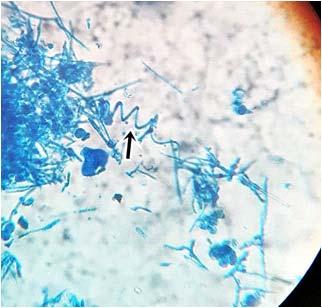
Growths of Non dermatophytic moulds were also identified by LPCB mounting morphologically.
Yeasts were identified by putting on germ tube tests, Dalmau on corn meal agar, sugar fermentation and assimilation tests.
Culture :
Specimens were inoculated simultaneously onto Sabouraud dextrose agar with chloramphenicol (0.05mg/ml) and cyclohexamide (0.5mg/ml). Specimens were inoculated on DTM slopes also. The tubes were incubated at room temperature and at 28oC in BOD incubator and checked for fungal growth at
Slide Culture Technique :
All the isolates for which the morphology was not clear in LPCB were subjected to slide culture technique. The slide culture technique permits the microscopic observation of the undisturbed relationship of spores to hyphae6.
All culture media, reagents and chemicals were obtained from HI MEDIA Private Limited, Mumbai, India. All statistical analysis was done using Chi-square test. The software used for the statistical analysis was Graph Pad Prism 7.
RESULTS
A total of 1248 clinically suspected cases of Dermatophytosis were analysed in this study.
Age & Sex Distribution :
The study group comprised of 673 (53.93%) males and 575 (46.07%) females (Fig 1). Male outnumbered female with a ratio of 1.17:1. The commonest age group affected was 21-40 years (48.96%) followed by 41-60 years (32.05%) (Table 1).
Occupation and Socio-Economic Status :
Regarding occupation, it was commonest among daily labour (28.85%) followed by housewives (22.04%), farmers (18.59%) and students (10.98%). According to Modified BG Prasad Scale, maximum number of
Vol 121, No 8, August 2023Journal
of the Indian Medical Association
KOH mount of skin scraping showing fungal hyphae
50
LPCB mount showing tear drop micro conidia of T Rubrum and spiral hyphae of T Mentagrophyte
patients belongs to class IV Socio-economic class (53.69%) followed by class III (22.44%)(Table 2).
Risk Factors Analysis :
Various risk factors of Dermatophytosis were analysed thoroughly (Table 3). From this table, it was found that sharing of footwear (69.95%) and towel (69.31%) was the commonest and significant factors associated with this disease, followed by application of steroids (53.29%) and using tight clothes (51.28%).
Mycological Isolates :
Out of total 1248 cases of clinically suspected cases of Dermatophytosis, 968 (77.56%) cases were positive in culture. 280 (22.44%) cases were culture negative.
Out of 968 isolates, growth of dermatophytes was
observed in 674 (69.62%) cases, whereas 178 (18.39%) were Candida, 76 (7.85%) were Non dermatophytic moulds and 40 (4.13%) were Malassezia spp (Tables 4 & 5).
Clinical Presentation :
Regarding clinical presentation of the lesions, T corporis was the commonest (44.39%) followed by T cruris (12.98%) and Tunguium (9.70%) (Table 6).
Fungal Staining :
Out of total 1248 samples, 1103 (88.38%) were positive in KOH (Table 7).
Species of Dermatophytes :
Out of total 1248 samples, 280 (22.44%) cases were culture negative. Growths of Dermatophytes were seen in 674 (69.62%) cases. Among these, Trichophyton rubrum was commonest (21.96%) followed by Microsporumaudouinii (18.55%)(Fig 2).
Vol 121, No 8, August 2023Journal of the Indian Medical
Association
AgeCases% 0-20 years 18114.50 21-40 years61148.96 41-60 years40032.05 >60 years56 4.49 Total1248100
Fig 1 — Sex wise distribution Table 1 — Age wise distribution of Cases
to
Social ClassC ases(%) Class I10 0.80 Class II244 19.55 Class III280 22.44 Class IV670 53.69 Class V44 3.53 Total1248100
Table 2 — Socio-economic Status wise distribution of cases (n=1248)
[According
Modified BG Prasad Scale]
Risk factorsCases (%) p value Seasonal Exacerbation432 (34.62)0.4539 Family history +ve192 (15.38)0.8753 Skipping daily bath356 (28.33)0.2354 Not changing undergarments daily459 (36.78)0.1526 Using tight clothes640 (51.28)0.0001 Sharing of clothes224 (17.95)0.5913 Sharing of towels865 (69.31)0.0001 Sharing of footwear873 (69.95)0.0001 Application of Corticosteroid665 (53.29)0.0001
Table 3 — Showing various risk factors associated with Dermatophytosis
Clinical presentationNo% T. corporis 55444.39 T. cruris 16212.98 T. unguium 1219.70 T. manuum 262.08 T. pedis 221.76 T. capitis 655.21 T. faciei 181.44 T. barbae 110.88 Intertrigo214 17.15 Pityriasis versicolor 554.40
Table 6 — Distribution of cases according to clinical presentation
Isolates Number% Dermatophytes674 69.62 Candida 17818.39 Non dermatophytic moulds76 7.85 Malasseziaspp 404.13
Candida No% C. albicans 6838.20 C. tropicalis 4123.03 C. guilliermondii 3519.66 C. parapsilosis 3419.10 Non Dermatophytic MouldsNo% Aspergillus 2127.63 Fusarium 2938.15 Curvularia 1519.74 Penicillium 79.21 Scopulariopsis 45.26
Table 4 — Distribution of Mycological isolates
Table 5 — Distribution of Candida and isolates of Non dermatophytic moulds
Culture KOH +veKOH -veTotal +ve94325968 -ve160120280 Total11031451248 51
Table 7 — Distribution of samples according to KOH positivity and Culture positivity
DISCUSSION
The rapidly evolving Dermatophytosis since the last decade reflects the changes in behaviour, life style, Socio-economic conditions and migration. The higher incidence in tropical countries like India may be due to favourable environmental as well as Socio-economic conditions3,7
In the present study, 1248 clinically suspected case of Dermatophytosis were studied. Various earlier studies showed already that infection with dermatophytes was more frequent in males compared to females3,8-10. In our study, the males were 53.93% which is marginally higher than the percentage of females (46.07%) with the male to female ratio 1.17 :1. It may be due to that increased outdoor exposure and more physical work that results in increased sweating which makes Male more susceptible! The study shows that the infection is predominant in the adult age group (21- 40 years). This may be due to increased level of physical activity in this particular age group leading to excessive sweating which favours the growth. Socialization with different people is also high compared to other age groups which eventually help in spreading of infection. This finding correlates with the other studies like Ahuja S, et al13, Satpathi P, et al12, Sarkar M, et al 11 and Ghosh R, et al6
Regarding occupation, it was commonest among daily labour (28.85%) followed by housewives (22.04%) in our study. Sarkar M, et al11 and Ghosh R, et al6 showed in their study that housewives were the predominant population. This is probably due to more physical activity in outdoor and more sweating in case of daily labours. Maximum patients (53.69%) in our study belongs to Class IV Socio-economic status. This finding was similar with the study of Surekha A, et al14
In the present study, sharing of footwear (69.95%) and towel (69.31%) were the commonest and significant factors associated with this disease, followed by application of steroids (53.29%) and using tight clothes (51.28%). Similar findings were also noted in Pathania S, et al15. Such objects like clothing, bedsheets, and towel may harbour fungal pathogens and facilitate fomite transmission among family members. Fungal spores remain viable for months in household dust, which may lead to recurrent episodes of clinical disease16,17
Among all the clinical types, tinea corporis was the predominant one (44.39%). The finding was comparable with the studies of Srinivasan, et al3, Kumar K, et al1 and Venkatesan G, et al10. Verma, et al18 and Sardari, et al19 found tinea cruris as the commonest clinical type, which contrasts with our study, where tinea cruris was the second common clinical types.
Positivity rates of KOH mount in our study was found to be 88.38%. Out of which in 75.56% cases culture was positive and among KOH negative cases, 2.0% showed culture positivity. Higher positivity in KOH mount was also found in the study of Ghosh R, et al 6,20, Doddamani PV, et al 21 and Singh S, et al 22 who reported 65.0% KOH positive and 39.0% culture positive and 60.4% positive.
In the present study, dermatophytes were grown in 69.62% cases. This finding was comparable with the study of Sarkar M, et al11, Surekha A, et al14 and Sanjiv Grover, et al23 Candida albicans was the commonest (38.20%) isolate in our study among Candida, whereas Fusarium was the most common isolate (38.15%) among NDM in our study. Similar findings were also noted in Sarkar M, et al11
Trichophyton species are the most frequent isolate as compared to Microsporum and Epidermophyton species. This finding has been noticed by many Indian studies as well as the Western countries24 T rubrum was the most common isolate (21.96%) in our study followed by Microsporumaudouinii(18.55%). TRubrum was the commonest isolate in various studies like Lakshmanan, et al25, S Das, et al26, Janardhan, et al27, Aruna et al28, Manjunath, et al29, Ramraj et al4, and Sarkar M, et al11 Microsporum audouinii was the commonest isolate in the study of Ghosh R, et al20, although it was found to be the second common isolate in our study. T mentagrophytes has been the most frequently isolated organism in other reports from India 16,17,30-37 . In a study by Grover, et al23 the commonest isolate was Trichophyton tonsurans followed by Trichophytonrubrum. This difference could be attributed to changes in global climate and humidity favouring the of growth different pathogens over the years11
CONCLUSION
The present study gave clear picture regarding the causative agents of Dermatophytosis along with its
Vol 121, No 8, August 2023Journal of
the Indian Medical Association
52
Fig 2 — Distribution of various species of Dermatophytes
of the Indian Medical Association
risk factors and Socio-economic status5. Diagnostic methodology and fungal susceptibility testing lag therapeutic advances. Our attention in future should be focused on molecular characterization of fungal pathogens along with antifungal susceptibility testing as it has been observed that patients are not responding to commonly used antifungals indicating emergence of drug resistance11
REFERENCES
1Kumar K, Kindo AJ, Kalyani J, Anandan S — Clinico–Mycological Profile of Dermatophytic Skin Infections ina Tertiary Care Center–A Cross Sectional Study. Sri Ramachandra Journal of Medicine 2007; 1(2): 12-5.
2Ranganathan S, Menon T, Sentamil GS — Effect of socioeconomic status on the prevalence of Dermatophytosis in Madras. Indian J Dermatol Venereol Leprol 1995; 61: 16-8.
3Srinivasan B, Rajan S, Thiyagarajan T, Solomon J — Epidemiology of Dermatophytosis in and around Tiruchirapalli, Tamilnadu, India. Asian Pacific Journal of Tropical Disease 2012; 2(4): 286-9. DOI:10.1016/S2222-1808(12)60062-0.
4Emmons CW, Binford CH, Utz JP, Kwon-Chung KJ — Medical Mycology. 3rd ed. Philadelpia, Lea and Febiges. 1974. 117-67.
5Ramaraj V, Vijayaraman RS, Rangarajan S, Kindo AJ — Incidence and prevalence of Dermatophytosis in and around Chennai, Tamilnadu, India. IntJResMedSci2016; 4: 695-700.
6Ghosh R, Ray R, Ghosh TK, Ghosh AP — Clinico-mycological profile of Dermatophytosis In a Tertiary Care Hospital in West Bengal An Indian scenario. Int.J Curr MicrobiolApp Sci 2014; 3(9): 655-66.
7Mycological study on Dermatophytosisin Seoul. Korean Journal of Dermatology 1994; 32: 24-33.
8Bhaskaran CS, Rao PS, Krishnamoorthy T, Tarachand P — Dermatophytosis in Tirupathi. Indian J Pathol Microbiol 1977; 31: 251-9.
9Maheshwariamma SM, Paniker CKJ, Gopinathan T — Studies on dermatomycosis in Calicut. Indian J Pathol Microbiol 1982; 25: 11-7.
10Venkatesan G, Ranjit Singh AJA, Murugesan AG, Janaki C, Gokul Shankar S— Trichophytonrubrum–the predominant etiological agent in human dermatophytoses in Chennai, India. Afr J Microbiol Res 2007; 1(1): 9-12.
11Sarkar M, Ray (Ghosh) R, Halder P, Ghosh AP, Chatterjee M — Clinico- Mycological Profile of Onychomycosis –A Study in A Tertiary Care Hospital In Kolkata IOSR. Journal of Dental and Medical Sciences 2016; 15(9): 78-83.
12Satpathi P, Achar A, Banerjee D, Maiti A, Sengupta M, Mohata A — Onychomycosis in Eastern India - study in a peripheral tertiary care centre. Journal of Pakistan Association of Dermatologists 2013; 23(1): 14-9.
13Ahuja S, Malhotra S, Charoo H. Etiological agents of onychomycosis from a tertiary care Hospital in central Delhi, India. Indian J Fundamental and Appl Life Sci 2011; 1: 11-4.
14Surekha A, Ramesh Kumar G, Sridevi K, Murty DS, Usha G, Bharathi G — Superficial Dermatophytosis: a prospective clinico-mycological study. J ClinSci Res 2015; 4: 7-15.
15Pathania S, Rudramurthy SM, Narang T, Saikia UN, Dogra S — A prospective study of the epidemiological and clinical patterns of recurrent Dermatophytosis at a tertiary care hospital in India. Indian J Dermatol Venereol Leprol 2018; 84: 678-84.
16Bhatia VK, Sharma PC — Epidemiological studies on Dermatophytosis in human patients in Himachal Pradesh, India. Springerplus 2014; 3: 134.
17Havlickova B, Czaika VA, Friedrich M — Epidemiological trends in skin mycoses worldwide. Mycoses 2008; 51(4): 2 15.
18Sen SS, Rasul ES — Dermatophytosis in Assam. Indian Journal of Medical Microbiology 2006; 24(1): 77-8.
19Sardari L, Sambhashiva RR, dandapani R — clinico mycological study of dermatophytes in a coastal area. Ind J Dermatol Venereol Leprol 1983; 49:2: 71-5.
20Ghosh R — Changing trend in clinico-mycological profile of Dermatophytosis of skin in Eastern India: IP. International Journal of Medical Microbiology and Tropical Diseases 2018; 4(3): 159-65.
21Doddamani PV, Harshan KH, Kanta RC, Gangane Sunil R — Isolation, Identification and Prevalence of Dermatophytes in Tertairy care Hospital in Gulbarga District, People’sJScientific Res 2013; 6: 2.
22Beena P M, Singh S — Profile of dermatophyte infectins in Baroda, Ind J Derm Venereal Leprol 2003; 69: 281-83.
23WgCdr Sanjiv Grover, Lt Col P Roy — Clinico-mycological Profile of Superficial Mycosis in a Hospital in North-East India; MJAFI 2003; 59(2):
24Kumar K, Kindo AJ, Kalyani J, Anandan S — Clinico –Mycological Profile of Dermatophytic Skin Infections In A Tertiary Care Center – A Cross Sectional Study. Sri Ramachandra Journal of Medicine 2007; 1(2): 12-5.
25Lakshmanan A, Ganeshkumar P, Mohan SR, Hemamalini M, Madhavan R — Epidemiological and clinical pattern of Dermatophytosis in rural India. Indian J Med Microbiol 2015; 33: S134-6.
26Das S, De A, Saha R, Sharma N, Khemka M, Singh S, et al — The current Indian epidemic of Dermatophytosis: A study on causative agents and sensitivity patterns. Indian J Dermatol 2020; 65: 118-22.
27Janardhan B, Vani G — Clinico mycological study of Dermatophytosis. Int J Res Med Sci 2017; 5: 31-9.
28Aruna GL, Ramalingappa B — A clinico mycological study of human dfermatophytosis in Chitradurga, Karnataka, India. JMSCR 2017; 5: 25743-9
29Manjunath M, Mallikarjun K, Dadapeer, Sushma — Clinicomycological study of dermatomycosis in a tertiary care hospital. Indian J Microbiol Res 2016; 3: 190-3.
30de Freitas RS, Neves PS, Charbel CE, Criado PR, Nunes RS, Santos Filho AM, et al — Investigation of superficial mycosis in cutaneous allergy patients using topical or systemic corticosteroids. Int J Dermatol 2017; 56: e194-8.
31Morimoto K, Tanuma H, Kikuchi I, Kusunoki T, Kawana S — Pharmacokinetic investigation of oral itraconazole in stratum corneum level of tinea pedis. Mycoses 2004; 47: 104-14.
32Hanumanthappa H, Sarojini K, Shilpashree P, Muddapur SB — Clinicomycological study of 150 cases of Dermatophytosis in a tertiarycare hospital in South India. Indian J Dermatol 2012; 57: 322 3.
33Bindu V, Pavithran K — Clinico mycological study of Dermatophytosisin Calicut. IndianJDermatolVenereolLeprol 2002; 68: 259-61.
34Vyas A, Pathan N, Sharma R, Vyas L — A clinicomycological study ofcutaneous mycoses in Sawai man singh hospital of Jaipur, North India. Ann Med Health Sci Res 2013; 3: 593-7.
35Bhatia VK, Sharma PC — Determination of minimum inhibitoryconcentrations of itraconazole, terbinafine and ketoconazole againstdermatophyte species by broth microdilution method. Indian J Med Microbiol 2015; 33: 533 7.
36Adimi P, Hashemi SJ, Mahmoudi M, Mirhendi H, Shidfar MR,Emmami M, et al — In vitro activity of 10 antifungal agents against 320dermatophyte strains using microdilution method in Tehran. Iran J Pharm Res 2013; 12: 537-45.
37Mahajan S, Tilak R, Kaushal SK, Mishra RN, Pandey SS— Clinico mycological study of dermatophytic infections and theirsensitivity to antifungal drugs in a tertiary care center. Indian J Dermatol Venereol Leprol 2017; 83: 436-40.
Vol 121, No 8, August
2023Journal
53
Original Article
Oral and Systemic Symptoms of COVID-19 among Patients Vaccinated and Unvaccinated against SARS-CoV-2
Pankaj Goel1, Aman Kumar2
Background : Vaccination against SARS-CoV-2 has been developed and are being administered at rapid pace globally, however despite vaccination cases have been reported who got infected even after vaccination. It is important to find the effect of vaccination on symptoms experienced by COVID-19 positive patients.
Objective : The aim of the study was to assess the effect of vaccination on oral and systemic symptoms experienced by the patients.
Method : An observational study was carried out at a dedicated COVID care public hospital where the patients reporting to the Out Patient Department (OPD) for getting themselves tested for COVID-19 were interviewed regarding the symptoms experienced by them and data was recorded. Further investigation was carried out using Reverse Transcriptase Polymerase Chain Reaction (RTPCR) test.
Results : A total of 219 patients reported to the OPD between July 2021 to September 2021, out of which 119 were found to be COVID-19 positive and 100 were COVID-19 negative. Out of 119, COVID-19 positive patients, 45 were fully vaccinated (received both doses) against SARS-CoV-2, while 74 were unvaccinated or partially vaccinated (received single dose only). Statistically significant difference was found between fully vaccinated and unvaccinated / partially vaccinated patients in relation to ageusia, xerostomia, anosmia and fatigue in COVID-19 positive group.
Conclusion : Although there are chances of getting infected with SARS-CoV-2 even after receiving vaccination against it, the symptoms experienced by the patients are less.
[J Indian Med Assoc 2023; 121(8): 54-7]
Key words : COVID-19, Vaccination, Ageusia, Xerostomia, Anosmia.
SARS-CoV-2 is a single stranded RNA corona virus which has infected human beings1,2. It has a high probability of zoonotic origin3. It can be transmitted directly by coughing, sneezing or indirectly by contact with mucous membrane like oral, ocular or nasal mucosa4. Dentists are at high risk of getting infected with the virus because of it air borne route of transmission and huge amount of aerosol generated during dental treatment make the situation even more precarious for the dentists. Various guidelines has been issued by different dental societies for the safe and effective management of acute dental infections which if not adhered to increases the risk of infection3
Although devoid of any characteristic signs and symptoms, various systemic symptoms associated with COVID-19 are fever, dry cough, headache, tiredness, diarrhoea, sore throat or runny nose, however in certain cases it may cause pneumonia, kidney failure or even death4 . There are certain infected patients who are asymptomatic but possess a risk of spreading infection, identification of such patients is important for the control of the disease5.
Department of Dentistry, AIIMS, Bhopal, Madhya Pradesh 462026
1MDS, Professor and Head and Corresponding Author
2MDS, Junior Resident
Received on : 03/11/2022
Accepted on : 02/04/2023
Editor's Comment :
Although it is possible to get infected with SARS-CoV-2 even after vaccination, the oral and systemic symptoms experienced by the individual are less as compared to unvaccinated individuals. As oral symptoms like xerostomia increases the chances of dental caries, decreased oral symptoms in vaccinated individuals will improve the oral health related quality of life in such patients.
Oral symptoms associated with the disease are dysgeusia, xerostomia, chemosensory alterations, bruxism and bleeding gums6. SARS-CoV-2 has affinity for Angiotensin-converting Enzyme 2 (ACE 2) receptors found in salivary gland, respiratory tract, gastro intestinal tract, leading to various signs and symptoms7. Decrease in salivary flow due to attachment of SARS-CoV-2 to ACE 2 receptors of salivary gland may cause the possible oral symptoms of the disease3. Cells with ACE 2 receptors may act as a host for the virus, which causes further inflammatory process in the organ concerned like tongue mucosa and salivary gland3,8. Oral and systemic symptoms may be due to the infection by SARS-CoV-2 per se or may be the aftermath of treatment of the infection which may lead to immuno suppression causing various oral and systemic symptoms.
There can be many variables which influence the
Vol 121, No 8, August 2023Journal of the Indian Medical Association 54
oral and systemic symptoms of SARS-CoV-2 infection and one of the such variable is COVID-19 vaccination status. As the development of vaccination against SARS-CoV-2 has been fast and vaccination has been done at a rapid pace across the globe, it is imperative to assess the effect of vaccination on the oral symptoms.
The aim of this study was to assess the oral and systemic symptoms in COVID-19 vaccinated and unvaccinated patients of SARS-CoV-2 infection.
MATERIALS AND METHODS
An observational study was conducted in the Department of Dentistry and COVID Screening OPD of a dedicated COVID care public hospital during July 2021 to September, 2021.Convenience sample size was taken as all the patients who presented to the OPD were screened for enrolling into the study based on the eligibility and exclusion criteria.
Eligibility Criteria :
(a) Those patients who presented with classical symptoms of SARS-CoV-2 (b) Those having positive history of contact with COVID-19 positive patients.
Exclusion Criteria :
Those patients who are not willing to participate. Informed consent was taken from all the patients and only those willing to participate were included in the study. In the case of children assent form was used, which was filled by the parents of the children. A single investigator was trained to conduct the interview and perform oral examination of the participants to record symptoms associated with COVID-19 as reported by participants. Oral symptoms which were recorded were dysgeusia, xerostomia, halitosis, bruxism, ulcer in the oral cavity or at the corners of the mouth, bleeding gums and burning sensation in the oral cavity. Systemic symptoms which were recorded were altered smell, headache, diarrhoea, chills, dry cough, fatigue, nausea / vomiting, shortness of breath, fever, sore throat and body ache. All the symptoms were recorded in a survey sheet by the investigator after conducting the interview and oral examination.
Ethical clearance for the research was taken from the All India Institute of Medical Sciences Bhopal, Institutional Human Ethics Committee by letter number IHEC-LOP/2021/IM0378.
Socio-demographic variables such as age, gender, diet, smoking / alcohol habit, medical history of co morbidity and severity of COVID-infection was also recorded. Vaccination status of all the participants was recorded, however participants whose vaccination status was unknown were not included in the analysis of data. ChAdOx1 nCoV-19 Corona Virus Vaccine
(Recombinant) was used to vaccinate all those individuals who were in the vaccinated group. Those individuals who had received both the doses of vaccination and fifteen days had been passed post inoculation of second dose were considered to be vaccinated.
Reverse Transcriptase Polymerase Chain Reaction (RTPCR) test was done on all patients after the interview and examination of the patients was done. The participants got to know about the positive or negative diagnosis of COVID 24 hours after the RTPCR test was done. Based on the confirmatory investigation, the patients were divided into two groups; COVID confirmed (cases) and COVID suspect (controls). Statistical Package for the Social Sciences (SPSS) software version 25 was used for data analysis. ChiSquare test were applied and p-value <0.05 was considered to be statistically significant.
RESULTS
A total of 219 patients were included in the study who visited the OPD having age range of 13 to 74 years with the mean age of 29.9±11.9 years. Out of 219 participants, 119 participants tested COVID positive (52 males and 67 females). 100 participants tested negative (66 males and 34 females) (Table 1). There was a statistically significant difference between COVID-19 positive group and negative in relation to various oral and systemic symptoms (Table 2).
Among oral symptoms, ageusia, xerostomia, halitosis and bruxism were statistically significantly higher among COVID patients as compared to COVID negative patients (P = <0.001, 0.021, 0.002, 0.001 respectively). Among systemic symptoms, anosmia, headache, diarrhoea, chills and fatigue were statistically significantly higher among COVID positive patients as compared to COVID negative patients (P = <0.001, <0.001, 0.010, 0.025, 0.001 respectively).
Among those oral and systemic symptoms who were found to have statistically significant difference between COVID 19 positive and negative patients, further analysis was done according to vaccination status against SARS-CoV-2 in COVID-19 positive group (Tables 3, 4).
Among oral symptoms, there was statistically significant difference between vaccinated and unvaccinated participants in relation ageusia and xerostomia. Among systemic symptoms, there was statistically significant difference between vaccinated and unvaccinated participants in relation anosmia and fatigue.
DISCUSSION
This study reported the effect of vaccination status
Vol 121, No 8, August 2023Journal
of the Indian Medical Association
55
Table 2 — Difference in relation to various oral and systemic symptoms between COVID-19 positive and COVID-19 negative patients
breakthrough cases among 1497 fully vaccinated participants 9 . Taste alterations have been reported in 59.5% (n=66 out of 111) of patients and xerostomia in 45.9% (n= 51 out of 111) of COVID-19 patients by Fantozi, et al, 202010. Xerostomia found in SARS-CoV2 infection may play an aggravating role in halitosis11. Increase in bruxism and Temporo Mandibular Dysfunction (TMD) symptoms has been reported during corona virus pandemic12. The increase in bruxism can be attributed to adverse effects on the psycho-emotional status and increase in psychological stress during COVID-19 pandemic.
Human strains of the corona virus have been demonstrated to invade the Central Nervous System through the olfactory neuroepithelium and propagate from within the olfactory bulb. Furthermore, nasal epithelial cells display the highest expression of the SARS-CoV-2 receptor, angiotensin converting enzyme 2, in the respiratory tree13. As statistically significant
Oral symptoms in fully vaccinated and unvaccinated / partially vaccinated individuals among COVID-19 positive participants
partially
— Systemic symptoms in vaccinated and unvaccinated individuals among COVID-19 positive participants Systemic Fully
partially square
performed
against SARS-CoV-2 on oral and systemic symptoms experienced by the patients in COVID-19 positive group. Statistically significant difference was found in relation to ageusia and xerostomia between vaccinated and unvaccinated population. Our results are found to be consistent with Bergwerk, et al, 2021 who reported less symptoms or asymptomatic cases in
Vol 121, No 8, August 2023Journal
of the Indian Medical Association
VariableTotal n (%)Positive n (%)Negative n(%)P valuea Gender 0.001 Male118 (53.9)52 (43.7)66 (66) Female 101 (46.1)67 (56.3)34 (34) Age (Years) 26.15 (10.86)b 34.3 (11.6)b <0.001c Co morbidity 0.127 Co morbidity present16 (7.3)6 (5)10 (10) Co morbidity absent203 (92.7)113 (95)90 (90) COVID symptoms 0.019 COVID symptoms present189 (86.3)97 (81.5)92 (92) COVID symptoms absent30 (13.7)22 (18.5)8 (8) Vaccination status0.004 One dose39 (17.8)17 (14.3)22 (22) Both dose57 (26)45 (37.8)12 (12) Unvaccinated 123 (56.2)57 (47.9)66 (66) Smoking status 0.056 Yes20 (9.1)7 (3.2)13 (5.9) No199 (90.9)112 (51.1)87 (39.7) Alcohol consumption 0.048 Yes16 (7.3)5 (2.3)11 (5) No203 (92.7)114 (52.1)89 (40.6) Diet 0.485 Vegetarian 130 (59.4)70 (32)60 (27.4) Non vegetarian89 (40.6)49 (22.4)40 (18.3) a : Chi-square test was performed, b : Standard deviation, c : 't' test was performed
Table 1 — Sample Characteristic
and Systemic COVIDCOVIDP valuea symptoms Suspected Confirmed (Negative), (Positive), n (%)n (%) Ageusia19 (19)49 (41.2)< 0.001 Xerostomia30 (30)56 (47.1) 0.021 Halitosis4 (4)11 (9.2) 0.002 Bruxism3 (3)11 (9.2) 0.001 Ulcer at corner of mouth1 (1)7 (5.9) 0.102 Bleeding gums1 (1)4 (3.4) 0.244 Ulcer in oral cavity12 (12)15 (12.6) 0.647 Burning sensation in mouth3 (3)4 (3.4) 0.651 Anosmia10 (10)51 (42.9)< 0.001 Headache18 (18)52 (43.7)< 0.001 Diarrhoea5 (5)19 (16) 0.010 Chills3 (3)13 (10.9) 0.025 Dry cough30 (30)48 (40.3)0.112 Fatigue33 (33)65 (54.6) 0.001 Nausea/ Vomiting6 (6)13 (10.9) 0.197 Shortness of breath8 (8)16 (13.4) 0.199 Fever64 (64)69 (58) 0.364 Sore throat43 (43)56 (47.1) 0.548 Body ache46 (46)57 (47.9) 0.779 a : Chi-square
was
Oral
test
Unvaccinated/Chip
symptomsVaccinated
vaccinated Anosmia114010.01790.001 No anosmia3434 Headache18340.40210.526 No headache2740 Diarrhoea9100.87750.349 No diarrhoea3664 Chills310 1.34810.246 No chills4264 Fatigue1946 4.48880.034 No fatigue2628
: Chi-square test was performed
Table 4
valuea
a
Oral
symptomsVaccinated
Ageusia12376.290.012 No ageusia3337 Xerostomia1541 5.4720.019 No xerostomia3033 Halitosis380.57290.449 No halitosis4266 Bruxism380.57290.449 No bruxism4266
: Chi-square test was performed 56
Table 3 —
FullyUnvaccinated/Chip valuea
square vaccinated
a
difference was found between vaccinated and unvaccinated participants in COVID-19 positive group in relation to anosmia, it can be said that vaccination do have a protective role in mitigating the symptoms associated with infection.
Statistically significant difference was found in relation to fatigue between COVID-19 positive and negative participants as well as between vaccinated and unvaccinated participants in COVID-19 positive group. It can be said that although fatigue is common finding in covid positive patients, vaccinated patients are less likely to experience it.Fatigue has been reported with a prevalence of 52.3% (n= 67 out of 128) in COVID-19 positive patients14. Chronic Fatigue Syndrome (CFS) has been associated with a large number of differing changes in the inflammatory markers and immune cell population. The lack of distinct immune signature, coupled with the association with depression, lends credence to the multifactorial aetiology of CFS14
Headache has been reported in COVID-19 positive patient15. Headaches reported can be a mixture of tension-type headache and headaches attributed to systemic viral infection, heterophoria, cough or hypoxia15. A case has been reported of a 27 years old COVID-19 positive male with type 2 diabetes, who had presented with a complaint of watery diarrhoea five to six times per day16. The presence of gastrointestinal symptoms in corona virus infection (SARS-CoV-1 and SARS-CoV-2) can be linked to the distribution of ACE2 receptor, which is present in lung alveolar type 2 cells, as well as in enterocytes16
Caponio, et al 2022 reported a case of fully vaccinated individual who developed ageusia after 55 days post vaccination17
Although it is possible to develop symptomatic COVID-19 post vaccination, the findings of our study are in agreement with the studies which reported less symptoms in breakthrough cases of vaccinated individuals9,18
CONCLUSION
It can be concluded that patients who were fully vaccinated against SARS-CoV-2 experienced less oral and systemic symptoms of COVID-19 in relation to ageusia, xerostomia, anosmia and fatigue as compared to unvaccinated patients, which was found to be statistically significant. Although patient may get infected with SARS-CoV-2 even after receiving vaccination against it, less symptoms experienced by them.
Recommendation : Further studies on larger sample size are needed to corroborate these findings. Conflict of Interest : Authors declare no conflict of interest.
REFERENCES
1Andersen KG, Rambaut A, Lipkin W, Holmes EC, Garry RF — The proximal origin of SARS-CoV-2. Nat. Med 2020; 26: 4502.
2Wu F, Zhao S, Yu B, Chen YM, Wang W, Song ZG, et al — A new coronavirus associated with human respiratory disease in China. Nature 2020; 579: 265-9.
3Sinjari B, D’Ardes D, Santilli M, Rexhepi I, D’Addazio G, Di Carlo P, et al — SARS-CoV-2 and Oral Manifestation: An Observational, Human Study. Journal of Clinical Medicine 2020; 9(10): 3218.
4Chen N, Zhou M, Dong X, Qu J, Gong F, Han Y, et al — Epidemiological and clinical characteristics of 99 cases of 2019 novel coronavirus pneumonia in Wuhan, China: A descriptive study. Lancet Lond Engl 2020; 395: 507-13.
5Epidemiology Working Group for NCIP Epidemic Response, Chinese Center for Disease Control and Prevention. The epidemiological characteristics of an outbreak of 2019 novel coronavirus diseases (COVID-19) in China. Zhonghua Liu Xing Bing Xue Za Zhi Zhonghua Liuxingbingxue Zazhi 2020, 41: 145-51.
6Vinayachandran D, Balasubramanian S — Is gustatory impairment the first report of an oral manifestation in COVID19? Oral Dis 2021; 27 Suppl 3(Suppl 3): 748-9.
7Freni F, Meduri A, Gazia F, Nicastro V, Galletti C, Aragona P, et al — Symptomatology in head and neck district in coronavirus disease (COVID-19): A possible neuroinvasive action of SARS-CoV-2. Am J Otolaryngol 2020; 41: 102612
8Xu H, Zhong L, Deng J, Peng J, Dan H, Zeng X, et al — High expression of ACE2 receptor of 2019-nCoV on the epithelial cells of oral mucosa. Int J Oral Sci 2020; 12: 8.
9Bergwerk M, Gonen T, Lustig Y, Amit S, Lipsitch M, Cohen C, etal— COVID-19 BreakthroughInfections in Vaccinated Health Care Workers. N Engl J Med 2021; 385: 1474-84.
10Fantozzi PJ, Pampena E, Vanna DD, Pellegrino E, Corbi D, Mammucari S, et al — Xerostomia, gustatory and olfactory dysfunctions in patients with COVID19. Am J Otolaryngol 2020; 41(6): 102721
11Dziedzic A, Wojtyczka R — The impact of coronavirus infectious disease 19 (COVID19) on oral health. Oral Dis 2020; 27(3): 703-6.
12Emodi-Perlman A, Eli I, Smardz J, Uziel N, Wieckiewicz G, Gilon E, et al — Temporomandibular Disorders and Bruxism Outbreak as a Possible Factor of Orofacial Pain Worsening during the COVID-19 Pandemic—Concomitant Research in Two Countries. J Clin Med 2020; 9(10): 3250.
13Boscolo-Rizzo P, Borsetto D, Fabbris C, Spinato G, Frezza D, Menegaldo A, et al — Evolution of altered sense of smell or taste in patients with mildly symptomatic COVID-19. JAMA Otolaryngol Head Neck Surg 2020; 146(8): 1-5.
14Townsend L, Dyer AH, Jones K, Dunne J, Mooney A, Gaffney F, et al — Persistent fatigue following SARS-CoV-2 infection is common and independent of severity of initial infection. PLoS ONE. 2020; 15(11): e0240784
15Belvis R — Headaches during COVID-19: My clinical case and review of the literature. Headache 2020; 60(7): 1422-6.
16Ata F, Almasri H, Sajid J — BMJCaseRep 2020; 13: e235456
17Caponio VCA, Lipsi MR, Fortunato F, Arena F, Lo Muzio L — Symptomatic SARS-CoV-2 Infection with Ageusia after Two mRNA Vaccine Doses. Int J Environ Res Public Health 2022; 19: 886
18Hacisuleyman E, Hale C, Saito Y, Blachere NE, Bergh M, Conlon EG, et al — Vaccine Breakthrough Infections with SARSCoV-2 Variants. N Engl J Med 2021; 384: 2212-8.
Vol 121, No 8, August 2023Journal
of the Indian Medical Association
57
Original Article
Effectiveness of Nursing Care Bundles in Hospitalized Patients with Chronic Obstructive Pulomnary Disease (COPD)
Smita Roba Tirkey1, Rashmi P John2, Anuj Kumar Pandey3, Ajay Kumar Verma4, Surya Kant5
In Chronic Obstructive Pulmonary Disease (COPD) exacerbation care bundle is essential to reduce not only clinical manifestations but also the severity of the disease condition. Here, we aimed to explore the effectiveness of care bundles by comparing the pre-test and post-test level of Quality of Life (QOL) and Activities of Daily Living (ADL) among hospitalized COPD patients. In this study sixty-six hospitalized COPD patients were included. The care bundle intervention was given thrice a day for consecutive seven days. Data were collected 24 hours after admission by St. George Respiratory Questionnaire-COPD (SGRQ-C) for pre-test and the same tool was used after intervention for post-test. The results were analyzed using descriptive statistics and making comparisons between the groups with respect to various parameters. There was a significant difference in the mean score of pre-test and post-test QOL & ADL. Aftercare bundle intervention patients felt better in their current situation, so, it is interpreted that patient’s quality of life increased. The main conclusions drawn from this study was nursing care bundle can improve the care of COPD exacerbation. The use of a bundle of cares not only streamlines care but also has the potential to reduce the risk of adverse effects from unnecessary medications or treatments.
[J Indian Med Assoc 2023; 121(8): 58-63]
Key words :Chronic Obstructive Pulmonary Disease (COPD), Care bundles, Exacerbation, Quality of life, Activities of Daily Living.
Chronic Obstructive Pulmonary Disease (COPD) is the major cause of hospitalization and disability due to lung disease. Its chronic progressive and acute exacerbations will affect the quality of life and life expectancy of COPD patients. Respiratory tract infections commonly develop in COPD because of the changes in the normal defense mechanism and decreased immune resistance. Further, infection frequently leads to acute respiratory failure. COPD and emphysema may worsen at night with sleep onset dyspnea and frequent awakenings. Because of the decrease in the muscle tone and activity of respiratory muscles leads to hypoventilation, increases in the resistance of the airways and becomes hypoxemic.1 Exercise program is a very essential step to control COPD. For people with lung disease, exercise restriction is very common because they are anxious of shortness of breath, however routine physical work
King George’s Medical University, Uttar Pradesh, Lucknow 226003
1MSc Nursing, Department of Medical Surgical Nursing
2MSc Nursing, Acting Principal, Department of Medical Surgical Nursing
3PhD, Department of Respiratory Medicine
4MD, Professor (Junior Grade), Department of Respiratory Medicine
5MD, Professor & Head, Department of Respiratory Medicine and Corresponding author
Received on : 13/07/2022
Accepted on : 21/01/2023
Editor's Comment : The implementation of nursing care services may result in shorter hospital stays, lower readmission rates, and reduces the risk of adverse effects caused by unnecessary medications or treatments in hospitalized COPD patients.
can improve the heart, lungs, muscles, and can help breathing easier and healthier. Generally, people with COPD and chronic bronchitis produce mucus. Techniques to remove mucus are often done after using inhaled bronchodilator medication.
Care bundle is used to promote the health of the patients with COPD. The nursing care bundle includes: effective huff coughing exercise, breathing retraining and relaxation techniques energy conservation techniques and upper and lower limb exercise. These interventions help the patient to decrease the symptoms, improving exercise ability and physical functioning. The quality of nursing care for the patient with respiratory disease is ensured based on the evidence based practice and holistic approach. This approach encompasses a comprehensive assessment, planning, intervention and evaluation process. Today nursing seems to be more creative and nurses incorporate critical thinking in rendering comprehensive care to patients in pulmonary care unit. This critical analysis helps them to identify the problems of the patient in its early stage and institute appropriate interventions2
Vol 121, No 8, August 2023Journal of the Indian Medical Association
58
The respiratory rate of COPD patients increases and the exhalation time is extended to compensate for airflow obstruction that causes breathing difficulties. Long-term use of accessory muscles can lead to increased patient fatigue. Pursed lip breathing prevents the bronchioles from collapsing and air retention. Patient is taught to breathe in slowly through the nose, and then breathe out slowly through the pursed lips, just like blowing a whistle. Use lips for positive pressure, because excessive resistance may increase the work of breathing. An effective cough will clear the airways from the sputum. The main purpose of coughing is to save energy, reduce fatigue and promote the removal of secretions. In 2011, CTS developed guidelines for the treatment of shortness of breath in patients with advanced COPD. Learning new breathing retraining techniques (such as lips breathing and coughing cough exercises) will help move air in and out of the lungs, which is very effective. Using effective breathing techniques and exercise helps to minimize shortness of breath and adequate oxygen to working muscles3
The goal of the nursing care bundle is to reduce the clinical manifestations of COPD and reduce the severity of the disease conditions. It is very effective to use the selected nursing interventions in improving the quality of life as well as activities of daily living in COPD patients and it can reduce cost of care and easily available in all settings. Exercise, activities of daily living, and health related quality of life which can be severely affected by the disease. These are interrelated and that impact the lives of these patients. The nursing care bundle will improve the patients exercise tolerance and subsequently quality of life.1 However, data are lacking in this area. In the present study we aimed to evaluate the effectiveness of selected nursing interventions to improve the quality of life and activities of daily living in COPD patients.
MATERIALS AND METHODS
Study Design and Setting :
Quantitative pre experimental research approach was applied in the current study. The work was conducted at ward of respiratory medicine department of a tertiary care hospital. Participants between 30-60 years age-group, clinically diagnosed with COPD and fulfilled the selection criteria were included. After obtaining ethical approval from committee and formal administrative permission from the department of respiratory medicine study was conducted. Self introduction and purpose of the study was explained to the subject, willingness to participate in the study
was ascertained and written informed consent was taken from each participants. The demographic, and clinical characteristics of subjects was collected in preformed questionnaire which was formed by experts from respiratory medicine, nursing, medicine, and pharmacology. The present study included two variables: first, dependent variable- quality of life and activities of daily living among COPD patients; second, independent variables- nursing care bundle.
Operational Definitions and Tools :
Effectiveness refers to the optimising physical health, reducing symptoms and functional impairment, and increasing emotional and social well being after giving nursing care bundle.
Bundle care refers to nursing interventions those are used to promote the health of the patients with COPD. Those interventions are purse lip breathing, diaphragmatic breathing, huff coughing, chest physiotherapy and upper and lower limb exercise. Quality of life refers to self determined evaluation of satisfaction with issues important to person. Activities of daily living refers to daily activities which patient do in their daily life like brushing, toileting, bathing, exercise etc.
St. George Respiratory Questionnaire - COPD (SGRQ-C) :
The SGRQ-C was developed by SGRQ which was structured to understand the health status of asthma and COPD patients. A study found that SGRQ-C contains the best original items, no longer specifies the recall period and produces the same scores as the original product4. SGRQ-C is divided into two categories- Part 1 (Questions 1-7) which explains the frequency of resolving respiratory symptoms It was not intended to be an accurate epidemiological tool, but to assess patients’ perceptions of their recent respiratory illness. Part 2 (Questions 8-14) discusses the patient’s current status, the most recent condition. Activity scores were used to measure interference with daily physical activities. The impact score covers a series of psychosocial dysfunction. This component is partly related to respiratory symptoms, but also closely related to exercise performance, dyspnea in daily life and mood disturbances. Therefore, the impact score is the most extensive part of the questionnaire, covering all the disturbances experienced by respiratory patients during their lifetime. All of these depend on the quality of life and activities of daily living.
A total score was calculated in terms of symptoms, activity, and impacts. By dividing the total weight by the maximum possible weight of the component and
Vol 121, No 8, August 2023Journal
of the Indian Medical Association
59
expressing the result as a percentage, the score for each component was calculated separately. Score was inversely proportional to the health condition means, higher the score worsen the condition.
Inclusion and Exclusion Criteria :
Inclusion criteria was as follows : (1) age group 30-60 years, (2) patients admitted in the ward more than seven days, (3) patient who are literate, (4) subjects clinically diagnosed with COPD. Exclusion criteria was as : (1) other concomitant pulmonary diseases viz, tuberculosis, asthma, pneumonia etc, (2) patient is on ventilator support or critically ill. (3) patient denied to participate in the study.
Interventional Procedure :
Intervention was given to COPD subjects at their bedside in the direct observation of specialist in the ward for two times in a day for a total duration of six days. And the duration of each interventional procedure was between 15-45 min5. These nursing care was provided to each subjects : pursed lip breathing -4 minute; diaphragmatic breathing -4 minute (to better control over breathing); huff coughing -2 min (which adequately clears sputum from airway); chest physiotherapy- 4 min (to prevent infections and helps easy breathing); upper and lower limb exercise for 3 min to improve the activities of daily living.
RESULTS
Demographic Profile of COPD Subjects : Demographic characteristics of the studied population was shown in Table 1. Fourty-three (65.2%) subjects were in the age group of >50 years. Majority of the subjects were males with 95.5% proportion, while 4.5% were females. All the subjects were married. Majority of the subjects were farmers with 62.1% proportion.
Pre & Post test comparison of Quality of Life (QOL), Activities of Daily Life (ADL), and Overall Impact Score (OIS) :
As shown in Table 2 the total score of QOL among the patients at pre test level had a mean of 76.94±5.63 with minimum value 47.89 and maximum value 90.85. After post test level the impact score was changed to the mean value 53.63±4.50 with minimum value 44.41 and maximum value 84.89. After post test, this change of
30.30%, among the subjects was found highly significant (P<0.001).
Pre & Post Test Comparison of ADLs was represented in Table 2. The ADL score among the patients at pre test level had a mean 92.36±0.00 with minimum and maximum value 92.36 (pre test activity score was constant). After post test level the activity score was changed to the mean value 68.56±3.18 with minimum value 68.15 and maximum value 92.36. This percentage change of 25.77% after post test among the subjects was found to be highly significant (P<0.001).
As shown in Table 2, the impact score among the patients at pre test level had a mean 74.04±7.41 with minimum value 34.81 and maximum value 86.81. After post test level the impact score was changed to the mean value 43.03±4.31 with minimum value 42.47 and maximum value 75.27. Percentage change of 41.88% after post test among the subjects was found to be highly significant (P<0.001).
Pre & Post Test Comparison of Item Scores of Symptoms and Activity :
When we compared the item scores of symptoms (cough, sputum, shortness of breath, wheezing, chest trouble, good days, morning wheezing) the maximum % change was seen for the symptom cough with 0.46%
Vol 121, No 8, August 2023Journal
of the Indian Medical Association
CharacteristicsNo ofParticipants’s participants (n) percentage (%) Age group 30-3946 (years) 40-49 1929 >504365 Gender Male34.5 Female 63 95.5 Martial Status Married66100 Un-married0 Occupation Unemployed23 Unskilled23 Semi skilled46 Skilled23 Clerk/shop-owner/5685 farmer
Table 1 — Demographic profile of studied participants
Variables MeanSD MedianMinMaxStatistical analysis QOL Pre Test Level 76.945.6377.4947.8990.85Wilcoxon Signed P<0.001 Post Test Level 53.634.5053.3244.4184.89 Ranks Test, Z= -7.17 % Change 30.30 ADL Pre Test Level 92.36092.3692.3692.36Wilcoxon Signed P<0.001 Post Test Level 68.563.1868.1568.1592.36 Ranks Test, Z= -7.55 % Change 25.77 OIS Pre Test Level 74.047.4174.8934.8186.81Wilcoxon Signed P<0.001 Post Test Level 43.034.314.3142.4775.27 Ranks Test, Z= -7.15 % Change 41.88 60
Table 2 — Pre and post test comparison of Quality of Life (QOL), Activities of Daily Life (ADL), and Overall Impact Score (OIS)
change. However, no significant difference was found in any symptom score (Fig 1).
On comparing the item scores of activity, the maximum % change was seen for the activities of ‘washed/ dressed’, ‘take long time to washed/ dressed’ and ‘taking bath’ with 98.28% change, each. Significant difference were found in activity scores of these with P<0.001 for each (Fig 2).
Pre & Post Test Comparison of Item Scores of Impact
On comparing the item scores of impact, the maximum % change was seen for the impact on ‘cannot move far’ (P<0.001) depicted in Table 3. Other remarkable changes were found for breathless with talk, breathless when bend over, and safe exercise with 98.28% change (P<0.001). However, significant difference were found in impact scores of cough makes tired, breathless with talk, breathless when bend over, get afraid, control chest problem, safe exercise, can move far and describing chest effect (P<0.001).
Association of Post Symtoms, Activity, Impact and Quality of Life (QOL) Scores :
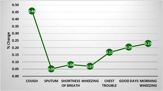
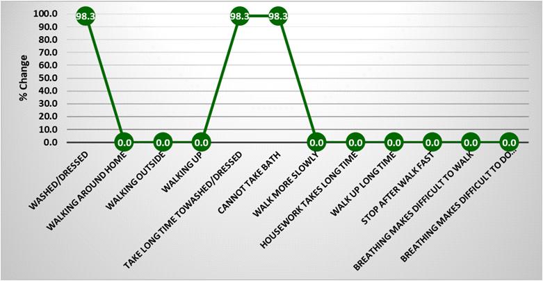
Association of post symtoms, activity, impact, and Quality of Life (QOL) was stated in Table 4. No post QOL domain was found to be significantly associated with age group (P=0.753). Activity and symptom domains were found significantly associated with gender (P<0.001). Activity and symptom domains were found significantly associated with occupation (P<0.001).
Association of age-group, gender and occupation with post Activities of Daily Living (ADL) scores:
As shown in Table 5, no ADL was found significantly associated with age-group (P=0.784). ADL domains was found associated with gender (P<0.001). Also, ADL domains was found to be significantly associated with occupation (P<0.001).
DISCUSSION
The present study demonstrated the role of care bundle in the assessment and management of patients with acute exacerbations of COPD. The use of a nursing care bundle is simple and easy to follow. Out
Vol 121, No 8, August 2023Journal of
the Indian Medical Association
Fig 1
Fig 2
Impact Pre test impactPost test impact%Z p-value MeanSDMeanSDChange Chest condition 35.386.1335.436.340.150.0001.000 Cough hurts 78.4814.4581.100.003.33 -1.4140.157 Cough makes tired 76.5514.091.3610.3998.22 -7.416 <0.001 Breathless when talk 84.500.001.4611.1098.28 -7.550 <0.001 Breathless when bend over 76.800.001.3210.0898.28 -7.550 <0.001 Breathing disturbs sleep 85.0615.6687.900.003.33 -1.4140.157 Exhausted daily 84.000.0084.000.000.000.0001.000 Breathing embarrassing in public 0.000.000.000.00-0.0001.000 Chest trouble is nuisance 0.000.000.000.00-0.0001.000 Get afraid or panic 87.700.001.5111.5298.28 -7.550 <0.001 Not in control of chest problem 90.100.001.5511.8398.28 -7.550 <0.001 Frail because of chest 87.0016.0189.900.003.33 -1.4140.157 Exercise is not safe 75.700.001.319.9498.28 -7.550 <0.001 Seems too much of an effort 81.7715.0584.500.003.33 -1.4140.157 Cannot go out for recreation 77.2314.2179.800.003.33 -1.4140.157 Cannot go out for shopping 79.6710.3781.000.001.67 -1.0000.317 Cannot do housework 77.8010.1379.100.001.67 -1.0000.317 Cannot move far 1.5211.9494.000.0061.00 -7.550 <0.001 Best describe chest affects 46.9714.1193.37 17.8098.79 -7.014 <0.001 61
Table 3 — Pre & post test comparison for impact scores
of the five elements in the nursing care bundle, all the elements that were correctly implemented, and after comparison of pre and post test the value was significant. The care package can improve the appropriateness of care and treatment, and allow for more effective long-term follow-up. In pre test assessment 15% patient perceives their health status very poor and 85% patient perceives poor, after intervention 3.1% patient perceive poor bur 96.9% patient perceives fair in their health status. The results of the study showed that after receiving the care bundle, the quality of life and activities of daily living increased significantly after the test.
A quasi-experiment showed that after choosing nursing interventions, COPD patients had differences in their health-related quality of life and activities of
daily living. The selected nursing interventions have effectively improved the health status, which is related to the quality of life and activities of daily living of COPD patients 6. An experimental study was carried out to study the effect of pursed lip breathing and diaphragm muscle breathing on the rehabilitation of COPD patients7. The author selected one group of pursed lip breathing and another group of diaphragm muscle breathing and concluded that diaphragm muscle breathing has both positive and positive effects but pursed lip breathing can improve people’s breathing. The use of nursing bundle improved the care of COPD worsening after implementation, and the average bundle score increased out of 10 (7 from 4.6)8. There was a significant reduction in the unnecessary use of intravenous corticosteroids from 60% to 32% and also a marked improvement in the use of oxygen therapy, with appropriate treatment increasing from 76% to 96%.These results were in line with our findings.
A study reported that the activity increased with a subsequent reduction from baseline to muscle activity at different time points. Rico-Blázquez M, et al conducted a study which shows that standardized intervention can enhance the quality of life9. Bendstrup KE, et al showed that economical, comprehensive, and well-tolerated rehabilitation programs can improve the daily life, quality of life and functional capacity of patients with moderate to severe COPD10. All the above studies have shown that the health-related quality of life and activities of daily living of COPD patients have improved after care.
Current study has some drawbacks. First, elderly more than 60 years old and critically ill patients were not enrolled in the present study. Second, the intervention was applied for limited period of time; however early intervention gives positive result. Third, sample size was low and study was single centre. Similar study with larger sample size with different care bundles are required.
The results of this research can be implemented in the fields of nursing, education, administration and research. This research provides evidence-based practice. Nurses can provide this kind of care package for COPD patients. The results of this study can be used to teach nursing students the appropriate teaching methods for
Vol 121, No 8, August 2023Journal of the Indian Medical Association
patient care packages in
COPD
Particulars Post symptomPost activityPost impact Post QOL score score scorescore Age group (years) (mean±SD) : 30-39 59.2768.1542.4753.32 40-49 56.34±13.18 68.1542.47 52.80±2.33 >5059.44±10.5868.77±3.8843.31±5.2553.98±5.28 Chi-square 0.570.490.490.57 P value 0.7530.7840.7840.753 Gender (mean±SD) : Female 79.64±28.80 80.25±17.1258.87±23.20 69.11±22.32 Male 57.87±9.58 68.1542.47 53.07±1.69 Mann-Whitney test 30.5028.0028.0030.50 P value 0.10 <0.001<0.001 0.10 Occupation (mean±SD) : Unemployed 79.64± 28.8080.25±17.12 58.87±23.2069.11±22.32 Unskilled 59.2768.1542.4753.32 Semi-skilled 59.2768.1542.4753.32 Skilled 60.34±1.86 68.1542.47 53.51± 0.33 Clerk/shopowner/ Farmer 57.50±10.54 68.1542.47 53.01± 1.86 Chi-square test 4.1728.0028.004.17 P value 0.383 <0.001<0.001 0.383 Table
Characteristics Post activityStatistical score (Mean) analysis Age group (Years) : 30-39 68.15 Chi-square 40-49 68.15 test=0.49 >5068.77 P=0.784 Gender : Male 80.25±17.12 Mann-Whitney test, Female 68.15 U=28P<0.001 Occupation : Unemployed 80.25±17.12 Unskilled 68.15 Chi-square Semi skilled 68.15 test=0.28 Skilled 68.15 P<0.001 Clerk/shop-owner/farmer68.15 62
Table 4 — Association of age, gender, and occupation with post Quality of Life (QOL) scores
5 — Association of Age, gender, and occupation with post activities of daily living scores
the classroom. A lot of work is needed in this area to educate the subjects of COPD on various aspects of nursing strategies. As a nurse researcher, nurses should encourage the improvement of medical care practices, so appropriate intervention measures need to be formulated, and the feasibility and effectiveness of their implementation should be required in in-depth research.
CONCLUSION
This study demonstrated that simple care bundle can improve care for COPD worsening. Using these nursing care bundle not only simplifies care, but also reduces the risk of adverse effects caused by unnecessary medications or treatments. The implementation of nursing care services may result in shorter hospital stays and lower readmission rates, but this requires further research. In addition care bundle can change the prescription of medicines, so it may have an impact on reducing unnecessary drug costs.
ACKNOWLEDGEMENTS
We thank to all the participants who had given their consent to participate in the study.
Ethics Approval : Study protocol was approved by Institutional Ethics Committee of the institution. Also, study was performed in the accordance of the approved protocol.
Consent to Participate : Informed written consent was taken from all the participants prior to enrolling in the study.
Conflicts of Interest: Authors declare no competing financial interest.
REFERENCES
1Black MJ, Hawkins J, Kane MA — Medical Surgical Nursing.8th edition. Missouri. Saunders Elsevier; 2005.
2Bunker C — Rosedale Text Book of Basic Nursing. 7th edition. Philadelphia. Lippincott Publishers. 1999.
3Pulaski LA,Taro ES — Linkman’s Core Principles and Practice of Medical Surgical Nursing.1st edition. India: WB Saunders Elsevier’s Publisher. 2010.
4Meguro M, Barley EA, Spencer S, Jones PW — Development and validation of an improved, COPD-specific version of the St. George Respiratory Questionnaire. Chest 2007; 132(2): 456-63.
5Gupta D, Agarwal R, Aggarwal AN, Maturu VN, Dhooria S, Prasad KT, et al — Guidelines for diagnosis and management of chronic obstructive pulmonary disease: Joint ICS/NCCP (I) recommendations. Lung India: official organ of Indian Chest Society 2013; 30(3): 228.
6Gopinath S, Malathi V, Quasi A — Experimental Study to Assess the Effectiveness of Selected Nursing Intervions on Health Related Quality Of Life and Activities Of Daily Living Among COPD Patients In Selected Teritary Hospital. IOSR Journal of Nursing and Health Science (IOSR-JNHS) 2015; 4(1): 1923.
7Parikh R, Shah TG, Tandon R — COPD exacerbation care bundle improves standard of care, length of stay, and readmission rates. International Journal of Chronic Obstructive Pulmonary Disease 2016; 11: 577.
8McCarthy C, Brennan JR, Brown L, Donaghy D, Jones P, Whelan R, et al — Use of a care bundle in the emergency department for acute exacerbations of chronic obstructive pulmonary disease: a feasibility study. International Journal of Chronic Obstructive Pulmonary Disease 2013; 8: 605.
9Rico-Blázquez M, Escortell-Mayor E, del-Cura-González I, Sanz-Cuesta T, Gallego-Berciano P, de Las Casas-Cámara G, et al — CuidaCare: effectiveness of a nursing intervention on the quality of life’s caregiver: cluster-randomized clinical trial. BMC Nursing 2014; 13(1): 1-0.
10Bendstrup KE, Ingemann Jensen J, Holm S, Bengtsson B — Out-patient rehabilitation improves activities of daily living, quality of life and exercise tolerance in chronic obstructive pulmonary disease. Eur Respir J 1997; 10(12): 2801-6.
Vol 121, No 8, August 2023Journal
of the Indian Medical Association
63 JIMA is now fully ONLINE and Publishes only ONLINE submitted Articles through https://onlinejima.com
Original Article
Study of Cardiac Dysfunction in Non-alcoholic Cirrhotic Patients in a Tertiary Care Hospital
Antarleena Ray1, Manjari Saha2, Tanmay Roy3, Jayanti Ray4, Udas Chandra Ghosh5, Soumya Sarathi Mondal5
Background : Cirrhotic Cardiomyopathy is one of the most important but apparently overlooked complications that significantly increase morbidity and mortality among patients of Liver Cirrhosis. Most studies are based on Alcoholic Cirrhosis and very limited data is available on non-alcoholic causes.
Aims and Objectives : The purpose of this study is to evaluate Cardiac Dysfunction among non-alcoholic cirrhotic patients and to correlate the severity of Cardiac Dysfunction with severity of Liver Cirrhosis assessed by Child Pugh scoring.
Methodology : In this cross sectional study 50 diagnosed patients of Chronic Liver Disease (CLD) were included who underwent Electrocardiography (ECG) and Echocardiography. Mean values of Left Ventricular Ejection Fraction (LVEF), Left Ventricular Internal Diameter-Diastolic [LVID (D)], E/A ratio and QTc interval were calculated and compared to Child Turcotte Pugh Class A, B and C to demonstrate correlation between Cardiac Dysfunction and severity of CLD.
Results : Significant association between the reduction of LVEF and Child Turcotte Pugh class A versus C (p<0.001) and B versus C (p<0.001) was found. Difference of mean LVID (D) among A versus C (p<0.001) and B versus C (p<0.001) was statistically significant. Relation between Diastolic Dysfunction and Child Pugh score in three groups was statistically significant, A versus C (p<0.001), B versus C (p<0.001).
Conclusion : Recognition of Cirrhotic Cardiomyopathy is suboptimal as it is mostly a subclinical cardiac failure. Significant Cardiac Dysfunction was found in our study as expressed by grade I Diastolic Dysfunction in 60% and grade II Diastolic Dysfunction in 20% of patients. Correlation between severity of Cardiac Dysfunction and severity of Liver Cirrhosis was also established.
[J Indian Med Assoc 2023; 121(8): 64-7]
Key words :Cirrhosis, Non-alcoholic Cirrhosis, Cardiac Dysfunction, Diastolic Dysfunction.
Cirrhosis of Liver is defined histopathologically as development of fibrosis to the point that there is architectural distortion with the formation of regenerative nodules. It has a variety of clinical manifestations and complications. Cirrhotic Cardiomyopathy is one of the most important but apparently overlooked complications that significantly increase morbidity and mortality among such patients.
In 2005, The World Congress of Gastroenterology in Montreal Working Group defined Cirrhosis associated cardiomyopathy as a form of chronic cardiac dysfunction in patients of cirrhosis, characterized by blunted contractile response to stress and/or altered
Department of General Medicine, Kolkata Medical College, Kolkata 700073
1MD, Postgraduate Trainee and Corresponding Author
2MD, Associate Professor
3MD, RMO cum Clinical Tutor, Department of General Medicine, Raiganj Government Medical College and Hospital, Raiganj 733134
4MD, Associate Professor, Department of General Medicine, College of Medicine & Sagore Dutta Hospital, Kolkata 700058
5MD, Professor
Received on : 13/07/2022
Accepted on : 21/01/2023
Editor's Comment :
Cirrhotic cardiomyopathy is mostly a subclinical heart failure that significantly increases morbidity in patients of liver cirrhosis.
There is very limited data available on cardiac dysfunction in non alcoholic cirrhosis.
ECG and echocardiography are the best screening methods, using which we found significant diastolic dysfunction correlating with the severity of liver cirrhosis.
diastolic relaxation with electrophysiological abnormalities in the absence of other known cardiac diseases.
Most studies are based on Alcoholic Cirrhosis and very limited data is available on non alcoholic causes. The purpose of this hospital based observational study is to evaluate prevalence of Cardiac Dysfunction among non-alcoholic cirrhotic patients and to correlate the severity of Cardiac Dysfunction with severity of Liver Cirrhosis assessed by Child Pugh scoring.
Pathophysiology :
•Systemic circulation in patients with decompensated Cirrhosis is hyper dynamic and
Vol 121, No 8, August 2023Journal of the Indian Medical Association 64
characterized by increased heart rate and cardiac output and decreased systemic vascular resistance with low-normal or decreased arterial blood pressure. The factors that increase the cardiac output are increased sympathetic nervous activity, increased preload and blood volume and the presence of arteriovenous communications. However, the Cardiac Stroke Index and Left Ventricular Ejection Fraction (LVEF) fall during exercise which indicates an abnormal ventricular response to an increased ventricular filling pressure.
•After exercise the LVEF increases significantly less in Cirrhosis patients as compared to matched controls. The reduced cardiac performance is probably caused by a combination of blunted heart rate response to exercise, reduced myocardial contractility and profound wasting of skeletal muscles with impaired extraction of peripheral oxygen. Reduced left ventricular end diastolic volume and LVEF after standing has also been noted which indicates an impaired myocardial response to erect posture.
•The fluidity of the Plasma membrane and the function of K and Ca ion channels have been shown to be impaired in Cirrhosis. Delayed repolarisation of cardiomyocytes secondary to defects in K+ channel function could lead to electrophysiological abnormalities in cardiac excitation. A prolonged QT interval (>440ms) has been previously described in Liver Disease leading to ventricular arrhythmias and sudden cardiac death.
•Response to Noradrenaline and other potent pressor substances such as Angiotensin II and Vasopressin is blunted in Cirrhosis patients.
Studies in animal models have shown down regulation with reduced beta adrenergic receptor density in cardiomyocytes and receptor desensitization. In non-alcoholic Cirrhosis cardio depressant substances are produced such as endotoxins, endothelins, bile acid and cardiac cytokines such as TNF Alfa and IL1b.
In its most benign form, Cirrhotic Cardiomyopathy manifests as subtle ECG and Echo abnormalities, most commonly as prolonged QT interval and Diastolic Dysfunction, but typically with preserved systolic function in the resting and pre transplant vasodilated state. At the extreme, Cirrhotic Cardiomyopathy progresses to fulminant Myocardial Failure under the hemodynamic stress of Liver Transplant Surgery, unmasked by the demands of bleeding, high doses of vasopressors, reversal of low to high afterload, withdrawal of cardio protective medications such as beta blockers and mineralocorticoid antagonists used
in End-stage Liver Disease (ESLD), and worsened by concomitant sepsis or Systemic Inflammatory Response Syndrome (SIRS).
MATERIALS AND METHODS
Study setting : Department of General Medicine at Kolkata Medical College, West Bengal, India
Study design : hospital based observational study
Time line: February 2016-January 2017
Definition of population : patients with Liver Cirrhosis attending Medicine OPD or admitted under the Department of Medicine
Sample size : 50
Sample design : all cirrhotic patients without having previous Cardiac Dysfunction attending Medicine OPD/admitted and given written informed consent will be made part of the study
Inclusion Criteria :
(1)Age 12-60
(2)Patient of CLD/Cirrhosis(documented by clinical/biochemical/radiological/histological criteria) who has no history of previous alcohol intake
Exclusion Criteria :
(1)Cirrhosis/CLD associated with alcohol intake
(2)Preexisting cardiac disease or obvious cardiac abnormality
(3)Preexisting Lung Disease or other systemic co morbidities affecting cardiac function
Parameters to be studied :
(1)Parameters to find out evidence of CLD : Clinical, Biochemical (Liver Function Tests), Radiological (USG) or Histopathological (Liver Biopsy).
(2)Parameters to find out Cardiac Dysfunction: Clinical, ECG, Echo.
Using basic 2D indices of Diastolic Dysfunction, pulsed wave Doppler at the mitral valve leaflet tips provides information about early and late diastolic filling in normal sinus rhythm with rapid passive filling (Ewave) followed by atrial contraction (A wave). Pulse wave Doppler estimates of Diastolic parameters can guide in categorizing patients on the spectrum of diastolic abnormalities using a validated grading system.
Study tools :
(1)Predesigned and pretested proforma.
(2)Tests to diagnose CLD (clinical, biochemical, Radiological and Histological).
(3)Tests to find out etiology of CLD[ Hepatitis B Surface Antigen(HBsAg), Hepatitis C Antibody(anti HCV), Anti Nuclear Antibody (ANA), serum ceruloplasmin, serum ferritin, anti-liver kidney
Vol 121, No 8, August 2023Journal
of the Indian Medical Association
65
microsomal antibody (anti LKM1), anti Smooth Muscle Antibody (anti SMA), anti soluble liver antigen (anti SLA antibodies)] according to clinical suggestion.
(4)Echo, ECG, Digital Chest X-ray.
(5)Tests to assess systemic disease status and co-morbidities (complete hemogram, lipid profile, Fasting Blood Sugar (FBS), Post Prandial Blood Sugar (PPBS), serum urea, serum creatinine, serum Na, serum K and other investigations as per clinical suggestion).
(6)Other relevant investigations to be done to rule out systemic conditions affecting cardiac function.
Diagnostic Criteria (Fig 1) :
Categorical data was analyzed by Chi square test and Cochran tests.
P value of <0.05 will be considered significant.
ANALYSIS AND RESULTS
In this cross sectional, observational, hospital based study- a total of 50 diagnosed patients of CLD were included. Patients having co morbid conditions that maybe associated with Cardiomyopathy such as Hypertension, Dyslipidemia and CKD- were excluded. Known cases of heart disease and pulmonary disease were excluded. Included patients underwent ECG and Echocardiography.

Child Turcotte Pugh class was calculated for each of the cases and was subdivided into 3 classes- A (56), B (7-9) and C (10-15). Mean values of Left Ventricular Ejection Fraction (LVEF), Left Ventricular Internal Diameter-Diastolic [LVID(D)], Left atrial diameter, E value, A value, E/A ratio and QTc interval were calculated. Mean values of these parameters were compared to Child Pugh class A, B and C to demonstrate whether there is any correlation between levels of these parameters and severity of CLD.
Out of 50 patients, 38 were Male and 12 were Females.
Median age of study was 44.50 ranging from 17-60 years.
42 patients had cryptogenic CLD, 5 had autoimmune Hepatitis and 3 had Chronic Hepatitis B contributing 84%, 10% and 6% to the etiology respectively (Tables 1-4).
Thus,
•Significant association between reduction of LVEF and Child Turcotte Pugh class A versus C (p<0.001) and B versus C (p<0.001) was found.
•Difference of mean LVID (D) among A versus C
Study Technique and Data Analysis :
Informed written consent was obtained from the patients and their relatives and the study protocol was approved by the Ethics Committee.
All data was tabulated in a master chart and analyzed by standard statistical procedures. Descriptive states expressed in terms of ratio, proportion, percentage for categorical data and in terms of mean, median, range for quantitative data.
Continuous variables are expressed as Mean and Standard Deviation (SD) and compared across the groups using MannWhitney U test/Kruskal Wallis Test as appropriate.
Vol 121, No 8, August 2023Journal
of the Indian Medical Association
Fig 1 — Diagnostic criteria for Cirrhotic Cardiomyopathy according to the World Gastroenterology Organization (Montreal 2005)
Child PughChild PughChild PughTotal Class AClass B Class C No Diastolic Dysfunction 6(60) 4(16.7)010(20) Diastolic Dysfunction Grade 1 4(40) 19(79.17)7(43.75)30(60) Diastolic Dysfunction Grade 2 01(4.17)9(56.25)10(20) Total10(100)24(100)16(100)50(100) A
B
versus C <0.001 Significant B versus C
Table 1 — Distribution Of Diastolic Dysfunction With Child Pugh Score
versus
0.034 Not SignificantA
<0.001 Significant
Child PughChild PughChild PughTotal Class AClass B Class C No QTc Prolongation 9(90) 22(91.6)11(68.75)43(86) QTc Prolongation 1(10) 2(8.3)5(31.25) 7(14) Total10(100)24(100)16(100)50(100)
C 0.210 Not Significant B versus C
66
Table 2 — Distribution Of QTc Prolongation With Child Pugh Score
A versus B 0.875 Not SignificantA versus
0.061 Not Significant
versus B 0.363 Not Significant
A versus C <0.001 Significant
B versus C <0.001 Significant
(p<0.001) and B versus C (p<0.001) was statistically significant.
•Relation between Diastolic Dysfunction and Child Pugh score in three groups was statistically significant, A versus C (p<0.001), B versus C (p<0.001).
•Association between QTc prolongation and Child Pugh score was not significant.
•No significant correlation between etiology of Cirrhosis and Cardiac Dysfunction was found.
SUMMARY AND CONCLUSION
Recognition of Cirrhotic Cardiomyopathy is suboptimal because of a lack of sensitive, noninvasive diagnostic tests. Similarly, management of Cirrhotic Cardiomyopathy is largely empirical because of a paucity of existing literature. It is a subclinical Cardiac Failure. The prevalence remains unknown mostly due to the disease being generally latent, overt Heart Failure is very rare. It is manifested only when the patient is subjected to stress such as exercise, drugs, hemorrhage, infections and surgery. Echocardiography allows the detection of LVDD with the E/A ratio being the best screening test to detect the condition. The severity of Cardiac Dysfunction may be a sensitive
marker for mortality. Treatment is non-specific but Orthotopic liver transplantation may normalize the Cardiac Function.
LIMITATIONS
Biggest limitation of this study is the small sample size. A large prospective and multicentric trial is necessary in this field.
Control population of healthy adults should have been incorporated to have a more reliable set of data. This study does not have a therapeutic arm. Whether available therapeutic options can modify Cirrhotic Cardiomyopathy or not should be properly studied.
REFERENCES
1Shorr E, Zweifach BW, Furchgott RF, Beaz S — Hepatorenal factors in circulatory homeostasis IV. Tissue origins of the vasotropic principles, VEM and VDM, which appear during evolution of hemorrhagic and tourniquet shock. 1951; 3: 4279.
2Lunseth JH, Olmstead EG, Abbound F — A study of heart disease in one hundred eight hospitalized patients dying with portal cirrhosis. Arch Intern Med 1958; 102: 405-13.
3Gould L, Schariff M, Zahir M, Lieto M, Di Lieto M — Cardiac haemodynamics in alcoholic patients with chronic liver disease and presystolic gallop. J Clin Invest 1969; 48: 860-8.
4Llach J,Gines P,Arroyo V — Prognostic value of arterial pressure, endogenous vasoactive system, and renal function in cirrhotic patients admitted to the hospital for the treatment of ascites. Gastroenterology 1988; 22: 88-95.
5Fernandez-Rodriguez CM, Prieto J, Zoyaya JM — Arteriovenous shunting, hemodynamic changes and renal sodium retention in liver cirrhosis. Gastroenterology 1993; 104: 1139-45
6Moller S,Wiinberg N,Henriksen JH — Noninvasive 24 hour ambulatory arterial blood pressure monitoring in cirrhosis. Hepatology 1995; 22: 88-95.
7Donovan CL, Marcovitz PA, Punch JD, Bach DS, Brown KA, Lucey MR — Two dimensional and dobutamine stress echocardiography in the preoperative assessment of patients with end stage liver disease prior to orthotopic liver transplantation. Transplantation 1996; 61: 1180-8.
8Pozzi M, Carugo S, Boardi G — Evidence of functional and structural cardiac abnormalities in cirrhotic patients with and without ascites. Hepatology 1997; 26: 1131-7
9Henriksen JH, Moller S, Ring Laesen H — The sympathetic nervous system in liver disease. J Hepatol 1998; 29: 328-41.
10Wong F, Liu P, Liily L, Bomzon A, Blendis L — Role of cardiac structural and functional abnormalities in the pathogenesis of hyperdynamic circulation and renal sodium retention in cirrhosis. Clin Sci (Lond) 1999; 97: 259-67.
Vol 121, No 8, August 2023Journal of
the Indian Medical Association
LVEF (%) Child Pugh Class A Mean55 Child Pugh Class A Standard Deviation 3.13 Child Pugh Class B Mean 53.33 Child Pugh Class B Standard Deviation 2.50 Child Pugh Class C Mean 46.19 Child Pugh Class C Standard Deviation 3.25 A versus B 0.136 Not Significant
versus C <0.001 Significant
versus C <0.001 Significant
Table 3 — Distribution of LVEF with Child Pugh Score
A
B
LVID (D) Child Pugh Class A Mean 44.90 Child Pugh Class A Standard Deviation 1.52 Child Pugh Class B Mean 45.50 Child Pugh Class B Standard Deviation 1.77 Child Pugh Class C Mean 54.44 Child Pugh Class C Standard Deviation 3.72 A
Table 4 — Distribution of LVID (D) with Child Pugh Score
67
Original Article
Summer Surge of Acute Appendicitis in Adults
Abdul Razack G S1, Harindranath H R2, Uday C3, Nashwath Kumar K H4
Background : The most common acute surgical condition of the abdomen is Appendicitis. There is growing evidence base to suggest the seasonal variation in causation of acute Appendicitis. Some studies have concluded that acute Appendicitis is a more common summer with reason still not clear. The rationale for our study is to determine whether there is seasonal association with the incidence of acute Appendicitis. The strong association if found will help in preplanning the management and preventing the incidence of severe complication of the disease.
Materials and Methods : This retrospective model was conducted from 01/05/2016 to 30/04/2021 in a patient who was diagnosed with acute Appendicitis. The total admission for acute Appendicitis were collated with calendar month to compare with seasonal variation. Then the number of cases of acute Appendicitis were segregated with respect to age and gender. Results were analysed in terms of number of cases and complications of acute Appendicitis in adults with respect to corresponding months in a year.
Results : During the observation period, 620 cases had been admitted with the diagnosis of acute appendicitis. The highest incidence of acute Appendicitis was seen in the 18-29 years age group (54%). In all age groups the incidence declined with age increment. A significant seasonal effect was also observed, the rate of acute Appendicitis was high in summer and was highest in the month of May (11.8%) and the lowest number of cases were in January (6%). The complication of acute Appendicitis was highest in the month of May (4.9%).
Conclusion : Acute Appendicitis and its complications is more common in the summer month and is maximum in the month of May. The clear association will help in preplanning the management and preventing the incidence of severe complication of the disease. [J Indian Med Assoc 2023; 121(8): 68-70]
Key words :Acute Appendicitis, Seasonal variation.
The most common acute surgical condition of the abdomen is appendicitis 1 The most common operation worldwide is Appendectomy2. The clinical syndrome which results in inflammation of vermiform appendix is called Appendicitis3. Among males and females there is equal distribution4
The causes of appendicitis are unclear and multifactorial. There are a variety of attributable causes like mechanical obstruction5, inadequate dietary fibre, smoking6 , air pollution and familial susceptibility7 There is growing evidence base to suggest the seasonal variation in causation of acute Appendicitis8.
Acute Appendicitis begins with the classical features of poorly localized colicky abdominal pain. The pain is first noticed in the periumbilical region with anorexia and nausea. Later due to involvement of somatic nerves because of irritation of parietal peritoneum pain gets shifted to the right iliac fossa. The cardinal features of acute Appendicitis is low grade pyrexia, localized abdominal tenderness, muscle guarding and rebound tenderness3
Department of General Surgery, Bangalore Medical College and Reearch Institute, Karnataka 560004
1MBBS, MS, Assistant Professor
2MBBS, MS, Professor
3MBBS, Postgraduate Student and Corresponding Author
4MBBS, Postgraduate Student
Received on : 03/12/2021
Accepted on : 14/04/2023
Editor's Comment :
In summer months, younger patients coming with pain abdomen and tenderness in right iliac fossa is more likely to be acute Appendicitis. Early preparedness and early intervention is necessary to avoid complications.
Variable results are obtained from various studies done to determine the seasonal variation of acute Appendicitis9 Some studies have concluded that acute Appendicitis is more common summer with reason still not clear. Most epidemiological studies of acute Appendicitis have been conducted and focused on western population10. The rationale for our study is to determine whether there is seasonal association with the incidence of acute Appendicitis. The strong association if found will help in preplanning the management and preventing the incidence of severe complication of the disease (Tables 1&2, Figs 1&2).
MATERIALS AND METHODS
This retrospective observational study was conducted at the Department of General Surgery, (Victoria Hospital and Bowring and Lady Curzon Hospital), Bangalore Medical College and Research Institute, Bengaluru, from 01/05/2016 to 30/04/2021. Ethical Committee and Medical records department permission was taken. Data was collected from the registers in the medical records department of both
Vol 121, No 8, August 2023Journal of the Indian Medical Association
68
hospitals. Registers include Surgery case completion register updated by General surgery residents, Anaesthesia case register updated by Anaesthesia residents, and Nurses case register updated by Floor nursing staff of the respective operation theatre complex. Inclusion criteria were set to identify the number of patients over the age of 18 years with an associated diagnosis of acute Appendicitis. There were total number of cases were further analysed to create anonymous database having admission number, age, sex, date of admission and final diagnosis. After entries were made of the patient’s data, date-resolved analysis was made. The total admission for acute Appendicitis were collated with calendar month to compare with seasonal variation. Then the number of cases of acute Appendicitis were segregated with respect to age and gender.
An expected number of cumulative admissions per month was established on the basis of a null hypothesis. This was taken to be that the incidence of acute Appendicitis is normally distributed across the year; that there is no seasonal variation over the 12 months and an equal number of episodes should be recorded in each month regardless of the analysed independent variables. An expected number of incident cases for each month was therefore one twelfth of the total figure, 620/12 = approximately 52 cases per calendar month.
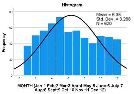
Based on the data collected from these registers, the total numbers of cases of acute Appendicitis were tabulated. The data collected was analysed. Results were analysed in terms of number of cases and complications of acute Appendicitis in adults with respect to corresponding months in a year. The study also analysed age wise distribution of acute Appendicitis in adults.
Statistical Analysis :
The summer surge in the incidence of acute Appendicitis in adults is presented as the number and percentage of total acute Appendicitis cases during the study period. Using the SPSS software package (SPSS 27.0 grad pack) the data were analysed. Count and frequency of patients in each gender, age groups and months were analysed. Comparisons between the frequencies were performed using Chi-square test.
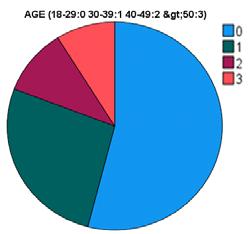
RESULTS
During the observation period, 620 cases had been admitted with the diagnosis of acute Appendicitis. Of these, 77.4% were males and 22.6% were females (Male: Female=3.424). The highest incidence of acute Appendicitis was seen in the 18-29 years age group (54%). The incidence was highest in males aged 1829 years. In all age groups the incidence declined with age increment. A significant seasonal effect was also observed, the rate of acute Appendicitis was high in summer and was highest in the month of May (11.8%) and the lowest number of cases were in January (6%). Age group of 18-29 years had the highest number of cases in the month of April (6.1%) and May (6.1%). There was significant association between complication of acute Appendicitis and age groups (p=0.040). The complication of acute Appendicitis was highest in the month of May (4.9%). The complication was lowest in the month of January (0.8%).
There was no significant association found between the seasonal changes and the sex of the patient of acute Appendicitis (p=0.783).
Vol 121, No 8, August 2023Journal of the Indian Medical Association
Month Abscess Mass PerforationTotal (Percentage) January0.30.20.30.8 February0.20.30.51.0 March0.50.31.01.8 April0.30.81.12.2 May0.81.82.34.9 June0.51.31.53.3 July0.60.60.61.8 August0.20.50.81.5 September0.50.00.51.0 October0.00.21.01.2 November0.20.30.00.5 December0.00.30.60.9 Total4.16.610.220.9
Table 2 — Month wise Distribution of Complication of Acute Appendicitis in Adults
Age (Years) Frequency Percentage 18-2933554 30-39166 26.8 40-4964 10.3 50 And Above558.9 Total620100
Table 1 — Age Wise Distribution of Acute Appendictis in Adults
Fig 2 — Age wise distribution of acute Appendicitis in adults
69
Fig 1 — Seasonal distribution of acute Appendicitis in adults
DISCUSSION
The demographic information from the contributing 620 admission episodes of acute Appendicitis appears consistent with that observed in prior work. In our study a significant seasonal effect was also observed, the rate of acute Appendicitis was high in summer and was highest in the month of May (11.8%) and the lowest number of cases were in January (6%). Age group of 18-29 years had the highest number of cases in the month of April (6.1%) and May (6.1%). The complication of acute Appendicitis was highest in the month of May (4.9%). The complication was lowest in the month of January (0.8%).
Many studies have shown seasonal variation in acute Appendicitis11-15 and a recent metanalysis, representing some of the strongest evidence for this, showed a peak incidence in the summer months for 9 of the 11 included studies16
A study was conducted by Babita, et al17 in India over from January 2003-July 2012. A total of 395 cases were included. The cases were maximum in the month of August and minimum was noticed in January17 while in our study maximum incidence was seen in May and minimum in January.
In a study conducted in by Rai, et al18 a clear seasonal variation was observed in the incidences of acute Appendicitis and Appendectomy for both genders. The peak incidence was seen in the summer season and decreased in the winter season. In addition, the present study observed a slight but consistent increase in the incidence of perforated appendicitis in the summer season18
A study was conducted in Taiwan by Lin, et al19 all in 2015. The highest incidence of Appendicitis was found in persons aged 15 to 29 years and males had higher rates of appendicitis than females at all ages except for 70 years and older. Appendicitis rates were 11.76 % higher in the summer than in the winter months19
In a study conducted at Finland by Imre, et al20 in various centres of Finland showed clear correlation of seasonality with acute Appendicitis with increased incidence in summer season.
CONCLUSION
Acute Appendicitis and its complications is more common in the summer month and is maximum in the month of May. The age group most commonly affected in adults is 18-29 years. The clear association will help in preplanning the management and preventing the incidence of severe complication of the disease. The seasonal variations of acute Appendicitis are consistent with other studies.
Conflicts of interest : There are no conflicts of interest.
REFERENCES
1Liu CD, McFadden DW — Acute abdomen and appendix. In:Greenfield LJ, Mulholland MW, Zelenock GB, Oldham KT,Lillemoe KD, editors.Surgery: scientific principles and practice. 3rd ed. Philadelphia: Lippincott-Raven; 1997. p. 1246e61
2Blomqvist P, Ljung H, Nyren O, Ekbom A — Appendectomy inSweden 1989e1993 assessed by the inpatient registry. J Clin Epidemiol 1998; 51(10): 859e65.
3Williams N, O’Connell PR, McCaskie AW — Bailey & Love’s Short Practice of Surgery, 27th Edition; Chapter 72 - The vermiform appendix. In: Bailey & Love’s Short Practice of Surgery 2018; p. 1329, 1302-03.
4Khattak S, Aslam S, Kamal A — Acute Appendicitis:An audit of 663 cases. Gomal J Med Sci 2010; 8: 209-11.
5Oldmeadow C, Wood I, Mengersen K, Visscher PM, Martin NG, Duffy DL — Investigation of the relationship between smoking and appendicitis in Australian twins. Ann Epidemiol 2008; 18: 631-6. doi:10.1016/j.annepidem.2008.04.004.
6Kaplan GG, Dixon E, Panaccione R, Fong A, Chen L, Szyszkowicz M, et al — Effect of ambient air pollution on the incidence of appendicitis. CMAJ2009; 181: 591-7. doi:10.1503/ cmaj.082068.
7Ergul E — Heredity and familial tendency of acute appendicitis. Scand J Surg 2007; 96: 290-2. doi:10.1177/ 145749690709600405.
8York TJ — Seasonal and climatic variation in the incidence of adult acute appendicitis: a seven year longitudinal analysis. BMC Emerg Med 2020; 20(1): 24.
9Noudeh YJ, Sadigh N, Ahmadnia AY — Epidemiologic features, seasonal variations and false positive rate of acute appendicitis in Shahr-e-Rey, Tehran. IntJSurg 2007; 5: 95-8. doi:10.1016/j.ijsu.2006.03.009.
10Rai R, D’Souza RC VV, Sudarshan SH, PSA, Pai JR, et al — An Evaluation of the Seasonal Variation in Acute Appendicitis. J Evol Med Dental Sci 2014; 3: 257-60. doi:10.14260/jemds/ 2014/1818.
11Luckmann R, Davis P — The epidemiology of acute appendicitis in California.Epidemiology 1991; 2(5): 323-30.
12Körner H, Söndenaa K, Söreide J, Andersen E, Nysted A, Lende T, et al — Incidence of acute nonperforated and perforated appendicitis: age-specific and sex-specific analysis. World J Surg 1997; 21(3): 313-7.
13Ahmed W, Akhtar M, Khan S — Seasonal variation of acute appendicitis. Pakistan J Med Sci 2018; 34(3): 564-7.
14Ilves I, Paajanen H, Herzig K, Fagerström A, Miettinen P — Changing incidence of acute appendicitis and nonspecific abdominal pain between 1987 and 2007 in Finland. World J Surg 2011; 35(4): 731-8.
15Stein G, Rath-Wolfson L, Zeidman A, Atar E, Marcus O, Joubran S, et al — Sex differences in the epidemiology, seasonal variation, and trends in the management of patients with acute appendicitis. Langenbeck’s Arch Surg 2012; 397(7): 1087-92.
16Fares A — Summer appendicitis. Ann Med Health Sci Res 2014; 4(1): 18.
17Jangra B, Jangra M, Rattan KN, Kadian Y — Seasonal and day of week variations in acute appendicitis in north Indian children. J Indian Assoc Pediatr Surg 2013; 18(1): 42.
18Rai R, D’Souza RC, Vijin, Sudarshan SH, Aithala, Pai. J R, etal An evaluation of the seasonal variation in acute appendicitis. J Evol Med Dent Sci 2014; 3(2): 257-60.
19Lin KB, Lai KR, Yang NP, Chan CL, Liu YH, Pan RH, et al — Epidemiology and socioeconomic features of appendicitis in Taiwan: a 12-year population-based study. World J Emerg Surg 2015; 10: 42. doi: 420.1186/s13017-015-0036-3.
20Ilves I, Fagerström A, Herzig KH, Juvonen P, Miettinen P, Paajanen H — Seasonal variations of acute appendicitis and nonspecific abdominal pain in Finland. World J Gastroenterol 2014; 20: 4037-42. doi: 10.3748/wjg.v20. i14.4037.
Vol 121, No 8, August 2023Journal
of the Indian Medical Association
70
Original Article
Post Mortem Renal Pathological Changes in Snake bite Patients and Association with Clinical Features
Pinaki Mukhopadhyay1, Joydeep Khan2, Piyali Banerjee3, Palas Bhattacharya4, Manik Kataruka5, Suparna Datta6
Snake bite is very common in rural India and incidence of renal failure and death following snake bite is high. We have studied the different histopathological changes in renal tissue in deceased patient following snake bite. Acute interstitial nephritis was the most common histological feature followed by acute tubular necrosis. Presence of DIC was most commonly associated with acute tubular necrosis.
[J Indian Med Assoc 2023; 121(8): 71-4]
Key words :Snake bite, Post-mortem, Renal histology.
Snakes have always managed to grab human attention and being an object of fear and veneration since historic civilizations. Snake bite is a common medical emergency and an occupational hazard, more so in tropical India, where farming is a major source of employment1. India has the highest mortality in the world, nearly 35000-50000 per year2. The World Health Organization (WHO) has estimated that nearly 1,25,000 deaths occur among 2,50,000 poisonous Snake bites worldwide every year, of which India accounts for 10,000 deaths1. Snake bite is a real problem in rural and urban India as mainly involve healthy, young, working population in their working area. There are nearly 3000 species of snake are known worldwide, of which nearly 600 are conserved poisonous. Snake bite results in multisystem involvement including kidneys. Renal manifestation ranges from AKI, Thrombotic Microangiopathy to Chronic kidney disease. Renal failure is mostly associated with Russell's Viper and E Carinatus bites (13-32%) 3 . Several mechanisms including
Department of Nephrology, Sir Nil Ratan Sircar Medical College & Hospital, Kolkata 700014
1MBBS, DCH, MD, DM (Nephrology), Professor & Head
2MBBS, MD (FSM), Assistant Professor, Department of Forensic Medicine and State Medicine, Santiniketan Medical College, Bolpur, West Bengal 731204
3DA, MD (Anaesthesia), Medical Officer, Lady Dufferin Victoria Hospital, Kolkata 700009
4MBBS, MD (Pathology), Associate Professor, Department of Pathology, Sir Nil Ratan Sircar Medical College & Hospital, Kolkata 700014
5MBBS, MD (Paediatric Medicine), DM (Nephrology), Assistant Professor and Corresponding Author
6MBBS, MD (FSM), Professor and Director, State Institute of Health and Family Welfare, West Bengal
Received on : 02/08/2022
Accepted on : 18/09/2022
Editor's Comment :
Post mortem Renal histopathology following Snake bite AKI varies. Though different series showed ATN as commonest, our series AIN predominant and DIC and shock was the surrogate clinical Predictor.
haemorrhage, hypertension, haemolysis, haemoglobinuria, rhabdomolysis and DIC as well as the direct effect of the venom have been incriminated in the pathogenesis of Snake bite related nephropathy. Renal replacement therapy, bite to needle time, hospital stay, bleeding manifestations, shock and DIC, number of vials AVS are known factors affecting the outcome in Snake bite patients. This study has been done to evaluate the different renal histopathological changes on Post mortem and associated clinical findings in Snake bites patients who had renal failure at presentation.
AIMS AND OBJECTIVE
The present study has been done to evaluate the surrogate predictors of mortality and the spectrum of histopathological changes of Kidney in death due to venomous snake bite.
MATERIALS AND METHODS
The study was conducted on death due to venomous Snake bite at Hospital ward of NRS Medical College & Hospital and cases sent for medicolegal autopsy at the hospital mortuary of l during the period September, 2017 to August, 2018. It was a Descriptive, Observational, Cross-sectional, Hospital based study. Any patient with previous history of renal dysfunction were excluded from the study. Informed consent had been taken from the legal representative of the deceased. Kidneys were preserved in 10% formalin and histopathological studies has been done by with
Vol 121, No 8, August 2023Journal of the Indian Medical Association 71
Hematoxylin and Eosin (H&E) and Periodic Acid Schiff (PAS) stains. All the clinical and laboratory findings were noted in a pre-specified proforma and Post mortem findings were also studied. Gross examination and histopathological findings of kidneys in Post mortem period were recorded and all data were analysed in SPSS software version 16 (SPSS, Chicago, IL, United States).
Inclusion Criteria :
All dead bodies with history of death following a venomous Snake bite.
Exclusion Criteria :
Decomposed bodies.
History of renal disease prior to Snake bite. Doubtful cases of Snake bite.
OBSERVATIONS
Total 49 patients were analysed. Out of 49 cases, 29 patients were admitted in NRS Medical College, while 20 were brought from outside. Among these patients, 34 (69.4%) were male. Most of the patients are in age group of 20 to 30 years (26.5%) followed by 10 to 20 years (24.5%). There were no patients below 10 years and above 70 years. Most of the snake bite cases occurred between June to October (n=37,75.5%). Highest number (36.7%) of Snake bite occurred between 6am to 12pm, most being between 6am to 6:30am, followed by those between 6pm to 12midnight (28.6%). Highest number of bite were in the lower limb (91.8%), followed by upper limb (6.1%) and (2.0%) in the abdomen. Out of 49 Snake bite, 47 involved vasculotoxic snake, while 2 were involved neurotoxic snake. 24.5% of victims received Anti Snake Venom (AVS) between 2 to 3 hours. The maximum time to receive AVS was between 10 to 11 hours Post Snake bite. Most of the patients who succumb to death, were within 1 to 3 day of hospitalization. Longest stay to hospital before death was 29 days.
Among clinical manifestation, shock needing vassopressor was present in 57% of patients, bleeding manifestation in 73.5% cases and DIC was present in 47%.
Most of the patients (73.5%) does not required Haemodialysis (HD) before death and rest received haemodialysis varying from 1 to 5 sessions.
Gross examination and histopathology of kidney performed in all cases in Post mortem. On gross examination, congestion with diffuse haemorrhage over kidney surface was the most common Post mortem finding (51%) followed by congestion with focal medullary haemorrhage (36.7%), congestion with patchy cortico- medullary haemorrhage (8.2%) and only
external congestion in 4.1%. Two patients with neurotoxic bite had shown only external renal congestion (Table 1).
On histopathology, interstitial inflammation was found in all the renal biopsy followed by acute tubular necrosis (57.1%), thrombotic microangiopathy in 14.3% and acute cortical necrosis in 8.2% cases (Tables 2&3).
On multivariate analysis, presence of shock and DIC was significantly associated with acute tubular necrosis. Acute cortical necrosis was positively associated with DIC and thrombotic microangiopathy was found to be significantly associated with presence of shock.
Renal Histopathological SurrogateP Value Findings Predictors(2-sided)
Tubular NecrosisBite to Needle Time0.020
Number of HD0.010
Shock0.000
DIC0.001
Cortical NecrosisBite to Needle Time0.004
Number of HDNS ShockNS
DIC0.026
Thrombi In Blood Bite To Needle Time0.030
Vessels
Number of HD0.038
Shock0.013
HD- Haemodialysis, DIC- Disseminated Intravascular coagulation, NS-Non Signifcant
Table
Renal Histopathological SurrogateP Value Findings Predictors(2-sided)
Tubular NecrosisBite to Needle Time0.206
Number of HD0.822
Shock0.000
DIC0.000
Cortical NecrosisBite to Needle Time0.906
Number of HD 0.311
Shock0.073
DIC0.026
Thrombi In Blood Bite To Needle Time0.153
Vessels
Number of HD0.219
Shock0.013
DIC0.820
HD- Haemodialysis, DIC- Disseminated Intravascular coagulation
Vol 121, No 8, August 2023Journal
of the Indian Medical Association
Histopathology Number Percentage Interstitial Inflammation49 100.0 Tubular Necrosis28 57.1 Cortical Necrosis48.2 Thrombi In Blood Vessels7 14.3
Table 1 — Histopathological Findings In Kidney
Table 2 — Correlation Between Risk Factors And Renal Histopathologial Findings (Chi Square Test)
3 — Multivariate Analysis of Risk Factors and Renal Histopathologial Findings
72
DISCUSSION
The present study was descriptive, cross sectional, hospital and mortuary based study, conducted at NRS Medical College & Hospital (NRSMC&H) during September 2017 to August 2018 and included cases which fulfilled the selection criteria. During this period of 1 year, there were 49 cases of death due to Snake bite, comprising 2.02% of all the autopsies done at NRSMC&H morgue.
Snake bite poisoning is a known problem in rural India. Most of the Snake bite cases are found during monsoon season4. In our study, 75.5% cases occurred in monsoon season. Most of the victims in our study were male. In the present study, the population comprises of 34(69.4%) males and 15(30.6%) females with a sex ratio of 2.26:1 in the study group. The probable reason for male predominance is males are more involved in outdoor activities compared to females. Similar results were seen from other studies5-7 in India except studies from hilly areas were the main workforce is female8,9. Mean age of subjects was 44.62 years with a range between 14 to 68 years consistent with other studies from India5,7,9
Snake venom causes damage to multiple organs in the body. Kidney being one of the most vascular tissue in the body, it receives a significant burden of venom, resulting in damage of kidney tissue, leading to Acute Kidney Injury (AKI). The exact pathogenesis of AKI following Snake bite is not well established, due to the lack of a reproducible animal model. The factors that may contribute are direct cytotoxicity, bleeding, hypotension, circulatory collapse, Intravascular haemolysis, disseminated intravascular coagulation and Micro-angiopathic Haemolytic Anaemia (MAHA). Doing renal biopsy is the most definite method to know the exact pathogenesis of renal failure. But doing renal biopsy may not always possible due to bleeding manifestations and co morbidity in Snake bite patients. So Post mortem renal biopsy gives us an insight in the renal pathology and possible pathogenesis. Incidence of AKI post Snake bite, has been reported to be 8-45% in different studies10-13. This study being primarily a Post mortem study, all of deceased had some degree of renal failure. During hospital stay, 26.5% patients received haemodialysis. Earlier studies had shown much higher requirement of dialysis in snake bite AKI patients ranging from 45% to 90.9%8. This low number of patients requiring dialysis may be explained by the fact that most of the patients in our study were too sick to initiate haemodialysis or died before dialysis can be initiated. Probably the patients who received
haemodialysis has better outcome and survivability, and so does not qualify for our study. Similarly, the patients in our study had much higher incidence of shock (57%) and DIC (73.5%) compared to older studies. In the study by Chugh, et al10 incidence of shock was 16% and DIC 43%. In the study by Athappan, et al14 incidence of shock and DIC were 17.7% and 27.8% respectively. Study by Dharod, et al13 had shown incidence of shock and DIC were 19.5% and 55.2% respectively.
On Post mortem examination, congestion with diffuse haemorrhage of the kidney parenchyma was the most common (50.2%) feature in our patients followed by focal medullary haemorrhage and patchy cortico medullary haemorrhage. As most of the studies are Pre-mortality in nature, gross description of kidney was absent. Only one study by Pal, et al15 described diffuse haemorrhage to be the most common finding. Among the histo-pathological findings, all patient’s biopsies showed acute interstitial inflammation (AIN) followed by Acute Tubular Necrosis (ATN) in 57% cases. Thrombotic microangiopathy was present in 14.3% and diffuse cortical necrosis in 8.2% cases. Neurotoxic cases showed only congestion & inflammation with no haemorrhagic features. Chugh, et al10 had reported ATN in 73% cases and cortical necrosis in 18% cases. In the study by Vikrant, et al8, 91% has ATN followed by AIN in 41% patients. In a study from Pakistan by Naqvi, et al16, ATN and cortical necrosis were most common finding. Pal, et al15 had reported ATN in 100% biopsies, cortical necrosis in 25% cases and proliferative glomerular changes in 1 case. Sathish, et al17 had reported interstitial congestion as most common finding (65.8%) followed by ATN in 55.3% cases and glomerular changes in 4.3% cases. Mittal BV18 in his study has shown cortical necrosis as predominant finding in 60.9% cases and ATN in 12.1% cases.
In our study, on multivariate analysis, presence of shock and DIC was significantly associated with acute tubular necrosis. Acute cortical necrosis was positively associated with DIC and thromotic microangiopathy and cortical necrosis was found to be significantly associated with presence of shock. Number of haemodialysis and bite to needle time had not been found to be associated with renal histo-pathological outcome.
Hypotension is the most common cause resulting in ATN and ACN. Acute tubular necrosis is reversible in most of the cases, while acute cortical necrosis results in residual renal damage and patient may need lifelong renal replacement therapy. Acute interstitial inflammation is usually secondary to direct venom
Vol 121, No 8, August 2023Journal
of the Indian Medical Association
73
of the Indian Medical Association
toxicity and cytokine activation 19 . Thrombotic microangiopathy is now a more and more getting recognised in Snake bite patients, characterised by thrombocytopenia and renal failure. Pathogenesis of thrombotic microangiopathy is supposed to be secondary to direct endovascular damage by venom or secondary to cytokine reaction Post envenomation.
CONCLUSION
Our study is one of the largest study including 49 post Snake bite victims with detailed pathological analysis of renal tissue. Contrast to previous studies, acute interstitial inflammation was the most common finding followed by acute tubular necrosis. Presence of shock and DIC was associated with poorer histological description.
Limitation : Being a Post mortem study, detailed clinical evaluation was not possible in all cases. We have to rely on documentation in in-patients records and history given by the patient relatives. Victim bodies, which were referred from other centre had poor clinical data. So detail renal parameter study and their association with pathological finding was not possible.
Conflicts of Interest : The authors declare no conflict of interest.
REFERENCES
1Kasturiratne A, Wickremasinghe AR, de Silva N, Gunawardena NK, Pathmeswaran A, Premaratna R, etal—The global burden of snakebite: a literature analysis and modelling based on regional estimates of envenoming and deaths. PLoS Med 2008; 5: e218 [PMID: 18986210 DOI: 10.1371/ journal.pmed.0050218]
2Mohapatra B, Warrell DA, Suraweera W, Bhatia P, Dhingra N, Jotkar RM, et al — Snakebite mortality in India: a nationally representative mortality survey. PLoSNegl Trop Dis 2011; 5: e1018 [PMID: 21532748 DOI: 10.1371/journal.pntd.0001018]
3Mulay DV, Kulkarni VA, Kulkarni SG, Kulkarni ND, Jaju RB — Clinical profile of snakebite at SRTR Medical College Hospital, Ambajogai (Maharashtra). Indian Medical Gazette 1986; 131: 363-6.
4Ahuja ML, Singh G — Snake bite in India. Indian J Med Res 1954; 42: 661-686 [PMID: 13232717]
5Hati AK, Mandal M, Mukherjee H. Epidemiology of Snakebite in the district of Burdwan. J Indian Med Assoc 1992; 90: 1457.
6Suchitra N — Snakebite Envenoming in Kerala, South India: Clinical profile and factors involved in adverse outcomes. EMJ 2008; 25: 200-4.
7Kulkarni M Ances S — Snake venom Poisons, experience with 633. Indian Paed 1994; 31: 1239-43.
8Vikrant S, Jaryal A, Parashar A — Clinicopathological spectrum of snake bite-induced acute kidney injury from India. World J Nephrol 2017; 6(3): 150-61.
9Monterio NP, Kanchan T, Bhagavath P, Pradeep Kumar G — Epidemiology of Cobra bite in Manipal, Southern India. JIndian Acad Forensic Med 2010; 32(3): 224-27.
10Chugh KS — Snake-bite-induced acute renal failure in India. Kidney Int 1989; 35: 891-907.
11Pinho FM, Zanetta DM, Burdmann EA — Acute renal failure after Crotalusdurissus snakebite: a prospective survey on 100 patients. Kidney Int 2005; 67: 659-667.
12Harshavardhan L, Lokesh AJ, Tejeshwari HL, Halesha BR, Siddharama SM — A study on the acute kidney injury in snake bite victims in a tertiary care centre. J ClinDiagn Res 2013; 7: 853-856.
13Dharod MV, Patil TB, Deshpande AS, Gulhane RV, Patil MB, Bansod YV — Clinical predictors of acute kidney injury following snake bite envenomation. N Am J Med Sci 2013; 5: 594-9.
14Athappan G, Balaji MV, Navaneethan U, Thirumalikolundusubramanian P — Acute renal failure in snake envenomation: a large prospective study. Saudi J Kidney Dis Transpl 2008; 19: 404-10.
15Pal M, Maiti AK, Roychowdhury UB, Basak S, Sukul B — Renal PathologicalChanges in Poisonous Snake Bite: J Indian Acad Forensic Med 2010; 32(1): 19-21.
16Naqvi R — Snake-bite-induced Acute Kidney Injury. J Coll Physicians Surg Pak 2016; 26: 517-20.
17Sathish K, Shaha KK, Patra AP — Histopathological profile of fatal snake bite autopsy cases in a tertiary care center in South India. Egypt J Forensic Sci 2021; 11: 3.
18Mittal BV — Acute renal failure following poisonous snake bite. J Postgrad Med 1994; 40: 123.
19Priyamvada PS, Shankar V, Srinivas BH, Rajesh NG, Parameswaran S — Acute Interstitial Nephritis Following Snake Envenomation: A Single-Center Experience. Wilderness Environ Med 2016; 27: 302-6.
Vol 121, No 8, August
2023Journal
74
Original Article
Evaluation of Organoleptic Properties and Compliance with Marketed Cough Syrups : A Cross Sectional Consumer Survey in Indian Patients
Ketan K Mehta1, Akil Contractor2, Sanjiv Maniar3, Atul Mashru4, Manoj Kumar Singh5, Atul Gogia6, Rohan Mahajan7, Harsh Mittal8, Monil Yogesh Neena Gala9, Mayank R Dhore10, Snehal Muchhala11, Gauri Dhanaki12, Arti Sanghavi13, Rahul Rathod10, Bhavesh Kotak14
Background : With growing burden of respiratory symptoms, it is crucial to develop cough syrups with appealing organoleptic properties. This study investigated the consumer feedback on various organoleptic properties and patient compliance to eight leading marketed cough syrups prescribed in routine clinical practice in India.
Materials and Methods : In this cross-sectional 3-months survey, adult patients prescribed with leading cough syrups were enrolled from 8 sites. A survey questionnaire was administered to collect data on the color, taste, viscosity, mouth feel, flavor, aroma, and aftertaste of the syrups using Likert scale ranging from 1-5.The compliance with prescribed regimens were also assessed. AP<0.05 was considered statistically significant.
Results : In total, 240 participants were enrolled in this survey. The syrups were generally prescribed for dry cough, fever, headache, productive cough, shortness of breath, and sneezing. Brand C (Bromhexine+ Guaiphenesin+Terbutaline) was the most commonly prescribed cough syrups (n=47). A significant association was found between the dose frequency and duration of cough syrups across all groups (P=0.005 for both). Brand D (Ambroxol+Levosalbutamol+Guaiphenesin), Brand A (Chlorpheniramine Maleate+Dextromethorphan), Brand C (Bromhexine+Guaiphenesin+Terbutaline)received the highest responses for “I liked it somewhat” and “I liked it extremely” for different organoleptic features. 97.1% of patients had consumed the cough syrup were compliant.
Conclusions : We found that Brands A, B, C, D (marketed by Dr. Reddy’s laboratory Ltd.) were the most accepted and liked cough syrups compared to other 4 leading marketed cough syrup brands.A high percentage of patients (ie, 97.1%) adhered to the recommended dosage and duration.
[J Indian Med Assoc 2023; 121(8): 75-80]
Key words :Cough syrups, Dr Reddy’s laboratories, India, Organoleptic, Sensory properties.
The acceptability of cough syrups to the Indian patients not only depends on its efficacy, but also on organoleptic properties such as taste, smell, color, and texture. Pharmaceutical companies often focus
1MD, Health Harmony, Dattani Chambers, West Mumbai 400064
2MBBS, Sai Clinic, Railway colony, Santacruz, West Mumbai 400054
3MBBS, Dr. Sanjiv Maniar Clinic, Somwari bazar Malad, West Mumbai 400064
4MBBS, Mumbai Citi Clinic, Khetwali Girgaon, Mumbai 400004
5MD, Flores Hospital, Pratap Vihar, Ghaziabad, Uttar Pradesh 201009
6MBBS, DNB, Good Health Clinic, Rajouri Garden, New Delhi 110027
7MD, Mahajan Hospital, Uttam Nagar, New Delhi 110059
8MBBS, DNB, Panchsheel Hospital, Yamuna Vihar, Shahdara, Delhi 110053
9MD, Dr Reddy’s Laboratory Ltd, Hyderabad and Corresponding
Author
10MD, Dr Reddy’s Laboratory Ltd, Hyderabad
11MBBS, MBA, Dr Reddy’s Laboratory Ltd, Hyderabad
12MSc, PGDCR, Dr Reddy’s Laboratory Ltd, Hyderabad
13BHMS, PGD, Dr Reddy’s Laboratory Ltd, Hyderabad
14MS, FRCS, Dr Reddy’s Laboratory Ltd, Hyderabad
Received on : 10/06/2023
Accepted on : 06/07/2023
Editor's Comment : Organoleptic properties significantly affect patient compliance and overall effectiveness of cough syrups Pharmaceuticals prioritize developing cough syrups with attractive organoleptic properties.
Dr. Reddy’s Laboratory Ltd.s cough syrups (Brand A, Brand B, Brand C, and Brand D) were highly accepted and preferred, surpassing four other leading cough syrup brands in the market
on developing cough syrups with appealing organoleptic properties to improve patient compliance and ultimately, treatment outcomes. Several excipients such as colors, flavors and pleasant aroma are used to enhance the palatability of cough syrups as these can significantly affect a patient’s willingness to take it, hence, play a crucial role in producing overall effectiveness. Cough syrups have varying textures from thicker and more viscous to thinner and more liquidlike, which make them easier to pour and swallow1
Organoleptic properties are vital as they can affect a patient’s adherence to prescribed medication and improve patient complaince2-6 There have been clinical studies that suggest that organoleptic properties can
Vol 121, No 8, August 2023Journal of the Indian Medical Association
75
have a significant impact on patient compliance6.
We undertook a survey to understand consumer feedback upon organoleptic properties and compliance of various leading marketed cough syrups prescribed in routine clinical practice in India. Also, we compared consumer survey response to the various cough syrups based on their organoleptic properties.
MATERIALS AND METHODS
Eight leading marketed cough syrup Brands were evaluated in this cross-sectional, observational, multicenter consumer survey to provide an initial assessment of their organoleptic properties. Adult patients aged 18 years or older, who were prescribed leading cough syrups, willing to complete the survey and provide data disclosure consent, were enrolled from 8 study centers located across 2 cities in India (New Delhi and Mumbai). Patients with unsuitable medical histories were excluded from the survey.
The study was approved by an independent ethics committee via a letter dated 17 May 2022 and 22 June 2022 (CTRI/2022/06/043040). All activities related to the collection, access, processing, and transfer of protected health information or sensitive personal data were conducted in compliance with applicable regulations. Each patient was assigned a unique patient identifier.
Survey duration and data collection :
The survey had a duration of ~3 months, during which patients attended 2 visits to the site/clinic. A follow-up visit was conducted either on-site or by phone after 5 days of enrollment. Patients were contacted by phone to complete the organoleptic questionnaire, but if they visited the site/clinic for follow-up, they completed the questionnaire during their visit. At baseline, demographic data such as age, gender, city/state, height, weight, BMI, presenting symptoms, clinical signs, and diagnosis
were collected by investigators or study personnel, along with comorbid diseases and vitals such as pulse rate, oral body temperature, and respiratory rate. Patients were also asked about the Brand, dosage, and duration of the prescribed cough syrup, and any concomitant medication.
Survey questionnaire :
The survey questionnaire contained a Likert scale ranging from 1-5 for participants to respond to various organoleptic properties of the syrup. These included the color, taste, viscosity, mouth feel, flavor, aroma, and aftertaste of the syrup. The scoring was mapped against the following responses: “I dislike it extremely,” “I dislike it somewhat,” “Neutral,” “I like it somewhat,” and “I like it extremely.”
Statistical analysis
Statistical analyses were performed usingR software Version 4.1.2. Descriptive statistics, such as mean and Standard Deviation (SD) are used to present continuous data. Categorical data are summarized using counts. The survey questionnaire data were presented descriptively. Comparisons were made using the Chi square/ Fisher’s exact test and t–test for categorical and continuous variables respectively. The p-value less than 0.05 was considered statistically significant.
RESULTS
In total, 240 participants prescribed with 8 cough syrupswere enrolled. The details of ingredients for each cough syrup are listed in Fig 1. Of total participants, 122 (50.8%) were male and 118 (49.2%) were female. The mean ± SD age of patients was 45.52 ± 16.54 years and mean ± SD BMI was 26.60 ± 4.76 kg/m2
Table 1 summarizes the demography and clinical characteristics of patients as per cough syrups. The age, gender and BMI were comparable across all eight groups (P>0.05).
Vol 121, No 8, August 2023Journal
of the Indian Medical Association
Brand ABrand BBrand CBrand DBrand EBrand FBrand GBrand HP-value (n=28)(n=26)(n=47) (n=27) (n=28)(n=28)(n=29)(n=27) Age (years) 46.75±16.8142.35±17.6748.74±18.3145.63±17.4446.57±16.27 47.11±15.08 41.76±15.8442.89±14.01 0.6128 Gender, n (%) Female9 (32.1)18 (69.2)22 (46.8)14 (51.9)14 (50.0)14 (50.0)15 (51.7)12 (44.4)0.3371 Male19 (67.9)8 (30.8)25 (53.2)13 (48.1)14 (50.0)14 (50.0)14 (48.3)15 (55.6) BMI26.52±4.22 25.28 ± 4.8726.63 ±5.0528.37 ± 5.5827.12 ±4.4825.42 ±3.3726.23 ±3.7927.26 ±5.980.2955 Presenting Symptoms, n (%) Dry Cough26 (92.9)19 (73.1)21 (44.7)13 (48.1) 25 (89.3)23 (82.1)14 (48.3)8 (29.6)<0.0001 Fever10 (35.7)8 (30.8)17 (36.2)9 (33.3)13 (46.4)9 (32.1)14 (48.3)15 (55.6) Headache2 (7.1)6 (23.1)13 (27.7)5 (18.5)8 (28.6)7 (25.0)8 (27.6)5 (18.5) Productive Cough2 (7.1)5 (19.2)23 (48.9)15 (55.6)1 (3.6)5 (17.9)15 (51.7)19 (70.4) Shortness of breath04 (15.4)6 (12.8)4 (14.8)2 (7.1)4 (14.3)2 (6.9)1 (3.7) Sneezing9 (32.1)8 (30.8)16 (34.0)8 (29.6)8 (28.6)13 (46.4)7 (24.1)4 (14.8) 76
Table 1 — Summary of demographic characteristics and clinical characteristics as per cough syrups
The symptoms for which cough syrups were generally prescribed were fever, headache, dry and productive cough, shortness of breath, and sneezing. Brand C was mostly prescribed for productive cough (n=23; 48.9%), followed by dry cough (n=21; 44.7%). The other Brands were prescribed for both dry and productive cough (Fig 1).
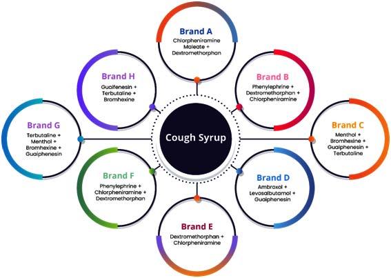
Brand C was the most prescribed cough syrup (n=47; 19.6%, males: 25 [53.2%] and females: 22 [46.8%]) followed by Brand G (n=29; 12.1%, males: 14 [48.3%] & females: 15 [51.7%]) and Brand A (n=28; 11.7%, males: 19 [67.9%] & females: 9 [32.1%].
No significant association was observed for medical history of patients across all cough syrup groups (P=0.988). Hypertension was the most frequently reported medical history across all the cough syrup groups. None of the participants taking Brand G had any reported medical history (Table 2).
A significant association was found between the dose frequency and duration of cough syrups across all groups (P=0.005 for both). The most frequently prescribed dose and frequency across all cough syrups was 1 TSF (Table Spoon Full) twice daily, with 17 patients (60.7%) receiving this dose for Brand E, followed by 16 patients (57.1%) for Brand Aand 15 patients (55.6%) for Brand H. Brand C was most commonly prescribed as 1 TSF thrice daily. The majority of cough syrups were prescribed for 5 days in over 50% of patients (Table 3).
The study evaluated patient preferences for different aspects of cough syrups, including aftertaste, aroma, color, flavor, mouthfeel, taste, and viscosity (Table 4). Brand D received the highest number of responses for “I liked it somewhat” and “I liked it extremely” regarding the aftertaste of the syrup (n=16; 59.3%), followed by Brand B (n=14; 53.8%), Brand A (n=12; 42.9%), and Brand C (n=20; 42.6%).
For aroma, Brand A (n=19; 67.9%) had the highest responses for “I liked it somewhat” and “I liked it extremely”, followed by Brand C (n=28; 59.5%), Brand D(n=16; 59.2%), and Brand B (n=14; 53.8%).
Brand C (n=19; 40.4%) received the highest number of responses for “I liked it somewhat” and “I liked it extremely” regarding the color of the syrup, followed by Brand A (n=10; 35.7%), and Brand B (n=7; 26.9%).
Brand A (n=15; 53.6%) and Brand C (n=24; 51.0%)
Brand B (N=26) :
Brand C (N=47) :
Brand D (N=27) :
Brand E (N=28) :
BrandF (N=28) :
Brand H (N=27) :
(14.3)
were the cough syrups which received the highest number of responses for “I liked it somewhat” and “I liked it extremely” regarding the flavor of the syrup, followed by Brand D (n=12; 44.4%) and Brand B (n=11;
Vol 121, No 8, August 2023Journal of the Indian Medical Association
(%)P
A (N=28) : Hypertension3 (10.7)0.9880 Hypothyroidism1
Diabetes
Table 2 — Medical history of patients by cough syrup
Cough syrups Medical Historyn
value Brand
(3.6)
Mellitus1 (3.6)
Hypertension1
(3.8)
Bronchitis1
Chronic kidney disease1
Cold and Fever1 (2.1) Coronary artery disease 1 (2.1) Hypothyroidism1 (2.1) Dyslipidemia1 (2.1) Hypertension5 (10.6) Type 2 diabetes mellitus3 (6.4) Thyroid1 (2.1) Osteopetrosis1 (2.1) Vertigo 1 (2.1)
(2.1)
(2.1)
Arthritis1
Hypertension6
(3.7)
(22.2)
Hypertension1
Diabetes1
(3.6)
(3.6)
Hypertension4
Hypertension3
(11.1) Type 2 diabetes mellitus1 (3.7)
Fig 1 — Compositions of cough syrups used in the study
77
42.3%). For remaining cough syrups, patients ranging from 32–39% had such responses.
For mouthfeel, Brand A (n=14; 50%) had the highest responses for “I liked it somewhat” and “I liked it extremely”, followed by Brand C (n=22; 46.8%), and Brand D (n=12; 44.4%). Brand E (n=5; 17.8%) and Brand G (n=5; 17.2%) had least number of responses for “I liked it somewhat” and “I liked it extremely” regarding the mouthfeel of the syrup. Brand C (n=26; 55.3%), Brand A (n=13; 46.4%) and Brand D (n=11; 40.7%) cough syrups received the highest number of responses for “I liked it somewhat” and “I liked it extremely” regarding the taste of the syrup.Brand G (n=5; 17.2%) received the least number of such responses for the taste.Brand C (n=23; 48.9%), and Brand D (n=13; 48.1%) cough syrups received the highest number of responses for “I liked it somewhat” and “I liked it extremely” regarding the viscosity of the syrup.
Overall, 233 (97.1%) patients had consumed the cough syrup in the same dosage and duration as prescribed and were compliant. Patients consuming Brands C, E, F, and H reported 100% compliance to the dosage and duration as prescribed (Fig 2).
DISCUSSION
The study is the first of its kind to assess the organoleptic features of 8 leading marketed cough syrups and compared them to understand the patients’ preference. A cough syrup with good organoleptic properties can improve patient compliance, satisfaction, and overall health outcomes.
If a medication has an unpleasant taste or odor, the patient may be less likely to comply with the prescribed regimen, leading to suboptimal
treatment outcomes. The aftertaste of cough syrups can be impacted by several factors, including the active ingredients, excipients, sweeteners, flavorings, and preservatives used in the formulation. Our results indicate that Brand D received the highest number of positive responses for aftertaste, followed by Brands B, A, and C. Most of these cough syrups have used mentholated flavored sugar base and menthol can improve the aftertaste of cough syrups by providing a refreshing sensation and masking unpleasant flavors, making the syrup more palatable and easier to swallow6. Flavored syrupy bases are often added to cough syrups to improve the aftertaste of the active ingredients. These bases typically contain sweeteners to make the syrup taste better. The addition of a syrupy base also helps to make the cough syrup thicker and more viscous, which can provide a more soothing effect on the throat5.
Our study demonstrated the highest positive responses for aroma for Brands A and C which could be attributed to the presence of menthol as one of the excipients. Brand C received the highest positive responses for color. Brand A and C had the highest
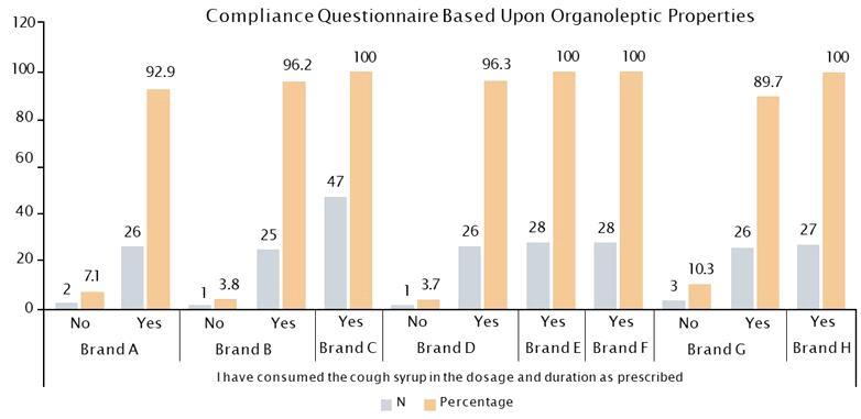
Vol 121, No 8, August 2023Journal of the Indian Medical Association
Brand ABrand BBrand CBrand D Brand EBrand FBrand GBrand HP-value (n=28)(n=26)(n=47) (n=27) (n=28)(n=28)(n=29)(n=27) Dose & Frequency, n (%) 1 TSF twice daily16 (57.1)11 (42.3)11 (23.4)11 (40.7)17 (60.7)9 (32.1)12 (41.4)15 (55.6)0.0052 1 TSF thrice daily5 (17.9)7 (26.9)22 (46.8)4 (14.8)04 (14.3)3 (10.3)5 (18.5) 2 TSF twice daily3 (10.7)5 (19.2)10 (21.3)9 (33.3)6 (21.4)9 (32.1)8 (27.6)3 (11.1) 2 TSF thricedaily 4(14.3)3 (11.5)4 (8.5)3 (11.1)5 (17.9)6 (21.4)6 (20.7)4 (14.8) Duration, n (%) 5 days14 (50.0)15 (57.7)34 (72.3)23 (85.2)15 (53.6)20 (71.4)18 (62.1)18 (66.7)0.0057 < 5 days7 (25.0)7 (26.9)6 (12.8)3 (11.1)8 (28.6)4 (14.3)9 (31.0)7 (25.9) > 5 days to < 10 days7 (25.0)4 (15.4)7 (14.9)1 (3.7)2 (7.1)4 (14.3)2 (6.9)2 (7.4) > 10 days----3 (10.7)--TSF,Table spoon full.
Table 3 — Dose frequency and duration of cough syrups
78
Fig 2 — Compliance Questionnaire Based on Organoleptic Properties – Overall
Vol 121, No 8, August 2023Journal
Table 4 — Organoleptic properties of cough
Cough SyrupQuestionI dislike itI dislike it NeutralI like itI like it extremely n (%) somewhat n (%)n (%) somewhat n (%)extremely n (%) Brand A(N=28)After Taste of the syrup04 (14.3)12 (42.9)8 (28.6)4 (14.3) Aroma of the syrup04 (14.3)5 (17.9)12 (42.9)7 (25.0) Color of the syrup2 (7.1)2 (7.1)14 (50.0)9 (32.1)1 (3.6) Flavour of the syrup2 (7.1)2 (7.1)9 (32.1)12 (42.9)3 (10.7) Mouth feel of the syrup03 (10.7)11 (39.3)13 (46.4)1 (3.6) Taste of the syrup03 (10.7)12 (42.9)11 (39.3)2 (7.1) Viscosity of the syrup04 (14.3)17 (60.7)7 (25.0)0 Brand B (N=26)After Taste of the syrup1 (3.8)2 (7.7)9 (34.6)9 (34.6)5 (19.2) Aroma of the syrup1 (3.8)2 (7.7)9 (34.6)7 (26.9)7 (26.9) Color of the syrup4 (15.4)2 (7.7)13 (50.0)5 (19.2)2 (7.7) Flavour of the syrup07 (26.9)8 (30.8)9 (34.6)2 (7.7) Mouth feel of the syrup07 (26.9)9 (34.6)8 (30.8)2 (7.7) Taste of the syrup2 (7.7)4 (15.4)10 (38.5)8 (30.8)2 (7.7) Viscosity of the syrup05 (19.2)11 (42.3)9 (34.6)1 (3.8) Brand C(N=47)After Taste of the syrup08 (17.0)19 (40.4)13 (27.7)7 (14.9) Aroma of the syrup06 (12.8)13 (27.7)16 (34.0)12 (25.5) Color of the syrup2 (4.3)3 (6.4)23 (48.9)11 (23.4)8 (17.0) Flavour of the syrup1 (2.1)6 (12.8)16 (34.0)16 (34.0)8 (17.0) Mouth feel of the syrup1 (2.1)5 (10.6)19 (40.4)16 (34.0)6 (12.8) Taste of the syrup1 (2.1)5 (10.6)15 (31.9)22 (46.8)4 (8.5) Viscosity of the syrup1 (2.1)4 (8.5)19 (40.4)19 (40.4)4 (8.5) Brand D (N=27)After Taste of the syrup06 (22.2)5 (18.5)14 (51.9)2 (7.4) Aroma of the syrup04 (14.8)7 (25.9)13 (48.1)3 (11.1) Color of the syrup4 (14.8)3 (11.1)16 (59.3)2 (7.4)2 (7.4) Flavour of the syrup03 (11.1)12 (44.4)8 (29.6)4 (14.8) Mouth feel of the syrup06 (22.2)9 (33.3)9 (33.3)3 (11.1) Taste of the syrup1 (3.7)3 (11.1)12 (44.4)8 (29.6)3 (11.1) Viscosity of the syrup02 (7.4)12 (44.4)11 (40.7)2 (7.4) Brand E(N=28)After Taste of the syrup6 (21.4)7 (25.0)7 (25.0)5 (17.9)3 (10.7) Aroma of the syrup2 (7.1)7 (25.0)9 (32.1)9 (32.1)1 (3.6) Color of the syrup4 (14.3)3 (10.7)14 (50.0)6 (21.4)1 (3.6) Flavour of the syrup1 (3.6)4 (14.3)12 (42.9)10 (35.7)1 (3.6) Mouth feel of the syrup2 (7.1)8 (28.6)13 (46.4)3 (10.7)2 (7.1) Taste of the syrup1 (3.6)8 (28.6)10 (35.7)9 (32.1)0 Viscosity of the syrup09 (32.1)13 (46.4)4 (14.3)2 (7.1) BrandF (N=28)After Taste of the syrup09 (32.1)6 (21.4)9 (32.1)4 (14.3) Aroma of the syrup1 (3.6)1 (3.6)13 (46.4)11 (39.3)2 (7.1) Color of the syrup5 (17.9)3 (10.7)13 (46.4)2 (7.1)4 (14.3) Flavour of the syrup2 (7.1)5 (17.9)12 (42.9)6 (21.4)3 (10.7) Mouth feel of the syrup05 (17.9)12 (42.9)10 (35.7)1 (3.6) Taste of the syrup3 (10.7)3 (10.7)13 (46.4)8 (28.6)0 Viscosity of the syrup011 (39.3)9 (32.1)8 (28.6)0 Brand G(N=29)After Taste of the syrup4 (13.8)8 (27.6)8 (27.6)6 (20.7)3 (10.3) Aroma of the syrup1 (3.4)2 (6.9)16 (55.2)5 (17.2)5 (17.2) Color of the syrup6 (20.7)2 (6.9)14 (48.3)5 (17.2)2 (6.9) Flavour of the syrup3 (10.3)1 (3.4)14 (48.3)11 (37.9)0 Mouth feel of the syrup2 (6.9)8 (27.6)14 (48.3)4 (13.8)1 (3.4) Taste of the syrup2 (6.9)10 (34.5)12 (41.4)5 (17.2)0 Viscosity of the syrup1 (3.4)9 (31.0)14 (48.3)5 (17.2)0 Brand H(N=27)After Taste of the syrup4 (14.8)6 (22.2)9 (33.3)7 (25.9)1 (3.7) Aroma of the syrup2 (7.4)8 (29.6)8 (29.6)6 (22.2)3 (11.1) Color of the syrup5 (18.5)2 (7.4)14 (51.9)4 (14.8)2 (7.4) Flavour of the syrup06 (22.2)10 (37.0)7 (25.9)3 (11.1) Mouth feel of the syrup1 (3.7)4 (14.8)12 (44.4)9 (33.3)1 (3.7) Taste of the syrup1 (3.7)7 (25.9)11 (40.7)7 (25.9)1 (3.7) Viscosity of the syrup2 (7.4)6 (22.2)11 (40.7)7 (25.9)1 (3.7) 79
of the Indian Medical Association
syrups
positive responses for flavor, and Brand A had the highest positive responses for mouthfeel. Brand C and D had the highest positive responses for taste and viscosity.
Menthol is a commonly used ingredient in cough syrups because it provides a cooling and soothing sensation in the throat and nasal passages. When combined with a flavored syrup base, it can improve the taste and odor of the cough syrup, making it more palatable for patients. In addition to menthol, some cough syrups, such as Brand C, contain Carmoisine. Carmoisine is a food coloring that is added to improve the color and appearance of the cough syrup. This can have a positive impact on patients, as colors are known to evoke emotions and feelings. For example, warm colors such as red and orange can evoke feelings of warmth and comfort, which may help patients feel more at ease when taking the cough syrup.7
The study found that a high percentage of patients, 97.1%, were adhering to the recommended dosage and duration of the cough syrup. This is a positive finding because it suggests that these patients are following the instructions provided by their healthcare provider, and are taking the cough syrup as intended. It’s worth noting that the study also found that the cough syrup had unsatisfactory organoleptic properties and despite this, the patients still adhered to the prescribed regimen. This indicates that the patients may be placing more importance on the effectiveness outcomes of the cough syrup rather than its sensory properties. Overall, the study suggests that patients are willing to follow the prescribed regimen for cough syrup, even if they find it unpleasant to take. This is an important finding for healthcare providers, as it highlights the importance of providing effective treatments that patients are willing to adhere to, even if they are not always palatable.
Our study had several limitations. Firstly, the study was observational in nature, which means that we did not control for other confounding factors that could have influenced the subject’s response. This limits the ability to establish a cause-and-effect relationship between the variables. It is important to acknowledge that subjective responses are subject to individual interpretation and may be influenced by various factors, such as personal beliefs and expectations. Lastly, the study sample was limited to patients from only two cities, which may affect the generalizability of the findings to other populations. Differences in demographics, cultural background, and healthcare practices between different regions can influence patient behavior.While the study provides valuable insights into patient adherence to cough syrup
regimens, further research is necessary to validate these findings.
CONCLUSION
In our study, we observed that Dr Reddy’s laboratories marketed syrups (Brands A, B, C and D) were the most accepted and liked cough syrups among the studied population as compared to other 4 leading marketed cough syrup brands. Overall, a high percentage of patients (ie, 97.1%) adhered to the recommended dosage and duration of the cough syrup, indicating that patients may prioritize the effectiveness outcomes of the cough syrup over its sensory properties.
Disclosures : Dr Monil, Dr Mayank, Dr Snehal, Gauri, Dr Arti, Dr Rahul, and Dr Bhavesh are employees of Dr Reddy’s laboratory Ltd. The other authors declare no conflict of interest
ACKNOWLEDGEMENTS
We would like to thank Dr Archana Karadkhele, Colette Pinto and Dr Amey Mane for being a part of the initial conceptualization of the study protocol. We thank NeoCrest Life Sciences Consulting (P) Ltd. for providing the medical writing support and IR Innovate Research Pvt Ltd for overall co-ordination and conduct of the study.The study and publication were financially supported by Dr Reddy’s Laboratories.
REFERENCES
1Eccles R — What is the Role of Over 100 Excipients in Over the Counter (OTC) Cough Medicines? Lung 2020; 198(5): 727-34.
2Kashyap B — Evaluation of Organoleptic Properties Preffered for Oral Dosage form by Students and Faculty Members of ADIT Campus (Gujarat Technological University/Sardar Patel University), New VV Nagar. Inventi Rapid: Pharmacy Practice, 2012(4): 1-3.
3Soares N, Mitchell R, McGoff T, Bailey T, Wellman GS — Taste Perceptions of Common Pediatric Antibiotic Suspensions and Associated Prescribing Patterns in Medical Residents. J Pediatr Pharmacol Ther 2022; 27(4): 316-23.
4Raji MIO, Ibikunle MT — Organoleptic Property and Bacteriological Analysis of Cough Syrups Sold in Sokoto Metropolis, North-Western Nigeria. Journal of Medical Sciences 2022; 22: 113-8.
5Sohi H, Sultana Y, Khar RK — Taste masking technologies in oral pharmaceuticals: recent developments and approaches. Drug Dev Ind Pharm 2004; 30(5): 429-48. doi: 10.1081/ddc120037477. PMID: 15244079.
6Sana S, Rajani A, Sumedha N, Mahesh B — Formulation and evaluation of taste masked oral suspension of dextromethorphan hydrobromide. IntJDrugDev&Res 2012; 4(2): 159-72.
7Biswal PK, Mishar MK, Bhadouriya AS, Yadav VK — An Updated Review On Colorants As The Pharmaceutical Excipients. International Journal of Pharmaceutical, Chemical & Biological Sciences 2015; 5(4).
Vol 121, No 8, August 2023Journal of the Indian Medical Association
80
Case Series
Gossypiboma : Word of Caution for an Unexpected Guest in the Abdomen — A Case Series and a Review of the Literature
Afzal Anees1, Yaqoob Hassan2, Shereen Fatima3, Surbhi Gupta3
Gossypiboma, an infrequent surgical complication, is defined as retained surgical sponge in the abdominal cavity following any operation. Aside from being medico-legal trouble for an operating surgeon, the implications for both the patient and the surgeon are grave. We present three cases of Gossypiboma admitted and managed over a period of 8 years. A 55 year- old female was referred for a mass in her right upper quadrant of abdomen that had been present for four months. She had surgical history of open cholecystectomy five months back in peripheral health care centre. On Laparotomy removal of sponge with primary repair of transected gastroduodenal junction was made (Billroth 1 procedure). A 42-year-old lady was referred for colicky pain in the abdomen and bleeding per rectum of 3 weeks duration. She had an open abdomen hysterectomy six months prior in a private hospital. Exploration revealed a walled off necrotic collection with spong in the pelvis and blow out of the rectosigmoid junction. Rectosigmoidectomy with primary anastomosis and spong removal was performed. A 43-year-old woman presented with a history of open cholecytectomy in private centre, presented with right upper abdominal pain and decreased appetite. Computed Tomography (CT) confirmed the presence of gossypiboma. The patient underwent exploration with Billroth 11 procedure. All the three patients are doing well and on regular follow up. Though rare, Gossypiboma should be included in the differential diagnosis of postoperative cases presenting as vague pain or chronic lump even years after the operation .The risk that every surgical procedure carries is inherent but Gossypiboma remains largely an easily preventable hazard. Strict adherence to the standard protocols of gauze count and better display of team work by everyone present in the operating room is important in decreasing the incidence.
[J Indian Med Assoc 2023; 121(8): 81-5]
Key words : Gossypiboma, Foreign body, Retained surgical mops.
Gossypiboma is the presence of cotton based foreign body that is in place of concealment following surgery. It is an uncommon but a grave surgical complication that has repercussions for both the patient as well the surgeon. The true incidence of Gossypiboma is almost certainly underestimated due to medicolegal implications, however it is reported that one in every 3000 to 5000 abdominal procedures will result in a retained surgical sponge 1. Retained foreign bodies (mops or gauzes) can be found in any body cavity (thorax, pelvis, pericardium), but they are most frequently discovered in the intra-peritoneal cavity2. Because of the wide range of non-specific symptoms associated with Gossypibomas, an attending surgeon may struggle to make a diagnosis. The clinical presentation can range from a subacute exudative reaction resulting in an abscess or a fistula formation to chronic aseptic fibrinous adhesions or granuloma formation. Gossypiboma can have
Department of Surgery, Jawaharlal Nehru Medical College Aligarh, Uttar Pradesh 202001
1MBBS, MS, FACS, FRCS, FICS, Professor and Chairman
2MBBS, MS, “Fellow of National Board (FNB), Minimal Access Surgery”, Department of General and Minimal Invasive Surgery, Sher-i-Kashmir Institute of Medical Sciences (SKIMS), Srinagar, Jammu and Kashmir 190011 and Corresponding Author
3MBBS, MS, Resident
Received on : 12/01/2022
Accepted on : 29/06/2023
Editor's Comment :
Gossypibomas are rare, largely asymptomatic, and difficult to diagnose. Therefore, a high index of suspicion is needed to make a diagnosis and prompt commencement of intervention. Although it is unlikely to be completely eradicated, the risk can essentially be avoided by following established gauze count practises in operating rooms.
disappointing, unfavorable and serious medicolegal repercussions for a surgeon, hence it should be avoided by performing adequate routine theatre swab counts before closing the abdomen. This article discusses the clinical presentation, diagnosis, treatment and complications of Gossypibomas, in addition to adding three cases to the existing literature. A brief review in the context is also present.
CASE 1
A 55-year-old female was referred to our tertiary care centre after experiencing a right upper quadrant mass, early satiety, and dull aching pain for four months. There was no history of constipation, loose stools, fever, rigors, or rectal bleeding; however, had an open cholecystectomy five months previously. The detailed surgical, medical, personal and family history was not clinically relevant. nothing significant. The patient was thoroughly examined, resuscitated and subjected to standard baseline investigations. On physical examination, the patient was
Vol 121, No 8, August 2023Journal of the Indian Medical Association 81
found to be haemodynamically stable, a pulse rate of 82 beats per minute, a blood pressure of 120/86 mm Hg, respiratory rate of 16 breaths per minute, temperature recorded was 98.80°F and saturation of 96 percent on room air. There was no pallor, icterus, cyanosis or pedal edema. The Respiratory and Cardiovascular examinations were unremarkable. The biochemical investigations and abdominal radiograph were normal. A surgical scar mark in the right subcostal region was discovered during an abdominal examination. A mobile 5x7cm lump was present in the right hypochondrium and epigastric region, adjacent to the scar mark. Ultrasonography revealed heterogenicity in the anterior abdominal wall. Computed Tomography with oral contrast revealed a walled-off lesion with admixed air and soft tissue particles abutting pancreas and compressing the second part of the duodenum, causing luminal narrowing,
however distal passage of contrast was present. During an exploratory laparotomy, walled-off infected collection with surgical sponge was discovered embedded in the anterior wall of 1st part of the Duodenum (Fig 1). The sponge that was embedded in the anterior wall of the first part of the duodenum was removed and the transected gastroduodenal junction was repaired (Billroth 1procedure). A feeding jejunostomy was performed to expedite enteral nutrition. The abdomen was closed back in layers and the patient was extubated and shifted to the postoperative care ward. The postoperative period was uneventful; liquid orals were introduced after 48 hours, followed by semisolids and solids. On the fourth postoperative day, the patient was discharged and was assigned to our Out Patient Department (OPD) for weekly follow-up. The patient is doing well and is being monitored on a regular basis.

CASE 2
A 42-year-old lady, P2L2 (Para 2 Live 2) was referred to our surgical clinic in view of recurrent colicky pain abdomen, constipation, mucous discharge and bleeding per rectum of 3 weeks duration. She had history of open abdomen hysterectomy done six months before at a
private hospital. The patient had history of lower abdominal discomfort in the postoperative period which was attributed to wound site pain by the attending doctor. Following discharge after hysterectomy patient had always experienced colicky pain abdomen relieved by simple over the counter medications. However since the

Vol 121, No 8, August 2023Journal of the Indian
Medical Association
Fig 1 — Contrast Enhanced Tomography Scan showing well defined walled off lesion (yellow arrow) with admixed air and soft tissue particle. Intra-operative photograph showing surgical mop (black arrow) and disrupted gastrodudonal junction. Diagrammatic representation showing surgical mop.
Fig 2 — Contrast Enhanced Tomography Scan showing a round well-defined soft-tissue mass containing an internal high-density area in the lower abdomen. Intra-operative picture showing retained sponge and disrupted recto-sigmoid junction.
82
3 weeks, patient had been concerned about the episodes of bleeding per rectum and difficulty in defecation. She was stable with pulse 86bpm, blood pressure 136/78 mmHg, respiratory rate 16 breaths/minute and Oxygen saturation 96% on room air. The physical examination was unremarkable except infra-umbilical surgical scar and tenderness was present. Digital rectal examination was unremarkable. Radiograph of abdomen revealed gaseous dilatation of large gut loops and an abdominopelvic ultrasound suggested thickened sigmoid wall and mild free fluid in the pelvis. Contrast enhanced Computed
Tomography scan revealed a cystic lesion of 3*4 cm with internal spongiform appearance abutting the sigmoid colon and surrounded by omentum. The patient was optimized and taken up for elective exploratory laparotomy as we did not have availability of Laparoscopy at that time. On exploration the spong was present in the pelvis with blow out of rectosigmoid junction (Fig 2). Localised faecal contamination was also present. Rectosigmoidectomy was done; continuity of remaining hollow viscera was restored with stapling device. Postoperatively, the patient recovered uneventfully and is on regular outpatient follow-up.
CASE 3
A 43 year old woman presented with a history of open cholecystectomy 3 months before in a private hospital and was admitted to our tertiary care institute in view of eight days history of pain in right upper abdomen, vomiting, nausea, decreased appetite and early satiety. Abdominal pain was continuous, non-radiating and relieved by medication. On general examination, she was stable, obese with a Body Mass Index (BMI) of 32. As regards to her vital signs, her Blood Pressure (BP) was 130/88 mmHg, respiratory rate of 18 breaths per minute and Oxygen saturation 98% on room air. Local per abdominal examination showed right sub-costal incision scar, and tender right upper abdomen. A plain abdominal radiograph was normal and abdomen-pelvic ultrasound suggested an echoic mass surrounded by irregular hyperechoic areas in right hypochondrium. In the abdominal Computed Tomography (CT) scan, a heterogeneous cystic soft tissue mass of 24*12 cm size was found in the right upper quadrant of abdomen and diagnosis of Gossypiboma was made. The patient was taken for exploratory laparotomy under General Anesthesia. Abdomen was opened with upper midline incision. A sponge was discovered between the duodenum and the stomach and the anterior wall of the Stomach and Duodenum was disrupted (Fig 3). The damaged and gangrenous portions were excised followed by Billroth - II Gastrojejunostomy and abdominal washes were given (Fig 3). A tube drain was kept in situ and abdomen was closed back in layers. Postoperative
period was uneventful, drain was removed on 3rd day, and patient was discharged on 5th day. Patient is doing well, and on regular follow-up.
DISCUSSION
Following surgery, leaving surgical sponge in the abdominal cavity is a serious but generally avoidable complication. Despite the fact that it is preventable, it still happens, causing mental anguish, embarrassment, humiliation, job loss, legal action and undesirable consequences for the operating surgeon, as well as exacerbating the patient's morbidity and death. These retained surgical sponge were named as Gossypiboma in 1978, a Latin term ‘Gossipium’ meaning cotton3,4 and the first case was reported by Wilson in 18845. Because of the concern of medico-legal implications, the exact incidence of retained foreign bodies is difficult to verify6 The current reported rate is 0.01 percent to 0.001 percent following surgery with Gossypiboma accounting for 80 percent of cases7
Pathologically, retained surgical mops cause two types of foreign body reactions; exudative inflammatory reactions with abscess formation and aseptic fibrinous responses with granuloma formation 5,8-10. The patients with Gossypiboma often remain asymptomatic or have non-specific symptoms and they may present immediately in postoperatively period or decades later11 A 66-year-old man with Gossypiboma was discovered after 24 years of Gastrectomy12. However, the signs and

Vol 121, No 8, August 2023Journal
of the Indian Medical Association
Fig 3 — Abdominal CT scan showing a heterogeneous cystic soft tissue mass in the right upper quadrant of abdomen. Intra-operative picture showing surgical sponge and disrupted gastro-duodenal junction.
83
symptoms depend upon the location, duration and body response to the retained foreign body. All postoperative patients who present with non-specific symptoms such as abdominal pain, infection or a palpable mass should be evaluated for the possibility of a retained foreign body. In the literature, patients with Gossypiboma have been reported with Gastrointestinal bleeding, bowel perforation, peritonitis, obstruction, sepsis and even mortality 11 . Two cases of intraperitoneal Gossipiboma have been described by Biswas RS, et al one of which had an intraluminal surgical sponge 15 cm proximal to the ileocaecal junction and required resection anatomosis; the other had eroded the duodenum and died as a result of a postoperative duodenal fistula and septicemia 13 . There have also been reports of intraperitoneal Gossypibomas that spontaneously passed through the rectum after eroding in the bowel14
Due to silent nature and non-specific symptoms, gossypiboma is often difficult to diagnose pre-operatively. Radiological imaging including Radiographam of abdomen, Ultrasonography and Computed Tomography contribute significantly to the detection of Gossypiboma. Detection by abdominal radiograph is impossible in cases where spong with no radio-opaque marker is retained or fragmented with time. Ultrasonography shows echogenic area with distinct posterior shadow or a complex mass with internal hyperechoic wavy stripped pattern15. Computed Tomography shows well capsulated mass with presence of air and internal heterogenous density5.
The prevention of retained surgical mops by following proper standard guidelines during operation can significantly decline the occurrence of such regrettable complications 16,17. The operating surgeons and team should be aware of the risk factors that lead to retained sponges and make concerted efforts to avoid them. Emergency surgeries, prolonged procedures, unplanned changes in procedure or operating team, patients with higher BMI, female gender, inexperienced staff, improper counting of surgical towels/sponges and poor communication among the surgical team all increase the risk of foreign body retention after surgery18,19.The retention of a foreign body is reported to be nine times more common in emergencies operation and four times more likely when an operation involves an unexpected change in procedure19
Keeping a meticulous pack and instrument count by hand before and after surgeries, tagging the packs with radio-opaque markers, avoidance of small sponges during Laparotomy and avoidance of staff changes during procedures, open communication and teamwork all are some of the key steps to avoid occurrence of retained foreign body. Four separate counts are recommended: one when the instruments and sponges are first unpacked and set up, a second before the surgical procedure begins, a third while closure begins, and a fourth as the final skin closure occurs20. The American College of Surgeons and the Joint Commission have
both advocated for additional guidelines21. Some of the new technologies that can be used to reduce the incidence of retained foreign bodies include radiofrequency identification systems, electronic article surveillance and the application of bar codes to surgical sponges 22
Once diagnosed, the only treatment option for reducing morbidity and mortality in such patients is prompt implementation of appropriate surgical intervention, either open or Laparoscopy. Laparoscopic removal of Gossypibomas is more difficult, but it has advantages such as less post-operative pain, a faster return of bowel function, smaller incisions, and a better cosmetic result23
CONCLUSION
Gossypiboma, a rare but a grave postoperative complication, is associated with a significant morbidity and medico-legal litigations against the operating surgeon. The risk that every surgical procedure carries is inherent but Gossypiboma remains largely an easily preventable hazard. Therefore, it can be avoided with strict adherence to the standard protocols of gauze count and better display of team work by everyone present in the operating room, after all an ounce of prevention is worth a pound of cure.
Disclosures Human subjects : Consent was obtained from all the participants in this study.
Ethical Issue : None
Financial and competitive interest : None
The authors declare that no financial support was received from any organization for the submitted work.
Other Relationships : The authors declare that there are no other relationships or activities that could appear to have influenced the submitted work.
Conflict of Interest : On behalf of all authors, the Corresponding author states that there is no conflict of interest.
The authors have no other disclosure.
REFERENCES
1Kiernan F, Joyce M, Byrnes CK, O'Grady H, Keane FB, Neary P — Gossypiboma: a case report and review of the literature. Irish Journal of Medical Science 2008; 177(4): 389-91.
2Manzella A. Filho P B, Albuquerque E, Farias F, Kaercher J — Imaging of gossypibomas: pictorial review. AJR. American Journal of Roentgenology 2009; 193(6 Suppl): S94–S101.
3Williams RG, Bragg DG, Nelson JA — Gossypiboma—the problem of the retained surgical sponge. Radiology 1978; 129: 323-6.
4O'Connor AR, Coakley FV, Meng MV — Imaging of retained surgical sponges in the abdomen and pelvis. AmJRoentgenol 2003; 2013: 481-9.
5Lauwers PR, Van Hee RH — Intraperitoneal gossypibomas: the need to count sponges. World J Surg 2000; 2013: 521-7.
6Uluçay T, Dizdar MG, SunayYavuz M, Asirdizer M — The importance of medico-legal evaluation in a case with intraabdominal gossypiboma. Forensic Sci Int 2010; 198: e15–8.
Vol 121, No 8, August 2023Journal
of the Indian Medical Association
84
7Kim HS, Chung TS, Suh SH, Kim SY — MR imaging findings of paravertebral gossypiboma. AJNR. American Journal of Neuroradiology 2007; 28(4): 709-13.
8Gibbs VC, Coakley FD, Reines HD— Preventable errors in the operating room: retained foreign bodies after surgery. Curr Probl Surg 2007; 44: 281-337.
9Lauwers PR, Van Hee RH — Intraperitoneal gossypibomas: the need to count sponges. World J Surg 2000; 2013: 521-7.
10Yamamura N, Nakajima K, Takahashi T — Intra-abdominal textiloma. A retained surgical sponge mimicking a gastric gastrointestinal stromal tumor: report of a case. Surg Today 2008; 2013: 552-4.
11Düx M, Ganten M, Lubienski A, Grenacher L — Retained surgical sponge with migration into the duodenum and persistent duodenal fistula. Eur Radiol 2002; 12: 874-7.
12Kubota A, Haniuda N — A case of retained surgical sponge (gossypiboma) and MRI features. Jpn J Gastroenterol Surg 2000; 33: 1719-23
13Biswas RS, Ganguly S, Saha ML, Saha S, Mukherjee S, Ayaz A — Gossypiboma and surgeon- current medicolegal aspect - a review. Indian J Surg 2012; 74(4): 318-22.
14Crossen HA, Crossen DF — Foreign bodies left in the abdomen. CV Mosby Co, St Louis, 1940; p 49.
15Karasaki T, Nomura Y, Nakagawa T, Tanaka N — Beware of gossypibomas. BMJ Case Rep 2013; 2013: bcr2013010059. Published 2013 Jun 21.
16Yamato M, Ido K, Izutsu M, Narimatsu Y, Hiramatsu K — CT and ultrasound findings of surgically retained sponges and towels. J Comput Assist Tomogr 1987; 11: 1003-6.
17Operating Room Nurses Association of Canada (2007) Surgical counts. In: Recommended standards, guidelines, and position statements for perioperative nursing practice. Canadian Standards Association, Mississauga
18South African Theatre Nurse (2007) Swab, instrument and needle counts. In: Guidelines for basic theatre procedures. Panorama, South Africa
19Patial T, Thakur V, Vijhay Ganesun N, Sharma M— Gossypibomas in India - A systematic literature review. J Postgrad Med 2017; 63(1): 36-41. doi:10.4103/00223859.198153
20Gawande AA, Studdert DM, Orav EJ, Brennan TA, Zinner MJ — Risk Factors for Retained Instruments and Sponges after Surgery. N Engl J Med 2003; 348: 229-35.
21Gibbs VC, Auerbach AD — The retained surgical mops (chapter 22) In retained surgical surgical foreign body. Prevention and severity of target safety problem .Agency for Healthcare Research and Quality (2008). From the AHRQ publication.
22Aminian A — Gossypiboma: a case report. Cases Journal 2008; 1(1): 220.
23Soori M, Shadidi-Asil R, Kialashaki M, Zamani A, Ebrahimian M — Successful laparoscopic removal of gossypiboma: A case report. International Journal of Surgery 2022; Case Reports; Volume 91.
Ifyouwanttosendyourqueriesandreceivethe responseonanysubjectfromJIMA,pleaseuse the E-mail or Mobile facility.
Know Your JIMA
Website:https://onlinejima.com
For Reception:Mobile : +919477493033
For Editorial:jima1930@rediffmail.com
Mobile : +919477493027
For Circulation:jimacir@gmail.com
Mobile : +919477493037
For Marketing: jimamkt@gmail.com
Mobile : +919477493036
For Accounts: journalaccts@gmail.com
Mobile : +919432211112
For Guideline:https://onlinejima.com
121, No 8,
Vol
August 2023Journal of the Indian Medical Association
85
Case Report
Angina Ludovici — A Rare Case Report
Ishwariya1, Madhan Raja2, Jude Vinoth3
Here we elaborate a case report on Ludwig’s angina, a diffuse cellulitis of the neck that has an acute onset that spreads rapidly affecting the sublingual, submandibular and submental spaces resulting in a state of emergency1. It is an uncommon cause of upper airway obstruction whose instantaneous diagnosis and treatment in the emergency department could be life-saving.
[J Indian Med Assoc 2023; 121(8): 86-7]
Key words :Ludwig’s angina, Cellulitis, Odontogenic, Upper airway obstruction, Life-saving ED treatment.
CASE REPORT
A66-year-old gentleman presented to the emergency department with breathing difficulty. He was on antibiotics for sore throat that developed after dental extraction 2 days ago. His breathing worsened rapidly with increase in swelling of the neck past 12 hours with decrease in level of consciousness. He is a known diabetic.
On arrival, his airway was not patent. His vitals were, heart rate 106/mt, Blood pressure of 110/70 mmHg, temperature 101.8°F and oxygen saturation was 94% in room air. His GCS was low 6/15. On examination he was dyspneic, anterior swelling of upper neck and back fall of tongue, trismus and stridor were noted. With effective hydration, his sensorium improved. He was immediately started on antibiotics after the collection of blood samples for cultures. His initial blood reports disclosed elevated leucocytes, liver and renal parameters. He was then electively intubated with preparedness for difficult intubation. He was started with Inj. Teicoplanin and Inj. Metronidazole, intravenous steroids (inj. Hydrocort 50 mg 6 th hourly) were given to bring down airway edema, nebulizers, gastroprotectives, analgesics and insulin. CT chest and CT neck showed extensive subcutaneous fat stranding and soft tissue emphysema involving all free spaces of neck extending up to the superior mediastinum. Diffuse circumferential wall thickening and edema of the walls of the pharynx is seen. ENT opinion was obtained
Department of Emergency Medicine, Apollo hospitals, Madurai, Tamilnadu 625020
1MBBS, MEM, Resident and Corresponding Author
2MD, FAECS, Consultant,
3MD, MRCEM, Senior Consultant and Head
Received on : 21/07/2022
Accepted on : 25/08/2022
Editor's Comment : Early recognition is required for prompt treatment and management.
and patient underwent Ludwig’s abscess drainage with cervical decompression and debridement. He improved clinically after the surgery. In spite of vigorous management, he showed only partial improvement due to underlying sepsis. He was then started on inotropes to maintain Mean Arterial Pressure above 65mm/Hg. He then gradually showed improvement and discharged after 1 week.
DISCUSSION
Ludwig’s angina is an infection of the submandibular space. This space lies between the floor of mouth and tongue on one side and cervical fascia ranging between the hyoid bone and mandible on the other. Mylohyoid muscle splits it into two as:
Above - Sublingual compartment
Below - Submental and submaxillary compartment
These two compartments are unremitting in the vicinity of the posterior border of mylohyoid muscle
Dental infections credit for about 80% of the etiology. Roots of premolars lie above the mylohyoid cause sublingual space infection while roots of the molar teeth extending below the mylohyoid primarily cause submaxillary space infection. Other causes include submandibular sialadenitis, injuries to oral mucosa and fractures of the mandible. In children, it can occur de novo, without any noticeable cause. Streptococci viridans (40.9%), staphylococcus aureus(27.3%), staphylococcus epidermis (22.7%) and pigmented bacteroids were isolated from the infections of deep neck2. Most patients report neck swelling frequently with either dental pain or
Vol 121, No 8, August 2023Journal of the
Indian Medical Association
86
following a recent dental procedure followed by sore throat, dysphagia (35%) pharyngodynia (25%), fever (25%), trismus (15%) and dysphonia (15%). Involvement of sublingual space bring about swelling of the structures in the floor of the mouth and tongue appears to be pushed upwards and backwards menacing the airway. The involvement in other spaces causes the swelling of soft tissues, woody hard in consistency, cellulitis instead of frank pus, occasionally with palpable crepitus. In ER perspective, airway compromise is synonymous with the name Ludwig’s angina, which should be managed immediately and appropriately. The stage of the disease at the time of presentation, available resources, physician experience, are all key factors in the decision for airway management either by elective intubation or tracheostomy. Tracheostomy remains the gold standard when stridor is present but sometimes it may be impossible in cases of anatomical distortion and in advanced cases of the disease. Blind nasotracheal intubation should not be endeavored in patients with Ludwig’s angina given the possibility for bleeding and abscess rupture. Early antibiotic therapy is of critical
importance for successful treatment. Penicillin G, metronidazole or clindamycin are suitable as initial coverage. Additionally, intravenous steroids and nebulized adrenaline use helps to allow easier intubation avoiding tracheostomy or cricothyroidotomy and letting antibiotic penetration into the facial spaces by reducing airway edema. Complications of Ludwig’s angina consist of thrombophlebitis of the internal jugular vein, carotid artery rupture or sheath abscess, empyema, subphrenic abscess, pericardial effusion, pleural effusion, mediastinitis, osteomyelitis of the mandible and aspiration pneumonia 1,3
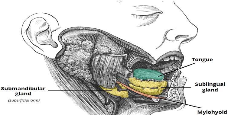
REFERENCES
1Candamourty R, Venkatachalam S, Babu M, Kumar G — Ludwig’s angina–an emergency: a case report with literature review. J Nat Sci Biol Med 2012; 3: 206-8. [PMC free article] [PubMed] [Google Scholar]
2jnsbm.com/jnsbmsite/wpcontent/uploads/2021/07/ JNatScBiolMed-3-2-206.pdf
3Saifeldeen K, Evans R — Ludwig’s angina. BMJ; 21(2): emj.bmj.com/content/21/2/242. http://dx.doi.org/10.1136/ emj.2003.012336
Vol 121, No 8, August 2023Journal
of the Indian Medical Association
Fig 1 — Images of the Patient and Saces of Neck
87
Commentary
Health Care Systems during Pandemic : Role of Government and Regulatory Authorities
Ankur Gupta1, Alok Pandey2, Puja Bansal3, Anureet Kaur4, Pranava Prakash5
This paper presents a review of literature on resilience of health care systems during pandemic. Resilience of health care systems through the review of literature has been defined as capacity of a system to bounce back from an adverse event such as a pandemic. We have reviewed 35 articles published on the issue during last five years (ie, 2018-2022) from different countries, both developed and developing. Most researchers have undertaken studies in this area and have conceptualized resilience of health systems with respect to infrastructure development, safety, awareness, connectivity, self-regulation, adaptability, integrity, disaster management, inventory and stock management of essential supplies, quality of services, community involvement, reactive capabilities and risk management. The aim of this paper is to describe how health care system resilience is defined and prepare an introductory conceptual review as a preliminary step for further studies. This paper helps us in identifying the key research themes that characterize the inquiry around resilience in context of a health care system during and post COVID-19 and also the structural improvements and characteristics to improve the competence of a health care system to deem it resilient during pandemic. [J Indian Med Assoc 2023; 121(8): 88-90]
Key words :Health Care Systems, Post COVID-19.
During December 2019, novel ‘‘coronavirus’’ (COVID19) which is a highly transmittable and pathogenic viral infection emerged first in China and rapidly spread around the world. On March 11, 2020 WHO declared the COVID-19 as a global outbreak of a pandemic (Padhan R, Prabheesh KP, 2021). The COVID-19 pandemic presented an unprecedented challenge to the global health care system. The pandemic exposed inherent weaknesses in our preparedness and response. Also, the health systems were grossly overwhelmed by the pandemic.
The novel coronavirus, or COVID-19, has resulted in several human deaths and socio-economic issues that have heavily damaged community health systems. India’s health care system now faces structural issues due to the pandemic. A number of immediate and long-term repercussions, primarily on the healthcare sector, have been affected by this global pandemic (Ayati, et al 2020).
The pandemic COVID-19 has brought the spot light on India as well. While there were some successes in the response to the pandemic, there were also several ways in which the health care system fell short. Overall, the COVID-19 pandemic exposed several weaknesses and flaws in the global health care system. There is much work to be done to address these issues and prepare for
1Research Scholar, Department of Management, GD Goenka University, Gurugram, Haryana 122103
2Professor and Vice Dean, Department Jindal School of Banking and Finance, OP Jindal Global University, Sonipat 131001
3Research Scholar, Guru Jambheshwar University of Science and Technology, Hisar, 125001 and Corresponding author
4Assistant Professor, G D Goenka Institute of Management, Gurugram, Haryana 122103
5MBBS, Associate Professor & Head, Public Health, GD Goenka University, Gurugram Haryana 122103
Received on : 06/04/2023
Accepted on : 07/05/2023
future pandemics.Financial and logistical resources should be accessible and well exploited so that health care systems can bring about immense benefit for the general public. Health care systems would also benefit from good governance and control systems along with sufficient funding for emergency medical care programme. Both public and private sectors should be involved in Infrastructure development so as to expand access to life-saving medical care through upgraded facilities (Shukla, et al 2021).
In India, the COVID-19 pandemic has also exposed several weaknesses and flaws in the health care system. The first wave presented a livelihood disaster due to an ill-planned lockdown. The second wave presented a health disaster that was due to government inaction (Pellissery S, et al 2021).
Some of the areas in which healthcare failed during the COVID-19 pandemic in India are:
Lack of preparedness : The health care system in India was not adequately prepared for the scale and severity of the pandemic. There was a shortage of critical resources such as Personal Protective Equipment (PPE), ventilators and hospital beds.
Testing and contact tracing failures : Initially, there were significant delays in testing and contact tracing in India, which contributed to the rapid spread of the virus. The testing capacity was inadequate, and there were issues with the quality of tests and delays in receiving results.
Overwhelmed health care systems : The rapid surge in COVID-19 cases in India overwhelmed the health care system, with hospitals and health care workers stretched thin. There were shortages of critical resources, such as oxygen cylinders and medication, which led to many deaths. During April 2021 the number of reported positive cases crossed 200,000 per day, hospitals had no capacity
Vol 121, No 8, August 2023Journal of the Indian Medical Association 88
to admit patients and also where beds were available, oxygen and life-supporting machines, were absent (Pellissery S, et al 2021). In such conditions individuals kept sickloved ones at home and started procuring medical essentials from the black market. The rule of law also becomes ineffective when citizens are left to their own devices to manage their health during a pandemic.
Inadequate communication and coordination : There were issues with communication and coordination between different health care organizations and the Government in India. This led to confusion and delays in implementing effective measures to control the spread of the virus. While during the first wave of the COVID-19 crisis, the government was visible through the lockdown measures but the second wave made it invisible.There was huge gap between public information on bed availability and the reality on the ground during COVID19, an important debate occurred in India among doctors as to whether CT scans could be used for COVID-19 diagnosis(Pellissery S, et al 2021)
Health disparities : The pandemic highlighted existing health disparities in India, with certain communities and populations being disproportionately affected by COVID19. This was due to a variety of factors, including socioeconomic status, lack of access to healthcare and underlying health conditions. India with its deep economic and social inequality was in shambles as the pandemic left vulnerable citizens to look on while well-off citizens grabbed the available resources (be it a hospital bed or a respectable funeral) and also hoarding and black marketing of medicines like Remdesivir (Pellissery S, et al 2021).
India’s Burial Infrastructure : The way in which the pandemic brought pressure on India’s burial infrastructure created graded dignity. Those who could afford the expenses of burial were able to respect their emotions toward the dead body of a relative. Impoverished villagers without resources to buy wood for cremation, had floated the dead bodies down rivers. Bribes were demanded both in public crematoriums as well as by middle-men to execute the task (Pellissery S, et al 2021).
These are some examples of how the system as a whole did actual harm, while most individual doctors were caring. Overall, the COVID-19 pandemic has exposed several weaknesses in the health care system in India. There is a need for significant investment in healthcare infrastructure and resources, as well as better coordination between different healthcare organizations and the Government to prepare for future pandemics and provide better healthcare to the population.
A regulatory framework is essential for ensuring the quality and safety of healthcare services. The health care system must have appropriate regulations and standards for healthcare providers, facilities, and medical products. It should also be sustainable, adaptable, and able to respond to the changing healthcare needs of the population. Appropriate response and responsibility by the Government towards health-related shock creates a resilient response by a nation (Augustynowicz A, et al,
2022; Khan Y, et al 2018; Rogers HL, et al 2021; Thomas S, et al 2020). Issue of policy guidelines and development of various health related clinical protocols for hospitals should be created (Burke DS, et al, 2021; Øyri SF, Wiig S, 2022; Barbash IJ, Kahn JM, 2021). The involvement of top policy makers in expanding of Government budget on health and provision for health facilities before and during a pandemic and creation of long-term plans and policies and tertiary level interventions at hospital level can improve healthcare capabilities of a nation (Burke DS, et al 2021; Khetrapal SR, Bhatia, 2020; Rogers HL, et al 2021; Thomas S, et al 2020).
The role of Government during a pandemic is multifaceted and can involve a range of activities and interventions. Some ways in which the Government can play a role in managing a pandemic is by providing accurate and timely information the pandemic, how it spreads, how to protect oneself and what measures are being taken to control the spread of the virus. This information should be communicated clearly and effectively, and in a manner that is accessible to everyone, regardless of their level of education or literacy.
There is a need to implement public health measures like travel restrictions, mandatory quarantine for those who have been exposed to the virus, mandatory maskwearing, social distancing measures, and restrictions on public gatherings. These measures can help slow the spread of the virus and prevent the health care system from becoming overwhelmed.
Mobilization of the healthcare resources is another important areawhere the Government can play a critical role to respond to the pandemic. This may involve procuring critical resources such as Personal Protective Equipment (PPE), ventilators and medication, as well as recruiting and training health care workers. The Government can also set up temporary healthcare facilities to accommodate the surge in patients during a pandemic.
As Pandemics are global in nature and coordination with other countries and International Organizations is essential to manage the spread of the virus. The Government can play a key role in coordinating with other countries to share information and resources, as well as collaborate on research and development of treatments and vaccines. International cooperation can also help to prevent the spread of the virus across borders.
The vulnerable populations of a country require Government support which may involve providing financial assistance to those who have lost their jobs or are unable to work due to the pandemic, as well as ensuring access to healthcare services for marginalized communities. The Government can also set up support systems for elderly people, disabled individuals, and those with pre-existing medical conditions who may be particularly vulnerable to the virus.
Roll out vaccines: As vaccines become available, the Government can play a critical role in rolling them out to the population. This may involve setting up vaccination centers, coordinating with healthcare providers, and communicating the importance of vaccination to the
Vol 121, No 8, August 2023Journal
of the Indian Medical Association
89
public. The government can also work to ensure equitable distribution of vaccines across different regions and populations.
A society’s ability to respond collectively, to pool resources and risk while safeguarding its population, especially the most vulnerable, will determine how much of an impact a pandemic or similar threat will have on society and the economy. These factors include public confidence in the Government’s actions, a balance between values and policies that promote health, wealth, and prosperity and a society’s willingness to take collective action. These characteristics define a strong and harmonious community (Etienne, 2020). Governments and other institutions cannot exclusively achieve resilience through top-down, unilateral strategies. Community involvement is as important to resilience as rules and hospital capacity. Managing challenges to health as well as other threats, such as climate and environmental change, requires a strong focus on community participation and how it relates to community resilience (Haldane, 2021). The national Governments must create and put into action plans to strengthen and bolster the resilience of their health systems with the aid of outside partners.
It is vital to implement new frameworks and policy instruments affecting health care organisations to adequately plan for anticipated crises and other difficulties that may impact health care delivery (Rogers, 2021). While according to Biddle, et al, 2020, resilience is no longer seen as a quality of the health care system but as a strength that depends on actors’ ability to adapt and adjust. So, the country’s National Health plans must prioritize the resilience of the health-care system (Arsenault, et al 2022). This is because all system levels and sectors’ stakeholders must see volatility as an opportunity to improve the health system rather than as a danger to its viability and integrity. Resilience must be understood as a multi-tiered notion in which resilience requirements at one level may be influenced by activities at other levels (Turner, 2022). Therefore, efforts to improve the resilience of health systems should place a major emphasis on state, Non-governmental Organisations (NGOs), as well as private health personnel.
The considerable research on flexibility of hospitals in terms of safety precautions, reorganizing spaces, managing resources, using technology and training and development of workforce is required. Also, disaster management systems, disaster resources, and disaster medical care capabilities should be prioritized by Government and regulatory authorities of a nation.
Overall, the Government plays a crucial role in managing a pandemic like COVID-19. By providing accurate information, implementing public health measures, mobilizing healthcare resources, coordinating with other countries and organizations, supporting vulnerable populations and rolling out vaccines, the Government can help to slow the spread of the virus and protect public health
REFERENCES
1Arsenault C — COVID-19 and resilience of healthcare systems in ten countries. Nat Med 1922; 28: 1314-24 https:/ /doi.org/10.1038/s41591-022-01750-1
2Augustynowicz A — Resilient Health and the Healthcare System. A Few Introductory Remarks in Times of the COVID19 Pandemic. Int. J. Environ. Res. Public Health 2022; 19: 3603. https://doi.org/10.3390/ijerph19063603
3Ayati N, Saiyarsarai P, Nikfar S — Short and long term impacts of COVID-19 on the pharmaceutical sector. DARU Journal of Pharmaceutical Sciences 2020; 28(2): 799-805.
4Barasa E, Mbau R, Gilson L — What Is Resilience and How Can It Be Nurtured? A Systematic Review of Empirical Literature on Organizational Resilience. International journal of health policy and management 2018; 7(6): 491-503. https:/ /doi.org/10.15171/ijhpm.2018.06
5Biddle L — Health system resilience: a literature review of empirical research. Health policy and planning 2020; 35(8): 1084-109. https://doi.org/10.1093/heapol/czaa032
6Burke DS — Building health system resilience through policy development in response to COVID-19 in Ireland: From shock to reform / The Lancet Regional Health 2021; - Europe 9 (2021) 100223.
7Etienne CF — COVID-19: transformative actions for more equitable, resilient, sustainable societies and health systems in the Americas. BMJ Global Health 2020; 5(8): e003509.
8Haldane V — Health systems resilience in managing the COVID-19 pandemic: lessons from 28 countries. Nature Medicine 2021; 27(6): 964-80.
9Khan Y, O’Sullivan T, Brown A, Tracey S, Gibson J, Généreux M, et al — Public health emergency preparedness: a framework to promote resilience. BMC public health 2018; 18(1): 1-16.
10Khetrapal S, Bhatia R — Impact of COVID-19 pandemic on health system & Sustainable Development Goal 3. Indian Journal of Medical Research 2020; 151(5): 395-9. | DOI: 10.4103/ijmr.IJMR_1920_20
11Øyri SF, Wiig S — Linking resilience and regulation across system levels in healthcare – a multilevel study. BMC Health Services Research 2022; 22: 510 https://doi.org/10.1186/ s12913-022-07848-z
12Padhan R, Prabheesh KP — The economics of COVID-19 pandemic: A survey. Econ Anal Policy 2021; 70: 220-37. doi: 10.1016/j.eap.2021.02.012. Epub 2021 Feb 25. PMID: 33658744; PMCID: PMC7906538.
13Pellissery S — The Case of India A Moral Foundation for the Impact of COVID-19 on Health and Society in the World’s Largest Democracy. International Journal of Social Quality 2021; Volume 11, Issues 1 & 2, Summer & Winter 2021: 6384.
14Rogers HL — Resilience testing of health systems: How can it be done? International Journal of Environmental Research and Public Health 2021; 18(9): 4742.
15Shukla D, Pradhan A, Malik P — Economic impact of COVID19 on the Indian healthcare sector: an overview. Int J Comm Med Public Health 2021; 8(1): 489-94.
16Thomas S — Strengthening health systems resilience: Key concepts and strategies [Internet]. Copenhagen (Denmark): European Observatory on Health Systems and Policies 2020; PMID: 32716618
17Turner S — We are all vulnerable, we are all fragile: COVID19 as opportunity for, or constraint on, health service resilience in Colombia? Public Management Review 2022; 1-22.
Vol 121, No 8, August 2023Journal of the
Medical
Indian
Association
90
Quiz 1
Image in Medicine
Bhoomi Angirish1, Bhavin Jankharia2
A 23-year-old male present with Recurrent Headache which Worsened since 15 days.
Questions :
(1)What is the diagnosis ?
(2)If such lesions are multiple in number, what syndrome are usually associated ?
Answers :
(1)An intracranial Arteriovenous Malformation (AVM) is seen composed of enlarged feeding arteries, a nidus of vessels and draining veins. In this case, the feeding arteries are seen arising from cortical branches of middle cerebral artery and draining veins are seen draining into superior sagittal sinus through cortical veins.
(2)Usually arteriovenous malformations are solitary, however multiple AVM are seen associated with Hereditary HemorrhagicTelangiectasia and Craniofacial arteriovenous metameric syndrome.
Quiz 2
A 19-year-old male present with Subcutaneous Fatty Tissue with Abnormal Hair Tuft in Lower Back.
Questions :
(1)What is the diagnosis ?
(2)What are the different types of spinal dysraphism ?
Answers :
(1)Spina bifida is seen at L3 and L4 vertebral levels. Low lying cord is seen with herniation of cord and meninges through the defect in posterior elements , however it is covered by overlying subcutaneous tissue and skin. These findings are suggestive of closed spinal dysraphism (lipomyelomeningocele).
(2)Spinal dysraphism can be broadly divided into open spinal dysraphism and closed spinal dysraphism. In open spinal dysraphism, the cord and its covering are exposed and there is no overlying skin. These include myelomeningocele, myelocele etc. In closed spinal dysraphism, the cord is covered by normal mesenchymal elements. These include lipomyelomeningocele, lipomyelocele, meningocele,isolated spina bifida, intradural lipoma, dorsal dermal sinus, diastematomyelia etc.
Department of Radiology, Picture This by Jankharia, Mumbai, Maharashtra 400004
1MD, DNB (Radiology)
2MD, DMRD (Radiology)
Vol 121, No 8, August 2023Journal of
the Indian Medical Association
91
Letter to the Editor
[The Editor is not responsible for the views expressed by the correspondents]
Dengue — The Remedial Steps
S IR , — The dengue recently have become more rampant and awareness regarding remedial steps both by public and health workers will be helpful in long way in curbing the dreadful disease.
(1) People need to be aware that dengue is of four serotypes. Infection from one serotype gives life long immunity to that but only cross protection to other 3 serotypes for 2 years, due to sharing of seventy percent aminoacid sequences identity. Secondary infection is more severe due to present antibodies. Primary prevention is best solution.
(2) Destroying breeding of dengue vector and emphasis on proper sanitation, avoiding exposure of body and use of mosquito nets and repellants specially when dengue is rampant.
(3) Notification of disease to government authorities for better policy making.
(4) Educating people about prevention and primary care of the disease as delay in treating patients increases mortality from 1-5 percent to 20 percent.
(5) Patients having comorbidities like diabetes, kidney disease, hypertension, elderly, obese, pregnant women, etc should be taken care at the earliest by the physicians and not home treated.
(6) People should be made aware that late presentation of dengue fever to the hospital leads to increased development of dengue haemorrhagic fever, dengue shock syndrome, multi-organ involvement like acute kidney injury, and increased mortality.
(7) The guide lines for hospital admission — if the platelet count is <100,000, or platelet count between 100,000-150,000 with a rapid drop in platelets, fever for three days with any warning signs such as abdominal pain, persistent vomiting, mucosal bleeding, lethargy and restlessness.
(8) Elisa based NS1 tests best and can not be false positive. NS1 antigen is tested positive in first few days and has a sensitivity of 60-90%. IgM antibody become positive after the 5th day of illness. Timing of testing should be made aware. In primary dengue IgG becomes + at the end of 7 days while IgM+ is after day 4.
Value of IgG and IgM with thrombocytopenia and looking sick on day 3 or 4 of illness, a very high titre of IgG with borderline rise in IgM signifies secondary dengue infection and are more prone to complications.
If Immature platelet fraction IPF is >10% despite a platelet count of less than 20,000, one is out of danger
and platelet will rise in 24 hours. However if less than 5% then bone marrow not responding for at least 3-4 days and is the candidate for platelet transfusion. Better to do IPF even in borderline low platelet count. Low mean platelet volume MPV are functionally not good and need more attention.
(9) Public awareness through social media is fruitful.
(10) Treatment of choice is paracetamol, plenty of liquid diet to prevent hypotension.
(11) Fluid of choice in is normal saline.
(12) Platelet transfusion when platelet count is less than 10,000 or when active bleeding or purpuric spots occur.
(13) Drugs to be avoided like aspirin, ibuprofen, nimuselide, acenofenac and steroid.
(14) Storage of platelets at 24 to 26 degrees Celsius. Half life of platelets is 6 to 9 days.
(15) Goat milk, papaya leaf, kiwi fruits have no role and any benefit is due to antioxidants but can affect liver which is often deranged.
(16) While treating dengue patients, formula of 20 is very handy. That is rise in pulse by more than 20, fall of BP by more than 20, difference between upper and lower BP of less than 20, and presence of more than 20 haemorrhagic spots on the arm after a torniquet test suggest a high risk situation and the person requires care.
(17) The primary cause of mortality is due to capillary leakage causing hypotension due to decrease in intravascular compartment volume leading to multiple organ failiure. Fluid replacement amounting to 20 ml per kg body weight per hour must be administered. This must be continued till difference between the upper and lower blood pressure is over 40 mm of mercury or the patient passes adequate urine. This is sufficient and giving unnecessary platelet transfusion can make patient more deteriorated.
Eye sees what the mind knows. So education and awareness of dengue control and management is key to curbing effectively this dreaded disease.
1MBBS, MS, MCh (Plastic Surgery), Sudhir Singh1
Hony IMA Professor, Manoj Kumar Srivastava2
Department of Plastic Surgery, Getwell Hospital, Varanasi
2MBBS, MD, Professor, Department of Medicine, Narayan Medical College, Bihar
Vol 121, No 8, August 2023Journal of the Indian Medical Association
92
