Endometriosis, a recognized clinical condition, is associated with pain and subfertility in 10-15% of women of the reproductive age group globally1. The pathophysiolology of this disease still remains elusive. The varied clinical presentations of the disease and the immense impact it has on the physical, psychological, domestic, social and professional lives of those suffering from it put challenges on the treating gynaecologists to find a solution to this perplexing problem. Currently clinical diagnosis and medical treatment are the pillars of management of symptomatic endometriosis only giving way to surgical intervention if the conservative methods are inadequate. Artificial Reproductive Techniques (ART) should be the preferred approach in associated subfertility.
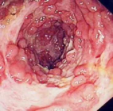
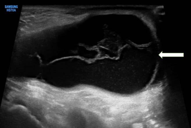
Pain is the hallmark of the disease. Most of the patient with endometriosis present with dysmenorrhoea, dyspareunia, dyschezia, lower abdominal and pelvic pain, either alone or in combination with subfertility. The classical triad of pelvic endometriosis is progressive dysmenorrhoea, dyspareunia and heavy menstrual bleeding. Therefore, in majority of cases the diagnosis is made clinically. However, 30-40% of pelvic endometriosis also present with ovarian endometrioma where pelvic ultrasonography and MRI may become helpful. Laparoscopy is no more a gold standard for the diagnosis of endometriosis in modern medicine. Serum biomarker like CA125 does not have any diagnostic or prognostic value in the disease management.
Medical management is the mainstay of treatment in endometriosis associated pain. It is also useful in pre-operative and post-surgical adjuvant therapy as well as in adolescent disease. Out of these, Pre-operative medical therapy is used in severe endometriosis for better surgical dissection in restoration of the pelvic anatomy. However, it has not been shown to have any immediate improvement on the outcome of pain. On the other hand, post-operative adjuvant therapy is known to have positive impact on immediate pain relief. It has also been shown to be very useful in the prevention of recurrent disease. Hence, it should be offered to every postsurgical patient where pregnancy is not desired. Symptomatic relief of pain associated with endometriosis can be achieved by NSAIDS, SSRIs and antidepressants without any effect on the progression of the disease. A basket of hormonal preparations containing Progestogens (oral/injectables/implants), Combined Oral Contraceptives (COC) pills, GNRH agonists and antagonists, aromatase inhibitors and danazol are available. COC pills either cyclical or continuous are known to work as long term medical therapy in endometriosis and has been shown to significantly reduce dysmenorrhoea, dyspareunia, dyschezia and cyclical pelvic pain2. Continuous COC pills however, work better in dysmenorrhoea than cyclical pills. Combined
contraceptives used via other routes eg, transdermal or vaginal are also equally effective3. This group of hormonal preparations are also effective in long term use for secondary prevention of recurrent disease. Progestogens are available in oral (Medroxyprogesterone Acetate / Dienogest) Injectable like Depo-Medroxyprogesterone Acetate (DMPA), and as implant such as Implanon /LevoNorgestrel releasingIntra Uterine System (LNG-IUS). Continuous Progestogens and anti-progestogen (Gestrinone) are equally effective. Out of all Progestogen preparations Oral Dienogest4 and LNG-IUS have been found to cause significant reduction of postoperative pain, comparable to GnRH agonist5. They are also more effective in reducing pain and/or disease recurrence with significantly higher patient satisfaction comparable to COC Pill and Danazol. Etonorgestrel releasing subdermal implant, like LNG-IUS has also been reported to cause significant reduction of endometriosis associated pain, dysmenorrhoea and chronic pelvic pain6. Amenorrhea, irregular bleeding, acne, weight gain are usual side effects. Effect on BMD is a concern in long term use of DMPA. Danazol is no longer a preferred medical treatment ofendometriosis because of it’s serious androgenic side effects. GnRH agonists (Leuprolide Acetate, Goserelin/ Triptorelin/Nafarelin) are potent agents, equally effective as oral Dienogest and areknown to reduce endometriotic implants with associated inflammation and adhesion. Theycan significantly reduce endometriosis associated pelvic pain and can cause delay in recurrence of the disease but shown to beless effective than LNG-IUS7. They are equally effective irrespective of route of administration. Add-back therapy is recommended to prevent side effects like vaginal dryness, hot flushes andloss of bone mineral density 8 . Aromatase inhibitors, Letrozole and Anastrozole do not have strong evidence to support their efficacy in endometriosis and should be reserved as the last option in the treatment of the disease.
The basic principle of surgical treatment of endometriosis is the removal or destruction of all endometriotic tissue including ovarian endometrioma, peritoneal implants and Deep Infiltrating Endometriosis (DIE). The main indication of surgical intervention in endometriosis is pain. Endometriotic implantsare best treated by surgical excision than ablation9. There are convincing evidences to support surgical excisionof ovarian endometrioma over drainage and coagulation10. However, any surgical excision of ovarian endometrioma has its negative impact on ovarian reserve and this has to be thoroughly explained to the patient before
surgery. Sub-fertile patient trying for pregnancy should not have surgical excision as first line of therapy unless she is in pain or the endometrioma is large enough to make ovum pick up difficult.Pelvic denervation by Presacral Neurectomy (PSN) has been reported as an effective procedure in reducing pain in recurrent endometriosis following first-line surgical treatment. However PSN can be associated with denervation of bowel and bladdercausing constipation and urinary dysfunction11
Deep Infiltrating Endometriosis (DIE) extends beneath the peritoneum and may affect the uterosacral ligaments, pelvic side walls, rectovaginal septum, vagina, bowel, bladder, or ureter. Excision of these nodules is usually performed when surgical treatment is chosen. The extent of surgical excision of DIE depends on the organ involved and the depth of infiltration and can lead even upto bowel resection. Hence DIE is best managed by multidisciplinary approach in tertiary referral centres.
Increased incidence of subfertility associated with Endometriosis is best treated with artificial reproductive techniques. Pre- and Post Operative medical treatment does not enhance the outcome of subfertility treatment and is not recommended. Repeated surgery for recurrent ovarian endometrioma can hamper the fertility outcome12. However, if surgery is needed for subfertile patients, as in the above mentioned conditions, post operative ART should be considered sooner.
Endometriosis is being recognised more frequently as a cause of abdominal pain and dysmenorrhoea in adolescents. The Gynaecologist has to be “Endometriosis Minded” to make an early clinical diagnosis and try to arrest diseaseprogression. Laparoscopy is not appropriate for diagnosis. Combined oral contraceptive pills 13 and progestogens like Dienogest14 are effective as first line of treatment in adolescent endometriosis. NSAIDs can be used for pain relief liberally for symptomatic relief. GNRH analogue usage should be reserved only for second line therapy due to its adverse effects.
Endometriosis is known to be a benign disease. However, endometriosis is associated with increased risk of ovarian cancer specially Clear cell carcinoma and Endometroid carcinoma.
Endometriosis is common in the reproductive age group. However, nowadays endometriosis is being diagnosed in adolescent girls. Hence, clinical diagnosis should be encouraged followed by first line medical treatment. Complete Surgical treatment should be reserved for resistant cases only. Subfertility is best treated by ART.
As endometriosis remains an enigmatic disease collaborative scientific efforts and research fundings should be directed towards the disease to seek a cure for the same.
FURTHER READINGS
1SE Bulun,”Endometriosis”. The New England Journal of Medicine 2009; 360(3): 268-79.
2Jensen JT, Schlaff W, Gordon K — Use of combined hormonal contraceptives for the treatment of endometriosis-related pain: a systematic review of the evidence. FertilSteril 2018; 110: 137-52.e131.
3Brown J, Crawford TJ, Datta S, Prentice A — Oral contraceptives for pain associated with endometriosis. Cochrane Database Syst Rev 2018; 5.
4Andres Mde P, Lopes LA, Baracat EC, Podgaec S — Dienogest in the treatment of endometriosis: systematic review. Arch Gynecol Obstet 2015; 292: 523-9.
5Lan S, Ling L, Jianhong Z, Xijing J, Lihui W — Analysis of the levonorgestrel-releasing intrauterine system in women with endometriosis. J Int Med Res 2013; 41: 548-58.
6Margatho D, Carvalho NM, Bahamondes L — Endometriosisassociated pain scores and biomarkers in users of the etonogestrel- releasing subdermal implant or the 52-mg levonorgestrel-releasing intrauterine system for up to 24 months. Eur J Contracept Reprod Health Care 2020; 25: 133-40.
7Brown J, Pan A, Hart RJ — Gonadotrophin-releasing hormone analogues for pain associated with endometriosis. Cochrane Database Syst Rev 2010.
8Wu D, Hu M, Hong L, Hong S, Ding W, Min J, et al — Clinical efficacy of add-back therapy in treatment of endometriosis: a meta-analysis. Arch Gynecol Obstet 2014; 290: 513-23.
9Pundir J, Omanwa K, Kovoor E, Pundir V, Lancaster G, Barton-Smith P — Laparoscopic Excision Versus Ablation for Endometriosis- associated Pain: An Updated Systematic Review and Meta-analysis. J Minim Invasive Gynecol 2017; 24: 747-56.
10Hart RJ, Hickey M, Maouris P, Buckett W — Excisional surgery versus ablative surgery for ovarian endometriomata. Cochrane Database of Systematic Reviews 2008.
11Vercellini P, Somigliana E, Daguati R — The second time around: reproductive performance after repetitive versus primary surgery for endome- triosis. Fertil Steril 2009; 92: 1253-5.
12Vercellini P, Somigliana E, Vigano‘ P — The effect of secondline surgery onreproductive performance of women with recurrent endometriosis: a systematic review. Acta Obstet Gynecol Scand 2009; 88: 1074-82.
13Davis AR, Westhoff C, O’Connell K, Gallagher N — Oral contraceptives for dysmenorrhea in adolescent girls: a randomized trial. Obstet Gynecol 2005; 106: 97-104.
14Ebert AD, Dong L, Merz M, Kirsch B, Francuski M, Bottcher B, et al — Dienogest 2 mg Daily in the Treatment of Adolescents with Clinically Suspected Endometriosis: The VISanne Study to Assess Safety in ADOlescents. J Pediatr Adolesc Gynecol 2017; 30: 560-7.
Perceptions of Medical Educators Regarding the Integration of Standardized Teaching-learning Modules for Training Communication Skills in Medical Undergraduate Students
Praveen S Ganganahalli1, Rekha Udgiri2
Background : The doctor-patient relationship is profoundly influenced by effective communication, leading to positive outcomes in terms of patient health and satisfaction. When medical undergraduates are trained in Communication Skills through a standardized module, it results in improved treatment adherence, decreased malpractice claims and notable advancements in psychological and physical well-being across various health conditions.
Aims and Objectives : This study aims to evaluate the perceptions of medical teachers in India concerning the training of medical undergraduates in Communication Skills using a standardized module.
Materials and Methods : An observational survey was conducted to assess the views of medical college teachers regarding the training of Communication Skills to medical undergraduate students. The survey utilized a pre-structured proforma containing questionnaires and employed a standardized teaching-learning module delivered through an online link-sharing platform.
Results : The faculty members hold a clear and robust belief in the importance of integrating Communication Skills training directly into the medical curriculum, with particular emphasis on community-based training. They firmly believe that such training not only enhances students’ communication abilities but also fosters empathy towards patients.
Conclusion : The perceptions of medical educators highlight the potential benefits of incorporating Communication Skills training modules into the medical curriculum. By doing so, it promotes better patient care and contributes to overall improved healthcare outcomes.
[J Indian Med Assoc 2024; 122(4): 15-8]
Key words :Perception, Communication Skills, Module, Faculty, Students.
Effective communication with patients significantly impacts the doctor-patient rapport, fostering positive health outcomes and patient satisfaction. It promotes better treatment adherence, reduces malpractice claims and enhances psychological and physical well-being across diverse health issues. Nevertheless, mastering communication in clinical settings is challenging, demanding clinicians to cultivate a repertoire of intricate skills. While these skills may not be innate, they are acquirable through education and practice. Numerous studies demonstrate that educational interventions typically enhance communication proficiency among medical trainees1
Patients should actively participate in both diagnosis and treatment, acting as partners in their healthcare journey. This involvement not only empowers patients to take ownership of their well-being but also
Department of Community Medicine, BLDEDU Shri B M Patil Medical College, Vijayapura, Karnataka 586103
1MD, Associate Professor and Corresponding Author 2MD, Professor
Received on : 18/08/2023
Accepted on : 23/09/2023
Editor's Comment :
Community-based modules designed to improve communication skills among medical undergraduate students positively impact their development. These skills are essential for fostering strong doctor-patient relationships, which are vital for effective healthcare.
Medical educators believe that integrating these modules into the curriculum could benefit significantly, ultimately improving patient care and healthcare outcomes.
enhances compliance and engagement within a patientcentered healthcare system. Physicians bear the responsibility of facilitating this collaborative approach, known as ‘shared decision making,’ through effective communication techniques. The Kalamazoo Consensus Statement outlines seven fundamental communication tasks crucial for physician-patient interactions: building rapport, initiating dialogue, collecting relevant information, understanding the patient’s viewpoint, exchanging information, agreeing on solutions and concluding the discussion2
The incorporation of Communication Skills (CS) into academic instruction reveals notable deficiencies and challenges. One critical oversight in designing and
executing such programs is the insufficient consideration of their effects on the recipients. This oversight is particularly pertinent in the case of medical students and their perception of such educational approaches1.
A study was undertaken to evaluate the perception and attitude of medical students in Central India towards the Communication Skills Lab (CSL) and its associated teaching module. A significant majority of the students (96.43%) expressed that the training had notably enhanced their communication abilities with patients. Furthermore, they advocated for the integration of such training into the standard teaching curriculum nationwide. The implementation of basic Communication Skills training could be seamlessly incorporated into the early stages of undergraduate medical education through a well-designed, engaging, and widely accepted teaching module3
In this context, a research project has been developed to explore how faculty members perceive the teaching of Communication Skills to medical undergraduates. The project will employ standardized modules for teaching, learning and assessment to facilitate this investigation.
MATERIALS AND METHODS
A survey was conducted to gauge the opinions of medical college educators in India regarding the instruction of Communication Skills to undergraduate medical students. This observational study utilized a predetermined questionnaire format, incorporating a standardized teaching module. Before initiating the study, approval was obtained from the Institutional Ethics Committee and participating teachers provided informed consent.
The survey was disseminated to educators from various medical colleges throughout India via a Google Form link shared across social media platforms including WhatsApp, Telegram and email. Subsequently, the data collected through the Google Form were exported into an Excel spreadsheet for analysis, wherein frequency distributions and correlations among variables were examined.
OBSERVATIONS
The survey received participation from a total of 105 teachers from various regions across India. After analysing the information collected, the following observations were made (Table 1).
Among those involved, the predominant group comprises faculty members (56%) holding titles such as assistant, lecturer, or tutor within the Department of Community Medicine (52%) across both
Table 1 — Distribution of the Participants according to Academic Characteristics
VariablesFrequency Percentage
Designation : Professor3533
Associate Professor1111
Assistant Professor4845
Lecturer/SR1111
Department :
Community Medicine5552
Biochemistry0808
Microbiology1010
Physiology1111
Pathology0404
Clinical dept1010
Dentistry0505
Type of Institute : Government4240
Private4038
Deemed to be University2322
State of Working : Karnataka6561
Andhra Pradesh0808 Kerala0505 Maharashtra1716
Tamil Nadu0505
West Bengal0505
Medical Education Unit member : Yes6561 No4139
Medical Education Training : RBCW only8277
RBCW + ACME1918
RBCW + FAIMER1010
Governmental and private institutions, with a nearly balanced representation (40% versus 38%) in Karnataka state (61%). Within this cohort, 61% were affiliated with the medical education unit and nearly all have completed fundamental training in medical education, with 28% having pursued advanced training.
As per Table 2, the faculty holds a firm conviction regarding the vital need for integrating Communication Skills training directly into undergraduate courses, particularly emphasizing community-based training. They argue that such training not only improves students’ communication prowess but also nurtures empathy towards patients. Additionally, the faculty acknowledges the significance of instituting a standardized Teaching-learning Module to provide students with communication skills uniformly. This method enables them to embrace emerging teaching and assessment methodologies, essential for successfully implementing a Competency-based Medical Education (CBME) curriculum.
Table 3 outlines various challenges encountered, including logistical hurdles in coordinating sessions to accommodate all participants, managing diverse group dynamics, addressing time constraints for
Table 2 — Perception of faculty about training UGs in Communication Skills
QuestionStronglyDisagree
disagree
Good communication skills offer advantages beyond just fostering a better doctor-patient relationship---11%89% Is the inclusion of communication skills training in the undergraduate medical course perceived as effective?--11%27%61%
The skill required for taking a good history is not as extensive as that needed for conducting a physical examination of the patient.50%
Training in communication skills is found to be more effective in a clinical setting compared to a community setting.27.8%
When taught in a community setting, communication skills training can significantly enhance empathy in medical students.
Utilizing a standardized Teaching-learning Module for skills training leads to improved outcomes.
The implementation of a standardized Teaching-Learning Module will aid faculty in uniformly and effectively training students.-- 5.6%38.9%55.6%
The utilization of a standardized Teaching-learning Module will facilitate the appropriate training of skills and ensure their effective acquisition.-- 5.6%44.4%50%
Employing formative assessment methods for in-course evaluation of students’ Communication Skills will prove to be more effective.--
Table 3 — challenges faced & opinion about small group community-based training
Challenges faced during implementation of small group teaching (Multiple answers) :
Faculty deficiency80%
Inadequate infrastructure54%
Stick to traditional method of teaching61%
Resistance to adapt newer techniques of teaching65%
Poor active participation of students54%
Student faculty ratio58%
Teachers training into newer techniques of teaching60%
Time constraints74%
covering essential content, ensuring sufficient resource allocation, effectively assessing individual student performance, providing valuable feedback, training faculty members in small group teaching methodologies, overcoming resistance to change, dealing with student preparedness issues, and determining the ideal group size to encourage meaningful discussions and active learning.
DISCUSSION
The current research explored the viewpoints of faculty members engaged in teaching medical undergraduates. These faculty members express a strong acknowledgment of the importance of integrating Communication Skills training into the undergraduate curriculum, particularly emphasizing community-based training. Additionally, the study highlighted several hurdles that need attention, such as logistical challenges in scheduling sessions to suit all participants, effectively managing time constraints to cover essential content, ensuring proper resource allocation and overcoming resistance to change and embracing active learning methodologies.
Educators’ appreciation for the relevance and
authenticity of Simulated Patient training grows with teaching tenure. This trend seems to be shaped by various factors, including the educator’s internal or external status, their own encounters with communication training during medical education, and the specific medical discipline they teach. The utilization of Simulated Patients in communication training holds significant value for medical educators due to its adaptable nature and wide-ranging relevance across medical fields4
Richard S and colleagues conducted a study on the perception of doctor-patient communication training among medical students. Their findings revealed that 55.6% of respondents felt adequately trained in this aspect. While 85.9% received the oretical courses, only 64.6% had the chance to supplement their learning with practical experience. A significant majority expressed a need for more hands-on practice in Communication Skills. Moreover, all participants agreed on the necessity of integrating more practical communication training into the curriculum5
Ruiz-Moral and colleagues investigated the viewpoints of fourth-year medical students regarding a Communication Skills training course featuring experiential learning elements. They discovered that while students found this approach beneficial, it also induced significant stress, particularly during smallgroup sessions where they interacted with standardized patients and during summative assessments6
In their research aimed at crafting, introducing and assessing a structured, validated module on Communication Skills for interns, Sinjita Dutta and colleagues discovered notable findings. They observed
122, No 04, April 2024Journal of the Indian Medical Association
a significant increase in post-training knowledge scores (16.68±2.5), which were notably higher than the pretraining scores (15.45±2.9). Moreover, there was a substantial rise in self-assessed knowledge (11.08±3.7 to 17.23±3.3) and skills (9.60±4.6 to 16±2.9) before and after the training, respectively. Impressively, all interns exhibited a positive attitude towards Communication Skills, as evidenced by scores on the Communication Skills Assessment Scale (CSAS). Interns also performed well on assessment using the SEGUE framework, with a mean score of 16.6±3.59. Feedback from interns, as indicated by the satisfaction index of survey items, ranged from 82.5% to 93%, reflecting high levels of satisfaction. Faculty members unanimously agreed on the relevance, usefulness and potential applicability of the module across other Departments for Communication Skills training7.
Aggarwal and colleagues conducted a study to evaluate the influence of training on clinical skills among Phase I MBBS students at a Government Medical College. Participants welcomed this participant-centric, assessment-based approach to teaching and learning. They found the sessions on effective communication engaging and enjoyable and expressed a strong commitment to applying the knowledge gained in practical settings8
CONCLUSION
The results highlight how modules focused on enhancing Communication Skills positively influence the development of medical undergraduate students. These skills are crucial for building robust doctorpatient relationships. The views of medical educators suggest that integrating these modules into the curriculum could bring about significant benefits, leading to better patient care and ultimately improving healthcare outcomes. Nevertheless, additional
research and implementation endeavours are necessary to thoroughly evaluate and refine the integration of these modules into medical education.
REFERENCES
1Ruiz-Moral R, Gracia de Leonardo C, Caballero Martínez F, Monge Martín D — Medical students’ perceptions towards learning communication skills: a qualitative study following the 2-year training programme. Int J Med Educ 2019; 10: 9097. doi: 10.5116/ijme.5cbd.7e96. PMID: 31055522; PMCID: PMC6766390.
2Graf J, Loda T, Zipfel S — Communication skills of medical students: survey of self- and external perception in a longitudinally based trend study. BMC Med Educ 2020; 20: 149. https://doi.org/10.1186/s12909-020-02049-w
3Tanwani R, Chandki R, Joshi A, Arora VK, Nyati P, Sutay S — Perception and Attitude of Medical Students towards Communication Skills Lab and Teaching Module. JClinDiagn Res 2017; 11(6): JC12-JC14. doi: 10.7860/JCDR/2017/ 24858.10120. Epub 2017 Jun 1. PMID: 28764200; PMCID: PMC5535393.
4Alvarez S, Schultz JH — Medical educators’ perception of communication training with simulated patients: an explorative study approach. BMC Res Notes 2017; 10(1): 650. doi: 10.1186/s13104-017-2988-8. PMID: 29187258; PMCID: PMC5707823.
5Richard S, Pardoen D, Piquard D, Fostier P, Thomas JM, Vervier JF, et al — Perception of training in doctor-patient communication for students at faculty of medicine. Revue Medicale de Bruxelles 2012; 33(6): 525-30.
6Ruiz-Moral R, Gracia de Leonardo C, Caballero Martínez F, Monge Martín D — Medical students’ perceptions towards learning communication skills: a qualitative study following the 2-year training programme. Int J Med Educ 2019; 10: 907. doi: 10.5116/ijme.5cbd.7e96. PMID: 31055522; PMCID: PMC6766390.
7Dutta S, Mukherjee M, Shukla V, Mishra A, Saha R, Basu SS, et al — Introduction of Module-based Training on Communication Skills among Interns in a Tertiary Care Teaching Hospital of Kolkata, India. Journal of Clinical & Diagnostic Research 2022; 16(3)
8Aggarwal P, Rawekar A, Dey SK, Roy R — Impact of an assessment-based training module on communication skills in phase I indian medical undergraduates. Acta Med Int 2023; 10: 9-13.
Original Article
Prevalence of Diabetic Kidney Disease and Its Associated Risk Factors in Type-2 Diabetes Mellitus — A Tertiary Care Experience
Shaila Jay Shah1, Jay Harishbhai Shah2, Parth Prajapati3
Background : The burden of diabetes is increasing in India with its associated complications. Diabetic Kidney Disease (DKD) is one of the microvascular complications of Diabetes Mellitus (DM) which leads to End Stage Renal Disease (ESRD). Regional and ethnic differences have been noted globally and within India regarding diabetes, its risk factors and its ensuing complications. DKD is identified clinically by a persistently elevated Urine Albumin/ Creatine Ratio (UACR) of >30mg/g and/or a persistently decreased eGFR below 60 ml/min/1.73m 2
Materials and Methods : This was a single centre cross-sectional observational study which included 150 patients. All patients with known type 2 diabetes or newly diagnosed diabetes presenting to the Department of Medicine for the first time were included after screening and fulfilling the said inclusion and exclusion criteria. Primary objectives were to estimate the prevalence of DKD in type 2 diabetes mellitus and to determine the CKD stage of patients with DKD. Secondary objectives were to determine the association of various risk factors with DKD and to determine correlation between UACR and HbA1c, e-GFR and serum creatinine.
Results : DKD was found in 111(74%) of patients. 99(66%) of patients had hypertension, 81 of whom had DKD. There were 59(39.33%) patients with stage G2 and 58(38.67%) in stage G3 out of which 34 patients were in stage G3a and 24 were in G3b. Fifty (33.33%) patients had A1, 89(59.33%) patients had A2 and 11(7.34%) had A3 stages of albuminuria. Presence of hypertension, retinopathy and duration of DM were found to have a significant association with DKD prevalence. Hypertension also had a significant negative correlation with eGFR.
Conclusion : The prevalence of DKD in the population may be under-estimated. It remains imperative to detect and control diabetes early and treat associated risk factors like hypertension diligently to delay the occurrence and progression of DKD.
[J Indian Med Assoc 2024; 122(4): 19-25]
Key words :Diabetes, Kidney Disease, Hypertension, Prevalence, Stages of CKD.
The burden of diabetes is increasing in India as well as globally. It has been estimated that India had 77 million diabetics in 2019, 57% of which supposedly remain undiagnosed1. It is expected that by 2030 and 2045, India shall have 101.0 and 134.2 million diabetics respectively surpassed only by China2 . Regional differences are found across various states in India itself owing to cultural and genetic differences3 . According to the ICMR-INDIAB study 9.5% of urban and 5.1% of rural population was found to have diabetes in the state of Gujarat while it ranged from 3.5%-8.7% in rural and 5.8-15.5% in urban population across the country4. This highlights the need to carry out various studies related to diabetes at regional levels to help manage diabetes and its various complications in a better manner.
Department of General Medicine, GCS Medical College, Hospital and Research Centre, Ahmedabad, Gujarat 380025
1MD, Associate Professor and Corresponding Author
2MD, Senior Resident
3MBBS, Junior Resident
Received on : 21/07/2022
Accepted on : 21/01/2023
Editor's Comment :
The prevalence of diabetic nephropathy may be higher than expected in the population.
Hypertension needs to be diligently controlled in all diabetic patients.
Early detection of nephropathy needs to be encouraged and measures taken to halt its progression in these patients.
Diabetes Mellitus (DM) is known to have microvascular and macrovascular complications out of which Diabetic Kidney Disease (DKD) is one of the microvascular complications leading to End Stage Renal Disease (ESRD). The clinical diagnosis of DKD is done on the basis of measurement of estimated glomerular filtration rate (eGFR) and the presence of albuminuria. It is identified clinically by a persistently elevated urine albumin/creatine ratio (UACR) of >30mg/ g and/or a persistently decreased eGFR below 60 ml/ min/1.73m2. The urinary albumin to creatine ratio done on a spot urine sample preferably a morning sample is the preferred test for albuminuria. Two measurements of atleast 3 months apart, of eGFR and albuminuria are required to confirm the diagnosis of DKD5
122, No 04, April 2024Journal
It has been reported that 80% cases of ESRD globally are due to either diabetes or hypertension6 Two studies, one from United States and India each, found that 44% of patients with ESRD were due to Diabetes Mellitus 7-8. However, the prevalence of diabetes and its complications vary as per regional and ethnic differences as demonstrated by various studies. In a cross-sectional study involving 32,208 type 2 diabetics without known albuminuria across 33 countries, it was found that the Asian-Hispanic population had the highest prevalence of a raised urine albumin/creatine ratio at 55% and Caucasians had the lowest at 40.6%9. It is estimated that 25% of diabetics in the United Kingdom and 36% of diabetics in the United States have diabetic nephropathy 10. In a systematic review done for 32 countries of Africa, the incidence of DKD disease varied between 11 to 83.7%11
In the START-INDIA study, performed at 30 different sites and having 3000 subjects the prevalence of DKD was found to be 48.4%12. In another study, involving two centres the prevalence of DKD was found to be 62.3%13. In a study of 100 newly diagnosed type 2 diabetic patients, the prevalence of DKD was found to be 43%14. Owing to the increasing prevalence of type 2 diabetes in India and a paucity of national data on the prevalence of DKD, we decided to conduct a study regarding diabetic kidney disease in type 2 diabetes attending our institution.
MATERIALS AND METHODS
Study Type :
This was a single centre observational crosssectional study conducted at a Tertiary Care Hospital in Ahmedabad carried during the period of September, 2019 to August, 2021 after getting the approval by the Institutional Ethics Committee vide no. GCSMC/EC/ Dissertation/APPROVE/2019/0066.
Study Objectives :
Primary objective
•To estimate the prevalence of DKD in type 2 diabetes mellitus at our institution
•To determine the CKD stage of patients with DKD.
Secondary objectives
•To determine the association of various risk factors with DKD
•To determine correlation between UACR and HbA1c, e-GFR and serum creatinine
Study Population :
All patients with known type 2 diabetes or newly
diagnosed diabetes presenting to the Department of Medicine for the first time were included after screening and fulfilling the said inclusion and exclusion criteria. All patients who participated gave prior consent for enrolment in the study.
Inclusion Criteria :
•Patients with Type 2 Diabetes Mellitus (DM) of age equal to more than 18 years.
Exclusion Criteria :
•Patients with Type 1 DM
•Patients not willing to get enrolled in the study
•Patients with non-diabetic kidney disease
•Patients on maintenance dialysis
•Patients with urinary tract infection
•Patients with obstructive uropathy
•Patients presenting with acute febrile illness
•Other acute illnesses
•Cancer patients
•Pregnant patients
•Patients on steroids
Data Collection :
Demographic data in the form of age and gender, data regarding co-morbid illness like hypertension and IHD, family history, habits of the patients etc. was recorded. Duration of diabetes and treatment details of patients were also noted. Vital data in the form of blood pressure and Body Mass Index (BMI) were noted. Laboratory investigations were carried out and recorded. All patients underwent fasting and postprandial blood glucose, glycosylated hemoglobin, serum creatinine level, ultrasound of kidney, echocardiography and fundus examination were carried out and recorded.
A single spot morning sample of urine for collected and UACR calculated by the immunoturbidometric method. eGFR was calculated with the help of Chronic Kidney Disease Epidemiology Collaboration (CKD-EPI) equation. The tests were repeated after three to six month sunless the patients had previous records regarding the same. The latest results were considered for study purposes. DKD was diagnosed and classified as per KDIGO 2012 classification15. Patients were classified as per the albuminuria as A1(mildly increased), A2(moderately increased) and A3(severely increased) as per KDIGO 2012 guidelines. Patients were said to be hypertensive if patients were already on medication for hypertension or previously had been on treatment for hypertension. For newly diagnosed hypertensives, two random office measurements in sitting position were recorded and a mean of both readings was taken. Patients were considered to be
hypertensive if either Systolic Blood Pressure (SBP) was above 140 mm Hg or Diastolic Blood Pressure (DBP) was above 90 mm Hg. Diabetes Mellitus (DM) was diagnosed as per latest guidelines of ADA16 HbA1c of more than 7.0% was considered as uncontrolled diabetes. Patients were classified as normal, overweight or obese as per the Indian consensus guidelines17
Statistical Analysis :
Categorical data was analysed using proportions and percentages. Chi-square test was used for analysis for single variables and multivariable logistic regression was used for studying the association of DKD with multiple variables. The Hosmer-Lemeshow model for goodness of fit was used. Continuous data was analysed in the forms of mean and Standard Deviation (SD). The Pearson’s correlation coefficient was utilised to study the association between continuous variables. The two-tailed Student’s t-test was utilised where required. Limits were set at 95% confidence intervals and p<0.05 was considered statistically significant.
RESULTS
During the study period a total of 362 patients were screened for the eligibility of the study out of which 218 patients were found eligible. Finally, 150 patients could be a part of the study as 68 patients were lost to follow-up.
Demographic Data :
The study population comprised of 85(56.67%) females and 65(43.43%) males. The M:F ratio in our study was 0.76. The mean age, BMI, duration of diabetes, FBS, PPBS HbA1c, creatinine, eGFR and UACR are depicted in Table 1. Eightyone patients (54%) had normal creatine levels while 69 (46%) patients had raised creatinine levels. (upper limit of normal for S.creatinine being 1.2 mg/ dl). HbA1c was more than 7% in 120(80%) patients while 30(20%) patients had a HbA1c of less than or equal to 7%.
Primary Objectives :
Prevalence of DKD.
DKD was found to be present in 111 of the study participants and the prevalence of DKD was 74% in our study.
Prevalence of Various CKD Stages :
The prevalence of various CKD
Table 1 — Demographic and Laboratory parameters of study participants Parameters
stages is shown in Table 2. There were 59(39.33%) patients with stage G2 and 58(38.67%) in stage G3 out of which 34 patients were in stage G3a and 24 were in G3b. Fifty (33.33%) patients had A1, 89(59.33%) patients had A2 and 11(7.34%) had A3 stages of albuminuria. Out of the 89 patients in A2 category 77(86.5%) patients were in stages G2 and G3 combined. Out of the 150 patients almost half (77 patients, 51.33%) were in category A2, G2 or G3 staging of CKD. As far as risk of progression of CKD was concerned, 38(25.33%) were at mildly increased risk, 45(30%) were at moderately increased risk, 27(18%) were at high risk and 40(26.67%) were at very high risk of disease progression.
Secondary Objectives :
Risk factors associated with DKD
Age and Gender :
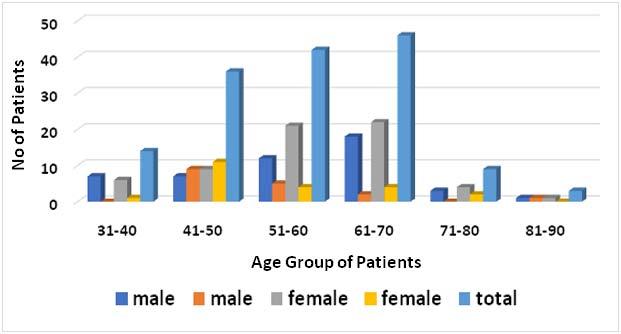
The mean (SD) age of our study patients was 56.5(11.38) years. One hundred twenty-four(82.67%) of patients were between the age group of 41 and 70 years. Out of 111 patients with DKD, 73(65.76%) belonged to the age group between 51 to 70 years. (Fig 1). There were only 12 patients in the age group
Table 2 — Prevalence of various CKD stages and distribution and risk of progression of study participants as per KDIGO 2012
Prognosis of CKD byPersistent albuminuria/ GFR and albuminuria proteinuria categories categories:KDIGO 2012 (description and range) A1<30mg/g A2 30-299mg/gA3>300mg/gTotal (%)
GFReGFRNormal to mildly moderately severely categories ml/min/1.73m-2 increaseincreasedincreased G1
Green : Low risk (if no other marker of kidney disease, no CKD); Yellow : moderately increased risk; Orange : high risk; Red : very high risk.
of 71-90 years of which 9(75%) patients had DKD as opposed to 50 out of which 29(58%) had DKD in the age group of 31-50 years and 88 out of which 70(79.54%) had DKD in the age group of 51-70 years.
We found that this difference in the prevalence of DKD was statistically significant (χ2=7.39, p=0.02), however, it failed to show significance on multivariable regression analysis (Table 3).
In our study we found a predominance of female gender as previously mentioned however, we did not find a significant association between gender and the prevalence of DKD in our study (Table 4).
Blood Pressure :
Ninety-nine (66%) of patients in our study were hypertensive out of which 81 patients had DKD and they comprised 72.97% of the patients having DKD. Presence of hypertension was significantly associated with DKD on both univariable analysis and multivariable logistic regression analysis. The mean (SD) duration of hypertension was 9(5.90) years. There was no significant correlation found between the duration of hypertension and UACR (R=-0.0314, p=0.75) or eGFR (R=-0.1632, p=0.10). The UACR between the hypertensive and normotensive group showed no significant difference of means (t=1.210, p=0.229) but a significant difference was noted in the eGFR between the normotensive and hypertensive groups (t=-2.744,p=0.007).
Body Mass Index (BMI) :
There were 90(60%) patients in the obese category and 30(20%) patients each in the normal and overweight categories. There was no significant difference noted in the prevalence of DKD within the three groups (Table 4).
Duration of Diabetes Mellitus :
The maximum number of patients, 45(30%) in our study had a duration of diabetes of less than five years. Twelve (8%) patients had newly diagnosed diabetes(<6 months), out of which five(41.67%) patients had DKD. We found that the duration of diabetes had a significant effect on the prevalence of DKD both by univariable and multivariable regression analysis.
Diabetic Retinopathy :
Forty-two (28%) of the study patients were found
Table 3 — Multivariable logistic regression for factors influencing the prevalence of DKD
Variable β -Standard p-value Odds 95% Confidence CoefficientError Ratio Interval
to have both DKD and Diabetic Retinopathy (DR) in our study. The association was found to be highly significant (p=0.0001). Even if the prevalence of retinopathy was not very high there was a high chance (95.45%) of diabetic nephropathy being present in these patients. The commonest form of retinopathy was non-proliferative present in 27(18%) patients followed by clinically significant macular edema in 9(6%) and proliferative retinopathy in 6(4%) patients. Correlation between UACR and HbA1c, eGFR and S creatinine :
There was a positive correlation between UACR and HbA1c (R=0.184, p=0.066) levels and serum creatinine (R=0.4563, p=0.00001) according to Pearson’s correlation co-efficient and a negative correlation between the UACR and eGFR (R=-0.366, p=0.00018). However, while the correlation between UACR and HbA1c was not significant, the correlation between the UACR and serum creatinine as well as eGFR was significant to a huge extent, with rising UACR, serum creatinine showed a rise and eGFR a fall in value.
DISCUSSION
We found a 74% prevalence of diabetes as per the prevailing definition which is alarmingly high as compared to the usual figures of DKD prevalence. However, similarly high prevalence rates have been found in other studies also. In an Indian study it was found that the prevalence of DKD in Delhi was 68.4% 11,13 . Another Indian study also showed a prevalence of 68.86%18. There is a recent unreasonable rise in the prevalence of diabetes in India and type 2 diabetes in Asian Indians differs from their Caucasian counterparts in terms of earlier age of onset, obesity is less common and genetic predisposition, (59% patients in our study gave a positive family history of diabetes). It has been reported that Asian Indians have the highest prevalence of T2DM world over19. Apart from that, a high prevalence in our study might have been a result of ours being a tertiary care referral centre which caters to the lower socio-economic class. Our patients were likely to have low awareness of disease leading to longer and poor control of diabetes alongwith financial restraints for follow-up and treatment of the same. Most of our patients were in KDIGO stage G2 and G3 which is in contrast to study by Farah, et al where 55% of patients were in stage G1 and prevalence of DKD was 50.14%20
In our study, we found a female predominance with 85 females in our study and a higher prevalence of DKD within females. A meta-analysis of 10 studies
involving more than 5,00,000 subjects showed that the pooled adjusted risk ratio of 3.34 in women and 2.84 in men without any difference in diabetes related risk of DKD. However, it has been found that women with DKD but without End Stage Renal Disease (ESRD) have better survival than men due to a more rapid and steeper decline in the eGFR in men21
The mean age of patients in our study was 56.5 years and the mean duration of diabetes was 10.34 years. Considering the fact that the mean age of diabetes onset in India is 40 years22 and the peak incidence of DKD is around after 10 to 20 years of onset of diabetes23 after which there is a progressive decline. Our study results are consistent with this fact given the mean age, mean duration of DM and also having maximum DKD patients in the age group of 5170 years of age. We also found a significant relationship of duration of DM with the presence of DKD both by univariate and multivariable analysis hence proving it to be a strong predictor of DKD.
The prevalence of hypertension in our study was 66% which is consistent with studies conducted by Farah, et al20 where hypertension was reported in 69% patients and in 67.14% patients in a study by Bhaisare, et al24 who has also quoted similar other studies having similar prevalence of hypertension. Verma, et al also found 66.3% of patients with DKD to have hypertension18. We found a significant correlation of hypertension with the prevalence of DKD in our study. Hypertension is a well-known risk factor for microalbuminuria however, we did not find any significant difference in UACR between normotensive and hypertensive patients in our study. This could be due to various other factors influencing the same like the glycemic control, duration of diabetes and age influencing UACR. However, there was a significant difference in the mean eGFR of both the groups, this was also found in the study by Verma, et al. Whether this difference is a cause or effect remains controversial but it does suggest that the presence of hypertension might indicate a progression of DKD and remains a strong predictor for the same.
We did not find BMI to be a predictor for DKD in our study. Although, several studies have showed BMI to be positively associated with prevalence of DKD, our study failed to do so. A study by Huang, et al showed that in normal weight T2DM patients, higher HOMA-IR, leptin and resistin levels were associated with higher risk of nephropathy while this was not seen with overweight and obese patients25. This remains an area of potential investigation in Indian patients and might be a reason for the finding in our study. Also, the use of Indian BMI classification may have 23
122, No 04, April 2024Journal
attenuated the significance of association as seen in the study by Man REK, et al26
There is a positive relation between HbA1c and diabetic nephropathy. Surprisingly, in our study we did not find this to be true. This might have been due to the reason that HbA1c though a central biomarker is not a perfect one27. Both, anaemia, especially iron deficiency or hemolytic as well as renal failure can lead to falsely elevated HbA1c. Also, HbA1c reflects glucose control over the preceding 3 months more so of the preceding 6 weeks and is not a predictor of very long-term glucose control28. It has been found that patients of DKD tend to have more incidence of anaemia as compared to their healthier counterparts29
We found a significant association of Diabetic Retinopathy (DR) with DKD in our study. It has been suggested that DR is a strong predictor of DKD progression and presence of severe DR increases the risk of DKD progression30
LIMITATIONS
The limitations of our study were a relatively small sample size from a single centre. The strengths of our study were that it was a prospective study which could generate current data. Most studies of this type are usually of a retrospective nature. We also included only those patients who had visited us for the first time so as to get a better idea about the population dynamics regarding prevalence and control of the disease.
CONCLUSION
In Conclusion, the prevalence of DKD is alarmingly high in type 2 DM patients and goes parallelly with the prevalence of diabetes mellitus. Duration of diabetes, hypertension and advancing age remain important risk factors for the development of DKD. Good glycaemic control remains the mainstay of prevention but HbA1c may not be a helpful biomarker for the same. The best strategy remains early detection of diabetes through screening, probably from the age of 30 onwards in our population and probably earlier with other risk factors. Prevention of diabetes remains the best intervention by following a healthy lifestyle and keeping the modifiable risk factors in check. Stringent control of hypertension remains paramount in prevention of DKD.
REFERENCES
1Pradeepa R, Mohan V — Epidemiology of type 2 diabetes in India. Indian Journal of Ophthalmology 2021; 69(11): 2932.
2Saeedi P, Petersohn I, Salpea P, Malanda B, Karuranga S, Unwin N, et al — Global and regional diabetes prevalence estimates for 2019 and projections for 2030 and 2045: Results
from the International Diabetes Federation Diabetes Atlas. Diabetes Research and Clinical Practice 2019; 157: 107843.
3Kaveeshwar SA, Cornwall J — The current state of diabetes mellitus in India. AustralasMedJ2014; 7(1): 45-8. doi: 10.4066/ AMJ.2013.1979. PMID: 24567766; PMCID: PMC3920109.
4Anjana RM, Pradeepa R, Deepa M, Datta M, Sudha V, Unnikrishnan R, et al — ICMR–INDIAB Collaborative Study Group. Prevalence of diabetes and prediabetes (impaired fasting glucose and/or impaired glucose tolerance) in urban and rural India: phase I results of the Indian Council of Medical Research-INdiaDIABetes (ICMR-INDIAB) study. Diabetologia 2011; 54(12): 3022-7. doi: 10.1007/s00125-011-2291-5. Epub 2011 Sep 30. PMID: 21959957.
6Roglic G — WHO Global report on diabetes: A summary. International Journal of Noncommunicable Diseases 2016; 1(1): 3.
7Burrows NR, Hora I, Geiss LS, Gregg EW, Albright A — Incidence of End-Stage Renal Disease Attributed to Diabetes Among Persons with Diagnosed Diabetes - United States and Puerto Rico, 2000-2014. MMWR Morb Mortal Wkly Rep 2017; 66(43): 1165-70. doi: 10.15585/mmwr.mm6643a2. PMID: 29095800; PMCID: PMC5689212.
8Modi GK, Jha V — The incidence of end-stage renal disease in India: a population-based study. Kidney Int 2006; 70(12): 2131-3. doi: 10.1038/sj.ki.5001958. Epub 2006 Oct 25. PMID: 17063176.
9Parving HH, Lewis JB, Ravid M, Remuzzi G, Hunsicker LG — DEMAND investigators. Prevalence and risk factors for microalbuminuria in a referred cohort of type II diabetic patients: a global perspective. Kidney Int 2006; 69(11): 2057-63. doi: 10.1038/sj.ki.5000377. PMID: 16612330.
10Kebede SA, Tusa BS, Weldesenbet AB, Tessema ZT, Ayele TA — Incidence of Diabetic Nephropathy and Its Predictors among Type 2 Diabetes Mellitus Patients at University of Gondar Comprehensive Specialized Hospital, Northwest Ethiopia. J Nutr Metab 2021; 2021: 6757916. doi: 10.1155/ 2021/6757916. PMID: 34497725; PMCID: PMC8419489.
11Noubiap JJ, Naidoo J, Kengne AP — Diabetic nephropathy in Africa: A systematic review. World J Diabetes 2015; 6(5): 759-73. doi: 10.4239/wjd.v6.i5.759. PMID: 26069725; PMCID: PMC4458505.
12Rajput R, Prasanna Kumar KM, Seshadri K, Agarwal P, Talwalkar P, Kotak B, et al — Prevalence of Chronic Kidney Disease (CKD) in Type 2 Diabetes Mellitus Patients: STARTIndia Study. J Diabetes Metab 2017; 8: 2. DOI: 10.4172/21556156.1000722
13Dash SC, Agarwal SK, Panigrahi A, Mishra J, Dash D — Diabetes, Hypertension and Kidney Disease Combination “DHKD Syndrome” is common in India. J Assoc Physicians India 2018; 66(3): 30-3. PMID: 30341865.
14Patel V, Shastri M, Gaur N, Jinwala P, Kadam A — A study in prevalence of diabetic nephropathy in recently detected cases of type 2 diabetes mellitus as evidenced by altered creatinine
Vol 122, No 04, April 2024Journal of the Indian Medical Association
clearance, urinary albumin and serum creatinine, with special emphasis on hypertension, hypercholesterolemia and obesity. International Journal of Advances in Medicine 2018; 5(2): 351-5. doi:http://dx.doi.org/10.18203/2349-3933.ijam20180999
15Levin A, Stevens PE — Summary of KDIGO 2012 CKD Guideline: behind the scenes, need for guidance, and a framework for moving forward. Kidney Int 2014; 85(1): 4961. doi: 10.1038/ki.2013.444. Epub 2013 Nov 27. PMID: 24284513.
16American Diabetes Association. Standards of Medical Care in Diabetes-2019 Abridged for Primary Care Providers. Clin Diabetes 2019; 37(1): 11-34. doi: 10.2337/cd18-0105. PMID: 30705493; PMCID: PMC6336119.
17Misra A, Chowbey P, Makkar BM, Vikram NK, Wasir JS, Chadha D, Joshi SR, et al — Concensus Group. Consensus statement for diagnosis of obesity, abdominal obesity and the metabolic syndrome for Asian Indians and recommendations for physical activity, medical and surgical management. J Assoc Physicians India 2009; 57: 163-70. PMID: 19582986.
18Verma A, Vyas S, Agarwal A, Abbas S, Agarwal DP, Kumar R — Diabetic Kidney Disease and Hypertension: A True Love Story. J Clin Diagn Res 2016; 10(3): OC11-3. doi: 10.7860/ JCDR/2016/18806.7511. Epub 2016 Mar 1. PMID: 27134912; PMCID: PMC4843298.
19Unnikrishnan R, Anjana RM, Mohan V — Diabetes mellitus and its complications in India. Nat Rev Endocrinol 2016; 12(6): 357-70. doi: 10.1038/nrendo.2016.53. Epub 2016 Apr 15. PMID: 27080137.
20Farah RI, Al-Sabbagh MQ, Momani MS, Albtoosh A, Arabiat M, Abdulraheem AM, et al — Diabetic kidney disease in patients with type 2 diabetes mellitus: a cross-sectional study. BMC Nephrol 2021; 22(1): 223. doi: 10.1186/s12882-021-024294. PMID: 34134654; PMCID: PMC8207700.
21Giuffrida AE, Gembillo G, Cucinotta D, Squadrito G, Santoro D, Russo GT — Gender Differences in Diabetic Kidney Disease: Focus on Hormonal, Genetic and Clinical Factors. Int J Mol Sci 2021; 22(11): 5808. doi: 10.3390/ijms22115808. PMID: 34071671; PMCID: PMC8198374.
22India State-Level Disease Burden Initiative Diabetes Collaborators. The increasing burden of diabetes and variations among the states of India: the Global Burden of Disease Study 1990–2016. Lancet Glob Health 2018; 6(12): e1352-62.
23Shahbazian H, Rezaii I — Diabetic kidney disease; review of the current knowledge. J Renal Inj Prev 2013; 2(2): 73-80. doi: 10.12861/jrip.2013.24. PMID: 25340133; PMCID: PMC4206005.
24Bhaisare SD, Rao AK, Jog AS — Clinical study of urine albumin creatinine ratio as an earlier predictor of diabetic nephropathy. JEvolutionMedDentSci2020; 9(09): 598-602, DOI: 10.14260/ jemds/2020/133
25Huang J, Peng X, Dong K, Tao J, Yang Y — The Association Between Insulin Resistance, Leptin, and Resistin and Diabetic Nephropathy in Type 2 Diabetes Mellitus Patients with Different Body Mass Indexes. Diabetes Metab Syndr Obes 2021; 14: 2357-65. doi: 10.2147/DMSO.S305054. PMID: 34079314; PMCID: PMC8163637.
26Man REK, Gan ATL, Fenwick EK, Gupta P, Wong MYZ, Wong TY, et al — The Relationship between Generalized and Abdominal Obesity with Diabetic Kidney Disease in Type 2 Diabetes: A Multiethnic Asian Study and Meta-Analysis. Nutrients2018; 10(11): 1685. doi: 10.3390/nu10111685. PMID: 30400648; PMCID: PMC6266073.
27Kaiafa G, Veneti S, Polychronopoulos G, Pilalas D, Daios S, Kanellos I, et al — Is HbA1c an ideal biomarker of wellcontrolled diabetes? Postgrad Med J 2021; 97(1148): 380383. doi: 10.1136/postgradmedj-2020-138756. Epub 2020 Sep 10. PMID: 32913038.
28Radin MS — Pitfalls in hemoglobin A1c measurement: when results may be misleading. J Gen Intern Med 2014; 29(2): 388-94. doi: 10.1007/s11606-013-2595-x. Epub 2013 Sep 4. PMID: 24002631; PMCID: PMC3912281.
29Martynov SA, Shestakova MV, Shilov EM, Shamkhalova MS, Vikulova OK, Sukhareva OYu, et al — Prevalence of anemia in patients with type 1 and type 2 diabetes mellitus with chronic renal disease. Diabetes Mellitus 2017; 20(5): 31828. https://doi.org/10.14341/DM9369
30Gupta M, Rao IR, Nagaraju SP, Bhandary SV, Gupta J, Babu GTC — Diabetic Retinopathy Is a Predictor of Progression of Diabetic Kidney Disease: A Systematic Review and MetaAnalysis. Int J Nephrol 2022; 2022: 3922398. doi: 10.1155/ 2022/3922398. PMID: 35531467; PMCID: PMC9076335.
Original Article
A Study on Association of Heinous Offences with Demographic, Socio-economic Factors and Personality Traits among the Children in Conflict with Law Staying in Juvenile Justice Homes
Background : Children in Conflict with Law (CCL) are those children between 7years to 17years who have committed some offence and have been placed in Juvenile Justice (JJ) Homes. Heinous offences are the most severe among all the offences committed by those CCLs. For prevention of those types of crimes by CCLs, some data were required on the demographic, Socio-economic and personality traits of those JCLs to find any association between heinous crimes and those factors. This study was undertaken to do that.
Materials and Methods : The study was a descriptive epidemiological study with cross-sectional design undertaken among 125 inmates staying in JJ Homes of West Bengal, India, between August, 2017 to October, 2018. Each of the 125 inmates were interviewed with a predesigned and pre-tested questionnaire and also with the Junior Eyesenck Personality (JEP) questionnaire and analysed by appropriate statistical methods.
Results : Significantly more Indian heinous offenders were males demographically and significantly more of them were either student or employed as child workers socio-economically. Most of the Indian heinous offenders were neurotic with strong association. Significantly more of them were introvert and the association was also strong. Most of the heinous offenders had average or low social desirability. Recidivism was significantly associated with absence of substance abuse and absence of gangsterism. Watching adult movies or pornography, had no association with sexual offences.
[J Indian Med Assoc 2024; 122(4): 26-30]
IKey words :CCL, JJ Home, Heinous, JEP questionnaire, Recidivism.
n the Juvenile Justice (Careand Protection of Children) Act, 1986 (JJ Act), which was amended in 2000 and 2015, the term “Child in Conflict with Law (CCL)” or “Juvenile in Conflict with Law” was used, for children above 7 years and below 18 years committing some offence classified as petty, serious and heinous offence by the court of law resulting in difference in duration of imprisonment for punishment. For heinous offences, the most severe type of offences, they are tried like adult heinous offenders and are sentenced for seven years or more1 A statistic published in India by National Crime record Bureau showed that in 2005, about 18939 juveniles were in conflict with law but the
1MBBS, DMCW, MD, FM, Assistant Professor, Department of Forensic Medicine and Toxicology, Barasat Government Medical College, Barasat, West Bengal 700124 and Corresponding Author
2MBBS, MD (Psychiatry), Professor, Department of Psychiatry, Bankura Sammilani Medical College and Hospital, Bankura, West Bengal 722102
3MBBS, MD, SPM, Associate Professor, Department of Community Medicine, RG Kar Medical College & Hospital, Kolkata 700004
4MBBS, MD, FM, Professor and Principal, KPC Medical College & Hospital, Jadavpur, Kolkata 700032
Received on : 22/11/2023
Accepted on : 19/12/2023
Editor's Comment :
It is seen from this study that among CCLs, boys, especially of higher age-group, originating from nuclear families, having lower education levels, either student or employed as child labourers, with neurotic and introvert personality mostly commit heinous offences.
Recidivism of offences has no significant association with substance use, gangsterism or nature of offence. Watching pornography or adult movies isn’t associated with sexual offences.
number had increased upto 35849 in 20162 It was also seen that sexual offences like rape were also on the rise and was more in number than other heinous crimes. So, there must have been some factors behind it. If any association could be established between juvenile delinquency with any modifiable factor, then those factors could have been controlled. There was paucity of scientific data in this field in our country. That was the reason behind undertaking the present study.
MATERIALS AND METHODS
It was a descriptive study with cross-sectional design undertaken among the inmates of five juvenile justice homes of West Bengal, run by Government of West Bengal from 10th August, 2017 to 31st October,
2018. The sample size was calculated using the formula (Zα /2 2 PQ)/L2 where, Zα/2 = Standard normal deviate and has a value of 1.96 at 95% confidence level; P= Expected proportion of juveniles committing a particular crime (rape) among accused of that age group, Q=100-P; L= allowable error. According to National Crime Records Bureau (NCRB), India, data in 2016, total number of juveniles in conflict with law (JCL) in India was 44171 and 2054 were accused of rape3. Therefore, P=4.65%, Q=95.35% +and Z α/2 2 =3.84. Considering the allowable error as 5 percentage points, a design effect of 1.16 [based on the formula, DEFF = 1+(m -1)*ICC] and a non-response rate of 14% (based on a pilot study), the sample size was calculated to be 125. Capacity of the JJ Homes situated in 14 districts of West Bengal, ranged from 25 to 250. Out of 14 districts with JJ Homes, 20% were selected by simple random sampling technique at first stage. From the observational homes and special homes (separate enclosures in a JJ Home) of selected three districts, inmates were selected following probability proportionate to the size principle. JJ Homes of selected three districts were visited fortnightly till desired sample size of a particular home was reached. In a particular JJ Home, list of inmates used to be prepared afresh with the help of officials for each day of visit as new inmates were being placed in those homes and old inmates were being released on completion of their tenure of stay there on a regular basis. On each day of visit to the JJ Home, six inmates could be interviewed taking time required to complete an interview of an inmate into account. From the prepared list of the inmates, six were to be selected by systematic random sampling technique. Informed consent for the study was sought from the legal guardians of the juvenile inmates. Study subjects who had completed 18 years of age but not yet shifted to adult correctional homes, physically ill inmates, inmates summoned to attend court of law on the day of interview and inmates already interviewed during previous visits to that particular home, were excluded from the list. Before undertaking the study, permission from Director, Child Rights and Trafficking, Government of West Bengal (GOWB), a State Government under the Indian Union, was obtained to visit five JJ Homes. Approval for the study was obtained from the Institutional Ethics Committee of RG Kar Medical College and Hospital (RGKMC&H), Kolkata, West Bengal, India. Necessary permission from EdITS, P O BOX 7234, San Diego, CA92167, US, (copyright owner) was obtained on procurement of the study tool, Junior Eyesenck Personality (JEP) questionnaire for assessing the personality trait of the inmates.
On each day of visit, responses from inmates were recorded in the predesigned, pretested questionnaire framed by the investigators and in the JEP questionnaire. Psychological analysis of the respondents was carried out subsequently based on the scoring system of JEPQ. Collected data were compiled in MS-excel spread sheet and analysed using SPSS version 26, Jamovi. Qualitative variables were expressed as frequencies and percentages. For summarization of quantitative variables mean± Standard Deviation/median with inter quartile range were used. Chi square test, Fisher Exact test, Mann Whitney U test was used to find out association along with Logistic Regression analysis. A p-value <0.05 was considered as statistically significant.
OBSERVATIONS
From Table 1, demographically, it was seen that among the respondents of the study, 100 (80%) were Indians and 25 (20%) were foreign inmates. Among the Indian respondents, 77 (77%) were involved in heinous offences whereas only 3 (12%) of their foreign counterparts committed heinous crimes. Heinous offenders were mostly from higher age group. Heinous offences were significantly (p=<0.05) higher among Indian males, 74(84.1%) in number, compared to females, 3(25%) in number. More heinous offenders came from rural areas and nuclear families.
Socio-economically, it was seen that very few among the Indian heinous offenders passed secondary level (class 10 standard) of education. Proportion of heinous offenders, 74 (79.6%) among students or children employed in any work as child labourer was significantly higher compared to 3(42.9%) among beggars or ragpickers. Among all Socio-economic classes according to the classification of modified BG Prasad scale 3 , applicable only in Indian context, proportions of heinous offenders were much higher compared to non-heinous offenders in class II (upper middle) and III (middle). Heinous offenders were found to be more among inmates living in kutcha houses, without any previous history of substance use or without any history of physical abuse but number of heinous offenders were significantly (P = <0.05) more among inmates without major familial or personal problem like broken homes, death or separation of parents compared to those inmates having such type of family problems.
Regarding the association of personality trait evaluated by JEP questionnaire it could be observed that, in terms of psychoticism, most of the Indian inmates who were either emotionally well-adjusted or emotionally constricted were heinous offenders, whereas 27
Table 1 — Association of Socio-demographic, Economic factors and Personality traits with heinous offences (n=125) Indian Inmates (n=100)Foreign Inmates (n=25)
Social Class# (BGI 7(77.8)2(22.2) 0.84@(2)NANANA** Prasad scaleII & III 48(78.7)13(21.3)NANA only for Indians)IV & V 22(73.3)8(26.7)NANA Housing Pucca37(78.7) 10(21.3) 0.70@(1)3(20) 12(80)0.53* Kutcha/ Mixed
* By Fisher’s exact test; @ By chi-square test; **NA= Not applicable; DF = Degree of Freedom; # I=upper, II=upper middle, III=middle, IV=lower middle, V=lower. (Figures in parentheses indicate row percentage)
a few psychotic inmates committed such crime. Regarding extroversion, significantly (P = <0.05) more heinous offenders, 53(86.9%) in number, were seen among introvert Indian inmates. Only 24 (61.5%) of the inmates were extrovert or ambient in nature. Based on lie scale, which judged social desirability of inmate’s responses, it could be said that Indian inmates committing heinous offences, mostly had average or low social desirability. All foreign heinous offenders hailed from nuclear families, were males, were in the age group of 13-17 years,were educated below secondary level. Majority of the heinous offender foreign inmates came from rural area, were either student or employed, had history of substance use, had major familial or personal problem, were emotionally well adjusted or constricted, were ambient or extrovert, were neurotic and had average or low social desirability but the sample size of foreigners was too small for any proper statistical evaluation. Table 2 shows different factors found to be
significantly associated with heinous crime among Indian inmates. It was observed that odds of committing heinous crime was significantly higher among males, among student or employed, among introvert inmates and among neurotics assuming that there is no change in other predictor variables.
From Table 3 it was observed that, absence of substance use and also absence of gangsterism were significantly associated with recidivism. Adjusted odds ratio revealed that both of these two factors independently retained their significant association with recidivism in assumption of no change in other predictor variables.
From Table 4, It was observed that, there was no significant association between viewing adult movies or pornography and committing heinous offences.
DISCUSSION
The findings of the present study was supported by a research work which also showed that CCLs of
122, No 04, April 2024Journal
higher age group were involved in heinous offences 4 The large scale Denver, Rochester and Pittsburgh longitudinal studies, also supported the findings of the present study5.
Table 2 — Measuring Strength of the Association of Significant Factors with Heinous Offences only among Indian Inmates (n=100)
Factors P-valueAdjusted Odds Ratio (with 95% confidence interval)
Sex [Male (as reference) & Female]<0.050.111 (0.022-0.555)
Among JCLs heinous offences were found to be significantly higher among boys compared to girl inmates with strong association. This was also supported by the data published in National Crime Records Bureau (NCRB), India, both in 20162 and 20216.
Table 3 — Association of Recidivism with Nature of Offences and Different Factors (n=125)
Recidivism among inmatesPresent (%) Absent (%) P-valueAdjusted Odds Ratio (with 95% CI)
Table 4 — Association of sexual offences with viewing adult movies/ pornography by Indian inmates (n=100)
In this study, more Indian heinous offenders came from rural areas than urban areas but that was to be considered as a reflection of normal population distribution of India according to census data in 20117
Sexual Other P-value Offences (%)Offences (%)
Viewed adult film/pornography14 (45.2)17 (54.8)0.32@ Did not view adult film/ pornography24 (34.8)45 (65.2) @ By Chi-square test; (percentage calculated as row percentage)
The observation that more Indian inmates involved in heinous crimes came from nuclear families than from joint families was clearly in excess of the normal proportion of nuclear families in our country as evident by the data collected in India between 2019 to 2021 through National Family Health Survey 5 (NFHS 5).
The observation in this study that most of the heinous offenders were either illiterate or just literate or educated up to primary level (class IV standard) but did not pass secondary (class X standard) board examination was supported by a study in Netherlands9
It was also seen that heinous offences were significantly higher among inmates who were either student or employed as child labourer and the association was also strong. This was supported by a study in UK10
The observation that most of the heinous offenders came from social class II (upper middle) and III (middle) followed by IV (lower middle) and V (lower) and then by I (upper) according to the BG Prasad scale based on Indian context11 was supported by a study in UK12
The observation that substance use were absent among majority of heinous offenders was supported by a study on JCLs in Sweden13 and another study in UK14
The observation that, heinous crime was significantly more common among inmates not having history of major familial or personal problems was partially supported by a study in Carolina, US15 Regarding personality trait, in terms of extroversion, significantly more heinous offenders from India were introvert in comparison to extrovert and ambient inmates and the association was also strong. Heinous offences were significantly more among neurotics and this also showed strong association. In a study among the JCLs in Sweden in 2008 it was seen that, those who were in judicial custody, suffered more from depression and childhood developmental disorder but with lower rates of psychosis and bipolar disorder than adult forensic psychiatric examinees13. In another study among JCLs in UK, it was found that 7% of them had some psychiatric problem necessitating further treatment14
Recidivism was significantly associated with absence of substance use and absence of gangsterism among inmates and the association was also strong. But in a study, it was observed that there is a possibility of a positive substance use and recidivism among incarcerated delinquents16. Another study in US showed that gangsterism had increased the proportion of juvenile delinquency specially among younger juveniles17
122, No 04, April 2024Journal
From the present study, no association could be established between committing sexual offences and viewing sexually explicit materials which was supported by a study in India18
CONCLUSION
From this study, it can finally be concluded that among CCLs, boys were significantly more involved in heinous offences. CCLs of higher age-group, belonging to nuclear families and educated below secondary level were more involved in heinous offences. Significantly more heinous offenders were found among students or inmates employed as child labourers previously. Inmates without the history of major familial or personal problems and belonging to upper middle and middle social class were also involved in heinous offences. Recidivism of any type of offence was found to be significantly associated with absence of any substance use and absence of gangsterism also. Majority of CCLs committing heinous offence did not experience any physical abuse.
By psychological evaluation of the CCLs, it was seen that regarding extroversion, inmates with introvert personality had a significantly strong association with heinous crime. Regarding Neuroticism, neurotics also had a significantly strong association with heinous crime. Regarding psychoticism, most of the Indian heinous offenders were either emotionally welladjusted or emotionally constricted. Most of the Indian and foreign heinous offenders had average or low social desirability according to the lie scale.
No significant association was found between watching adult movies or pornography with involvement in sexual offences among the JCLs.
ACKNOWLEDGEMENT
Dr Narendra Kumar Tiwary, Statistician cum Assistant Professor, Department of Community Medicine, RG Kar Medical College, Kolkata, West Bengal, India.
Conflict of interest : Nil
REFERENCES
1The Juvenile Justice (Care and Protection of Children) Act, 2015. GOI. Available from: www.indiacode.nic.in/bitstream/ 123456789/8864/1/201602.juvenile2015pdf.pdf [Accessed on 20 Nov 2023]
2Juveniles in Conflict with Law, Crime in India statistics. 2016. National Crime records Bureau, Ministry of Home affairs, New Delhi, GOI. Available from: https://ncrb.gov.in/sites/default/ files/crime_in_india_table_additional_table_chapter_reports/ Table%205A.4.pdf [Accessed on 16 Apr 2023]
3Pandey VK, Aggrawal P, Kakkar R — Modified BG Prasad’s Socio-economic Classification-2018: The need of an update in
the present scenario. IndianJournalofCommunityHealth 2018; 30(1): 82-4. doi: https://doi.org/10.47203/IJCH.2018.v30i01.014
5Huizinga D, Loeber R, Thornberry TP — Longitudinal Study of Delinquency, drug use, sexual activity and pregnancy among children and youth in three cities. Public Health Reports 1993; 108: 90-6.
6National Crime records Bureau, Ministry of Home affairs, Government of India, New Delhi. Juveniles in Conflict with Law, Crime in India statistics. 2021 Available from: https:// view.officeapp s.live.com/op/view.aspx?src=https% 3A%2F%2Fncrb.gov.in%2Fsites%2Fdefault%2Ffiles%2FCII2021%2FTABLE%25205A.4A.xlsx&wdOrigin=BROWSELINK [Accessed on16 April, 2023]
7Census of India. Govt. of India. Table-A-01: Number of villages, towns, households, population and area (India, states/UTs, districts & sub-districts). 2011 Available from: https:// censusindia.gov.in/census.website/dat a/census-tables [Accessed on 1 May, 2023].
8National Family Health Survey 5 (2019-2021).Ministry of H & FW, GOI.Table:2.14: Household Composition:2022. Available from: https://dhsprogram.com/pubs/pdf/FR375/FR375.pdf [Accessed on 21 Apr, 2023].
9Rud I, Klaveren C V, Groot W, Maassenvandenbrink H — Education and Youth Crime: A review of the Empirical Literature. Tier Working Paper 16/6:2013. Available from: https://www.researchgate.net/publication/269392011_ Education_and_Youth_Crime_A_review_of_the_Empirical_ Literature [accessed on18th April, 2023]
10Farrington DP — The psychosocial milieu of the offender. In: Gunn J, Taylor PJ editors- Forensic Psychiatry. 2nd Ed. New York: CRC Press. 2014: 177.
11Khairnar MR, Kumar PG, Kusumakar A — Updated BG prasad socioeconomic status classification for the year 2021. J Indian Assoc Public Health Dent 2021; 19: 154-5.
12Wilson H — Juvenile Delinquency, Parental Criminality and Social Handicap. British Journal of Criminology 1975; 15(3): 241-50.
13Fazel M, Langstrom N, Grann M, Fazel S — Psychopathology in adolescent and young adult criminal offenders (15-21 years) in Sweden. Social Psychiatry and Psychiatric Epidemiology 2008; 43(4): 319-24.
14Dolan M, Holloway J, Bailey S, Smith C — Health status of juvenile offenders. A survey of young offenders appearing before the juvenile courts. Journal of Adolescence 1999; 22(1): 137-44.
15Boccio CM, Beaver KM — The Influence of Family Structure on Delinquent Behavior. Youth Violence and Juvenile Justice, 2019; 17(1): 88-106. Available from: https://doi.org/10.1177/ 1541204017727836.
16Welner M, DeLisi M, Baglivio MT, Guilmette TJ, Knous-Westfall HM — Incorrigibility and the Juvenile Homicide Offender: An Ecologically Valid Integrative Review. Youth Violence and Juvenile Justice 2022; 20(1): 22-40. https://doi.org/10.1177/ 15412040211030980
17Zachary AP, Jessica MC, Alex RP, Michael TB, Nathan E — Delinquent Youth Concentration and Juvenile Recidivism. Deviant Behavior 2021: 42(7): 821836, DOI: 10.1080/01639625.2019.1701622
18Math SB, Viswanath B, Maroky AS, Kumar NC, Cherian AV& Nirmala MC — Sexual Crime in India: Is it Influenced by Pornography? Indian Journal of Psychological Medicine 2014; 36(2): 147-52. DOI: 10.4103/0253-7176.130976.
Original Article
Knowledge, Attitude and Practice on Breastfeeding in the Rural Field Practice Area in Tamil Nadu
Breastfeeding is the primary event of the utmost importance that is required to be executed within one hour of birth. Exclusive breastfeeding up to a certain age is considered to hold significant importance. The study would investigate the Knowledge, Attitude and Practice of breastfeeding in the rural field practice area in Tamil Nadu. Exclusive breastfeeding has been proposed as the best nutritional option for infants up to six months of age. In the age of 19-32 years, female groups the 230; 19-21 year’s age group showing 43 percent. Hindu, Muslim and Christian in the ratio 182:21: 27. Pre-lacteal feed as Factor affecting the Breastfeeding practices- in accordance with the duration of each breastfeeding characterization which is less than 10 mins. Out of 230 females, 35 breastfed their children from one side with a 15 percent outcome. In 195 females who are breastfeeding their children from both sides have 85 percent. Study shows that socio-economic factors are important for long-term breastfeeding. Findings suggest that a mother’s sense of control over breastfeeding and perceived acceptance will affect the intentions and behavior of mothers.
[J Indian Med Assoc 2024; 122(4): 31-7]
Key words :Breastfeeding, Knowledge, Attitude, Practice, Tamil Nadu, Pre-lacteal Feed.
Breastfeeding is the primary event of the utmost importance that is required to be executed within one hour of birth1. For the newborn, the only compatible source of nutrients is the breast milk of the lactating mother. Breast milk fed children have a greater chance of survival than a child artificially fed2. Further, it also renders protection to the newborn from malnutrition and other infections. Exclusive breastfeeding up to a certain age is considered to hold significant importance in not only reducing the mortality and morbidity rates of the children but also their effective development3,4 Breastfeeding should begin within one hour of birth, breast milk is the ideal food for the infant5,6
Giving breastfeeding immediately after delivery prevents PPH in large amounts7. Given the importance of breastfeeding the current study would investigate the Knowledge, Attitude and Practice on breastfeeding in the rural field practice area in Tamil Nadu. This section of the study would elaborate on breastfeeding, it’s the appropriate time of initiation, the importance of breastfeeding and its implications in the growth and development of the child. The section would conclude with the rationale behind the genesis of the study.
Department of Community Medicine, Government Medical College and Hospital, Krishnagiri, Tamil Nadu 635115 1MD, Associate Professor and Corresponding Author 2MD, Associate Professor 3MS (ENT), Postgraduate Trainee, JJM Medical College, Davanagere, Karnataka 577004
Received on : 14/10/2022
Accepted on : 05/11/2022
Editor's Comment :
Socio-economic factors, sense of control and perceived acceptance of social breast feeding will affect the intention and behaviour of the Mother's.
Research Problem :
Exclusive breastfeeding or the practice of feeding the infant for the first 6 months of life on breast milk only without any other type of food, not even water has been recommended as the best feeding alternative for infants up to 6 months owing to its protective effect against mortality and morbidity 2,4 . In this regard recommendations from international bodies such as the American Academy of Pediatrics (AAP) and World Health Organization (WHO) states exclusive breastfeeding for the first 6 months and continued breastfeeding for at least 12 months to be compulsory. The same could be continued up to the age of 2 years or beyond depending upon the desire of the child5
However, advocates of breastfeeding have noticed a significant decline in the exclusive breastfeeding behavior among nursing mothers specifically in developing countries6. Global statistics have shown that only 46.4% of the mothers in India exclusively breastfed their children in the first 6 months. Whereas the rates of breastfeeding in 0-6 months are 56.2 percent in Kerala and 0-3 month’s exclusive breastfeeding is 68.5%1
Knowledge about breastfeeding has been identified to have a significant correlation with the attitude and practice of breastfeeding. Improving women’s
122, No 04, April 2024Journal
educational level has clear impacts on the health of the newborn 2,5 . There have been studies that investigate the knowledge, attitude and practice of breastfeeding of lactating mothers in different parts of India, there has been no specific research that focuses on rural field practice areas in Tamil Nadu. The current study intends to fill the gap by conducting a crosssectional study that would investigate the knowledge, attitude, and practice on breastfeeding among the mothers in Dharmapuri, a city in the western part of Tamil, Nadu, India.
AIMS AND OBJECTIVES
The objectives of the study will be listed in this section of the study.
To highlight the status of breastfeeding and associated health of infants in rural India.
To emphasize the importance of breastfeeding in ensuring the healthy development of the child.
To investigate the impact of the demographic factors on the breastfeeding trend among mothers.
To investigate the knowledge, attitude and practice of breastfeeding among the mothers.
To propose the probable measures that could enhance the status of breastfeeding.
Literature Review :
It is necessary to understand the determinants of the decisions of the mothers regarding long-term breastfeeding in order to encourage breastfeeding for up to 12 months after and beyond2,5,7. In a retrospective study on feeding children, 80 participants who breastfed nine-month-old infants were used to illustrate the expected behavioral theory. The longer they were breastfed, the more participants received less approval for breastfeeding. The approval received did not explain the intended prenatal duration but strongly explained the expected duration at 9 months after the first day5,7 Nevertheless, the perceived number of control mothers during breast-feeding clarified the prenatal and 9-month breastfeeding intentions. The weaning reasons for 9 to 12 months showed the effects of perceived power but not perceived acceptance. Such results indicate the importance of (1) measures to make long-term breastfeeding socially acceptable and (2) ongoing guidance and support for long-term breastfeeding7
As per the World Health Organization, neonatal deaths account for 45 percent of the under-five. More than one percent of these deaths occur in the first 24 hours of birth, while three-quarters of neonatal deaths occur in the first seven days. The aim of this study is to assess the awareness, attitudes and practices of mothers in the province of Badin Sindh in Pakistan
regarding the treatment of young people and their related factors27. Within 48 hours of delivery, the survey examined the factors related to the breastfeeding decision8,13. The Mother’s response to the issue of child feeding preference was tested against food records. A number of issues related to perceived maternal lactation support for six persons. There was also a collection of issues affecting the decision of the mother on breast or bottle feeding. There have been a number of different trends in bivariate connections between assistance and breastfeeding between ethnic and maritime groups. In order to find out where breastfeeding support was most relevant, single and multiple logistic analyzes were conducted within ethnic groups 8,14. The support of the male partner was significant among the Anglo-Americans. A wide range of research questions that follow has been raised. This suggests that effective support may be a significant intervention variable, but that the individual supporting the intervention program differs from ethnic to ethnic groups8.
In-hospital self-efficacy in breastfeeding has been measured and 4 weeks of insufficient milk perception has been measured. Although most mothers plan to breastfeed exclusively, less than 40% were breastfeeding at 4 weeks of age9,15,16. In 73% of mothers using the formula considered the primary cause of supplementation or complete cessation of breastfeeding to be inadequate milk. In the immediate postpartum period (r=0.45, p<0.001), mothers were significantly associated with their perception of insufficient milk for hospital breastfeeding in 4 weeks postpartum. Multiple hierarchical regression has shown that lactating self-efficacy is 21 percent different from the mother’s expectations of insufficient milk and is independent of socio-demography9. Modifiable factors affecting women’s breastfeeding are: focus on breastfeeding, self-efficacy of breastfeeding and social support. Additional stereo promotion strategies often involve social support and do not address sufficient attempts to change the purpose and self-efficiency of breastfeeding10,17
During the period from 1 August to 30 September, 2005, a survey of questionnaires administered by health personnel was carried out. The levels of initiation were 72.7 and 84.4 percent, respectively, within 1 hour and 24 hours of delivery. Within two months of birth, 82.3 percent of mothers had exclusive breastfeeding11,18. As the first feed, 332 babies (86.2 percent) had breast milk/colostrum, but 17.2 percent had breast milk or breast milk from another mother. 14 percent of infants received a pre-lacteal feed.
Formula feed (6.2 percent), sugar water (5.9 percent), and cow’s milk (2.8 percent) are all popular pre-lactate feeds. 12.7 percent of mothers had additional feed added. The study of logistic regression affected the exclusive breastfeeding habits and forms of delivery of the mother and the first feeding of the baby11
The frequency of delays in LS-II ranges from 11 to 44 percent, which is most likely to occur with primiparity12,19. Delayed LS-II was correlated with a variety of factors with mixed evidence: Cesarean section, prolonged phases of work II, stress, maternal age, diabetes, obesity, inadequate breastfeeding, and pre-onset supplementation12,20-22. The objective was to determine how primary breastfeeding mothers affect the influence of birth and maternal / infant factors on the LS-II Na+ marker. The involvement of gestational diabetes poses a risk of high na+ breast milk for 48 hours 12,23-25 . This reduces the development of breastfeeds, indicating the initiation of lactogenesis II (mean=8,91, SD=3,77)12,26
MATERIALS AND METHODS
This cross-sectional research study was conducted among 230 pregnant mothers in Government Dharmapuri Medical College and Hospital. Research classes included a 3 months delivery of pregnant women’s and a study period of a purposive random sampling of 4 months. The analysis was completed one month after October 2017 with 230 participants. Inclusion criteria: Mothers came for delivery from rural field practice area to the government dharmapuri medical college and hospital and those mothers who are willing to take part in the study are included. Participants were fully informed and written consent was obtained on the nature and purpose of the study. The research was conducted using an anonymous questionnaire specifically designed to obtain qualitative and quantitative data for the classification of semidesigned semi-structured, pre-tested proforma, simple random sampling, frequency and percentage only predictions. Knowledge concerns related to preventive measures such as the use of safety tools and intervention in the event of an unknown source have also been addressed.
Before the study was conducted, the Institutional Ethics Committee received clearance of the research procedure. In order to identify the different aspects of the Institutional Clearance Obtained, the results were evaluated under different headings. The methods used were MS Excel and other simple data analysis techniques.
RESULTS
A Socio-economic of the Mother as Factor Affecting the Breastfeeding Practices —
As shown in Table 1, the socio-economic factors ie, age, religion, income, employment, type of delivery, education and gravida. In the age of 19-32 years, female groups the 230; 19-21 years age group showing 43 percent, 22-24 years group showing 21 percent, 2527 years group shows 22 percent and 28-32 years group shows 14 percent. In religion, the groups were taken Hindu,Muslim,andChristian in the ratio 182:21: 27 showing 79, 9, 12 percent respectively.
While when we compare the income (Rs/month) category there is a variation from low to high-income status. From 600-1500 income categories have 122 females with a percentage of 53 percent. From the 1501-4000 income category, we’ve 27 females with 27 percent. From the 4001-7000 income category, we’ve 47 females with 21 percent. From the 7001-12000 income category, we’ve 26 females with 11 percent. From the 12001-15000 income category, we’ve 5 females with 2 percent. From the 15001-20000 income category, we’ve 3 females with 1 percent.
Here’s another category which is on the basis of employment, those who are unemployed are 156 females and 68 percent, and when compared to employed that would be 38 percent in 74 females. The
Table 1 — Showing the factors as, Age, Religion, Income and many other factors Affecting the Breastfeeding Practices FactorsFrequencyPercentage
Income (Rs/month) 600-150012253% 1501-40002727% 4001-70004721% 7001-120002611% 12001-1500052% 15001-2000031% EmploymentEmployed7432% Not employed15668% Type of Delivery Normal22096% LSCS 104%
Education : Illiterate42% 1st to 5th (primary school)3113% 6th to 10th (middle school)7834% 11th to 12th (high school)5624% Degree6127% Gravida Primi9441% Multi13659%
next category is the type of delivery; in a normaldelivery, we have 220 females with 96 percent and the other one is via LSCS includes 10 females with 4 percent.
On the basis of education, we’ve illiterate,primary school, middle school, high school, degree category females in which out of 230 only 4 are illiterate with 2 percent, and 31 females have primaryschool education and 13 percent outcome. In 78 females have middle school education showing 34 percent. 56 females have highschool education showing 24 percent. 61 females have adegree qualification 27 percent. The last factor is on the basis of gravida we have two forms which are 94 primi females with 41 percent and 136 multi females with 59 percent.
Pre-lacteal Feed as Factor Affecting the Breastfeeding Practices —
In accordance with the duration of each breastfeeding characterization which is less than 10 mins we’ve 41 females with 18 percent outcome; other is on the basis of tillthebabysleeps we’ve 179 females with 78 percent outcome, next is leaves on its own females we’ve 10 females with 4 percent outcome. The next category is the frequency of breastfeeding is 52 females with on-demand breastfed with 23 percent; and 178 other females who are breastfeeding at regular intervals with 77 percent outcome.
Another category is on the basis of Mode of breastfeeding out of 230 females 35 females are breastfed their children from one side with 15 percent outcome and meanwhile, the 195 females who are breastfeeding their children from both sides have 85 percent. This category is Exclusive breastfeeding means herein, this 218 females said yes with 95 percent outcome and yet those who said no are 12 females and have 5 percent outcome.
Herein the next category is colostrumfed in 198 females and others discarded have 32 females, resulting in 86 percent and 14 percent respectively. The next category is pre-lacteal breastfeeding given to 143 females and notgiven to 187 females, resulting in 19 percent and 81 percent.
The next category is pre-lacteal feeds range in 14 females with healthy fed, 179 females with custom fed, and 10 females for others feeds; resulting in 18 percent in healthy fed, 78 percent with custom feed, and 4 percent with others feeds. The next category is what kind of pre-lacteal feed given with sugar in 41 females with 18 percent, and with honey 32 females with 14 percent outcome. Next category in Positioning during breastfeeding with the Sitting position in 208 females with 90 percent and in Lying position in 22 females with 10 percent resulting.
The next category is Does breastfeeding reduce mothers’ beauty11 with13 females saysYes with 6 percent, and 217 females with 94 percent say no. The last category is what care should be given after breastfeeding, Put on the shoulder till burping in 210 females with 91 percent outcome and Puttingbabyin lolly(cradle) to sleep in 20 females with 9 percent outcomes (Table 2).
DISCUSSION
A group of mothers was strongly encouraged to breastfeed and volunteered to take part in a cross sectional study. The study included However, only a small part of these inspired mothers had a breastfed year. What factors have affected these long-term breastfeeding decisions? The Governement Dharmapuri Medical College and Hospital, Infant Feeding Survey provided quantitative and qualitative evidence to address this important issue by analyzing the purpose of breastfeeding and the actions of breastfeeding mothers for 4 months.
Table 2 — Pre-lacteal feed as Factor affecting the Breastfeeding practices FactorsFrequency
Duration of each breastfeeding <10 min4118%
breastfeeding
Exclusive breastfeeding means Yes21895% No125% Colostrum
feed
feeds
What kind of pre-lacteal feed given Sugar water4118%
Positioning during breastfeeding
Does breastfeeding reduces mothers beauty Yes136%
What care to be given after breastfeeding Put on the shoulder till burping21091%
baby in lolly(cradle) to sleep209%
A Socio-economic of the mother as Factor affecting Breastfeeding practices. In the age of 19-32 years, female groups the 230; 19-21 years age group showing 43 percent. Hindu, Muslim and Christian in the ratio 182:21: 27 showing 79, 9, 12 percent respectively. From 600-1500 income categories have 122 females with a percentage of 53 percent. In a normal delivery, we have 220 females with 96 percent and the other one is via LSCS. Pre-lacteal feed as Factor affecting the Breastfeeding practices- in accordance with the duration of each breastfeeding characterization which is less than 10 mins. Out of 230 females, 35 females have breastfed their child from one side with 15 percent outcome and 195 females who are breastfeeding their child’s from both sides with 85 percent. Next category is Positioning during breastfeeding with the Sitting position in 208 females with 90 percent and in the Lying position in 22 females with 10 percent. Muthulakshmi M, Gopalakrishnan S, study says 72 percent have good knowledge, 92 percent have a good practice and 94 percent have a good practice. The good practice among mothers can be seen to exceed the level of knowledge between them. Mothers have a positive attitude and good practice despite a slightly lower level of knowledge.
Descriptive research has been conducted with the aim of exploring breastfeeding awareness, attitudes, and practices among postnatal mothers and factors affecting them in the Pediatrics Department of South India’s Neonatal Division. Data were obtained from qualified interviewers using a standardized proforma of 100 postnatal mothers. In addition to demographics, mothers were asked about their breastfeeding experience and their behaviors and practices. In areas of breastfeeding time (92 percent), colostrum feeding (56 percent), and exclusive breastfeeding time (38 percent), as well as breastfeeding time (51 percent) and breast-saving breastfeeding, maternal awareness, was inadequate 2,4,20 . Better outcomes were substantially linked to higher maternal age, better maternal education, higher socio-economic status and prenatal care. Programs that promote and encourage breast-saving of younger, less well-educated women and those of lower social and financial classes continue to be important, particularly at the primary care level2. Girish, Seena and M Gandhimathi study say, Understanding for mothers was not sufficient in areas such as lactation (92 percent), colostrum lactation (56 percent), exclusive lactation (38 percent), breastfeeding (51 percent) and breastfeeding during childhood. Higher maternity age and improved maternity education were significantly linked to improved scores, higher socio-
economic status and antenatal care2. Research has been conducted on infant feeding behavior among antenatal women in Belgaum, Karnataka, a selected maternity hospital. They found that 91.9 percent of women in antennas thought breast milk was better for infants, 85.1 percent felt breast milk was easier to digest, 21.4 percent felt that the formula was safer for a baby, 86.1 percent felt sheep were more convenient and 79 percent felt it was cheaper. There has been a significant correlation between infant feeding behavior and a variety of variables, such as age, self-education and spousal education, and self-employment2,12,18. Researchers also reported a moderately positive outlook for infant feeding among the majority of antenatal mothers. This means that more effort is needed from healthcare professionals, in particular healthcare professionals, who take care of antenatal women in the hospital and, in turn, educate and change their actions in a very positive way 2. George, Neethu, et al study says, Exclusive breastfeeding of 31.85 percent and exclusive breastfeeding of only 61.1 percent of participants felt that all participants had original milk removed, 8.6 percent knew breastfeeding was cancer-protective, 23.2 percent thought that baby and breast milk needed water, 25.8 percent felt breastfeeding was essential for breastfeeding and most mothers had neutral breastfee. There is still a low level of attitude and knowledge with regard to breastfeeding, which means that this issue needs to be addressed urgently. “Unmet breastfeeding fitness” needs to be assessed and addressed as unsatisfactory for family planning. Our study shows that long-term breastfeeding is affected by socio-economic factors. Findings suggest that a mother’s sense of control and perceived acceptance of breastfeeding may affect the behavior of the mothers. In a study done by Al Ketbi MI, et al 72.6 percent of breastfeeding was taken within 1 hour; this figure was higher than that recorded in the Saudi Arabian survey (31 percent) and focused on WHO guidelines for breastfeeding. This difference in breastfeeding may represent the efforts of Abu Dhabi hospitals to achieve child-friendly practices and to set up a 10-stage WHO breastfeeding initiative2. 16.9 percent of the mothers in our sample reported continuous exclusive breastfeeding for up to 6 months without any supplement. This figure was comparable in several other studies in the region and in the US (15.9%) to that reported in another Report in Saudi Arabia, but was lower than the figure reported in studies in Uganda (49.8%). In contrast to the Saudi Arabia reports, the higher exclusive breastfeeding rate at 6 months may
be explained by the fact that an increase in our sample (81.2%) assumed breast milk alone was adequate for the child during the first 6 months of his or her life compared with a lower percentage (28%), in a Saudi Arabia report3.
Krishnendu M, Devaki G, 2017 say in their study. In total, 84.1 percent of lactating mothers were given breast milk for the first feed. The findings are almost as follows: in the survey, 19 percent of pre-lactate feed use15, the survey provided almost twice as many prelactate feeds to children in Kerala, the provision of prelactate feed to children is customary17, with a 95 percent negative attitude towards feeding their children. Pre-lactate foods are the most common feeds found in this sample, mostly as honey or sugar water, and are recorded in the 2007 study3. Pre-lactate feeds are considered to be laxative when the meconium is extracted. Unfortunately, mothers are not aware of the pollution that can be caused by pre-lacteal feed. Studies show that previous breastfeeding begins to consolidate the cycle earlier and more successfully, resulting in a stronger post-natal effect resulting in the earlier initiation of breast milk secretion1,3
Breast milk is the safest way to feed and provide the nutrients needed by infants for healthy development and growth, according to the WHO 26 . As for breastfeeding awareness, our study showed that 54.6 percent of mothers knew they were expected to start breastfeeding within an hour of birth. 57.5 percent knew that neonates did not receive pre-lacteal feeding and 55.6 percent said that their neonates would receive colostrum. These figures did not match the findings of the study in Ethiopia; there was a better awareness of breastfeeding among respondents12,27. Pathak, Nitin Kumar, and Pankaj Kumar’s study say, this is consistent with a study that has shown that 75 Percent of mothers know that they must continue breastfeeding until they are 2 years of age. There was a significant difference (p<0.05). Prior literature has also shown that mothers who do not breastfeed have negative breastfeeding behaviors9.
In the report, approximately 80.9% claimed that breastfeeding would start within the first hour of birth, 95.9% reported that the newborns should not have the pre-lacteal feed, while 96.4% claimed that the newborn should have colostrum27. The explanation that the research was performed in Ethiopia, relative to the analysis carried out in rural areas, maybe because the information surrounding neonatal treatment differentiates from that in urban areas14. Some studies have also shown that both mother and infant have psychological advantages because breastfeeding is a
time of fitness and closeness that nurtures the relationship between the two. Mothers from urban backgrounds, in line with previous research, have a higher IIFAS score and a more positive breastfeeding attitude. Maternal education was one of the most important determinants of EBF practice9
Our study showed that 23 percent of nursing mothers provided pre-lactate food. The results of the study were similar to those of Bhale P, Kishore S, Garg BS and Parmar VR, which accounts for 43.96 percent of pre-lactate feed prevalence, 45 percent and 42 percent. Compared to 84.5 percent of mothers offering baby demand, 62.8 percent of lactating mothers feed their infants on request12-14,18,20,22,24,26
CONCLUSION
In conclusion, this study has shown that socioeconomic factors that are important for long-term breastfeeding and how these factors can be modified over time can be used as a conceptual context. These findings, in particular, suggest that a mother’s sense of control over breastfeeding and the perceived acceptance of social breastfeeding will affect the intentions and behavior of the mothers. These findings found that measures to change social expectations and increase social assistance could significantly improve breastfeeding practices over a period of 12 months or longer. The development and evaluation of these approaches should be included in future studies. Empowering mothers with trust and social support to continue breastfeeding would bring significant benefits of long-term breastfeeding to an increasing number of mothers and children.
REFERENCES
1Al Ketbi MI, Al Noman S, Al Ali A, Darwish E, Al Fahim M, Rajah J — Knowledge, attitudes, and practices of breastfeeding among women visiting primary healthcare clinics on the island of Abu Dhabi, United Arab Emirates. International Breastfeeding Journal 2018; 13(1): DOI:10.1186/s13006018-0165-x
2Balogun MR, Okpalugo OA, Ogunyemi AO, Sekoni AO — Knowledge, attitude, and practice of breastfeeding: A comparative study of mothers in urban and rural communities of Lagos, Southwest Nigeria. Niger Med J 2017; 58: 123-30.
3Cai X, Wardlaw T, Brown DW — Global trends in exclusive breastfeeding. International Breastfeeding Journal 2012; 7(1): 12. doi:10.1186/1746-4358-7-12
4Gupta A, Dadhich JP, Faridi MMA — Breastfeeding and complementary feeding as a public health intervention for child survival in India. The Indian Journal of Pediatrics 2010; 4: 413-8.
5Garg M, Marriyah H, Deeksha K — Infant and Young Child Feeding (IYCF) practices in Udupi district, Karnataka. Journal of Nutrition Research 2015; 1: 38-44.
6Krishnendu M, Devaki G — Knowledge, Attitude and Practice TowardsBreasfeeding Among Lactating Mothers in Rural
Vol 122, No 04, April 2024Journal of the Indian Medical Association
Areas of Thrissur District of Kerala, India: A Cross-Sectional Study. Biomed Pharmacol J 2017; 10(2).
7Rempel LA — Factors influencing the breastfeeding decisions of long-term breastfeeders. Journal of Human Lactation 2004; 20(3): 306-18.
8Baranowski T, Bee DE, Rassin DK, Richardson CJ, Brown,JP, Guenther N, Nader PR — Social support, social influence, ethnicity and the breastfeeding decision. Social Science & Medicine 1983; 17(21): 1599-611.
9Otsuka K, Dennis CL, Tatsuoka H, Jimba M — The relationship between breastfeeding self-efficacy and perceived insufficient milk among Japanese mothers. JournalofObstetric, Gynecologic & Neonatal Nursing 2008; 37(5): 546-55.
10Scott JA, Binns CW, Aroni RA — The influence of reported paternal attitudes on the decision to breast feed. Journal of Paediatrics and Child Health 1997; 33(4): 305-7.
11Odom EC, Li R, Scanlon KS, Perrine CG, Grummer-Strawn L, — Association of family and health care provider opinion on infant feeding with mother’s breastfeeding decision. Journal of the Academy of Nutrition and Dietetics 2014; 114(8): 1203-7.
12Meedya S, Fahy K, Kable A — Factors that positively influence breastfeeding duration to 6 months: a literature review. Women and Birth 2010; 23(4): 135-45.
13Semenic S, Loiselle C, Gottlieb L — Predictors of the duration of exclusive breastfeeding among first time mothers. Research in Nursing & Health 2008; 31(5): 428-41.
14McCarter Spaulding DE, Kearney MH — Parenting self efficacy and perception of insufficient breast milk. JournalofObstetric, Gynecologic, & Neonatal Nursing 2001; 30(5): 515-22.
15Galipeau R, Goulet C, Chagnon M — Infant and maternal factors influencing breastmilk sodium among primiparous mothers. Breastfeeding Medicine 2012; 7(4): 290-4.
16Chandrashekhar TS, Joshi HS, Binu VS, Shankar PR, Rana MS, Ramachandran U — Breast-feeding initiation and determinants of exclusive breast-feeding–a questionnaire survey in an urban population of western Nepal. Public Health Nutrition 2007; 10(2): 192-7.
17Leahy-Warren P, Mulcahy H, Phelan A, Corcoran P — Factors influencing initiation and duration of breast feeding in Ireland. Midwifery 2014; 30(3): 345-52.
18Alikasþifoðglu M, Erginoz E, Gur ET, Baltas Z, Beker B, Arvas A — Factors influencing the duration of exclusive breastfeeding in a group of Turkish women. Journal of human lactation 2001; 17(3): 220-6.
19Blyth R, Creedy DK, Dennis CL, Moyle W, Pratt J, De Vries, SM — Effect of maternal confidence on breastfeeding duration: An application of breastfeeding self efficacy theory. Birth 2002; 29(4): 278-84.
20Walingo MK, Mutuli LA — Influence of maternal beliefs, attitude, perceived behavior on breast-feeding among post partum mothers in Western Kenya. 2014.
21Turner CT, Papinczak TA — An analysis of personal and social factors influencing initiation and duration of breastfeeding in a large Queensland maternity hospital. Breastfeeding Review 2000; 8(1): 25.
22Dunn S, Davies B, McCleary L, Edwards N, Gaboury I — The relationship between vulnerability factors and breastfeeding outcome. Journal of Obstetric, Gynecologic & Neonatal Nursing 2006; 35(1): 87-97.
23Nesbitt SA, Campbell KA, Jack SM, Robinson H, Piehl K, Bogdan JC — Canadian adolescent mothers’ perceptions of influences on breastfeeding decisions: a qualitative descriptive study. BMC Pregnancy and Childbirth 2012; 12(1): 1-14.
24Tarrant RC, Younger KM, Sheridan-Pereira M, Kearney JM — Factors associated with duration of breastfeeding in Ireland: potential areas for improvement. Journal of Human Lactation 2011; 27(3): 262-71.
25Maonga AR, Mahande MJ, Damian DJ, Msuya SE — Factors affecting exclusive breastfeeding among women in Muheza District Tanga northeastern Tanzania: a mixed method community based study. Maternal and Child Health Journal 2016; 20(1): 77-87.
26Kimani-Murage EW, Madise NJ, Fotso JC, Kyobutungi C, Mutua, MK, Gitau TM, Yatich N — Patterns and determinants of breastfeeding and complementary feeding practices in urban informal settlements, Nairobi Kenya. BMCPublic Health 2011; 11(1): p.396.
27Memon J, Holakouie-Naieni K, Majdzadeh R, Yekaninejad MS, Garmaroudi G, Raza O, Nematollahi S — Knowledge, attitude, and practice among mothers about newborn care in Sindh, Pakistan. BMC Pregnancy and Childbirth 2019; 19(1): 329.
Original Article
High Sensitive CRP and Platelet Volume Indices in Acute Coronary Syndrome and their Impact in Prognostication
Background : Elevated levels of serum highly sensitive C-Reactive Protein (hs-CRP) serve as a strong independent predictor of risk for Myocardial Infarction (MI), stroke, peripheral arterial disease and overall cardiovascular mortality. Studies have demonstrated that hs-CRP, measured at either presentation or discharge, may have prognostic value in patients with acute coronary syndromes. Platelets and their activity have an important role in initiation of atherosclerotic lesions and coronary thrombus formation.
Aims and Objective : The primary objective was to estimate the average of hs CRP among patients with Acute Coronary Syndrome (ACS) and the correlation of hs-CRP and platelet volume indices with conventional risk factors. The secondary objective was to assess its role as a prognostic marker in patients with ACS.
Materials and Methods : Hundred subjects admitted with ACS and equal number of age and sex matched healthy control were studied. Diagnosis of ACS was made based on the American College of Cardiology and European Society of Cardiology guidelines for acute MI. The baseline clinical characteristics were analysed. Blood samples were collected for estimation of hs-CRP, platelet volume indices and other routine parameters. Patients were followed up for a period of 1 month for adverse cardiac events. A probability value of <0.05 at 95% Confidence Interval (CI) was considered significant.
Observation : Mean hs-CRP level in the cohort of ACS patients (7.053±4.833) was higher than the control group (1.942±1.099). Additionally hs-CRP was found to be a significant prognostic marker for predicting the adverse events in patients of ACS (p<0.01). It was also observed that ACS patients had higher Mean Platelet Volume (MPV) (10.97) compared with control groups (9.21)(p<0.001). Further, on logistic regression analysis significant positive correlation existed between Mean Platelet Volume (MPV) (14.942±2.842) and adverse outcomes (p<0.01), whereas platelet count, Platelet to Large Cell Ratio (PLCR), Platelet Distribution Width (PDW) and Plateletcrit were not.
Conclusion : hs-CRP level and MPV are simple, yet reliable indicators for adverse cardiac outcomes and can be used in risk stratification of ACS patients.
Key words :ACS, Platelet Volume Indices, hs-CRP.
Cardiovascular Disease (CVD) is major global health problem and reaching epidemic proportions in the Indian subcontinent and low and middle income countries, accounting for majority of all deaths. Elevated levels of serum C-Reactive Protein (CRP) serve as a strong independent predictor of risk of MI, stroke, peripheral arterial disease and cardiovascular mortality. The risk factors for cardiovascular disease such as age, obesity, smoking and diet are all factors that would increase CRP levels in the body. An association between minor CRP elevation and future
Department of Medicine, Tripura Medical College and Dr BRAM Teaching Hospital, Hapania, Agartala 799014
1MBBS, MD (Med), Ex Postgraduate Trainee
2MBBS, MD (Medicine), Professor and Corresponding Author
3MBBS, MD (Pathology), Assistant Professor, Department of Pathology
4MBBS, MD (Med), Postgraduate Trainee (Hematology), Cambridge (UK), Ex Professor and Head
Received on : 13/04/2023
Accepted on : 06/12/2023
[J Indian Med Assoc 2024; 122(4): 38-42]
Editor's Comment :
Markers like hs-CRP and MPV are simple, yet reliable indicators for predicting adverse cardiac events. Elevated levels of these markers may play a significant role for risk stratification in patients with acute coronary syndrome.
cardiovascular events has been recognised, leading to a recent recommendation to measure CRP in patients at risk for heart disease1. A high sensitivityCRP (hs-CRP) measures low levels of CRP with a sensitivity as low as 0.04 mg/dl. Several studies have demonstrated that hs-CRP measured at either presentation or discharge, may have prognostic value in patients with Acute Coronary Syndromes2
Platelets activation is a hallmark of ACS. Platelets and their activity have an important role in initiation of atherosclerotic lesions and coronary thrombus formation. Platelet size when measured as Mean Platelet Volume (MPV), is a marker of platelet function and is positively associated with indicators of platelet
Vol 122, No 04, April 2024Journal of
activity. An increased MPV, an indicator of larger and more reactive platelets has been associated with myocardial damage in ACS and has been found to be predictive of an unfavourable outcome among survivors of AMI3.
Another platelet volume index, Platelet Distribution Width (PDW) reflects the variance in platelet size due to active platelet release. The PDW increases when there are increased number of larger platelets as well as smaller platelets in circulation. It indicates heterogenicity of platelet sizes by providing the relative width of distribution of platelets by volume. It could be used as a marker platelet reactivity as well as index of short term mortality4
AIMS AND OBJECTIVE
The primary objective was to estimate the average of hs-CRP among patients with acute coronary syndrome and the correlation of hs-CRP and platelet volume indices with conventional risk factors. The secondary objective was to assess its role as a prognostic marker in patients with acute coronary syndrome.
MATERIAL AND METHODS
This study was conducted at Tripura Medical College and Dr BRAM Teaching Hospital as a prospective hospital based case control study. The ethical approval for the study was obtained from Ethical Review Committee of our institute.
A total of hundred subjects admitted with ACS in our hospital and equal number of age and gender matched controls were studied after obtaining informed consent. Sample size was calculated by using statistical formula :
Sample size (n)={Z (1-α /2)}2 x SD2 / d2 =1.962 x 5.012 / 12
=96 (Rounded off to 100)
Where,Z(1-α/2)=1.96SD(SampleStandardDeviation) = 5.015d = 1 mg/1 on either side specified precision
Sampling method : Convenience non random sampling technique was used to achieve the sample size. Diagnosis of acute coronary syndrome was made based on the American College of Cardiology and European Society of Cardiology guidelines for acute MI6
Inclusion Criteria :
(1) Patients above 18 years of age.
(2) Having ACS including unstable angina, STEMI, NSTEMI enrolled as per ESC, ACCF, AHA, third universal definition of MI, 2012.
Exclusion Criteria :
(1) Diagnosis of ACS not confirmed.
(2) Subjects not willing to give consent.
(3) Primary platelet disorder, bleeding and clotting disorder.
(4) Patients suffering from CKD, cancer.
(5) Patients discharged before completion of the treatment or lost in follow-up.
At the time of admission, a detailed clinical history, including presenting complaints, risk factors, family history and clinical examination with special reference to cardiovascular system was conducted. Various anthropometric measurements including waist to hip ratio was calculated. The normal WHR for males was taken at <0.9 and for females <0.857. Blood samples were collected on admission for hs-CRP, platelet volume indices and other routine parameters. Fully automated five partanalyzer was used for Complete Blood Count including platelet indices. Normal reference range of platelet count was taken at 150450x109/L, for MPV : 6.1-12fl, for PDW :9-17fl, for Plateletcrit : 0.19-0.39% and for PLCR 19.7-42.4%.
All patients were followed up for a period of 1 month for any adverse cardiac outcome (Pumpfailure, requirement of rescue interventional procedures and mortality from cardiovascular cause).
Statistical Analysis :
Statistical analysis was performed using the software microsoft excel office 2016 and IBM SPSS software for windows version 22. The categorical variables were compared by chi square test. Continuous variables were presented as mean (±SD) and were compared by unpaired t test. Multiple logistic regression analysis was used to analyze the association between parameters.
A probability value of <0.05 at 95% Confidence Interval (CI) was considered significant.
OBSERVATION
Out of hundred patients, majority of cases were above 60 years of age. As per AHA ACS criteria, 33% had NSTEMI, 57% had STEMI and 10% had unstable angina.
With regard to distribution of risk factors significant difference were observed in the prevalence amongst Cases Versus Controls (Table 1). Amongst adverse events, pump failure was seen in 15%, failed thrombolysis in 11%, requirement of rescue interventional procedures in 1% and mortality due to cardiovascular cause were 3% in our study.
It was observed that hs-CRP level was significantly elevated among ACS cases than controls (Table 2). Further. doing multiple logistic regression analysis it was observed that probability of adverse cardiac outcome increases with increase in hs-CRP level (Fig 1).
122, No 04, April 2024Journal
Table 1 — The risk factors exposures of the Case and Control subjects are enumerated VariablesTotal
Mean age54.31± 63.12±59.97± (0.08)NS 15.22(SD)14.10(SD)10.89(SD)
Age group : <40 years3969NS 41-60973744NS >60645747NS
Smoking history7550 25 <0.01
Alcoholism2826 2<0.01
Hypertension5453 1<0.01
Diabetes Mellitus2524 1 <0.01
Dyslipidaemia8269 13 <0.01
CAD44 0Obesity5743 14<0.01
Significance at p<0.05 at 95% CI
Table 2 — Comparison of hs-CRP and platelet indices among Case and Control
Variables Controls CaseP value Mean±SDMean±SD hs-CRP 1.942±1.0997.053±4.833 <0.01
After analysing the data on platelet, we found that platelet count was not significantly different between groups, although it was numerically higher in subjects with ACS than control subjects. Platelet indices like MPV, PDW, PLCR and plateletcrit were significantly elevated in cases compared to control subjects (Table 2).
With regard to post-discharge cardiac events it was observed that platelet volume indices were higher in patients with adverse cardiac events compared to those without. However, out of all the indices which were numerically higher, only mean platelet volume level was found to be statistically significant (Table 3).
Further, applying multiple logistic regression analysis to determine the correlations between platelet indices and cardiac events it was observed that Mean Platelet Volume (MPV) remained statistically significant predictor of adverse outcomes in ACS cases (p<0.01) whereas platelet count, PLCR, PDW and plateletcrit were not (Figs 2 & 3).
DISCUSSION
With the present epidemiological transition from infectious diseases to noncommunicable diseases, cardiovascular diseases remains a major cause of morbidity and mortality, accounting for 53% of all deaths in India8
Our study shows that increase in hs-CRP is significantly elevated in ACS patients and it significantly increases the risk of developing ACS events (P<0.01). Further it was found that hs-CRP level was
Fig 2 — Multiple logistic regression analysis shows that probability of adverse cardiac outcome increases with increase in MPV level
Fig 1 — Multiple logistic regression analysis showing increasing probability of adverse cardiac outcome with increase in hs-CRP
a significant prognostic indicator for predicting the adverse outcome of ACS events (p<0.01) which was in concurrence with studies by Raju H Badiger, et al9
Frits Haverkate, et al10 similarly in their study showed that raised circulating concentrations of hsCRP are predictors of coronary events in patients with both stable or unstable angina. However, in contrary Daniel Rios Pinto Ribeiro, et al11 showed that although the hs-CRP level was not a good marker in predicting combined major cardiovascular events within 30 days after ST elevation Myocardial Infarction but it was an independent predictor of 30 day mortality.
In our study, platelet count was no different between cases and controls. In contrast, several previous trials depicted significant difference in platelet count in patients of Myocardial Infarction compared to controls12
Our data showed that the platelet count was not significant as a prognostic indicator of combined ACS events. However, Q Ly Hung, et al2 showed that only in STEMI, a higher platelet count on presentation was independently associated with adverse clinical outcomes.
Platelet volume indices shows that the mean MPV, PLCR and plateletcrit were significantly higher in ACS patients than in control subjects whereas differences in platelet count were not. Several previous studies have also supported the notion that MPV is significantly high in patients of STEMI compared to matched controls13,14. In contrary in the work done by Akula, et al15 could not find similar difference. Strong positive correlations between MPV and PDW in patients of Acute Coronary Syndrome indicate that these parameters directly reflect platelet characteristics. While highly prothrombotic larger platelets increase the MPV, simultaneously the PDW is increased because of heterogeneity in platelet size.
Logistic regression analysis showed that only MPV has significant positive predictive correlation with adverse outcomes in ACS, whereas the correlation with other indices is insignificant. Our results are similar with the findings of Tomasz Rechcinski, et al16. Platelet volume indices particularly MPV are indicators of the
Fig 3 — Multiple
Vol 122, No 04, April 2024Journal of the Indian Medical Association
degree of damage already done and that these markers maintain their strength and predictive value for a long time. Large platelets are metabolically and enzymatically more active than small platelets and produce more thromboxane A2 which could also explain the significant correlation between mean platelet volumes and adverse cardiac outcomes among the ACS patients.
The measured indices in our study is in tune with previous studies, with has shown that MPV can be used in diagnostic workup because of their prognostic value17,18
LIMITATION
Our study has several limitations. Firstly, our sample size was small, hence, it is not empowered to draw a definite conclusion. Second, as it was observational study, strength of our conclusions can be challenged. Finally, as we did not follow up our patients with coronary angiogram, the atherosclerotic burden and the culprit lesion could not be ascertained.
CONCLUSION
This study depicts that hs-CRP level and MPV are simple, yet reliable indicators for adverse cardiac outcomes in ACS patients. Inflammatory markers like hs-CRP, an easily available test has shown robust correlation with clinical events in such patients. Likewise Mean Platelet Volume (MPV) is useful marker to predict the adverse clinical events in the entire spectrum of ACS patients. Similar studies done on larger group of patients, followed up for a longer duration can offer better understanding on the relationship of platelet volume indices and hs-CRP in acute coronary patients and their impact on outcome.
REFERENCES
1Volanakis E, John Human C — Reactive protein: expression, structure and function. The Journal of Biomedical Science 2001; 38: 189-97.
2Ly Hung Q, Kirtane AJ, Murphy SA, Burose J, Cannon CP, Braunwald E, et al — Association of platelet counts on presentation and clinical outcomes in STEMI. The American Journal of Cardiology 2006; 98(1): 1-5.
3Khode V, Sindhur J, Kanbur D, Ruikar K, Nallulwar S — Mean platelet volume and other platelet volume indicesin patients with stable coronary artery disease and acute myocardial infarction: A case control study. Journal of Cardiovascular Disease Research 2012; 3(4): 272-5.
4Vagdatli E, Gounari E Lazaridou, Katsibbourlia E, Tsikopoulou F, Labrianau I — Platelet distribution width:a simple practical and specificmarker of activation of coagulation. Hippok 2010; 14(1): 28-32.
5Shakthi Saranya Devi K, Sasi Rekha B, Vijay Thyagarajan J, Dhivya R, S Santhosh S — Comparative Evaluation of Salivary and Serum High-Sensitive C-Reactive Protein in Acute Myocardial Infarction. J Pharm Bioallied Sci 2022; 14(Suppl 1): S127-S130.
6Thygesen K, Alpert JS, Jaffe AS, Simoons ML, Chaitman BR, White HD, et al — ESC/ACCF/AHA/WHF Expert Consensus Document on Third Universal Definition of Myocardial Infarction. Circulation 2012; 126: 2020-35.
7WHO Waist circumference and waist–hip ratio:report of a WHO expert consultation, Geneva 2008; 8-11 December.
8Chakma JK, Gupta S — Lifestyle and non-communicable disease: adoubleedged sword for future India. Indian Journal of Community Health 2014; 26(4): 25-32.
9Badiger RH — hs-C reactive protein as an indicator for prognosis in acute myocardial infarction. Journal of the Scientific Society 2014; 41(2): 118.
10Haverkate F, Thompson SG, Pyke SD, Gallimore JR, Pepys MB — Production of C-Reactive protein and risk of coronary events in stable and unstable angina. European Concerted Action on Thrombosis and Disabilities Angina Pectoris Study Group. Lancet 1997; 349: 462-6.
11Rios Pinto RD — High sensitivity C-Reactive Protein as a predictor of cardiovascular events after ST-elevation MI. Arquivosbrasileiros de Cardiologia 103. 2014; 1: 69-75.
12Sharma D, Pandey M, Rishi J — Study of platelet volume indices in patients of coronary artery diseases. J Sci Innov 2016; 5(5): 161-4.
13Adel MH, Seyedian M, Hafersakehi M — Is any correlation between platelet indices with extent of coronary artery involvement in ischemic heart diseases? Health 2016; 8(12): 1266-74.
14Patil SK, Karachi SD — A comparative study of platelet indices in acute coronary syndrome. Int J Contemp Med Res 2017; 4(3): 77-83.
15Akula SP, Krishna VSK, Krishna JR — A study of platelet indices in acute myocardial infarction: An observational study. J Med Dent Sci 2017; 16(6): 10-3.
16Tomasz R — Prognostic value of platelet indices after acute myocardial infarction treated with primary percutaneous coronary intervention. Cardiology Journal 2013; (20.5): 4918
17Ridker PM — High sensitivity C-Reactive protein:potential adjunct for global risk assessment in the primary prevention of cardiovascular disease. Circulation 2001; 103: 1813-8.
18Khandekar MM, Khurana AS, Deshmukh SD, Kakrani AL — Platelet volume indices in patients with coronary artery disease and acute myocardial infarction:An Indian scenario. Journal of Clinical Pathology 2006; 59(2): 146-9.
Original Article
An Exploratory Study on Factors Affecting Patient Attitude towards Private Clinical Establishments in India
Background & Objective : The satisfaction of patients and their families is integral in healthcare settings. The relationship between healthcare service providers and their service recipients is age-old and does not require any interface for its existence. Majority of the conflicts are largely a result in different attitudes regarding goals, interests and trust related issues. This study explored the different factors related to healthcare provisions that affect patient attitude towards healthcare personnel/facility.
Materials and Methods : The study was conducted using Likert scale questionnaires administered to 700 patients with a return rate of 77%. The initial model consisted of 6 latent constructs with 48 items. Eventually, only 38 items were kept for the final construct. The data was empirically validated using Structural Equation Modelling (SEM). The ‘Lavaan’ package in R has been used.
Result : The final results indicated that arrival convenience, attention of the personnel and adequate communication affected the patient attitude towards healthcare facilities.
Interpretation and Conclusion : The healthcare service acceptors and service recipients share a mutual relationship for each other’s wellbeing. In Indian context, with a significantly high Out-of-Pocket expenditure (OOP), facilities and services hold prominence as patients have options while accessing healthcare services. Positive experience and engagement of patients and families shall go a long way in instilling positive attitude of patients towards healthcare facilities and personnel.
[J Indian Med Assoc 2024; 122(4): 43-50]
Key words :Arrival Convenience, Attention, Communication, India, Patient Attitude.
Evolution of medicine, as a separate discipline has significantly evolved and transformed the attitude and expectations of patients and their family members in terms of provisions for a disease free life, optimal care and support during disease recovery 1 . The healthcare establishments cater to emergency and non-emergency services focusing on achieving Universal Health Coverage (UHC) and eventually the Sustainable Millennium Goals (SDGs). To address the same, strategic implementations for expansion, specialization and availability of manpower has been done to meet the expectations of the stakeholders2 While higher expectations and fulfillment positively influence the reputation of patients, low expectations and negative experiences deter patients and their families from seeking timely help from medical agencies3
1 PhD, Associate Professor, Department of Business Administration, Institute of Engineering and Management, and University of Engineering and Management, Kolkata 700091 and Corresponding Author
2 PhD, Assistant Professor, Department of Business Administration, IIT Guwahati, Guwahati, Assam 781039
3MBBS, DCP, DNB, PGMLE, Consultant Pathologist, Department of Pathology, Chikitsa Medicare Pvt. Limited, Kolkata 700034
4PhD, Professor, Department of Business Administration, University of Engineering and Management, Kolkata 700091
5PhD, Professor, Amity Institute of Information Technology (AIITK), Amity University, Kolkata 700135
Received on : 07/09/2023
Accepted on : 15/11/2023
Editor's Comment :
Positive patient attitude towards healthcare personnel and establishments is integral to sustainable provider- recipient relationships.
In private healthcare settings, arrival convenience, attention of the personnel and efficient communication helps to develop positive patient attitude.
Healthcare establishments must work together with patients and families to ensure that customised solutions and significant attention is given to them to ensure patient retention and revisit intention in private healthcare settings.
The healthcare delivery system and patient engagement varies across the World. The Indian healthcare system largely encompasses the public and private hospitals which cater to the needs of the common man across Rural and Urban sector. A large section of the Indians are not insured via any Government insurance policy thereby compelling them to buy private health insurance policies or remain uninsured, thereby escalating Out-of-Pocket expenditure (OOP). The patient, therefore, has a choice regarding the healthcare facility for seeking treatment, which is linked to previous interactions and experiences.
Positive patient attitude towards healthcare facilities is important for a sustainable healthcare service provider-recipient relationship. It is one of the most important indicators to assess healthcare quality and outcome4. Over the last few decades, patient attitude, expectations and underlying satisfaction has taken a noteworthy position in medical research. The dimensions of healthcare establishments that affect patient attitude have been found to be different across regions. Commonly, patients that are satisfied with information sought on disease and treatment, customized care and interactive communications showcase better psychological adjustments and low stress levels compared to those whose expectations remain unmet 5 . Attitude of patients are largely reflections of their medical experience, treatment attributes, associated costs, healthcare delivery and its cumulative effect on their quality of life6,7 have reported that the attitude of doctors, professionalism and patient handling during treatment impacted patient attitude and satisfaction. Additionally, non-clinical attributes like promptness, empathy, staff involvement, professionalism and overall gesture also influence patient attitude8. Therefore, it is critical to identify attributes of medical professionals and healthcare establishments that affect patient attitude. It will further help to lessen episodes of interpersonal healthcare service provider-acceptor conflict.
Various studies have highlighted factors that affect patient attitude and satisfaction in relation to physicians or particular departments or service units4,5. The role of socio-demographics, physician performance, hospital care and its effect on patient engagement have also been explored.
In order to understand the different factors that affect patient attitude towards medical professionals and private healthcare establishments in India, this study has attempted to develop a conceptual framework and test the same empirically. The subsequent sections detail the conceptual framework, hypotheses formulation, methodology, data analysis, discussion, conclusion and limitations of the current research.
Proposed Conceptual Framework :
Arrival convenience and patient attitude — Arrival convenience to the healthcare establishment is a multidimensional factor that includes geographical location, patient criticality, vicinity, access and presence of appropriate signboard/guiding personnel to help reach the specific department of interest, amongst others 9 . Patients that required regular healthcare facility visit stressed more on arrival convenience compared to those that required lesser
visit. Numerous studies globally and in India have highlighted the importance of healthcare facility proximity, access and easy access to affect patients’ positively10-13
Based upon the extant literature, the following hypothesis can be framed
H1 : Arrival convenience to the healthcare facility has a significant positive affect towards patient attitude
Medical Facility and Patient Attitude —
Medical facility refers to the ‘state of the art’ technology, faster disease detection and diagnosis, medical and ambulatory services as well as ancillary facilities like blood bank, pathology, pharmacy, ambulance etc. all available at one location. Availability of good medical facility directs impacts patient attitude, as documented by14-18
Based on this literature, we can hypothesize the following.
H2 : Medical Facility provisions of the healthcare establishment has a significant positive affect towards patient attitude
Basic Infrastructure and Patient Attitude — Infrastructure of hospitals directly affects perception and attitude of patients and their family members. Comfortable seating, lifts, waiting area and physical infrastructure positively impact patient attitude 19, 20
The following hypothesis can therefore be formulated.
H3 : Basic Infrastructure of the healthcare facility has a significant positive affect towards patient attitude
Attention and Patient Attitude —
Attention to details during routine and emergency examination and addressing concerning issues with customized solutions influence patient attitude. It is an important criterion to determine patient attitude especially in Indian context where out of pocket expenditure is very significant thereby patients having a choice to choose health establishments. Spending quality time and attending to details has been identified to influence patient attitude21, 8
Based on information from extant literature, the following hypothesis has been framed.
H4 : Attention given to patients and caregivers has a significant positive affect towards patient attitude
Communication and Patient Attitude —
The therapeutic ‘doctor-patient’ relationship is integrally intertwined to adequate communication.
Many of the complaints, grievances and causes can be adequately dealt through constructive communication. This has been identified by numerous researchers where the role of communication adequacy has been highlighted21-23.
The following hypothesis can thus be formulated
H5 : Communication of healthcare providers has a significant positive affect towards patient attitude
Customer Service and Patient Attitude —
Good customer service is the hallmark to positive patient attitude and loyalty towards health establishments. Various researchers have recognized the role of customer service that includes attributes of interpersonal relationships, specific information availability, privacy maintenance, short wait times and queues, customized care, patient engagement and delivering quality outcome 24, 25 .
On the basis of information available in literature, the following hypothesis has been formulated.
H6 : Customer service of healthcare providers has a significant positive affect towards patient attitude
The conceptual model is depicted in Fig 1.
MATERIALS AND METHODS
Data and Sample :
Sample consisted of respondents that visited private healthcare establishments. Data was collected online using a questionnaire administered to people seeking treatment in the states of West Bengal, Assam, Odisha and Jharkhand respectively. Data collection period was from March, 2019 to December, 2020. The sociodemographic details are given in Table 1. Initially, the study was piloted with 100 respondents to understand
Table 1 — Socio-demographic details of the respondents (n=542)
Demographic DescriptionNo of% Profile Respondents
Age (in years) 18-2526348 26-3518434 36-455210
46 and above438
Total542
Gender Male28452
Female25747
Prefer Not to Say1<1
EducationNo Formal Education00
Primary School234
High School24746
College and above27250
OccupationStudent10720
Unemployed438
Entrepreneur/ Business5610
Salaried33662
Monthly Income 1,000-20,000 INR11221 20,000-60,000 INR23944
60,000-1,00,000 INR8916 1,00,000 and above10219
MedicalYes26148
InsuranceNo28152
Chronic IllnessYes7614 No46686
the content and clarity of the scale items. The feedback revealed understanding about the subject of current research.
Structural Equation Modelling (SEM) was employed. Regarding sample size and optimal size for SEM, the opinions were different; for instance, a sample size of (N) to the total number of items (q) should have a ratio of 20:1 for a good analysis26 have identified that a sample size of 10 per item is considered good for SEM analysis.
The current study has utilized 38 valid scale items which is indicative of an approximate sample size of 390 respondents26. Likert scale ranging from strongly agree (5) to strongly disagree (1) has been used to capture the data. This questionnaire was administered to 700 people out of which 542 completed and returned the questionnaire. This indicated a response rate of 77%. Datasets with omitted values were deleted as per27
Measurement Model, Tools and Techniques
:
The initial model consisted of 6 latent constructs with 48 items. However, 10 items were removed as it did not meet the threshold Cronbach’s alpha value of 0.528
The data was empirically validated using Structural Equation Modelling (SEM). The ‘Lavaan’ package in R has been used. Composite Reliability (CR), Average
Fig 1 — The Conceptual Framework
122, No 04, April 2024Journal
Variance Extracted (AVE) was calculated to confirm reliability as well as convergent and discriminant validities of the constructs. The goodness-of-fit was checked utilizing multiple indices that include Root Mean Square Error of Approximation (RMSEA), ChiSquare (for degree of freedom), Standardized Root Mean Square Residual (SRMR), Comparative Fit Index (CFI) and Tucker-Lewis Index (TLI) respectively.
A comprehensive literature search revealed that there is limited research on aspects of patient attitude and satisfaction in India pertaining to private healthcare establishments. The existing scales on patient satisfaction are limited in their approach and do not operationalize the different study constructs as per the conceptual framework developed by the researchers. The current questionnaire is therefore finalized considering a few items from the current available scales and also adding a few as per expert suggestions. This scale was also translated in Bengali language and back translation was used to maintain consistency between English and Bengali. The measurement scale items are given in Table 2.
Ethical Consideration :
All efforts to maintain ethicality has been strictly followed. The institutional ethics committee reviewed the research protocol and approved the same.
RESULTS
The conceptual framework was tested using 542 respondent’s data. The socio-economic distribution highlighted at least 48% of the samples to be representative of 1825 years population where 52% were males and 47% were females. Around 50% respondents were graduate and above. Approximately 48 % had medical insurance whereas around 14% of the population had some medical issue.
The empirical analysis was done utilizing the measurement model and the structural model. Measurement model confirmed the reliability as well as convergent and discriminant validity of the model. The final measurement model rejected one
item from arrival convenience (AC: AC1), five items from medical facility (MF: MF1, MF2, MF3, MF5, MF8), one item from communication (COM: COM2), two items from attitude (ATT: ATT8, ATT9) and one item from customer service (CS: CS6) respectively. These items did not meet the minimal standard value of 0.5. Rest of the items has successfully satisfied all conditions of reliability, discriminant validity and convergent validity respectively.
The structural model helps to specifically predict the relationship between different constructs. Barring the following hypotheses, ie, Medical facility positively impact patient’s attitude, third, ie, basic infrastructure positively impacts patient’s attitude and the last, ie, customer service positively impacts patient’s attitude, all other constructs showcased a satisfactory ‘p’ value at 95% confidence level.
Table 2 — Measurement scale items based upon literature and context
ConstructMeasurement Scale
Arrival
Approach Roads leading to a Healthcare facility is smooth (AC2)
Convenience Usually, there is an acceptable level of traffic on the road leading to a Healthcare facility (AC3)
Availability of public transport to a Healthcare facility (AC4)
Medical FacilityAmbulance services (MF4)
Scheduling of OPD appointments (MF6)
Ease of accessing a healthcare facility over telephone (MF7)
Safety and security (MF9)Emergency services (MF10)
Basic Availability of Lift (BI1)
Infrastructure Availability of safe and hygienic food (BI2)
Enough seating arrangement in the waiting lounge (BI3)
Availability of socially distant seating arrangement (BI4)
Public address system for announcements (BI5)
Availability of hygienic infrastructure (BI6)
AttentionTime spent by Doctor during history-taking/examination (ATTN1)
Doctor review of past medical history/documents/medications (ATTN2)
Level of follow up assessments (ATTN3)
Proactiveness of doctors (ATTN4)
The extent to which doctors respected your privacy (ATTN5)
Emotional support by doctors (ATTN6)
Overall attention provided by medical personnel (ATTN7)
Communication Explanation before hospital discharge (COM1)
Informed consent before procedures with explanations on risks, benefits, alternatives (COM2)
Discussion regarding disease prognosis (COM3)
Ease to approach medical personnel (COM4)
Addressing Grievance/ Feedback (COM5)
Attitude
Professional bearing and courtesy shown by doctors (ATT1)
Empathy shown by the doctors (ATT2)
Professional bearing and courtesy shown by hospital (ATT3)
Help-desk/Reception personnel are friendly and helpful (ATT4)
Billing staff is swift and helpful with queries/information (ATT5)
Behavior of Security personnel (ATT6)
General attitude of the medical personnel (ATT7)
Customer Provision to obtain detailed breakup bill (CS1)
Service Provision to obtain detailed estimation (CS2)
Issuing of relevant medical documents (CS3)
Duration of wait-time for assessment by doctor (CS4)
Post treatment communication/feedback (CS5)
Table 3 — Factor Loading, Composite Reliability and AVE
ArrivalAC20.803
Convenience (AC)AC3 0.743
AC40.759
Medical Facility (MF)MF4 0.7010.860.56
MF60.725
MF70.726
MF90.767
MF10 0.807
BasicBI1 0.7170.900.60
Infrastructure (BI)BI2 0.769
BI30.769
BI40.775
BI50.780
BI60.811
Attention (ATTN)ATTN1 0.7880.920.64 ATTN2
Table 4 — Measurement Model detailing the outcomes of discriminant validity ACMFBIATTNCOMCS
AC0.77
MF0.580.74
BI0.570.730.77
ATTN0.570.720.730.80
COM0.600.690.700.790.80
CS0.630.710.750.760.770.78
reliability and AVE values are given in Table 3.
As a rule, to confirm the criteria for discriminant validity, the square root of AVE of any of the constructs must be higher than the correlation of that particular construct with any other construct29. Discriminant validity is well satisfied for this model and details about the same have been given in Table 4.
The datasets have satisfied the parameters of reliability and validity. The Goodness of Fit (GOF) indices has also been checked, upon achieving satisfactory reliability and validity indices outcomes. Table 5 has the values for gof indices and this model has achieved the standard acceptable criteria.
(B) Structural Model
ATT20.797
ATT30.816
ATT40.817
ATT50.794
ATT60.775
ATT70.7
CustomerCS1 0.8080.890.61
Service (CS)CS2 0.816
CS30.785
CS40.744
CS50.764
Further analysis of this model was done using Measurement model and Structural model.
(A) Measurement Model
The associated items of the measurement scale to confirm the reliability and validity indices have factor loading values that are at least 0.5 or more28. The current model has rejected 10 out of 48 items since they did not attain the satisfactory factor loading of at least 0.5. The composite reliability score for each of these constructs have been found to be higher than the acceptable value of 0.7 28,29 . According to 29 , conditions pertaining to convergent validity can be satisfied if the AVE value is atleast 0.5. Details about factor loadings, composite
The structural model helps to check the validity of the relationships that has been specified for the various constructs. The validity of the proposed model was determined using 542 valid data-points. The model checked the relationship amongst the various predictors and the outcome variable. The conceptual model is depicted in Fig 1.
A ‘p’ value that is lower than 0.05 at 95% in regression model supports the hypothesis. The results obtained support a few hypotheses at 95% confidence interval whereas reject the others. Table 6 documents in details the areas of estimate, standard error, ‘z’ value
Table 5 — Measurement Model fit indices
Fit-IndexObtained AcceptableReference Value Criteria
RMSEA 0.06<0.07Hu and Bentler, 1999 SRMR 0.035<0.08Hu and Bentler, 1999
Chi-Square/ Degrees of Freedom 0.000<5 Wheaton, et al, 1977
CFI0.920 Close to or >0.9Hu and Bentler, 1999
TLI0.912 Close to or >0.9Hu and Bentler, 1999
Table 6 — Regression parameter values
122, No 04, April 2024Journal
and ‘p’ value for all the predictors used in this model. The goodness of fit indices for structural model is the same as that of measurement model. The estimates for the structural model have been depicted in Table 7.
The RMSEA and SRMR values are 0.06 and 0.035. The values obtained for CFI and TLI are 0.926 and 0.916 respectively representing acceptable model fit indices.
Table 7 — Structural Model fit indices
Fit-IndexObtained AcceptableReference Value Criteria
RMSEA 0.06<0.07Hu and Bentler, 1999
SRMR 0.035<0.08Hu and Bentler, 1999
Chi-Square/ Degrees of Freedom 0.000<5 Wheaton, et al, 1977
CFI0.923 Close to or >0.9Hu and Bentler, 1999
TLI0.916 Close to or >0.9Hu and Bentler, 1999
The ‘R square’ value for Attitude (ATT) has been found to be 0.909. This represents approximately 90.9% of the variance of Attitude (ATT) has been explained by Arrival Convenience (AC), Communication (COM) and Attention (ATTN).
DISCUSSION
The current research revolved around exploring factors that affect patient attitude towards healthcare workers and facilities in the private healthcare establishments. Various researchers have underlined the role of patient attitude towards overall patient satisfaction which helps to lessen episodes of interpersonal conflicts amongst healthcare service providers and service recipients8. Effect of different constructs like Arrival Convenience (AC), Medical Facility (MF), Basic Infrastructure (BI), Attention (ATTN), Communication (COM) and Customer Service (CS) has been checked on overall patient Attitude (ATT). The detailed discussion regarding each of these constructs has been done in the next paragraphs. The result summary of the proposed theoretical framework is highlighted in Table 8.
The empirical results demonstrate that arrival convenience to healthcare facility and the layout of the departments within establishments with appropriate signs and signage strongly impact patient attitude positively. A few studies conducted in this aspect have obtained similar results. Emergency situations, unforeseen circumstances or planned hospitalizations demand an easy to go location which is convenient for patients and families. The location of health facility and nearby transport connectivity, ease of access to health facility has positively impacted patients12,13
The medical facility includes diagnostic and curative
Table 8 — Results of Proposed Framework Independent VariableDependent Variable Result
Arrival Convenience (AC) Attitude(ATT) Significant Medical Facility (MF) Attitude(ATT) Not Significant
Basic Infrastructure (BI) Attitude(ATT) Not Significant
Attention (ATTN) Attitude(ATT) Significant
Communication (COM) Attitude(ATT) Significant
Customer Service (CS) Attitude(ATT) Not Significant
provisions that help to ease out availing of medical services. Globally, various researchers have identified the importance of medical facilities and the various associated services that impact patient attitude. However, in this study, we did not find any significant association between diagnostic and curative provisions of medical facility with that of patient attitude. One of the plausible reasons for the same could possibly be the dis-proportionate doctor-patient ratio in India. While people struggle to reach healthcare facility and seek treatment of their ailments, sometimes these ancillary facilities do not directly impact patient attitude in situations where seeking specific treatment and care seems to be paramount. Similar results have been obtained by11.
The availability of requisite infrastructure, for instance, appropriate seats, hygienic food, clean rest rooms are integral facilities expected in health facilities. However, the results of this research reflect that patients primarily look forward to diagnosis, treatment and related aspects18,19 have highlighted the role of timely medical support and assistance in situations of need.
Attention and support to patients and their families as per need, creates a positive doctor- patient interaction. Positive attitude of healthcare personnel and an eye for minute details definitely creates a positive attitude and enhances satisfaction3,5,21. In this regard, the role of physicians and their teams is critical, especially during history taking and interactions. Considerable time spent with the patients lead to developing positive attitude and patient engagement. In India, with a significant Out-of-Pocket expenditure (OOP), paying attention to the needs of people enhances the chances of patient compliance18. Similar results have also been obtained in the current research where customized support and individual attention have been found to directly influence patient attitude.
Communication plays an important role in healthcare delivery process. Different areas of communication help to meet the need of everyone associated. It also helps to obtain clarity on the context. Researchers have identified the importance of
communication delivery in all strata of healthcare delivery process21 . Effective communication can address grievance and ensures a positive patient interaction and lessen the chances of conflict and dissatisfaction22. Current research has identified the importance of dialogues between healthcare service providers and recipients. Detailed explanations of consent, post discharge care, disease prognosis and planned treatment help to positively impact patients and their families.
The relationship between customer service and patient attitude has been studied in details. Customer service involves providing correct and timely information on billing, costs and all other associated parameters of quality of care. As reported by14,15,17, attributes of customer service and their subsequent effect on patient attitude and satisfaction has been linked to competency of the employees who manage these departments. Higher levels of competency positively influence the patients and their families. However, our results did not show any significant relationship of customer service with patient attitude. One of the possible reasons for the same could be that in India, due to issues with access to healthcare, patients and their families are prepared mentally to incur a sizeable expenditure towards healthcare treatment or hospitalization30. They look forward to a conducive environment which humanize the relationship between the stakeholders and address their issues, concerns and vulnerabilities. Humanitarian experiences, specific support and post hospitalization follow ups are more important than bill details and break-ups. Therefore, being available for the patients and attending to their needs, communicating aspects of healthcare goes a long way in building positive outlook of patients towards healthcare establishments.
CONCLUSION
The health services revolve around providing the best of services and treatment to different patients and their families. Healthcare privatization has led to enhanced competition amongst the different service providers as there is an urgent need to meet the requirements of a common Indian. The healthcare costs are largely borne out of pocket triggering a feeling of choosing the healthcare facility as per experience and suggestions. In such a scenario, engaging patients and their families in a positive manner is one of the vital requirements of healthcare facilities. Patient compliance can be maintained with a positive patient attitude and experience.
The modern day healthcare services rely upon patient’s attitude, satisfaction as well as a strong
relationship amongst the different stakeholders. The quality of care does not only include quality treatment, but also the overall engagement of patients within the healthcare facility. The current study has successfully identified the importance of geographical location, arrival options to the healthcare facility and the road conditions and transport availability to be important determinants of patient attitude.
There is a major role of individual attention and customized approach linking behavioral attributes to make patients and families feel valued and addressing their concerns. The role of effective and efficient communication definitely improves patient experience and positively impacts the attitude.
The current research has tried to provide newer insights to the limited literature available on factors that influence patient attitude and also could possibly affect patient satisfaction. A total of 542 data points were considered in this context and a significant role of arrival convenience, communication, attention and overall attitude of patients have been considered to be important parameters that determine patient attitude towards healthcare personnel and private establishments. However, interestingly, compared to other available literature, this research did not identify any significant relationship between latent constructs of medical facility having diagnostic or curative provisions, basic infrastructural support or aspects of customer service to influence patient attitude. Further in-depth studies in this area can definitely enhance the robustness of existing information on healthcare service recipient’s needs, perceptions and attitude and thereby lessen the significant number of provideracceptor conflicts.
LIMITATIONS
This research has considered only those respondents that have the experience of visiting private healthcare establishments in Eastern India. The publicly funded Government set-ups are beyond the purview of the current research. Newer insights and possible implications can be drawn if all facilities across the country are considered along with respondent population across all socio-economic strata and location.
Acknowledgement : The authors would like the Head of the Department for supporting the research initiative,Chikitsa Medicare Centre Pvt Limited, AMRI Hospitals, Kolkata, Institute of Engineering and Management, Kolkata and University of Engineering and Management, Kolkata for necessary infrastructure support and approvals.
122, No 04, April 2024Journal
Funding : None
Conflict of Interest : None
REFERENCES
1Kruk ME, Gage AD, Arsenault C, Jordan K, Leslie HH, RoderDeWan S, et al — High-quality health systems in the Sustainable Development Goals era: time for a revolution. Lancet Glob Health 2018; 6(11): e1196-252.
2Cantiello J, Kitsantas P, Moncada S, Abdul S — The evolution of quality improvement in healthcare: patient-centered care and health information technology applications. JHospAdmin 2016; 5(2): 62-8.
3Lateef F — Patient expectations and the paradigm shift of care in emergency medicine. J Emerg Trauma Shock 2011; 4(2): 163.
4Crawford MJ, Rutter D, Manley C, Weaver T, Bhui K, Fulop N, et al — Systematic review of involving patients in the planning and development of health care. BMJ 2002; 325(7375):1263.
5Fröjd C, Lampic C, Larsson G, Birgegård G, Essen LV — Patient attitudes, behaviours, and other factors considered by doctors when estimating cancer patients’ anxiety and desire for information. Scand. J Caring Sci 2007; 21(4): 5239.
6Tang L — The influences of patient’s trust in medical service and attitude towards health policy on patient’s overall satisfaction with medical service and sub satisfaction in China. BMC Public Health 2011; 11(1): 1-8.
7Chandra S, Ward P, Mohammadnezhad M — Factors associated with patient satisfaction in outpatient department of Suva Sub-divisional Health Center, Fiji, 2018: a mixed method study. Front Public Health 2019; 7: 183.
8Prakash B — Patient satisfaction.J Cutan Aesthet Surg2010; 3(3): 151.
9Karaca A, Durna Z — Patient satisfaction with the quality of nursing care. Nurs Open 2019; 6(2): 535-45.
10Raza M, Hoa Le M, Aslam N, Hieu Le C, Tam Le N, Ly Le T — Tele-health technology: Potentials, challenges and research directions for developing countries. Springer Singapore. In 6th International Conference on the Development of Biomedical Engineering in Vietnam (BME6) 2018; 6: 523-8.
11Kumari R, Idris MZ, Bhushan V, Khanna A, Agarwal M, Singh SKSpringer Singapore.Study on patient satisfaction in the government allopathic health facilities of Lucknow district, India. Indian J Community Med 2009; 34(1): 35.
12Deshmukh M, Joseph MA, Verdecias N, Malka ES, LaRosa JHSpringer Singapore.Acute coronary syndrome: factors affecting time to arrival in a diverse urban setting. JCommunity Health 2011; 36: 895-902.
13Puri N, Gupta A, Aggarwal AK, Kaushal V — Outpatient satisfaction and quality of health care in North Indian medical institute. Int J Health Care Qual Assur 2012; 25(8): 682-97.
14George A, Sahadevan J — A conceptual framework of antecedents of service loyalty in health care: Patients’ perspective. IIM Kozhikode Society & Management Review 2019; 8(1): 50-9.
15Mindaye T, Taye B — Patients satisfaction with laboratory services at antiretroviral therapy clinics in public hospitals, Addis Ababa, Ethiopia. BMC Research Notes 2012; 5: 1-7.
16Abaerei A, Ncayiyana J, Levin J — Health-care utilization and associated factors in Gauteng province, South Africa. Glob. Health Action 2017; 10(1): 1305765.
17Kamra V, Singh H, Kumar De K — Factors affecting patient satisfaction: an exploratory study for quality management in the health-care sector. Total Qual Manag 2016; 27(9-10): 1013-27.
18Kamra V, Singh H, De KK — Factors Affecting Hospital Choice Decisions: an exploratory study of healthcare consumers in Northern India. Asia Pac. JHealthManag 2016; 11(1): 76-84.
19Luxon L — Infrastructure–the key to healthcare improvement. Future Hosp J 2015; 2(1): 4.
20Saaty AS, Ansari ZA — Patient’s Satisfaction from the Infrastructure Facilities of the Government Hospitals in Saudi Arabia. MAGNT Research Report (ISSN. 1444-8939) 2014; 2(6): 531-9.
21Mehra P, Mishra A — Role of communication, influence, and satisfaction in patient recommendations of a physician. Vikalpa 2021; 46(2): 99-111.
22Ha JF, Longnecker N — Doctor-patient communication: a review. Ochsner J 2010; 10(1): 38-43.
23Duffy FD, Gordon GH, Whelan G, Cole-Kelly K, Frankel R — Assessing competence in communication and interpersonal skills: the Kalamazoo II report. Academic Medicine 2004; 79(6): 495-507.
24Nordrum JT, Kennedy DM — Seven principles for improving service and patient satisfaction. Fam Pract Manag 2016; 23(3): 15-9.
25Murti A, Deshpande A, Srivastava N — Service quality, customer (patient) satisfaction and behavioural intention in health care services: exploring the Indian perspective. J Health Manag 2013; 15(1): 29-44.
26Bentler PM, Chou CP — Practical issues in structural modeling. Sociol Methods Res 1987; 16(1): 78-117.
27Yadav ML, Roychoudhury B — Handling missing values: A study of popular imputation packages in R. KnowlBasedSyst 2018; 160: 104-18.
28Hair JFJ, Black WC, Babin BJ, Anderson RE, &Tatham RL — Multivariate data analysis. 2006. 6th Edition.Pearson Prentice Hall: New Jersey
29Fornell C, Larcker DF — Evaluating structural equation models with unobservable variables and measurement error. J Mark Res 1981; 18(1): 39-50.
30Arora V, Moriates C, Shah N — The challenge of understanding health care costs and charges. AMA J Ethics 2015; 17(11): 1046-52.
Original Article
An Observational Study on Neurological Complications among Patients with Dengue Fever
Prakhar Kumar1, Kamble Suchita Sambhaji2
Background : Dengue is the most rapidly spreading vector-borne viral disease of mankind. It is a major public health concern throughout the tropical and subtropical regions of the world. According to recent studies by different authors, incidence of neurologic manifestation in Dengue ranges from 2.6%-13.6%. However, neurological complications are not well described and very little is known about these manifestations. Here we present our study of Dengue fever patients with neurological manifestations in view of their presentation treatment and status of recovery at the time of discharge.
Aims and Objective : To study incidence and spectrum of neurological complications in dengue fever.
Materials and Methods : An Observational Analytical Study was conducted on Dengue fever patients admitted to VSSIMSAR, Burla, Odisha. All patients admitted to Dengue ward with age more than 14 years with serum NS1 Ag positive by immuno-chromatography method and/or serum IgM antibody positive by MAC ELISA were included in the study. Thorough examination of the patients with relevant blood investigations, CSF analysis, imaging studies as well as electrodiagnostic tests were carried out in all patients showing neurological manifestations.
Results : Among 106 patients studied we found 8 patients who showed neurological symptoms. Out of 8 patients 6 patients showed Central Nervous System affection and 2 patients showed peripheral nervous system affection. Six cases who showed Central Nervous System affection included 4 cases of encephalitis, 1 case of Encephalopathy and One case of Cerebellar syndrome. Two cases affecting Peripheral Nervous System were of Mononeuropathy and Demyelinating Polyneuropathy in the form of right foot drop and Gullain Barre Syndrome respectively. Of all the cases studied 6 patients recovered completely in reference to neurological sympotoms at the time of discharge, 1 patient showd partial recovery and 1 patient died in hospital in second week of his illness.
Conclusion : Although neurological involvement is not very frequent but is widespread and may involve almost all parts of nervous system in patients with Dengue fever. Incidence of neurological complications in our study was 7.54%. There are several forms of neurological involvement in patients with acute Dengue virus infection. The diagnosis of Dengue should always be considered in patients with fever and acute neurological symptoms during Dengue epidemic.
[J Indian Med Assoc 2024; 122(4): 51-6]
IKey words :Dengue, Polyradiculopathy, Encephalopathy, Neuropathy.
n recent years, neurological manifestations of Dengue infection have been increasingly reported; however, their precise incidence rates remain undefined. Neurological signs were first reported in 1976 as atypical symptoms of Dengue infection 1 ; their incidence rates varied from 0.5 to 20% in recent years 2-5 . Neurological manifestations have been reported in 25 countries spanning almost all continents and involve individuals aged 3 months to 60 years6. High body temperature, elevated hematocrit, thrombocytopenia, rash and liver dysfunction are independent risk factors for neurological
1MD (General Medicine), Assistant Professor, Department of Medicine, Jawaharlal Nehru Medical College, Aligarh, Uttar Pradesh 202001 and Corresponding Author
2DNB, Senior Resident, Department of Cardiology, Deenanath Mangeshkar Hospital and Research Center, Pune, Maharashtra 411004
Received on : 05/08/2022
Accepted on : 21/01/2023
Editor's Comment :
Study shows that Dengue has a varied neurological complication from polyradiculopathy to transverse myelitis to encephalopathy. After the study, the most pertinent thing that we learn is that Dengue is a preventable disease and thus lots of neurological complications that can cause significant morbidity and mortality can be prevented by simple measure of curtailing spread of dengue.
complications7. Almost 20 years ago, Dengue virus neurotropism in the human host was considered an opportunistic characteristic7. However, more and more evidence strongly supports the notion that the virus is directly neurovirulent 8. The DENV-2 and DENV-3 serotypes are mostly related to neurological complications.
Neuropathogenesis is likely associated with direct invasion of the Central Nervous System (CNS) by the virus, autoimmune reactions and metabolic alterations. Most neurological manifestations of dengue virus
122, No 04, April 2024Journal
infection have been reported in case reports or short series and its spectrum is diverse. Neurological involvements has been categoried as Central Nervous System (CNS) Syndomes, Peripheral Nervous System (PNS) syndromes and convalescent or post-dengue immune-mediated syndromes9
CNS complications are diagnosed by assessing anti-DENV immuno-globulin (Ig) M, detecting viral RNA or Non-structural Protein 1 (NS1) in the CSF, isolating the virus from the CSF and after excluding other causative agents of brain diseases9
Encephalitis and encephalopathy are the most common neurological presentations of Dengue infection 10 . Apart from Encephalitis and Encephalopathy, Stroke Acute Disseminated Encephalomyelitis (ADEM), Cerebellar syndrome, Transeverse myelitis or Longitudinally Extensive Transeverse Myelitis (LTEM) are manifestations of CNS in Dengue fever.
Neuro-imaging of Dengue encephalitis yields divergent data, with normal findings in most cases11. Changes in MRI brain are usually non-specific. Decisive characterizations of MRI properties in Dengue encephalitis remain undefined 12 . Treatment is nonspecific with mostly symptomatic treatment.
Reviews indicated that Peripheral Nervous System (PNS) signs comprise 5% of neurological symptoms in dengue fever. The associated peripheral syndromes mainly include Guillain-Barre syndrome, quadriparesis or plegia, mononeuritis multiplex, brachial plexitis, diaphragmatic paralysis and myositis13-18
MATERIALS AND METHODS
• Study Population : Study was carried out on 106 patients admitted to dengue fever ward in VSSIMSAR, Burla.
• Subjects : 106
• Study Design : Observational analytical hospital based study.
Inclusion Criteria :
(1)All patients admitted to Dengue ward and general medicine wards with •serum NS1 antigen positive by immunochromatography method and/or
•serum IgM antibody positive by MAC ELISA method
(2) Age - >14 years.
Exclusion Criteria :
(1)Patients of Age <14 years.
(2)Patient with pre-existing neurological deficits.
(3)Patient with malaria positive by MP- QBC or slide test.
Ethics Approval :
Study protocol was approved by Board of Ethical Committee VSSIMSAR
MATERIALS AND METHODS
This study was conducted on 106 patients of either gender admitted to Dengue ward and Medicine wards with Dengue NS1 Ag positive by immunochromatography test and/or Dengue Ig M positive by MAC ELISA.
Detailed history and thorough physical examination of all system was carried out for every subjects enrolled in the study as per pre-designed proforma. Routine blood investigations were done in the hospital laboratory.Imaging studies like Computed Tomography (CT) scan and / or Magnetic Resonance Imaging(MRI) of brain or spinal cord was done.Electrodiagnostic tests such as Nerve Conduction Study (NCS) and Electroencephalogram (EEG)of relevant patients was also done. In patients showing neurological manifestation underwent lumbar puncture for cerebrospinal fluid analysis.Spectrum of neurological manifestation was studied by tracking the progress of disease.
STASTICAL ANALYSIS
All stastical data was analysed using SPSS software version 26. Continuous variables were expressed as mean or Standard Deviation (SD) while poportions were expressed as percentages.
RESULTS
Spectrum of Neurological Complications (Table 1).
In this study 6 cases showed Central Nervous System (CNS) affection which included 4 cases of encephalitis, 1 case of encephalopathy and 1 case of cerebellitis. Peripheral Nervous System (PNS) affection by Dengue virus is also seen and 2 cases of Mononeuropathy and Demyelinating Polyneuropathy in the form of right foot drop and Gullain Barre Syndrome respectively.
Out of these 6 cases affecting CNS, 5(83.33%) cases were having Thrombocytopenia. One patient was having DHF and 1 patient fulfilled criteria of DSS. Only 2 (33.33%) cases showed positive findings on MRI. CSF study was showing Dengue IgM in 3(50%) cases. 5 patients had presented with convulsion but only 2(40%) patient’s EEG was showing seizure activity. This study shows 2 patients of Peripheral Nervous System affection. Both of them were having Thrombocytopenia. One patient was having DSS. NCS was done in both the cases and in both of them (100%) it was suggestive of demyelinating neuropathy. CSF
study was done in only one cases and it correlated with the diagnosis but Dengue IgM was negative in CSF.
All the patient with neurological complications
recovered with conservative management except 1 patient who died on day 12 of illness. Associated Acute Renal Failure was contributing factor for this patient with DSS and Encephalopathy. Other two patient Table 1 —
having DHF and DSS survived with treatment. In remaing 7 (87.5%) patients who survived 6 (75%) patients showed full recovery till day of discharge. One patient (12.5%) patient showed significant but partial recovery till day of discharge.
DISCUSSION
The present study was conducted on 106 cases of Dengue fever admitted to Department of general medicine of VSSIMSAR during year 2015-2017. Patients diagnosed to have Dengue fever as per inclusion criteria mentioned before. All these cases of Dengue were studied using clinical examination, biochemical tests, imaging studies and electrodiagnostic tests whenever needed.
In neurological manifestations, our study describes 5 (4.7%) patients presented with altered sensorium and convulsion. Study shows 1 (0.9%) had GCS 8 who succumbed on day 12. In 4( 3.7%) patients were having GCS in range of 9-12. In this study 1(0.9%) patient presented with quadriparesis, 1 (0.9%)patient had right foot drop and 1 more patient had difficulty in walking 'n' speech.
In this study during last two years we found one case of Cerebellar syndrome due to Dengue fever. Patient presented with fever and had developed difficulty in walking and speech. On examination patient was showing cerebellar signs in the form of dysmetria, dysarthria, gait and limb ataxia, hypotonia bilaterally. All blood parameters were in normal range except low platelet count. Patients CT brain was normal but MRI showed bilateral cerebellar hemisphere altered signal intensity. CSF study was showing normal cell and protein but showed Dengue IgM both in serum and CSF. Patient recovered with short course of steroid therapy and discharged on day12.
A case of Dengue Encephalopathy was found who presented with altered behaviour and convulsion. At the time of admission patient was having low platelet count and internal organ bleed in the form of hematuria. Patient had developed Acute Kidney Injury also. Patient’s MRI and CSF study was normal. Patient was treated conservatively but patient could not survive and died on day 12 in ICU.
We found 4 cases of Dengue Encephalitis, all of them presented with fever, headache, altered sensorium and convulsion. All of them had GCS less than 11. 3 of them had hypertonia in all 4 limbs and extensor plantar response and 1 patient was having normal power in limbs and extensor plantar response. All 4 patients had Thrombocytopenia. Hematocrit was normal 2(50%) in patients and increased in 2(50%)
patients. RBS, RFT and LFT was normal in all 4 patients. CSF study was positive for Dengue IgM in 2(50%) patients. MRI of brain was suggestive of encephalitis in only 1(25%) case, 3(75%) were having normal MRI brain. EEG of 1(25%) patient was showing abnormal seizure activity, rest 3(75%) patients had normal EEG. All 4 (100%) patients recovered completely without any residual neurological deficit.
In last two years one case of right foot drop was also found as a complication of Dengue fever. Patient presented with right foot drop on day 5 of fever. Patient was having rash all over the body and Thrombocytopenia. Patient’s weakness did not improve with correction of electrolytes which were marginally lower than normal limit. Motor weakness did not progress proximally. On Nerve conduction study demyelination of right common peroneal nerve was present. Patient recovered partially with minimal residual weakness without specific treatment.
We also had a case of Gullain Barre Syndrome. Patient presented with fever, vomiting and headache. On day 4 fever subsided and on day 8 patient developed weakness of bilateral lower limbs which progressed in 48 hours to quadriparesis hospital stay. On detailed examination, patient had autonomic dysfunction in the form of tachycardia and hypotension, areflexic qudriparesis with absent plantar reflex bilaterally. All biochemical and electrolyte abnormalities ruled, NCS came to be suggestive of demyelinating Polyneuropathy. CSF study showed albumino-cytological dissociation. MRI cervical spine was normal. Patient treated with intravenous immuno-globulin therapy. Patient responded to treatment well and recovered fully without residual neurological deficit.
CONCLUSION
Dengue fever is emerging as a disease of significant public health problem throughout globe. Although neurological involvement is not very frequent but there are several forms of neurological involvement in patients with acute Dengue virus infection. The diagnosis of Dengue should always be considered in patients with fever and acute neurological symptoms during Dengue epidemics. Incidence of neurological complications in our study was 7.54%.
REFERENCES
1Sanguansermsri T, Poneprasert B, Phornphutkul B — Acute Encephalopathy Associated with Dengue Infection. Bangkok: Seameo Tropmed 1976, 10-11.
2Murthy JM — Neurological complications of dengue infection. Neurol India 2010; 58(4): 581-4. 10.4103/0028-3886.68654
3Carod-Artal FJ, Wichmann O, Farrar J, Gascón J —
Vol 122, No 04, April 2024Journal of the Indian Medical Association
4Sahu R, Verma R, Jain A, Garg RK, Singh MK, Malhotra HS, et al — Neurologic complications in dengue virus infection: a prospective cohort study. Neurology 2014; 83: 1601-9. 10.1212/WNL.0000000000000935
5Saini L, Chakrabarty B, Pastel H, Israni A, Kumar A, Gulati S — Dengue fever triggering hemiconvulsion hemiplegia epilepsy in a child. Neurol India 2017; 65: 636-8. 10.4103/ neuroindia.NI_1367_15
6Qureshi NK, Begum A, Saha PR, Hossain MI — Guillain–Barre syndrome following dengue fever in adult patient. J Med 2012; 13: 246-9. 10.3329/jom.v13i2.12772
7Ramos C, Sánchez G, Pando RH, Baquera J, Hernández D, Mota J, et al — Dengue virus in the brain of a fatal case of hemorrhagic dengue fever. J Neurovirol 1998; 4: 465-68. 10.3109/13550289809114548
8Rosen L, Khin MMUT — Recovery of virus from the liver of children with fatal dengue: reflections on the pathogenesis of the disease and its possible analogy with that of yellow fever. Res Virol 1989; 140: 351-60. 10.1016/S09232516(89)80115-3
9Solbrig MV, Perng GC — Current neurological observations and complications of dengue virus infection. Curr. Neurol Neurosci Rep 2015; 15: 29. 10.1007/s11910-015-0550-4
10World Health Organization — Dengue Haemorrhagic Fever: Diagnosis, Treatment, Prevention and Control, 2nd Edition. Geneva: WHO. 1997.
11Baldaçara L, Ferreira JR, Filho LC, Venturini RR, Coutinho OM, Camarço WC, etal—Behavior disorder after encephalitis caused by dengue. J Neuropsychiatry Clin Neurosci 2013; 25: E44. 10.1176/appi.neuropsych.12020040
12Mathew T, Badachi S, Sarma GR, Nadig R — “Dot sign” in dengue encephalitis. Ann Indian Acad Neurol 2015; 18: 77-9. 10.4103/0972-2327.165475
14Sharma CM, Kumawat BL, Ralot T, Tripathi G, Dixit S — Guillain Barre syndrome occurring during dengue fever. J Indian Med Assoc 2011; 109: 675-82.
15Verma SP, Himanshu D, Tripathi AK, Vaish AK, Nirdesh J — An atypical case of dengue haemorrhagic fever presenting as quadriparesis due to compressive myelopathy. BMJ Case Rep 2011; 3421. 10.1136/bcr.10.2010.3421
16Gutch M, Agarwal A, Amar A — Hypokalemic quadriparesis: an unusual manifestation of dengue fever. J Nat Sci Biol Med 2012; 3: 81-3. 10.4103/0976-9668.95976
17Ratnayake EC, Shivanthan C, Wijesiriwardena BC — Diaphragmatic paralysis: a rare consequence of dengue fever. BMC Infect Dis 2012; 12: 46. 10.1186/1471-2334-1246
18Jain RS, Handa R, Prakash S, Nagpal K, Gupta P — Acute hypokalemic quadriparesis: an atypical neurological manifestation of dengue virus. J Neurovirol 2014; 20: 103-4. 10.1007/s13365-014-0232-z
Ifyouwanttosendyourqueriesandreceivethe responseonanysubjectfromJIMA,pleaseuse the E-mail or Mobile facility.
Know Your JIMA
Website:https://onlinejima.com www.ejima.in For Reception:Mobile : +919477493033 For Editorial:jima1930@rediffmail.com
Mobile : +919477493027
For Circulation:jimacir@gmail.com
Mobile : +919477493037
For Marketing: jimamkt@gmail.com
Mobile : +919477493036
For Accounts: journalaccts@gmail.com
Mobile : +919432211112 For Guideline:https://onlinejima.com
Original Article
Bacteriological Profile and Antibiotic Susceptibility Patterns of Clinical Isolates of Broncho Alveolar Lavage Fluid (BAL) in Patients with Lower Respiratory Tract Infections (LRTIs)
Sangeeta Panigrahy1, Pilli Hema Prakash Kumari2, P Samatha3
Background : Lower Respiratory Tract Infections (LRTIs) are among the most common infectious diseases of humans worldwide. LRTIs produce between 5 and 10% of all deaths reported to the Center for Disease Control and Prevention (CDC) via the 122 Cities Mortality Reporting System. Broncho Alveolar Lavage (BAL) of a lung sub segment samples a large area of alveolar surface and sensitive tool in diagnosing pulmonary infections.
Aims and Objectives : The present study aimed to isolate and identify the bacteria from BAL samples to know the incidence of different conditions in LRTIs.
Materials and Methods : Prospectively BAL samples were collected under aseptic precautions based on protocol from clinically suspected cases of LRTIs who attended the Pulmonology Department ASRAM Hospital and microbiologically processed using standard microbial methods.
Results : Out of 150 cases of LRTIs, incidence of pneumonia was high 61(40.6%). Among 150 cases 120 were bacterial culture positives and among them Gram-positive isolate Staphylococcus aureus 40(26.6%) was predominant and among Gram-negative isolate the common isolate was Klebsiella Pneumoniae 20(13.3%). In the antimicrobial susceptibility testing, Gram-positive isolates showed maximum sensitivity to Cefotaxime (95%), Ceftriaxone (95%), Cefoxitin (90%), Vancomycin (100%), Amoxicillin (100%). The Methicillin resistant Staphylococcus aureus incidence was 10% in the current study. The Gram-negative isolates showed maximum sensitivity of 90% to Cefoperazone/ Sulbactam, 80% sensitive each to Imipenem and Ciprofloxacin and at the same time Pseudomonas species showed 100% sensitive to Piperacillin/ Tazobactam, Carbenicillin, Tobramycin.
Conclusions : Early diagnosis and proper choice of antimicrobials is crucial for management of LRTI cases to reduce morbidity and mortality in the present clinical scenario.
[J Indian Med Assoc 2024; 122(4): 56-60]
Key words :BAL, Bacterial Isolate, Staphylococcusaureus, Antibiotic Sensitivity.
Infections of the respiratory tract are an unrelenting and insidious health concern that puts an immense strain on society. Consultation and hospitalization are common causes. Lower Respiratory Tract Infections (LRTIs) are the world’s most prevalent human infectious diseases and are responsible for highest morbidity and mortality, including in India. Infections of the lower respiratory tract cause a variety of diseases, from acute bronchitis to cases of pneumonia1 For all patients with infectious diseases visiting Outpatient clinics in tertiary care hospitals, LRTIs are responsible for 6 percent and 4.4 percent of hospital admissions2. They account for 3%-5% deaths in adult up to the age of 60 years3 According to WHO (2004) figures, chronic
Department of Microbiology, GEMS Medical School and Hospital, Andhra Pradesh 532484
1MBBS, MD, Assistant Professor and Corresponding Author
3MBBS, MD, Professor and Head, Department of Microbiology, GITAM Institute of Medical Sciences and Research, Andhra Pradesh 530045
3MBBS, MD, Professor and Head
Received on : 24/12/2021
Accepted on : 14/04/2023
Editor's Comment :
The emergence of resistant strains poses a major threat to the patients globally, Broncho Alveolar Lavage (BAL) has improved sensitivity and specificity in the diagnosis of pulmonary infections to strat initial empiric therapy, to reduce drug resistance and reduce morbidity and mortality.
respiratory diseases account for 4 million deaths annually in India, leading to 5% of global deaths1. The burden of chronic respiratory diseases was estimated to account for 4% of the global burden and 8.3% of the burden of chronic diseases in 2005, calculated in Disability-adjusted Life Years (DALY)1. In less than 50 percent of patients with pneumonia, sputum culture produces diagnosis4. In hospitals with bronchoscopy facilities, samples of Broncho Alveolar Lavage (BAL) can be collected and the relevant bacterial pathogens isolated to direct therapy1 Lower Respiratory Tract Infection management is a challenge in terms of reasonable antimicrobial use, especially with regard to a wide range of antimicrobial agents. In addition, the advent of resistance to a wide variety of antibiotics has drawn attention to the need for improved testing
122, No 04, April 2024Journal
techniques and the development of new drugs to allow for much more precise therapy. This knowledge will enable doctors to understand the bacterial pathogens among the different isolates, along with the pattern of antibiotic sensitivity.
The present study was aimed to isolate and identify the bacteria from BAL specimens and to observe the antimicrobial susceptibility testing pattern which will help to control the incident rate of Lower Respiratory Tract Infection so as to reduce the morbidity and mortality of the lower respiratory tract infected patients. The study also helps to know the incidence of different conditions in the LRTIs.
MATERIALS AND METHODS
This present study was a prospective study conducted in the Department of Microbiology, Alluri Sita Rama Raju Academy of Medical Sciences (ASRAM), Eluru, Andhra Pradesh, India. The total number of 150 bronchial wash (BAL) samples were collected with aseptic precautions based on protocol from clinically suspected cases of LRTIs from both Outpatients (OP) and Inpatients (IP) who attended the Pulmonology Department, GEMS Hospital, Srikakulam from April, 2021 to October 2021, ie, for a period of 7 months. Exclusion criteria includes age younger than 15 years of age, use of anti-platelet drugs, use of antivitamin K medications, coagulation failure, opposition from patient, bronchospasm.
on
The protocol was approved by the ethics review committee of the hospital. Review of clinical records patient demographics (gender, age and smoking status) and clinical characteristics (co morbidities, symptoms and physical signs) were obtained and reviewed. The bronchoscopy was performed by the physician by fiber optic flexible bronchoscopy and transported to microbiology lab immediately. Samples were carefully observed for consistency, specific color and odour. All the samples were cultured on MacConkey agar, sheep blood agar and chocolate agar. Plates were incubated at 37°C overnight. The next day, the growth on the petridishes was observed and the bacterial isolates were identified according to standard protocol (as shown in Figs 1-7). For aerobic bacterial isolates, identification was done by culture 104 CFU/ml. Sensitivity to various antibiotics was assessed using the disk-diffusion method by Kirby-Bauer disc diffusion method.The procedure was performed according to CLSI guidelines (CLSI Catalogue, 2016).
RESULTS
Out of 150 cases of LRTI majority of cases 65(43.33%) belong to age group ie, 45 to 59 years of age and 112 (74.67%) were males. Majority of cases 116 (77.3%) belong to rural area. Out of 150 cases of LRTIs, incidence of pneumonia was high 61(40.6%).
2 — Bio-chemical Reactions of Escherichia coli. Indole +ve, MR +ve, VP –ve, Citrate –ve, TSI A/A with Gas, Urease –ve
Fig 3 — Bio-chemical reactions of klebsiellapneumoniae from left to right – sugar fermentation of glucose, lactose, sucrose, mannitol with the production of acid and abundant Gas, nitrate reduction, indole –ve, MR -Ve, VP +ve, citrate +ve,urease +ve, TSI – K/A with gas
Fig 1 — Bacterial growth
different culture media (Nutrient Agar, Blood Agar, Mac Conkey Agar)
Fig
Fig 4 — Identification of pseudomonas spp
Fig 5 — Growth And Identification of Staphylococcus Aureus
Fig 6 — Identification of Streptococcus Pneumoniae
Fig 7 — Positive Bile Esculin Test for Enterococcus spp
122, No 04, April 2024Journal
Smoking was found to be the most Common risk factors among all cases. Among different groups of lower respiratory tract cases incidence of bacterial culture positives was highest in pneumonia cases 48(40%) followed by COPD 32 (26.6%). Among 150 cases 120 were bacterial culture positives, predominance isolate was Staphylococcusaureus 40 (26.6%), followed by Streptococcus Pneumoniae 24 (16.0%), Klebsiella Pneumoniae 20(13.3%), Pseudomonas species 15 (12.5%), Enterococcus species 11 (7.3%), Escherichia coli 10(6.6%) (Table 1). Antimicrobial resistance among Gram-positive and Gram-negative organisms in isolates from BAL fluid was shown in Tables 2 & 3 and Figs 8&9 respectively.
Table 1 — Spectrum of bacterial isolates from BAL fluid
Staphylococcus aureus 281240(26.6%)
Streptococcus pneumonia 121224(16.0%)
Enterococcus spp. 5611(7.3%)
Klebsiella Pneumoniae 14620(13.3%)
Pseudomonas species 10515(12.5%)
Escherichia coli 7310(6.6%)
Table 2 — Antimicrobial sensitivity pattern of Gram-positive isolates from BAL fluid
DISCUSSION
Due to their prevalence and economic effects and a significant cause of mortality and morbidity worldwide, Lower Respiratory Tract Infections are a public health problem in both developed and developing countries. This research was conducted to test infectious bacterial agents in patients with LRTIs and to determine their susceptibility to various antibiotics. In this study, majority of cases 65(43.33%) belong to age group 45-59 years with male predominance which correlated with the study conducted by Tripathi Purti, et al (2014)5 and Dey, et al (2007)6 and reason might be due to age related physiological changes, reduced immunity, malnutrition. Majority of the cases belong to rural area compare to urban area which is similar to Agnihotram, et al (2005)7 and might be due to lack of knowledge of the occurrence of infections of the lower respiratory tract with their lifestyle in unhealthy areas, overcrowding, indoor pollution exposure, soil-cooking fuels, poor sanitation conditions. The present study yielded positive bacterial BAL cultures in 80% of the cases of LRTIs, incidence of bacterial culture positives was highest 48(40%) in pneumonia cases followed by
COPD 32 (26.6%) which was similar to Goto, et al (2006)8 (50.8%) in pneumonia. Out of 120 bacterial isolates among Gram-positive bacteria, Staphylococcus aureus was highest 40(26.6%) followed by StreptococcusPneumoniae24(16.0%) and Enterococcus species 11(9.1%). Other studies Bajpai, et al (2013)9 (3.55%), Goto, et al (2006)8 (20.9%), Moisoiu, et al (2007)10 (54.1%), Abdul Kashmet, et al (2014)11 (20.6%) also showed Staphylococcusaureus as most common isolated organism, reason might be due to multidrug resistant Staphylococcusaureus, high rate of carriers for Staphylococcus among hospital staffs and nosocomial infections. But it is dissimilar to study done by Latabaswanna and Pradnyashankar (2015)12 where Streptococcus pneumoniae is the dominant isolate. Among Gram-negative organisms, the isolation of Klebsiella pneumoniae was high 20(16.6%) followed by Pseudomonas Species 15(12.5%), E coli 10(6.6%) but other studies Bajpai, et al (2013)9, Latabaswanna and Pradnyashankar, et al (2015)12 showed Pseudomonas Species as most common which is dissimilar to this study. Another study Abdul Kashmet, et al (2014)11 showed equal number of Klebsiella species and Pseudomonas species. Klebsiella is a part of normal flora of the mouth and most widely associated with pneumonia in a hospitalized patients and elderly. Hence, its predominance may be related to more elderly population in our study. Among Gram-positive antibiogram Staphylococcusaureus more sensitive to cefotaxime (95%), ceftriaxone (95%), cefoxitin (90%) followed by amikacin (90%) Azithromycin (87.5%) and
low sensitivity rate was seen in ciprofloxacin (57.5%). In this study, methicillin resistant Staphylococcus aureus was 10% similar to study done by Kitara Anywar, et al (2011) 13, Maciel, et al (2012)14, Masood and Nousheen, etal(2010)15, Manikandan and Amsath, et al (2013)16, but not similar to study done by Falcone, et al (2002)17 where methicillin resistance was 30%. In this study StreptococcusPneumoniae showed more sensitivity to Amoxycillin/Clavulanate (95.8%), Penicillin (91.6%), Cefuroxime (87.5%), cefotaxime (83.3%) highest resistance was seen in Trimethoprim/ sulfamethoxazole (25%) similar to Tribuddharat, et al (2008)18, Naaber, et al (2006)19. Enterococcus species showed 100% sensitive to Amoxicillin and Vancomycin dissimilar to study done by Oncu, et al20 (2004), where Enterococcus showed maximum resistance to Vancomycin. Among Gram-negative antibiogram Klebsiella Pneumoniae showed 90% Cefoperazone/ Sulbactam, 80% sensitive each to Imipenem and Ciprofloxacin,75% sensitive to Gentamycin similar to study done by Ravi Chitra, et al (2014)21, Archana Singh, et al (2011) 22 which shows high sensitive to Quinolones, Aminoglycosides, Amikacin, Gentamycin. Escherchia coli isolates showed maximum sensitivity to Imipenem, Cefoperazone-Sulbactam, Cefotaxime similar to Kibert, et al (2011)23, Kmar, et al (2015)24 but dissimilar to Chaudhary, et al(2012)25 which shows low sensitive to Imipenem, Cefoperazone/Sulbactam, Gentamycin.in this study Pseudomonas species showed 100% sensitive to Piperacillin/Tazobactam,
Fig 9 — Antimicrobial sensitivity pattern of Gram-negative Organisms
Fig 8 — Antimicrobial sensitivity pattern of Gram-positive Organisms
Vol 122, No 04, April 2024Journal of the Indian Medical Association
Carbenicillin, Tobramycin, 93.3% sensitive to Imipenem which was similar to study done by VireJaviya, etal (2008) 26, Rakesh Kumar, etal(2015)27 but dissimilar to Latabaswanna and Pradnyashankar, et al (2015) 12 showing maximum resistance to Piperacillin/Tazobactam.
CONCLUSION
So based on the observations in this study it was customary for pulmonologist that all the clinically suspected LRTI cases attending the Department of Pulmunology should be tested with patients BAL specimens by Bronchoscopy and those specimen should be processed in the Microbiology Department by various microbiological diagnostic tests for the confirmation of LRTI as observed in this study so as to start early specific treatment with specific antibiotics to treat LRTI cases to reduce morbidity and mortality.
Conflict of Interest : The authors declared no conflict of interest
Sources of Support: The work was not supported by any funds
Institutional Review Board Statement : This study was reviewed and approved by The Ethics Committee of ASRAM hospital, Eluru, Andhra Pradesh, India where the study was conducted
REFERENCES
1George B, Ronald, light W, Richward, Matthau A, Michael, et al — Chest medicine: essential of Pulmunory and critical care medicine. 3rd edition. Elsevier publications. 2007; 423-27.
2Hosker RSH, Jones GM, Hikey — Review: management of community acquired lower respiratory tract infection. BMJ 1994; 308: 701-5.
3Mandell SF, Dowell JG, Caroll DR, et al — Antibiotics and respiratory illness. BMJ 1974; III: 125-9.
4Arancibia F, Ewig S, Martinez JA, Ruiz M, Bauer T, Marcos MA, et al — Antimicrobial treatment failures in patients with community-acquired pneumonia: Causes and prognostic implications. Am J Respir Crit Care Med 2000; 162: 154-60.
5Tripathiputi C, Kiran D — Lower respiratory tract infections: current etiological trends and antibiogram. J Pharm Biomed Sci 2014; 4(3): 249-55.
6Dey A — Incidence of multi drug resistant organisms causing ventilator associated pneumonia in a tertiary care hospital: A nine months prespective study. Ann Thoracic Med 2007; 2(2): 52-7.
7Agnihotram VR — Respiratory Disease Burden In Rural India: A Review From multiple data sources 2005; 357.
8Goto H, Takeda — Susceptibility of bacterial isolates from patients with lower respiratory tract diseases to antibiotics. Jpn J Antibiotics 2006; 59(5): 353-8.
9Bajpai T, Shrivastav G — Microbiological profile of lower respiratory tract infections in neurological intensive care unit of a tertiary care center from central India. J Basic Clin Phram 2013; 4(3): 51-5.
10Moisoiu A, Dorobat OM — Incidence and resistance patterns of pathogens from lower respiratory tract infections (LRTI). 2007; 56(1); 7-15.
11Mannan AM, Kashem AM — Microbiological profile of lower respiratory tract infections in intensive care unit of a tertiary care cente of Dhaka, Bangladesh. Crit Care J 2014; 2(2): 536.
12Latabaswannagalate, Gajbhiye PS — Microbiological profile and antibiogram pattern of lower respiratory tract infection. 2015; 3(4): 1-6.
13Kitara LD, Anywar AD — Antib iotic susceptibility of Staphylococcus aureus in suppurative lesions in Lacor hospital, Uganda. Afr Health Sci 2011; 11 suppl 1: S34-9.
14Maciel MA, Caraciolo FB — Antimicrobial resistance profile of Staphylococcus aureus isolates obtained from skin and soft tissues infections of outpatients from a university hospital in Recife–PE, Brazil. An Bras Dermatol 2012; 87(6): 857-61.
15Syed HM, Nousheenaslam — In vitro suspectibity test of different isolates against Ceftriaxone. Oman Med J 2010; 25(3): 199-202.
16Mnikandan C, Amsath A — Antibiotic susceptibility of bacterial strains isolated from wound infection patients in Pattukkottai, Tamilnaidu, India. 2013; 2(6): 195-203.
17Falcone M, Carfagna P — Staphylococcus aureus sepsis in hospitalized non neutropic patients: retrospective clinical and microbiological analysis. Ann Ital Med Int 2002; 17(3): 16672.
18Srifuengfung S, Tribuddhart C — Antimicrobial susceptibility of Streptococcus Pneumoniae isolated from patients with respiratory tract infections in Thailand. Southeast Asian J Trop Med Public Health 2008; 39(3): 461-6.
19Naaber AP — Antimicrobial susceptibility of common pathogens from community acquired lower respiratory tract infections in Estonia 2006; 18(6): 603-9.
20Oncu S, Punar M — Susceptibility patterns of Enterococci causing infections. Thokou Journal Med 2004; 202: 23-9.
21Ravichitra KN, Kumari HP — Isolation and antibiotic sensitivity of Klebsiellae Pneumoniae from pus, sputum and urine samples 2014; 3(3): 115-9.
22Singh AS, Vardhanbatra H — Prevalence of antimicrobial drug resistance of Klebsiellae Pneumoniae in India. International Journal of Bioscience, Biochemistry and Bioinformatics 2011; 1(3): 1-6
23Kibert M, Abera B — Antimicrobial susceptibility patterns of E Coli from clinical sources in Northeast Ethiopia. Afr Health Sci 2011; 11(supp l1): s40-s45.
24Kumar D, Singh AK — Antimicrobial susceptibility pattern of extended spectrum beta-lactamase producing Klebsiella pneumoniae clinical isolates in an Indian Tertiary Care Hospital 2015; 41(31): 153-9.
25Chaudhary M, Payasi A — Prospective study for antimicrobial susceptibility of EscherchiaCcoli isolated from various clinical specimens in India. J Microb Biochem Technol 2012; 15760.
26Vire A, Javiya, Somsurva B, Ghatak — Antibiotic susceptibility patterns of Pseudomonas Aeruginosa at a tertiaty care hospital in Gujrat, India. Indian J Pharmacol 2008; 40(5): 230-4.
27Kumar R, Srivastav P — Detection and antimicrobial susceptibility pattern of Pseudomonas Aeruginosa isolates in various clinical samples with special references to Metallo Beta Lactamase from a tertiary care hospital in Jaipur, India. Nat J Med Res 2014; 4(2): 128-13.
Original Article
Dengue Fever seen through the Eyes : Ocular Manifestations of Patients with Dengue Fever with Thrombocytopenia
Background & Objective : To study the ocular manifestations in Dengue fever patients with Thrombocytopenia.
Methods : The study was conducted over a period of two months at our Tertiary Care Centre. Hundred, serology confirmed cases of Dengue fever with Thrombocytopenia admitted in medicine department were included in the study. Complete Ophthalmic examination was done. Data were entered and analyzed in Statistical Package for the Social Science (SPSS) version 12 statistical software.
Results : Sample size was of 100 patients in our study. There were 62 males and 38 females. Maximum number of cases was found in age group 30-40 years. There were 54 urban and 46 rural patients. Twenty (20%) of our patients had ocular complaints. Anterior segment finding was seen in 15% of which Sub-conjunctival Hemorrhage (SCH) was commonest clinical presentation noted in 14 patients (14%). Posterior segment findings were present in 11 patients (11%). Retro-orbital pain was noted in seven patients (7%). Five had redness, eight had blurring of vision. Four patients (4%) had Superficial Retinal Hemorrhage (SRH), One patient (1%) had Vitreous Hemorrhage (VH), Two patients had Pre-retinal Haemorrhage (PRH)(2%), Two (2%) patient had hard exudates.
Interpretation & Conclusion : Ophthalmic manifestations are usually seen in patients who present with severe and moderate Thrombocytopenia. Despite good visual recovery and resolution of clinical signs in most patients, ophthalmologists and physicians should be cautious as isolated reports of cases of Dengue related ophthalmic complications with poor visual acuity refractory to treatment have been reported.
Key words :Dengue, Ocular manifestations, Retinal Hemorrhage.
Dengue fever is a mosquito borne viral disease of tropical and subtropical area. It is clinically characterized by acute onset of biphasic, high-grade fever lasting for 3 days to 1 week associated with symptoms of malaise, fatigue, sore throat, rhinitis and cough, severe head ache, muscle ache, retro-orbital pain, joint pain, abdominal pain and rash. Dengue infection is usually a clinical diagnosis but can be confirmed with laboratory tests based on the time of presentation, virological method (RT-PCR) and serological method such as Enzyme-linked Immunosorbent Assay (ELISA) with the detection of Immunoglobulin M (IgM) or Immunoglobulin G (IgG)1. Presentation of Dengue can be classified into nonspecific Febrile Illness, Classic Dengue, Dengue Hemorrhagic Fever, Dengue Hemorrhagic Fever with
Department of Ophthalmology, Sardar Patel Medical College, Bikaner, Rajasthan 334001
1MS, Associate Professor
2MBBS, Junior Resident
3MS, Medical Officer
4MS, Senior Professor
5MD, Senior Professor, Department of Medicine
6DO, DNB, Medical Officer and Corresponding Author
Received on : 16/01/2023
Accepted on : 16/09/2023
[J Indian Med Assoc 2024; 122(4): 61-4]
Editor's Comment :
The spectrum of ocular manifestations of Dengue fever with thrombocytopenia is wide and physicians should be aware of the vision threatening complications.
Dengue Shock Syndrome and other rare complications such as Encephalopathy and hepatitis. Both Dengue fever and Dengue hemorrhagic fever are known to be associated with a generalized bleeding tendency secondary to Thrombocytopenia. Ocular involvement has also been reported in various studies2-8. The precise pathophysiology of Dengue ophthalmic complications is not well understood; however, many studies have pointed towards the possibility of an immune-mediated process as a likely mechanism7-9. Since, we had an epidemic of Dengue in India that clinched our attention towards study on ocular manifestations of Dengue at our Centre. In this article, we aim to study the ocular manifestations in patients of Dengue fever with thrombocytopenia.
MATERIAL AND METHODS
A Prospective Observational study was conducted at a Tertiary Care Centre over a period of two months June-July, 2021. During this period there was an
epidemic of Dengue. Hundred serological confirmed cases of Dengue fever with Thrombocytopenia were included in our study from Medicine Department, Dengue ward at our Tertiary Care Centre. Patients suffering from diabetes, hypertension, anaemia and other comorbid conditions were excluded from our study. A complete systematic evaluation was done including detailed clinical history systemic and ocular examination. Complete ocular examination like visual acuity, slit lamp examination, intra-ocular pressure by Goldman Applanation Tonometery and indirect Ophthalmoscopy were carried out. Detailed haematological evaluation with haemoglobin levels (Hb), Total Leukocyte Count (TLC), Platelet Count (PC), malarial antigen, slide test for malaria parasite, IgM antibodies and Widal test for typhoid was performed. All subjects were classified according to WHO guideline. Patients with Dengue fever with Thrombocytopenia were taken. Data were entered and analyzed in Statistical Package for the Social Science (SPSS) version 12 statistical software.
RESULTS
A total of 100 patients diagnosed as Dengue were enrolled in our study out of which 62 were males (62%) and 38 were females (38%). Mean age of presentation was 35 years (20-80years). Maximum number of cases was found in age group 30-40 years (Fig 1). Patient
mainly belonged to low socio-economic status with predominate labor and working class. There were 54 urban and 46 rural patients. Twenty (20%) of our patient had ocular complaints (Fig 2). Retro-orbital pain was noted in seven patients (7%). Five had redness and eight had blurring of vision. Ocular findings were present in 25 patients (25%) in which anterior segment was involved most-commonly (Table 1). Sub-conjunctival Haemorrhage (SCH)(Fig 3) was the most common ocular finding noted in fourteen patients (14%), periorbital oedema in one patient; Posterior segments finding was present in 11 patients (11%). Most common posterior segment finding was superficial retinal haemorrhage that was noted in four patients (4%), Preretinal haemorrhage and hard exudates were noted in two patients(2%) each respectively. Least common finding vitreous haemorrhage was noted in one patient (1%). The superficial haemorrhage were scattered in fundus and also found in Macula. Maculopathy was noted in two patients (2%). Association of Platelet Count with ocular finding was also seen. In our study thrombocytopenia was noted in all patients of whom 25 patients (25%) had ocular findings. 22 cases out of 25 patients we encountered in severe thrombocytopenia. Only 3 patients had moderate thrombocytopenia. We did not encounter single case
Table 1 — Ocular findings in Dengue Patients
Ocular findingsNo of cases of% out of total cases ocular findingsof Dengue fever
Anterior Segment Findings1515% SCH1414%
Periorbital Oedema11%
Posterior Segment Findings1111% SRH44%
Macular Oedema22% PRH22% Hard Exudate22% VH11%
Fig 3 — Sub-conjunctival Hemorrhage
Fig 2 — Showing Ocular Symptoms in Dengue Cases
Fig 1 — Showing Age at Presentation
Vol 122, No 04, April 2024Journal
with mild form of Thrombocytopenia (Table 2). DHF is defined by WHO as DF associated with Thrombocytopenia (<100 × 109 cells/L) and hemoconcentration (hematocrit >20% above baseline). Its most severe form, Dengue Shock Syndrome (DSS), is associated with hypotension, narrowing of pulse pressure (<20 mm Hg)1. All patients of Dengue shock syndrome had ocular findings, 53.33% cases of dengue hemorrhagic fever and 18.07% cases of dengue fever had ocular manifestations (Table 3).
DISCUSSION
The incidence, clinical manifestation and geographical distribution of Dengue have increased due to increase in global temperature and population growth rate, unplanned urbanization, inefficient mosquito control, frequent air travel, and lack of health care facilities10-12. Dengue affects human of all age group. In our study the mean age of presentation is 35 years (30-40 years) which is similar to other studies13 with a male preponderance14-15. Dengue has diverse of clinical manifestations starting from simple fever to severe encephalopathy. In our study all patient presented with fever (100%), followed by Myalgia (85%). Headache is one of common presentation found in 60% but retroorbital pain which is a classical feature of Dengue was also seen in 7% of cases which is much less than other authors16-17 more than Kapoor, et al14 Most common complaint in our study was blurring of vision. All the patients, complaining of blurring vision had severe Thrombocytopenia. It was supported by similar studies in Singapore3,18,19 . In our study cause of blurring of vision was associated with posterior segment finding as discussed in later section. We reported SCH in 14 patients that constituted the most common anterior segment finding out of which five had diffuse SCH and rest had petechial haemorrhages. A study by Kapoor, et al accounted for the majority of cases of Dengue-related sub-conjunctival haemorrhage, in the 50 patients with sub-conjunctival haemorrhage, 42 (84%, n=50) patients had petechial haemorrhages present in the conjunctivae and eight (16%, n=50) patients had diffuse haemorrhages noted in one to four quadrants 14. Another study in India reported subconjunctival haemorrhage as the only anterior segment finding in three out of five patients with dengue hemorrhagic fever20. One other study reported subconjunctival haemorrhage present in 3 eyes of 50 patients (65 eyes) diagnosed with Dengue fever and had visual symptoms18. One patient had peri-orbital oedema pathogenesis of which is unknown and may be attributed to release of inflammatory mediators.
Table 2 — Platelet Count association with Ocular Manifestation
Laboratory parameter With ocularWithout ocular FindingFinding
Platelets count (<150000 mm3 2575
Mild (100000-150000)025
Moderate (50000-100000)337
Severe (<50000)2213
Table 3 — Ocular manifestations in different Dengue patients DFDHFDSS
With ocular findings1582
Without ocular findings6870
Total case83152
Similarly one patient presented with periorbital lid oedema in a prospective observational based study conducted by Srikant, et al done in 2018. A case series by Gupta, et al21 reported dengue-related uveitis in six patients, with five isolated in the anterior chamber and only one with pan uveitis. Although Dengue related uveitis has been reported in few studies we did not encounter any such case in our study. In our study 11% cases have posterior segment finding which is almost similar to Kapoor, et al14. In our study most common posterior segment finding of superficial retinal haemorrhage in four patients. We reported maculopathy in two patients, hard exudate in two patients, pre-retinal haemorrhage in two patients. Least common finding vitreous haemorrhage was noted in one patient. All the posterior segment findings were noted in severe form of Thrombocytopenia. In our study 88% patients with ocular finding had severe Thrombocytopenia ie <50,000. This was consistent with Kapoor, et al. Of all laboratory parameters evaluated by them, marked thrombocytopenia (Platelet Count <50,000/ μ L) emerged to be significantly associated with ocular haemorrhage in their study too. None of the patients in our study had retro-bulbar haemorrhage, optic neuritis and retinal vasculitis. The exact pathogenesis of these findings is not clearly defined in the literature. Multiple hypotheses have been suggested including low platelet counts pre-disposing to bleeding, and leakage due to increased permeability mediated by pro-inflammatory like IL-6 cytokines aided by coexistent inflammation as an immune-mediated hypothesis has also been suggested21-22 The usual timeline of eye involvement is seen in close association with the lowest platelet count values in both DF and DHF, a phenomenon observed by Chan, et al18 Autoantibodies against endothelial cells and platelets as a result of increased interleukin (IL)-6 production or molecular mimicry against Dengue virus structural proteins have also been reported. However, raised
Vol 122, No 04, April 2024Journal
cytokine levels and auto-antibodies alone cannot explain the manifestations seen as this is also a feature in many infective diseases that do not result in increased vascular permeability. Infection of cells with Dengue virus has been postulated to cause a shift in balance of the cell-mediated immunity from Th1 and Th2 resulting in CD4/CD8 inversion and release proinflammatory mediators including IFNγ and Tumour Necrosis Factor (TNF- α ) that can directly affect vascular endothelial cell apoptosis resulting in increased permeability23-24 . Hence, probably the pathogenesis of these ocular manifestations is directly related to the immuno-pathogenesis of Dengue fever, which is a subject of ongoing research.
CONCLUSION
Multiple ocular complications are related to Dengue infection with most of them confined to the posterior pole of the fundus. The mechanism behind ocular involvement in Dengue is probably related to an immune-mediated process. DHF and DSS are mostly prone to develop ocular complications. These patients along with those having ocular complaints should have an early evaluation by ophthalmologist to prevent any vision compromising complications. Further studies are needed to evaluate the mechanism of ocular manifestations of Dengue.
Acknowledgmen t : Nil
Financial Interest : Nil
Disclosure : None
REFERENCES
1World Health Organization. Dengue and dengue haemorrhagic fever—Fact sheet. 2009.
2Haritoglou C, Dotse SD, Rudolph G, Stephan CM, Thurau SR, Klauss V — A tourist with dengue fever and visual loss. Lancet 2002; 360: 1070.
3Lim WK, Mathur R, Koh A, Yeoh R, Chee SP — Ocular manifestations of dengue fever. American Academy of Ophthalmology 2004; 111&11: 2057-64.
9Su DH, Bacsal K, Chee SP, Policarpio JV, Lim WK, Li Chang BC, et al — Prevalence of dengue maculopathy in patients hospitalized for dengue fever. American Academy of Ophthalmology 2007; 114&9: 1743-47.
10Gubler DJ — Dengue and dengue Hemorrhagic fever. Clinical Microbiology Reviews 1998; 11&3: 480-96.
11New ed. Geneva, Switzerland: World Health Organization; 2009. World Health Organization (WHO). Dengue- Guidelines for Diagnosis, Treatment, Prevention and Control.
13Yip VC, Sanjay S, Koh YT — Ophthalmic complications of dengue Fever: A systematic review. Ophthalmol Ther 2012; 1&1: 2.
14Kapoor HK, Bhai S, John M, Xavier J — Ocular manifestations of dengue fever in an East Indian epidemic. Canadian Journal of Ophthalmology 2006; 41&6: 741-6.
15Islam MN, Halder P, Mukherji S, Khanam BS — International Journal of Scientific Study 2017; 4&12: 204-6.
16Sharp TW, Wallace MR, Hayes CG, Sanchez JL, DeFraites RF, Arthur RR, et al — Dengue fever in U.S. troops during Operation Restore Hope, Somalia, 1992-1993. Am J Trop Med Hyg 1995; 53&1: 89-94.
17Narayanan M, Aravind MA, Thilothammal N, Prema R, Sargunam CS, Ramamurty N — Dengue fever epidemic in Chennai—a study of clinical profile and outcome. Indian Pediatr 2002; 39&11: 1027-33.
18Teoh SC, Chan DP, Nah GK, Rajagopalan R, Laude A, Ang SP, et al — Eye institute dengue-related ophthalmic complications workgroup. A re-look at ocular complications in dengue fever and dengue haemorrhagic fever. Dengue Bulletin 2006; 30: 184-93.
19Teoh SC, Chee CK, Laude A, Goh KY, Barkham T, Ang BS, et al — Optical coherence tomography patterns as predictors of visual outcome in dengue-related maculopathy. Retina 2010; 30&3: 390-8.
20Mehta S — Ocular lesions in severe dengue hemorrhagic fever (DHF). J Assoc Physicians India 2005; 53: 656-57.
21Gupta A, Srinivasan R, Setia S, Soundravally R, Pandian DG — Uveitis following dengue fever. Eye (London) 2009; 23: 873-6.
22Teoh CB, Chan PL, Laude A, Chee KLC, Lim HT, Goh YK — Dengue chorioretinitis and dengue-related ophthalmic complication. The eye institute dengue-related ophthalmic complications workgroup.
23Lei HY, Yeh TM, Liu HS, Lin YS, Chen SH, Liu CC — Immunopathogenesis of dengue virus infection. J Biomed Sci 2001; 8: 377-88.
24Stephenson JR — Understanding dengue pathogenesis: implications for vaccine design. Bull WHO 2005; 83&4: 30814.
Invasive Fungal Sinus Diseases with Maxillary Bone Erosion : Is Aggressive Maxillectomy Justified in all Cases ?
Sathiyabama Sethuraman1, Adithya
S2
Background : Successful disease control of Sino Nasal Mucormycosis is challenging but the role of total maxillectomy for all patients is controversial.
Materials and Methods : This cross-sectional study included patients complaining of nasal problems from an ENT Outpatient Department of a Tertiary Care Centre. Acute sinusitis patients with Diabetes Mellitus, and those who underwent minimal surgical procedure like Functional ESS (FESS), without total maxillectomy were included in the study. Initial symptoms of the patients, blood sugar level, extent of infection and invasiveness of the infection were noted. Computed Tomography (CT) scans were recorded for each patient, FESS debridement was carried out and tissue material obtained was sent for histopathological staining and evaluation. The management included cleaning of maxillary and hard palate with conventional amphotericin on alternative days. The patients were followed up for a period of 1-5 years after they were completely symptom-free.
Results : Thirty patients with mean age of 50.13 years were recruited for the study. Majority of the patients had nasal block (83.33%, n=25) and erosion (86.67%, n=26). On histopathological examination, 96.67% (n=29) had invasive fungal sinusitis. On examining pre-operative CT all patients had paranasal sinuses showing bone erosion. Healthy tissues were observed after cleaning sinuses post-treatment and blood sugar levels reduced postmanagement with insulin or oral hypoglycemic drugs. On follow-up after one year, the eroded bone had regenerated, filling the defective eroded part, and giving its normal appearance.
Conclusions : The hypothesis was proved by patients’ clinical well-being and Quality of Life without having any difficulty in continuing their occupation.
Fungal sinusitis could be invasive and non-invasive depending on the fungal species outside the paranasal sinuses. Aspergillus and Mucoraceae species are said to be the infective species in most cases. A high mortality of 50%-80% has been reported, however, due to improved understanding and timely diagnosis, the mortality rate has come down to 18%1. Overall mortality is reportedly high in Mucor infections when compared to Aspergillus sp1. Hence, we studied patients with Mucor infections, as also due to recent advent of the COVID-19 pandemic that has increased opportunistic infection by Mucormycosis, which predominantly affected the maxillary sinus2
Acute fungal sinusitis is fatal in immunocompromised individuals, like those with hematological malignancies, poorly controlled Diabetes Mellitus and those on immuno-suppressants post organ transplant3 The non-specific presentation of symptoms like
Department of ENT, PSG Institute of Medical Science and Research, Coimbatore, Tamil Nadu 641004
1MS (ENT), Assistant Professor and Corresponding Author 2MS, Resident
Received on : 18/10/2023
Accepted on : 25/02/2024
Editor's Comment :
The study highlights the unwanted use of aggressive maxillectomy in all invasive fungal diseases with maxillary bone erosion.
Routine management with cleaning of maxillary and hard palate with conventional amphotericin improved the outcomes without affecting the quality of life of the patients.
rhinorrhea, nasal congestion, facial pain or pressure, and fever delays diagnosis, worsening the infection and hence, invading the local anatomy4
The Para nasal sinuses are the air containing spaces which frequently affected through the nasal inspiratory air containing the organisms. After COVID the incidence of invasive fungal sinusitis cases are increasing. Controlling the infection will be easy once the metabolic imbalance addressed early. Amphotericin is drug of the choice for invasive fungal disease.The trends of Maxillectomy done invariably for all invasive fungal sinus disease. So, a routine treatment directed at resolution of AIFD may not be suitable for all patients. Treatment has to be directed according to the response of patients.
In my study, I have justified Invasive Fungal
Sinusitis with erosive bone disease can be treated by surgery like middle meatal antrostomy, ethmoidectomy and sphenoidotomy with debridement as per disease involved. I stressed the point of Maxillectomy which is not a needed surgery for all Invasive Fungal Sinusitis which produce lifelong implication and impairment in their livelihood like job, social involvement. The major management strategy of acute invasive fungal sinusitis involves reversing immuno-deficiency, surgical debridement and systemic antifungal therapy. Early surgical debridement includes Endoscopic Sinus Surgery (ESS) and maxillectomy for complete removal of infected tissue5. A crucial treatment modality of necrotic tissue is surgical resection3, which reduces quality of life due to difficulty in chewing or swallowing, leakage when eating and impairment of speech, in addition to elevated psychological and physical trauma6,7. Hence, we hypothesized that acute invasive fungal sinusitis can be treated by medical management and simple debridement of necrotic tissues, with maxillectomy being dispensable.
MATERIALS AND METHODS
This was a cross-sectional study, that included patients complaining of nasal problem screened from ENT Outpatient Department of a tertiary care centre. The patients were recruited between 2015-2020. This study was conducted after obtaining approval from Institutional Human Ethics Committee (IHEC) (reference number: PSG/IHEC/2022/Appr/Exp/240), dated October 20, 2022.
Patients suffering from acute sinusitis with Diabetes Mellitus, and those who underwent minimal surgical procedure like Functional ESS (FESS), without total maxillectomy were included in the study. Patients who underwent maxillectomy for invasive fungal disease and those with allergic non-invasive fungal sinusitis were excluded from the study. After screening patients for inclusion criteria, a total of 30 patients were enrolled for the study, after obtaining informed consent from the patients.
Initial symptoms of the patients were noted, followed by measurement of blood sugar level, extent of infection and invasiveness of the infection. Computed Tomography (CT) scans were recorded for each patient (Fig 1) and FESS debridement was carried out, post which tissue material obtained was sent for histopathological staining and evaluation. FESS was done by middle meatal antrostomy with debridement of maxillary sinus cavity. Periodic AcidSchiff (PAS) and Grocott’s Methenamine Silver (GMS) stains were used to confirm fungal infection in the
tissues. Postoperative review was done after the management of sinusitis. The management plan included cleaning maxillary and hard palate with conventional amphotericin on alternative days. A total dose of 100mg amphotericin was given intravenously every day or once in two days, depending on the variations in signs and symptoms.
The patients were followed up for 1-5 years after they were completely symptom-free.
Data were analyzed using statistical software R version 4.2.1. and Microsoft Excel. Categorical and continuous variables were represented by frequency, percentages, and mean (Standard Deviation, SD) or median (minimum, maximum) form, respectively.
Acute Invasive Fungal Sinusitis :
Before treating sinonasal Mucormycosis everyone should understand the characteristic features of Mucormycosis and Maxillary bone uniqueness. The Mucorales are ubiquitous in nature, It is an opportunistic infection caused by bread mold. They are thermotolerant and are usually found in decaying organic matter. Mucormycotina are no harm to immuno-competent individual, butthey infect immunesuppressed people causing frequently sinusitis and pneumonitis. Major pre-disposing factors are neutropenia, corticosteroid use, diabetes mellitus, iron overload, and breakdown of cutaneous barrier.
Pathogenesis :
Mucormycosis transmitted by airborne asexual spores. Inhaled spores commonly produce infection in the sinuses and the lungs but percutaneous exposure oringestion can also lead to infection. Macrophages provide the initial defence by
Fig 1 — CT scan of paranasal sinus of one of the patients
122, No 04, April 2024Journal
phagocytosis and non-oxidative killing of germinating sporangiospores. Mucor hyphal components are recognized by TLR2 which results in a pro-inflammatory cascade of cytokines including IL-6 &TNF- α Neutrophils have a great role in killing hyphae after germination by directly damaging hyphae walls. If the macrophages or neutrophils are compromised in numbers or function the probability of an established and then infectionis greatly increased. The availability of free iron increases probability of infection, as seen in patients on chronic iron chelation treatment.
Stages of Mucor Infection :
(1) Acute inflammation : suppuration milky white nasal sinus dischage
Serosanguineous - thin, watery and palered to pink in color.
Seropurulent - thin, watery, cloudy and yellow to tan in color
Mechanism of Cell Injury :
Cell injury are reversible, and irreversible. Cell death the end result of progressive cell injury. Cell death end as two types Necrosis and Apoptosis. Nutrient deprivation triggers an adaptive cellular response called autophagy. Mucor gives local tissue necrosis invade arterial walls and penetrate tissue and cranial vault. This fungus invades the arteries, forms thrombi within the blood vessels that reduce blood supply and cause necrosis of hard and soft tissues. The maxilla rarely undergoes necrosis due to its rich vascularity.
Host Response :
Immune dysfunction, whether overt or subtle, is the key factor predisposing to fungal invasion of sinonasal tissues and must be considered. Fungi are unable to penetrate the epithelial layer when the immune system is functioning normally.
Maxilla Uniqueness :
Maxilla is an irregular bone, containing a large air sinus cavity and walls are mainly composed of thin compact bone, below which lies a mass of spongy medullary alveolar bone where infection is more likely to gain a footing and spread in the alveolar bone mass, than in the relatively compact bone of the antral walls. Compared to the frontal bone, arrangement of the arteries and its lack of diploic veins make maxilla
vulnerable to infection. Infection may be derived from the antrum, lacrimal apparatus, the dental germ or may be blood-borne. Maxilla derives its arterial supply from the internal maxillary artery, whose branches form anastomosing loops or arcades. Hence, sequestra may be localized, but when the internal maxillary artery is itself thrombosed the whole maxilla sequestrates.
Bone Erosion :
Inflammatory cytokines stimulates bone resorption. RANKL exists as a homotrimeric protein, secreted by some cells, such as activated T cells. RANKL also stimulates the release of OCPs into the circulation.
Treatment provided to this study group patients :
(1) Anti-Diabetic drugs to control the blood sugar either oral or insulin.
(2) IV Amphotericin is superior than other drugs. so infection arrested from further spread by antifungal drugs like intravenous Amphotericin.
(3) Metabolic acidosis correction - The cell injury by mycotoxin the growth of fungus is abolished by correcting the acidosis.
(4) FESS without Total maxillectomy.
(5) OPD basis postoperative sinus wash to get rid of infective fungal load.
A dose of 100mg amphotericin was given intravenously every day or once in two days upto 13gm depending on the adverse reaction (high blood urea, creatinin level indicates nephrotoxicity). CT paranasal sinus has been taken for all patient reported as invasive fungal sinusitis. The debris obtained in maxillary sinus sent HPE. Reported as bone invasion positive in some cases,few angio invasion, others both.
Severity Assessment (Table 1) :
The treatment varies depends the types of infection. Severity based on involvement of teeth, bone erosion wound in palate.
Mild — Single tooth socket erosion or small discoloration (<0.5cm) with dent mucosa in clinical examination.
Table 1 — Severity Assessment
Mild erosionModerateS evere
No of patients6195
Days of hospital stay5 days12 days21 days
Period of recovery1 months2-3 months3-6 months
Control of blood sugar7 days15 days1 month
Total dose of Amphotericin200mg500mg1-2gram
No of patient had high ureanil105
No of patients review5 patients18 patients2 patients to hospital reviewcame review reviewed every week every every month 2 monthsmonth forfor 1 year 6 months
Moderate — Involvement of 1-3 upper row of teeth sockets erosion with 2-3cm wound in palate. Severe — One half of upper row of teeth sockets erosion with hard palate mucosa 3-5cm erosion.
RESULTS
This study included 30 patients with ages ranging from 33-72 years, with a mean (SD) age of 50.13 (10.04) years and majority of them patients were males (66.67%, n=20). The clinical parameters of all the patients are presented in Table 2. Majority of the patients complained of nasal block (83.33%, n=25), and had erosion (86.67%, n=26). Majority of the patients had invasive fungal sinusitis (Fig 2) on histopathological examination (96.67%, n=29). Mean (SD) blood sugar level was 256.8 (153.48) mg/dL. On examining pre-operative CT all patients had paranasal sinuses showing bone erosion. On histopathological examination, the tissue removed from sinus were dead and necrosed and varying in colour from white, grey to blackish in appearance. Tissues which were pink and not bleeding were not removed.
Healthy tissues were observed after cleaning sinuses post-treatment and blood sugar levels reduced postmanagement with insulin or oral hypoglycemic drugs. Earliest sign of improvement was observed in the form of decreased self-reported pain. All the patients were reviewed after 3 months of surgery, while only 15 were followed up after 6 months, 1 after one year and one failed to follow-up. In the one patient who followed up after 1 year, the eroded bone had regenerated, filling the defective eroded part and giving its normal appearance.
On assessing the improvement of various clinical presentations after management, palatal erosion healed among 20, alveolar erosion among five and orbital pain in three. However, there was no
Table 2 — Clinical parameters of Patients Variables Subcategory Number of subjects (%)
improvement in vision among the 2 patients with blindness.
DISCUSSION
In modern medical field, treatment concepts are evidenced based. Advanced medicine reduces mortality and morbidity by following newer methods of treatment. In view of that constructive surgeries are needed more than the destructive surgeries. In the present study, patients with invasive fungal sinusitis were successfully treated without radical surgical resection or maxillectomy. Multimodal treatment option with antifungal therapy, control of blood sugar, and simple debridement with regular follow-ups and review was effective in managing invasive sinusitis. The study findings are consistent with few other studies which support the use of timely endoscopic debridement, medical management using antifungal therapy and regular review for recurrence, which led to symptom-free follow-up and minimal to no mortality8,9.
HistopathologicalBone and angio invasion1 (3.33%) examination Invasive29 (96.67%)
*Total more than 30, as few patients had more than one symptom #Diabetic >126mg/dL, Prediabetic=100-125mg/dL, Normal = 7099mg/dL
CT scan helps identify the extent of fungal invasion by determining difference in sinus structure, and orbital and intracranial propagation if any 5 . Sequential diagnosis of invasive fungal sinusitis was carried out in the current study by recording their symptoms, followed by CT scan and FESS to remove tissue sample for further histopathological examination. Among symptoms, patients in the present study reported facial pain, headache, nasal block, etc, which were consistent with the invasive varieties of fungal sinusitis9 Fungal invasion results in vascular thrombosis giving a pale and necrotic appearance to mucosa and angio invasion results in luminal thrombosis, this is typical
Fig 2 — Palate erosion due to Fungal invasion
122, No 04, April 2024Journal
of Acute Invasive Fungal Sinusitis10. In the present study most of the patients had had fungal sinusitis with bone and angio invasion.
Medial and infrastructure maxillectomy are preferred procedures for bone invasion fungal sinusitis. Literature suggests aggressive surgical debridement for better prognosis and reduced mortality, than medical therapy alone5,11,12. However, from this study, it is evident that simple and minimal surgery can improve and relieve patients from acute Invasive Fungal Sinusitis, instead of removal of the whole maxilla. Maxilla, supported by rich vasculature, could regenerate faster once the infected tissue has been removed13. In concert with this, on follow-up, palatal and alveolar erosion had healed completely and there was relief from orbital pain, among patients in the present study.
LIMITATIONS
While the study confirms the improvement of Acute Invasive Fungal Sinusitis without maxillectomy, a control arm that underwent maxillectomy, could have helped determine the comparative effectiveness of FESS and maxillectomy. The current study did not evaluate the Quality of Life of patients post-treatment. Pain scores were not evaluated during the follow-ups.
CONCLUSIONS
In the management protocol of Acute Invasive Fungal sinusitis Maxillectomy is not a definitive treatment in all cases, I have proved in my study group patients. prolonged follow-ups necessary to observe the response for treating the eroded tissue & bone prognosis assessment
Should not be done by on the basis of frequent imaging, instead tissue response assessment by endoscopic examination and clinical improvement are ideal parameters. Bone erosion in CT imaging takes few months and years needed to become normal appearance.
REFERENCES
1Raz E, Win W, Hagiwara M — Fungal Sinusitis. Neuroimaging Clin N Am 2015; 25: 569-76.
2Nagalli S, Kikkeri NS — Mucormycosis in COVID-19: A systematic review of literature. Infez Med 2021; 29: 504.
3Nam SH, Chung YS, Choi YJ — Treatment outcomes in acute invasive fungal rhinosinusitis extending to the extrasinonasal area. Scientific Reports 2020; 10: 1-6.
4Deutsch PG, Whittaker J, Prasad S — Invasive and NonInvasive Fungal Rhinosinusitis—A Review and Update of the Evidence. Medicina (B Aires); 55. Epub ahead of print 1 July 2019. DOI: 10.3390/MEDICINA55070319.
5Luo Y tuo, Zhu C rui, He B — Diagnostic and therapeutic strategies of acute invasive fungal rhinosinusitis. Asian J Surg 2023; 46: 58-65.
6Peker K — Health-Related Quality of Life in Maxillectomy Patients Rehabilitated with Obturator Prostheses: A Literature Review. In: In Akarslan, Z Diagnosis and Management of Head and Neck Cancer. London: Intech Open, 2017. Epub ahead of print 6 September 2017. DOI: 10.5772/ INTECHOPEN.69099.
7Wang F, Huang W, Zhang C — Functional outcome and quality of life after a maxillectomy: a comparison between an implant supported obturator and implant supported fixed prostheses in a free vascularized flap. Clin Oral Implants Res 2017; 28: 137-43.
8Fadda GL, Martino F, Andreani G — Definition and management of invasive fungal rhinosinusitis: a single-centre retrospective study. Acta Otorhinolaryngologica Italica 2021; 41: 43.
9Monga S, Malik JN, Sharma A — Management of Fungal Rhinosinusitis: Experience From a Tertiary Care Centre in North India. Cureus; 14. Epub ahead of print 5 April 2022. DOI: 10.7759/CUREUS.23826.
10Montone KT — Pathology of Fungal Rhinosinusitis: A Review. Head Neck Pathol 2016; 10: 40.
11Rudagi BM, Goyal J, Palande C — Functional Endoscopic Sinus Surgery and Recurrence of Post-COVID Mucormycosis. J Maxillofac Oral Surg 2022; 1-8.
12Hathiram BT, Khattar VS — Surgical Management of Fungal Rhinosinusitis. Surgical Management of Fungal Rhinosinusitis Otorhinolaryngology Clinics: An International Journal 2009; 1: 69-75.
13Whyte A, Boeddinghaus R — The maxillary sinus: physiology, development and imaging anatomy. Dentomaxillofacial Radiology; 48. Epub Ahead of Print 2019. DOI: 10.1259/ DMFR.20190205.
Review Article
An Update on Oral & Systemic Manifestations of GIT Diseases : A Narrative Review
Puneeta Vohra1
It is often said that mouth is the mirror of one's own health. There are several manifestations, which occur in oral cavity specifically for GIT diseases which can be as simple as change in oral or perioral pigmentation, recurrent apthous ulcers, atrophy of tongue, dysguesia, xerostomia, increased dental caries and poor periodontal health. Proper oral examination is must for examining the oral mucosal lesions which are many times indicator for underlying GIT illness to make a suspected diagnosis. A changes in oral mucosa may develop because of complication of or as partial manifestation of underlying GIT disease. These may also occur due to patient's concurrent drug therapy for underlying GIT conditions. Oral cavity provides a window to GIT tract also being a part of 1st cycle of digestion that takes place in oral cavity itself hence by knowing the early signs and symptoms associated with oral cavity can help us to refer the patient to a gastroenterologist which can help the patient in early diagnosis in preliminary stage of disease which will prevent further spread of disease and related complications. Early diagnosis & referral in cases of GIT cancers can be life saving for the patients as initially in these cases there is only GERD leading to erosions on surface of teeth and dentinal hypersensitivity. Hence, proper and detailed examination of oral cavity by a oral or general physician cannot be ignored and should be given primary importance. The oral cavity should be thought to be the window to the GIT tract.
[J Indian Med Assoc 2024; 122(4): 70-2]
Key words :Gastric Diseases, Mouth Ulcers, Crohns Disease, Syndrome, Diagnostic.
It is often said that mouth is the mirror of one’s own health & also a window to examine the digestive health. The manifestations, which occur in oral cavity for any systemic disease, are due to its embryonic origin 1 . A complete oral cavity examination by a physician as a preliminary triage is of utmost importance as several oral mucosal lesions are many times indicator for underlying systemic illness to make a suspected diagnosis. A number of these may develop because of complication of or as partial manifestation of underlying systemic disease. These may also occur due to patient’s concurrent drug therapy for underlying systemic conditions. Although oral lesions and symptoms are typically the outcome of local disease, they can occasionally be the first signs of systemic disease or even its main symptoms in some patients. Oral symptoms can occasionally lead to a diagnosis, but systemic diseases may also call for changes in oral healthcare for the safety of patients or staff. Examination of the mouth and tongue was highly valued even in ancient times2. The oral tissues are physically connected to the rest of the body and they are also linked by nerve, blood and lymphatic pathways. A good
1MDS, Professor, Department of Oral Medicine and Radiology, SGT University, Gurgaon, Haryana 122505 and Corresponding
Author Received on : 02/10/2023
Accepted on : 13/10/2023
Editor's Comment :
It is of utmost importance to do a perfect oral examination of hard and soft tissue of oral cavity to find out the signs and symptoms of indigestion or malabsorption. As early referral to gastroenterologist can be a preventive measure in diagnosing the disease in early stage and will prevent the risk of further complications in oral as well as GI tract.
oral physician plays a significant part in preventive medicine as many GIT diseases have primary oral manifestations. These oral manifestations must be properly recognized if the patient is to receive appropriate diagnosis and referral for treatment. The lesions of the oral mucosa, tongue, gingiva, dentition, periodontium, salivary glands, facial skeleton, extra oral skin and other related structures are caused by some of the more common systemic/GIT diseases3. Most of these manifestations are nonspecific but should alert the physician to the possibility of concurrent GIT disease or systemic disease that may develop subsequently.
GIT DISEASES
The oral cavity is the portal of entry to the GI tract. Lined by stratified squamous epithelium, the tissues of the mouth are often involved when individuals have conditions affecting the GI system4. These may be immune- mediated or chemically mediated processes. GIT diseases will cause following alterations in oral mucosa of patient which can be manifested as —
Crohn Disease :
Idiopathic Crohn disease can cause transmural inflammation, noncaseating granulomas and fissures across the whole GI tract. The majority of cases of this disease occur in Western nations and white males are significantly more likely to contract it. The second and third decades of life see the highest incidence, with the sixth and seventh decades seeing the second highest prevalence. Intermittent bouts of diarrhoea, constipation, abdominal pain and fever are among the signs of Crohn’s disease. Patients might experience malabsorption, which would lead to malnutrition. People with chronic diseases may develop fissures or fistulas. Various oral lesions in Crohn disease will be seen in oral cavity as diffuse labial, gingival or mucosal swelling, cobble stoning of buccal mucosa and gingiva aphthous ulcers, mucosal tags, angular cheilitis, oral granulomas cobble stoning of gut mucosa (Figs 1-3).
Ulcerative Colitis :
An inflammatory disorder called ulcerative colitis resembles Crohn’s disease in several ways. However, it only affects the mucosa and submucosa of the colon, sparing the muscularis. Abscesses, regions of haemorrhage and ulcerations are all examples of lesions in the colon. Aphthous ulcerations or superficial hemorrhagic ulcers are two oral manifestations of similar disease. Periods of exacerbation and remission are characteristics of ulcerative colitis and oral lesions typically accompany these episodes of the intestinal disease. Similar ulcerations could develop on the face, thighs, abdomen, and buttocks (Handlers, 1999). Up to 5-10% of patients develop aphthous ulcers or angular stomatitis. When the condition is exacerbating, oral symptoms including aphtous ulceration, superficial hemorrhagic ulcers, angular stomatitis, pyostomatitis vegetans, pyostomatitis gangrenosum7.
Disease of Hepatobillary System :
Numerous bodily systems are impacted by chronic liver disease. One such system is the pathway for coagulation9. Many of the clotting factors required to
halt bleeding are produced by the liver. Additionally, for Vitamin K, a fat-soluble vitamin, to be properly absorbed from the intestines, healthy liver function is necessary. Patients with liver disease may experience increased gingival bleeding after minor trauma or petechiae as a result of the decreased hemostasis that results from this. If it does so without inflammation, it is even more suggestive. Because there aren’t many clotting factors in the body, serious haemorrhage can arise after any form of surgery, whether it’s oral or not.
Oral Manifestation :
Jaundice, a yellow tint caused by bilirubin buildup in the submucosa, is the sole sign of severe liver disease that can be seen in the oral mucosa. Disorders in bilirubin metabolism, synthesis, or secretion can result in jaundice. The rate-limiting phase in the metabolism of bilirubin is affected by hepatocellular injury, which causes conjugated bilirubin to flow out of the cells and into the blood. This water-soluble material is deposited in the mucus membranes all throughout the body and is only loosely attached to albumin. When liver disease is the cause of jaundice, the liver’s health is directly reflected by the colour yellow. Serum bilirubin levels that are more than 2.5-3 mg/dL or 2-3 times baseline are indicative of jaundice. The mucosae on the soft palate and sublingual region are thinner because of these regions are often first to reveal a yellow hue. With time, the yellow changes can be visible at any mucosal site. In patients Gastroesophageal reflux there is decrease of the pH of the oral cavity below 5.5 leads to enamel damage, damage of the dentin which causes hypersensitivity and dental caries (Figs 3,4)5,6
Peutz-jegher’s Syndrome :
Multiple intestinal polyps throughout the gastrointestinal system, but particularly in the small intestine, are a defining feature of Peutz-jeghers Syndrome. Approximately 10% of people with this disease have been documented to have gastrointestinal and other body-wide malignancies. This
Fig 1 — Pystomatitis vegetans
Fig 3 — Cobble stonning of mucosa
Fig 2 — Chelitis granulomatosa swelling of lower lip
122, No 04, April 2024Journal
syndrome is distinguished by facial, lip and oral cavity pigmentation (present from birth)12. It’s interesting to note that whereas intraoral mucosal pigmentation lasts throughout life, face pigmentation decreases as we age. No particular oral care is required (Fig 5).
Gardners Syndrome :
Osteomas, fibromas and epidermoid cysts are features of this hereditary disease, which also includes polyposis. Multiple impacted supernumerary (extra) teeth and intestinal polyposis, which reflect premalignant lesions, make up Gardner’s syndrome. Few patients with this condition live past the age of 50 without surgical intervention because it is an autosomal dominant illness15. Dental radiography, such as pantomography, can give the earliest indication of the presence of this disease process in a young child with a family history of Gardner’s syndrome10
Plummer-vinson Syndrome :
The majority of cases of Plummer-vinson Syndrome, also known as “hysterical dysphagia,” are found in women in their fourth and fifth decades of life. This disease is characterised by dysphagia brought on by oesophageal stricture, which makes many patients fearful of choking5,6 Patients may also have spoon-shaped fingernails, koilonychia, splenomegaly, dry skin and a pallor with a lemon hue. Iron deficiency anaemia is the cause of the oral symptoms. Atrophic glossitis with erythema or fissures, angular cheilitis, weakening of the lips’ vermilion margins and leukoplakia of the tongue are all oral symptoms. Oral mucous membrane examination will reveal atrophy and hyperkeratinization. These alterations in the mouth are comparable to those that occur in the pharynx and oesophagus. Upper digestive tract carcinoma has been reported in 10 to 30% of patients11. Thorough oral, pharyngeal and oesophageal examinations are mandatory to ensure that carcinoma is not present. Artificial saliva may lessen the fear of choking as well as the associated choking feeling7,8.
Cowden’s Syndrome :
Multiple hamartomas and neoplasia syndrome, also known as Cowden’s syndrome, is an autosomal dominant condition that is primarily characterised by oral abnormalities, gastrointestinal polyps, breast and thyroid neoplasms and facial trichilemmomas. According to some experts, Cowden’s Syndrome is a cutaneous indicator of internal cancers13 Numerous fibromas and lesions that resemble pebbly papillomas can be detected all over the oral cavity14,15
Pyostomatitis Vegetans :
Oral lesions in mouth, related to inflammatory bowel
Fig 5 — Peutz-jegher’s Syndrome Café-au-lit Pigmentation present on face and lip
disease, are termed pyostomatitis vegetans, include deep fissures, pustules, ulcers and papillary projections. The course of these lesions tends to follow that of bowel disease. Most patients with these lesions have ulcerative colitis or Crohns disease. Some have liver disease, Oral lesional biopsy and gastrointestinal investigation are required. Management is with sulphasalazine or systemic corticosteroids (Fig 1).
REFERENCES
1Manfred Strassbung, disease of oral mucosa. A colour atlas 2nd Quitencence books. 154.
2Lipsett J, Sparnon AL, Byard RW — Embryogenesis of enterocystomas-enteric duplication cysts of the tongue. Oral Surg Oral Med Oral Pathol 1993; 75: 626-30.
3Gorlin RJ, Jirasek JE — Oral cysts containing gastric or intestinal mucosa: unusual embryologic accident or heterotopia. J Oral Surg 1970; 28: 9-11.
4DeMeester SR, Campos GM, DeMeester TR — The impact of an antireflux procedure on intestinal metaplasia of the cardia. Ann Surg 1998; 228: 547-56.
5Wyngaarden JB, Smith LH — Cecil textbook of medicine. 16th ed. Philadelphia (PA): WB Saunders; 1982; 16(1): 1370-82.
6Richter JE — Typical and atypical presentation of gastroesophageal reflux disease. Gastroenterol Clin North Am 1996; 25(1): 75-102.
7Richter JE — Short segment Barrett’s esophagus: the need for standardization of the definition and of endoscopic criteria.
8Yagiela JA, Neidle EA, Dowd FJ — Pharmacology and theraputics for dentistry. 4th ed. St. Louis (MO): Mosby; 1998; 449-52.
10Arendt DM, Frost R, Whitt JC, Palamboro J — Multiple radiopaque masses in jaws. JAm Dental Assoc 1989; 118(3): 349-51.
11Chen TS, Chen PS — Rise and fall of the Plummer-Vinson syndrome. J Gastroenterol Hepatol 1994; 9(6): 654-8.
12Wescott WB, Correll RW — Oral and perioral pigmented macules in a patient with gastric and intestinal polyposis. J Am Dent Assoc 1984; 108(3): 385-6.
13Mignogna MD, Lo Muzio L, Ruocco V, Bucci E — Early diagnosis of multiple hamartoma and neoplasia syndrome (Cowden syndrome): the role of the dentist. Oral Surg Oral Med Oral Pathol Oral Radiol Endod 1995; 79(3): 295-9.
Acinetobacter baumannii, gram negative bacteria has been a major health threat attributed to its ability to develop resistance to one of the most effective antibiotics- carbapenems. Carbapenem-resistant A baumannii (CRAB) caused infections are hard to manage and might be a potential life threat in those vulnerable as in-hospital patients, prolonged ill patients with organ transplants, weakened immune systems. The various mechanisms of drug resistance in A baumannii included and discussed in the article are Beta-lactamase production, Efflux pumps, Outer membrane permeability modifications, Antibiotic modification or inactivation and Genetic modifications. Acinetobacter species were the second amongst Gram negative non-fermenters category with prevalence of 45% as reported in India. The sensible use of antibiotics and application of antimicrobial stewardship programs are essential to help to reduce the spread and emergence of CRAB. Molecular typing and infection control measures, such as proper sanitization, sterilization and increased usage of hand hygiene protocols are essential in prevention and transmission of interhospital infections of CRAB. Preventing the spread of CRAB is essential to control its prevalence and the development of new antibiotics is urgently needed to address this issue with timely availability of advanced level molecular diagnostics.
[J Indian Med Assoc 2024; 122(4): 73-7]
Key words : Acinetobacter baumannii, CRAB, Drug Resistance Mechanism.
ADepartment of Clinical Microbiology, JNMC, Datta Meghe Institute of Higher Education & Research (DMIHER), Sawangi(M), Wardha, Maharashtra 442001
1MBBS, Postgraduate Trainee and Corresponding Author
Editor's Comment :
Carbapenem-resistant Acinetobacter baumannii is a significant threat to public health and is emerging as a leading cause of nosocomial infections worldwide.
Antibiotic resistance mechanisms employed by MDR- A baumannii are multifactorial and complex.
The management of CRAB infections requires a multidisciplinary approach like infection control measures, antimicrobial stewardship, and the use of combination therapy with appropriate antibiotics according to their sensitivity patterns.
Preventing the spread of CRAB in India is crucial to controlling its prevalence. The development of new antibiotics and knowledge regarding their judicial use are mandatory. The availability of advanced-level molecular diagnostics must be incorporated to check the resistance pattern and use the antibiotics accordingly.
cinetobacter baumannii, gram negative bacteria has been a major health threat attributed to its ability to develop resistance to one of the most effective antibiotics- carbapenems. Carbapenem-resistant A baumannii (CRAB) caused infections are hard to manage and might be a potential life threat in those vulnerable as in-hospital patients, prolonged ill patients with organ transplants, weakened immune systems1. A baumannii is a gram-negative coccobacilli, capable of causing varied infections, including blood infections, meningitis, urinary tract and respiratory tract infections. The prevalence of CRAB is a matter of concern, especially in patients with critical illness and also have been correlated with severe complications as mortality and increased morbidity. The emergence of resistance of carbapenem in A baumannii has been found to be largely attributed by acquiring plasmids carrying carbapenemase genes, such as class B metallo-betalactamases and class D OXA-type genes2. These enzymes are capable of hydrolyzing carbapenems, rendering them ineffective in treating infections caused by CRAB. Additionally, CRAB is often multi-drug resistant, making it difficult to treat with other antibiotics.Several factors contribute to high prevalence of CRAB, majorly inadequate infection control and misuse or overuse of antibiotics. The overuse of antibiotics poses a pressure and has been identified as a significant contributor in spread of antimicrobial resistance variants, including carbapenem resistance1,2
2MD (Microbiology), Associate Professor
3MD (Microbiology), Professor and Head
4PhD (Medical Microbiology), Assistant Professor
Received on : 06/06/2023
Accepted on : 12/10/2023
CRAB is a significant health concern around the world with India as no exception. There are few options to treat CRAB making it difficult to treat. India is one 73
No 04,
of the countries with the highest burden of antibioticresistant microbial infections and CRAB being leading cause of in-hospital or hospital acquired infections3 CRAB infections are also found to be associated with increased healthcare costs due to delayed recoveries and prolonged hospital stays. The management of CRAB infections requires multidisciplinary approach, including infection control measures, antimicrobial stewardship and management by different drug dose combinations. Two or more antibiotics with different mechanisms of action, has been shown to improve outcomes in patients with CRAB infections.Along with carbapenem A baumannii has been noted to develop resistance against many other drugs and have been reported in different clinical settings and is on rise5 Several risk factors have been articulated with increasing prevalence of CRAB in India6. Main factors listed for the emergence and spread of CRAB are lack of effective infection control practices, overuse of antibiotics, previous exposure to antibiotics, prolonged hospitalization, invasive procedures, mechanical ventilation and the presence of comorbidities such as diabetes mellitus and chronic kidney disease and lack of awareness of preventive programs 6,7 Also, inadequate infection control measures, such as poor hand hygiene, inadequate disinfection of medical equipment and overcrowding in hospitals, promote the transmission of CRAB between patients8
Mechanisms of Drug Resistance in A baumannii :
Various evidence based mechanisms have reported A baumannii to have developed drug resistance to numerous drugs including carbapenem which allows it to proliferate and survive in harsh conditions as desiccation, major differences in pH and others as growth in presence of antibiotics. A baumannii strains have shown to develop resistance against broad range of antimicrobial molecules, including commonly used antibiotics such as penicillins, cephalosporins, fluoroquinolones, aminoglycosides and also to the lastresort treatments suchas carbapenems. Carbapenem resistant Acinetobacter has become major threat all around the world attributing to these factors4,9. There are several mechanisms that contribute in the development of drug resistance including carbapenem in A baumannii which is commonly termed as ‘MultiDrug Resistant AB’ (MDR-AB). The emergence and spread of MDR-AB is facilitated by a combination of intrinsic and acquired resistance mechanisms which includes efflux pumps, beta-lactamase enzyme production, alteration of porins, genetic mutations and modifications of antibiotic target sites.
(1) Beta-lactamase production :
Though A baumannii has varied apparatus which empowers it with multidrug resistance and a position in the ESKAPE, which is a group of six major antibiotic resistant nosocomial pathogens ( Enterococcus faecium, Staphylococcus aureus, Klebsiella pneumoniae,Acinetobacterbaumannii,Pseudomonas aeruginosa and Enterobacter species), beta-lactamase enzyme production is one of the most significant mechanism employed for the same 9,10 These enzymes contribute to the hydrolysis of beta-lactam ring present in some antibiotics, rendering them ineffective and inactive. Beta-lactamase production by A baumannii is a crucial factor contributing to its betalactam antibiotic resistance, such as penicillins, cephalosporins and pisaller ‘carbapenems’. A baumannii do have an ability to produce betalactamase enzymes as AmpC beta-lactamases, Extended-Spectrum Beta-Lactamases (ESBLs) and carbapenemases making it resistant to diverse antibiotics. A major type of beta-lactamase produced by A baumannii is OXA-type beta-lactamases. These enzymes can hydrolyze penicillins, cephalosporins, and carbapenems as it has a broad substrate profile. They are often classified into different groups, such as OXA-58, OXA-24/40,OXA-143 and OXA-23.The expression of OXA-type beta-lactamases can be constitutive or inducible and their production is often associated with resistance to carbapenem antibiotics11,12
Genes coding for the production of these enzymes are bla-OXA, bla-TEM, bla-SHV, blaCTX-M, bla-ADC and bla-CIT. Point of importance to note is that the presence of specific beta-lactamase genes might be different in the A baumannii strains based on its geographic location. Additionally, it may carry multiple beta-lactamase genes simultaneously augmenting its resistance potential12 Detection and characterization of genes encoding beta-lactamase enzymes in A baumannii strains is very important for appropriate management of MDR infections caused due to MDRAB and to implement effective control measures. Molecular techniques, such as PCR and genome sequencing are commonly used for the identification and analysis of beta-lactamase genes in clinical microbial isolates of CRAB9,13 It is important to note that beta-lactamase production in MDR-A baumannii is not limited to a single enzyme or gene but can involve multiple enzymes working together or in combination with other resistance mechanisms, such as alterations in outer membrane permeability or efflux pumps. Understanding the specific beta-lactamase profiles of
Vol 122, No 04, April 2024Journal
MDR-CRAB involves determining appropriate treatment strategies and implementing effective measures to put a check on its spread14,15
(2) Efflux Pumps :
As per the available scientific literature, MDR-A baumannii depicts a wide spectrum of efflux pump mechanisms that can contribute to drug resistance by expelling various classes of antibiotics, including fluoroquinolones, aminoglycosides and tetracyclines. The upregulation of efflux pumps leads to reduced antibiotic accumulation within the bacterium or facilitate expulsion, thus conferring drug resistance. Efflux pump mechanisms commonly associated with MDR- A baumannii are AdeABC efflux pump, AdeFGH efflux pump, AdeIJK efflux pump and AdeXYZ efflux pump and the MacAB-TolC efflux systems. Most of the efflux pumps belong to the Resistance-Nodulation-Division family of efflux pumps (AdeABC, AdeFGH, AdeIJK, CzcABCD, AbeD, ArpAB). These efflux pumps have been reported to confer resistance to numerous antimicrobial such as, Chlorhexidine, β -lactams, erythromycin, tetracycline, fusidic acid, chloramphenicol, rifampin, clindamycin, gentamicin, ceftriaxone,benzalkonium chloride and others16-18
(3) Outer Membrane Permeability Modifications :
A baumannii can block or retard the entry of antibiotics into the cells by altering its outer membrane permeability. Changes in porin proteins, which act as channels for antibiotic entry, can reduce outer membrane permeability and restrict the penetration of antibiotics. Consequently, the bacterium becomes less susceptible to the action of multiple classes of antibiotics. Outer membrane protein A (OmpA) is nonselective porin in A baumannii, which is also the major structural protein of outer membrane facilitating pathogenesis of eukaryotic hosts. This OmpA majorly is nonspecific channel porin related to lower permeability of outer membrane of Abaumannii, though it acts along with drug efflux pumps19,20.
(4) Antibiotic Modification or Inactivation :
MDR-A.baumanniican modify or acquire mutations in antibiotic target sites, rendering them less susceptible to drug action. For instance, alterations in Penicillin-Binding Proteins (PBPs), which are targeted by beta-lactam antibiotics, can diminish the binding affinity of antibiotics, reducing their effectiveness. These modifications can occur through enzymatic activities that chemically alter the antibiotic structure, rendering them ineffective against the bacterium. MDR-A baumannii may produce enzymes that modify or inactivate antibiotics, impairing their
pharmacological activity. Aminoglycoside-modifying enzymes, for example, can chemically modify aminoglycoside antibiotics, leading to decreased binding and subsequent resistance. These enzymes chemically modify aminoglycoside antibiotics, such as gentamicin and amikacin, by adding chemical groups or transferring phosphate or nucleotide moieties. These modifications reduce the affinity of aminoglycosides for their target sites, such as the bacterial ribosomes, thereby decreasing their bactericidal activity. Similarly, Chloramphenicol acetyltransferase and phosphotransferases have been reported to diminish the efficacy of drugs like chloramphenicol and fluoroquinolones respectively19,21
(5)
Genetic Modifications :
Spontaneous mutations in A baumannii genome can confer resistance to specific antibiotics or contribute to a more general increase in resistance. These mutations can occur in genes encoding antibiotic target sites or metabolic pathways, altering bacterial physiology and rendering antibiotics less effective. This can be a result of either a mutation or a gene transfer from modified to other species of A baumannii Carbapenem Resistance Gene (CRG) has been one of the most studied genetic element in CRAB which helps it acquire resistance against multiple drugs including carbapenem. A baumannii has a remarkable ability of acquisition of drug resistance genes from other bacteria as well through horizontal gene transfer which adds up to its ability to acquire drug resistance against multiple targets. This process involves gene transfer via plasmids, transposons, or other extranuclear gene elements. The transfer of resistance determinants can rapidly disseminate antibiotic resistance within bacterial populations. A research by Hamidian and Nigro (2019) reported 91 genomes out of 128 A baumannii genomes in GenBank had atleast 1 CRG in it. Another systemic review by Gupta, et al have also studied the role of mobile gene elements in wide spread of antimicrobial resistance some of which encoding enzyme carbapenemase 1,9,15 Also, production of OXA enzymes is typically mediated by insertion of transposable elements or integrons within bacterial genome, facilitating their horizontal transfer and dissemination among A baumannii strains.In addition to OXA-type enzymes, other beta-lactamases, such as TEM- and SHV-type enzymes, which are more commonly associated with Enterobacteriaceae, but have been occasionally detected in A baumannii strains. The presence of these enzymes further expands the spectrum of antibiotics that can be inactivated by A baumannii11,12,21
Prevalence of CRAB in India : CRAB is an opportunistic pathogen that primarily affects immuno-compromised individuals, particularly those with critical illness, those who underwent major surgeries or are on immuno-suppressants, patients with indwelling medical devices such as catheters or mechanical ventilators. The prevalence of CRAB varies widely depending on the geographic region and patient population studied. In general, CRAB is more prevalent in developing countries, particularly in Southeast Asia and the Middle East, where it has been reported to account for up to 80% of Acinetobacter infections in some hospitals. In contrast, the prevalence of CRAB in developed countries, such as the United States and Europe, is lower but increasing. CRAB is often transmitted via the hands of healthcare workers or contaminated medical equipment, particularly in settings with inadequate infection control measures. Outbreaks of CRAB have been reported in hospitals, particularly in ICUs, where patients are more likely to be colonized or infected with the bacterium. Additionally, CRAB has been found to persist in the hospital environment, particularly in water sources, where it can serve as a reservoir for infection1,5,14,23
National antimicrobial surveillance program has been initiated by Indian Council of Medical Research (ICMR) in the form of Antimicrobial Resistance Surveillance & Research Network (AMRSN) in year 2013 to study the prevalence and compile the data of antimicrobial resistance in six pathogenic groups in India. This includes pathogens from the WHO priority pathogen list (2017) such as Gram negative nonfermenters, Enterobacteriaceae causing sepsis,Gram positives: Staphylococci and Enterococci and others. Acinetobacterspecies were the second amongst Gram negative non-fermenters category with prevalence of 45% as reported. Several studies amongst different clinical set ups have been carried out across India22,23 Prevention Measures and Management of CRAB :
The prevalence of CRAB is a significant threat to public health that requires urgent attention. Understanding and deciphering these mechanisms is important for developing effective treatment strategies for MDR-Ab and CRAB infections. The prevention and control of CRAB require a comprehensive approach that involves several strategies. The sensible use of antibiotics and application of antimicrobial stewardship programs are essential to help to reduce the spread and emergence of CRAB. Molecular typing and infection control measures, such as proper sanitization, sterilization, and increased usage of hand
hygiene protocols are essential in prevention and transmission of inter-hospital infections of CRAB24,25
(1) Infection Control Measures :
The prevention and control of CRAB require a comprehensive approach that involves several strategies. Hand hygiene protocols, autoclaving and the use of contact precautions, are important to prevent the transmission of MDR Ab and CRAB. Several studies have reported efficacy of hand hygiene in reducing spread of A baumannii in healthcare settings. Additionally, targeting the underlying mechanisms of resistance, such as using beta-lactamase inhibitors, may improve the effectiveness of antibiotics against MDR Ab and CRAB. Also, the use of active surveillance cultures and molecular typing methods can help identify outbreaks of CRAB and implement appropriate control measures9,10,26
(2) Clinical Implications :
The high prevalence of CRAB has significant adverse outcomes in various clinical settings majorly due to prolonged illnesses and increased costs resulting in health and financial losses. Additionally, CRAB is often difficult to treat with standard therapy alone. In a research study, the mortality rate associated with CRAB bloodstream infections was reported to be as high as 70%. The management of CRAB infections requires a multidisciplinary approach, including infection control measures, antimicrobial stewardship and the use of combination therapy16,26
Conclusion :
CRAB is a significant threat to public health which is emerging as a leading cause of nosocomial infections worldwide. Antibiotic resistance mechanisms employed by MDR-A baumannii are multifactorial and complex. The bacterium’s ability to produce various beta-lactamases, alter outer membrane permeability, activate efflux pumps, modify target sites and acquire resistance genes through horizontal gene transfer highlights its remarkable adaptability and resilience. The management of CRAB infections requires a multidisciplinary approach, including infection control measures, antimicrobial stewardship and use of combination therapy.However, emergence of antibiotic resistance has been reported, and there is an urgent need for development of new treatment options. Preventing the spread of CRAB in India is crucial to control its prevalence. Preventing the spread of CRAB is essential to control its prevalence and the development of new antibiotics is urgently needed to address this issue with timely availability of advanced level molecular diagnostics.
Vol 122, No 04, April 2024Journal of
REFERENCES
1Vijayakumar S, Anandan S, Prabaa D, Kanthan K, Vijayabaskar S, Kapil A, et al — Insertion sequences and sequence types profile of clinical isolates of carbapenemresistant A. baumannii collected across India over four year period. Journal of Infection and Public Health 2020; 13(7): 1022-8.
2Hsu LY, Apisarnthanarak A, Khan E, Suwantarat N, Ghafur A, Tambyah PA — Carbapenem-resistant Acinetobacter baumannii and Enterobacteriaceae in south and southeast Asia. Clinical Microbiology Reviews 2017; 30(1): 1-22.
3Chandra P, Rajesh V, Surulivelrajan M, Shastry CS, Unnikrishnan MK — Multidrug-resistant Acinetobacter baumannii infections: looming threat in the Indian clinical setting. Expert Review of Anti-infective Therapy 2022; 20(5): 721-32. doi: 10.1080/14787210.2022.2016393. Epub 2021 Dec 27.
4Cerqueira GM, Peleg AY — Insights into Acinetobacter baumannii pathogenicity. IUBMB life 2011; 63(12): 1055-60.
5Sinha N, Agarwal J, Srivastava S, Singh M — Analysis of carbapenem-resistant Acinetobacter from a tertiary care setting in North India. Indian Journal of Medical Microbiology 2013; 31(1): 60-3.
6Joshi SG, Litake GM, Satpute MG — Clinical and demographic features of carbapenem-resistant Acinetobacter baumannii. Indian J Med Microbiol 2013; 31(2): 159-61. doi:10.4103/ 0255-0857.115636.
7Varghese JM, Roberts JA, Lipman J — Antimicrobial pharmacokinetic and pharmacodynamic issues in the critically ill with severe sepsis and septic shock. Crit Care Clin 2011; 27(1): 19-34. doi:10.1016/j.ccc.2010.09.002.
8Bandiæ-Pavloviæ D, Zah-Bogoviæ T, •i•ek M, Bielen L, Bratiæ V, Hrabaè P, Slaèanac D, et al — Gram-negative bacteria as causative agents of ventilator-associated pneumonia and their respective resistance mechanisms. Journal of Chemotherapy 2020; 32(7): 344-58.
9Hamidian M, Nigro SJ — Emergence, molecular mechanisms and global spread of carbapenem-resistant Acinetobacter baumannii. Microbial Genomics 2019; 5(10).
10Rice LB — Federal funding for the study of antimicrobial resistance in nosocomial pathogens: no ESKAPE. TheJournal of Infectious Diseases 2008; 197(8): 1079-81.
11Nagshetty K, Shilpa BM, Patil SA, Shivannavar CT, Manjula NG — An overview of extended spectrum beta lactamases and metallo beta lactamases. AdvancesinMicrobiology 2021; 11(01): 37.
12Yang Z, Wang P, Song P, Li X — Carbapenemase OXA-423: A Novel OXA-23 Variant in Acinetobacter baumannii. Infect Drug Resist 2020; 13: 4069-75. doi: 10.2147/IDR.S277364. PMID: 33204124; PMCID: PMC7666985.
13Özçelik HB, Yildirim T, Marakli S, Idil Ö — Investigation of oxacillinases type beta-lactamases in carbapenems resistant Acinetobacter baumannii clinical isolates. Reviews and Research in Medical Microbiology 2020; 31(4): 209-14.
14Hays JP, Safain KS, Almogbel MS, Habib I, Khan MA — Extended Spectrum-and Carbapenemase-Based â-Lactam Resistance in the Arabian Peninsula—A Descriptive Review of Recent Years. Antibiotics 2022; 11(10): 1354.
15El-Ageery SM, Al-Hazmi SS — Microbiological and molecular detection of VIM-1 metallo beta lactamase-producing Acinetobacter baumannii. European Review for Medical and Pharmacological Sciences 2014; 18(7): 965-70.
16Kakoullis L, Papachristodoulou E, Chra P, Panos G — Mechanisms of Antibiotic Resistance in Important GramPositive and Gram-Negative Pathogens and Novel Antibiotic Solutions. Antibiotics (Basel) 2021; 10(4): 415. doi: 10.3390/ antibiotics10040415. PMID: 33920199; PMCID: PMC8069106.
17Damier-Piolle L, Magnet S, Brémont S, Lambert T, Courvalin P — AdeIJK, a resistance-nodulation-cell division pump effluxing multiple antibiotics in Acinetobacter baumannii. Antimicrob Agents Chemother 2008; 52: 557-62. 10.1128/AAC.0073207.
18Kornelsen V, Kumar A — Update on Multidrug Resistance Efflux Pumps in Acinetobacter spp. Antimicrob Agents Chemother2021; 65(7): e0051421. doi: 10.1128/AAC.0051421. Epub 2021 Jun 17. PMID: 33903107; PMCID: PMC8218648.
19Kyriakidis I, Vasileiou E, Pana ZD, Tragiannidis A — Acinetobacter baumannii Antibiotic Resistance Mechanisms. Pathogens 2021; 10(3): 373. doi: 10.3390/ pathogens10030373. PMID: 33808905; PMCID: PMC8003822. 20Sugawara E, Nikaido H — OmpA is the principal nonspecific slow porin of Acinetobacter baumannii. J Bacteriol 2012; 194(15): 4089-96. doi: 10.1128/JB.00435-12. Epub 2012 May 25. PMID: 22636785; PMCID: PMC3416538.
21Gupta N, Angadi K, Jadhav S — Molecular Characterization of Carbapenem-Resistant Acinetobacter baumannii with Special Reference to Carbapenemases: A Systematic Review. Infection and Drug Resistance 2022; 31: 7631-50.
22Website: https://iamrsn.icmr.org.in/index.php/amrsn/amrsn. Accessed on 18 May 2023.
23Walia K, Madhumathi J, Veeraraghavan B, Chakrabarti A, Kapil A, Ray P, et al — Establishing Antimicrobial Resistance Surveillance & Research Network in India: Journey so far. Indian J Med Res 2019; 149(2): 164-79. doi: 10.4103/ ijmr.IJMR_226_18. PMID: 31219080; PMCID: PMC6563732.
24Mody L, Washer LL, Kaye KS, Gibson K, Saint S, Reyes K, et al — Multidrug-resistant organisms in hospitals: What is on patient hands and in their rooms? Clin Infect Dis 2019; 69: 1837-44.
25Perez S, Innes GK, Walters MS — Increase in HospitalAcquired Carbapenem-Resistant Acinetobacter baumannii Infection and Colonization in an Acute Care Hospital During a Surge in COVID-19 Admissions - New Jersey, February-July 2020. MMWR Morb Mortal Wkly Rep 2020; 69(48): 1827–31. https://doi.org/10.15585/mmwr.mm6948e1.
26Pogue JM, Mann T, Barber KE, Kaye KS — Carbapenemresistant Acinetobacter baumannii: epidemiology, surveillance and management. Expert review of anti-infective therapy 2013; 11(4): 383-93.
Case Report
A Rare Case of Encysted Type of Spermatic Cord Hydrocele in 2-year-old Child
Hydrocele of the Spermatic Cord is a rare anomaly which occurs when there is failure in closure of processus vaginalis. It usually manifests in infancy and childhood. There are two conditions of hydrocele of Spermatic Cord : (1) Encysted type and (2) Funicular type. We are reporting imaging findings in a 2-year male child who was brought to paediatric surgery department with chief complaints of swelling in right Inguino-scrotal region for 8 months. On high frequency ultrasound of Inguino-scrotal region, an anechoic oval cystic lesion with multiple septations within was present above the superior pole of right testis and extending up to the right inguinal region suggestive of spermatic cord hydrocele which later confirmed on surgery.
[J Indian Med Assoc 2024; 122(4): 78-9]
Key words :Spermatic Cord Hydrocele, Encysted Hydrocele, Funicular Hydrocele, Inguino-scrotal Swelling, Ultrasound of Scrotum.
When closure of processus vaginalis hampered then hydrocele of Spermatic Cord anomaly occurs. This anomaly presents as a swelling in inguinal region which can be extend towards the scrotum. This anomaly is rare. There are two types of Spermatic Cord Hydrocele. These are as follows: encysted type and funicular type1. First one does not communicate with peritoneal cavity while second one have’s connection with peritoneal cavity. Ultrasonography can be used to confirm the diagnosis. This case report depicts the clinical investigation of Spermatic Cord Hydrocele in 2-year-old boy1,2
CASE REPORT
A 2-year-old child was come to paediatric surgery OPD with chief complaints of swelling in right Inguino-scrotal region (Fig 1). This swelling is present since 8 -9 months. As per history told by mother that patient is not complaining of any tenderness over the swelling. The patient had no bowel or bladder complaints. Past history was insignificant.
On local examination, it shows positive transill umination test, cough impulse test was negative. Both testes show normal position. A swelling measurement comes around 5 x 2 cm. Manual reduction of swelling was tried but it fails to reduce it (Fig 2). On ultrasonography of Inguino-scrotal region: An anechoic
Department of Radiodiagnosis, NKP SIMS and Lata Mangeshkar Hospital, Nagpur, Maharashtra 440019
1MBBS, Junior Resident
2MD, Professor and Head
3MD, Professor and Corresponding Author
4MD, Professor
5MD, Professor and Dean
Received on : 14/06/2023
Accepted on : 28/11/2023
Editor's Comment :
Encysted spermatic cord hydrocele, although rare, should be considered in the differential diagnosis of scrotal swellings in young males.Radiological imaging, such as ultrasound, plays a crucial role in accurate diagnosis. Early detection and appropriate management are essential to prevent complications and ensure optimal outcomes.
oval cystic lesion of size (5.5 x 2.3 x 2.4) cm with multiple septations within is noted above the upper pole of right testis extending into the right inguinal region.
Bilateral testes are normal in position, size, shape, echo pattern and vascularity on colour doppler.
Based upon sonographic findings diagnosis of encysted type of Spermatic Cord Hydrocele was given which was confirmed on surgery.
DISCUSSION
According to embryological development testes are formed in retroperitoneal location and descends into scrotal sac between twenty-eight to thirty-two weeks of gestation 2
Fig 1 — Clinical image showing swelling in right inguinoscrotal region (white arrow)
Processus vaginalis has two layers of peritoneum which closes at proximal end near the internal inguinal ring and distal end is above the epididymis. The segment between proximal and distal end involutes. Defect in joining of proximal and distal end leads to Spermatic Cord Hydrocele.
Encysted hydrocele occurs when there is failure of involution involving the middle segment but both ends are closed 3 . Spermatic Cord Hydrocele generally presented as firm, mobile mass in inguinal region and it can extend towards the scrotum 4 . Spermatic cord hydrocele is classified into two varieties encysted with no communication of peritoneal cavity and funicular type has communication with peritoneal cavity. On Ultrasonography encysted type presents as loculated collection above the testis. While funicular type presented as an anechoic collection separated from the testis inferiorly but communicate with the peritoneal cavity5. In both types we can see septations, avascularity.
The differential diagnosis of Inguino-scrotal swellings is indirect inguinal hernia, undescended testis, epididymo-orchitis, inguinal lymphadenitis, Para testicular tumors such as lipoma Management is decided according to patency of processus vaginalis, conservative management is done in encysted variety hydrocele, because it generally resolves by 12 months of age. If it doesn’t resolve then we do surgical management. Funicular type requires surgery as they considered as potential hernia 6
CONCLUSION
Spermatic Cord Hydrocele is rare paediatric condition presented as firm, mobile mass in the inguinal region which can be extend to the scrotum. Ultrasonography is a simple, non-invasive, not using any radiation and highly accurate imaging modality for its early diagnosis and patient management.
REFERENCES
1Sugianto KY, Vijay Pramod S — Encysted spermatic cord hydroceles in 3-year old boy, case report. Urol Case Rep 2021; 38: 101652.
2Singh AK, Kao S, D’Alessandro M, Sato Y — Case 164: Funicular type of spermatic cord hydrocele. Radiology 2010; 257(3): 890-2.
3DM, M KT, Khan DM — Encysted Spermatic Cord Hydrocele in a 60-year-old, Mimicking Incarcerated Inguinal Hernia: A Case Report. J Clin Diagn Res JCDR 2014; 8(2): 153-4.
4Busigó JP, Eftekhari F — Encysted spermatic cord hydroceles: a report of three cases in adults and a review of the literature. Acta Radiol Stockh Swed 1987. 2007; 48(10): 1138-42.
5Chang YT, Lee JY, Wang JY, Chiou CS, Chang CC — Hydrocele of the spermatic cord in infants and children: its particular characteristics. Urology 2010; 76(1): 82-6.
6Rathaus V, Konen O, Shapiro M, Lazar L, Grunebaum M, Werner M — Ultrasound features of spermatic cord hydrocele in children. Br J Radiol 2001; 74(885): 818-20.
Fig 2 — On B-mode ultrasonography, an anechoic oval cystic lesion (white arrow) is seen with multiple septat
Case Report
Mandibular Osteomyelitis Secondary to Osteopetrosis : A Management Conundrum
Osteopetrosis is a rare congenital disorder of the skeletal system characterized by defects in osteoclast function. Presenting with a variety of clinical manifestations, the common features are increased density of bone and reduced marrow spaces caused by increased calcification resulting in sclerosis of bone. Osteomyelitis is a common and serious manifestation of osteopetrosis, seen to occur in about 10% of patients with osteopetrosis and commonly involving the mandible. Osteomyelitis of the mandible secondary to osteopetrosis can present a management conundrum. Defective bone turnover leads to decreased marrow spaces that hamper blood supply to the bone and restrict its healing potential even after debridement of necrotic bone and sequestrectomy for the removal of focus of infection. We present mandibular osteomyelitis in a 13-year-male patient with osteopetrosis which was surgically debrided. His acute infection underwent resolution but the chronic infection continues to persist. This case report highlights the need for preventive therapy and conservative management as the first choice of treatment in osteopetrosis to avoid the need for aggressive treatment and further complications as a consequence of impaired healing of bone.
[J Indian Med Assoc 2024; 122(4): 80-3]
Key words :Osteomyelitis, Osteopetrosis, Dental Complications, Mandibular Osteomyelitis.
Osteopetrosis, also known as marble bone disease, osteosclerosis fragilis generalisata or AlbersSchönberg disease, is ‘a group of rare, heritable disorders of the skeleton characterised by increased bone density on radiographs’as defined by the World Health Organisation (WHO) in International Classification of Diseases-11 (ICD-11)1,2
In 1904, Albers Schönberg, a German radiologist, reported the disease that we know today as ‘osteopetrosis’, a term coined by Karshner in 19261,3-8 Various genetic defects responsible for a single phenotype cause the group of diseases collectively termed osteopetrosis 9 . Despite the heterogeneity of genetic defects, osteopetrosis may be transmitted as Autosomal Recessive Osteopetrosis (ARO) and Autosomal Dominant Osteopetrosis (ADO). ARO, also known as malignant osteopetrosis, presents in infancy and has a poor prognosis. Obliteration of marrow spaces leads to severe anaemia while extramedullary haematopoiesis is responsible for hepatosplenomegaly in these patients. The benign variant of osteopetrosis, Autosomal Dominant Osteopetrosis (ADO) designated as Albers-Schönberg disease, presents in the third or fourth decade of life and is associated with fewer symptoms1,5,10. ADO can be further classified into two varieties differentiated based on clinical and radiological signs – ADO type I and ADO type II. ADO type I is the sole variant of osteopetrosis that is not associated with an
Department of Oral and Maxillofacial Surgery, Dr R Ahmed Dental College & Hospital, Kolkata 700014
1MDS, Assistant Professor
2BDS, Postgraduate Trainee and Corresponding Author
3BDS, Postgraduate Trainee
Received on : 08/03/2023
Accepted on : 13/11/2023
Editor's Comment :
Mandibular osteomyelitis in a patient with osteopetrosis can be a challenge to treat and cure due to the unique anatomical considerations. Palliative treatment may be the best possible option for the patient without having to resort to extreme modalities that are either inaccessible to the masses or cause a deterioration in the Quality of Life. Increased awareness coupled with preventive care is the silver bullet to managing mandibular osteomyelitis secondary to osteopetrosis.
increase in the rate of fracture. On the other hand, ADO type II presents with long bone fractures associated with or without trauma in 78% of patients 9 . ADO typically presents with many orofacial findings such as malformed, unerupted and delayed eruption of teeth, multiple carious teeth and osteomyelitis 5,9,11. Osteomyelitis is the most severe complication of ADO. An intermediate variety has also been reported which manifests with varying degrees of bony sclerosis, pathological fractures and anaemia9
Osteopetrosis has an overall incidence ranging from approximately 1 in 1,00,000 to 5,00,000 4,7 . Although osteopetrotic conditions present a great variety in their severity and expression of molecular lesions as well as clinical features, a single pathogenic nexus in the osteoclast is shared by all known forms6. The causative factor in humans for this group of disorders has been identified as mutations in at least 10 genes that lead to failure of osteoclast differentiation or function. These mutations involve the proton pump gene, the chloride channel gene or the gene encoding for carbonic anhydrase II 1.9 . However, the underlying genetic abnormality is not known in around 30% of patients1,7,8 Decreased bone resorption by osteoclasts resulting in sclerotic bone, either due to poor quantity of osteoclasts (osteoclast poor type due to a reduction or absence of
osteoclast precursors) or due to functional defects (osteoclast rich type - where osteoclasts are normal or increased in number but are unable to form the ruffle border necessary for bone resorption), is the characterizing feature of this disorder. The mutations that are known to date cause defects in ionic charge regulation across the cell membrane of osteoclasts, in osteoclastic proteins necessary for resorption lacunae acidification and the subsequent resorption of the inorganic matrix component of bone. Certain cases also exhibit mutations in genes encoding the cell surface receptor RANK or RANKL that further interferes with osteoclastogenesis 7 . These mutations lead to the development of bones that, as a result of bony sclerosis and modelling defects, are overly dense, typically brittle, and exhibit poor mechanical properties making them prone to fracture.
CASE PRESENTATION
A 13-year-old male patient was referred to our department for evaluation and treatment of a swelling of the right mandible (Fig 1) associated with persistent pus discharge intraorally (Fig 2) as well as extraorally (Fig 3). The patient reported a history of swelling of the right mandible since the last one and a half years that was insidious in onset and gradually increasing in size. He also reported undergoing extraction of a tooth present in the lower right jaw the previous month which was followed by the development of a persistent discharging sinus, communicating both intraorally and extraorally.
On general examination, the patient was seen to have partial blindness which he reported to be progressive in nature. He also presented with limb length discrepancy, lordosis, (Fig 4) scoliosis and pectus excavatum
(Fig 5). Patient was of normal intelligence. Extraorally, the patient presented with frontal bossing. He exhibited gross facial asymmetry due to the presence of a solitary, ill-defined swelling present in relation to the right side of the mandible with the expansion of buccal and lingual cortices. The swelling was firm in consistency, tender on palpation and the overlying skin was not fixed to the underlying swelling. A pus-discharging sinus was present on the right side of the mandible which exuded yellowish creamy pus since the extraction had been performed. On intraoral examination, the patient presented with partial anodontia, the presence of a malformed tooth – a pegshaped lateral incisor in his upper right quadrant and an ill-defined firm swelling present in the lower right posterior alveolar segment that was associated with pus discharge. Past medical history revealed pathological long bone (femur) fracture after trivial trauma. There was no reported history of prolonged fever, loss of weight, jaundice, haematemesis, haemoptysis, haematological disorder in the family, nor any history of previous tubercular infection. Patient was born of a nonconsanguineous marriage. Patient has a younger sibling who is a 5-year-old female.None of the family members had history of fractures or any significant history of a similar condition.
Cone Beam Computed Tomography (CBCT) of the face revealed increased radiodensity of the maxilla and mandible (Fig 6). Radiolucent foci associated with an expansion of the buccal cortex demonstrated evidence of sequestrum in the right side of the mandible suggestive of chronic osteomyelitis (Fig 7). Multiple tooth like structures were seen to be present in both the jaws which had roots indistinguishable from the surrounding bone. Other radiographic investigations revealed the presence of a
Fig 3 — Extraoral discharging sinus
Fig 1 — Extraoral swelling of right mandible
Fig 2 — Intraoral pus discharge from right alveolar segment
Fig 4 — LordosisFig 5 — Scoliosis and pectus excavatum
generalized increase in bone density, small and underpneumatized sinuses, evidence of previous femur fracture, and funnel-like appearance of long bones (Erlenmeyer flask deformity) with transverse banding (Fig 8).
Biochemical and Haematological Investigations
Revealed the following :
Hb 10 g/dL, haematocrit 30%, total leucocyte count of 5.6 x 10³/ μL with polymorphs 48%, lymphocytes 16%, eosinophil 2%, MCV 88.24fl, MCH 29.41pg, MCHC 33.33 g/dL, Erythrocyte Sedimentation Rate 45 mm (first hour), normal platelet count and coagulation profile and without apparent evidence of extramedullary haematopoesis.
Serum albumin was 4.2 g/dL, calcium 8.4 mg/dL, phosphorus 3.2 mg/dL, alkaline phosphatase 236 IU/L.
Biopsy was not performed for this patient as radiological investigations were diagnostic for osteopetrosis8,12. The swelling in the mandible which was suspected to be osteomyelitis was managed by thorough surgical debridement of necrotic bone and sequestrum till the exposure of fresh bone exhibiting healthy bleeding (Fig 9) followed by primary closure extraorally as well as intraorally. Bacterial culture was performed with antibiotic sensitivity testing which was followed by antibiotic therapy with gentamicin for 3 days and linezolid for 14 days, followed by clindamycin for another 7 days. The wound was also subjected to daily irrigation using povidone iodine solution.
The debrided bone, upon undergoing histopatho-logical examination, was diagnosed as chronic osteomyelitis, secondary to the extraction of a primary tooth on the right side of the mandible due to osteopetrosis.
The patient is currently under 6 months of follow-up. There has been complete
resolution of the acute infection which has resulted in the cessation of pus discharge and tenderness without resolution of chronic osteomyelitis (Fig 10). The patient reported with intraoral wound dehiscence one month postoperatively exposing underlying necrotic bone (Fig 11). The intraoral wound has since then been managed with regular application of a gauze pack impregnated with Bismuth-Iodoform-Paraffin Paste (BIPP). A second debridement was also carried out three months postoperatively to remove the newly formed necrotic bone but there has been recurrent necrosis of the healthy bone that was exposed upon debridement once again. Secondary healing of the intraoral wound by the formation of granulation tissue has not occurred as the underlying bone is necrotic in nature. The patient has currently been advised to remain under follow-up and maintain strict oral hygiene measures along with the regular application of BIPP-impregnated gauze to facilitate healing as best as possible.
DISCUSSION
The vascularity of bone is the most critical factor for the healing of bone. Patients with osteopetrosis present with varying degrees of bony sclerosis leading to poor blood supply to the bone. Consequently, the bone is more susceptible to infection and exhibits a delayed healing process despite removal of the source of infection, leading to an unfavourable outcome such as avascular
Fig 6 — Increased radiodensity of maxilla and mandible
Fig 9 — Necrotic bone and sequestrum debrided till exposure of fresh bone
Fig 11 — Intraoral wound dehiscence with necrotic bone exposure
Fig 10 — Resolution of extraoral sinus
Fig 8 — Erlenmeyer flask deformity of long bones of lower limb
Fig 7 — Expansion of buccal cortex with radiolucent foci suggestive of sequestrum
Vol 122, No 04, April 2024Journal
necrosis and infection after extraction of a carious tooth9. The maxilla rarely presents as a site for osteomyelitis as it has a rich blood supply and thin cortical plates9,11 The commonest site for osteomyelitis is the mandible, usually associated with dental extractions or surgical exposure of the pathological bone. Patients with osteopetrosis commonly present to the general dental practitioner with grossly carious teeth9. Tooth extraction or pulpal necrosis are the common causes of osteomyelitis in osteopetrosis. Extraction in these cases leads to the creation of a wound that has to undergo healing in an area of poor blood supply. Accompanying anaemia and neutropenia may lend it an even more severe form with a protracted course. Mandibular osteomyelitis, seen in 10% of osteopetrosis cases, presents a grave management conundrum. Lack of adequate blood supply to the mandible due to constriction of the inferior alveolar canal housing the neurovascular bundle and the marrow spaces is the leading cause of the increased rate of infection 9 . Management of the infection is challenging due to the poor vasculature as well as the progressive obliteration of marrow space in surrounding areas. Bony sequestrum and draining fistulae are common findings of osteomyelitis secondary to osteopetrosis9
Treatment regimens include bacterial culture for antibiotic sensitivity testing, thorough surgical debridement and primary closure (if possible), followed by prolonged high-dose systemic antibiotics5,9. Surgical debridement to remove the sequestrum and surrounding necrotic bone may act only as palliative care as the bone inherently lacks the capacity to heal the surgically created wound during debridement. The overlying soft tissue healing may also be hampered due to the presence of underlying necrotic bone causing wound dehiscence following primary closure.
Hyperbaric oxygen has also been used for the treatment of chronic osteomyelitis for its actions of increased osteoclastic resorption of the necrotic bone, enhanced leucocytic killing, neovascularisation, collagen production, fibroblastic division and enhanced permeation of certain antibiotics (aminoglycosides) across bacterial cell walls within the necrotic tissue5,9. Osteoclasts, being 100 times more metabolically active are highly dependent on oxygen for their function leading to the success of hyperbaric oxygen therapy as reported in the literature9 However, it has a restricted role due to its limited infrastructural presence and the high associated cost making it inaccessible in many areas and amongst certain demographics of patients.
Medical management of osteomyelitis secondary to osteopetrosis has been reported with varying success in literature. Restriction of calcium intake, parathyroid hormone, steroids, high-dose calcitriol therapy and recombinant human interferon gamma-1b are some of the interventions used for the modulation of osteoclasts. Recently, bone marrow transplantation has also been performed successfully for the treatment of malignant osteopetrosis 7-9 .
CONCLUSION
The definitive treatment of osteomyelitis secondary to osteopetrosis is unfortunately an enigma without the complete removal of the affected mandible or maxilla. Preventive care such as maintenance of good oral hygiene should be encouraged. Carious teeth should preferably be treated endodontically rather than opting for extraction. Any surgical procedure should be the last resort to prevent the promotion of osteomyelitis resulting from the periosteal stripping of bone which may render even asymptomatic bone to become necrotic. Surgical debridement, when absolutely necessary, should be performed in a conservative fashion with restricted flap elevation and periosteal stripping. Palliative treatment is the method of choice in most cases to avoid the promotion of the disease process and manage the patient’s discomfort. Increased awareness could play a crucial role in the early diagnosis of osteopetrosis and the identification of its proclivity to cause osteomyelitis can avoid inappropriate treatments, unnecessary surgical intervention and further complications.
4Lam DK, Sándor GK, Holmes HI, Carmichael RP, Clokie CM — Marble bone disease: a review of osteopetrosis and its oral health implications for dentists. JCanDentAssoc 2007; 73(9): 839-43. PMID: 18028760.
5Makarem A, Lotfi N, Danesh-Sani SA, Nazifi S — Osteopetrosis: Oral and Maxillofacial Manifestations. Int J Head Neck Surg 2012; 3(2): 115-7.
6Mahmoud Adel AH, Abdullah AA, Eissa F — Infantile osteopetrosis, craniosynostosis, and Chiari malformation type I with novel OSTEM1 mutation. J Pediatr Neurosci 2013; 8(1): 34-7. doi: 10.4103/1817-1745.111420. PMID: 23772242; PMCID: PMC3680893.
7Neville BW, Damm DD, Allen CM, Chi A — Bone Pathology. In: Oral and Maxillofacial Pathology. First South Asia Edition. New Delhi: Reed Elsevier India Pvt Ltd 2015. 574-6.
8Sivapathasundaram B, editor. Bone and Joints Diseases. In: Shafer’s Textbook of Oral Pathology. 8th Edition. New Delhi: Elsevier, Inc; 2016. 460-2.
9García CM, García MA, García RG, Gil FM — Osteomyelitis of the Mandible in a Patient with Osteopetrosis. Case Report and Review of the Literature. J Maxillofac Oral Surg 2013; 12(1): 94-9. doi: 10.1007/s12663-011-0196-y. Epub 2011 Apr 20. PMID: 24431821; PMCID: PMC3589508.
10Beighton P, Horan F, Hamersma H — A review of the osteopetroses. Postgrad Med J 1977; 53: 507-16.
11Wu CC, Econs MJ, DiMeglio LA, Insogna KL, Levine MA, Orchard PJ, et al — Diagnosis and Management of Osteopetrosis: Consensus Guidelines From the Osteopetrosis Working Group. J Clin Endocrinol Metab 2017; 102(9): 311123. doi: 10.1210/jc.2017-01127.
12Wu CC, Econs MJ, DiMeglio LA, Insogna KL, Levine MA, Orchard PJ, et al — Diagnosis and Management of Osteopetrosis:Consensus Guidelines From the OsteopetrosisWorking Group. J Clin Endocrinol Metab 2017; 102(9): 3111-23. doi: 10.1210/jc.2017-01127.
Case Report
Cutaneous Venous Malformation Masquerading as Hematoma in a Newborn
Chetan Khare1, Aman Kumar2
A term newborn was suspected for coagulopathy due to a persisting cutaneous hematoma in the first month of life. The infant was healthy and growing well. The striking rubbery consistency and a careful clinical examination made the diagnosis.
Key words :Venous Malformation, Low Flow Lesions, Doppler.
Afour-week-old female infant was referred to our Tertiary Care Hospital for a persisting subcutaneous hematoma on the trunk raising the suspicion of coagulopathy in the newborn. The infant was born at term gestation and grew well on the mother’s milk. The infant had normal baseline coagulation parameters and a reassuring platelet count. No family history of clotting disorder was present in the family. An isolated non-tender 1cm blue nodule was seen over the lateral abdominal wall. On initial screening, no similar lesion was present on the skin or oral mucosa. A striking rubbery consistency on palpation with a visual appearance of vascular channels draining to a vascular plexus in a telangiectatic arrangement, suggested it to be a cutaneous vascular malformation1 This was non-pulsatile and did not increase in size with crying or activity of the infant. As low flow pattern on Doppler ultrasound is consistent with this information 1 No communication to the deeper vasculature was visualized. These vascular channels lacked arterial circulation and possibly had only one variety of vascular dysplasia comprising veins or venules, hence, a venous malformation. The parents were educated about the condition and counselled for a watchful observation. The nodule partially regressed in size but could be palpated at deeper subcutaneous tissues at six months of age.
[J Indian Med Assoc 2024; 122(4): 84]
Editor's Comment : The case illustrates a common venous malformation that presented itself as a cutaneous hematoma. With the high uptake of Vitamin K at birth at delivery sites in India, venous cutaneous malformations may become more visible to healthcare professionals. A careful inspection is all that is needed for diagnosis in addition to an Ultrasound. In complicated cases, a referral to a specialist is needed.
Ultrasound of the nodule, suggesting a slow flow pattern of the vascular malformation and no communication with arterial circulation (the yellow arrow points towards the vascular malformation)
Venous malformations are the most frequent vascular anomaly, with a predilection for the facial region. A simple venous malformation comprises dysplastic veins devoid of arterial or lymphatic communication 2 These are commonly seen at birth and continue to increase in size with somatic growth. Unlike infantile capillary hemangioma, spontaneous regression is uncommon 3 . It is often misdiagnosed but a Doppler ultrasound or MRI imaging is essential for diagnosing and assessing its deeper tissue involvements1 Treatment of larger lesions
AIIMS Bhopal, Madhya Pradesh 462026
1DM (Neonatology), Assistant Professor, Department of Neonatology and Corresponding Author
2MD, Associate Professor, Department of Radiodiagnosis
Received on : 30/08/2023
Accepted on : 08/12/2023
is sclero therapy or surgical excision. Minor isolated venous malformations are not usually associated with problems; however, multiple enlarging venous malformations should be explored for blue rubber bleb nevus syndrome and assessed for their thrombotic potential 2,4
REFERENCES
1Behravesh S, Yakes W, Gupta N — Venous malformations: clinical diagnosis and treatment. CardiovascDiagnTher 2016; 6: 557-69.
2Diociaiuti A, Paolantonio G, Zama M — Vascular Birthmarks as a Clue for Complex and Syndromic Vascular Anomalies. Front Pediatr 2021; 9: 730393.
3Lee BB — Venous malformation and haemangioma: differential diagnosis, diagnosis, natural history and consequences. Phlebology 2013; 28 Suppl 1: 176-87.
4Soudet S, Dakpe S, Le Gloan S — Thrombotic Complications in Venous Malformations: Are There Differences Between Facial and Other Localizations? Clin Appl Thromb Hemost 2020; 26: 1076029620968143.
Fig 1 — A,C Depicts a superficial venous malformation mimicking a cutaneous hematoma. The image is enlarged to visualize better superficial blood vessels arranged in a telangiectatic pattern draining to the venous malformation (orange arrow in Fig 1A & C). Fig 1B is a Doppler
Letters to the Editor
[The Editor is not responsible for the views expressed by the correspondents]
Challenges in Healthcare Reforms in India
SIR, — Voice of expert “Robust Indian Healthcare Reforming towards UHC” by Prof Dr S Arulrhaj, National President, API, Past President, IMA (JIMA, Vol 118, No 12, December, 2020) is an excellent article discussing each and every aspect of health care system from rural health care to medical education and the challenges faced along with logical solutions to strengthen health care in India. As health and human rights activist I strongly feel that health care is not a fundamental right, but Indian State is committed to provide universal, equitable and quality health care. Bhore Committee report (1946)1 suggested the idea of health for all with multilevel public health care system to provide health care irrespective of ability to pay. Since, India’s economic liberalization programme started in 1991, the Government’s commitment to public health has sharply declined, with one of the lowest spending in the world at 1.04% of the GDP, leading to gradual and progressive deterioration, decline and virtual collapse more so exposed by COVID pandemic. More over under the influence of free market economy, health care has rapidly emerged as one of the most important sectors with estimated worth of ~280 billion US$ by 2020. Definitely India’s public healthcare has achieved a lot, but health indicators are still one of the worst among developing countries. Apprehensions about the possible adverse effect of market economy on health sector was expressed by NHP–2002.2 Health activists have expressed concern that various insurance schemes including Ayushman Bharat are being used to hand over public money to the private sector. At present healthcare system in India is one of the most unorganized, fragmented, unregulated, with vast regional and social disparities with very poor public healthcare and world class, expansive commercialized private health-care. No doubt corporatization and privatization has improved health services and will continue to flourish, but India needs to resist the market hegemony and change its priorities to provide universal public health care without ignoring city-centric super-specialty hospitals. Health is a complex issue. A simplistic approach to it is bound to fail. We need to reform our institutions and evolve a system where the private and public health care are complimentary in a common pursuit to provide universal health care and not only to those who can pay. ‘Human welfare’ has to take priority over ‘Productivism’. I strongly feel that unless there is a paradigm shift in understanding of health and its implications and the political will to provide health services to all, there cannot be any substantial improvement in the current pathetic status of public health care. Such excellent article by Prof Dr S Arulrhaj should provide a road map and serve a wakeup call to strengthen our public health care and medical education system.
FURTHER READINGS
1Bhore Committee. Report of the Health Survey and Development Committee. 1946. http://nihfw.org/NDC/ DocumentationServices/Committe_and_commission.html
2National Health Policy – 2002 (NHP-2002), Govt of India (GOI), 2002, Department of Health, New Delhi.
1MD (Medicine), FICP, FCSI, FACP, FIACM, Vitull K Gupta1 Health and Human Rights activist, Consultant, Kishori Ram Hospital & Diabetes Care Centre, Basant Vihar, Bathinda, 151001, Punjab
Truth Unveiled
: ICMR Guidelines
Debunks Common Misconceptions about Umbilical Cord Blood Banking
SIR, — The Indian Council of Medical Research (ICMR) has recently published “Guidelines for Umbilical Cord Blood Banking, (Collection, Processing, Testing, Storage, and release for Clinical Application) 20231. Umbilical cord blood contains hematopoietic stem cells which can self-renew and differentiate into myeloid and lymphoid lineages. In addition to that it has self-renewing mesenchymal cells which are less immunogenic with the potential to be of great rescue in the management of Graft Versus Host Disease (GVHD).
In India, the Drugs and Cosmetics Act 1940, Rules 1945 (Amendments 2016) currently governs the regulatory framework of umbilical cord blood banking, and new guidelines have been issued to ensure the quality and ethical regulation of the cord blood banking process.There has been a huge unscientific persuasive market-driven approach towards umbilical cord blood banking in India so far. In a nutshell, the purpose of the guidelines is to dismantle the pseudoscience behind the propaganda that cord blood banking is ‘biological insurance for a lifetime’ without undermining the actual benefits of utilizing cord blood cells in clinical practice and regenerative medicine research.
The document released by the ICMR emphasizes that the only accepted standard use of cord blood cells is allogeneichematopoietic cell transplantation.Intriguingly, the availability of HLA haploidentical stem cell transplantation from a family member makes the scientific rationale for cord blood banking questionable. The guidelines also contended that consensus emerged in the scientific community that autologous transplant of cord blood cells for treating one’s genetic condition is flawed sincethe cells would still harbour the same genetic abnormality that causes the primary disease. Furthermore, it has been put forward that the data from the registry of the Indian Society of Blood and Marrow Transplantation shows that only sixty unrelated cord blood transplants were undertaken from 2012 to 2020 which makes it clear that there is a meagreutilization of stored cord blood for therapeutic purposes. The guidelines have also brought forward that the chance of using stored blood for a Haematopoietic Stem Cell Transplant (HSCT) is as low as 0.005% to 0.04% in the first 20 years of life. This data is collated from consensus statements of experts in the fields of haematology, clinical genetics, obstetrics, and paediatrics.In the present scenario, private banking is suggested only if there is a relative or sibling in the family suffering from a malignant or genetic disorder requiring HSCT. With the recent progress in utilizing induced Pluripotent Stem Cells (iPSC) for regenerative medicine, the significance of umbilical cord blood in certain applications is expected to decline2
The guidelines exhorted for quality and ethics in cord blood banking in India considering the prevalent unethical advertisement of the issue coupled with poor quality compliance of existing cord blood banks.The principles laid down by the ICMR concerningdonor management, collection procedure, sample processing, cryopreservation, storage and release are worth practicing for all the existing umbilical cord blood banks in India. As regards misleading advertisements, one should not hesitate to seek legal remedies as stated in the document which includes prosecuting the culprits under various laws like the Drugs and Magic Remedies Act and Consumer Protection Act. The practical point that all of us should bear in mind is the operational quality of private cord blood
bankscontemplatingthe lack of a third-party quality control/ assurance mechanism. Even these ICMR guidelines are not legally binding on the market players except for compliance under the Drugs and Cosmetics Act 1940.
So far, the cord blood banking companies successfully misled several gullible expectant parents by promoting cord blood banking as a ‘once in a lifetime opportunity’. One point worth mentioning here is that the actual therapeutic use of stored cord blood for autologous purposes has been very minimal in practice and the yield of viable cells that remain utilizable is always a question. As practitioners of allopathic medicine, all of ushave a responsibility to dispel the myths behind such practices by using the ICMR guidelines as a vision document. In any case, there is a strong need to set up a national network of just a handful of umbilical cord blood banks by the government for therapeutic and research purposes and the role of private players in this sector should be slowly phased out.
REFERENCES
1Mehra NK, Jotwani G, Kjarlwa G, Dalal V, Hemlata — Guidelines for Umbilical Cord Blood Banking, Collection, Processing, Testing, Storage, Banking and Release for Clinical Application (2023) [Internet], [Cited 2023 12 March] Indian Council of Medical Research, New Delhi, India, Jan 2023. Available from: Microsoft Word - Cover (icmr.nic.in). Last Accessed 2023 12 March 11:45 IST.
2Glicksman MA — Induced Pluripotent Stem Cells: The Most Versatile Source for Stem Cell Therapy. Clin Ther 2018; 40(7): 1060-5.
1MD, Assistant Professor, Ananth Rupesh Kattamreddy1 Department of Forensic Medicine and Toxicology, ACSR Government Medical College, Andhra Pradesh 524004
Evolution of Clinical Competence Assessment
S IR , — The traditional long-case examination has been a cornerstone in evaluating medical student’s clinical competence for decades. However, concerns about its subjectivity, reliability, and limited assessment scope have prompted educators to explore alternative methods1
The long-case examination boasts historical value and practicality, yet its shortcomings are undeniable. The 45-minute solo interaction with a patient, followed by a 20-minute assessment, introduces inconsistencies due to case complexity, examiner bias, and time constraints. These factors raise concerns about objectivity, validity, and reliability – crucial for exams impacting medical certification.
In response, the Objective Structured Long Examination Record (OSLER) emerged as a structured and objective approach. This 10-item analytical record standardises assessments across students by employing examiner-observed history-taking and physical examinations. By evaluating diverse criteria like communication skills, systematic approach, and clinical understanding, OSLER strives to overcome the limitations of the long case. Initial feedback from students and faculty has been positive, highlighting fairness, comprehensiveness, and a wellstructured format. While not flawless, OSLER represents a significant step towards more robust clinical competence assessments2-5
However, the search for improvement continues. Traditional methods, including long and short cases, often lack a comprehensive evaluation across all learning domains, failing to pinpoint specific areas of weakness. Furthermore, while addressing short-case limitations, the Objective Structured Clinical
Examination (OSCE) faces challenges in adapting to long-case assessments.
Recognising these gaps, the Structured Clinical Case Examination (SCCE) presents a novel approach. This examination seamlessly blends elements of traditional cases, OSLER and OSCE, to deliver a more effective and practical tool. Its structured format ensures consistency and reliability while incorporating comprehensive assessments of history-taking, communication, physical examination, and clinical reasoning. Studies comparing SCCE to the long case have shown promising results, demonstrating improved differentiation between competence levels and favourable student perceptions due to its inclusivity of communication skills. Faculty have also acknowledged its feasibility and practicality, emphasising the importance of standardised evaluation.
Despite its advantages, SCCE faces concerns regarding time investment. However, considering the critical evaluation of vital clinical competencies, this investment appears justified, especially with the addition of compulsory mini-DOPS and communication skills assessments.
As we move forward, it is crucial to acknowledge the limitations of traditional methods and embrace innovative approaches like OSLER and SCCE. These advancements offer structured, comprehensive, and reliable evaluations, paving the way for a more robust and future-proof assessment of clinical competence in medical students. Further research and implementation efforts are essential to solidify their role in medical education, ultimately ensuring the development of well-equipped healthcare professionals prepared to serve the evolving needs of patients6,7
REFERENCES
1Troncon EA, Dantas RO, Fe L — A standardized, structured long-case examination of clinical competence of senior medical students. Medical Teacher 2000; 22(4): 380-5.
2Prabhu GS, Abraham G, Nair LM — Comparative study of conventional assessment and OSLER conducted at the model clinical examination in a teaching hospital. Int J Surg Ortho 2020; 6(3): 154-8.
3Bhalerao P — Perception of medical teachers and students regarding objective structured long examination record as an assessment tool- a prospective study. Int J Res Med Sci 2017; 5(4): 1679.
4Gleeson F — AMEE Medical Education Guide No. 9. Assessment of clinical competence using the Objective Structured Long Examination Record (OSLER). Medical Teacher 1997; 19(1): 7-14.
5Wanjari SA, Vagha SJ — Utility of OSLER for assessing enhancement of learning in postgraduate students. SE Asian Jnl Med Educ 2020; 13(2): 37.
6Qureshi FU, Sohail S, Dar LR — Comparison of standardized, structured clinical case examination and traditional long-case examination for assessment of final-year medical students: a non-randomized control trial. BioMedica 2022; 38(2): 7782.
7Pandya H — Comparative evaluation of structured clinical case examination with traditional long-case examination for clinical competence assessment. J Integr Health Sci 2019; 7(1): 13.
Department of Pediatrics, Vinayaka G Bhat1 Subbaiah Institute of Darshan Rajatadri Rangaswamy2 Medical Sciences, Niranjan Kamble3 Karnataka 577222 1MBBS, MD, Professor 2MBBS, MD, DNB, Assistant Professor, 3MD, Assistant Professor