Overview of the Respiratory System: Function and Structure
OBJECTIVES
1.Introducethemajorfunctionsofrespiration.
2.Describethecomponentsoftheupperandlower airways.
3.Outlineandbrieflydescribethecomponentsofthe respiratorysystemincluding:
•Theconductingairways
•Thealveolar-capillaryunit
Theprincipalfunctionoftherespiratorysystemistobring oxygenfromtheexternalenvironmenttothetissuesinthe bodyandtoremovefromthebodythecarbondioxideproducedbycellmetabolism.Inaddition,respirationfunctionsinacid-basebalance(seeChapter9),inhostdefense, inmetabolism,andinthehandlingofbioactivematerials (seeChapter11).
Therespiratorysystemiscomposedofthlungs·the upperandlowerairways,includingthenose;hechestwall, includingthemusclesofrespiration(diar,hrag,intecostalmuscles,andabdominalmuscles)andthvribcage;the pulmonarycirculation;andthosepartsofthecentralnervoussystemthatregulaterespiration(ig.1.1).
BASICSTRUCTURE OFT E RESPIRATORY SYSTEM
Theairwaysaredividedintoupperandlowerairways.The upperairwayconsistsofallstructuresfromthenosetothe vocalcords,whereasthelowerairwayconsistsofthetracheaandthebronchialstructurestothealveolus.
Airflowstothelowerairwaysthrougheitherthemouth orthenose.Nasalbreathingisthepreferredroutefortwo reasons:first,thenosefiltersparticulatematterandplays amajorroleinlungdefense(seeChapter11);second,the nosehumidifiesinspiredairasaresultofthelargesurface areacreatedbythenasalseptumandthenasalturbinates. Thenosealsooffersahigherresistancetoairflowthanthe mouth,however,andthisresistanceisincreasedinthe
•Thealveolarsurface
•Thepulmonarycirculation
Thecellsoftheairwar.
•Themusclesofrespiration
•Thecentralnervoussystemandneuralpathways regulatingrespiration
4.Relatelungstru<l:turetolungfunction.
presenceonasalcongestion,largeadenoids,ornasalpolyps.Increasingairflowasoccursduringexerciseresultsin increasingresistanceinthenose,withaswitchfromnasalto mouthbreathingduringexercisearoundinspiratoryflow ratesof35L/min.
Thetracheobronchialtreeisanarrangementofbranchingtubesthatbeginsatthelarynxandendsinthealveoli. ThetracheabeginsatthelarynxandinthetracheobronchialtreenomenclaturehasbeendesignatedGeneration 0.Thetracheadividesatthecarina,or"keel"(sonamed becauseitlookslikethekeelofaboat),intotherightand leftmain-stembronchi(Generation1)thatpenetratethe lungparenchyma(tissueofthelung).Therightmain-stem bronchusislargerthantheleft,andtheangleofthetakeoffislessacute.Thishasimplicationsforaspirationofforeignbodies,whichmostoftenentertherightratherthan theleftmain-stembronchus.Main-stembronchibranch intolobarbronchi(threeontherightandtwoontheleft) (Generation2)thatinturnbranchintosegmentalbronchi (Generation3)andanextensivesystemofsubsegmental andsmallerbronchi.Asaroughrule,inthefirstsixairway generations,thenumberofairwaysineachgenerationis doublethatinthepreviousgenerationandthenumberof airwaysineachgenerationisequaltothenumber2raisedto thegenerationnumber.Airwaybranchingbeyondthesixth generationisasymmetricinbranchingangle,size,number ofbranches,andnumberofsubsequentgenerations.As aresult,althoughingeneraltherearebetween15and20 generationsofairwaysfromthetracheatothelevelofthe
Frontal sinus
Nasal cavity
Nasal vestibule
Hyoid bone
Thyroid cartilage
Cricothyroid membrane
Cricoid cartilage
Posterior tracheal wall (membranous portion)
Right main-stem bronchus
Right upper lobe
Right middle lobe
Right lower lobe
Sphenoid sinus
Pharyngeal tonsil (adenoids)
Nasopharynx
Oropharynx
Epiglottis
Esophagus
Trachea
Tracheal cartilage
Left main-stem bronchus
Fig. 1.1 Schematic diagram of the respiratory system including the bronchopulmonary segments; anterior view. Numbers refer to bronchopulmonary segments: 1, apical; 2, posterior; 3, anterior; 4, lateral (superior on the left); 5, medial (inferior on the left); 6, superior; 7, medial basal; 8, anterior basal; 9, lateral basal; 10, posterior basal (see Fig. 1.13).
Lingula
Carina
Left upper lobe
Left lower lobe
Fig. 1.2 Airway generations and approximate dimensions in the human lung. In the adult, alveoli can be found as early as the 10th airway generation and as late as the 23rd generation. (Redrawn from Weibel ER. Morphometry of the Human Lung. Berlin: Springer Verlag; 1963. Data from Bouhuys A. The Physiology of Breathing. New York: Grune & Stratton; 1977.)
terminal bronchioles, there can be as few as 10 or as many as 20 generations (Fig. 1.2). With each airway generation, the airways become smaller and more numerous (Fig. 1.3) as they penetrate deeper into the lung parenchyma.
Both the right and the left lung are encased by two membranes—the visceral pleura and the parietal pleura. The visceral pleural membrane completely envelops the lung except at the hilum where the bronchus, pulmonary vessels, and nerves enter the lung parenchyma. The parietal pleural membrane lines the inner surface of the chest wall, mediastinum, and diaphragm and becomes continuous with the visceral pleura at the hilum. Under normal conditions, the space between the two pleuras contains a small amount of clear, serous fluid that is produced by filtration from the parietal pleural capillaries and is resorbed by the visceral pleural capillaries. This fluid facilitates the smooth gliding of the lung as it expands in the chest and creates a potential space that can be involved in disease. Air can
enter this potential space between the visceral and parietal pleuras because of trauma, rupture of a weakened area at the surface of the lung, or surgery producing a pneumothorax. Fluid can also enter this space, creating a pleural effusion. Because the pleuras of the right and left lung are separate, a pneumothorax involves only the right or the left hemithorax.
Structurally, the trachea is supported by C-shaped (sometimes referred to as U-shaped) cartilage anteriorly and laterally that prevents tracheal collapse and by smooth muscle posteriorly, which can invaginate and markedly decrease the cross-sectional area of the trachea. Like the trachea, cartilage in large bronchi is also semicircular, but as the bronchi enter the lung parenchyma, the cartilage rings disappear and are replaced by plates of cartilage. As the airways further divide, these plates of cartilage decrease in size and eventually disappear around the 11th airway generation. Airways beyond the 11th generation are
in
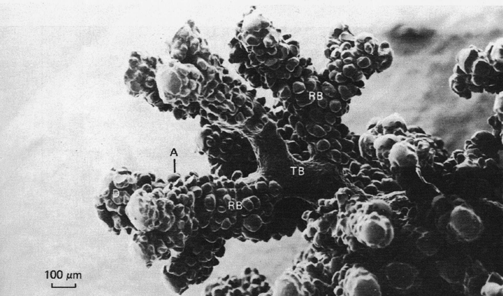
Fig. 1.3 Transition of terminal bronchiole. Scanning electron micrograph of airway branches peripheral to terminal bronchiole in a silicon-rubber cast of cat lung. Note multiple, smaller branches from respiratory to terminal bronchioles. A, alveolus; RB, respiratory bronchiole; TB, terminal bronchiole. Note absence of alveoli in terminal bronchiole (From Berne RM, Levy ML, Koeppen BM, Stanton BA (eds.). Physiology, 7th ed. St. Louis: Mosby; 2018.)
the lung parenchyma, and the caliber of their lumen is regulated by the elastic recoil of the lung and lung volume. In addition, the number of bronchioles increases beyond the 11th generation more rapidly than the diameter decreases. As a result the cross-sectional area increases rapidly at this point and is 30 times the cross-sectional area of the mainstem bronchi. This results in a marked decrease in airway resistance to approximately one-tenth of the resistance of the entire respiratory system (see Chapter 3).
The airways can thus be divided into two types: cartilaginous airways, or bronchi; and noncartilaginous airways, or bronchioles (Table 1.1). Bronchi contain cartilage and are the conductors of air between the external environment
and the distal sites of gas exchange. They do not participate in gas exchange. Bronchioles do not contain cartilage and are subdivided into terminal bronchioles, which do not participate in gas exchange; and respiratory bronchioles, which contain alveoli and alveolar ducts and function as sites of gas exchange.
The airways from the nose to and including the terminal bronchioles are known as the conducting airways because they bring (conduct) gas to the gas-exchanging units but do not actually participate in gas exchange. The conducting airways (primarily the nose) also function to warm and humidify inspired air. Because the conducting airways contain no alveoli and therefore take no part in gas
TABLE 1.1 Anatomic Features of Bronchi and Bronchioles
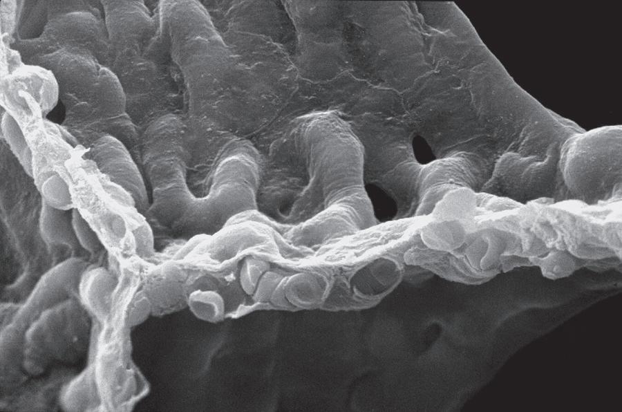
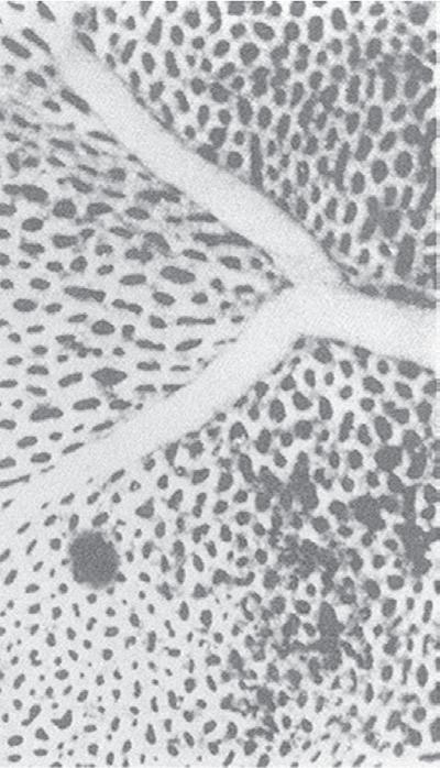
Fig. 1.4 Scanning electron micrograph of an alveolar surface demonstrating the alveolar septum. Capillaries (C) are seen in cross section in the foreground with erythrocytes (EC) in their lumen. At the circled asterisk, three septae come together. The septae are held together by connective tissue fibers (uncircled asterisks). A, alveolus; D, alveolar duct; PK, pores of Kohn. (Micrograph courtesy of Weibel ER, Institute of Anatomy, University of Berne, Switzerland.)
exchange, they constitute the anatomic dead space (see Chapter 5). In normal individuals, the first 16 generations of airway branchings, with a volume of 150 mL, constitute the anatomic dead space, whereas the next 7 generations contain an increasing number of alveoli and constitute the gas exchange unit.
Alveolar–Capillary Unit
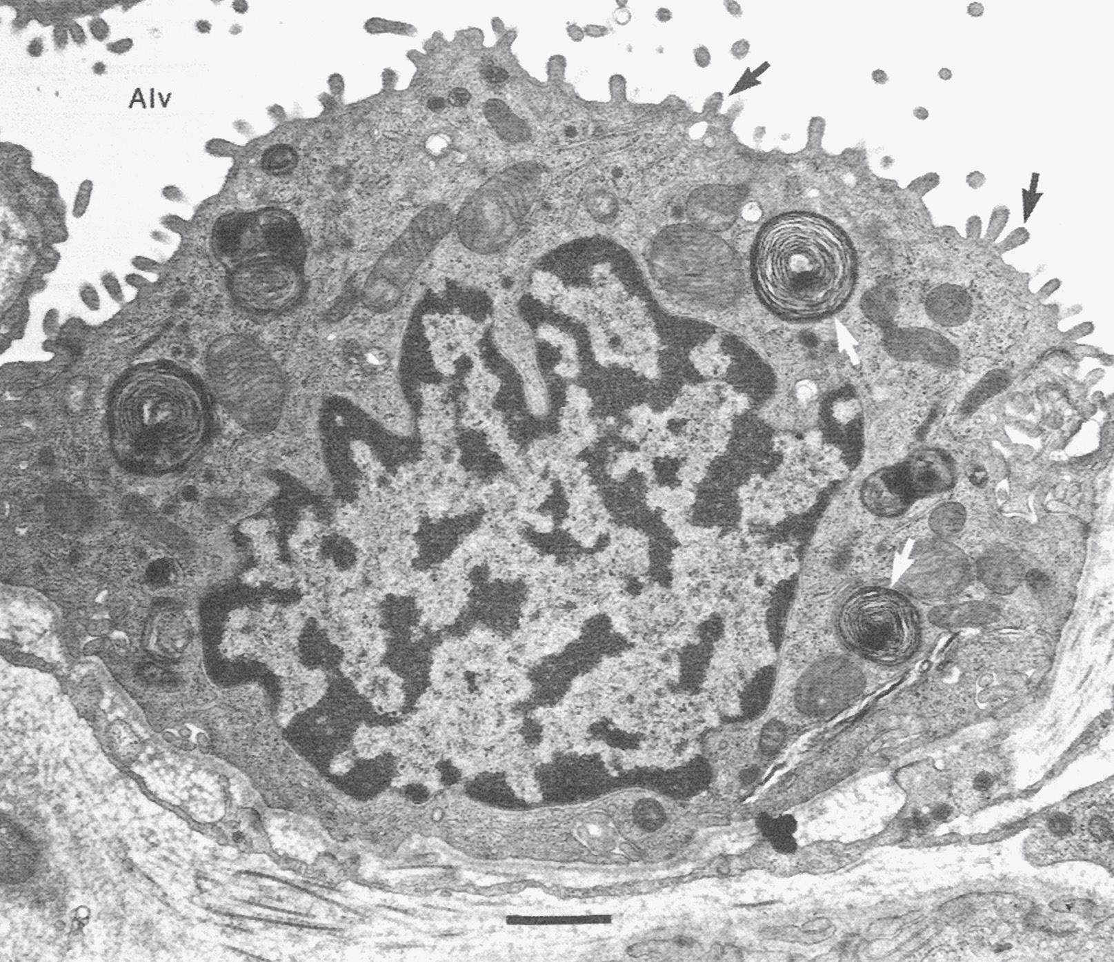
The terminal bronchioles divide into respiratory bronchioles, which contain alveolar ducts and alveoli and constitute the last three to five generations of the respiratory system. Gas exchange occurs in the alveoli through a dense meshlike network of capillaries and alveoli called the alveolar–capillary network (Fig. 1.4). The alveolar–capillary unit consists of the respiratory bronchioles, the alveolar ducts, the alveoli, and the pulmonary capillary bed. It is the basic physiologic unit of the lung and is characterized by a large surface area and a blood supply that originates from the pulmonary arteries. In the adult, there are approximately 300 million alveoli, which are 250 μm in size and are entirely surrounded by capillaries. In addition, there are 280 billion capillaries in the lung or almost 1000 capillaries for each alveolus. The result is a large surface area for gas exchange—approximately 50 to 100 m2, which occurs in a space that is only 5 mm in length. It is one of the most remarkable engineering feats in the body. The portion of the lung supplied by respiratory bronchioles is called an acinus. Each acinus contains in excess of 10,000 alveoli; gas movement in the acinus is by diffusion rather than tidal ventilation.
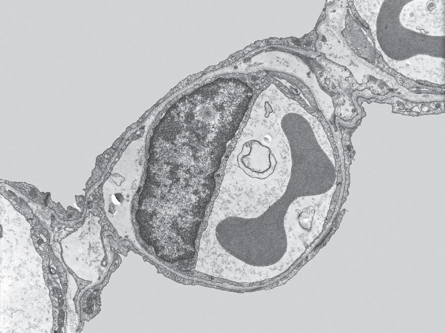
Fig. 1.5 Transmission electron micrograph of a pulmonary capillary in cross section. Alveoli (Alv) are on either side of the capillary that is shown with a red blood cell (RBC). The diffusion pathway for oxygen and carbon dioxide (arrow) consists of the areas numbered 2, 3, and 4, which are the alveolar–capillary barrier, plasma, and erythrocyte, respectively. BM, basement membrane; C, capillary; EN, capillary endothelial cell (note its large nucleus); EP, alveolar epithelial cell; FB, fibroblast process; IN, interstitial space. (Reproduced with permission from Weibel ER. Morphometric estimation of pulmonary diffusion capacity, I. Model & method. Respir Physiol. 1970;11:54–75.)
The barrier between the gas in the alveoli and the red blood cells is only 1 to 2 μm in thickness and consists of type I alveolar epithelial cells, capillary endothelial cells, and their respective basement membranes (Fig. 1.5). O2 diffuses across this barrier into plasma and red blood cells, whereas the reverse occurs for CO2 (see Chapter 8). Red blood cells pass through the pulmonary network in less than 1 second, which is sufficient time for CO2 and O2 gas exchange to occur.
In some regions of the alveolar wall there is nothing between the airway epithelial cells and the capillary endothelial cells other than their fused basement membranes. In other regions there is a space between the epithelial and endothelial cells called the interstitial space or interstitium (see Fig. 1.5). The interstitium is composed of collagen, elastin, proteoglycans, a variety of macromolecules involved with cell–cell and cell–matrix interactions, some nerve endings, and some fibroblast-like cells. The alveolar septum creates a fiber scaffold through which pulmonary capillaries are threaded and is supported by the basement membrane. There are also small numbers of lymphocytes that have migrated out of the circulation in the interstitium and capillary endothelial cells. The basement membrane is capable of withstanding high transmural pressures and sometimes is the only remaining separation between blood and gas.
Fig. 1.6 Structure of the normal alveolus.
The type I cell, with its long thin cytoplasmic processes, lines most of the alveolar surface, whereas the cuboidal type II cell, which is more numerous, occupies only about 7% of the alveolar surface. Capillaries (C) with red blood cells (RBC) are also shown. A, alveolar surface; IS, interstitial space; L, lamellar body, source of surfactant. (Modified from Weinberger S, Cockrill, BA, Mandel J. Principles of Pulmonary Medicine, 5th ed. Philadelphia: W.B. Saunders; 2008.)
Alveolar Surface
The alveolar epithelium is a continuous layer of tissue composed primarily of type I cells or squamous pneumocytes. These cells have broad, thin extensions that cover approximately 93% of the alveolar surface (Fig. 1.6). They are highly differentiated cells that do not divide, which makes them particularly susceptible to injury from inhaled or aspirated toxins and from high concentrations of oxygen (see Chapter 11). They are joined into a continuous sheet by tight junctions that prevent large molecules such as albumin from entering the alveoli, resulting in pulmonary edema. The thin cytoplasm of the type I cell is ideal for optimal gas diffusion.
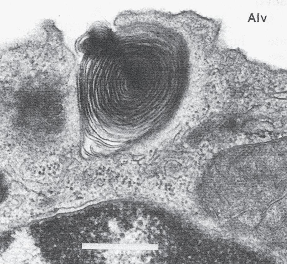
Type II cells, or granular pneumocytes, are more numerous than type I cells; however, because of their cuboidal shape, they occupy only approximately 7% of the alveolar surface and are located in the corners of the alveolus (see Fig. 1.6). The hallmarks of the type II cell are their microvilli and their osmiophilic lamellar inclusion bodies that contain surfactant, a compound with a high lipid content that acts as a detergent to reduce the surface tension of the alveoli (Fig. 1.7; also see Chapter 2). The type II cell is the progenitor cell of the alveolar epithelium. When there is injury to the type I cell, the type II cell multiplies and eventually differentiates into a type I cell. In a group of diseases that result in pulmonary fibrosis, the type I cell is injured and the alveolar epithelium is now lined entirely by type II cells, a condition that is not conducive to optimal
gas exchange. This repair system is an example of phylogeny recapitulating ontogeny, because the epithelium of the alveolus is composed entirely of type II cells until late in gestation.
The lumen of the alveolus is covered by a thin layer of fluid composed of a water phase immediately adjacent to the alveolar epithelial cell and covered by surfactant. Within the alveolar epithelium there are also a small number of macrophages, a type of phagocytic cell that patrols the alveolar surface and ingests (phagocytizes) bacteria and inhaled particles (see Chapter 11).
Pulmonary Circulation
The lung has two separate blood supplies (see Chapter 6 ). The pulmonary circulation brings deoxygenated blood from the right ventricle to the gas-exchanging units (alveoli). Pulmonary perfusion (Q ) refers to pulmonary blood flow, which equals the heart rate multiplied by the right ventricular stroke volume. The lungs receive the entire right ventricular cardiac output and are the only organ in the body that functions in this manner. The bronchial (or lesser) circulation arises from the aorta and provides nourishment to the lung parenchyma. The dual circulation to the lung is another of the unique features of the lung.
The pulmonary capillary bed is the largest vascular bed in the body, with a surface area of 70 to 80 m2. It is best
AB C
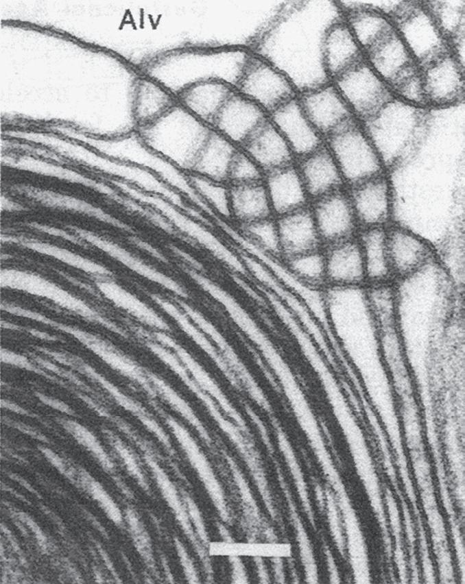
Fig. 1.7 Surfactant release by type II epithelial cells. Alv, alveolus. A, Type II epithelial cell from a human lung showing characteristic lamellar inclusion bodies (white arrows) within the cell and microvilli (black arrows) projecting into the alveolus. Bar = 0.5 μm. B, Early exocytosis of lamellar body into the alveolar space in a human lung. Bar = 0.5 μm. C, Secreted lamellar body and newly formed tubular myelin in alveolar liquid in a fetal rat lung. Membrane continuities between outer lamellae and adjacent tubular myelin provide evidence of intraalveolar tubular myelin formation. Bar = 0.1 μm. (Courtesy Dr. Mary C. Williams.)
viewed as a sheet of blood interrupted by small vertical supporting posts (Fig. 1.8). When the capillaries are filled with blood, about 75% of the surface area of the alveoli overlies the red blood cells. The capillaries allow red blood cells to flow through in single file only; this greatly facilitates gas exchange between the alveoli and the red blood cells. Once gas exchange is complete, the oxygenated blood returns to the left side of the heart through pulmonary venules and veins and is ready for pumping to the systemic
circulation. In contrast to the systemic circulation, the pulmonary circulation is a highly distensible, low-pressure system capable of accommodating large volumes of blood at low pressure. This is another unique feature of the lung.
Pulmonary arteries that contain deoxygenated blood follow the bronchi in connective tissue sheaths, whereas pulmonary veins cross segments on their way to the left atrium (Fig. 1.9). Bronchial arteries also follow the bronchi and divide with them. In contrast, one-third of the blood
Fig. 1.8 Pulmonary capillary surface of the lung. View of alveolar wall (in a frog) demonstrating the dense network of capillaries. A small artery (left) and vein (right) can also be seen. The individual capillary segments are so short that the blood forms an almost continuous sheet. (From Maloney JE, Castle BL. Pressure-diameter relations of capillaries and small blood vessels in frog lung. Respir Physiol. 1969;7:150–162.)
Fig. 1.9 The anatomic relation between the pulmonary artery, the bronchial artery, the airways, and the lymphatics. A, alveoli; AD, alveolar ducts; RB, respiratory bronchioles; TB, terminal bronchioles. (From Berne RM, Levy ML, Koeppen BM, Stanton BA (eds.). Physiology, 7th ed. St. Louis: Mosby; 2018.)
Pulmonary
from the bronchial veins (deoxygenated blood) drains into the right atrium, and the remainder drains into pulmonary veins that drain into the left atrium. Thus a small amount of deoxygenated blood that has nourished the lung parenchyma mixes with oxygenated blood in the left atrium. Pulmonary capillaries, on the other hand, are not confined to a single alveolus but pass from one to another as well as to adjacent alveolar septae before emptying into a venule. This improves the efficiency of gas exchange and minimizes the effect of alveolar disease on gas exchange.
Cells of the Airways
The respiratory tract (with the exception of the pharynx, the anterior one-third of the nose, and the area distal to the terminal bronchioles) is lined by a pseudostratified, ciliated, columnar epithelium interspersed with mucus-secreting goblet cells and other secretory cells (Fig. 1.10; also see Chapter 11). In the distal airways, the columnar epithelium gives way to a more cuboidal epithelium. The airway epithelial cells are responsible for maintaining a thin, aqueous layer of fluid adjacent to the cells (periciliary fluid) in which the cilia can function. The depth of this periciliary fluid is maintained by the movement of ions across the epithelium.
Interspersed among the epithelial cells are surface secretory cells, which are also known as goblet cells. In general, there is one goblet cell per five to six ciliated cells. Goblet cells decrease in number between the 5th and 12th lung generation and in normal individuals disappear beyond the 12th tracheobronchial generation. Both goblet cell number and secretions increase in many diseases including asthma and cystic fibrosis. Secretions also increase by rapid exocytosis in response to chemical irritation, inflammatory cytokines, and neuronal stimulation. In the bronchioles, goblet cells are replaced by Clara cells, another type of secretory cell.
Basal cells are located underneath the columnar epithelium and are responsible for the pseudostratified appearance of the epithelial surface. They are absent in the bronchioles and beyond. Although their function is not clear, they appear to be the stem cells for the airway epithelium and the goblet cells.
Submucosal tracheobronchial glands are present wherever there is cartilage in the tracheobronchial tree. These glands empty to the surface epithelium through a ciliated duct and are lined by mucous and serous cells. Submucosal tracheobronchial glands increase in number and size in chronic bronchitis, a chronic lung disease primarily occurring in smokers, and extend down to the bronchioles in disease.
The ciliated epithelium, goblet cell, Clara cell, and tracheobronchial glands are important in host defense and are discussed in Chapter 11.
The Muscles of Respiration
The chest wall encases the lung, and normally the two structures move together. The lungs do not self-inflate. The force for lung inflation is supplied by the muscles of respiration, which are skeletal muscles. Like all skeletal muscles, their force of contraction increases when they are stretched and decreases when they are shortened. Thus the force of contraction of the respiratory muscles increases with increasing lung volume.
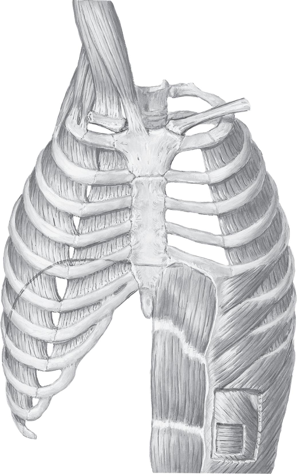
Dividing the thoracic cavity from the abdominal cavity is the diaphragm, the major muscle of respiration (Fig. 1.11). The diaphragm is a thin, musculotendinous, domeshaped sheet of muscle that is inserted into the lower ribs and separates the thoracic from the abdominal cavity. It is supplied by the phrenic nerve that arises from the second cervical vertebra. When it contracts, the abdominal contents are forced downward and forward and the vertical dimension of the chest cavity is increased. In addition, the rib margins are lifted and moved out, causing an increase in the transverse diameter of the thorax. In adults, the diaphragm is capable of generating airway pressures of 150 to 200 cm H2O during a maximal inspiratory effort. During quiet breathing (known as tidal volume breathing), the diaphragm moves approximately 1 cm, but during large-volume breathing, the diaphragm can move as much as 10 cm. If the diaphragm is paralyzed, it moves higher up in the thoracic cavity during inspiration because of the fall in intrathoracic pressure. This paradoxical movement of the diaphragm can be demonstrated using the radiographic technique called fluoroscopy.
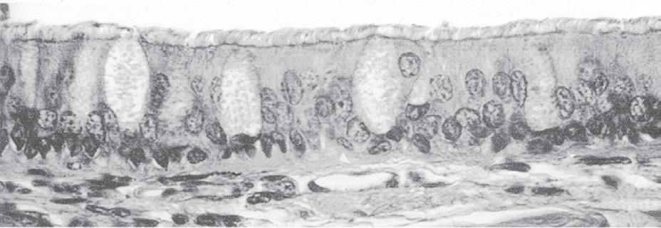
Fig. 1.10 Scanning electron micrograph of airway, showing the ciliated, pseudostratified, columnar epithelium of a bronchus. Each cilium is connected to a basal body (BB), which collectively appears at the base of the cilia (C) as a dark band. Goblet cells (GC) and basal cells (BC), the potential precursors of the ciliated cells, are shown. CT, connective tissue. (From Berne RM, Levy ML, Koeppen BM, Stanton BA (eds.). Physiology, 5th ed. St. Louis: Mosby; 2004.)
Fig. 1.11 The diaphragm. View from the inside of the thorax illustrates the position of the diaphragm in the thorax. (From Grippi MS, Elias JA, Fishman JA, et al. Fishman’s Pulmonary Diseases and Disorders, 5th ed. New York: McGraw-Hill; 2015.)
The other significant muscles of inspiration are the external intercostal muscles that pull the ribs upward and forward during inspiration, causing an increase in both the side-toside and front-to-back diameters of the thorax (Fig. 1.12). Innervation of these muscles originates from intercostal nerves that originate from the spinal cord at the same level. Paralysis of these muscles has no significant effect on respiration because of the dominance of the diaphragm as the major muscle of respiration. Accessory muscles of inspiration (scalene muscles, which elevate the sternocleidomastoid; the alae nasi, which cause nasal flaring; and small muscles in the neck and head) are quiet during quiet breathing but contract vigorously during exercise and with significant airway obstruction.
The upper airway must remain patent during inspiration; therefore the pharyngeal wall muscles, the genioglossus, and the arytenoid muscles are also considered muscles of inspiration.
Exhalation during quiet breathing is passive but becomes active during exercise and hyperventilation. The most important muscles of exhalation are those of the abdominal wall (rectus abdominis, internal and external oblique, and transversus abdominis) and the internal intercostal muscles that oppose the activity of the external intercostal muscles (i.e., pull the ribs downward and inward).
The Central Nervous System and Neural Pathways
The central nervous system (CNS), and in particular the brainstem, functions as the main control center for
respiration (see Chapter 10). Breathing is both voluntary and automatic. Each breath begins in the brain, where the signal to breathe is carried to the respiratory muscles through the spinal cord and the nerves that innervate the respiratory muscles. It is remarkable that despite widely varying demands for O2 uptake and CO2 removal, the arterial levels of O2 and CO2 are normally maintained within tight limits. Regulation of respiration requires three components (see Chapter 10):
1. Generation and maintenance of a respiratory rhythm (respiratory control center)
2. Modulation of the respiratory rhythm by sensory feedback loops and reflexes that allow adaptation to various situations and minimize energy costs
3. Recruitment of respiratory muscles that can contract appropriately for effective gas exchange.
Unlike the heart, which begins beating at approximately 6 weeks’ gestation, rhythmic respirations do not begin until birth.
ANATOMIC AND PHYSIOLOGIC CORRELATES
Lung structure is closely correlated with lung function in health and disease. Because lung disease is described in anatomic terms (e.g., right middle lobe pneumonia), knowledge of lung anatomy is essential. The bronchopulmonary segment is the region of the lung supplied by a segmental bronchus. It is the functional anatomic unit of the lung , so named because disease usually involves one segment at a time and because surgical resection follows along segments. When using a stethoscope (auscultation), all of the bronchopulmonary segments can be examined with one exception, namely the hilar segments of the lower lobes ( Fig. 1.13 ). The hilum is the area of the lung where the main-stem bronchi and pulmonary arteries and veins enter and leave the right and left lung. These segments have no topographic relationship to the chest.
The various lobes of the lung (three on the right and two on the left) are subdivided by fissures . The division into the lobes, however, is incomplete, which allows for collateral ventilation. Collateral ventilation is an accessory pathway that connects airspaces supplied by other airways. There are two types of accessory pathways in the lung: (1) canals of Lambert , which connect respiratory bronchioles and terminal bronchioles to airspaces supplied by other airways; and (2) pores of Kohn , which are openings in the alveolar walls that connect adjacent alveoli. These accessory pathways help prevent collapse of terminal respiratory units ( atelectasis ) when their supplying airway becomes obstructed and are particularly important in individuals with lung diseases such as emphysema
Diaphragm
Muscles of inspiration
Accessory
Sternocleidomastoid (elevates sternum)
Scalenes
Anterior Middle Posterior (elevate and fix upper ribs)
Principal
External intercostals (elevate ribs, thus increasing width of thoracic cavity)
Interchondral part of internal intercostals (also elevates ribs)
Diaphragm (domes descend, thus increasing vertical dimension of thoracic cavity; also elevates lower ribs)
MUSCLES OF RESPIRATION
Muscles of expiration
Quiet breathing
Expiration results from passive recoil of lungs and rib cage
Active breathing
Internal intercostals, except interchondral part
Abdominals (depress lower ribs, compress abdominal contents, thus pushing up diaphragm)
Rectus abdominis
External oblique
Internal oblique
Transversus abdominis
Fig. 1.12 Muscles of respiration. Diagram of the anatomy of the major respiratory muscles. Left side, inspiratory muscles; right side, expiratory muscles. (Kaminsky D. The Netter Collection of Medical Illustrations: Respiratory System, vol. 3, 2nd ed. Philadelphia: Elsevier; 2011.)
Fig. 1.13 Topography of the lung demonstrating the lobes, segments, and fissures. Numbers refer to specific bronchopulmonary segments that are also shown in Fig. 1.1. SVC, superior vena cava. (From Berne RM, Levy ML, Koeppen BM, Stanton BA (eds.). Physiology, 7th ed. St. Louis: Mosby; 2018.)
Physiologically, the lung demonstrates functional unity; that is, every alveolar unit has the same structure and the same function as every other alveolar unit. This is in contrast to the heart, in which the various chambers have both
a different structure and a different function. The significance of functional unity is that a large portion of the lung can be removed without significantly compromising overall lung function (i.e., gas exchange).
CLINICAL BOX
Understanding lung topography is useful in both diagnosing and localizing disease. For example, a 2-year-old boy presents with a 2-day history of fever, cough, and recent onset of tachypnea (an increased respiratory rate). On examination, there are intercostal muscle retractions and nasal flaring, and the child appears ill. On auscultation, breath sounds are decreased over the right upper lobe anteriorly.
A chest x-ray reveals opacification (known as consolidation) over the right upper lobe anteriorly consistent with lobar (specifically right upper lobe) pneumonia.
SUMMARY
1. The principal function of the respiratory system is gas exchange. Other functions include acid–base balance, host defense and metabolism, and the handling of bioactive materials.
2. Gas exchange occurs in the alveolar–capillary unit, the basic physiologic unit of the lung.
3. The bronchopulmonary segment is the segment of the lung supplied by a segmental bronchus. It is the functional anatomic unit of the lung.
4. The alveolar surface is lined by type I and type II cells. The thin cytoplasm of the type I cell is ideal for optimal gas diffusion, whereas the type II cell is important for the production of surfactant, which decreases the surface tension of the alveolus.
5. The lung has two separate circulations. The pulmonary circulation brings deoxygenated blood from the right ventricle to the gas-exchanging units. The
bronchial circulation arises from the aorta and nourishes the lung parenchyma.
6. The circulation to the lung is unique in its dual circulation and in its ability to accommodate large volumes of blood at low pressure.
7. The anatomic dead space is composed of all of the airways that do not participate in gas exchange—that is, the airways to the level of the respiratory bronchioles.
8. The cells of the conducting airways include the pseudostratified, ciliated, columnar epithelial cells, surface secretory cells, Clara cells, and submucosal tracheobronchial gland cells.
9. The diaphragm is the major muscle of respiration.
10. Breathing is both voluntary and automatic.
11. The lung demonstrates both anatomic and physiologic unity—that is, each unit is structurally identical and functions just like every other unit.
KEY WORDS AND CONCEPTS
Alveolar macrophage
Alveolar–capillary unit
Alveolus
Anatomic dead space
Atelectasis
Bronchial circulation
Bronchiole
Bronchopulmonary segment
Bronchus
Canals of Lambert
Chemoreceptor
Clara cell
Collateral ventilation
Diaphragm
Emphysema
Fissure
Functional unity
Glottis
Hilum
SELF-STUDY PROBLEMS
1. What anatomic features of the alveolar–capillary unit make it appropriate to function as the gas-exchanging unit?
2. How can you distinguish type I cells from type II cells?
3. If the pulmonary artery that supplies the left lung was occluded for a short period of time and the cardiac
ADDITIONAL READINGS
Baile EM. The anatomy and physiology of the bronchial circulation. J Aerosol Med. 1996;9:1–6.
Boggs DS, Kinasewitz GT. Review: pathophysiology of the pleural space. Am J Med. 1995;309:53–59.
Broaddus VC, Mason RJ, JD Ernst, et al., eds. Murray & Nadel’s Textbook of Respiratory Medicine. 6th ed. Philadelphia: WB Saunders; 2016.
Fehrenbach H. Alveolar epithelial type II cell: defender of the alveolus revisited. Respir Res. 2001;2:33–46.
Gandevia SC, Allen GM, Butler J, et al. Human respiratory muscles: sensations, reflexes and fatigability. Clin Exp Pharm Physiol. 1998;25:757–763.
Grippi MA, Elias JA, Fishman JA, et al. Fishman’s Pulmonary Diseases and Disorders. 5th ed. New York: McGraw Hill; 2015.
Hlastala MP, Berger AJ. Physiology of Respiration. 2nd ed. New York: Oxford University Press; 2001.
Horsfield K, Cumming G. Morphology of the bronchial tree in man. J Appl Physiol. 1968;24:373–383.
Interstitium/interstitial space
Lower airway
Parenchyma
Parietal pleura
Partial pressure
Periciliary fluid
Pleural effusion
Pneumothorax
Pores of Kohn
Respiratory control center
Surface secretory cells (goblet cells)
Surfactant
Tracheobronchial glands
Turbinates
Type I cell
Type II cell
Upper airway
Visceral pleura
output remained unchanged (that is, all of the blood from the right ventricle now goes to the right lung), what would be the effect on the pressure inside the right pulmonary artery?
4. What are the components of the blood–gas barrier?
5. What are the anatomic features that make the lung ideally suited for its principal function?
Leff AR. Schumacker PT. Respiratory Physiology: Basics and Applications. Philadelphia: WB Saunders; 1993.
Lumb AB. Nunn’s Applied Respiratory Physiology. 8th ed. Philadelphia: Elsevier; 2017.
Massaro D, Massaro GD. Invited review: pulmonary alveoli: Formation, the “call for oxygen,” and other regulators. Am J Physiol Lung Cell Mol Physiol. 2002;282:L345–L358.
Nettesheim P, Koo JS, Gray T. Regulation of differentiation of the tracheobronchial epithelium. J Aerosol Med. 2000;13: 207–218.
Poole DC, Sexton WL, Farkas GA. Diaphragm structure and function in health and disease. Med Sci Sports Exerc. 1997; 29:738–754.
Rogers DE. Airway goblet cells: Responsive and adaptable frontline defenders. Eur Respir J. 1994;7:1690–1706.
Weibel ER The Pathway for Oxygen: Structure and Function of the Mammalian Respiratory System. Cambridge, MA: Harvard University Press; 1984.
Mechanical Propertiesof the Lung and Chest Wall
OBJECTIVES
1.Describestaticlungmechanicsandthemeasurement oflungvolumes.
2.Definelungcomplianceanditsmeasurement.
3.Relatelungandchestwallcompliancetolungvolumes.
STATIC LUNG MECHANICS
Airmovementinandoutofthelungiscontrolledbythe mechanicalpropertiesofthelungandchestwall.Static lungmechanicsisthestudyofthemechanicalproperties ofthelungandchestwallwhosevolumeisnotchanging withtimeandisdiscussedinthischapter.Dynamiclung mechanics,whichisthestudyofthelungandchestwallin motion(i.e.,changingvolume),isdiscussedinChape
Themechanicsofthelungarecomposedofthecombinedmechanicalpropertiesoftheairways,lungpar@:chyma,interstitialmatrix(composedoffibrin,collagen, andafewcells),alveolarsurface,andpulmonai:rcirculation.Themechanicalpropertiesofthechestwallinclude thepropertiesofallofthestructuresoutsideofthelungs thatmoveduringbreathing,includingtheribcage,diaphragm,abdominalcavity,andanteriorabdominalmuscles.TheinteractionbetweentBelungandthechestwall determineslungvolumes,andstaticlungvolumesplaya majorroleingasexchangeandintheworkofbreathing. Theycanbemeasuredandareabnormalinmanylung diseases.
LUNG VOLUMES
ThestaticvolumesofthelungsareshowninFig.2.1.All lungvolumesaresubdivisionsofthetotallungcapacity (TLC)andaremeasuredinliters.Theyarereportedeither asvolumes(e.g.,residualvolume)orcapacities(e.g.,vital capacity).Acapacityiscomposedoftwoormorevolumes.
4.Characterizelungandchestwallinteractionsin termsofpressuregradientsandpressurevolume relationships.
5.Describesurfactantanditsroralteringsurfacetension.
Thetotalvolmeofairthatiscontainedinthelungis calledthe'fLG.Itiscomposedofthevolumeofairthat anindividu�anexhalefromamaximuminspirationto amaximumexhalation,knownasthevitalcapacity(VC), andthevolumeofairthatisleftinthelungafteramaximalexhalation,knownastheresidualvolume(RV).Two otherimportantlungvolumesarethetidalvolume(TV, or,VT)andthefunctionalresidualcapacity(FRC).TheTV ·sthevolumeofairthatisbreathedintoandoutofthe lungduringquietbreathing.TheFRCisthevolumeofair containedinthelungafteranormalexhalation.TheFRC iscomposedoftheresidualvolumeandthevolumeofair thatcanbeexhaledfromtheendofanormalexhalation toresidualvolume.Thislattervolumeiscalledtheexpiratoryreservevolume(ERV).TheFRCrepresentstheresting volumeoftherespiratorysystem,inwhichtheforcesof thechestwalltoincreaseinsizeandtheforcesofthelung todecreaseinsizeareequalbutopposite(seelaterinthis chapter).
Togetasenseoftheimportanceoflungvolumesinrespiration,breathequietlyclosetoTLC(takeadeepbreath in,andbreatheatthishighlungvolumeforafewminutes).Nowbreatheoutuntilyoucannotforceanymore airout,andtrybreathingatthisvolume,whichiscloseto yourRV.Bothofthesemaneuversshouldbeuncomfortableandassociatedwithincreasedwork;bothincreases anddecreasesinlungvolumeoccurinlungdiseaseasa resultofachangeinlungmechanics.Themeasurementof lungvolumesisusedtodetectandfollowtheprogression oflungdiseaseandisdiscussedinChapter4.
Fig. 2.1 The various lung volumes and capacities. ERV, expiratory reserve volume; FRC, functional residual capacity; FVC, forced vital capacity; IC, inspiratory capacity; IRV, inspiratory reserve volume; RV, residual volume; TLC, total lung capacity; VC, vital capacity; VT, tidal volume. (From Koeppen BM, Stanton BA, eds. Berne and Levy’s Physiology, 7th ed. Philadelphia: Elsevier; 2018.)
USING AND INTERPRETING RESULTS OF LUNG VOLUME MEASUREMENTS
Two major types of pathophysiologic abnormalities involving the lung and chest wall can be described using lung volumes. One group of diseases is called obstructive pulmonary disease (OPD). In OPD, during exhalation the airways close (premature airway closure, the hallmark of OPD) trapping air behind them (see Chapter 3). This results in an increase in TLC, RV, and FRC. In contrast, in restrictive pulmonary disease, the other major pathophysiologic abnormality involving the lung and chest wall, lung volumes are reduced.
One of the most useful tests for distinguishing obstructive and restrictive types of lung disease is the measurement of the RV/TLC ratio. In normal individuals, the RV/TLC ratio is less than 0.25, that is, approximately 25% of the air in the lungs is trapped and cannot be exhaled. An elevated RV/TLC ratio, characterized by an increase in RV out of proportion to any increase in TLC, is due to air trapping secondary to airway obstruction and is seen in individuals with OPD. An elevated RV/TLC ratio due to a decrease in TLC out of proportion to any change in RV is seen in individuals with restrictive types of pulmonary disease.
LUNG COMPLIANCE AND LUNG ELASTIC PROPERTIES
Lung compliance (Cl) is a measure of the elastic properties of the lung and is a reflection of lung distensibility. These distensibility properties of the lung are seen in the pressure
volume relaxation curve for the lung that is called the compliance curve of the lung Compliance of the lungs is defined as the change in lung volume resulting from a change in the distending pressure of the lung equal to 1 cm H2O. The units of compliance are mL (or L)/cm H2O. A lung with high lung compliance refers to a lung that is easily distended. A lung with low compliance or a “stiff” lung is the one that is not easily distended. Thus the compliance of the lung (Cl) is: CL = ΔV/ΔP
where ΔV is the change in volume and ΔP is the change in pressure.
The compliance of the isolated lung is measured in animals by removing the lung and measuring the changes in lung volume that occur with each change in the pressure between the inside of the lung and the outside (also known as transpulmonary or translung pressure). As transpulmonary pressure increases, lung volume increases (Fig. 2.2A). The line that is generated, however, is curvilinear, not linear. That is, at low lung volumes, the lung distends easily, but at high lung volumes, larger increases in transpulmonary pressure are needed to produce only small changes in lung volume. This is in part because at high lung volumes all of the elastic fibers in the alveolar units and airways have been maximally stretched. More important than elastic recoil in the determination of compliance is the surface tension at the air–water interface lining the alveoli due to surfactant (see later in this chapter).
Fig. 2.2 Deflation pressure volume (PV) curve of the lung (A) and chest wall (B). A, The compliance of the lung at any point along the curve is the change in volume (ΔV) per the change in pressure (ΔP). From the curve, it is apparent that the compliance of the lung changes with lung volume. By convention, the deflation pressure volume curve is used, and lung compliance is the change in pressure when going from functional residual capacity (FRC) to FRC + 1 L. RV, residual volume; TLC, total lung capacity. B, PV curve of the chest wall demonstrating a change in compliance with change in lung volume. Note that at volumes greater than 60% of the TLC, the pressure needed to expand the chest wall is positive (inward recoil), whereas at lower lung volumes, the chest wall pressure is negative (outward recoil).
Lungs that are highly compliant will have a steeper slope than lungs with a low compliance. Lung compliance or distensibility is the inverse of elasticity or lung elastic recoil (P el). Compliance is the ease with which something is stretched, whereas elastic recoil is the tendency to resist or oppose stretching and return to its previous configuration when the distorting force is removed.
Fig. 2.3 Inflation: deflation pressure–volume curve of the lung. The direction of inspiration and exhalation is shown by the arrows. The difference between the inflation and deflation pressure-volume curves is due to surface tension variation with changes in lung volume. FRC, functional residual capacity; TLC, total lung capacity; Vt, tidal volume. (From Koeppen BM, Stanton BA, eds. Berne and Levy’s Physiology, 7th ed. Philadelphia: Elsevier; 2018.)
By convention, the compliance of the lung is measured as the slope of the line between any two points on the deflation limb of the pressure volume loop. The compliance of the lung is greater when measured from TLC to RV (deflation) than from RV to TLC (inflation) (Fig. 2.3). This is due in large part to the changes in surface tension with changing lung volume and is discussed later in this chapter. This difference between the inflation and exhalation curve is called hysteresis. As we
Fig. 2.4 The relaxation pressure–volume curve of the lung, chest wall, and respiratory system. The curve for the respiratory system is the sum of the individual curves (Prs = Pl + Pw). The curve for the lung is the same as in Fig. 2.2A, and the curve for the chest wall is the same as in Fig. 2.2B FRC, functional residual capacity; Pw, chest wall pressure; Pl, transpulmonary pressure; Prs, respiratory system pressure; TLC, total lung capacity. (Koeppen BM, Stanton BA, eds. Berne and Levy’s Physiology, 7th ed. Philadelphia: Elsevier; 2018.)
will see later in this chapter, the most important reason for hysteresis is changes in surfactant. Other reasons include redistribution of gas and recruitment of alveoli.
COMPLIANCE OF THE CHEST WALL
When the lungs are removed, the chest wall has a springlike character with a relatively high resting volume. In much the same way as the lung, the compliance curve of the chest wall relates the volume of gas enclosed by the chest wall to the pressure across the chest wall. The curve (see Fig. 2.2B) is relatively flat at low volumes; that is, the chest wall is stiff with the shortened respiratory muscles maximally contracted. The curve is also flat at high lung volumes where the respiratory muscles are maximally stretched. At both high and low lung volumes, large changes in pressure across the chest wall result in small changes in the volume enclosed by the chest wall.
COMPLIANCE OF THE RESPIRATORY SYSTEM
Both the lungs and the chest wall contribute to the compliance of the respiratory system (Fig. 2.4). The lung and chest wall are held together by the thin layer of pleural fluid that functions like a liquid film holding two pieces of glass together. The glass pieces slide easily relative to each other, but it is difficult to pull them apart. The compliance of the respiratory system is also analogous to electrical capacitance, and in the respiratory system the compliances of the lung and chest wall are in parallel. Thus their individual compliances
Pressure (cm H2O) (PL, PW, or PRS)
add as reciprocals; that is, 1/compliance of the respiratory system = 1/compliance of the lung + 1/compliance of the chest wall or
1/CRS = 1/CL + 1/CW
In contrast, the reciprocal of compliance is elastance, and the elastance of the lung and chest wall add directly. In addition, compliances in series add directly. For example, the compliance of the lungs in the two hemithoraces that are in series is the sum of the compliances of the lung in each hemithorax.
As noted previously, lung compliance varies with lung volume (see Fig. 2.2 ). It is greater at lower lung volumes than at higher lung volumes. For this reason, specific compliance, or compliance divided by the lung volume at which it is measured (usually FRC), is used ( Fig. 2.5 ). As an example, consider the individual with chronic bronchitis in whom FRC is increased. As a result, pulmonary compliance, which is now being measured at this higher lung volume, would also be increased. However, when corrected for the FRC (specific compliance), the compliance is normal. In individuals with normal FRC, the compliance of the lung is about 0.2 L/cm H 2O, of the chest wall is 0.2 L/cm H 2O, and of the respiratory system is 0.1 L/cm H 2O. Note that the compliance of the respiratory system is lower than the compliance of either the lung or the chest wall. Lung compliance is not affected by age.
CLINICAL BOX: CLINICAL USE OF COMPLIANCE
The compliance of the lung is not altered by airflow per se, but the compliance of the lung and chest wall is affected by a number of respiratory disorders. In emphysema, the lung is more compliant because of destruction of lung elastic tissue; that is, for every 1 cm of H2O pressure increase, there is a larger increase in volume than in the normal lung. In contrast, a proliferation of connective tissue in the lung called pulmonary fibrosis can be seen in lung diseases such as interstitial pneumonitis and sarcoidosis or in association with chemical or thermal lung injury. The lungs in these diseases are “stiff,” or noncompliant; that is, for every 1 cm H2O pressure change, there is a smaller change in volume. Similarly, in diseases associated with increased fluid in the interstitial spaces such as pulmonary edema or in diseases associated with fluid, blood, or infection in the intrapleural space (pleural effusion, hemothorax, or empyema, respectively), lung compliance is reduced. The compliance of the chest wall is decreased in individuals with obesity in whom adipose tissue results in an additional load on the chest wall muscles and the diaphragm. Individuals with decreased mobility of the rib cage such as in kyphoscoliosis or other types of musculoskeletal diseases that affect chest wall movement also have decreased chest wall compliance.
Individuals with decreased compliance must generate greater transpulmonary pressures to produce changes in lung volume than individuals with normal compliance. This results in increased work associated with breathing (see Chapter 3).
Fibrosis/emphysema pressure–volume curve. TLC, total lung capacity.
(cm H2O)
From Koeppen BM, Stanton BA, eds. Berne and Levy’s Physiology, 7th ed. Philadelphia: Elsevier; 2018.
FACTORS DETERMINING LUNG VOLUME
Why can’t we inspire above TLC or exhale beyond RV? The answers lie in the properties of the lung parenchyma and in the interaction between the lungs and the chest wall. Both the lungs and the chest wall have elastic properties. Both have a resting volume (or size) that they would assume if there were no external forces or pressures exerted on them. Both expand when stresses are applied and recoil passively when stresses are released. If the lungs were removed from the chest and no external forces were applied, they would become almost airless. To expand, these lungs would require either the exertion of a positive pressure on the alveoli and airways or the application of a negative pressure from outside the lungs. Either would result in a positive transpulmonary pressure. These situations are analogous to the balloon and the vacuum canister. A balloon is airless until positive pressure is exerted at the opening to distend the balloon walls (positive-pressure “ventilation”). In the case of the vacuum, negative external pressure is applied and results in sucking materials (air) into the canister (negative-pressure “ventilation”).
The lungs are enclosed by the chest wall, which expands during inspiration. The lungs and chest wall always move together in healthy individuals. Lung volumes are determined by the balance between the lung’s elastic recoil properties and the properties of the muscles of the chest wall. TLC occurs when the forces of inspiration decrease because of chest wall muscle lengthening and are insufficient to overcome the increasing force required to distend the lung and chest wall (see Fig. 2.4). Thus TLC is limited by the distensibility of both the lungs and the chest wall and the amount of force that the inspiratory muscles can generate. Disease that affects any of these three components will affect TLC.
At RV, a significant amount of gas remains within the lung. As RV is approached, the chest wall becomes so stiff that additional effort by the expiratory chest wall muscles to contract is unable to further reduce the volume. Thus RV occurs when the expiratory muscle force is insufficient to cause a further reduction in chest wall volume (see Fig. 2.4). As the chest wall is squeezed by the expiratory muscles, the recoil pressure of the chest wall (the chest wall wanting to increase in size) increases. The expiratory muscles
Lung volume⁄ Pressure
Compliance = Specific Compliance =
Lung compliance⁄Lung volume
Fig. 2.5 Relationship between compliance and lung volume. Imagine a lung in which a change in pressure of 5 cm H2O results in a change in volume of 1 liter. If half of the lung is removed (Situation 2), the compliance will decrease, but when corrected for the volume of the lung, there is no change (specific compliance). Even when the lung is reduced by 90% (Situation 3), the specific compliance is unchanged. R, right lung; L, left lung. (From Koeppen BM, Stanton BA, eds. Berne and Levy’s Physiology, 7th ed. Philadelphia: Elsevier; 2018.)
shorten, and their capacity to generate force decreases; the point at which the force generated by the expiratory muscles is insufficient to overcome the outward recoil of the chest wall determines the RV. This simple model of RV applies to (young) individuals with normal lungs. In older individuals and in individuals with lung disease, premature airway closure, a property of the lung (see Chapter 3), becomes the major determinant of RV rather than outward chest wall recoil.
The FRC is the volume of the lung at the end of a normal exhalation and is determined by the balance between the elastic recoil pressure generated by the lung parenchyma to become smaller and the pressure generated by the chest wall to become larger (see Fig. 2.4). FRC occurs when these two forces are equal and opposite. In the presence of chest wall weakness, the FRC decreases (lung elastic recoil is greater than chest wall muscle force). In the presence of airway obstruction, the FRC increases because of premature airway closure that traps air in the lung. Always, however, the FRC occurs at the lung volume at which the outward recoil of the chest wall is equal to the inward recoil of the lung.
LUNG–CHEST WALL INTERACTIONS
The lung and chest wall move together in healthy people. The pleural space that separates the lung and the chest wall is best thought of as a “potential” space because of its small volume. Because the lung and chest wall move together, changes in their respective volumes are the same. The pressure changes across the lung and across the chest wall are defined as the transmural pressures. Transmural pressure refers to any pressure difference across a wall and by convention represents the inside of the wall pressure minus the outside of the wall pressure. For the lung, this transmural pressure is called the transpulmonary pressure (Pl; also called the translung pressure) and is defined as the pressure difference between the airspaces (alveolar pressure [Pa]) and the pressure surrounding the lung (pleural pressure [Ppl]); that is,
PL = PA PPL
The lung requires a positive Pl to increase its volume and lung volume increases with increasing Pl. The lung assumes its smallest size when the transpulmonary