Preface to the Ninth Edition
Over the past five decades Nunn’s Applied Respiratory Physiology has developed into a renowned textbook on respiration, providing both physiologists and clinicians with a unique fusion of underlying principles and their applications. After writing four editions, Dr John Nunn retired in 1991, and a new author was required. As Dr Nunn’s final research fellow in the Clinical Research Centre in Harrow, AL was honoured to be chosen as his successor. AL has now also completed four editions and has chosen a successor to lead the project into the future whilst maintaining the fundamental ethos of the book. As practising clinicians with a fascination for physiology, the authors of the ninth edition have again focussed on combining a clear, logical and comprehensive account of basic respiratory physiology with a wide range of applications, both physiological and clinical. This approach acknowledges the popularity of the book amongst doctors from many medical specialities, but also provides greater insight into the applications of respiratory physiology to readers with a scientific background. The clinical chapters of Part 3 are not intended to be comprehensive reviews of the pulmonary diseases considered, but rather to provide a detailed description of physiological changes, accompanied by a brief account of the clinical features and treatment of the disease.
In this edition, the number of references provided has been reduced by around a third in recognition of the ease with which online searches may now be performed. References retained are either historical or seminal papers, or recent high-quality publications. Key references are identified by bold type in the reference list following each chapter. These highlighted references either provide outstanding recent reviews of their subject or describe research that has made a significant impact on the topic under consideration.
Advances in respiratory physiology since the last edition are too numerous to mention individually. Appreciation of the impact of air quality on the lungs continues to develop, and there is increasing awareness of the global health burden of pollution. Chapter 20 has been updated in recognition of this and the publication of new worldwide guidelines on pollution levels. The harmful effects of hyperoxia are becoming more accepted in clinical practice, and the physiological mechanisms of these are described in Chapter 25. There is much recent literature on this topic, exemplified by the U-shaped curve of oxygen levels and mortality in critical care patients (see Fig. 25.6). The optimal strategy for
artificial ventilation in healthy lungs, for example, during anaesthesia, remains disputed; the section in Chapter 21 has been updated to reflect the physiological effects of these strategies, for example, the recent focus on driving pressure as the potentially damaging component. The role of intraoperative ventilation strategy in the prevention of postoperative pulmonary complications is becoming more clear.
In keeping with its increasing worldwide prevalence, the effects of severe obesity on the respiratory system are now brought together into a new chapter for this edition. Chapter 15 covers the predictable aspects of obesity on respiration, such as the effect of the mass of the chest wall and abdomen on lung mechanics and lung volumes. Less predictable topics include the effects of obesity hormones on respiratory control. The chapter also covers the impact of childhood obesity on lung development, which may lead to lung dysanapsis in which airway and gas exchange tissues grow disproportionately.
For this edition the book is printed with a larger page format to improve the clarity of the figures and tables, and remains available in both print and electronic format. This allows readers wishing to dip into the book access to chapter summaries or individual chapters. For those who own a print copy, online access is automatically available. This content includes additional chapters and self-assessment material, useful for students approaching exams and, new for this edition, a series of 24 mini-lectures by AL to enhance the information provided in print.
We wish to thank the many people who have helped with the preparation of the book at Elsevier and our colleagues who have assisted our acquisition of knowledge in subjects not so close to our own areas of expertise. We are indebted to Professor Peter Slinger for his kind words in the Foreword and would like to thank Drs B. Oliver, J. Black, K. McKay and P. Johnson for permission to use the images in Figure 28.3. Last, but by no means least, we thank our families for their continuing encouragement and for tolerating preoccupied and reclusive parents/spouses for so long. AL’s daughter Jenny, when aged 5, often enquired about his activities in the study, until one evening she nicely summarized the years of work by confidently stating that ‘if you don’t breathe, you die’. So what were the other 423 pages about?
Andrew Lumb and Caroline Thomas Leeds 2019
Foreword by Professor Peter Slinger, vi Preface, vii
Part 1: Basic Principles
1. Functional Anatomy of the Respiratory Tract, 2
2. Elastic Forces and Lung Volumes, 14
3. Respiratory System Resistance, 27
4. Control of Breathing, 42
5. Pulmonary Ventilation, 59
6. The Pulmonary Circulation, 73
7. Distribution of Pulmonary Ventilation and Perfusion, 88
8. Diffusion of Respiratory Gases, 111
9. Carbon Dioxide, 122
10. Oxygen, 136
11. Nonrespiratory Functions of the Lung, 164
Part 2: Applied Physiology
12. Pregnancy, Neonates and Children, 175
13. Exercise, 183
14. Sleep, 191
15. Obesity, 199
16. High Altitude and Flying, 205
17. High Pressure and Diving, 218
18. Respiration in Closed Environments and Space, 225
19. Drowning, 233
20. Smoking and Air Pollution, 236
21. Anaesthesia, 244
22. Changes in the Carbon Dioxide Partial Pressure, 268
23. Hypoxia, 273
24. Anaemia, 279
25. Oxygen Toxicity and Hyperoxia, 285
26. Comparative Respiratory Physiology, 299
Part 3: Physiology of Pulmonary Disease
27. Ventilatory Failure, 316
28. Airways Disease, 324
29. Pulmonary Vascular Disease, 339
30. Diseases of the Lung Parenchyma and Pleura, 349
31. Acute Lung Injury, 365
32. Respiratory Support and Artificial Ventilation, 375
33. Pulmonary Surgery, 398
Appendix A Physical Quantities and Units of Measurement, 412
Appendix B The Gas Laws, 415
Appendix C Conversion Factors for Gas Volumes, 417
Appendix D Symbols and Abbreviations, 418
Appendix E Mathematical Functions Relevant to Respiratory Physiology, 419
Online Content
34. The Atmosphere
35. The History of Respiratory Physiology
Test your Knowledge
Access via Expert Consult – see inside front cover for instructions.
Index, 425
1 Functional Anatomy of the Respiratory Tract
KEY POINTS
• In addition to conducting air to and from the lungs, the nose, mouth and pharynx have other important functions including speech, swallowing and airway protection.
• Starting at the trachea, the airway divides about 23 times, terminating in an estimated 30 000 pulmonary acini, each containing more than 10 000 alveoli.
This chapter is not a comprehensive account of respiratory anatomy but concentrates on those aspects that are most relevant to an understanding of function. The respiratory muscles are covered in Chapter 5.
Mouth, Nose and Pharynx
Breathing is normally possible through either the nose or the mouth, the two alternative air passages converging in the oropharynx. Nasal breathing is the norm and has two major advantages over mouth breathing: filtration of particulate matter by the vibrissae hairs and better humidification of inspired gas. Humidification by the nose is highly efficient because the nasal septum and turbinates increase the surface area of mucosa available for evaporation and produce turbulent flow, increasing contact between the mucosa and air. However, the nose may offer more resistance to airflow than the mouth, particularly when obstructed by polyps, adenoids or congestion of the nasal mucosa. Nasal resistance may make oral breathing obligatory, and many children and adults breathe only or partly through their mouths at rest. With increasing levels of exercise in normal adults, the respiratory minute volume increases, and at a level of around 35 L.min 1 the oral airway comes into play. Deflection of gas into either the nasal or the oral route is under voluntary control and accomplished with the soft palate, tongue and lips. These functions are best considered in relation to a midline sagittal section (Fig. 1.1).
Figure 1.1, A, shows the normal position for nose breathing: the mouth is closed by occlusion of the lips, and the
• The alveolar wall is ideally designed to provide the minimal physical barrier to gas transfer, whilst also being structurally strong enough to resist the large mechanical forces applied to the lung.
tongue is lying against the hard palate. The soft palate is clear of the posterior pharyngeal wall. Figure 1.1, B, shows forced mouth breathing, for instance when blowing through the mouth without pinching the nose. The soft palate becomes rigid and is arched upwards and backwards by contraction of tensor and levator palati to lie against a band of the superior constrictor of the pharynx known as Passavant’s ridge, which, together with the soft palate, forms the palatopharyngeal sphincter. Note also that the orifices of the pharyngotympanic (Eustachian) tubes lie above the palatopharyngeal sphincter and can be inflated by the subject only when the nose is pinched. As the mouth pressure is raised, this tends to force the soft palate against the posterior pharyngeal wall to act as a valve. The combined palatopharyngeal sphincter and valvular action of the soft palate is very strong and can easily withstand mouth pressures in excess of 10 kPa (100 cmH2O).
Figure 1.1, C, shows the occlusion of the respiratory tract during a Valsalva manoeuvre. The airway is occluded at many sites: the lips are closed, the tongue is in contact with the hard palate anteriorly, the palatopharyngeal sphincter is tightly closed, the epiglottis is in contact with the posterior pharyngeal wall, and the vocal folds are closed, becoming visible in the midline in the figure.
During swallowing the nasopharynx is occluded by contraction of both tensor and levator palati. The larynx is elevated 2 to 3 cm by contraction of the infrahyoid muscles, stylopharyngeus and palatopharyngeus, coming to lie under the epiglottis. In addition, the aryepiglottic folds are approximated, causing total occlusion of the entrance to the larynx. This extremely effective protection of the larynx is
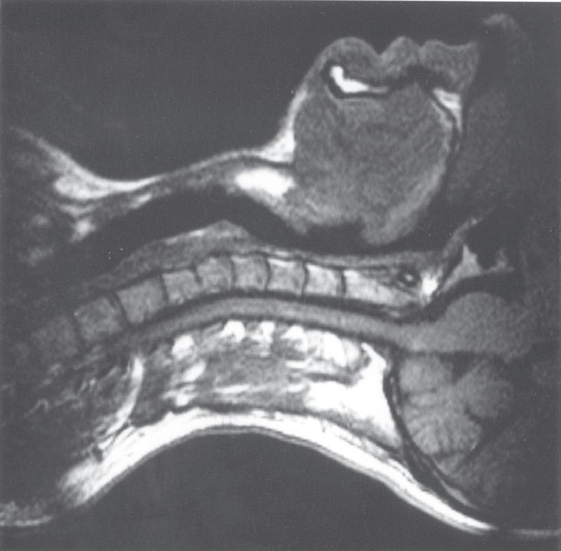
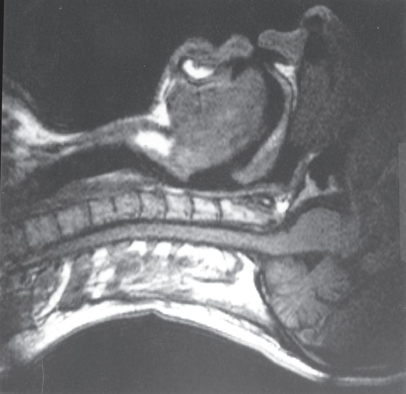
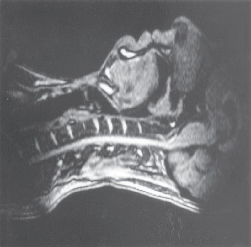
• Fig. 1.1 Magnetic resonance imaging scans showing median sagittal sections of the pharynx in a normal subject. (A) Normal nasal breathing with the oral airway occluded by lips and tongue. (B) Deliberate oral breathing with the nasal airway occluded by elevation and backwards movement of the soft palate. (C) A Valsalva manoeuvre in which the subject deliberately tries to exhale against a closed airway. Data acquisition for scans (A) and (B) took 45 s, so anatomical differences between inspiration and expiration will not be visible. I am indebted to Professor M. Bellamy for being the subject. E, Epiglottis; L, larynx; NC, nasal cavity; SP, soft palate; T, tongue; VF, vocal fold.
capable of withstanding pharyngeal pressures as high as 80 kPa (800 cmH2O), which may be generated during swallowing.
Upper airway cross-sectional areas can be estimated from conventional radiographs, magnetic resonance imaging (MRI) as in Figure 1.1 or acoustic pharyngometry. In the latter technique, a single sound pulse with a duration of 100 ms is generated within the apparatus and passes along the airway of the subject. Recording of the timing and frequency of sound waves reflected back from the airway allows calculation of the cross-sectional area, which is then presented as a function of the distance travelled along the airway (Fig. 1.2). Acoustic pharyngometry measurements correlate well with MRI scans of the airway, and the technique is now sufficiently developed for use in clinical situations such as estimating airway size in patients with sleep-disordered breathing (Chapter 14).1
The Larynx
The larynx evolved in the lungfish for the protection of the airway during such activities as feeding and perfusion of the gills with water. Although protection of the airway remains important, the larynx now has many other functions, all involving some degree of laryngeal occlusion.
Speech
Phonation, the laryngeal component of speech, requires a combination of changes in position, tension and mass of the vocal folds (cords). Rotation of the arytenoid cartilages by the posterior cricoarytenoid muscles opens the vocal folds, while contraction of the lateral cricoarytenoid and oblique arytenoid muscles opposes this. With the vocal folds almost closed, the respiratory muscles generate a positive pressure of 5 to 35 cmH2O, which may then be released by slight opening of the vocal folds to produce sound waves. The cricothyroid muscle tilts the cricoid and arytenoid cartilages backwards and also moves them posteriorly in relation to the thyroid cartilage. This produces up to 50% elongation and therefore tensioning of the vocal
• Fig. 1.2 Normal acoustic reflectometry pattern of airway crosssectional area during mouth breathing.
Trachea
Structural Characteristics of the Air Passages
U-shaped Links open end of cartilage
Columnar ciliated epithelium Main bronchi
Conducting airways
From the bronchial circulation
Within connective tissue sheath alongside arterial vessels
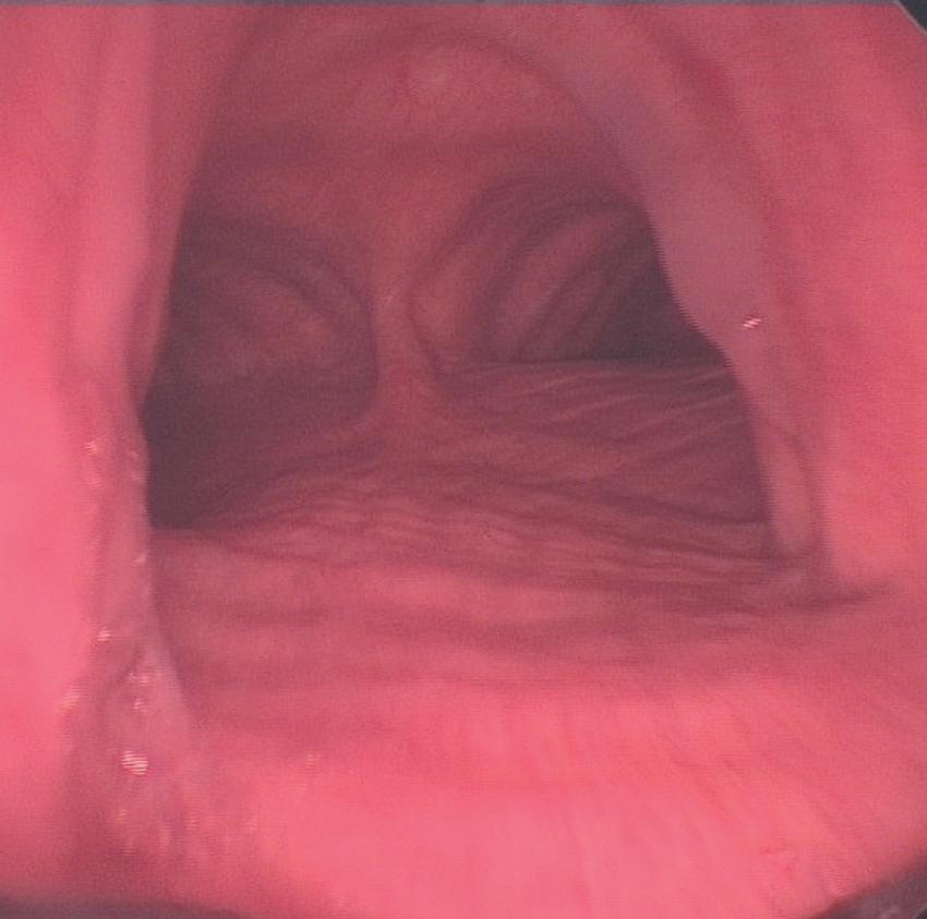
• Fig. 1.3 The normal trachea as viewed during a rigid bronchoscopy (page 399). The ridges of the cartilage rings are seen anteriorly, and the longitudinal fibres of the trachealis muscle are seen posteriorly, dividing at the carina and continuing down both right and left main bronchi. The less acute angle of the right main bronchus from the trachea can be seen, with its lumen clearly visible, illustrating why inhaled objects preferentially enter the right lung.
to direct traction and rely for their patency on cartilage within their walls and on the transmural pressure gradient, which is normally positive from lumen to intrathoracic space. In the normal subject this pressure gradient is seldom reversed and, even during a forced expiration, the intraluminar pressure in the small bronchi rapidly rises to more than 80% of the alveolar pressure, which is more than the extramural (intrathoracic) pressure.
Bronchioles (Generations 12–14)
An important change occurs at about the 11th generation, where the internal diameter is around 1 mm. Cartilage disappears from the airway wall below this level and
ceases to be a factor in maintaining patency. However, beyond this level the air passages are directly embedded in the lung parenchyma, the elastic recoil of which holds the air passages open like the guy ropes of a tent. Therefore the calibre of the airways beyond the 11th generation is mainly influenced by lung volume because the forces holding their lumina open are stronger at higher lung volumes. The converse of this factor causes airway closure at reduced lung volume (see Chapter 3). In succeeding generations, the number of bronchioles increases far more rapidly than the calibre diminishes ( Table 1.1). Therefore the total cross-sectional area increases until, in the terminal bronchioles, it is about 100 times the area at the level of the large bronchi ( Fig. 1.5). Thus the flow resistance of these smaller air passages ( , 2 mm diameter) is negligible under normal conditions. However, the resistance of the bronchioles can increase to very high values when their strong helical muscular bands are contracted by the mechanisms described in Chapters 3 and 28. Down to the terminal bronchiole the air passages are referred to as conducting airways, which derive their nutrition from the bronchial circulation and are thus influenced by systemic arterial blood gas levels. Beyond this point the smaller air passages are referred to as acinar airways and rely upon the pulmonary circulation for their metabolic needs.
Respiratory Bronchioles (Generations 15–18)
Down to the smallest bronchioles, the functions of the air passages are solely conduction and humidification. Beyond this point there is a gradual transition from conduction to gas exchange. In the four generations of respiratory bronchioles there is a gradual increase in the number of alveoli in their walls. Like the bronchioles, the respiratory bronchioles are embedded in lung parenchyma; however, they have a well-defined muscle layer with bands which loop over the opening of the alveolar ducts and the mouths of the mural alveoli. There is no significant change in the
the acinus and the most distal alveolus, which in humans is between 5 and 12 mm.
Respiratory Epithelium
Before inspired air reaches the alveoli it must be ‘conditioned’, that is, warmed and humidified, and airborne particles, pathogens and irritant chemicals removed. These tasks are undertaken by the respiratory epithelium and its overlying layer of airway lining fluid, and are described in Chapter 11. To facilitate these functions the respiratory epithelium contains numerous cell types.
Ciliated Epithelial Cells5
These are the most abundant cell type in the respiratory epithelium. In the nose, pharynx and larger airways the epithelial cells are pseudostratified, gradually changing to a single layer of columnar cells in bronchi, cuboidal cells in bronchioles and finally thinning further to merge with the type I alveolar epithelial cells (see later). They are differentiated from either basal or secretory cells (see later) and are characterized by the presence of around 300 cilia per cell (page 165). The ratio of secretory to ciliated cells in the airway decreases in more distal airways from about equal in the trachea to almost three-quarters ciliated in the bronchioles.
Goblet Cells
These are present at a density of approximately 6000 per mm2 (in the trachea) and are responsible for producing the thick layer of mucus that lines all but the smallest conducting airways (page 165).
Airway Glands6
Submucosal glands occur predominantly in the trachea and larger bronchi, diminishing in both size and numbers in more distal airways. The glands consist of a series of branching ducts, ending with a single terminal duct opening into the airway and contain both serous cells and mucous cells, with serous cells occurring in the gland acinus, whereas mucous cells are found closer to the collecting duct. The serous cells have the highest levels of membrane-bound cystic fibrosis transmembrane conductance regulator in the lung (Chapter 28).
Basal Cells
These cells lie underneath the columnar cells, giving rise to the pseudostratified appearance, and are absent in the bronchioles and beyond. They are the stem cells responsible for producing new epithelial and goblet cells.
Mast Cells
The lungs contain numerous mast cells which are located underneath the epithelial cells of the airways and in the alveolar septa. Some also lie free in the lumen of the airways and may be recovered by bronchial lavage. Their important role in bronchoconstriction is described in Chapter 28.
Club Cells (Formerly Clara Cells)
These nonciliated bronchiolar epithelial cells are found in the mucosa of the terminal bronchioles, where they may be the precursor of epithelial cells in the absence of basal cells. They are metabolically active, secreting a club cell secretory protein which has antioxidant and immune-modulatory functions.7
Neuroepithelial Cells
These cells are found throughout the bronchial tree but occur in larger numbers in the terminal bronchioles. They may be found individually or in clusters as neuroepithelial bodies, and are of uncertain function in the adult lung. Present in foetal lung tissue in a greater number, they may have a role in controlling lung development. Similar cells elsewhere in the body secrete a variety of amines and peptides such as calcitonin, gastrin-releasing peptide, calcitonin gene-related peptide and serotonin.
The Alveoli
The mean total number of alveoli has been estimated as 400 million, but ranges from about 270 to 790 million, correlating with the height of the subject and total lung volume.8 The size of the alveoli is dependent on lung volume, but at functional residual capacity (FRC) they are larger in the upper part of the lung because of gravity. At total lung capacity this situation reverses, and there are estimated to be 32 alveoli per mm3 at the lung apices compared with 21 at the lung bases.9 At FRC the mean diameter of a single alveolus is 0.2 mm, and the total surface area of the alveoli is around 130 m2
The Alveolar Septa
The septa are under tension generated partly by collagen and elastin fibres, but more by surface tension at the air–fluid interface (page 14). They are therefore generally flat, making the alveoli polyhedral rather than spherical. The septa are perforated by small fenestrations known as the pores of Kohn (Fig. 1.7), which provide collateral ventilation between alveoli. Collateral ventilation also occurs between small bronchioles and neighbouring alveoli (Lambert channels) and through interbronchiolar pathways of Martin, and is more pronounced in patients with emphysema (page 332) and in some other species of mammal (page 310).
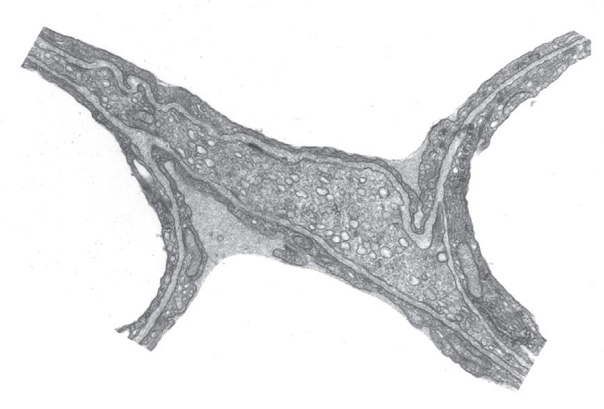
On one side of the alveolar wall the capillary endothelium and the alveolar epithelium are closely apposed, with almost no interstitial space, such that the total thickness from gas to blood is around 0.3 mm (Figs 1.8 and 1.9).10 This may be considered the ‘active’ side of the capillary, and gas exchange must be more efficient on this side. The other side of the capillary, which may be considered the ‘service’ side, is usually more than 1- to 2-mm thick, and contains a recognizable interstitial space containing elastin and collagen fibres, nerve endings and occasional migrant
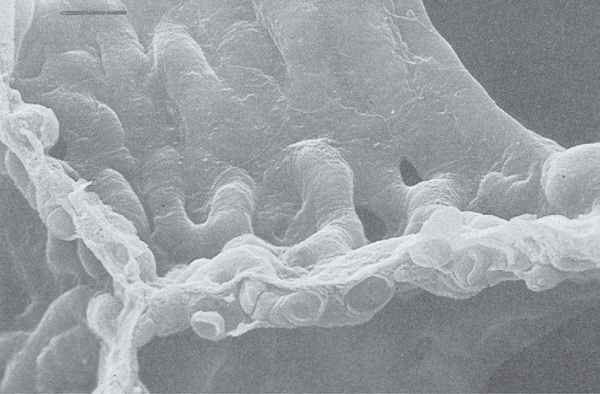
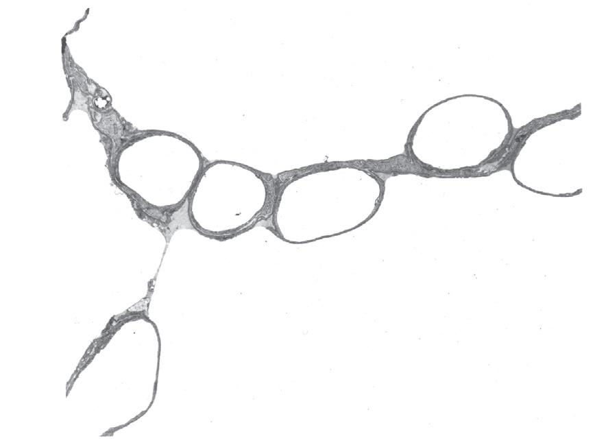
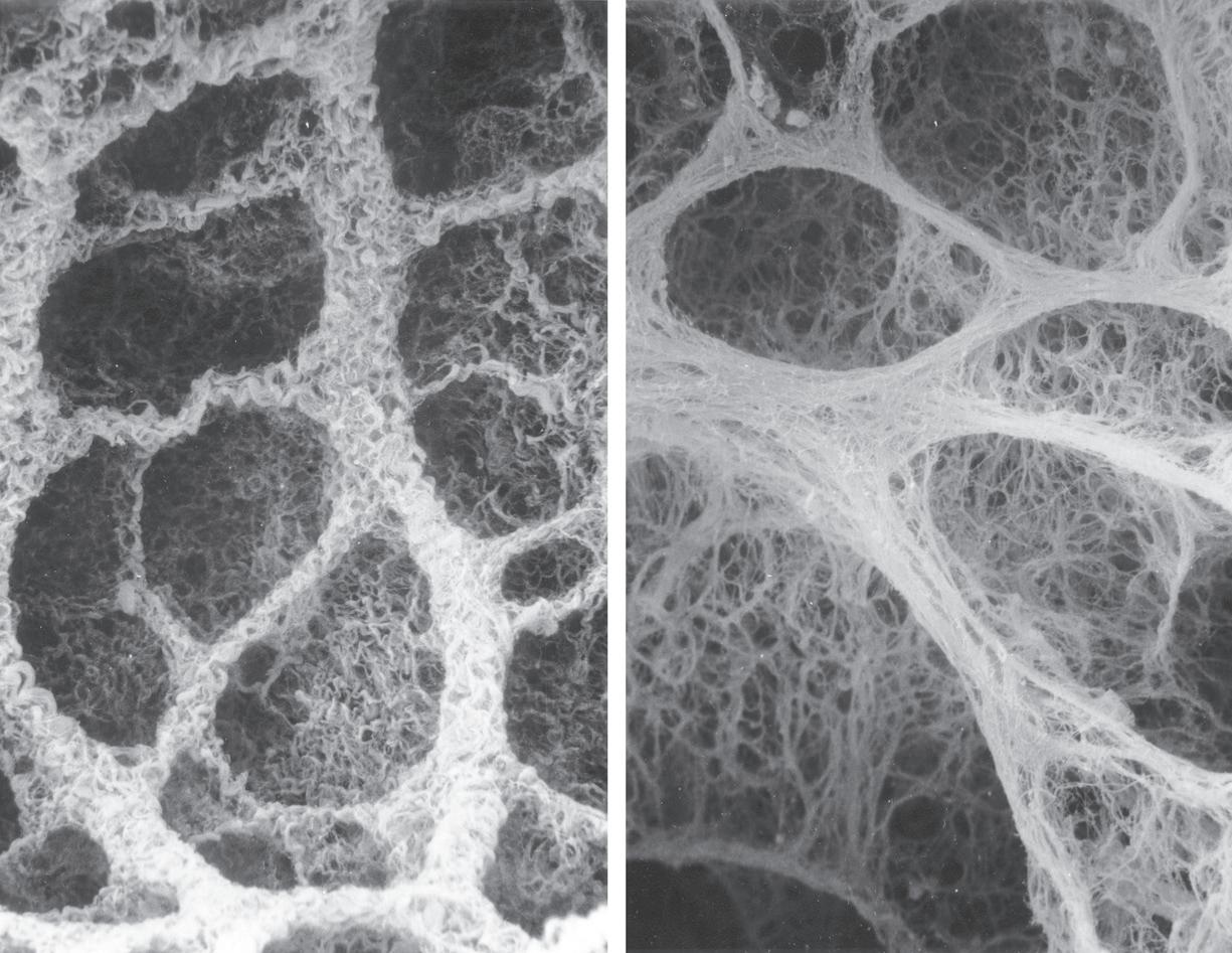
• Fig. 1.7 Scanning electron micrograph of the junction of three alveolar septa which are shown in both surface view and section view showing the polyhedral structure. Two pores of Kohn are seen to the right of centre. Red blood cells are seen in the cut ends of the capillaries. Scale bar 5 10 mm. (From Weibel ER. The Pathway for Oxygen Cambridge, Mass.: Harvard University Press; 1984. With permission. © Harvard University Press.)
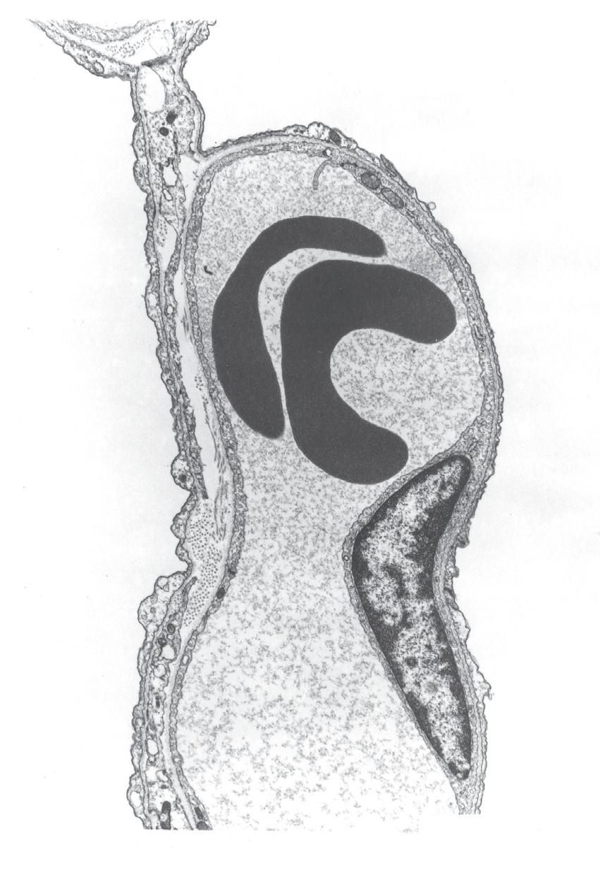
• Fig. 1.8 Details of the interstitial space, the capillary endothelium and the alveolar epithelium. Thickening of the interstitial space is confined to the left of the capillary (the service side), whereas the total alveolar/ capillary membrane remains thin on the right (the active side), except where it is thickened by the endothelial nucleus. Alv, Alveolus; BM, basement membrane; EN, endothelial nucleus; End, endothelium; Ep, epithelium; FB, fibroblast process; IS, interstitial space; RBC, red blood cell. (Electron micrograph kindly supplied by Professor E. R. Weibel.)
polymorphs and macrophages. The distinction between the two sides of the capillary has considerable pathophysiological significance, as the active side tends to be spared in the accumulation of both oedema fluid and fibrous tissue (Chapter 29).
• Fig. 1.9 (A) Transmission electron micrograph of alveolar septum with lung inflated to 40% of total lung capacity. The section in the box is enlarged in (B) to show alveolar lining fluid, which has pooled in two concavities of the alveolar epithelium and has also spanned the pore of Kohn in (A) There is a thin film of osmiophilic material (arrows), probably surfactant, at the interface between air and the alveolar lining fluid. (From reference 10 by permission of the authors and the editors of Journal of Applied Physiology.)
The Fibre Scaffold11
The connective tissue scaffold which forms the lung structure has three interconnected types of fibre:
1. Axial spiral fibres running from the hilum along the length of the airways
2. Peripheral fibres originating in the visceral pleura and spreading inwards into the lung tissue
3. Septal fibres, a network of which forms a basket-like structure12 of alveolar septa, through which are threaded the pulmonary capillaries, which are themselves a network
As a result, capillaries pass repeatedly from one side of the fibre scaffold to the other (Fig. 1.7), the fibre always residing on the thick (or service) side of the capillary, allowing the other side to bulge into the lumen of the alveolus. The left side of the capillary in Figure 1.8 is the side with the fibres. Structural integrity of the whole fibre scaffold is believed to be maintained by the individual fibres being under tension, referred to as a tensegrity structure, 11 such that when any fibres are damaged, the alveolar septum disintegrates and adjacent alveoli change shape, ultimately leading to emphysema.
• Fig. 1.10 Electron micrographs of the collagen fibre network of rat lung at low lung volume (A) and when fully inflated (B) 12 Note the folded, zigzag shape of the collagen at low lung volume in (A) (Photograph from Professor Ohtani. Reproduced by permission of Archives of Histology and Cytology.)
How the shape of this complex structure changes with breathing remains uncertain.13 Increasing lung volume may be achieved by increasing the size of alveolar ducts, expanding alveoli or recruiting previously collapsed alveoli. All three undoubtedly contribute, as lung volume increases approximately fivefold from residual volume to total lung capacity (page 22). Recent work using a new imaging technique demonstrated only small changes in alveolar size at different lung volumes, but a large change in alveolar numbers, indicating recruitment as the main mechanism for increasing lung volume.8,13 Change in alveolar size is facilitated by the molecular structures of both the elastin and collagen that make up the fibre scaffold, with the collagen forming helices or zigzags at lower lung volumes (Fig. 1.10).12
At the cellular level, the scaffolding for the alveolar septa is provided by the basement membrane, which provides the blood–gas barrier with enough strength to withstand the enormous forces applied to lung tissue.14 At the centre of the basement membrane is a layer of type IV collagen, the lamina densa, which is around 50 nm thick and made up of many layers of a diamond-shaped matrix of collagen molecules. On each side of the lamina densa the collagen layer is attached to the alveolar or endothelial cells by a series of proteins collectively known as laminins, of which seven subtypes are now known. The laminins are more than simple structural molecules, having complex interactions with membrane proteins and the intracellular cytoskeleton to help regulate cell shape, permeability and so on. These aspects of the function of the basement membrane are important. Increases in the capillary transmural pressure gradient greater than around 3 kPa (30 cmH2O) may cause disruption of
endothelium and/or epithelium, whereas the basement membrane tends to remain intact, sometimes as the only remaining separation between blood and gas.
Alveolar Cell Types
Capillary Endothelial Cells
These cells are continuous with the endothelium of the general circulation and, in the pulmonary capillary bed, have a thickness of only 0.1 mm, except where expanded to contain nuclei (Fig. 1.8). Electron microscopy shows the flat parts of the cytoplasm are devoid of all organelles except for small vacuoles (caveolae or plasmalemmal vesicles) which may open onto the basement membrane or the lumen of the capillary or be entirely contained within the cytoplasm (Fig. 1.9). The endothelial cells abut one another at fairly loose junctions of the order of 5 nm wide. These junctions permit the passage of quite large molecules, and the pulmonary lymph contains albumin at about half the concentration as that found in plasma. Macrophages pass freely through these junctions under normal conditions, and polymorphs can also pass in response to chemotaxis (page 353).
Alveolar Epithelial Cells: Type I15
These cells line the alveoli and exist as a thin sheet of around 0.1 mm in thickness, except where expanded to contain nuclei. Like the endothelium, the flat part of the cytoplasm is devoid of organelles, except for small vacuoles. Epithelial cells each cover several capillaries and are joined into a
continuous sheet by tight junctions with a gap of around 1 nm. These junctions may be seen as narrow lines snaking across the septa in Figure 1.7. The tightness of these junctions is crucial for preventing the escape of large molecules, such as albumin, into the alveoli, thus preserving the oncotic pressure gradient essential for the avoidance of pulmonary oedema (page 340). Nevertheless, these junctions permit the free passage of macrophages, and polymorphs may also pass in response to a chemotactic stimulus. Figure 1.9 shows the type I cell covered with a film of alveolar lining fluid. Type I cells are end cells and do not divide in vivo.
Alveolar Epithelial Cells: Type II
These are the stem cells from which type I cells arise.16 They do not function as gas exchange membranes and are rounded in shape and situated at the junction of septa. They have large nuclei and microvilli (Fig. 1.11). The cytoplasm contains characteristic striated osmiophilic organelles that contain stored surfactant (page 16). Type II cells are also involved in pulmonary defence mechanisms in that they may secrete cytokines and contribute to pulmonary inflammation. They are resistant to oxygen toxicity, tending to replace type I cells after prolonged exposure to high concentrations of oxygen.
Alveolar Macrophages
The lung is richly endowed with these phagocytes which pass freely from the circulation, through the interstitial space and thence through the gaps between alveolar epithelial cells
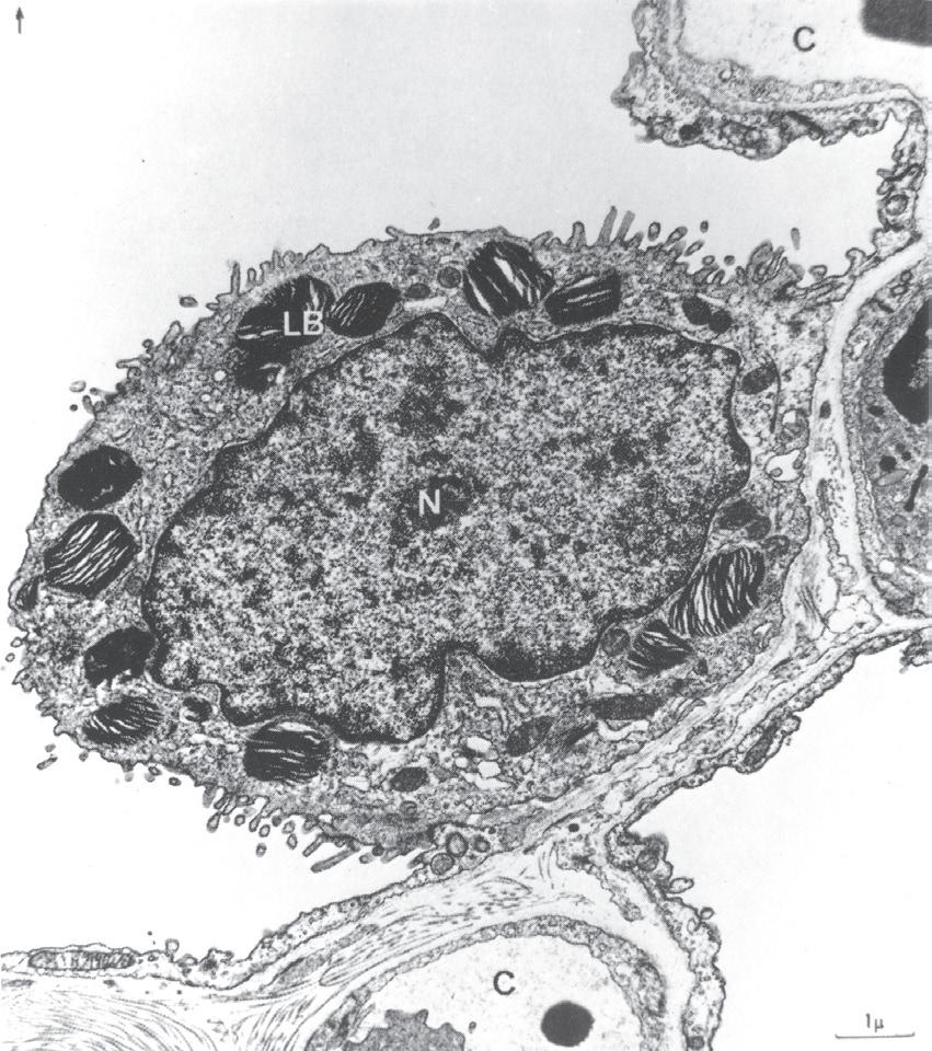
• Fig. 1.11 Electron micrograph of a type II alveolar epithelial cell of a dog. Note the large nucleus, the microvilli and the osmiophilic lamellar bodies thought to release surfactant. Alv, alveolus; C, capillary; LB, lamellar bodies; N, nucleus. (From reference 17 by permission of Professor E. R. Weibel and the editors of Physiological Reviews.)
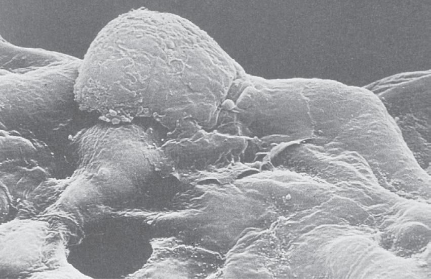
• Fig. 1.12 Scanning electron micrograph of an alveolar macrophage advancing to the right over epithelial type I cells. Scale bar 5 3 mm. (From Weibel ER. The Pathway for Oxygen Cambridge, Mass.: Harvard University Press; 1984. With permission. © Harvard University Press.)
to lie on their surface within the alveolar lining fluid (Fig. 1.12).18 They are remarkable for their ability to live and function outside the body. Macrophages form the major component of host defence within the alveoli, being active in combating infection and scavenging foreign bodies such as small dust particles. They contain a variety of destructive enzymes but are also capable of generating reactive oxygen species (Chapter 25). These are highly effective bactericidal agents, but their presence in lung tissue may rebound to damage the host. Dead macrophages release the enzyme trypsin, which may cause tissue damage in patients who are deficient in the protein a1-antitrypsin.
The Pulmonary Vasculature
Pulmonary Arteries
Although the pulmonary circulation carries about the same flow as the systemic circulation, the arterial pressure and the vascular resistance are normally only one-sixth as great. The media of the pulmonary arteries is about half as thick as in systemic arteries of corresponding size. In the larger vessels it consists mainly of elastic tissue, but in the smaller vessels it is mainly muscular, the transition is in vessels of around 1 mm diameter. Pulmonary arteries lie close to the corresponding airways in connective tissue sheaths. Table 1.2 shows a scheme for consideration of the branching of the pulmonary arterial tree. This may be compared with Weibel’s scheme for the airways (Table 1.1).
Pulmonary Arterioles
The transition to arterioles occurs at an internal diameter of 100 mm. These vessels differ radically from their counterparts in the systemic circulation and are virtually devoid of muscular tissue. There is a thin media of elastic tissue separated from the blood by endothelium. Structurally there is no real difference between pulmonary arterioles and venules.
of tracheobronchial lymph glands, where they receive tributaries from the superficial subpleural plexus. Most of the lymph from the left lung usually enters the thoracic duct, whereas the right side drains into the right lymphatic duct. However, the pulmonary lymphatics often cross the midline and pass independently into the junction of the internal jugular and subclavian veins on the corresponding sides of the body.
References
1. Patel SR, Frame JM, Larkin EK, et al. Heritability of upper airway dimensions derived using acoustic pharyngometry. Eur Respir J. 2008;32:1304-1308.
2. Weibel ER. Why measure lung structure? Am J Respir Crit Care Med. 2001;163:314-315.
3. Sauret V, Halson PM, Brown IW, et al. Study of the three-dimensional geometry of the central conducting airways in man using computed tomographic (CT) images. J Anat. 2002;200:123-134.
4. Sapoval B, Filoche M, Weibel ER. Smaller is better—but not too small: a physical scale for the design of the mammalian pulmonary acinus. Proc Natl Acad Sci USA. 2002;99:10411-10416.
5. Tilley AE, Walters MS, Shaykhiev R, et al. Cilia dysfunction in lung disease. Annu Rev Physiol. 2015;77:379-406.
6. Widdicombe JH, Wine JJ. Airway gland structure and function. Physiol Rev. 2015;95:1241-1319.
7. Barnes PJ. Club cells, their secretory protein, and COPD. Chest 2015;147:1447-1448.
8. Hajari AJ, Yablonskiy DA, Sukstanskii AL, et al. Morphometric changes in the human pulmonary acinus during inflation. J Appl Physiol. 2012;112:937-943.
9. McDonough JE, Knudsen L, Wright AC. Regional differences in alveolar density in the human lung are related to lung height. J Appl Physiol. 2015;118:1429-1434.
10. Gil J, Bachofen H, Gehr P, et al. Alveolar volume-surface area relation in air and saline filled lungs fixed by vascular perfusion. J Appl Physiol. 1979;47:990-995.
11. Weibel ER. It takes more than cells to make a good lung. Am J Respir Crit Care Med. 2013;187:342-346.
12. Toshima M, Ohtani Y, Ohtani O. Three-dimensional architecture of elastin and collagen fiber networks in the human and rat lung. Arch Histol Cytol. 2004;67:31-40.
13. Nieman G. Amelia Earhart, alveolar mechanics, and other great mysteries. J Appl Physiol. 2012;112:935-936.
14. Maina JN, West JB. Thin and strong! The bioengineering dilemma in the structural and functional design of the bloodgas barrier. Physiol Rev. 2005;85:811-844.
*15. Weibel ER. On the tricks alveolar epithelial cells play to make a good lung. Am J Respir Crit Care Med. 2015;191:504-513.
16. Hogan B. Stemming lung disease? N Engl J Med. 2018;378: 2439-2440.
17. Weibel ER. Morphological basis of alveolar-capillary gas exchange. Physiol Rev. 1973;53:419-495.
18. Staples KJ. Lung macrophages: old hands required rather than new blood? Thorax. 2016;71:973-974.
19. Kennedy JM, Foster GE, Koehle MS, et al. Exercise-induced intrapulmonary arteriovenous shunt in healthy women. Respir Physiol Neurobiol. 2012;181:8-13.
20. Duke JW, Davis JT, Ryan BJ, et al. Decreased arterial PO2, not O2 content, increases blood flow through intrapulmonary arteriovenous anastomoses at rest. J Physiol. 2016;594:4981-4996.
21. Duke HN. The site of action of anoxia on the pulmonary blood vessels of the cat. J Physiol. 1954;125:373.
22. Paredi P, Barnes PJ. The airway vasculature: recent advances and clinical implications. Thorax. 2009;64:444-450.
23. Mitzner W, Wagner EM. Vascular remodeling in the circulations of the lung. J Appl Physiol. 2004;97:1999-2004.