Top 10 Hacking Scripts in Python, C#, and ASP.NET: 2 Books in 1: Unmasking Cyber Secrets: Python, C#, and ASP.NET Scripts to Propel Your Hacking Journey Devwebtuts
No part of this publication may be reproduced or transmitted in any form or by any means, electronic or mechanical, including photocopying, recording, or any information storage and retrieval system, without permission in writing from the publisher. Details on how to seek permission, further information about the Publisher’s permissions policies and our arrangements with organizations such as the Copyright Clearance Center and the Copyright Licensing Agency, can be found at our website: www.elsevier.com/permissions
This book and the individual contributions contained in it are protected under copyright by the Publisher (other than as may be noted herein).
Notice
Practitioners and researchers must always rely on their own experience and knowledge in evaluating and using any information, methods, compounds or experiments described herein. Because of rapid advances in the medical sciences, in particular, independent verification of diagnoses and drug dosages should be made. To the fullest extent of the law, no responsibility is assumed by Elsevier, authors, editors or contributors for any injury and/or damage to persons or property as a matter of products liability, negligence or otherwise, or from any use or operation of any methods, products, instructions, or ideas contained in the material herein.
Previous edition copyrighted 2018.
International Standard Book Number: 978-0-323-76174-1
Publisher: Sarah Barth
Senior Content Development Specialist: Jennifer Shreiner
Publishing Services Manager: Catherine Jackson
Health Content Management Specialist: Kristine Feeherty
Design Direction: Renee Duenow Printed in India
This book is dedicated to the residents at Children’s Wisconsin. Their enthusiasm, thirst for knowledge, and desire to become outstanding pediatricians inspire us.
We further dedicate this textbook to the many patients and their families who continue to teach us important diagnostic considerations during their odyssey toward improved health.
Omar Ali, MD
Division of Pediatric Endocrinology
Valley Children’s Hospital Madera, California
Louella B. Amos, MD
Associate Professor of Pediatrics Division of Pulmonary and Sleep Medicine
Medical College of Wisconsin Milwaukee, Wisconsin
Bethany Auble, MD
Assistant Professor of Pediatrics
Medical College of Wisconsin Milwaukee, Wisconsin
Donald Basel, MD Professor of Pediatrics
Section Chief, Division of Medical Genetics
Medical College of Wisconsin Milwaukee, Wisconsin
Shannon H. Baumer-Mouradian, MD
Director of Quality Improvement
Assistant Professor of Pediatrics
Medical College of Wisconsin Milwaukee, Wisconsin
Ashley Beattie, MD
Fellow in Child and Adolescent Psychiatry
Medical University of South Carolina Charleston, South Carolina
Geetanjali Bora, MD
Fellow in Pediatric Gastroenterology
Medical College of Wisconsin Milwaukee, Wisconsin
Brett J. Bordini, MD
Associate Professor of Pediatrics
Section of Hospital Medicine
Nelson Service for Undiagnosed and Rare Diseases
Medical College of Wisconsin Milwaukee, Wisconsin
Brian R. Branchford, MD
Assistant Professor of Pediatrics
Division of Pediatric Hematology/Oncology/ Bone Marrow Transplant
Medical College of Wisconsin Milwaukee, Wisconsin
Amanda M. Brandow, DO, MS
Associate Professor of Pediatrics
Medical College of Wisconsin Milwaukee, Wisconsin
Ryan Byrne, MD
CONTRIBUTORS
Child and Adolescent Psychiatrist
Coastal Empire Community Mental Health Center
Beaufort, South Carolina
Gisela G. Chelimsky, MD Professor of Pediatrics
Medical College of Wisconsin Division of Pediatric Gastroenterology
University of Wisconsin School of Medicine and Public Health Madison, Wisconsin
Gary Cohen, MD, MS
Associate Professor of Pediatrics
Medical College of Wisconsin Milwaukee, Wisconsin
Deborah M. Costakos, MD, MS Professor of Ophthalmology
Medical College of Wisconsin Milwaukee, Wisconsin
Emily M. Densmore, MD, MS Assistant Professor of Pediatrics
Medical College of Wisconsin Milwaukee, Wisconsin
John C. Densmore, MD Professor of Surgery
Medical College of Wisconsin Milwaukee, Wisconsin
Patricia A. Donohoue, MD Professor and Section Chief
Pediatric Endocrinology and Diabetes
Medical College of Wisconsin Milwaukee, Wisconsin
Amy L. Drendel, DO, MS Professor of Pediatrics
Medical College of Wisconsin Milwaukee, Wisconsin
Garrett Elsner, MD
Fellow in Child and Adolescent Psychiatry
Medical University of South Carolina Charleston, South Carolina
Raquel Farias-Moeller, MD
Assistant Professor of Neurology and Pediatrics
Medical College of Wisconsin Milwaukee, Wisconsin
Shayne D. Fehr, MD
Associate Professor of Orthopaedic Surgery
Medical College of Wisconsin Milwaukee, Wisconsin
Susan Feigelman, MD Professor of Pediatrics
University of Maryland School of Medicine
Baltimore, Maryland
Veronica H. Flood, MD Professor of Pediatrics
Division of Pediatric Hematology/Oncology/ Bone Marrow Transplant
Medical College of Wisconsin Milwaukee, Wisconsin
Jessica Francis, MD
Assistant Professor Director, Residency Program Department of Obstetrics and Gynecology
Medical College of Wisconsin Affiliated Hospitals Milwaukee, Wisconsin
Julia Fritz, MD Pediatric Gastroenterology
Maine Medical Partners
Portland, Maine
Sandra Gage, MD, PhD Associate Professor of Pediatrics
University of Arizona College of MedicinePhoenix Phoenix, Arizona
Bhaskar Gurram, MBBS, MD
Associate Professor of Pediatrics
Division of Pediatric Gastroenterology, Hepatology, and Nutrition
University of Texas Southwestern Medical Center
Dallas, Texas
Matthew M. Harmelink, MD
Assistant Professor, Section of Child Neurology
Department of Neurology
Medical College of Wisconsin
Milwaukee, Wisconsin
Kristen E. Holland, MD
Associate Professor of Dermatology
Medical College of Wisconsin
Milwaukee, Wisconsin
Stephen R. Humphrey, MD
Assistant Professor of Dermatology
Children’s Hospital of Wisconsin
Medical College of Wisconsin Milwaukee, Wisconsin
Anna R. Huppler, MD
Assistant Professor
Pediatrics (Infectious Diseases) and Microbiology & Immunology
Medical College of Wisconsin Milwaukee, Wisconsin
Susan L. Jarosz, DO
Assistant Professor, Pediatric Urology
Baylor College of Medicine
Texas Children’s Hospital Houston, Texas
S. Anne Joseph, MD
Attending Physician
Neurology
Associate Professor of Pediatrics
Northwestern University Feinberg School of Medicine
Chicago, Illinois
Alvina R. Kansra, MD
Associate Professor of Pediatrics
Medical College of Wisconsin Wauwatosa, Wisconsin
Virginia Keane, MD
Mt. Washington Pediatric Hospital Baltimore, Maryland
Robert M. Kliegman, MD
Professor of Pediatrics
Nelson Service for Undiagnosed and Rare Diseases
Medical College of Wisconsin Milwaukee, Wisconsin
Julie M. Kolinski, MD
Assistant Professor
Department of Pediatrics and Internal Medicine
Medical College of Wisconsin Milwaukee, Wisconsin
Chamindra G. Konersman, MD
Associate Professor of Neurosciences
University of California, San Diego
San Diego, California
Kathleen A. Koth, DO, DFAACAP
Associate Professor of Psychiatry and Behavioral Medicine
Medical College of Wisconsin Milwaukee, Wisconsin
Katja Kovacic, MD
Associate Professor of Pediatrics
Division of Pediatric Gastroenterology
Medical College of Wisconsin Milwaukee, Wisconsin
Amornluck Krasaelap, MD, FAAP
Assistant Professor of Pediatrics
Children’s Mercy Hospital Kansas City, Missouri
John V. Kryger, MD
Chief of Pediatric Urology
Children’s Hospital of Wisconsin Professor
Department of Urology
Medical College of Wisconsin Milwaukee, Wisconsin
Sara M. Lauck, MD
Assistant Professor of Pediatrics
Medical College of Wisconsin Milwaukee, Wisconsin
Tracey H. Liljestrom, MD
Assistant Professor of Medicine and Pediatrics
Medical College of Wisconsin Milwaukee, Wisconsin
Ahmad Marashly, MD
Assistant Professor
Department of Neurology
University of Washington/Seattle Children’s Hospital Seattle, Washington
Seema Menon, MD
Pediatric and Adolescent Gynecology
Program Director, Children’s Hospital of Wisconsin
Associate Professor
Obstetrics and Gynecology
Medical College of Wisconsin
Milwaukee, Wisconsin
Adrian Miranda, MD
Professor of Pediatrics
Division of Pediatric Gastroenterology, Hepatology, and Nutrition
Medical College of Wisconsin Milwaukee, Wisconsin
Michelle L. Mitchell, MD
Assistant Professor of Pediatrics
Division of Pediatric Infectious Diseases
Medical College of Wisconsin Milwaukee, Wisconsin
Amy Moskop, MD, MS
Assistant Professor
Pediatric Bone Marrow Transplant and Cellular Therapy
Medical College of Wisconsin
Milwaukee, Wisconsin
Michael Muriello, MD
Assistant Professor of Pediatric Genetics
Medical College of Wisconsin
Milwaukee, Wisconsin
James J. Nocton, MD
Professor of Pediatrics
Medical College of Wisconsin
Milwaukee, Wisconsin
Joshua Noe, MD
Associate Professor of Pediatric Gastroenterology, Hepatology, and Nutrition
Department of Pediatrics
Medical College of Wisconsin
Milwaukee, Wisconsin
Cynthia G. Pan, MD
Professor Department of Pediatrics
Medical College of Wisconsin Milwaukee, Wisconsin
Andrew N. Pelech, MD Professor of Pediatrics
University of California, Davis Sacramento, California
Brittany Player, DO, MS
Fellow
Section Pediatric Infectious Disease
Medical College of Wisconsin Milwaukee, Wisconsin
Jacquelyn M. Powers, MD, MS
Assistant Professor of Pediatrics
Baylor College of Medicine Houston, Texas
Angela L. Rabbitt, DO
Associate Professor of Pediatrics
Medical College of Wisconsin
Milwaukee, Wisconsin
Amanda Rogers, MD
Assistant Professor of Pediatrics
Medical College of Wisconsin Milwaukee, Wisconsin
John M. Routes, MD Professor of Pediatrics
Medical College of Wisconsin Milwaukee, Wisconsin
Mark Simms, MD, MPH Emeritus Professor of Pediatrics Medical College of Wisconsin Milwaukee, Wisconsin
Rajasree Sreedharan, MD
Associate Professor Pediatrics/Nephrology
Medical College of Wisconsin Wauwatosa, Wisconsin
Alyssa Stephany, MD, SFHM, FAAP
Associate Professor
University of Kansas School of Medicine
Associate Professor
University of Missouri-Kansas City School of Medicine
Children’s Mercy Kansas City Kansas City, Missouri
Julie Talano, MD Professor of Pediatrics
Pediatric Bone Marrow Transplant and Cellular Therapy
Medical College of Wisconsin Milwaukee, Wisconsin
Grzegorz W. Telega, MD
Associate Professor of Pediatrics
Medical College of Wisconsin Milwaukee, Wisconsin
Heather Toth, MD Professor of Pediatrics
Program Director, Internal MedicinePediatrics Residency Program
Hospitalist, Departments of Medicine and Pediatrics
Medical College of Wisconsin Milwaukee, Wisconsin
Scott K. Van Why, MD Professor
Department of Pediatrics
Medical College of Wisconsin Milwaukee, Wisconsin
Sarah Vepraskas, MD
Associate Professor of Pediatrics
Section of Hospital Medicine
Medical College of Wisconsin Milwaukee, Wisconsin
James W. Verbsky, MD, PhD Professor of Pediatrics
Medical College of Wisconsin Milwaukee, Wisconsin
Bernadette Vitola, MD, MPH Associate Professor of Pediatrics
Pediatric Gastroenterology, Hepatology, and Nutrition
Medical College of Wisconsin Milwaukee, Wisconsin
Kevin D. Walter, MD, FAAP
Associate Professor of Pediatric Orthopedics and Pediatric Sports Medicine
Medical College of Wisconsin
Milwaukee, Wisconsin
Michael Weisgerber, MD, MS
Associate Professor of Pediatrics
Section of Hospital Medicine
Program Director, Pediatric Residency Program
Program Director, Pediatric-Anesthesia Combined Program
Medical College of Wisconsin Milwaukee, Wisconsin
Peter M. Wolfgram, MD
Associate Professor of Pediatrics
Medical College of Wisconsin Milwaukee, Wisconsin
Sarah C. Yale, MD
Assistant Professor of Pediatrics
Medical College of Wisconsin
Division of Hospital Medicine
Children’s Wisconsin Milwaukee, Wisconsin
Alicia C. Zolkoske, MD
Assistant Professor of Orthopaedic Surgery
Medical College of Wisconsin Milwaukee, Wisconsin
This book is intended to help the reader begin with a specific chief complaint that may be seen in many different disease entities. It is arranged in chapters that cover specific symptoms mirroring clinical practice. For example, patients do not usually present with a chief complaint of cystic fibrosis; rather, they may present with a cough, respiratory distress, or chronic diarrhea.
With a user-friendly, well-tabulated, illustrated approach, this text will help the reader differentiate between the many disease states causing a common symptom. The inclusion of many original tables and figures will help the reader identify distinguishing features of diseases and work through a diagnostic approach to the symptom. Modified and borrowed figures and tables from other outstanding current sources have been added as well. The combination of these illustrations and tables with diagnostic clues within the text will help provide a quick visual guide to the differential diagnosis of the various diseases under discussion. The diagnostic approach includes standard laboratory and radiologic testing, as well as advanced imaging studies and geneticbased analysis.
In addition, we have incorporated our experience with patients from our Undiagnosed and Rare Disease Program and have included uncommon disorders and those diseases that often remain undiagnosed, which may present with common symptoms. Furthermore, we discuss disease mimics or look-alike disorders as well as distinguishing features that may suggest a less common disease presenting with a common symptom.
We appreciate the hard work of our contributing authors. Writing a chapter in this type of format is quite different from writing in the format of a disease-based book. In addition, we thank Sarah Barth, Jennifer Shreiner, and Kristine Feeherty of Elsevier, whose patience and expertise contributed to the publication of this book. We are all also greatly appreciative of Carolyn Redman at the Medical College of Wisconsin Department of Pediatrics, whose editorial assistance and organization have made this new edition a reality. Finally, we are ever grateful for the understanding and patience of Diane Basel, Jessica Bordini, Ryan Festerling, and Sharon Kliegman in supporting this work.
Disease Mimics: An Approach to Undiagnosed Diseases
Brett J. Bordini and Donald Basel
“Getting the right diagnosis is a key aspect of health care, as it provides an explanation of a patient’s health problem and informs subsequent health care decisions.”
The Institute of Medicine; The National Academies of Sciences, Engineering, and Medicine, 2015
At its core, medicine is committed to identifying, preventing, and treating disease to maintain or restore health. Fundamental to this commitment is diagnosis, the process of uncovering the cause of a patient’s health-related concerns. Patients rarely present to their physicians with a diagnosis already identified, even with the widespread and increasing availability of health-related information and technology. Rather, patients present with a symptom: a subjective physical finding, sensation, or phenomenon that indicates to them that they may no longer be in their baseline state of health. A symptom may be accompanied by a sign: an objective finding on physical examination, laboratory investigation, or imaging study that indicates an abnormal deviation from homeostasis. Some signs, like elevated blood pressure, may not be associated with any noticeable symptoms. While not all symptoms or signs denote underlying pathophysiology, their mere presence is often unsettling to the person experiencing them, requiring either reassurance in instances of benign etiologies or a specific diagnosis and management plan in instances of pathology. Diagnosis is the process by which the health care team uncovers the precise explanation for those symptoms and signs so as to inform any subsequent health care–related decisions. The diagnostic process is complex, iterative, collaborative, often subject to revision, and fraught with the possibility of diagnostic error, yet when properly engaged holds the potential for restoring a patient to health.
DIAGNOSIS: IT ALL STARTS WITH A SYMPTOM
While asymptomatic individuals may have abnormalities detected on screening investigations, most patients enter into the diagnostic process by virtue of symptoms. With the exception of certain pathognomonic findings, such as Koplik spots in measles, presenting features rarely correlate directly and discretely to one individual disease process. It is up to the diagnostician to methodically gather information, categorizing and assigning importance to each historical and physical finding, noting both what is present and what is absent, so that this information can define the patient’s phenotype, the unique constellation of
subjective and objective information indicating the functional health status of the patient, how current physiology differs in ways that may suggest pathology, and the context in which these changes are occurring. Establishing this phenotype is the starting point for developing a structured working differential diagnosis, the possible diseases and mechanisms by which the patient’s symptoms are produced, which then drives additional investigation. This process is not random but rather has evolved through years of collective experience and analysis into the evidence-based standard of care (Fig. 1.1). Advances in medical science have been accompanied by refinements and improvements in diagnosis, ranging from enhanced understanding of physiology, allowing for more rapid and sophisticated identification of pathology, to the development of diagnostic technologies, such as MRI, molecular genetic assays, or the use of data aggregation and machine learning, allowing for the implementation of artificial intelligence–assisted diagnosis (Fig. 1.2).
DIAGNOSIS AS AN ITERATIVE PROCESS
Diagnosis is an iterative process, progressing in a stepwise fashion from an undifferentiated primary medical concern, or chief complaint, to an identified cause of the patient’s concerns with an associated management plan. The iterative process often requires one to go back to the beginning and, based on new data from the history, physical exam, imaging, or diagnostic tests, to re-evaluate the original hypothesis. Regardless of the ways in which diagnostic techniques and modalities have advanced over time, the fundamental components of diagnosis remain relatively unchanged and begin with establishing the history of the presenting concern.
History
The goal of obtaining the medical history is to develop a comprehensive yet concise narrative summary of the patient’s primary health concern, while placing that concern in the context of the patient’s past medical history, determining what social and environmental factors may be contributing to the patient’s health, eliciting which health conditions may have a familial tendency, and delineating what other symptoms or signs may be associated with the presenting concern. The scope of history gathering during any patient encounter is highly individualized and depends on both the clinician’s experience and the circumstances of the encounter; for example, an annual health supervision visit in an
Clinical history and interview Physical exam
Patient experiences a health problem Patient engages with health care system Communication of the diagnosis
Referral and consultation Diagnostic testing
Hassufficientinformation beencollected? Information integration & interpretation gathering I nformation diagnosis Working
The explanation of the health problem that is communicated to the patient
The planned path of care based on the diagnosis Patient and system outcomes Learning from diagnostic errors, near misses, and accurate, timely diagnoses Outcomes Treatment
Fig. 1.1 An overview of the diagnostic process. (From Balogh EP, Miller BT, Ball JR, Committee on Diagnostic Error in Health Care; Board on Health Care Services; Institute of Medicine. Improving Diagnosis in Health Care. Washington, DC: National Academies Press; December 29, 2015: Fig. 2.1.)
A century ago:
Diagnostic standard:
Bedside observation
Technologic test
Modern times:
Bedside observation is diagnostic standard
Classic rash (measles, zoster, lupus)
Neurology (Bell palsy, peripheral neuropathy)
Infection (otitis media, scarlet fever)
Immune (Kawasaki disease)
Technologic test or imaging is diagnostic standard
Fig. 1.2 The evolution of the diagnostic evaluation over time and with advances in diagnostic technology. (Modified from McGee S. Evidence-Based Physical Diagnosis. 3rd ed. Philadelphia: Elsevier; 2012.)
outpatient clinic and an emergency department visit for acute lateralized weakness would generate vastly different sets of questions. While matching the pace and scope of history gathering to the acuity of the patient’s presentation, the physician should conduct the medical interview in a manner that establishes trust, confidence, and rapport with the patient; creates sufficient space for the patient to tell
the story; and focuses on eliciting high-yield information in a timely fashion. To accomplish these aims, a combination of patient-driven, clinician-driven, and evidence-based medical interview techniques are employed. Typically, these interview styles are not implemented in isolation, but rather are blended to nurture conversational flow while allowing for comprehensive information gathering. The patientcentered portions focus on encouraging the patient to express what is most important, while the clinician-centered portions allow the physician to expand, clarify, and refine the history so that it is both comprehensive and specific. Evidence-based elements allow for probing aspects of the history and physical examination with higher-yield questions that help distinguish diagnostic possibilities with a high likelihood from those with a lower likelihood. This discrimination is best accomplished through the use of key pointer questions. These questions are often disease or specialty specific and are designed to tease out the nuances of relevant or pertinent information during history taking. When deciding if a suggestive history and physical examination are consistent with Kawasaki disease, a rheumatologist knows to investigate not simply whether the patient does or does not have conjunctivitis, but rather whether conjunctivitis is purulent (exclude), nonpurulent (include), limbic sparing (include), or not (exclude). Such questions leverage high likelihood ratios for crucial aspects of the history and physical examination to allow for more rapid refinement of the differential diagnosis (Fig. 1.3).
In addition to providing crucial information regarding the patient’s concerns, the medical history is an interactive process that allows the clinician to establish rapport and hopefully to avoid scenarios in which the patient or family state that the clinician “is a good doctor but never listens to what I say.” Building trust, setting expectations, and establishing the primacy of the patient-clinician relationship can be just as crucial as collecting informative data. Listening and being aware of nonverbal cues such as vocal inflection and body language can inform the data collection process as much as directive questioning. In some cases, obtaining that critical piece of information is highly dependent on the tone of the relationship between the two parties.
The medical history, and the quality of data obtained from it, can also be enhanced by the electronic medical record and the ways in which technology has evolved to facilitate good patient care and a good patient experience. Pre-charting, or familiarizing oneself with what is already known of the patient’s medical history, should be encouraged for any
Review working differential diagnosis
Narrow options with high LR
History of presenting complaint
EMR review
Collaborative narrative
Create a working differential diagnosis Data gathered from physical examination
“Key pointer” questions
Review pretest probabilities
System 1 process
Review options to exclude with low LR Pattern recognition
Exclude from working differential diagnosis Choose relevant investigations with high sensitivity and specificity for primary working diagnosis
Data to support current working diagnosis
nonurgent health visit, as a comprehensive chart review can save a significant amount of time during the patient encounter and can create an immediate sense of clinician engagement for the patient, wherein the patient knows that the physician has taken an interest in and has an immediate knowledge of the past history. This preparatory information should always be confirmed though, as diagnostic labels and medical problem lists can sometimes be inaccurate and perpetuate incorrect information about the patient that can increase the risk of diagnostic error.
Physical Examination
While advances in medical technology have changed the degree to which diagnosis is solely reliant on physical examination findings, comprehensive physical examination remains a cornerstone in identifying key phenotypic features that can then be further informed by
applying appropriate technologic testing (see Fig. 1.2). The office visit is a snapshot in time of the person being evaluated, and not all pertinent features are necessarily present for this evaluation: a fever noted at home may resolve or a rash may fade by the time of the appointment; new features may evolve following the medical evaluation. These reported findings, even if not present during the actual physical examination, should still be considered in the diagnostic formulation, given that many diseases and their associated findings are dynamic. When examining a patient, the traditional dichotomous concept of “positive” and “negative” findings either supporting or negating the possibility of a diagnosis remains important; however, the presence or absence of findings is not weighted equally and requires the application of evidence to determine significance. When evaluating the significance of a finding or result of a test, there are four concepts to consider:
Fig. 1.3 The use of “key pointer” questions in developing a deliberate and analytical diagnosis. EMR, electronic medical record; LR, likelihood ratio.
Diagnostic specificity Test + Test –
Fig. 1.4 The extended Fagan nomogram for the use of likelihood ratios and pretest probability to determine post-test probability according to Bayes’ theorem. LR, likelihood ratio.
pretest probability, sensitivity, specificity, and likelihood ratios (Figs. 1.4 and 1.5).
Pretest Probability
The pretest probability is the general prevalence of the condition within the patient population being evaluated. In this context it is the population risk prior to any bias of the examination or testing.
Sensitivity
Sensitivity refers to the ability of a test to correctly identify the presence of disease in a population of people who are affected by the disease.
Specificity
Specificity refers to the ability of a test to accurately identify the absence of disease through a negative test result.
Likelihood Ratio
Likelihood ratios (LRs) synthesize sensitivity and specificity of examination findings or test results to inform the probability of the presence or absence of disease for the individual patient. The greater the absolute value of the LR, the higher the clinical utility of the test in establishing or excluding a particular disease. Thus, it could be seen as a measure
Sensitivity/ (1-specificity)
(1-sensitivity)/ specificity
Fig. 1.5 Formulas used to calculate sensitivity, specificity, positive predictive value, negative predictive value, accuracy, and likelihood ratios.
of how “useful” a particular test is in the context of the diagnosis in question. Considered from a diagnostic perspective, it is also a measure of the likelihood of the presence or absence of disease in a patient after the test has been performed (i.e., the post-test probability of a specific diagnosis). The accuracy of an LR is entirely dependent on the quality of data used to generate the LR.
Relying purely on clinical findings, without the aid of additional testing, can on occasion lead the physician to the wrong diagnosis. Fever, cough, rhinorrhea, abdominal pain, headache, tonsillar exudate, and palatal petechiae all have low sensitivity and specificity when considering streptococcal pharyngitis, despite being common findings in patients with streptococcal pharyngitis. The positive predictive value of these signs and symptoms is no more than 50%; other clinical features carry more diagnostic weight: sore throat, tonsillar swelling, anterior cervical adenopathy, and scarlatiniform rash have all been shown to have a significant correlation with culture-positive group A βhemolytic streptococcal pharyngitis. Therefore, it is important to focus on features with high likelihood ratios when formulating differential diagnoses and testing strategies. When doing so, it may be tempting to aggregate likelihood ratios into clinical reasoning, using the post-test probability of one finding to serve as the pretest probability for the next finding, but when using these tools, each point needs to be considered in isolation and cannot be cascaded to inform one another. In addition, diseases are often complex, and thinking of disease paradigms as either present or absent does not account for the real-life variability in clinical presentation and severity that results from the unique interplay between the patient’s individual health status and the dynamic nature of many diseases. It is precisely this interplay that can lead to certain unrelated disorders presenting with similar features (mimic), or the same disorder presenting differently in different patients or with changing features over time in the same patient (chameleon) An example of considering mimics in the differential diagnosis while looking for distinguishing features is noted in Table 1.1. An example of the same disease with different manifestations acting as chameleons is noted in Table 1.2. At the gene level there are multiple genes producing the Noonan phenotype, while one gene (ADA-2 deficiency) may
TABLE 1.1 MRI Red Flags for the Diagnosis of Children with Acquired Demyelinating Syndromes
MRI Finding Differential Diagnosis Distinguishing Features
Leptomeningeal enhancement
Lesion expansion
SVcPACNS Infection
Tumor
HLH
Tumor
Lymphoma
PML
Sarcoidosis
Hemorrhage ANE
Stroke
Cerebellitis
AHLE
Large-vessel CNS vasculitis
SVcPACNS
Leptomeningeal enhancement is not a feature of MS in adults and emerged as a red flag for vasculitic or malignant processes in the pediatric cohort.
Increased size of T2 lesions on serial imaging is well recognized in MS, although this should always prompt consideration of malignancy. Increasing size of a white matter–predominant lesion without lesion enhancement in a patient treated with immunosuppressant therapy (or a patient with known HIV) should prompt consideration of PML. PML is a risk for MS patients exposed to more intense immunosuppressive therapies.
Although susceptibility-weighted imaging reveals tiny microfoci of hemosiderin in MS patients, hemorrhage large enough to be visible on conventional MRI sequences is not a feature of ADS or MS and should prompt consideration of disorders in which the cerebral vasculature is specifically involved.
ADS, acquired demyelinating syndrome; AHLE, acute hemorrhagic leukoencephalitis; ANE, acute necrotizing encephalopathy; CNS, central nervous system; HLH, hemophagocytic lymphohistiocytosis; MS, multiple sclerosis; PML, progressive multifocal leukoencephalopathy; SVcPACNS, smallvessel childhood primary angiitis of the central nervous system.
From O’Mahony J, Shroff M, Banwell B. Mimics and rare presentations of pediatric demyelination. Neuroimag Clin N Am. 2013;23(2):321–336 (Table 2, p. 323).
APPENDICITIS
TABLE 1.2 Guillain-Barré and Miller Fisher Syndromes and Their Subtypes
Guillain-Barré Syndrome
• Paraparetic variant*
• Pharyngeal-cervical-brachial weakness*
Acute pharyngeal weakness* †
• Bifacial weakness with paraesthesias*
Miller Fisher Syndrome
• Acute ataxic neuropathy†
• Acute ophthalmoparesis†
• Acute ptosis†
• Acute mydriasis†
• Bickerstaff brainstem encephalitis‡
Acute ataxic hypersomnolence† ‡
*Localized forms.
†Incomplete forms.
‡Central nervous system form.
From Wakerley BR, Yuki N. Mimics and chameleons in Guillain-Barré and Miller Fischer syndromes. Pract Neurol. 2015;15:90–99, Box 1, p. 91.
produce what appears as unrelated phenotypes (stroke, cytopenias, intestinal lesions). The application of sensitivity, specificity, and LRs thus needs to be judicious, with insight into what is being offered to aid the question at hand. Applied appropriately, these tools offer an established methodology for adjusting disease probability in the context of a specific defined parameter (see Fig. 1.4).
Physical examinations have two paradigms, one in which no additional evidence is required and one that is reliant on advanced testing. Certain presenting disorders remain clinical diagnoses. The diagnosis of otitis media relies on clinical features: the presence of fever along with an erythematous tympanic membrane that has limited mobility on pneumatic otoscopy or a middle ear effusion on visual inspection. Conversely, the definitive diagnosis of a structural malformation in congenital heart disease requires advanced imaging technology such as an echocardiogram to accurately make the diagnosis. When there is a recognized standard-of-care evaluation or investigation that is codependent on the physical findings to achieve a clear clinical diagnosis, this element of the physical examination is considered evidence based. When considering the example of an adolescent biological male presenting to the emergency room with right-sided lower quadrant pain, requesting a pregnancy test is unlikely to yield an informative result as pretest probability is negligible. Rather, applying appropriate clinical reasoning and looking for information to support a more decisive diagnosis may lead to the consideration of appendicitis (Fig. 1.6).
Incorrect application of these diagnostic aids or inaccurate assimilation of phenotypic data forms the basis of diagnostic error. Highquality information, properly synthesized, is important. Diagnostic aids such as artificial intelligence (e.g., machine learning), even when using natural language processing algorithms, require accurate subjective and objective phenotypic data to perform well.
DIAGNOSIS AS A COLLABORATIVE PROCESS AND THE TEAM-BASED APPROACH
The success of any diagnostic endeavor depends on the nature of the relationship forged with the patient, any of the patient’s family members or caregivers, and the health care team. The value of any subjective information gathered, whether it be the narrative history of the
Alvarado score, 4 or less
Absence of severe right lower quadrant tenderness
Probability
Absence of McBurney point tenderness
McBurney point tenderness
Alvarado score, 7 or more
Rovsing sign
Psoas sign
Fig. 1.6 Clinical evidence-based evaluation of the likelihood of appendicitis. LR, likelihood ratio. (From McGee S. Evidence-Based Physical Diagnosis. 3rd ed. Philadelphia: Elsevier; 2012.)
primary medical concern, the features and nuances of symptoms, the context of any associated signs, or the impact on daily life, depends on the strength of that relationship and the quality of communication that relationship affords.
The success of this team-based approach to diagnosis extends beyond the physician, patient, and the patient’s family and caregivers. When presentations are atypical or when patients have endured diagnostic delays or diagnostic errors, incorporating other health care providers into the patient’s care team enhances diagnostic accuracy beyond that achieved even by individual experienced expert clinicians. The primary aphorism that should drive the evaluation of every patient is that “none of us is as smart as all of us”; indeed, collective intelligence-based medical decision-making has consistently and significantly outperformed even the most accurate single diagnostician in certain clinical contexts. To the degree possible, each patient evaluation team should be composed of individuals from a wide breadth of disciplines and perspectives so as to broaden the collective knowledge base and solicit a variety of perspectives on pathophysiology and differential diagnoses that may not have otherwise been considered had the patient been evaluated in individual specialty settings. This collaborative and more deliberate approach to diagnosis additionally encourages the generation of multiple diagnostic hypotheses and testing strategies. The end goal and product of this process is the group phenotype, in which the evaluation team has collectively—as a group—analyzed the patient’s primary concerns, physical findings, and objective data into discrete phenotypic phenomena. These phenomena can then be explored further in attempts to uncover hypothesized underlying and unifying pathophysiologic mechanisms that can be investigated with further testing.
Diagnostic Error
Despite the best efforts and intentions of the health care team, the diagnostic process is at risk for error: depending on the clinical setting, diagnostic errors have been identified in up to 15% of patient encounters. Diagnostic error may consist of a missed, delayed, or wrong diagnosis, and the error may be attributable to faults within the structure and function of the health care system, such as the unavailability or inaccuracy of a particular diagnostic modality, or to factors beyond the control of the patient or clinician (e.g., “no fault” error), such as a disease process presenting with such atypical or subtle features as to completely preclude proper diagnosis. Most diagnostic error is attributable to cognitive errors related to cognitive bias
Dual process theory holds that clinicians engaging in medical reasoning typically utilize either a predominantly intuitive approach, termed a system 1 process, or a more analytical approach, termed a system 2 process. System 1 processes are based in heuristics, which rely on pattern recognition and rules of thumb to rapidly sort large amounts of clinical information into an illness script that allows for the
TABLE 1.3 Cognitive Biases Related to Heuristic Failure Bias Definition
Anchoring Locking into a diagnosis based on initial presenting features, failing to adjust diagnostic impressions when new information becomes available
Confirmation bias
Diagnostic momentum
Expertise bias/yinyang out
Overconfidence bias
Premature closure
Looking for and accepting only evidence that confirms a diagnostic impression, rejecting or not seeking contradictory evidence
Perpetuating a diagnostic label over time, usually by multiple providers both within and across health care systems, despite the label being incomplete or inaccurate
Believing that a patient who has already undergone an extensive evaluation will have nothing more to gain from further investigations, despite the possibility that the disease process or diagnostic techniques may have evolved so as to allow for appropriate diagnosis
Believing one knows more than one does, acting on incomplete information or hunches, and prioritizing opinion or authority as opposed to evidence
Accepting the first plausible diagnosis prior to obtaining confirmatory evidence or considering all available evidence; “when the diagnosis is made, thinking stops”
Unpacking principle Failing to explore primary evidence or data in its entirety and subsequently failing to uncover important facts or findings, such as accepting a biopsy report or imaging study report without reviewing the actual specimen or image
TABLE 1.4 Cognitive Biases Related to Errors of Attribution
Bias Definition
Affective bias
Appeal to authority
Ascertainment bias
Allowing emotions to interfere with a diagnosis, either positively or negatively; dislikes of patient types (e.g., “frequent flyers”)
Deferring to authoritative recommendations from senior, supervising, or “expert” clinicians, independent of the evidentiary support for such recommendations
Maintaining preconceived expectations based on patient or disease stereotypes
Countertransference Being influenced by positive or negative subjective feelings toward a specific patient
Outcome bias
Psych-out bias
Minimizing or overemphasizing the significance of a finding or result, often based on subjective feelings about a patient, a desired outcome, or personal confidence in one’s own clinical skills; the use of “slightly” to describe abnormal results
Maintaining biases about people with presumed mental illness
quick elaboration of a diagnosis. System 2 processes, on the other hand, rely on deliberate counterfactual reasoning and hypothesis generation tailored to individual patient circumstances to arrive at a more robust differential diagnosis. While clinicians predominantly engage system 1 processes and achieve relatively accurate diagnoses for most patients under most circumstances, heuristics can fail when patient presentations are multisystem, atypical, complex, or evolving, instead becoming a form of bias that can result in diagnostic error (Table 1.3). Biases may also be the result of errors of attribution, in which perceived characteristics or motivations of patients, family members, or members of the medical evaluation team are given undue weight in the diagnostic formulation. These factors can influence the affective state of the clinician and the integrity of cognition, increasing the likelihood of error. Examples of attribution-related errors are listed in Table 1.4
Cognitive bias may also be a product of the context in which a diagnostic evaluation takes place. With context-related biases, the setting of the diagnostic evaluation influences how clinicians perceive and process the information used in medical decision-making. External factors such as an increased patient volume, higher patient acuity, or staffing shortages, as well as internal factors such as sleep deprivation, stress, and physician burnout, can amplify individual cognitive burden and increase the likelihood of diagnostic error. Independent of cognitive burden, context can introduce bias by causing physicians to consciously or subconsciously de-emphasize relevant information and amplify impertinent information while formulating a diagnosis. Most common among context-related biases is the framing effect, in which the manner or setting of a patient’s presentation implicitly restricts the breadth of differential diagnoses considered. This framing effect is often
more prominent in contexts where patient handoffs occur frequently and diagnostic labels can take on an independent momentum, leaving little opportunity for physicians to challenge their appropriateness, or in specialty care settings, where patient symptoms are more likely to be considered primarily within the scope of that specialty’s pathophysiologic mechanisms. Specialty-specific settings may also increase the risk of availability bias, in which familiar and more frequently encountered diagnoses are more readily recalled and are given greater weight in the differential diagnosis. When framing effects and availability heuristics are invoked early, they may perpetuate anchoring biases and result in a diagnostic momentum that precludes consideration of alternate diagnoses, producing both erroneous diagnoses and significant delays in proper diagnosis. Further examples of context-related biases are listed in Table 1.5
The Well-Calibrated Diagnostician
Effective diagnosis extends beyond applying factual recall to the information gathered from a standardized medical history and instead requires the integration of experiential knowledge with individualized information derived from the patient, family, members of the care team, and diagnostic testing to develop diagnostic considerations based on evidence. The general challenge is being able to access information quickly, at the appropriate time, and in a manner that enables seamless patient care. It is not uncommon that clinicians need to engage these more analytical diagnostic approaches while simultaneously focusing on and reacting to tenuous physiology in a critically ill patient. Under circumstances such as these, determining whether the patient warrants a more deliberate, analytical evaluation may be difficult. Confidence
TABLE 1.5 Cognitive Biases Related to Errors of Context
Bias Definition
Availability bias
Base-rate neglect
Basing decisions on the most recent patient with similar symptoms, preferentially recalling recent and more common diseases
Over- or underestimating the prevalence of a disease, typically overestimating the prevalence of common diseases and underestimating the prevalence of rare diseases
Framing effect Being influenced by how or by whom a problem is described, or by the setting in which the evaluation takes place
Frequency bias
Hindsight bias
Posterior probability error
Representative bias
Believing that common things happen commonly and are usually benign in general practice
Reinforcing diagnostic errors once a diagnosis is discovered in spite of these errors; may lead to clinicians overestimating the efficacy of their clinical reasoning and may reinforce ineffective techniques
Considering the likelihood of a particular diagnosis in light of a patient’s prior or chronic illnesses (new headaches in a patient with a history of migraines may in fact be a tumor)
Basing decisions on an expected typical presentation; not effective for atypical presentations; overemphasis on disease diagnostic criteria or “classic” presentations; “looks like a duck, quacks like a duck”
Sutton’s slip Ignoring alternate explanations for “obvious” diagnoses (Sutton’s law is that one should first consider the obvious)
Thinking in silo
Zebra retreat
Restricting diagnostic considerations to a particular specialty or organ system; each discipline has a set of diseases within its comfort zone, which reduces diagnostic flexibility or team-based communication
Lacking conviction to pursue rare disorders even when suggested by evidence
in a proposed or working diagnosis may bias or impede consideration for a more appropriate diagnosis. The degree of concordance between diagnostic confidence and diagnostic accuracy is termed diagnostic calibration. Given the magnitude of this challenge, in the context of a busy service, be it the intensive care unit, emergency room, or routine office visit in the middle of a pandemic, a variety of cognitive biases may result in poor diagnostic calibration, as a result of either biasinduced overconfidence or cognitive burden–related complacency, in which a clinician may be aware of the possibility of diagnostic error but underestimates or trivializes its frequency or impact. One mechanism to avoid such diagnostic errors is the diagnostic timeout. When the natural history or progression of an illness fails to conform to the original diagnosis, or when new physical examination, laboratory, or imaging findings contradict that diagnosis or suggest alternate diagnoses, the entire diagnostic formulation must be re-evaluated without being anchored to the original and possibly wrong diagnosis (Table 1.6).
These biases may interfere with the ability to recognize when a patient’s physiology or response to treatment fails to support the
SUMMARY AND RED FLAGS
Diagnosis most often begins with a symptom and is a complex, iterative, and collaborative process. Despite the incorporation of evidence-based tools, the risk of diagnostic error is always present and most often results from cognitive biases that can lead the clinician to de-emphasize relevant clinical information or overemphasize impertinent findings. The path from symptom to diagnosis is rarely straightforward, and it is improved by building strong relationships with patients, employing a team-based approach whenever possible, and being aware of the cognitive biases that can increase the risk of
BIBLIOGRAPHY
A bibliography is available at ExpertConsult.com
TABLE 1.6 Diagnostic Timeout
• Identify the clinical issues, dilemmas, or concerns needing a timeout.
• Remove all previous diagnoses and re-examine all labs, imaging, and other information including the history and physical exam.
• Did we consider the risks of heuristic (intuitive) thinking? (See Table 1.3.)
• Do we have biases? (See Tables 1.4 and 1.5.)
• What are the diseases we must not miss?
proposed diagnosis, such as when a child with known asthma does not respond to bronchodilation in the treatment room and aspiration of a foreign body is not considered, or when difficulty ventilating indicates interstitial lung disease or surfactant protein deficiency as opposed to bronchopulmonary dysplasia in a neonate. Revisiting the initial working diagnosis and reassessing when these red flags indicate a parallel diagnosis is an essential part of nurturing the skills required to being a well-calibrated diagnostician.
diagnostic error. Clinicians should also be aware of and consider rarer disorders that may mimic more common diseases, as well as atypical, variable, or slowly evolving presentations of common diseases (chameleons). When presentations are complex or confounding, pausing the diagnostic process to engage more deliberate analytical approaches is warranted. One must always consider the complexity of the diagnostic process and emphasize the importance of developing a differential diagnosis that includes common disorders, atypical presentations, imitative mimics, and rare zebras
BIBLIOGRAPHY
Balogh EP, Miller BT, Ball JR, eds. Committee on Diagnostic Error in Health Care, Board on Health Care Services, Institute of Medicine, the National Academies of Sciences, Engineering, and Medicine. Improving Diagnosis in Health Care. National Academies Press (US); 2015
Barnett ML, Boddupalli D, Nundy S, et al. Comparative accuracy of diagnosis by collective intelligence of multiple physicians vs individual physicians. JAMA Netw Open. 2019;2(3):e190096
Berkwitt A, Grossman M. Cognitive bias in inpatient pediatrics. Hosp Pediatr. 2014;4(3):190–193
Berner ES, Graber ML. Overconfidence as a cause of diagnostic error in medicine. Am J Med. 2008;121(suppl 5):S2–S23
Bordini BJ, Stephany A, Kliegman R. Overcoming diagnostic errors in medical practice. J Pediatr. 2017;185:19–25.e1
Brenner H, Gefeller O. Variation of sensitivity, specificity, likelihood ratios, and predictive values with disease prevalence. Stat Med. 1997;16:981–991
Croskerry P. Bias: a normal operating characteristic of the diagnosing brain. Diagnosis. 2014;1(1):23–27
Custer JW, Winters BD, Goode V, et al. Diagnostic errors in the pediatric and neonatal ICU: a systematic review. Pediatr Crit Care Med. 2015;16(1):29–36
Ely JW. Preventing diagnostic errors in primary care. Amer Family Phys. 2016;94(6):426–432
Fagan TJ. Letter: nomogram for Bayes theorem. N Engl J Med. 1975;293:253
Grimmes DA, Schulz KF. Refining clinical diagnosis with likelihood ratios. Lancet. 2005;365:1500–1505.
Henderson M, Tierney L, Smetana GW, et al. The Patient History: EvidenceBased Approach. 2nd ed. McGraw-Hill Education/Medical; 2012. ISBN-10: 0071624945
Kämmer JE, Hautz WE, Herzog SM, et al. The potential of collective intelligence in emergency medicine: pooling medical students’ independent decisions improves diagnostic performance. Med Decis Making 2017;37(6):715–724
Kohn LT, Corrigan JM, Donaldson MS, eds. Institute of Medicine (US) Committee on Quality of Health Care in America. To Err Is Human: Building a Safer Health System. National Academies Press (US); 2000
Kurvers RHJM, Herzog SM, Hertwig R, et al. Boosting medical diagnostics by pooling independent judgments. Proc Natl Acad Sci USA 2016;113(31):8777–8782
Kurvers RHJM, Krause J, Argenziano G, et al. Detection accuracy of collective intelligence assessments for skin cancer diagnosis. JAMA Dermatol 2015;151(12):1346–1353
Lighthall GK, Vazquez-Guillamet C. Understanding decision making in critical care. Clin Med Res. 2015;13(3–4):156–168.
Lin MH, Fong WK, Chang PF, et al. Predictive value of clinical features in differentiating group A beta-hemolytic streptococcal pharyngitis in children. J Microbiol Immunol Infect. 2003;36(1):21–25
Mamede S, Gog T van, Berge K van den, et al. Effect of availability bias and reflective reasoning on diagnostic accuracy among internal medicine residents. J Am Med Assoc. 2010;304(11):1198–1203.
Mandin H, Harasym P, Eagle C, et al. Developing a “clinical presentation” curriculum at the University of Calgary. Acad Med. 1995;70(3):186–193
Marsicek SM, Odom B, Woodard A, et al. Time for a time-out: the value of a diagnostic time-out in prolonged fever and lymphadenopathy. Hospital Pediatr. 2019;9(2):139–141
McDonald CJ. Medical heuristics: the silent adjudicators of clinical practice. Ann Intern Med. 1996;124(1 Pt 1):56–62
Moayyeri A, Soltani A, Moosapour H, et al. Evidence-based history taking under “time constraint.” J Res Med Sci. 2011;16(4):559–564
Moons KG, van Es GA, Deckers JW, et al. Limitations of sensitivity, specificity, likelihood ratio, and Bayes’ theorem in assessing diagnostic probabilities: a clinical example. Epidemiology. 1997;8:12–17
Norman G. Dual processing and diagnostic errors. Adv Health Sci Educ Theory Pract. 2009;14(suppl 1):37–49
O’Mahony J, Shroff M, Banwell B. Mimics and rare presentations of pediatric demyelination. Neuroimag Clin N Am. 2013;23(2):321–336
Rajkomar A, Dhaliwal G. Improving diagnostic reasoning to improve patient safety. Perm J. 2011;15(3):68–73
Reiswich A, Haag M. Evaluation of chatbot prototypes for taking the virtual patient’s history. Stud Health Technol Inform. 2019;260:73–80
Sackett DL, Haynes RB, Guyatt GH, et al. A Basic Science for Clinical Medicine New York: Little, Brown and Co.; 1991:109–167. Clinical Epidemiology
Saposnik G, Redelmeier D, Ruff CC, et al. Cognitive biases associated with medical decisions: a systematic review. BMC Med Inform Decis Mak. 2016;16:138
Sweller J. Cognitive load theory, learning difficulty, and instructional design. Learn InStruct. 1994;4(4):295–312
Wakerley BR, Yuki N. Mimics and chameleons in Guillain-Barré and Miller Fischer syndromes. Pract Neurol. 2015;15:90–99
Wolf M, Krause J, Carney PA, et al. Collective intelligence meets medical decision-making: the collective outperforms the best radiologist. PloS One 2015;10(8):e0134269
Woloschuk W, Harasym P, Mandin H, et al. Use of scheme-based problem solving: an evaluation of the implementation and utilization of schemes in a clinical presentation curriculum. Med Educ. 2000;34(6):437–442
Most causes of sore throat are nonbacterial and neither require nor are alleviated by antibacterial therapy (Tables 2.1, 2.2, and 2.3). Accurate diagnosis is essential. Acute streptococcal pharyngitis warrants proper diagnosis and prompt therapy to prevent serious suppurative and nonsuppurative sequelae. Life-threatening complications of oropharyngeal infections, whether caused by streptococci or other pathogens, may manifest initially with poorly differentiated symptoms of mouth pain and pharyngitis and then extend into the parapharyngeal space, cause jugular venous thrombophlebitis, or result in critical airway obstruction (Tables 2.4 and 2.5). Noninfectious causes of sore throat, such as solid tumors, hematologic malignancy, or Kawasaki disease, if not appropriately diagnosed, may similarly carry a high risk of morbidity and even mortality. In most cases, history and physical examination identify the etiology of a sore throat or sufficiently narrow the differential diagnosis so as to direct an evaluation strategy or empiric treatment. It is important to note that in many of these diseases sore throat is not the defining symptom but part of a systemic illness (e.g., measles, COVID-19, Kawasaki disease).
VIRAL PHARYNGITIS
Most episodes of pharyngitis are caused by viruses (see Tables 2.2 and 2.3). It is difficult to precisely distinguish between viral and bacterial pharyngitis on clinical grounds, though certain clues may help. Accompanying findings of rhinitis, conjunctivitis, cough, croup, laryngitis, or discrete oropharyngeal ulcerations are common with viral infection but are rare in bacterial pharyngitis (Table 2.6).
Many viruses can produce pharyngitis (see Tables 2.2 and 2.3). Some cause distinct clinical syndromes that are readily diagnosed without laboratory testing (Table 2.7; see also Tables 2.1 and 2.4). In pharyngitis caused by parainfluenza viruses, influenza viruses, rhinoviruses, coronaviruses, and respiratory syncytial viruses, symptoms of coryza and cough often overshadow sore throat, which is generally mild. Parainfluenza viruses are associated with croup and bronchiolitis; minor sore throat and signs of pharyngitis are common at the outset but rapidly resolve. Influenza infection is often associated with high fever, cough, headache, malaise, myalgia, and cervical adenopathy in addition to pharyngitis. In young children, croup or bronchiolitis may develop. When influenza is suspected on clinical and epidemiologic grounds or confirmed by testing (polymerase chain reaction [PCR] is most accurate), specific antiviral therapy is available for treatment of
SECTION 2 Respiratory Disorders
patients and prophylaxis of family members and other close contacts. In young children, respiratory syncytial virus (RSV) infection is associated with bronchiolitis and, to a lesser degree, pneumonia and croup. In older children, RSV infection is usually indistinguishable from a simple upper respiratory tract infection. Pharyngitis is not a prominent finding of RSV infection in any age group. Infections caused by parainfluenza, influenza, and RSV are often seen in seasonal (winter) epidemics. Many viral pathogens can be identified using multiplex or targeted PCR testing, but there is rarely reason to test patients in the outpatient setting and infrequent benefit to testing in the inpatient setting, except to confirm and treat influenza.
Adenoviruses can cause upper and lower respiratory tract disease, ranging from ordinary colds to severe pneumonia and multisystem disease, including hepatitis, myocarditis, and myositis. The incubation period of adenovirus infection is 2–4 days. Upper respiratory tract infection typically produces fever, erythema of the pharynx, and follicular hyperplasia of the tonsils, together with exudate. Enlargement of the cervical lymph nodes occurs frequently. When conjunctivitis occurs in association with adenoviral pharyngitis, the resulting syndrome is called pharyngoconjunctival fever. Pharyngitis may last as long as 7 days and does not respond to antibacterials. There are many adenovirus serotypes; adenovirus infections may therefore develop in children more than once. Outbreaks have been associated with swimming pools and contamination in health care workers. If obtained, laboratory studies may reveal a leukocytosis and an elevated ESR.
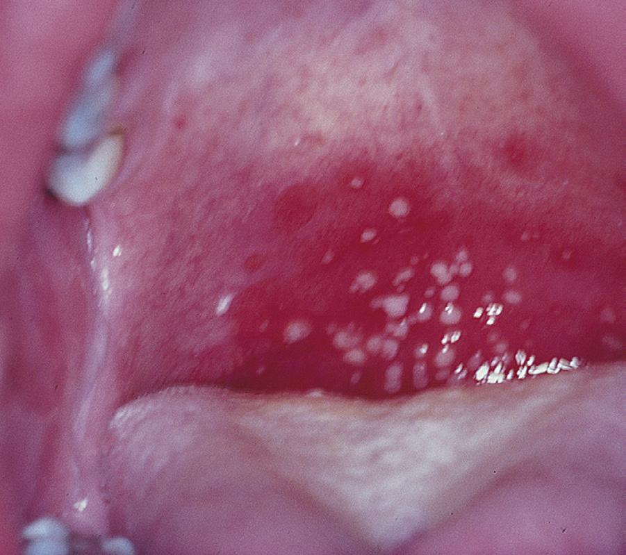
The enteroviruses can cause sore throat. High fever is common, and the throat is mildly erythematous; tonsillar exudate and cervical adenopathy are unusual. Symptoms usually resolve within a few days. Enteroviruses historically were classified as nonpolio enteroviruses, coxsackie A viruses, coxsackie B viruses, and echoviruses, though with increasing recognition of the multitude of enterovirus serotypes they are now numbered consecutively as novel serotypes are identified. Enteroviruses can also cause meningitis, acute flaccid myelitis, myocarditis, rash, and two specific syndromes that involve the oropharynx: herpangina and hand-foot-mouth disease. Herpangina is characterized by distinctive discrete, painful, gray-white papulovesicular lesions distributed over the posterior oropharynx (see Table 2.7). The vesicles are 1–2 mm in diameter and are initially surrounded by a halo of erythema before they ulcerate (Fig. 2.1). Fevers are typically present at illness onset, last several days before abating, and may reach 39.5°C or higher. The illness is typically caused by coxsackie A viruses and
Vincent angina (mixed anaerobic bacteria–gingivitis–pharyngitis)
Irritation
Cigarette or electronic cigarette/vaping use
Inhaled irritants; occupational, environmental
Reflux esophagitis
Allergic rhinitis
Chemical toxins (caustic agents including household cleaners, laundry detergent pods)
Medications
Paraquat ingestion
Smog, air pollutants
Dry, hot air
Hot foods, liquids
Shouting
Other
Tumor, including Kaposi sarcoma, leukemia
Granulomatosis with polyangiitis (formerly Wegener granulomatosis)
Sarcoidosis
Glossopharyngeal neuralgia
Foreign body
Stylohyoid syndrome
Behçet disease
Kawasaki disease
Posterior pharyngeal trauma—pseudodiverticulum
Pneumomediastinum with air dissection
Hematoma
Systemic lupus erythematosus
Bullous pemphigoid
Syndrome of periodic fever, aphthous stomatitis, pharyngitis, cervical adenitis (PFAPA)
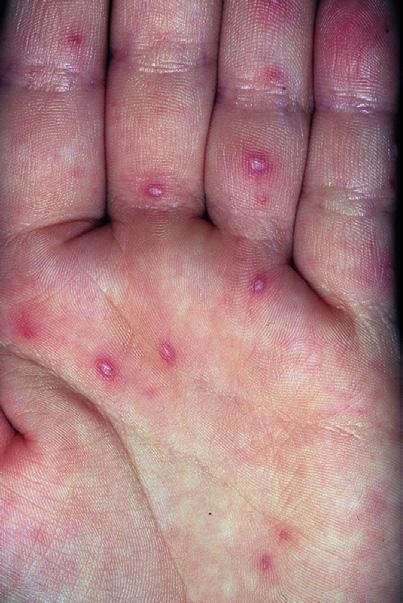
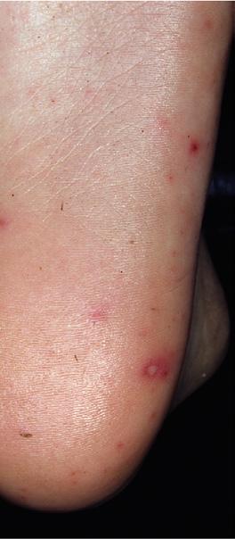
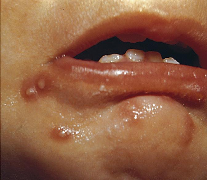
generally lasts <7 days, but severe pain may impair fluid intake and occasionally necessitates medical support. Hand-foot-mouth disease is also caused by enteroviruses, most commonly coxsackievirus A16. Painful vesicles that ulcerate can occur throughout the oropharynx, as opposed to the lesions of herpangina, which tend to be restricted to the posterior oropharynx. Vesicles also develop on the palms and soles and, less often, on the trunk, extremities, or diaper region (Fig. 2.2). Fever is present in most cases, but many children do not appear seriously ill. Illness typically lasts <7 days.
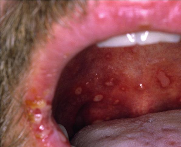
Primary infection caused by herpes simplex virus (HSV) (Fig. 2.3) usually produces high fever with acute gingivostomatitis, involving
TABLE 2.2 Infectious Etiology of Pharyngitis
Definite Causes
Streptococcus pyogenes (group A streptococci)
Corynebacterium diphtheriae
Arcanobacterium haemolyticum
Neisseria gonorrhoeae
Epstein-Barr virus
HIV (primary infection)
Parainfluenza viruses (types 1–4)
Influenza viruses
Rhinoviruses
Enteroviruses
Coronavirus, including SARS-CoV-2
Adenovirus (types 3, 4, 7, 14, 21, others)
Respiratory syncytial virus
Herpes simplex virus (types 1, 2)
Probable or Occasional Causes
Group C streptococci
Group G streptococci
Chlamydia pneumoniae
Chlamydia trachomatis
Mycoplasma pneumoniae
Toxoplasma gondii
SARS-CoV-2, severe acute respiratory syndrome-coronavirus 2.
TABLE 2.3 Additional Potential Pathogens Associated with Sore Throat
Bacteria
Fusobacterium necrophorum (Lemierre syndrome)
Neisseria meningitidis
Yersinia enterocolitica
Tularemia (oropharyngeal)
Yersinia pestis
Bacillus anthracis
Chlamydia psittaci
Secondary syphilis
Mycobacterium tuberculosis
Lyme disease
Corynebacterium ulcerans
Leptospira species
Mycoplasma hominis
Coxiella burnetii
Virus
Cytomegalovirus
Viral hemorrhagic fevers
HIV (primary infection)
Human herpesvirus 6
Measles
Varicella
Rubella
Fungus
Candida species
Histoplasmosis
Cryptococcosis
Respiratory Tract Infections
Distinguishing Features of Parapharyngeal–Upper
Postanginal Sepsis (Lemierre Syndrome)
Bacterial Tracheitis
Laryngotracheo bronchitis (Croup)
Masticator Space * Epiglottitis
Submandibular Space (Ludwig Angina) * Lateral Pharyngeal Space
Retropharyn geal Abscess (Cellulitis)
Peritonsillar Abscess
Moraxella catarrhalis, S. aureus, H. influenzae type b or nontypeable
Parainfluenza virus; influenza, adenovirus, and respiratory syncytial virus less common
Etiology Group A streptococci, oral anaerobes † Staphylococcus aureus, oral anaerobes, † group A streptococci, “suppurative adenitis” Oral anaerobes † Oral anaerobes † Oral anaerobes † Haemophilus influenzae type b (rarely), group A streptococci, Streptococcus pneumoniae , Staphylococcus aureus , and non–type b H. influenzae
Fusobacterium necrophorum Age Teens Infancy, preteens, occasionally teens
Prior pharyngitis with suddenonset fever, chills, odynophagia, neck pain, septic thrombophlebitis of internal jugular vein with septic emboli (e.g., lungs, joints), bacteremia
Prior history of croup with sudden onset of respiratory distress, high fever, “toxic” appearance, hoarseness, stridor, barking cough, tripod sitting position; radiograph as per croup plus ragged tracheal air column
Lowgrade fever, barking cough, hoarseness, aphonia, stridor; mild retractions; radiograph shows “steeple sign” of subglottic narrowing on anteroposterior neck view
Pain, prominent trismus, fever Swelling not always evident
Severe pain, fever, trismus, dysphagia, edematous appearing, painful lateral facial (jaw) or neck swelling (induration) May lead to Lemierre syndrome
Fever, dysphagia, odynophagia, stiff neck, dyspnea; airway obstruction, swollen tongue and floor of mouth (tender) Muffled voice
Usually not hoarse or coughing
Lateral neck radiograph shows “thumb sign” of swollen epiglottis
Initial episode of pharyngitis, followed by sudden worsening of unilateral odynophagia, trismus, hot potato (muffled) voice, drooling, displacement of uvula Fever, dyspnea, stridor, dysphagia, drooling, stiff neck, pain, cervical adenopathy, swelling of posterior pharyngeal space
Manifestations
Descending mediastinitis (rare)
Lateral neck radiograph reveals swollen retropharyngeal prevertebral space: infants, >1× width of adjacent vertebral body (>2–7 mm); teens, >1/3× width of vertebral body (>1–7 mm) CT distinguishes cellulitis from abscess
*Often odontogenic; check for tooth abscess, caries, tender teeth.
† Peptostreptococcus, Fusobacterium, Bacteroides
TABLE 2.5 Red Flags Associated with Sore
Throat
Toxic appearance
Shock Fever >2 wk
Duration of sore throat >2 wk
Dysphagia
Trismus
Stridor
Drooling
Dysphonia
Cyanosis
Hemorrhage
Asymmetric tonsillar swelling or asymmetric cervical adenopathy
Respiratory distress (airway obstruction or pneumonia)
Suspicion of parapharyngeal space infection
Suspicion of diphtheria (bull neck, uvula paralysis, thick membrane)
Apnea
Severe, unremitting pain
Tripod sitting position
“Hot potato” or muffled voice
Chest or neck pain
Weight loss
Systemic lymphadenopathy
Pharyngeal pseudomembrane
Marked neck swelling (bull neck)
Uvula paralysis
Travel or exposure to individuals from diphtheria endemic region
HIV behavioral risk
TABLE 2.6 Findings Suggestive of Group A Streptococcus and Viral Pharyngitis
Suggestive of Group A Streptococcus
Sudden onset
Sore throat
Fever >100.4°F
Headache
Nausea, vomiting, and abdominal pain
Erythema of pharynx and tonsils
Patchy discrete exudates
Tender, enlarged anterior cervical nodes
Patient aged 3–15 yr
Presentation in winter or early spring
History of exposure
Scarlet fever
Suggestive of Viral Etiology
Conjunctivitis
Coryza
Cough
Hoarseness
Diarrhea
Discrete ulcerative lesions
Myalgia
Typical viral rash (measles, etc.)
Posterior cervical or generalized lymphadenopathy
Hepatosplenomegaly
EBV-ampicillin rash
EBV, Epstein-Barr virus.
vesicles throughout the anterior portion of the mouth, including the lips; vesicles eventually ulcerate. There is sparing of the posterior pharynx in herpes gingivostomatitis; the infection usually occurs in young children. High fever is common, pain is intense, and intake of oral fluids is often impaired, which may lead to dehydration and need for medical support. In adolescents, HSV may manifest as poorly differentiated pharyngitis. Approximately 35% of new-onset HSV-positive adolescent patients have herpetic lesions; most teenage patients with HSV pharyngitis cannot be distinguished from patients with other causes of pharyngitis. The classic syndrome of herpetic gingivostomatitis in infants and toddlers lasts up to 2 weeks; data on the course of more benign HSV pharyngitis infections in older children and adolescents are lacking. The differential diagnosis of vesicular-ulcerating oral lesions is noted in Tables 2.7 and 2.8
Infants and toddlers with measles often have prominent oral findings early in the course of the disease. In addition to high fever, cough, coryza, and conjunctivitis, the pharynx may be intensely and diffusely erythematous, without tonsillar enlargement or exudate. Koplik spots, the pathognomonic white or blue-white enanthem of measles, appear on the buccal mucosa near the mandibular molars, generally before the typical measles rash develops. Complications of measles include ear infections, pneumonia, and encephalitis. In the United States, widespread measles vaccination virtually eliminated transmission of natural measles infection except among unvaccinated subpopulations (e.g., children younger than 12 months old, families who have refused immunization). Most recent cases are related to unimmunized visitors from countries with endemic measles, although there has been a rise in cases within the United States due to increasing rates of unvaccinated children.
The lesions of herpangina, hand-foot-mouth disease, herpes gingivostomatitis, and measles should be distinguished from noninfectious aphthous ulcers, colloquially referred to as canker sores. Aphthous ulcers are typically discrete, 3–5 mm in diameter, round or ovoid, with a peripheral erythematous halo and a white to yellow-white covering exudate. Lesions tend to be painful, though some may be noted only incidentally. An isolated episode of individual or clustered lesions may be related to physical or chemical irritation, a reaction to an allergic or infectious exposure, nutritional deficiency, or immune-mediated inflammation. Individuals with recurrent aphthous stomatitis may lack associated features during or between episodes and experience one to several outbreaks per year, a condition known as simple aphthosis. The outbreaks of simple aphthosis tend to start in late childhood or adolescence, last up to 2 weeks, and abate by adulthood. Patients with complex aphthosis tend to have more frequent outbreaks, have more numerous and painful lesions, and occasionally have lesions on the genital mucosa as well. In such circumstances, complex aphthosis must be distinguished from Behçet disease, the latter of which is a systemic inflammatory disorder that may include arthritis, neurologic manifestations, and cutaneous lesions, in addition to orogenital mucosal aphthae (see Table 2.6).
An autoinflammatory periodic fever syndrome known as PFAPA (periodic fever, aphthous stomatitis, pharyngitis, and cervical adenitis) occurs predictably every 2–8 weeks. The onset of PFAPA is usually before the age of 5 years. In addition to aphthous stomatitis and pharyngitis, PFAPA is characterized by high fever lasting 4–6 days. The diagnosis is clinical and is typically made in patients with a phenotype consistent with PFAPA after excluding cyclic neutropenia, other periodic fever syndromes, infections, malignancy, and the persistence of elevated acute-phase reactants between episodes. Individual episodes resolve spontaneously but may respond to oral prednisone. Cimetidine in PFAPA syndrome is ineffective. As corticosteroids do not prevent future fever cycles, long-term intervention may also
Fever, mouth pain, toxic, fetid breath, drooling, anorexia, cervical lymphadenopathy; cracked, swollen hemorrhagic gums; secondary inoculation possible (fingers, eye, skin); reactivation with long latency (any age)
Manifestations
include tonsillectomy with or without adenoidectomy, although risks of surgical intervention such as anesthetic and postoperative complications must be considered. In most patients PFAPA completely resolves without sequelae before puberty; a few persist into adulthood. Some patients initially diagnosed as having PFAPA actually had a monogenetic recurrent fever syndrome (see Chapter 54).
INFECTIOUS MONONUCLEOSIS
Pathogenesis
Acute exudative pharyngitis commonly occurs with infectious mononucleosis caused by primary infection with the Epstein-Barr virus (EBV). Mononucleosis is a febrile, systemic, self-limited lymphoproliferative disorder that is usually associated with hepatosplenomegaly and generalized lymphadenopathy. Acute pharyngitis may be mild or severe, with erythema, impressive tonsillar exudates, and significant tonsillar hypertrophy that can result in airway obstruction. Regional lymph nodes may be particularly enlarged and slightly tender. Infectious mononucleosis usually occurs in adolescents and young adults; EBV infection is generally milder or subclinical in preadolescent children. In U.S. high school and college students, attack rates are 200–800 per 100,000 per year. EBV is transmitted primarily by saliva.
A B C
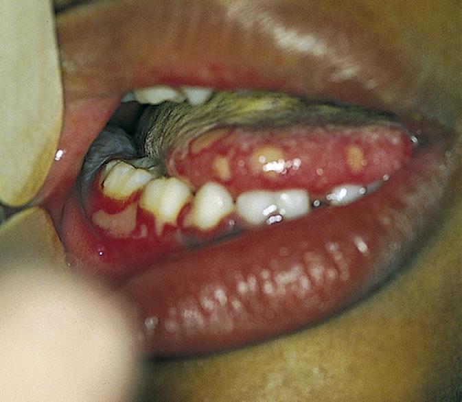
Fig. 2.2 A, Hand-foot-mouth disease. Aphthae-like erosions may appear anywhere in the oral cavity. B, Hand-foot-mouth disease. Cloudy vesicles with a red halo are highly characteristic of this disease. C, Handfoot-mouth disease. The pale, white, oval vesicles with a red areola are a distinguishing feature of this disease. (From Dinulos JGH. Habif’s Clinical Dermatology. 7th ed. Philadelphia: Elsevier; 2021, Figs. 14.4, 14.6, and 14.7.)
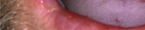
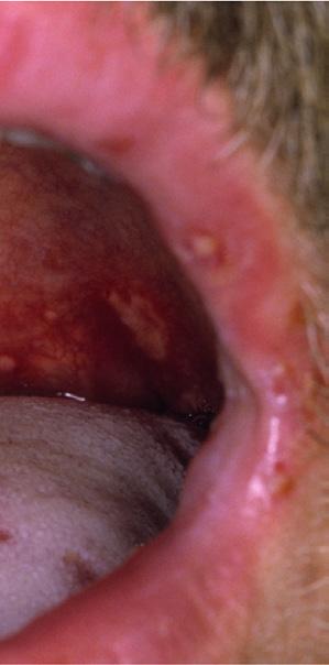
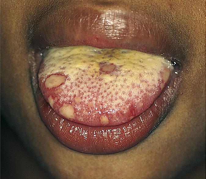
Fig. 2.3 Herpes simplex infections. A, Herpetic gingivostomatitis is characterized by discrete mucosal ulcerations and diffuse gingival erythema, edema, and friability in association with fever, dysphagia, and cervical adenopathy. B, Numerous yellow ulcerations with thin red halos are seen on the patient’s tongue as well. C, Thick-walled vesicles on an erythematous base were noted in this child, who showed early findings of intraoral involvement. (From Michaels MG, Williams JV. Infectious diseases. In Zitelli BJ, McIntire SC, Nowalk AJ, eds. Zitelli and Davis’ Atlas of Pediatric Physical Diagnosis. 7th ed. Philadelphia: Elsevier; 2018, Fig. 13.3.)
Fig. 2.1 Herpangina with shallow ulcers in the posterior oropharynx. (From Cohen J, Powderly WG. Infectious Diseases. 2nd ed. St Louis: Mosby; 2004.)
TABLE 2.8
Broad
Differential Diagnosis of Oral Ulceration
Condition Comment
Common
Aphthous ulcers (canker sores)
Traumatic ulcers
Hand-foot-mouth disease
Herpangina
Herpetic gingivostomatitis
Recurrent herpes labialis
Other viruses
Chemical burns
Heat burns
Drugs
Uncommon
Neutrophil defects
Systemic lupus erythematosus
Behçet syndrome
Necrotizing ulcerative gingivostomatitis
Syphilis
Oral Crohn disease
Histoplasmosis
Pneumococcal sepsis or pneumonia
Pemphigus
Stevens-Johnson syndrome
Mycoplasma (MIRM)
RIME
PFAPA
Celiac disease
Nutritional deficiencies
Painful circumscribed lesions; recurrences
Accidents, chronic cheek biter, after dental local anesthesia
Painful; lesions on tongue, anterior oral cavity, hands, and feet
Painful; lesions confined to soft palate and oropharynx
Vesicles on mucocutaneous borders; painful, febrile
After a 2–4-week incubation period, patients with infectious mononucleosis usually experience an abrupt onset of malaise, fatigue, fever, and headache, followed closely by pharyngitis. The tonsils are enlarged with exudates and cervical (often posterior) adenopathy. More generalized adenopathy with hepatosplenomegaly often follows. Fever and pharyngitis typically last 1–3 weeks, and lymphadenopathy and hepatosplenomegaly resolve over 3–6 weeks. Malaise and lethargy can persist for several months and can affect school or work performance.
Diagnosis
The findings of acute exudative pharyngitis together with hepatomegaly, splenomegaly, and generalized lymphadenopathy suggest infectious mononucleosis. Early in the disease and in cases without liver or spleen enlargement, differentiation from other causes of pharyngitis, including streptococcal pharyngitis, is difficult. Indeed, a small number of patients with infectious mononucleosis have a throat culture positive for group A streptococci, though these patients are likely streptococcal carriers. Patients with infectious mononucleosis treated for presumed streptococcal pharyngitis with antibacterials, particularly with ampicillin or amoxicillin, frequently develop a diffuse, erythematous, papular exanthem during the course of therapy that retrospectively suggests infectious mononucleosis due to
EBV. An indistinguishable mononucleosis syndrome can occur with cytomegalovirus, but differentiation is rarely of clinical importance. Acute toxoplasmosis may also present similarly. Serologic evidence of mononucleosis should be sought when splenomegaly or other features are present or if symptoms persist longer than expected. Laboratory studies of diagnostic value include atypical lymphocytosis; these lymphocytes are primarily EBV-specific, cytotoxic T lymphocytes that represent a reactive response to EBV-infected B lymphocytes. A modest elevation of serum aminotransferase levels, reflecting EBV hepatitis, is common. Tests useful for diagnosis include detection of heterophile antibodies that react with bovine erythrocytes (most often detected by the monospot test) and a specific antibody against EBV viral capsid antigen (VCA), early antigen (EA), and nuclear antigen (EBNA).
Acute infectious mononucleosis is usually associated with a positive heterophile antibody test result and the presence of antibodies to VCA and EA in a specific pattern; subacute and remote infections produce distinct antibody profiles (Fig. 2.4). In young children, the monospot may be negative.
Primary infection with HIV may produce a mononucleosis-like illness with sore throat, fever, lymphadenopathy, rash, myalgia, and hepatosplenomegaly. Early infection may be detected by viral RNA or DNA load because immunoglobulin M (IgM) or immunoglobulin G (IgG) titers may have not yet developed.
BACTERIAL PHARYNGITIS
Group A Streptococcal Infection
In evaluating a patient with sore throat, the primary concern is usually accurate diagnosis and treatment of pharyngitis caused by group A streptococci (GAS), also known as Streptococcus pyogenes, which accounts for up to 30% of all episodes of pharyngitis in children in the United States. The sequelae of GAS pharyngitis, especially acute rheumatic fever (ARF) and poststreptococcal glomerulonephritis (PSGN), at one time resulted in considerable morbidity and mortality in the United States and continue to do so in other parts of the world. Prevention of ARF in particular depends on timely diagnosis of streptococcal pharyngitis and prompt antibacterial treatment. Group A streptococci are characterized by the presence of group A carbohydrate in the cell wall, and they are further distinguished by several cell wall protein antigens (M, R, T). These protein antigens, especially the M protein, a virulence factor, are useful for studies of epidemiology and pathogenesis but are not used in clinical care.
Epidemiology
Group A streptococcal pharyngitis is endemic in the United States; epidemics occur sporadically. Episodes peak in the late winter and early spring. Rates of GAS pharyngitis are highest among children aged 5–11 years old. Spread of GAS in classrooms and among family members is common, especially in the presence of crowded living conditions. Transmission occurs primarily by the inhalation of organisms in large droplets or by direct contact with respiratory secretions. Pets do not appear to be a frequent reservoir. Untreated streptococcal pharyngitis is particularly contagious early in the acute illness and for the first 2 weeks after the organism has been acquired. Antibacterial therapy effectively prevents disease transmission: Within 24 hours of
institution of therapy with penicillin, it is difficult to isolate GAS from patients with acute streptococcal pharyngitis, and infected children can return to school.
Molecular epidemiology studies of streptococcal pharyngitis have shown that the prevalent M protein types vary among communities and over time. Numerous distinct strains of GAS can circulate simultaneously in a community during the peak season. Molecular genetic studies can establish the specific M protein gene (i.e., emm gene); M protein identification is not available or useful for clinical care. Children with streptococcal pharyngitis can serve as a local reservoir for strains that cause invasive disease (e.g., sepsis, streptococcal toxic shock syndrome, cellulitis, necrotizing fasciitis) in the same geographic area and season.
Clinical Features
Acute streptococcal pharyngitis classically presents with the sudden onset of fever and sore throat (see Table 2.6). Headache, malaise, abdominal pain, nausea, and vomiting occur frequently. Cough, rhinorrhea, conjunctivitis, stridor, diarrhea, discrete ulcerated lesions, and hoarseness are distinctly unusual and suggest a viral etiology (see Table 2.6).
Physical examination reveals marked pharyngeal erythema. Petechiae may be noted on the palate but can also occur in viral pharyngitis, especially mononucleosis. Tonsils are enlarged, symmetric, and bright red, with patchy exudates on their surfaces. Anterior cervical lymph nodes are often tender and enlarged.
Combinations of these signs can be used to assist in diagnosis; in particular, tonsillar exudates in association with fever, palatal petechiae, and tender anterior cervical lymphadenopathy strongly suggest infection with GAS. However, other diseases can produce this constellation of findings, including infectious mononucleosis. Some or all
Fig. 2.4 Schematic representation of the evolution of antibodies to various Epstein-Barr virus antigens in patients with infectious mononucleosis. The titers are geometric mean values expressed as reciprocals of the serum dilution. The minimal titer tested for viral capsid antigen (VCA) and early antigen antibodies was 1:10; for Epstein-Barr nuclear antigen, it was 1:2.5. The immunoglobulin M (IgM) response to capsid antigen was divided because of the significant differences noted according to age. IgG, immunoglobulin G. (From Jenson HB, Ench Y, Sumaya CV. Epstein-Barr virus. In: Rose NR, de Macario EC, Folds JD, et al., eds. Manual of Clinical Laboratory Immunology. 5th ed. Washington, DC: American Society for Microbiology; 1997:634–643.)