No part of this publication may be reproduced or transmitted in any form or by any means, electronic or mechanical, including photocopying, recording, or any information storage and retrieval system, without permission in writing from the publisher. Details on how to seek permission, further information about the Publisher’s permissions policies and our arrangements with organizations such as the Copyright Clearance Center and the Copyright Licensing Agency, can be found at our website: www.elsevier.com/permissions
This book and the individual contributions contained in it are protected under copyright by the Publisher (other than as may be noted herein).
Notices
Knowledge and best practice in this field are constantly changing. As new research and experience broaden our understanding, changes in research methods, professional practices, or medical treatment may become necessary.
Practitioners and researchers must always rely on their own experience and knowledge in evaluating and using any information, methods, compounds, or experiments described herein. In using such information or methods they should be mindful of their own safety and the safety of others, including parties for whom they have a professional responsibility.
With respect to any drug or pharmaceutical products identified, readers are advised to check the most current information provided (i) on procedures featured or (ii) by the manufacturer of each product to be administered, to verify the recommended dose or formula, the method and duration of administration, and contraindications. It is the responsibility of practitioners, relying on their own experience and knowledge of their patients, to make diagnoses, to determine dosages and the best treatment for each individual patient, and to take all appropriate safety precautions.
To the fullest extent of the law, neither the Publisher nor the authors, contributors, or editors, assume any liability for any injury and/or damage to persons or property as a matter of products liability, negligence or otherwise, or from any use or operation of any methods, products, instructions, or ideas contained in the material herein.
Previous edition copyrighted 2006.
Library of Congress Cataloging-in-Publication Data
Names: Malanga, Gerard A., editor. | Mautner, Kenneth R., editor.
Title: Musculoskeletal physical examination : an evidence-based approach / [edited by] Gerard A. Malanga, Kenneth Mautner.
Description: Second edition. | Philadelphia, PA : Elsevier, [2017] | Includes bibliographical references and index.
Identifiers: LCCN 2016022179 | ISBN 9780323396233 (hardback : alk. paper)
Subjects: | MESH: Musculoskeletal System | Physical Examination | Musculoskeletal Diseases—diagnosis | Diagnostic Tests, Routine—methods | Range of Motion, Articular | Evidence-Based Medicine—methods
Classification: LCC RC925.7 | NLM WE 141 | DDC 616.7/0754—dc23 LC record available at https://lccn.loc.gov/2016022179
Orthopaedic Surgeon, Duke Orthopaedics, Durham, North Carolina
Keith Bengtson, MD
Clinical Director, Hand Rehabilitation, Physical Medicine and Rehabilitation, Mayo Clinic, Rochester, Minnesota
Anthony Beutler, MD
Sports Medicine Fellowship Director and Associate Professor, Family Medicine, Uniformed Services University, Bethesda, Maryland
Amrut Borade, MD
Clinical Research Fellow, Division Of Shoulder Surgery, The Johns Hopkins School Of Medicine, Baltimore, Maryland
Clinical Research Fellow, Department of Orthopaedic Trauma, Geisinger Health System, Danville, Pennsylvania
Jay E. Bowen, DO
Assistant Professor, Department of Physical Medicine and Rehabilitation, Rutgers—New Jersey Medical School, Newark, New Jersey
J. W. Thomas Byrd, MD
Clinical Professor, Department of Orthopaedic Surgery and Rehabilitation, School of Medicine, Vanderbilt University, Nashville, Tennesee
Larry H. Chou, MD
Medical Director, Sports and Spine Rehabilitation Division, Premier Orthopaedic and Sports Medicine Associates, LTD, Havertown, Deleware
Clinical Assistant Professor, Department of Physical Medicine and Rehabilitation, University of Pennsylvania School of Medicine, Philadelphia, Pennsylvania
Matthew J. Fanous, MD
Resident Physician, Physical Medicine and Rehabilitation, The Ohio State University Wexner Medical Center, Columbus, Ohio
Heather R. Galgon, DO
Attending Physician, Swedish Spine, Sports and Musculoskeletal Medicine, Swedish Medical Group, Providence Healthcare System, Issaquah, Washington
Mederic M. Hall, MD
Assistant Professor, Department of Orthopaedics and Rehabilitation, University of Iowa, Iowa City, Iowa
Contributors
Lisa Huynh, MD
Clinical Assistant Professor, Division of Physical Medicine and Rehabilitation, Department of Orthopaedics, Stanford University, Redwood City, California
David J. Kennedy, MD
Clinical Assistant Professor, Division of Physical Medicine and Rehabilitation, Department of Orthopaedics, Stanford University, Redwood City, California
John Kincaid, MD
Nerve Conduction Studies and Needle EMG, Department of Neurology, Indiana University, Indianapolis, Indiana
Brian Krabak, MD, MBA, FACSM
Clinical Professor, Department of Rehabilitation, Orthopedics and Sports Medicine, University of Washington and Seattle Children’s Sports Medicine, Seattle, Washington
Gerard A. Malanga, MD
New Jersey Regenerative Institute, Cedar Knolls, New Jersey
Clinical Professor, Department of Physical Medicine and Rehabilitation, Rutgers School of Medicine, New Jersey Medical School, Newark, New Jersey
Kenneth Mautner, MD
Associate Professor, Department of Physical Medicine and Rehabilitation, Emory University, Atlanta, Georgia
Edward G. McFarland, MD
The Wayne Lewis Professor of Shoulder Surgery Director, Division of Shoulder Surgery, The Department of Orthopaedic Surgery, The Johns Hopkins University, Baltimore, Maryland
Matthew S. Mendez-Zfass, MD
Physician, Department of Orthopedic Surgery, Lenox Hill Hospital, New York, New York
Kenneth D. Montgomery, MD
Head Team Physician and Chairman, Medical Department, New York Jets Football Team Orthopedic Surgeon, Tri-County Orthopedics, Cedar Knolls, New Jersey
Francis G. O’Connor, COL, MC, USA, MD, PhD
Professor and Chair, Military and Emergency Medicine
Associate Director, Consortium for Health and Military Performance (CHAMP), Uniformed Services University of the Health Sciences, “America’s Medical School”, Bethesda, Maryland
Tutankhamen Pappoe, MD
Medical Director, Pain Management, Phoenix Neurological and Pain Institute, Phoenix, Arizona
Joel M. Press, MD
Medical Director, Spine and Sports Rehabilitation Centers, Rehabilitation Institute of Chicago Professor, Physical Medicine and Rehabilitation, Feinberg School of Medicine, Chicago, Illinois
Lt Col Ross A. Schumer, MD
Department of Orthopaedic Surgery and Rehabilitation, San Antonio Military Medical Center, San Antonio, Texas
Jeffrey A. Strakowski, MD
Clinical Associate Professor, Department of Physical Medicine and Rehabilitation, The Ohio State University Associate Director of Medical Education, Physical Medicine and Rehabilitation, Riverside Methodist Hospital
Director of Musculoskeletal Research, The McConnell Spine Sport and Joint Center, Columbus, Ohio
Walter Sussman, DO
Primary Care Sports Medicine Fellow, Department of Sporrts Medicine, Physical Medicine and Rehabilitation, Emory University, Atlanta, Georgia
Andrew Willis, MD
Faculty, Orthopedic Sports Medicine Fellowship, Department of Orthopedic Surgery, Lenox Hill Hospital, New York, New York
Attending Orthopedic Surgeon, Department of Orthopedic Surgery, Morristown Medical Center, Morristown, New Jersey
David N. Woznica, MS, MD
Clinical Instructor, Department of Orthopaedics and Rehabilitation, Yale University School of Medicine, New Haven, Connecticuit
Assessment in musculoskeletal physical diagnosis in the United States is rapidly changing. As the use of advanced diagnostic imaging, including “sports ultrasound,” has grown in clinical musculoskeletal care, it has become a tool that far too often is a “go to” resource for the clinician to facilitate a diagnosis. Most seasoned practitioners, however, are well aware that these imaging techniques often confound the assessment effort, and truly have their greatest utility when used in conjunction with point of care physical diagnosis. When I completed my sports medicine fellowship in the early 1990s under the tutelage of Dr. Robert P. Nirschl, which now seems like ancient history, obtaining an MRI was truly a rare event and only used to unravel a diagnostic mystery. In the community where I presently practice, all too often an orthopedic consult or surgical procedure is only contemplated or approved if there is advanced imaging confirmation. As medical assessment is rapidly evolving, so too is medical care delivery and reimbursement. Multiple forces, including the implementation of the Affordable Care Act and the promotion of patient-centered medical care, are calling on clinicians, in particular those who deliver primary care, to optimize diagnosis and treatment at the initial point of medical entry. As clinicians in a brave new world of limited and constrained health-care dollars, we are additionally being challenged to be good stewards of resources and being scrutinized for overuse of diagnostic tests.
As a long-time educator in musculoskeletal medicine, physical examination has been a skill set I have always enjoyed studying, and most importantly, teaching. As an allopathic physician, not trained in manipulation, the physical examination is a foundational skill. The interaction of “laying hands” on a patient after a well-performed history is much more than confirming a diagnosis; it is also an opportunity to assess structure and function. In addition to integrating a clinician’s knowledge of functional anatomy and biomechanics is also the need to utilize their skill in assessing a patient’s effort and pain. The physical examination also demonstrates to the patient that one is a true practitioner of the medical arts; I have seen far too many patients in consultation where the patient is quick to report that “the last doctor never even bothered to examine me.”
In teaching medical students, residents, fellows, and colleagues, I am invariably asked as I demonstrate a Neer test or discuss a Thessaly test just how good the test is. What are the test discriminators such as sensitivity, specificity, and predictive value: We are clearly in the era of evidence-based medicine, and these questions are first and foremost concerns of eager learners. Accordingly, educators and experts in musculoskeletal medicine seek out resources that “answer the mail” in addressing core questions on the validity of the tests we employ in musculoskeletal assessment. As a student myself, I have engaged numerous texts and resources to identify the best information to accurately describe the test
and describe its discriminators. Before the first edition of Malanga and Nadler’s Physical Examination, there was a clear “gap” in this literature. The first text was an unparalleled entry into the academic arena that offered the reader the original description of all the special tests we incorporate in the clinical examination, as well as a detailed summary of current evidence-based literature to help the end user assess the test’s value added. The first edition has literally become one of my favorite resources and one I would frequently recommend to both students and educators. In the years since the first edition, I have had the privilege to engage with Dr. Gerry Malanga at sports medicine meetings, and I always have complemented his contribution to our field. I am thrilled to be a contributing author to this updated edition.
The second edition builds on the success of the first and provides a complete resource for those who administer a musculoskeletal examination. The second edition continues to build and expand on physical examination techniques with up-to-date literature on more recent exam maneuvers such as the Thessaly test and tests for athletic pubalgia. In addition, this edition has gained the resources of some of the experts in the field outside of just PCSM, including notable orthopedic physicians such as Tom Byrd and Ned Amendola. Lastly the video section that precisely explains how to perform the physical examinations has been greatly expanded with many new additional video clips and now will be easier to access from the eBook version, which was not available with the first edition.
In musculoskeletal and sports medicine, many core texts are easily recognized and referred to by the original authors’ last names. Examples include Rockwood and Green’s textbook of fracture care, DeLee and Drez’s textbook of orthopedic sports medicine. I have no doubt that this resource will emerge, if it has not already done so, as “the” resource on evidenced-based physical assessment for the musculoskeletal provider, and quite simply be referred to as Malanga and Mautner’s. I congratulate Drs. Malanga and Mautner on yet again a wonderful contribution to our discipline because I know this textbook will be an invaluable resource for providers and serve to improve care for thousands of patients and for educators who will instruct the next generation of clinicians.
Francis G. O’Connor, COL, MC, USA, MD, PhD Professor and Chair
Military and Emergency
Medicine
Associate Director
Consortium for Health and Military Performance (CHAMP) Uniformed Services University of the Health Sciences “America’s Medical School” Bethesda, MD
Preface
It has been 10 years since the publication of the first edition of Musculoskeletal Physical Examination: An Evidenced-based Approach. Since then, many students, residents, and attending physicians have expressed their gratitude for this type of textbook and its associated videos. Since the initial publication, there has been renewed interest in musculoskeletal physical examination with the development of many additional physical examination maneuvers supported by scientific evidence. The Decade of Bone and Joint has been extended with the knowledge that musculoskeletal conditions are the second most common reason for primary care visits and a major cause of disability and limitation in daily function. In addition, the past decade has seen a greater emphasis on cost-effective and evidence-based medicine with a goal of decreasing unnecessary testing, especially unnecessary imaging studies for musculoskeletal conditions.
This second edition of Musculoskeletal Physical Examination: An Evidenced-based Approach is based on the same premise as the first edition: descriptions of how to properly perform various musculoskeletal physical examination
maneuvers AND understanding the scientific validity of a “positive” or “negative” test. This edition contains many new physical examination tests that were not previously described at the time of the first edition. We have many new authors of various specialties from Family Practice, Sports Medicine, Physical Medicine, and Rehabilitation as well as Orthopedics. Many additional video clips of physical examination tests have been added to the original set of videos. This edition of the textbook continues to have expanded descriptive photographs and figures, as well as summary tables of the various physical examination tests. The memory and contribution of Dr. Scott Nadler continue to permeate the pages of this new edition as well.
We hope the readers (students, residents, attending physicians, athletic trainers, physical and occupational therapists, etc.) will continue to find this textbook useful in the skillful evaluation of their patients to establish an accurate diagnosis that will facilitate optimal treatment outcomes.
Gerard A. Malanga, MD Kenneth Mautner, MD
Video Table of Contents
Video 2-1
Sensory Exam
Video 2-2 Manual Muscle Testing
Video 2-3 Reflex Exam
Video 2-4 Hoffman’s Sign
Video 3-1 Spurling Test
Video 3-2 Shoulder Abduction Test
Video 3-3 Neck Distraction Test
Video 3-4 Lhermitte Sign
Video 3-5 Adson Test
Video 3-6 Roos Test
Video 4-1 Empty Can Test
Video 4-2 Drop Arm Test
Video 4-3 Patte Test
Video 4-4 Lift-off Test
Video 4-5 Bear Hug Test
Video 4-6 Lateral Scapular Slide Test
Video 4-7 Scapular Assistance Test
Video 4-8 Yergason’s Test
Video 4-9 Speed’s Test
Video 4-10 Neer Sign Test
Video 4-11 Hawkins Test
Video 4-12 Yocum’s Test
Video 4-13 Fulcrum Test
Video 4-14 Apprehension Test
Video 4-15 Relocation Test
Video 4-16 Sulcus Sign Test
Video 4-17 Active Compression Test
Video 4-18 Biceps Load Test
Video 4-19 Biceps Load Test II
Video 4-20 New Pain Provocation Test
Video 4-21 Crank Test
Video 4-22 Anterior Slide Test
Video 4-23 Compression-Rotation Test
Video 4-24 Scarf Test
Video 5-1 Tinel’s Cubital Tunnel
Video 5-2 Cozen Test
Video 5-3 Resisted Middle-Finger Extension Test
Video 5-4 Resisted Wrist Extension Test
Video 5-5 Biceps Hook Test
Video 5-6 Biceps Squeeze Test
Video 5-7 Varus Stress Test
Video 5-8 Pivot Shift Test For Posterolateral Rotatory Instability
Video 5-9 Posterolateral Drawer Test
Video 5-10 Valgus Test
Video 5-11 Milking Maneuver
Video 6-1 Allen Test
Video 6-2 Tinel Sign at Wrist
Video 6-3 Phalen’s Test
Video 6-4 Reverse Phalen’s Test
Video 6-5 Carpal Compression Test
Video 6-6 Finkelstein’s Test
Video 6-7 Thumb Basilar Joint Grind Test
Video 6-8 Watson Test
Video 6-9 Lunotriquetral Test
Video 6-10 Shear Test
Video 6-11 Ulnocarpal Stress Test
Video 6-12 Ulnar Collateral Test
Video 7-1 Inspection of the Lumbar Spine
Video 7-2 Lower Extremity Manual Motor Testing
Video 7-3 Lower Extremity Sensory Examination
Video 7-4 Lower Extremity Reflex Examination
Video 7-5 Straight-Leg Raise Test
Video 7-6 Crossed Straight-Leg Raise Test
Video 7-7 Bowstring Sign
Video 7-8 Slump Test
Video 7-9 Ankle Dorsiflexion Test
Video 7-10 Femoral Nerve Stretch Test
Video 7-11 Crossed Femoral Nerve Test
Video 7-12 Waddell Signs
Video 7-13 Hoover Test
Video 7-14 Standing Flexion Test
Video 7-15 Seated Flexion Test
Video 7-16 Gillet Test
Video 7-17 Compression Test
Video 7-18 Gapping Test
Video 7-19 Patrick Test
Video 7-20 Gaenslen Test
Video 7-21 Thigh Thrust Test
Video 7-22 Active Straight-Leg Raise and Active Assisted Straight-Leg Raise
Video 8-1 Thomas Test
Video 8-2 Ely Test
Video 8-3 Rectus Femoris Contracture Test
Video 8-4 Ober Test
Video 8-5 Piriformis Test
Video 8-6 Popliteal Angle Test
Video 8-7 Stinchfield Test
Video 8-8 Log Roll Test
Video 8-9 Quadrant Test (Scour Test)
Video 8-10 Axial Hip Distraction Test
Video 8-11 Resisted Sit-Up Test
Video 8-12 Resisted Hip Adduction Test
Video 8-13 Leg-Length Discrepancy Test
Video 9-1 Anterior Drawer Test
Video 9-2 Lachman Test
Video 9-3 Pivot Shift Test
Video 9-4 Posterior Sag Sign
Video 9-5 Posterior Drawer Test
Video 9-6 Quadriceps Active Test
Video 9-7 Valgus Stress Test
Video 9-8 Varus Stress Test
Video 9-9 Patellofemoral Grinding Test
Video 9-10 Patellar Compression Test (Clarke Sign)
Video 9-11 Patellar Apprehension Test
Video 9-12 Joint Line Tenderness
Video 9-13 McMurray Test
Video 9-14 Bounce-Home Test
Video 9-15 Thessaly Test
Video 10-1 Anterior Drawer Test
Video 10-2 Talar Tilt Test
Video 10-3 External Rotation or Kleiger Test
Video 10-4 Syndesmotic Ligament Palpation Test
Video 10-5 Syndesmosis Squeeze Test
Video 10-6 Thompson Test
An Evidence-Based Approach to the Musculoskeletal Physical Examination
Gerard A. Malanga, MD | Kenneth Mautner, MD
Since the publication of the first edition of this textbook, much has changed in the world, including the passing of our dear colleague and coeditor of this textbook, Dr. Scott Nadler. This is not only a great personal loss but also a loss to the many residents and fellows who will never benefit from his teaching skills; still others who will be deprived of his masterful speaking skills; and the medical community in general, which has lost a great mind and researcher.
We are pleased that over the past decade, there appears to be an increased interest in orthopedic physical examination with a great deal of peer-reviewed literature published in orthopedic, rehabilitation, physical therapy, and many other journals and textbooks. New tests have been developed and studied for various orthopedic conditions.1,2 In addition, a greater emphasis has been placed on education in the performance of the orthopedic physical examination in medical schools and in the related specialty areas of physical medicine rehabilitation; rheumatology; and obviously, orthopedic surgery.
Unfortunately, as noted in the introduction of the first edition of this textbook, there continues to be overemphasis on and increased use of imaging studies such as magnetic resonance imaging (MRI), and recently, musculoskeletal ultrasonography. While these technologies offer incredible resolution of musculoskeletal pathology, the medical literature has clearly demonstrated a significant number of falsepositive findings with their use. Moreover, the escalation in the use of MRI and other imaging studies is believed to be due to their indiscriminate use without appropriate indications. It is therefore clear that the appropriate use of imaging studies should require a thorough history and physical examination beforehand.
Overreliance on imaging and other diagnostic studies can additionally result in improper diagnosis and unnecessary treatment. Multiple studies have demonstrated the incidence of MRI abnormalities in normal subjects. This has been reported in the spine,3 shoulder,4 knee,5 and other areas. LaPrade and colleagues5 noted that 24% of normal individuals have findings consistent with grade II meniscal tears on MRI, and they recommended that clinicians match clinical signs and symptoms with MRI findings before surgical intervention. O’Shea and coworkers6 noted that the correct diagnosis was made in 83% of patients using the history and physical examination alone in the diagnosis of knee injuries. This, along with the significant findings on MRI in asymptomatic individuals, brings into question the need for MRI or ultrasonography as part of a standard
INTRODUCTION
screen for musculoskeletal injury. It also highlights the importance of a properly performed clinical examination.
Musculoskeletal complaints represent some of the most common reasons for patient encounters by primary care physicians.7 This trend is likely to increase secondary to societal changes in health and fitness, which has led our aging population to be more physically active than in years past. A poor history or physical examination can lead to inappropriate diagnostic testing and will influence patient outcome because treatment may be directed toward abnormal findings on imaging study rather than being based on the patient’s complaint and physical exam findings.8 The history can quickly produce a more discrete differential diagnosis and includes the mechanism of injury; the quality, location, and referral of pain; and the associated functional deficits. The fact that the physical examination is not performed in isolation but rather in conjunction with the history needs to be stressed. The information gained from the history helps focus the physical examination, especially if the physician has a good understanding of the underlying anatomy and biomechanics.
The physical examination of the musculoskeletal system is often limited by a lack of research into the sensitivity and specificity for the disease processes that these tests are used to assess. Sackett and Rennie9 noted that studies have been limited in regard to the physical exam, though the capability of reporting sensitivity, specificity, and predictive power would be similar to commonly studied laboratory tests. Reliability or reproducibility in regard to the translation of skills between the same or different clinicians at various time points during the course of disease is also poorly defined. Finally, there is no true gold standard by which to assess the validity of the different maneuvers because even reported surgical pathology may be a normal anatomic or age-related change in a variety of disease processes. This does not imply that physical examination maneuvers should be abandoned but rather that they need to be more completely understood and, ultimately, refined. Unfortunately, these issues are not understood by clinicians in various specialties and disciplines, leading to a promulgation of myths rather than facts.
Another issue arises when clinicians fail to recognize the importance of using more than one examination maneuver to make their diagnosis. Andersson and Deyo10 identified improved sensitivity, specificity, and positive predictive value with the utilization of combinations of tests rather than tests in isolation. This has also been shown in the physical examination of the shoulder4,7 and other joints. Although this
makes the scientific validation of any particular test challenging, it also reinforces the fact that, when properly performed and used in combination, the physical examination can be a powerful clinical tool.6,11,12 Clearly, improved understanding of the science behind physical examination maneuvers should be part of the evaluation of all clinicians in training and should be initiated at the outset of training rather than after habits have been established and become difficult to change.
Education becomes an important issue when considering musculoskeletal physical examination.13 Medical students and residents, especially those choosing primary care as a specialty, are poorly trained in the basics of diagnosis and treatment of musculoskeletal problems.14,15 Freedman and Bernstein examined the basic competency of internal medicine residents and found 78% failed to demonstrate basic competency on a validated musculoskeletal examination with a criterion set by their program directors.14 These authors had previously noted deficiencies in musculoskeletal knowledge using this same competency examination and noted better scores in residents who had rotated through orthopedics while in medical school.15 Those who had rotated through rheumatology, physical medicine and rehabilitation, or neurology unfortunately did not have scores significantly different from those who did not rotate in these specialties. Even those who rotated through orthopedics scored lower than the recommended passing score. This suggests that there may be a problem with course content in addition to musculoskeletal exposure in medical school and residency education. Exposure to an outpatient orthopedics or physical medicine and rehabilitation rotation would appear to be beneficial to medical students, residents, and fellows. Mazzuca and Brandt surveyed 271 rheumatology fellows regarding their experience in various aspects of musculoskeletal care; 60% desired more experience in nonoperative sports medicine and indicated that they would have opted for a 3-year fellowship for additional training.16
In addition to exposure to musculoskeletal problems, there are issues regarding the knowledge and experience of those who teach these skills. These educators need to have a broad knowledge base, which should include an understanding of the techniques as originally described, test limitations, and educational strategies to relay this information. Overall, the lack of proper training in the diagnosis and treatment of musculoskeletal problems is distressing given the high incidence of patient visits to primary care physicians for musculoskeletal complaints. The simplest route of treatment has been early imaging and referral to a specialist when, in many cases, an appropriate diagnosis based a proper history and physical examination can provide a discrete diagnosis that can be readily treated without the need of imaging studies or referral to a specialist.
Proficiency in physical examination skills has not been extensively evaluated. Clinical competency can be measured in many different ways, including the use of traditional multiple choice tests, bedside assessment, and the objective structured clinical examination (OSCE). When a physical examination skill is taught or performed inaccurately, the results are inaccurate information that is communicated to patients and poor examination skills that are disseminated to clinicians in training. Multiple-choice examination questions do not capture the hands-on skills required during
physical exam. Bedside evaluation is probably the best assessment of examining skills, but unfortunately, changes in health care leading to increased busywork and patient load have resulted in less time to evaluate these skills at the bedside. Utilizing an OSCE format, Petrusa and associates17 demonstrated excellent agreement (0.80) in evaluation of resident physical examination skills between patient and faculty evaluators. Utilizing the OSCE to assess interrater reliability of physical examination skills of the ankle, hand, knee, shoulder, and lower back, excellent reliability was demonstrated for examination of the lower back (0.837) and good reliability for the knee (0.582) and hand (0.622), with fair agreement for the ankle (0.460) and shoulder (0.463).18 Physical examination skills were later compared with the existing gold standard, the board certification examination results, and scores on the test poorly correlated with the physical examination skills of the lower back (0.15) and ankle ( 0.64), and only fair agreement was demonstrated with shoulder examination skills (0.44).18 This tells us that we need better ways to assess examination skills of individuals we are training and that the current means of verifying competency (board certifying examinations) do not currently assess these skills.
Physical examination skills remain a vital part of the art of medicine, which must be supported by as much science as possible. As such, they require proper instruction, practice, and feedback to ensure that they are done correctly. We must take a thoughtful look at the physical exam and ask some important questions. How, why, and what are we doing, and where are we going in regard to the educational needs of those learning these skills? This book was initially undertaken and now has been revised with the goal of improving not only the competency of musculoskeletal physical exam skills but also to assist readers in better understanding the current exams that we perform in order to make them more valuable in the decision-making process. We hope that this text spurs further interest in additional research on the reliability and validity of the existing musculoskeletal physical exam maneuvers, which has increased greatly since the initial publication of this textbook. Potentially, utilizing a scientific approach that is supported by anatomic, biomechanical, and clinical validation, we may be able to develop more sensitive, specific, and reliable tests for the musculoskeletal physical examination. We believe this will lead to better and more cost-effective health care for patients with musculoskeletal conditions.
REFERENCES
1. Karachalios T, Hantes M, Zibis AH, et al. Diagnostic accuracy of a new clinical test (the Thessaly test) for early detection of meniscal tears. J Bone Joint Surg Am. 2005;87:955-962.
2. Kim YS, Kim JM, Ha KY, et al. The passive compression test: a new clinical test for superior labral tears of the shoulder Am J Sports Med. 2007;35:1489-1494.
3. Jensen MC, Brant-Zawadzki MN, Obuchowski N, et al. Magnetic resonance imaging of the lumbar spine in people without back pain. N Engl J Med. 1994;331:69-73.
4. McFarland EG, Garzon-Muvdi J, Jia X, et al. Clinical and diagnostic tests for shoulder disorders: a critical review. Br J Sports Med 2010;44:328-332.
5. LaPrade RF, Burnett QM, Veenstra MA, et al. The prevalence of abnormal magnetic resonance imaging findings in asymptomatic knees, with correlation of magnetic resonance imaging to arthroscopic findings in symptomatic knees. Am J Sports Med. 1994; 22:739-745.
6. O’Shea KJ, Murphy KP, Heekin RD, et al. The diagnostic accuracy of history, physical examination, and radiographs in the evaluation of traumatic knee disorders. Am J Sports Med 1996;24:164-167.
7. Jia X, Petersen SA, Khosravi AH, et al. Examination of the shoulder: the past, the present, and the future. J Bone Joint Surg Am 2009;91(suppl 6):10-18.
8. Solomon DH, Simel DL, Bates DW, et al. The rational physical exam. Does this patient have a torn meniscus or ligament of the knee? Value of the physical examination. JAMA. 2001;286:1610-1620.
9. Sackett DL, Rennie D. The science of the art of the clinical examination. JAMA. 1992;267:2650-2652.
10. Andersson GB, Deyo RA. History and physical examination in patients with herniated lumbar discs. Spine. 1996;21(suppl 24): 10S-18S.
11. Ahern MJ, Scultz D, Soden M, et al. The musculoskeletal examination: a neglected clinical skill. Aust NZ J Med. 1991;21:303-306.
12. Karpman RR. Musculoskeletal disease in the United States. Clin Orthop Rel Res. 2001;385:52-56.
13. Branch VK, Graves G, Hanczyc M, et al. The utility of trained arthritis patient educators in the evaluation and improvement of musculoskeletal examination skills of physicians in training. Arthritis Care Res. 1999;12:61-69.
14. Freedman KB, Bernstein J. Educational deficiencies in musculoskeletal medicine. J Bone Joint Surg. 2002;84A:604-608.
15. Freedman KB, Bernstein J. The adequacy of medical school education in musculoskeletal medicine. J Bone Joint Surg. 1998;80A: 1421-1427.
16. Mazzuca SA, Brandt KD. Clinical rheumatology training in an uncertain future: opinions of recent and current rheumatology fellows about an extended fellowship in musculoskeletal medicine. Arthritis Rheum. 1994;37:329-332.
17. Petrusa ER, Blackwell TA, Rogers LP, et al. An objective measure of clinical performance. Am J Med. 1987;83:34-42.
18. Jain SS, DeLisa JA, Eyles MY, et al. Further experience in development of an objective structured clinical examination for physical medicine and rehabilitation residents. Am J Phys Med Rehabil 1998;77:306-310.
Reliability and Validity of Physical Examinations
Heather R. Galgon, DO | Larr y H. Chou, MD
INTRODUCTION
In 1880, John Venn, a priest and lecturer in Moral Science at Caius College, Cambridge University, England, introduced and popularized Venn diagrams (Fig. 1.1).1 Each circle represents a distinct domain that interacts with and is overlapped by other domains. The areas of overlap are more significant than the circles themselves, for within the overlapping areas “truth” can be found. While these diagrams were originally designed as models for mathematics and logic, they can also be used in the philosophy and practice of modern clinical medicine.
The “truth” in medicine represents the underlying diagnosis giving rise to a patient’s symptoms and signs. In this version of the Venn diagram, the large circle represents a patient’s relevant clinical history. It is the largest of the circles and where the majority of useful information can be found. Partially overlapping the patient’s history is a smaller circle representing the physical examination. The exam substantiates findings from the clinical history, but the nonoverlapping area represents its ability to identify issues not uncovered in the patient’s history. Last, the smallest circle represents additional clinical testing—whether laboratory, imaging, or electrophysiologic—that can further refine and confirm the true diagnosis denoted by the black dot. The true diagnosis lies within the clinical history, is supported by the physical examination, and is corroborated with other clinical studies.
In modern medicine, the bulk of clinical practice is predicated on the research question and the quality of support by which the question is answered. When critically reviewing the literature, it is paramount to understand these concepts. Indeed, an appreciation of the scientific method is necessary to fully understand the merits and pitfalls of the medical literature such that a conclusion can be properly applied to a specific clinical scenario. This chapter discusses the concepts of validity and reliability, how they give rise to sensitivity and specificity for diagnostic tests, and how the reported statistics of the various diagnostic tests should be interpreted in the clinical setting.
In contrast to observational cohort, case-control, and cross-sectional studies, the evaluation of diagnostic tests is different. Most observational studies attempt to show an association between the test result (a predictor variable) and the disease. In contrast, diagnostic studies attempt to discriminate between the diseased and the nondiseased. It is insufficient to merely identify an association between the test result and the disease.2 The concepts of specificity and
sensitivity as well as positive and negative predictive value are discussed here.
VALIDITY
Validity represents the truth, whether it is deduced, inferred, or supported by data. There are two types of validity: internal and external.3 Internal validity is the degree to which the results and conclusions from a research study correctly represent the data that were measured in the study. That is, the truth in the study can be correctly explained by the results of the study. However, it is important to recognize that this may not correctly answer the clinical question at hand. While a conclusion can be properly reached based on the available study findings, if the question asked or methods used are incorrect, then meaningful interpretation of the results is suspect. Once internal validity issues are satisfied, then the greater issue is that of external validity.
External validity is the degree to which the internal validity conclusions can be generalized to situations outside of the study. This is the sine qua non of meaningful clinical research. That is, can the conclusion of a study that has correctly interpreted its results be used outside of that specific research setting? The variables designed in a study must correctly represent the phenomena of interest. A research study that is so contrived or so artificially oversimplified to a degree that does not exist in the real world clinical setting is of guarded value.
Errors in study design and measurement tools greatly affect validity. How well a measurement represents a phenomenon is influenced by two major sources of error: sampling error and measurement error. In order for a study to be generalizable, the study population needs to parallel the target population. That is, the inclusion criteria for entrance into the study must represent the clinical characteristics and demographics of the population for which the study is intended. The sample size needs to be sufficiently large to avoid bias and increase power (see the following section). It is important to recognize that reporting errors can also occur, though these should be, and often are, identified in the peer review process.
Likewise, measurement errors need to be avoided so that valid conclusions can be drawn from the results. This brings up the concept of the accuracy and precision of a measurement (Fig. 1.2). Accuracy is the degree to which the study measurement reflects the actual measurement. In other words, accuracy represents the validity of the study, whether
Studies
internal or external. Greater accuracy increases the validity of the study.
Accuracy is influenced by systematic errors or bias. Limiting consistent distortion from the observer, subject, or instrument reduces accuracy. Observer distortion is a systematic error on the part of the observer in data gathering or reporting data. Subject bias refers to the consistent distortion of the facts as recalled or perceived by the subject. Instrument bias results from an error in the measurement device, either by malfunctioning or inappropriate usage for a study purpose for which it was not designed. Comparing the measurement to a reference standard best assesses accuracy.
RELIABILITY
Reliability, and the related concept of precision, represents the reproducibility of a test. A test is considered reliable if repeated measurements consistently produce similar results. These results do not need to be compared with a reference standard. Precision refers to the uniformity and consistency of the repeated measurement. It is affected by random error whereby the greater the error, the lower the precision.4 Standard deviations are typically used to describe precision.
The three primary sources of precision error are observer, subject, and instrument variability. Observer variability is dependent on the observer in gathering data points, whereas subject variability refers to innate differences within the subject population that can contribute to errors. Instrument variability is affected by environmental factors.
Research studies on diagnostic tests are inherently susceptible to random errors.2 Patients with positive findings may not have the disease by chance alone and vice versa. Because random errors are difficult to control, confidence intervals for sensitivity and specificity should be reported.
Confidence intervals allow for the possibility of random errors given the study’s sample size. The ranges of these confidence intervals are perhaps even more important than the actual sensitivity and specificity score.
The degree of concordance between paired measurements is usually expressed as a correlation coefficient (R) or as a kappa statistic (κ). The correlation coefficient is a number between 1 and +1. The absolute value indicates the strength of correlation, where 0 is poor and 1 is high, that is, very precise. Various tests can be used, including the Pearson coefficient, where values are evaluated directly, and the Spearman rank test, where values are placed in rank order and then analyzed.
Reliability measurements need to be observed for test–retest, internal, and interobserver and intraobserver consistency. The test–retest reliability refers to the concordance among repeated measurements on a sample of subjects. Caution must be exercised especially with physical exam maneuvers because the test itself can create errors by factors such as the training effect and learning curve. Internal consistency indicates that separate measures of the same variable will have internal concordance. Intraobserver consistency indicates that repeated measurements by a single observer are reproducible whereas interobserver measurements are reproducible by separate observers of the same event.
Interobserver agreement is often reported as a kappa statistic, which provides a quantitative measure of the magnitude of agreement between observers. For example, the modified scapular assistance test (SAT), as described by Rabin and colleagues,5 reveals moderate interrater reliability with a kappa coefficient and percent agreement of 0.53 and 77%, respectively, when performed in the scapular plane and 0.62 and 91%, respectively, when performed in the sagittal plane. Based on a higher degree of interobserver agreement, the authors concluded that the modified SAT is more reliable when performed in the sagittal plane.
Precision strongly influences the power of a study.4 A more precise measurement lends greater statistical power. Power is the probability of rejecting the null hypothesis when it is in fact false. The null hypothesis suggests there is no association between the two variables in question. The power depends on the total number of end-points experienced by a population. By increasing the sample size, the power will increase.6 This will also decrease the probability that the null hypothesis will be incorrectly accepted.
Validity and reliability are not necessarily linked nor are they mutually exclusive. Although high accuracy and precision are ideal within a given test, unfortunately, this is not often the case. It is possible to have high accuracy yet low precision, and vice versa (Fig. 1.2).
SPECIFICITY AND SENSITIVITY
As mentioned previously, the outcome variable of a diagnostic test is the presence or absence of disease or injury when compared with the ideal reference standard known as the “gold standard.” By convention, the gold standard is always positive in patients with the disease and negative in those without the disease. However, in the clinical setting, even the gold standard has its limitations and is not impervious
to error. Generally, the quality and efficacy of a diagnostic test is obtained by calculating its sensitivity and specificity.
The outcome variable of a diagnostic test falls into one of four situations (Table 1.1):
1. A true-positive result, where the test is positive for the patient who has the disease
2. A false-positive result, where the test is positive but the patient does not have the disease
3. A false-negative result, where the test is negative but the patient has the disease
4. A true-negative result, where the test is negative for the patient who does not have the disease.
Ideally, the best diagnostics tests have no false positives or false negatives. Sensitivities and specificities are unlinked and should not affect one other. It is possible to have any combination of sensitivities and specificities—high sensitivity with high or low specificity, and vice versa. The utility of a test with both low sensitivity and specificity has dubious value.
The sensitivity of a test represents how good it is at identifying disease. Andersson and Deyo7 used the mnemonic SnNout. If Sensitivity is high, a Negative test result rules out the target diagnosis. It is calculated by the proportion of patients with the disease who have a positive test:
Specificity, on the other hand, represents how good a test is at identifying those patients without disease. Using Andersson’s mnemonic, SpPin, if Specificity is high, a Positive test result rules in the target diagnosis. It is calculated as the proportion of patients without the disease who have a negative test:
In the chapter on knee examinations (Chapter 9), various physical exam maneuvers are used to assess the integrity of the anterior cruciate ligament (ACL). Using arthroscopy as the gold standard, the Lachman test was 81.8% sensitive and 96.8% specific, and the pivot shift was 81.8% sensitive and 98.4% specific, while the anterior drawer sign was only 40.9% sensitive yet 95.2% specific.8 With the high sensitivities of the Lachman and the pivot shift test, a negative result on physical examination essentially rules out an ACL tear. Likewise, with the high specificities, a positive finding on
the Lachman, pivot shift, and anterior drawer likely rules in the diagnosis. The low sensitivity of the anterior drawer test indicates that it is suboptimal at diagnosing ACL-deficient knees. Although this study was published in 1986, it is unclear why the anterior drawer test is still one of the most beloved tests of the ACL in clinical practice.
POSITIVE AND NEGATIVE PREDICTIVE VALUES
After the specificity and sensitivity of a test have been established, the predictive value of a positive test versus a negative test can be determined if the prevalence of the disease is known. When the prevalence of a disease increases, a patient with a positive test result is more likely to have the disease. It is therefore less likely for that test to represent a false negative. A negative result of a highly sensitive test will probably rule out a common disease. Conversely, however, if a disease is rare, the test must be much more specific for it to be clinically useful.
Predictive values are especially clinically relevant because they utilize information on both the test itself and the population being tested. This introduces the concept of prior probabilities, which is essentially the prevalence of a disease in a single test subject. Prior probability is determined based on the subject’s demographics and clinical presentation. Unfortunately, delineating these values on a single subject in order to calculate the positive predictive value in a population of patients is difficult. The calculation for positive predictive value (PV), which is beyond the scope of this chapter, is provided by Bayes theorem:
Positive PVLikelihood of a true positive
SUMMARY
Likelihood of a = [ ttrue positive
Likelihood of a false positive + ]
Correctly analyzing and interpreting conclusions is the cornerstone of modern medical practice. The use of the clinical history, substantiation with the clinical exam, and corroboration with clinical studies to diagnose a patient is predicated on the available scientific studies in the literature. The physical exam requires not only knowledge of how to perform a specific maneuver and its nuances but also knowledge of how the results of a specific test support or challenge a given diagnosis. The reliability and validity of a particular diagnostic exam maneuver will establish the sensitivity and specificity statistics. Understanding the scientific method of a particular study and how its results can be applied to the community at large is critical.
REFERENCES
1. Venn J. On the diagrammatic and mechanical representation of propositions and reasonings. Philosoph Magazine J Sci S. 1880;9: 1-18.
2. Browner WS, Newman TB, Cummings SR. Designing a new study. III: Diagnostic tests. In: Hulley SB, Cummings SR, eds. Designing Clinical Research. Baltimore: Williams & Wilkins; 1988:87-97.
3. Hulley SB, Newman TB, Cummings SR. Getting started: the anatomy and physiology of research. In: Hulley SB, Cummings SR, eds. Designing Clinical Research. Baltimore: Williams & Wilkins; 1988:1-11.
4. Hulley SB, Cummings SR. Planning the measurements: precision and accuracy. In: Hulley SB, Cummings SR, eds. Designing Clinical Research. Baltimore: Williams & Wilkins; 1988:31-41.
5. Rabin A, Irrgang JJ, Fitzgerald GK, et al. The intertester reliability of the Scapular Assistance Test. J Orthop Sports Phys Ther. 2006; 36:653-660.
6. Hulley SB, Gove S, Browner WS, et al. Choosing the study subjects: specification and sampling. In: Hulley SB, Cummings SR, eds.
Designing Clinical Research. Baltimore: Williams & Wilkins; 1988: 18-30.
7. Andersson GB, Deyo RA. History and physical examination in patients with herniated lumbar discs. Spine. 1996;21(suppl 24): 10S-18S.
8. Katz JW, Fingeroth RJ. The diagnostic accuracy of ruptures of the anterior cruciate ligament comparing the Lachman test, the anterior drawer sign, and the pivot shift test in acute and chronic knee injuries. Am J Sports Med. 1986;14:88-91.
Sensory, Motor, and Reflex Examination
Jeffrey A. Strakowski, MD | Matthew J. Fanous, MD | John Kincaid, MD
INTRODUCTION
The neurologic examination is an integral component of any musculoskeletal assessment. Determining the relative integrity of the neurologic system is an important step toward arriving at a proper diagnosis and ultimately appropriate management. Neurologic and musculoskeletal injuries can often mimic each other, and the symptoms from the patient’s history are not always reliable and specific. Objective findings from appropriately performed sensory, motor, and reflex testing can provide clarity for differentiating these categories of conditions. When a neurologic deficit is present, integration of the results of the different techniques of sensory, motor, and reflex testing should be used to localize the lesion to the extent possible. Additional diagnostic testing should be considered when further clarification of the clinical examination is needed.
SENSORY EXAMINATION
The sensory examination is often the most challenging and time-consuming portion of the neurologic evaluation. When assessing a sensory disturbance, the examination should always be performed in the context of a detailed history, including the nature, distribution, and pattern of onset. Sensory complaints can be characterized by positive or negative symptoms. Examples of positive symptoms include spontaneous sensations such has tingling or shocking. Paresthesias are examples of positive symptoms and are described as tingling or pins and needles occurring spontaneously. Symptoms like this arising from nonnoxious stimuli are termed allodynia. Examples of negative symptoms include the lack of normal cutaneous sensation in a certain distribution or inability to identify the location of a body area in space. Numb, dead, woody, or leathery are terms used by patients to report negative symptoms.
The role of formal sensory testing is to establish objective evidence of the function of the sensory system. Subjective reports of sensory disturbance have less specificity and frequently less accuracy for establishing and localizing a sensory deficit. By contrast, diagnostic certainty is substantially improved by establishing sensory deficit in a specific distribution. It is also clinically useful to determine if the distribution of complaint is the same as the actual sensory deficit. With this in mind, the examiner should make every effort to be consistent in examination technique and to demonstrate reproducibility in the findings.1 All sensory testing
requires cooperation from the patient; however, it should be performed in a manner that minimizes subjectivity from both the patient and the examiner. Actions to improve reliability include confirming the patient understands the test, shielding the examination from the patient’s line of site, and repeating the testing. Using tools such as SemmesWeinstein monofilaments can add objectivity to the evaluation when needed.2
Sensory testing is not considered highly sensitive for a neurologic deficit.3 It has been postulated that at least 50% of the sensory fibers of a peripheral nerve must be dysfunctional before a consistent clinical deficit is detectable. The sensory studies should always be used within the context of the motor and reflex examination as well. The extent of the sensory testing employed should usually be based on the context of the other examination findings.
Sensory deficits can occur as a result of CNS or peripheral nerve system injuries. Light touch and pin prick assessments are the most commonly used tests. Two-point discrimination can be valuable for assessment of both central and peripheral nerve lesions. Techniques to assess multiple sensory pathways are more frequently used in CNS lesions and include light touch, pressure, pain, temperature, vibration, and proprioception.4 A detailed knowledge of the sensory pathways is needed to reliably localize a lesion to the peripheral versus CNS insults. A detailed discussion of this anatomy is beyond the scope of this chapter. Peripheral nerve lesions should be differentiated between root level, plexus, main nerve trunk, or distal branch level. Knowledge of the dermatome patterns and peripheral nerve cutaneous patterns can help distinguish these potential lesion sites (Fig. 2.1; Video 2-1). The distribution of sensory disturbance can help localize the source of the insult. Sensory loss in the pattern of a dermatome suggests a radicular lesion, whereas a mononeuropathy will have a deficit limited to a peripheral nerve main trunk or one of its branches.5,6 When the pattern extends beyond a single nerve but remains in peripheral nerve patterns, a plexopathy or polyneuropathy is considered. Generalized, length-dependent neuropathies often produce a “stocking-glove” pattern. In this condition, the distal zone of maximum deficit gradually merges with a zone of less diminished sensation, and then into a region of normal sensation. When a generalized neuropathy is present, the examiner should be vigilant for focal neuropathies superimposed on a more generalized neuropathy. Identifying superimposed lesions requires appropriate history taking and often detailed side-to-side comparisons.
Spinal cord pathology should be considered when a sensory impairment is seen in a distribution below a specific dermatomal level. Loss of sensation of the upper limbs and/ or upper portion of the truncal area with sparing of the lower limbs or sacral sparing should raise the suspicion of an expanding intraspinal mass. The converse of this, with predominantly lower limb sensory loss, can be seen in the presence of a syrinx.7 Sensory disturbance on an entire side of the body suggests a central nervous system (CNS) lesion. Concomitant sensory loss on the same side of the face localizes the lesion to above the level of the pons. Sensory examination findings that do not follow physiologic boundaries can be suggestive of nonorganic etiology. Facial sensory disturbances that cross midline, as in a perioral pattern, can be associated with anxiety.
Distinguishing whether the sensory loss is across all modalities or selective can be useful to help determine the source of the deficit. For example, temperature and pain sensation are transmitted along small-diameter nerve fibers and then to the spinothalamic tract. Vibration perception is transmitted via large-diameter, heavily myelinated nerve fibers and then to the dorsal column–medial lemniscus tracts. Selective loss of sensation in these modalities can aid in localization of the lesion and in understanding its mechanism.8
Sensory function can be divided clinically into primary and secondary (aka cortical) modalities. Primary modalities include light touch, pressure, pain, temperature, proprioception, and vibration sense. Cortical modalities require the synthesis and integration of the input from the primary modalities. This includes modalities such as two-point discrimination, stereognosis, and graphesthesia. Damage at the level of the parietal lobe can cause impairment of the secondary modalities when the primary modalities are intact. The best screening tests for sensory abnormality in a typical musculoskeletal examination are pain and light
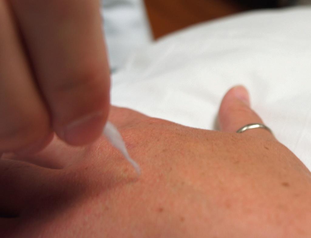
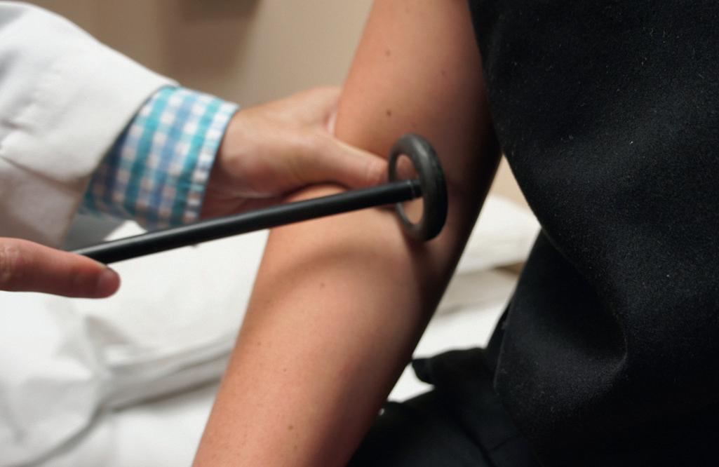
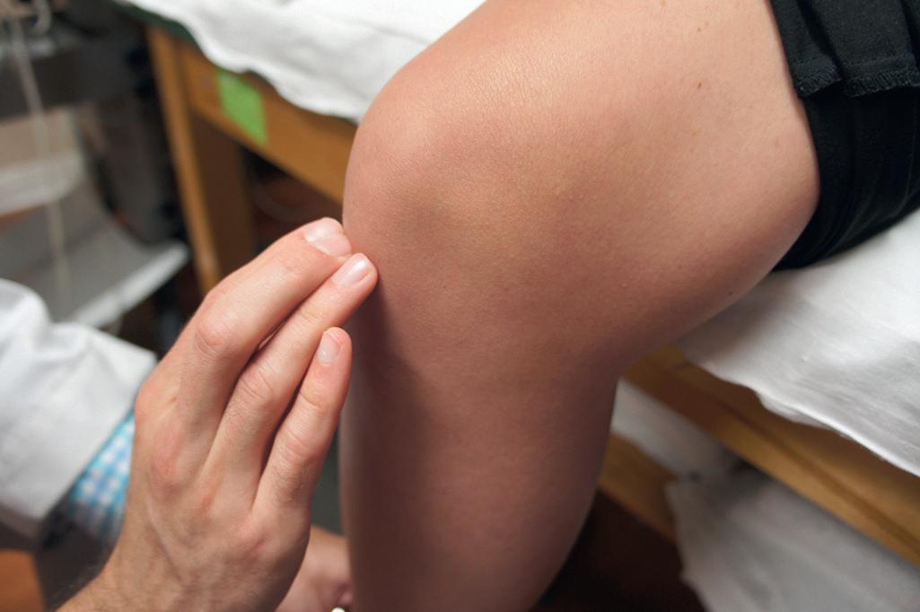
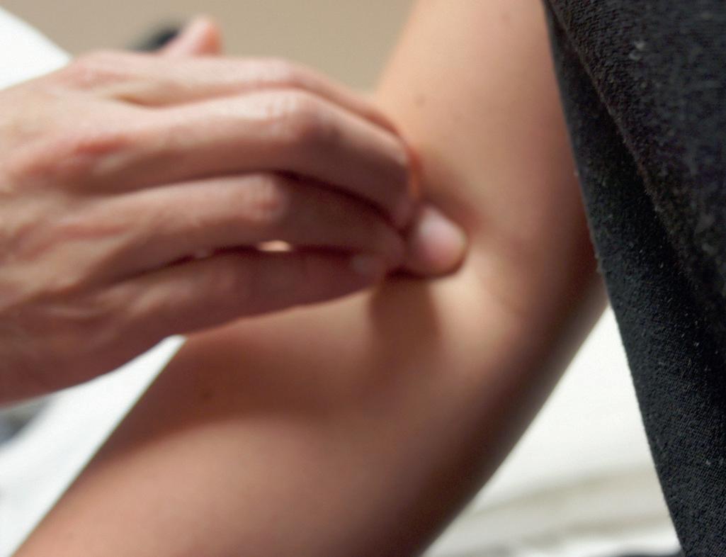
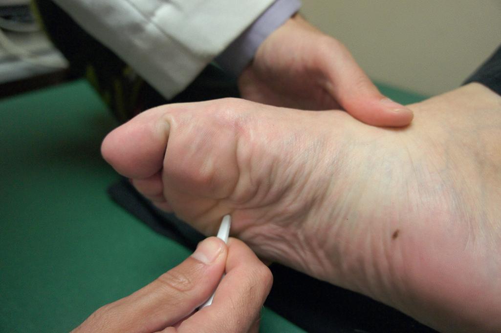
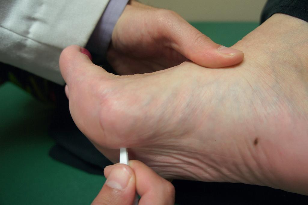
touch. The screening for pain sensory function is the use of a safety pin to lightly prick the regions of concern (Fig. 2.2). The patient is asked whether the pinprick feels sharp in the affected area and typically in the same location of the opposite limb. While the examiner uses both the sharp and dull portion of the safety pin, the patient, with eyes closed, is asked to report whether the sensation is sharp or dull. A cotton wisp can be used to test light touch. The patient, with eyes closed, is asked to report when the cotton is felt (Fig. 2.3).
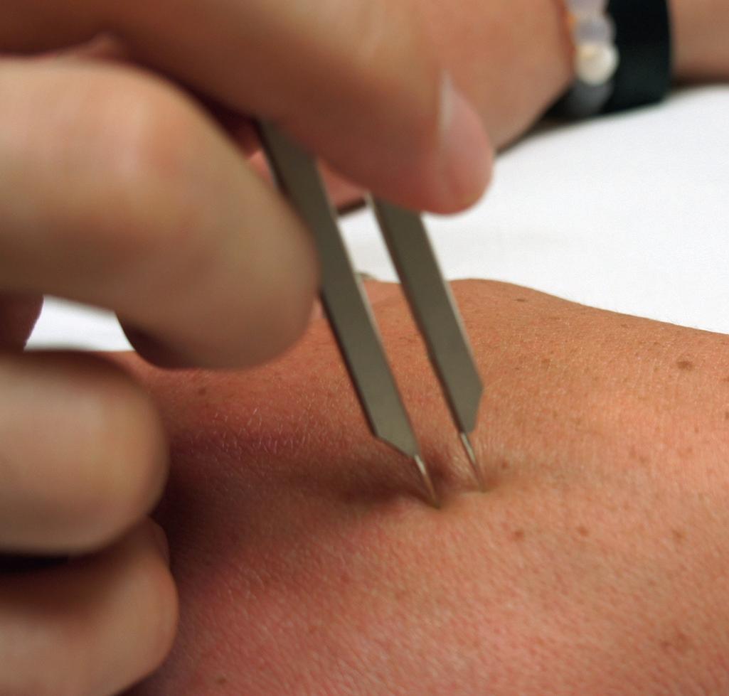
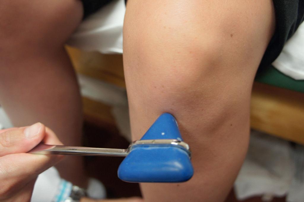
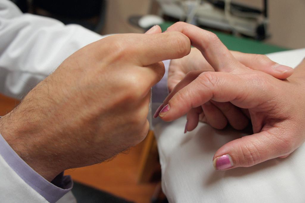
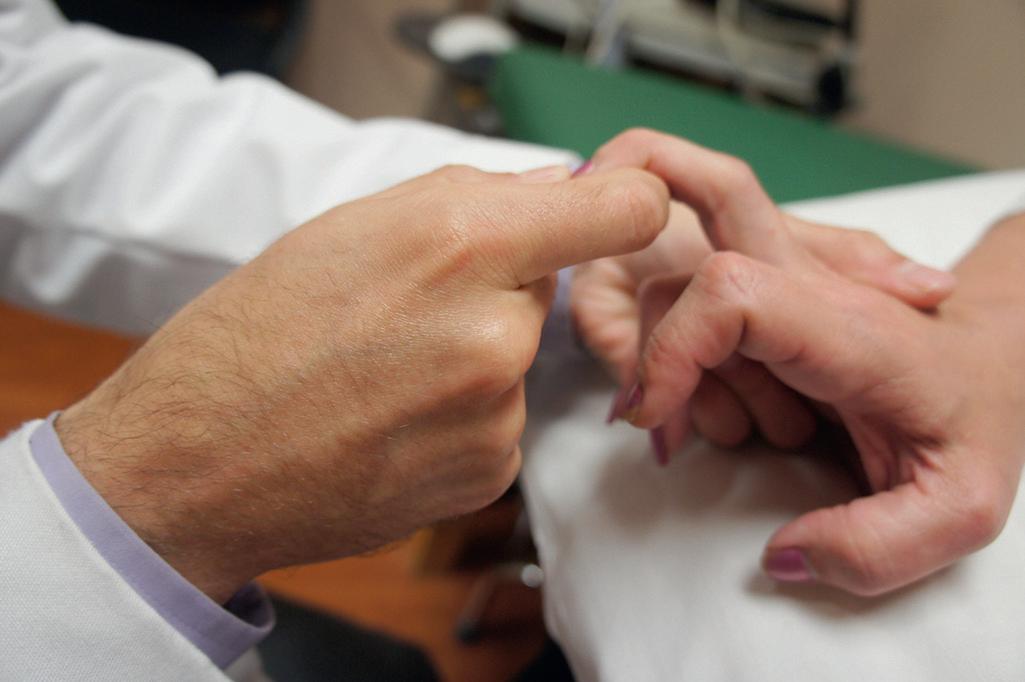
Cortical sensory function can be evaluated with two-point discrimination, stereognosis, and graphesthesia. Two-point discrimination is performed by using two points, such as the tips of an unfolded paper clip to touch two closely separated locations on the skin.9,10 The patient’s eyes should be closed, and he or she asked ask if he or she can feel one or two points11 (Fig. 2.4). Stereognosis testing is performed by asking the patient to identify a familiar object, such as a key or coin placed in the palm of the hand. Graphesthesia
Figure 2.1 Illustration of the dermatome map.
Figure 2.2 Demonstration of the use of a safety pin to assess pain sensation.
Figure 2.3 Demonstration of the use of a cotton wisp to assess light touch.
testing is performed by asking the patient to identify numbers that are written on the palm. Temperature sense can be tested with a cold tuning fork or with test tubes containing warm and cold water. When cold sensation is being tested, the body part must be normally warm.12
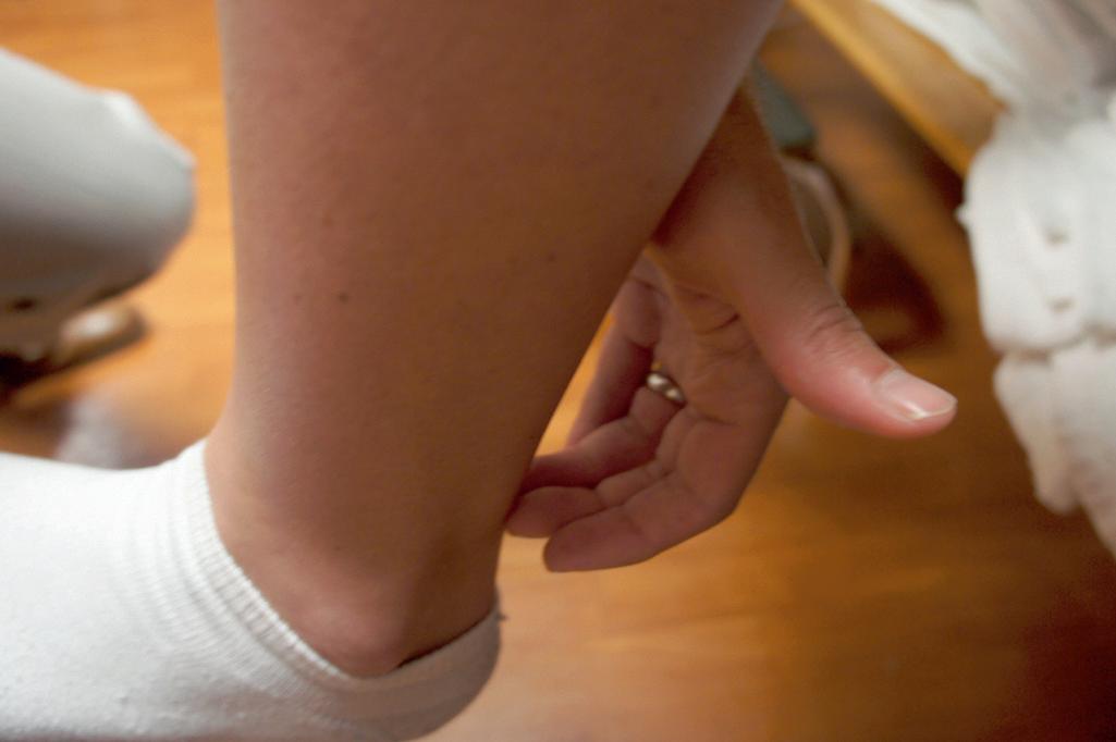
Joint position sense is tested by moving the terminal phalanx of a patient’s finger or toe up or down a few degrees. If the patient cannot identify these tiny movements with eyes closed, similar testing should be performed on the larger joints such as the metacarpal phalangeal joint or wrist. The body part being tested should be grasped on the sides rather than the dorsal or ventral aspect to prevent the patient from using pressure cues to detect movement.
To test vibration sense, the examiner places a finger under the patient’s distal interphalangeal joint and presses a lightly tapped 128-cycle tuning fork on top of the joint. The patient detects the vibration and then notes its extinction about the same time as the examiner, who feels it through the patient’s digit.13 The age and size of the patient should be considered when assessing abnormalities of vibration sense.14 Devices designed to improve quantitative measurement of vibration sensation can also be used.15,16
PROVOCATIVE MANEUVERS
Provocative maneuvers for eliciting sensory symptoms are notoriously nonspecific for reliably distinguishing true neurologic deficit, but they can sometimes provide clinical clues to the source of pain complaints. An example of this is a patient with chronic pain such as fibromyalgia, whose complaints of paresthesias are magnified by muscle palpation. Tender or “trigger” points in muscle can lead to reporting sensations described as paresthesias but are not related to an identifiable neurologic deficit.
Other maneuvers can potentially lead to dynamic nerve compression and provide clinical clues that contribute to
localization of the source of a pain generator, but they are not specific for either neurologic deficit or nerve entrapment. Examples include the Spurling test in cervical radiculopathy, Adson test in thoracic outlet entrapment, and Phalen sign in median nerve entrapment at the carpal tunnel. Techniques of this nature can potentially produce neurologic or neurologic-like symptoms, but they cannot be expected to alter the sensory examination.
Tapping over a suspected focal peripheral neuropathy to reproduce neuritic symptoms and assess for sensitivity is a frequently cited technique and has been termed the Tinel sign. Tinel originally described this technique as a method to determine the location of recovery of regenerating axons after trauma. In his description, the presence of the sign at the location that was being tapped was indicative that the nerve had regenerated to that position.17,18 Localized sensitivity can develop over an area of peripheral nerve injury; however, percussion over a normal peripheral nerve in a superficial location will also induce pain and paresthesias. The use of this technique should not be considered reliable confirmation of a focal entrapment neuropathy.
MOTOR EXAMINATION
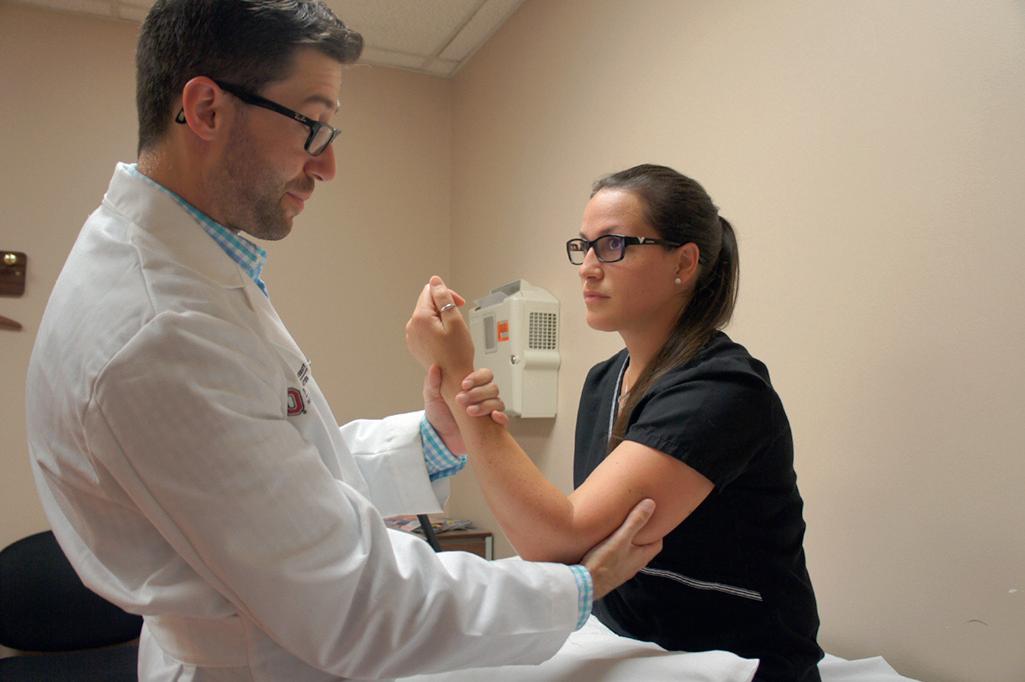
The role of motor testing is to assess the patient’s strength. Reports of weakness are not always due to a true motor deficit. Some complain of weakness when they are actually referring to fatigue, malaise, or incoordination. Strength testing is not the same as power, which refers to the rate of performing work. Manual muscle testing is the most commonly used technique for testing strength. With manual muscle testing, the strength of specific muscle groups is tested against resistance, and one side of the body is compared with the other. It is performed by providing a counterforce on a specific point on the limb against the patient’s best effort19 (Fig. 2.5; Video 2-2).
There are a variety of different classification systems for grading manual muscle testing. Most are based on the Medical Research Council 0–5 scale20 (Table 2.1).
There have been significant modifications of this format by many authors. Most of these scales give ordinal data. This
Figure 2.4 Demonstration of the use of calipers to assess two-point discrimination.
Figure 2.5 Demonstration of the technique of manual muscle testing.
Table 2.1 Medical Research Council 0–5 Scale
Objective
5 Normal Antigravity plus full resistance Full range of motion
4 Diminished Antigravity plus some resistance Full range of motion
3 At least antigravity Antigravity only Full range of motion
2 Poor Gravity omitted Full range of motion
1 Trace Evidence of activation Partial range of motion
0 No activation No evidence of activation n/a
n/a, not applicable.
means the values are on an arbitrary numerical scale where the exact numerical quantity has no significance beyond its ability to establish a ranking over the other values. Some of the modifications have been created to provide a designation between these ordinal measures.
In the Medical Research Council scale, the grades of 0, 1, and 2 are tested in a position that minimizes the effect of gravity. This is performed while having the patient’s contraction perpendicular to gravitational force. Grades of 3, 4, and 5 are tested with the contraction against gravity. One difficulty with the higher grades in this system is that gravity has more impact on heavier limbs or body regions compared with smaller areas. An example is testing hip flexion in a supine position in comparison with toe extension. Detecting subtle weakness in strong muscles with relatively short lever arms such as the gastroc-soleus complex can also be challenging with manual testing.
Another difficulty with the comparison of higher grades between different examiners is the relative subjectivity of the assessment. Distinguishing mild weakness can be dependent on multiple factors. This includes the size, age, fitness level, and general health of the patient.21,22 It also can be potentially affected by those same factors in the examiner.23,24 Other factors can complicate the assessment including orthopedic conditions that preclude full range of motion and other sources of pain or psychological factors that might limit full effort.
Some authors have presented other manual muscle testing protocols in an effort to minimize some of these limitations. Daniels and Worthingham proposed a more functional grading system by testing motion that uses all of the agonists and synergists involved in a particular motion.25 Kendall and McCreary proposed testing a specific muscle rather than a motion for strength.26 This method requires a considerably higher skill level with detailed knowledge of anatomy and kinesiology. Regardless of the method used, challenges of subjectivity and variability of patient effort remain.
It is best to use a grading system for motor testing that will be understood by other practitioners who might also be caring for the patient. It is therefore preferable to use a highly reproducible, easily performed examination. A consistent method of performing the examination is necessary for reproducible results.27
The testing should be explained to the patient in simple terms to invoke optimum cooperation. In the presence of asymmetric weakness, the testing should first be performed on the uninvolved or less involved side. This helps to properly gauge the contralateral strength and establish that the patient understands the directions for the involved side.
The limbs being examined should first be assessed for passive motion limitations such as joint deformity or joint or muscle contracture. Any orthopedic limitation affecting normal motion should be noted and accounted for when considering strength.
Each muscle group or limb motion should be tested in a consistent fashion. Differences in the point of application of the resistive force will result in varying assessments of strength.28 Shorter lever arms provide higher strength against the same resistance as longer lever arms. The application of resistance should be as distal as possible from the axis of movement on the moving segment without crossing another joint. The patient should be positioned comfortably on a firm surface with the limb in the correct testing position. The correct testing position ensures that the muscle fibers are correctly aligned. Resistance is applied in a direction opposite the muscle’s rotary component and at right angles to the line of the pull of the muscle fibers. The resistance should be applied gradually to give the patient sufficient time to provide resistance. The proximal segment that uses counterpressure to the examiner’s resistance should be stabilized to avoid unnecessary movement or muscle substitution. It can be beneficial to place some stronger muscles in a position of mechanical disadvantage when investigating subtle weakness. For example, when assessing strength of the triceps brachii, place the elbow in 90 degrees of flexion instead of full extension, thus limiting the resistance of the extended joint.
When the patient demonstrates that he or she can move the area of interest through full range of motion against gravity, then resistance is applied. If the patient is unable to oppose gravity, then the test movement is positioned in a direction, usually in the transverse plane, to minimize the influence of gravity. Some recommend that the test be repeated up to three times for consistency to determine the muscle strength grade.29 Manual muscle testing for weakness should include not only muscles in a pattern of the various myotomes, but consideration should also be given to the pattern of peripheral nerves and their branches when appropriate.
OTHER MEASUREMENT TOOLS
Motor testing can be better quantified by use of various measurement tools. A method of strength testing using dedicated measuring devices such as dynameters, strain gauges, and other apparatus is called dynamometry. There are tools available that provide both isometric and isokinetic assessments.30,31 Isometric testing provides information about force production with a specific, fixed joint angle.
Isokinetic testing measures torque across a joint as it moves through its range of motion.32,33
Advantages of testing with these tools include providing interval data with a continuous scale of grades in contrast to the more limited ordinal scale used with conventional manual muscle testing. For this reason, quantitative motor testing with dynamometry typically provides better information for subtle changes in strength that would not be reliably reflected in manual muscle tests.34,35 Dynamometry is also useful for greater reproducibility in clinical trials. Disadvantages of motor assessment with dynamometry include a relative limitation of muscle groups that can be reliably tested.36 Follow-up testing by another clinician would require that the clinician also have the same tools available. Additionally, some of the tools for testing, particularly for isokinetic assessment, can be relatively expensive.37
FUNCTIONAL MEASURES
Functional assessments are also an important component of motor testing both as a screening test and for identifying deficiencies that might not be evident with routine motor testing. Isolation of specific muscles is often challenging because they generally work in conjunction with synergistic and antagonist muscles.38 Additionally, the components of the motor cortex represent movements rather than contractions of individual muscles.39 Assessments of gait or active shoulder motion are examples of complex motor evaluations that can serve as a screen for underlying weakness. Tests such as single leg squats or toe raises can demonstrate side-to-side asymmetry in strength that might not be evident with routine manual muscle testing.
EXAMINATION OF REFLEXES
Reflex testing assesses both sensory and motor pathways and is considered one of the more objective components of the neurologic evaluation.40 The commonly used reflexes are known as muscle stretch reflexes, or they are sometimes referred to as myotatic reflexes. The muscle stretch reflex is a muscle contraction that occurs in response to stretching of the muscle.41 This monosynaptic reflex provides regulation of skeletal muscle length in healthy individuals and can be used for diagnostic assessment.42
When a muscle lengthens, receptors within the muscle fibers called muscle spindles are activated. This causes afferent sensory neuron depolarization, which travels through the dorsal root ganglion into the spinal cord and synapses with alpha motor neurons in the anterior horn of the spinal cord.43 This leads to depolarization of the alpha motor neurons at the same and adjacent spinal levels, causing the muscle fibers to contract and resist the stretch (Fig. 2.6). At the same time the reflex is activated, other neurons cause the antagonistic muscle to relax. The entire cascade serves to maintain the muscle at a constant length.
The term deep tendon reflex does not accurately reflect the mechanism of this phenomenon. The reflex’s response begins with stretch responsive receptors in the muscle fibers, not tendon. Additionally, some reflexes, such as the jaw jerk reflex, do not involve tendon at all. Therefore, the use of this term is discouraged.
Cell body of sensory
Sensory neuron
Sensory stretch receptor
Quadriceps muscle
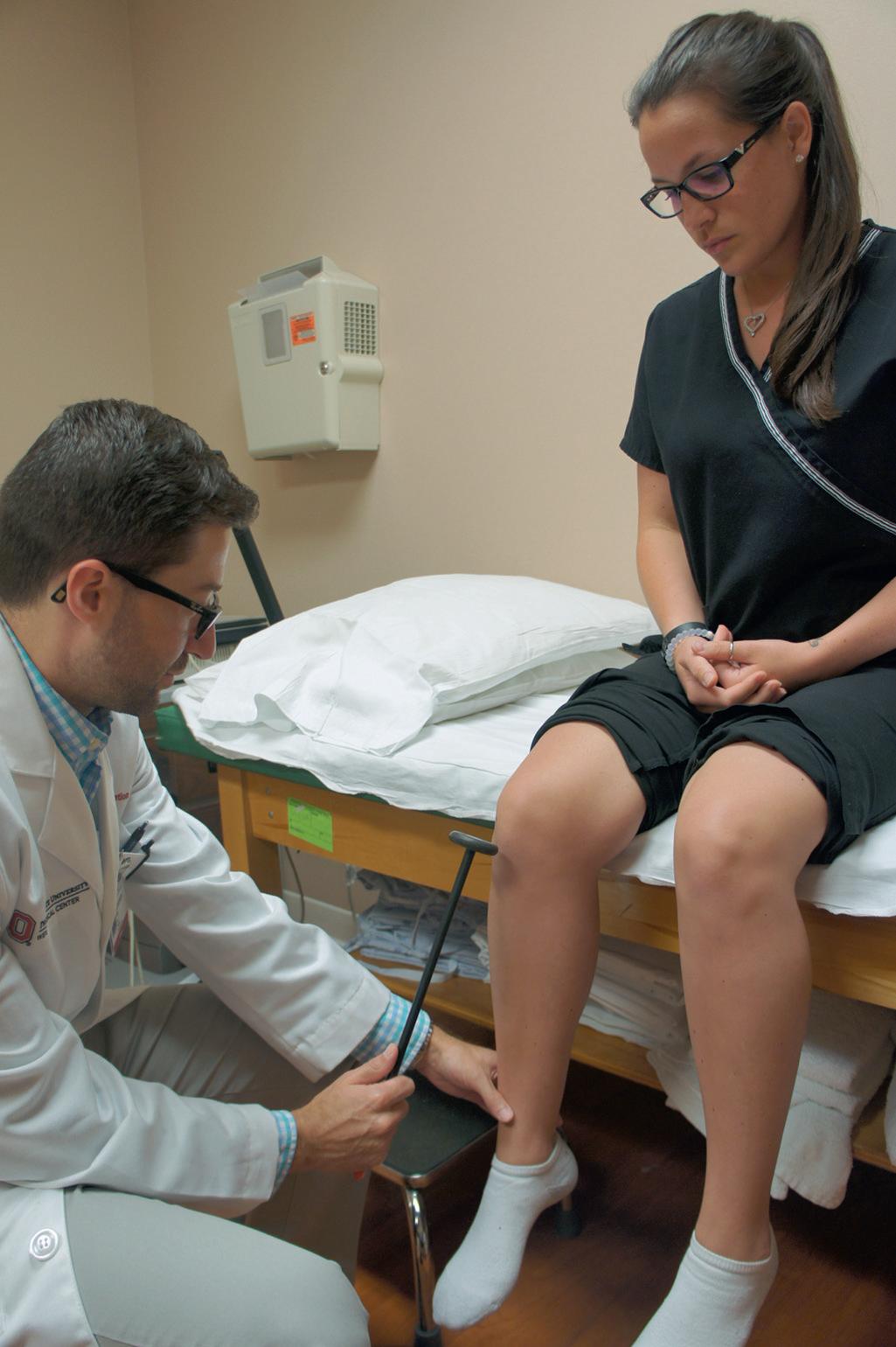
The clinical muscle stretch reflex is performed by rapidly striking the tendon of the muscle of interest (Fig. 2.7; Video 2-3). The rapid stretch on the tendon transmits the mechanical impulse to the muscle spindle, activating the reflex. In healthy patients, the response in the form of muscle contraction is seen within 25 milliseconds of the muscle stretch impulse.44 Muscle stretch reflexes can be performed with either a reflex hammer or with a manual technique. There are various types of reflex hammers available, and the type for clinical use generally is a result of personal preference.45 (Fig. 2.8). The author’s recommendation is to use a manual technique to remove an additional barrier between the patient and the examiner. This is performed by striking the tendon of interest with slightly curved and rigidly held long and ring fingers (Fig. 2.9). This method also alleviates the need to keep track of a reflex hammer.
With whatever technique is used, a short, sharp blow is applied to a tendon, with the muscle of interest kept in minimal extension. The hand that is not responsible for the tendon tap should be held on the muscle being tested. This allows the examiner to feel the muscle response instead of relying only on visual perception to assess for contraction. The patient should be properly positioned and kept as relaxed as possible. Muscle stretch reflexes can be inhibited by anxiety and tension.46 It is often helpful to use facilitation by placing tension on the tendon of the antagonist muscle or even distraction by having the patient contract muscles in other limbs.47-49
Muscle stretch reflexes are affected by the supraspinal CNS. This is done by influencing gamma motor neurons, which regulate the sensitivity of the muscle stretch reflex by tightening or relaxing the fibers within the muscle spindle.50-52 Common muscle stretch reflexes tested include the jaw jerk (CN V), biceps brachii (C5–C6), brachioradialis (C5–C6), extensor digitorum (C6–C7), triceps brachii
Figure 2.6 Illustration of the reflex arc.
neuron in dorsal root ganglion
Motor neuron serving quadriceps
tions, diseases, and drugs.60-62 The findings should always be used in clinical context. The diagnostic specificity of clearly abnormal reflexes is relatively good, but the presence of normal reflexes does not exclude a neurologic deficit.63
PATHOLOGICAL REFLEXES
CLONUS
The most frequently used grading system for muscle stretch reflexes is that of the National Institute of Neurological Disorders and Stroke (NINDS) scale.54 This is a 5-point scale that ranges from 0 to 5. Zero represents an absent reflex and is virtually always considered abnormal.55,56 Other values are defined as follows: A score of 1+ represents a diminished reflex and might be normal or abnormal; 2+ represents normal reflexes; 3+ represents a brisk reflex and might be normal or abnormal; 4+ represents a brisk reflex with the presence of clonus, a series of involuntary muscular contractions and relaxations; and 4+ is always considered abnormal.
Consideration should be given to the age of the patient when interpreting reflexes.57 Individuals without neurologic deficit will experience a decline in reflex response with aging.58,59 Multiple other factors can influence muscle stretch reflexes, including endocrine changes, medica-
Clonus is a series of involuntary rhythmic muscle contractions elicited by a rapid passive stretching of a muscle. It is due to a lesion in descending motor neurons and is often associated with increased muscle stretch reflexes and spasticity.64,65 Clonus is most commonly seen at the ankle but can be found at the ankle, knee, wrist, jaw, or elbow.66 Clonus can be seen in virtually any muscle group and generally repeats with a frequency of 5 to 8 Hz.67
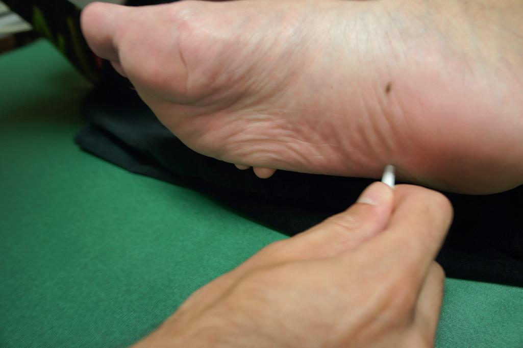
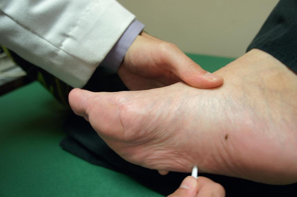
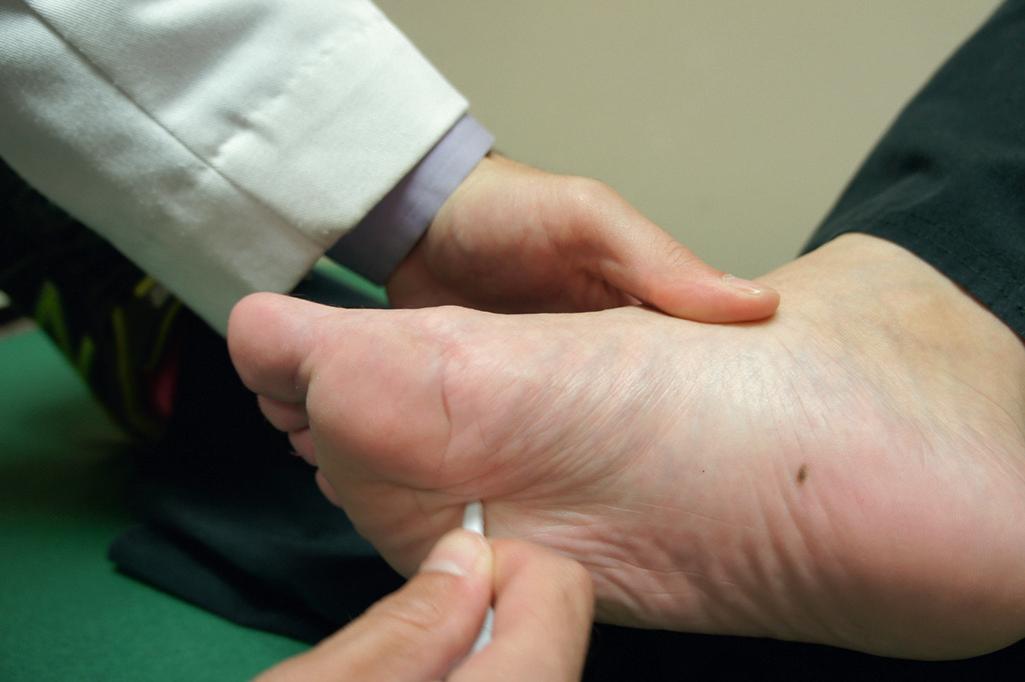
PLANTAR REFLEX
The plantar reflex is a reflex elicited when the sole of the foot is stroked, often with a blunt instrument (Fig. 2.10). In normal adults, the reflex causes flexion of the hallux (Fig. 2.11). An abnormal response, also known as the Babinski sign, named after Joseph Babinski,68,69 is an extension of the hallux (Fig. 2.12). An abnormal plantar reflex can represent dysfunction of the pyramidal tract. It can sometimes be the (C7–C8), quadriceps (ie, patellar) (L3–L4), semimembranosus (L5, S1), and gastrocnemius (aka Achilles or ankle) (S1, S2).53 Side-to-side comparisons should always be used with these reflexes to assess for variations.
Figure 2.8 Demonstration of the use of different reflex hammers for eliciting muscle stretch reflexes. A, Babinski reflex hammer. B, Taylor hammer.
Figure 2.7 Demonstration of the technique for eliciting the muscle stretch reflex. In this case, the quadriceps reflex is elicited by briskly striking the patellar tendon, with the patient’s knee in a relaxed and flexed position.
2.9 Demonstration of the manual technique for eliciting muscle stretch reflexes without a hammer. The fingers are tapped briskly on the tendon. A, Patellar. B, Achilles. C, Biceps Brachii.
first and only indication of a CNS lesion. The initial response of the toe should be carefully observed when the reflex appears abnormal. Normal withdrawal with extension of the toes can occur by the patient after the reflex is complete.
HOFFMAN’S REFLEX
Hoffman’s reflex (aka finger flexor reflex) is performed by tapping or flicking the terminal phalanx of the long or ring finger. The Hoffman’s sign is a positive test that is
Figure 2.10 Demonstration of the method for assessing the plantar reflex. The lateral side of the foot is stroked from the heel along a curve to the area of the metatarsal pads. The progression of movement is shown in A, B and C
demonstrated by reflexive flexion of the terminal phalanx of the thumb with this maneuver (Fig. 2.13, Video 2-4).
Unlike the Babinski sign, the Hoffman’s sign can be seen in normal adults that are naturally somewhat hyperreflexic. The finding more likely represents upper motor neuron pathology when it is an acute onset or unilateral. Hoffman’s sign should be interpreted within the context of other neurologic signs and is not considered a good screening test in asymptomatic individuals.70,71 The mechanism is
Figure
A
B C
A
B C
REFERENCES
different between the Hoffman’s reflex and the plantar reflex. Hoffman’s reflex is a monosynaptic muscle stretch reflex, and the plantar reflex is more complicated and not completely understood.72
CONCLUSION
The sensory, motor, and reflex examinations should all be performed and integrated to help localize a neurologic deficit to the extent possible. The underlying basis or source of the deficit is more frequently obtained from a detailed history, but the extent of the deficit is obtained from a properly performed physical examination. The history and physical examination should be used to create a differential diagnosis that should guide appropriate use of any additional diagnostic testing. The practitioner should resist relying on imaging and other testing without first creating the proper context with the history and physical examination. Each examination technique has limitations of specificity and sensitivity, but when taken together, sensory, motor, and reflex tests are invaluable components of the neurologic examination.
1. Dyck PJ, O’Brien PC, Kosanke JL, et al. A 4, 2, and 1 stepping algorithm for quick and accurate estimation of cutaneous sensation threshold. Neurology. 1993;43:1508-1512.
2. Bell-Krotoski J. Advances in sensibility evaluation. Hand Clin. 1991;7:527-546.
3. Bell-Krotoski J, Weinstein S, Weinstein C. Testing sensibility, including touch-pressure, two-point discrimination, point localization, and vibration. J Hand Ther. 1993;6:114-123.
5. Inouye Y, Buchthal F. Segmental sensory innervation determined by potentials recorded from cervical spinal nerves. Brain. 1977; 100:731-748.
6. Mazurek MT, Shin AY. Upper extremity peripheral nerve anatomy: current concepts and applications. Clin Orthop Relat Res. 2001; 383:7-20.
7. Freeman C, Okun MS. Origins of the sensory examination in neurology. Semin Neurol. 2002;22:399-408.
8. Dyck PJ, Dyck PJ, Larson TS, et al. Patterns of quantitative sensation testing of hypoesthesia and hyperalgesia are predictive of diabetic polyneuropathy: a study of three cohorts. Nerve growth factor study group. Diabetes Care. 2000;23:510-517.
9. Finnell JT, Knopp R, Johnson P, et al. A calibrated paper clip is a reliable measure of two-point discrimination. Acad Emerg Med 2004;11:710-714.
10. Lundborg G, Rosén B. The two-point discrimination test—time for a re-appraisal? J Hand Surg [Br]. 2004;29:418-422.
11. Dellon AL, Mackinnon SE, Crosby PM. Reliability of two-point discrimination measurements. J Hand Surg [Am]. 1987;12(Pt 1):693-696.
Figure 2.11 Demonstration of the plantar reflex in a normal adult. Note the flexion of the toes after the sole of the foot is stroked.
Figure 2.12 Demonstration of an abnormal plantar reflex (Babinski sign) with extension of the hallux after the foot is stroked.
Figure 2.13 A, Demonstration of testing of Hoffman’s reflex. B, A position of rapid flexion of the terminal phalanx of the thumb occurs in a positive test.
12. Dyck PJ, Zimmerman I, Gillen DA, et al. Cool, warm, and heat-pain detection thresholds: testing methods and inferences about anatomic distribution of receptors. Neurology. 1993;43:1500-1508.
13. Huizing EH. The early descriptions of the so-called tuning-fork tests of Weber, Rinne, Schwabach, and Bing. II. The “Rhine Test” and its first description by Polansky. ORL J Otorhinolaryngol Relat Spec. 1975;37:88-91.
14. Burns TM, Taly A, O’Brien PC, et al. Clinical versus quantitative vibration assessment: improving clinical performance. J Peripher Nerv Syst. 2002;7:112-117.
15. Gerr FE, Letz R. Reliability of a widely used test of peripheral cutaneous vibration sensitivity and a comparison of two testing protocols. Br J Ind Med. 1988;45:635-639.
16. Deng H, He F, Zhang S, et al. Quantitative measurements of vibration threshold in healthy adults and acrylamide workers. Int Arch Occup Environ Health. 1993;65:53-56.
17. Tinel J. Nerve Wounds. Symptomatology of Peripheral Nerve Lesions Caused by War Wounds. London: Bailliere, Tinall and Cox; 1918.
18. Spinner M. Injuries to the Major Branches of Peripheral Nerves of the Forearm. With a translation of J. Tinel’s original “Fourmillement” paper by Emanuel B. Kaplan, MD. 2nd ed. Philadelphia: W.B. Saunders Company; 1978:6-13.
19. Lilienfeld AM, Jacobs M, Willis M. A study of the reproducibility of muscle testing and certain other aspects of muscle scoring. Phys Ther Rev. 1954;34:279-289.
20. Medical Research Council. Aids to the Investigation of the Peripheral Nervous System. London: HMSO; 1975.
21. Jaric S. Muscle strength testing: use of normalisation for body size. Sports Med. 2002;32:615-631.
22. Vanpee G, Hermans G, Segers J, et al. Assessment of limb muscle strength in critically ill patients: a systematic review. Crit Care Med. 2014;42:701-711.
23. Mulroy SJ, Lassen KD, Chambers SH, et al. The ability of male and female clinicians to effectively test knee extension strength using manual muscle testing. J Orthop Sports Phys Ther. 1997;26:192199.
24. Lu TW, Hsu HC, Chang LY, et al. Enhancing the examiner’s resisting force improves the reliability of manual muscle strength measurements: comparison of a new device with hand-held dynamometry. J Rehabil Med. 2007;39:679-684.
25. Daniels K, Worthingham C. Muscle Testing Techniques of Manual Examination. 5th ed. Philadelphia: W.B. Saunders; 1986.
26. Kendall FP, McCreary EK, Provance PG. Muscles: Testing and Function. Baltimore: Williams & Wilkins; 1993.
27. Florence JM, Pandya S, King WM. Intrarater reliability of manual muscle test (Medical Research Council scale) grades in Duchenne’s muscular dystrophy. Phys Ther. 1992;72:115-122, discussion 122-126.
28. Barbic S, Brouwer B. Test position and hip strength in healthy adults and people with chronic stroke. Arch Phys Med Rehabil 2008;89:784-787.
29. Symons TB, Vandervoort AA, Rice CL, et al. Reliability of a singlesession isokinetic and isometric strength measurement protocol in older men. J Gerontol A Biol Sci Med Sci. 2005;60:114-119.
30. Emery CA, Maitland ME, Meeuwisse WH. Test-retest reliabilty of isokinetic hip adductor and flexor muscle strength. Clin J Sport Med 1999;9:79-85.
31. Gaines JM, Talbot LA. Isokinetic strength testing in research and practice. Biol Res Nurs. 1999;1:57-64.
32. Osternig LR. Isokinetic dynamometry: implications for muscle testing and rehabilitation. Exerc Sport Sci Rev. 1986;14:45-80.
33. Baltzopoulos V, Brodie DA. Isokinetic dynamometry. Applications and limitations. Sports Med. 1989;8:101-116.
34. Bohannon RW. Manual muscle test scores and dynamometer test scores of knee extension strength. Arch Phys Med Rehabil 1986;67:390-392.
35. Dekkers KJ, Rameckers EA, Smeets RJ. Upper extremity strength measurement for children with cerebral palsy: a systematic review of available instruments. Phys Ther. 2014;94:609-622.
36. Visser J, Mans E, de Visser M, et al. Comparison of maximal voluntary isometric contraction and hand-held dynamometry in measuring muscle strength of patients with progressive lower motor neuron syndrome. Neuromuscul Disord. 2003;13:744-750.
37. El Mhandi L, Bethoux F. Isokinetic testing in patients with neuromuscular diseases: a focused review. Am J Phys Med Rehabil 2013;92:163-178.
38. Kannus P. Isokinetic evaluation of muscular performance: implications for muscle testing and rehabilitation. Int J Sports Med 1994;15(suppl 1):S11-S118.
39. Ropper A, Samuels M, Klein J. Adams and Victor’s Principles of Neurology. 10th ed. New York: McGraw-Hill Education Medical; 2014.
40. Paulson GW. Reflexes and neurologists. Clin Neurol Neurosurg 1992;94(suppl):S133-S1366.
41. Louis ED, Kaufmann P. Erb’s explanation for the tendon reflexes. Links between science and the clinic. Arch Neurol 1996;53:1187-1189.
42. Litvan I, Mangone CA, Werden W, et al. Reliability of the NINDS Myotatic Reflex Scale. Neurology. 1996;47:969-972.
43. Morita H, Petersen N, Christensen LO, et al. Sensitivity of H-reflexes and stretch reflexes to presynaptic inhibition in humans. J Neurophysiol. 1998;80:610-620.
44. Burke D, Gandevia SC, McKeon B. Monosynaptic and oligosynaptic contributions to human ankle jerk and H-reflex. J Neurophysiol 1984;52(3):435-448.
45. Marshall GL, Little JW. Deep tendon reflexes: a study of quantitative methods. J Spinal Cord Med. 2002;25:94-99.
46. Dick JP. The deep tendon and the abdominal reflexes. J Neurol Neurosurg Psychiatry. 2003;74:150-153.
47. Bussel B, Morin C, Pierrot-Deseilligny E. Mechanism of monosynaptic reflex reinforcement during Jendrassik manoeuvre in man. J Neurol Neurosurg Psychiatry. 1978;41:40-44.
48. Burke D, McKeon B, Skuse NF. Dependence of the Achilles tendon reflex on the excitability of spinal reflex pathways. Ann Neurol. 1981;10:551-556.
49. Rossi-Durand C. The influence of increased muscle spindle sensitivity on Achilles tendon jerk and H-reflex in relaxed human subjects. Somatosens Mot Res. 2002;19:286-295.
50. Polus BI, Patak A, Gregory JE, et al. Effect of muscle length on phasic stretch reflexes in humans and cats. J Neurophysiol. 1991;66:613-622.
51. Gregory JE, Wood SA, Proske U. An investigation into mechanisms of reflex reinforcement by the Jendrassik manoeuvre. Exp Brain Res. 2001;138:366-374.
52. Enríquez-Denton M, Morita H, Christensen LO, et al. Interaction between peripheral afferent activity and presynaptic inhibition of ia afferents in the cat. J Neurophysiol. 2002;88:1664-1674.
53. Felsenthal G, Reischer MA. Asymmetric hamstring reflexes indicative of L5 radicular lesions. Arch Phys Med Rehabil. 1982;63: 377-378.
54. Manschot S, van Passel L, Buskens E, et al. Mayo and NINDS scales for assessment of tendon reflexes: between observer agreement and implications for communication. J Neurol Neurosurg Psychiatry 1998;64:253-255.
55. Bowditch MG, Sanderson P, Livesey JP. The significance of an absent ankle reflex. J Bone Joint Surg Br. 1996;78:276-279.
56. Vrancken AF, Kalmijn S, Brugman F, et al. The meaning of distal sensory loss and absent ankle reflexes in relation to age: a metaanalysis. J Neurol. 2006;253:578-589.
57. O’Keeffe ST, Smith T, Valacio R, et al. A comparison of two techniques for ankle jerk assessment in elderly subjects. Lancet 1994;344:1619-1620.
58. Kamen G, Koceja DM. Contralateral influences on patellar tendon reflexes in young and old adults. Neurobiol Aging. 1989;10: 311-315.
59. Chandrasekhar A, Abu Osman NA, et al. Influence of age on patellar tendon reflex response. PLoS ONE. 2013;8(11): e80799.
60. Moreau L, Philbert M. [Modifications in the Achillean reflexogram under the influence of quick-acting barbiturates]. [Article in French] Ann Endocrinol (Paris). 1968;29(6):781-798.
61. Shafer RB, Nuttall FQ. Achilles reflex in thyroid disorders: a 10-year clinical evaluation. Am J Med Sci. 1972;264(4):313-317.
62. Dry J, Leynadier F, Pradalier A, et al. [Changes in Achilles tendon reflex under influence of nonendocrine factors]. [Article in French] Sem Hop. 1976;52:1697-401.
63. Stam J. [Physical diagnostics—tendon reflexes]. [Article in Dutch] Ned Tijdschr Geneeskd. 1999;143:848-851.
64. Wallace DM, Ross BH, Thomas CK. Motor unit behavior during clonus. J Appl Physiol. 2005;99(6):2166-2172.
65. Rossi A, Mazzocchio R, Scarpini C. Clonus in man: a rhythmic oscillation maintained by a reflex mechanism. Electroencephalogr Clin Neurophysiol. 1990;75:56-63.
66. Uysal H, Boyraz I, Yağcıoğlu S, et al. Ankle clonus and its relationship with the medium-latency reflex response of the soleus by peroneal nerve stimulation. J Electromyogr Kinesiol. 2011;21: 438-444.
67. Boyraz I, Uysal H, Koc B, et al. Clonus: definition, mechanism, treatment. Med Glas (Zenica). 2015;12:19-26.
68. Bassetti C. Babinski and Babinski sign. Spine. 1995;20:25912594.
69. Furukawa T. [Joseph Babinski’s contribution to neurological symptomatology]. [Article in Japanese] Brain Nerve. 2014;66:12791286.
70. Grijalva RA, Hsu FP, Wycliffe ND, et al. Hoffmann sign: clinical correlation of neurological imaging findings in the cervical spine and brain. Spine. 2015;40:475-479.
71. Tejus MN, Singh V, Ramesh A, et al. An evaluation of the finger flexion, Hoffman’s and plantar reflexes as markers of cervical spinal cord compression—A comparative clinical study. Clin Neurol Neurosurg. 2015;134:12-16.
72. Hoffmann G, Kamper DG, Kahn JH, et al. Modulation of stretch reflexes of the finger flexors by sensory feedback from the proximal upper limb poststroke. J Neurophysiol. 2009;102:14201429.
Physical Examination of the Cervical Spine
Lisa Huynh, MD | David J. Kennedy, MD
INTRODUCTION
The annual prevalence of neck pain is estimated to range between 30% and 50%,1 and nearly half of all individuals will experience neck pain in their lifetime.2 History and physical examination can provide important clues in determining the etiology of symptoms. Many specialized provocative tests have been described for physical examination of the neck and cervical spine. These tests are routinely performed by clinicians with varying experience and skill. This may lead to error in both the technique and the interpretation of findings.
Several key principles exist in examination of the cervical spine: (1) The exam should be systematic to avoid missing key steps. (2) Generally, exam maneuvers should be done in a stepwise manner so that less painful movements are performed first and most painful movements are completed last; this ensures the least amount of pain carryover, which may confound exam findings. (3) Of crucial concern for any examination of the cervical spine is the ability to differentiate pathologies that merely cause pain from those that adversely affect sensitive neural tissues associated with the cervical spinal cord and its nerve roots.
This chapter provides a comprehensive overview of the physical examination of the cervical spine. For each test, the original description, currently performed technique, reliability, validity, and clinical significance are discussed, based on a comprehensive search of the existing literature. The goal is not necessarily to learn every examination maneuver performed for neck pain but rather to understand the limitations, reliability, and scientifically proven validity of some of the commonly used tests.
INSPECTION
Inspection should begin by noting the position of the head in relation to the line of gravity, which passes through the external auditory meatus; odontoid process; the cervical, thoracic, thoracolumbar, and lumbosacral spine; and the sacral promontory. One should carefully assess not only the upper cervical region but also the relative curvature of the thoracolumbar and lumbosacral spines because the relative positioning of the cervical spine may be influenced by the curvature below. The forward-head position can also be the direct cause of the loss of cervical motion. Caillet3
reported a 25% to 50% loss of head rotation with a forwardly protruded head and a significant increase in the gravity-induced weight of the head brought on by this postural abnormality. The forward-head posture thus increases the work requirements of the capital and cervical musculature. Additional features associated with damage to the neural structures may be noted by observation/inspection, including limb muscle atrophy, clumsiness, and balance problems on gait.
PALPATION
Palpation is a common component of cervical spine evaluation. It should be systematic and focus on palpation of the midline spinous processes, the paraspinal musculature, and the underlying zygapophyseal joints (z-joints), as well as the associated cervical spinal musculature. Studies have addressed the accuracy of palpation in identifying the structure or level of the spine and interexaminer reliability. In a study of 69 patients, experienced anesthesiologists were asked to identify the C7 spinous process by palpation, which was then compared with fluoroscopy.4 The C7 level was chosen because it was believed to be the most prominent and easiest level to identify. The physicians in this study were only able to correctly identify the C7 spinous process 47.9% of the time when compared with fluoroscopy. This was increased to 77.1% with the addition of neck flexionextension. Additional concerns with the reliability of the cervical spine palpatory examination have been highlighted in a systematic comprehensive review of the literature, which showed overall poor interexaminer reliability for all palpation.5 Reviewers also found that the level of clinical experience did not improve the reliability in that experienced clinicians fared no better than students in terms of palpatory reliability. Segmental range of motion as assessed by palpation was also found to have very low reliability. Additionally, palpation for soft tissue had very low reliability in all regions tested. Reviews of the literature on manual palpation of trigger points have shown poor reproducibility; however, the majority of studies had poor methodological quality.6
Studies on the validity of palpation in the cervical spine are lacking. In 1995, Sandmark and Nisell7 found in a study of 75 patients with self-reported pain that palpation over the facet joints was the most appropriate screening to corroborate self-reported neck dysfunction. This was based on a