ACKNOWLEDGMENTS
Clinical Computer Systems, Inc.
Elgin, Illinois
GE Medical Systems Information Technologies
Milwaukee, Wisconsin
Hill-Rom Company, Inc.
Batesville, Indiana
Huntleigh Healthcare Ltd.
Cardiff, United Kingdom
Philips Medizin Systemes
Böblingen, Germany


We are proud to continue nursing, midwifery, and physician collaboration in this new ninth edition of Mosby’s® Pocket Guide to Fetal Monitoring: A Multidisciplinary Approach, consistent with the interand intradisciplinary approach all of us bring to teaching this subject matter. Diligently revised, this new edition continues to emphasize standardized terminology and an evidence-based approach to interpretation and management. This text remains a key resource for the most clinically useful information for clincians of all levels on every aspect of fetal monitoring, including intermittent auscultation, assessment and management of uterine activity, and the crucial role of both communication and documentation in risk management related to fetal monitoring. Whether your practice is office-based, in a birth center, in a hospital serving the community, or in an academic tertiary care center, this text provides a relevant and easily understood reference for daily clinical practice as well as clinician orientation and ongoing education.
DESCRIPTION
Primarily an oxygen monitor, the electronic fetal monitor is a tool used to prevent fetal injury resulting from interruption of fetal oxygenation, whether used during labor or in the antepartum period. Key to this goal is standardization and simplification of clinical practices related to interpretation and management of fetal monitoring. This book provides clinicians with the tools needed to understand both the strengths and the weaknesses of both electronic fetal monitoring (EFM) and intermittent auscultation; as well as apply a collaborative approach to clinical practice that is evidence- and consensus-based. After a brief overview of the history of fetal monitoring, the text provides core clinical information on the physiologic basis for monitoring, reviews the newest instrumentation for uterine and fetal heart rate (FHR) monitoring, including the newer abdominal “patch” technology, and identifies key factors in the evaluation of uterine activity. In keeping with maintaining the legal standard of care in the United States, the National Institute of Child Health and Human Development (NICHD) definitions are presented and reviewed, and a standardized approach to interpretation and management is clearly outlined. The influence of gestational age on FHR is examined, along with the evaluation of fetal status outside the obstetric unit and in the antenatal setting. Documentation and risk management issues are
delineated, including issues of informed consent in choice of monitoring modality. An overview of fetal monitoring in Europe provides clinicians with a look at fetal monitoring outside the United States. Patient safety, communication, and clinical collaboration are the cornerstones of each chapter, and suggestions for practice improvement make this edition an invaluable resource for the busy clinician.
FEATURES
This book has a number of distinctive features:
n Content is organized in a manner that allows clinicians to build on key fundamental concepts and progress logically to advanced principles, making the text suitable for novices needing basic information as well as experienced practitioners seeking greater insight into clinical practice issues.
n Critical information is highlighted using illustrations, tables, and illustrative fetal monitor tracings.
n FHR characteristics are explained and the supporting level of evidence is provided, revealing a number of common myths regarding fetal monitoring.
n Evidence levels are provided for information regarding various FHR patterns, and several common obstetric myths are laid to rest.
n Appendices now include self-assessment questions as well as fetal monitor tracings for practice in application of the NICHD definitions and principles of standardized interpretation.
ORGANIZATION
Chapter 1 traces the history of fetal monitoring from the use of auscultation in the 17th century to present-day practice and includes a discussion of the resurgence of intermittent auscultation for fetal monitoring in low-risk women.
Chapter 2 provides a review of the physiologic basis for monitoring. The oxygen pathway is discussed, as well as the fetal response to interrupted oxygenation. These core physiologic concepts provide clinicians with the fundamentals of fetal oxygenation that serve as the basis for current practice.
Chapter 3 offers a detailed look at instrumentation for both intermittent auscultation and EFM, including newer approaches such as abdominal electrocardiogram and new display options for dopplers used in intermittent auscultation. Both external and internal monitoring devices and their application are covered in depth, including artifact detection, telemetry, and troubleshooting tips.
Chapter 4, on uterine activity, provides crucial information including a detailed discussion of normal versus excessive uterine activity and the limitations of the summary term tachysystole. Consensus guidelines for the diagnosis of active labor are presented, and the link between excessive uterine activity and fetal acidemia is elucidated. Evidence-based tips for managing uterine activity in clinical practice are offered, and oxytocin use is also addressed.
Chapter 5 breaks down clinical practice in fetal monitoring to its three core elements: definitions, interpretation, and management. This chapter includes the NICHD definitions with illustrations to aid in recognition and application. The role of NICHD categories is examined, and evidence- and consensus-based principles of interpretation are explained.
Chapter 6 presents the management of FHR tracings using a systematic approach based on principles of fetal oxygenation. This comprehensive model is based on EFM’s value as a screening tool (rather than a diagnostic tool). The management algorithm uses NICHD categories and a structured approach based on the oxygen pathway. Evidence-based corrective measures for hypoxemia are provided in a checklist format. An adapted model specific to the management of Category 2 FHR tracings with significant decelerations is included, with evidence supporting this approach. Chapter 6 elucidates the primary objective of intrapartum FHR monitoring: to prevent fetal injury that might result from the progression of hypoxemia during the intrapartum period.
Chapter 7 reviews FHR characteristics in the preterm, late-term, and postterm fetus, including implications for management in both antepartum and intrapartum settings. The chapter includes information on a variety of medications and clinical factors that can affect FHR at various gestational ages.
Chapter 8 explores non-obstetric settings and FHR evaluation, focusing on the importance of collaboration. Settings such as the emergency department, surgical suite, or intensive care unit are discussed with key points for clinical care and FHR assessment. Obstetric triage is reviewed, including the impact of the Emergency Medical Transport and Labor Act (EMTALA).
Chapter 9 focuses on antepartum testing, including the nonstress test, the contraction stress test, vibroacoustic stimulation, ultrasound, and the biophysical profile. Information regarding indication, frequency, and type of antepartum test based on the results of the most recent NICHD panel are provided in a clinically relevant manner. Chapter 10 focuses on documentation and risk management. Clinical tips for improving documentation are a highlight of this
new edition. Intermittent auscultation, EFM, and informed consent are discussed, with suggestions for inter- and intradisciplinary discussion points and patient education. Actual deposition testimony related to documentation reveals the importance of knowing both nomenclature and physiology in detail.
Chapter 11 provides a glimpse of FHR monitoring in select European countries, where paper speed is frequently 1 cm/minute versus the typical 3 cm/minute seen in the United States. Variations in obstetric care models and sample illustrations of a variety of fetal monitor tracings from our European colleagues are provided for review.
Appendix A reflects current practice regarding amnioinfusion, indicated as a potential corrective measure for variable decelerations.
Appendix B has been updated and includes 10 new fetal monitoring tracings, bringing the total number of FHR tracings in the appendix to 40. This appendix provides ample opportunities for education, review, and preparation for certification or credentialing exams. Clinicians can use the tracings to practice application of the NICHD definitions, as well as the principles of standardized interpretation, and an answer key allows clinicians to evaluate their skills.
Appendix C offers a self-assessment consisting of multiplechoice questions related to the content of the textbook. Helpful for reinforcement of information presented herein, it can also be used to study for certification or credentialing examinations or to develop internal competency assessment tools for clinical practice.
Mosby’s® Pocket Guide to Fetal Monitoring: A Multidisciplinary Approach continues to be written by clinicians, for clinicians. Nurses, nurse-midwives, medical students, physicians, resident physicians, clinical specialists, educators, and risk management and medicallegal professionals will gain a clear perspective on modalities of fetal monitoring, the role of standardization, as well as the keys to successful collaboration. Meticulously researched and revised, the ninth edition is the most portable and practical reference available for daily clinical practice, education, test prep, and habituation of both knowledge and skills. We are thrilled to be able to offer this valuable tool to all clinicians, in all practice settings.
LISA A. MILLER
DAVID A. MILLER
REBECCA L. CYPHER
1 A Brief History of Fetal Monitoring, 1
Historical Overview, 1
Randomized Trials of Electronic Fetal Monitoring, 3
Research at the End of the 20th Century, 3
Fetal Monitoring in the 21st Century, 4
Summary, 6
2 Physiologic Basis for Electronic Fetal Heart Rate Monitoring, 10
Transfer of Oxygen from the Environment to the Fetus, 10
External Environment, 12
Maternal Lungs, 12
Maternal Blood, 12
Maternal Heart, 14
Maternal Vasculature, 15
Uterus, 15
Placenta, 16
Fetal Blood, 21
Umbilical Cord, 22
Fetal Response to Interrupted Oxygen Transfer, 22
Mechanisms of Injury, 24
Injury Threshold, 24
Summary, 26
3 Methods and Instrumentation, 28
Intermittent Auscultation of Fetal Heart Rate, 28
Description, 28
Leopold’s Maneuvers, 31
Utilization, Procedure, and Frequency of Intermittent
Auscultation, 33
Documentation of Auscultated Fetal Heart Rate, 36
Benefits and Limitations of Auscultation, 37
Electronic Fetal Monitoring, 38 Overview, 38
Converting Raw Data Into a Visual Display of Fetal Heart Rate, 40
4
External Mode of Monitoring, 42
Ultrasound Transducer, 42
Tocotransducer, 45
Advantages and Limitations of External Transducers, 46
Internal Mode of Monitoring, 49
Fetal Spiral Electrode, 49
Contraindications, 50
Situations Requiring Caution, 50
Intrauterine Pressure Catheter, 51
Advantages and Limitations of Internal Monitoring, 53
Display of Fetal Heart Rate, Uterine Activity, and Other Information, 54
Monitor Tracing Scale, 54
Monitoring Multiple Gestations, 57
Artifact Detection and Signal Ambiguity (Coincidence) With Maternal Heart Rate, 59
Telemetry, 64
Electronic Fetal Monitoring Troubleshooting, 65
Troubleshooting Actions, 65
Computerized Perinatal Data Systems, 67
Computer Decision Analysis of the Fetal Heart Rate, 71
Data-Input Devices, 71
Summary, 72
Uterine Activity Evaluation and Management, 77
Assessment Methods: Palpation and Electronic Monitoring, 77
Manual Palpation, 78
Electronic Monitoring of Uterine Activity, 79 Parameters for Normal Labor, 81
Defining Adequate Uterine Activity, 83 Defining Excessive Uterine Activity, 88
Common Underlying Causes of Excessive Uterine Activity, 91
Corrective Measures to Decrease Excessive Uterine Activity, 92
Current Trends in Labor Support and Management, 92
Latent Phase Abnormalities, 93
Active Phase Abnormalities, 94
Second-Stage Abnormalities, 95
Uterine Activity and Oxytocin Use, 95 Summary, 99
5
Pattern Recognition and Interpretation, 104
The Evolution of Standardized Fetal Heart Rate Definitions, 104
The 2008 National Institute of Child Health and Human Development Consensus Report, 104
Evidence-Based Interpretation of Fetal Heart Rate Patterns, 107
National Institute of Child Health and Human Development Definitions: General Considerations, 109
Five Essential Components of a Fetal Heart Rate Tracing, 110
Definitions, Physiology, and Interpretation of Specific Fetal Heart Rate Patterns, 110
Baseline Rate, 110
Categories of Baseline Rate, 112
Tachycardia, 112
Bradycardia, 114
Baseline Fetal Heart Rate Variability, 115 Categories of Baseline Variability, 118 Absent Variability, 118
Minimal Variability, 120
Moderate Variability, 120
Marked Variability, 121
Sinusoidal Pattern, 121
Acceleration, 122
Decelerations, 123
Types of Decelerations, 124
Early Deceleration, 124
Late Deceleration, 125
Variable Deceleration, 127
Prolonged Deceleration, 130
Fetal Cardiac Arrhythmias, 132
Terms and Concepts Not Supported by Evidence or Consensus, 133
Wandering Baseline, 133
Lambda Pattern, 133
Shoulder, 133
Checkmark Pattern, 134
End-Stage Bradycardia and Terminal Bradycardia, 134
Uniform Accelerations, 134
Atypical Variable Decelerations, 135
Variable Deceleration With a Late Component, 135
Mild, Moderate, and Severe Variable Decelerations, 136
V-Shaped Variables and W-Shaped Variables, 136
Good Variability Within the Deceleration, 137
Other Mechanisms That Lack Scientific Basis, 137 Summary, 139
6 Intrapartum Management of the Fetal Heart Rate Tracing, 145
Fundamental Principles, 145
Standard of Care, 146
Confirm Fetal Heart Rate and Uterine Activity, 147
Evaluate Fetal Heart Rate Components, 147
A Standardized “ABCD” Approach to Fetal Heart Rate Management, 149
A: Assess the Oxygen Pathway and Consider Other Causes of Fetal Heart Rate Changes, 152
B: Begin Corrective Measures as Indicated, 153
C: Clear Obstacles to Rapid Delivery, 156
D: Delivery Plan, 157
Expectant Management Versus Delivery, 157
Other Methods of Fetal Monitoring, 160
Intrapartum Fetal Scalp pH and Lactate Determination, 160
Fetal Scalp Stimulation and Vibroacoustic Stimulation, 160
Computer Analysis of Fetal Heart Rate, 161
Fetal Pulse Oximetry, 161
ST Segment Analysis, 161
Umbilical Cord Blood Gas Analysis, 163
Summary, 165
7 Influence of Gestational Age on Fetal Heart Rate, 170
The Preterm Fetus, 170
Baseline Fetal Heart Rate in the Preterm Fetus, 172
Baseline Variability in the Preterm Fetus, 173
Periodic and Episodic Heart Rate Changes in the Preterm Fetus, 173
Behavioral States in the Preterm Fetus, 175
Preterm Uterine Activity, 177
Short-Term Tocolytic Therapy and Effect on Fetal Heart Rate, 178
Monitoring the Preterm Fetus, 184
The Late-Term and Postterm Fetus, 185
Fetal Assessment, 186
Risks Associated with Postterm Pregnancy, 186
Management of Postterm Pregnancy, 188
Summary, 188
8 Fetal Assessment in Non-Obstetric Settings, 197
Maternal Trauma Algorithm, 197
A Culture of Patient Safety, 197
Pregnancy Anatomy and Physiology, 198
Obstetric Patients in the Emergency Department, 198
Federal Law and Emergency Medical Treatment and Active Labor Act, 203
Pregnant Trauma Victim Assessment and Care, 203
Primary and Secondary Survey in the Emergency Department, 205
Emergent Cesarean Birth and Resuscitative Hysterotomy, 210
Stabilization and Discharge, 210
Non-Obstetric Surgical Procedures: Maternal–Fetal Assessment and Care, 212
Intraoperative Maternal–Fetal Assessment, 213
Tocolytic Agents and Antenatal Corticosteroids, 216
Fundamentals of Non-Obstetric Surgery, 216 Summary, 216
9 Antepartum Fetal Assessment, 221
Comparing Antepartum Testing Methods, 221 Methods of Testing, 225
Contraction Stress Test and Oxytocin Challenge Test, 225
The Nonstress Test, 228
The Biophysical Profile, 230
The Modified Biophysical Profile, 232
Fetal Movement Counts, 234
Doppler Velocimetry of Maternal and Fetal Blood Vessels, 235
Biochemical Assessment, 237
Summary, 239
10 Patient Safety, Risk Management, and Documentation, 243
Risk Management, 243
The Decision-Making Process in Electronic Fetal Monitoring and Intermittent Auscultation, 244
Central Concepts in Liability Claims, 246
Components of Care: Assessment, Communication, and Documentation, 248
Liability in Fetal Monitoring, 251
Fetal Monitoring Documentation, 251
Documentation Components of the Electronic Fetal Monitoring Evaluation, 253
Common Documentation Dilemmas, 255
Use of Fetal Heart Rate Categories, 255 Documentation of Uterine Activity, 257
Quantification of Decelerations, 258
Frequency of Electronic Fetal Monitoring Assessment and Documentation, 258
Electronic Fetal Monitoring Documentation Policies, 263
Summary, 266
11
Obstetric Models of Care and Electronic Fetal Monitoring Outside the United States, 269
Obstetric Models of Care, 269
Electronic Fetal Monitoring: Cardiotocography, 271
International Intermittent Auscultation and Cardiotocography Guidelines, 273 Guidelines for Terminology and Interpretation, 276
Methods of Determining Fetal Acid–Base Status, 279
Fetal Blood Sampling, 279
ST Analysis of the Fetal Electrocardiogram, 281 Summary, 282
Appendix A: Amnioinfusion, 288
Appendix B: Fetal Heart Rate Tracings Review, 291
Appendix C: Self-Assessment, 332
Index, 338
A Brief History of Fetal Monitoring
Shareddecision-making, informed choice, and greater public and consumer engagement in obstetrics and obstetric safety has shifted clinical focus in perinatal care. Electronic fetal monitoring (EFM) is only one of the issues being discussed by clinicians and the families they serve. Newer labor curves, intermittent auscultation (IA) of the fetal heart rate (FHR), and continuous labor support are becoming the new normal, especially for healthy, low-risk women [1–3]. The shift to IA for healthy low-risk women relates to a widespread recognition of these facts: research has been unable to definitively show that use of intrapartum FHR monitoring leads to a significant reduction in neonatal neurologic morbidity [4], and both early randomized trials and meta-analyses have shown a trend toward higher cesarean section rates for women who have continuous EFM during labor versus women who do not have continuous EFM [5]. This means that clinicians must be adept at both EFM and IA modalities, and for this reason, a brief overview of the history of FHR assessment is justified.
HISTORICAL OVERVIEW
Jean Alexandre Le Jumeau, Vicomte de Kergaradec, was the first person to speculate in print about the potential clinical uses for FHR auscultation. In 1822 he used a stethoscope hoping to hear the noise of the water in the uterus, and he identified the noise he heard as the FHR [6]. Confirmation of pregnancy, identification of twin gestation, and justification for a postmortem cesarean section were some of the early uses of FHR auscultation. William Kennedy, a British obstetrician, published a description of “fetal distress” in 1833 by describing what would later be classified as a late deceleration. Kennedy correctly associated late decelerations with poor prognoses. He also made the link between fetal head compression and decrease in FHR, now known as early decelerations [7]. Other discoveries from early use of FHR assessment via IA included identification of fetal tachycardia in response to maternal fever, FHR decelerations

after excessive uterine activity, and accelerations accompanying fetal movement (Fig. 1.1) [6].
In 1917, the head stethoscope, or DeLee-Hillis fetoscope, was first reported in the literature [8]. Fast forward to the 1950s: physicians throughout the world, including Edward Hon [9–11] in the United States, Caldeyro-Barcia [12,13] in Uruguay, and Hammacher [14] in Germany, developed electronic devices that were able to continuously measure and record the FHR and uterine activity. The simultaneous measurement of FHR and uterine activity came to be called EFM or cardiotocography (CTG). This new technologic capability permitted systematic study of the relationships between recorded FHR patterns and fetal physiology [10,11,15]. Although investigators worldwide made remarkably similar observations of FHR characteristics, dramatically different terms and definitions were being used and there was no significant standardization (Fig. 1.2).
Fig. 1.1 Early obstetric trumpet stethoscope. (Courtesy Wellcome Library, London.)
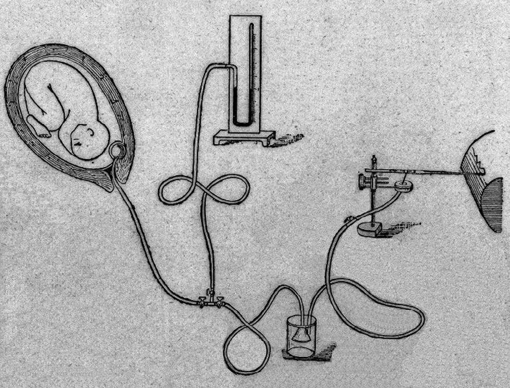
Fig. 1.2 Apparatus for studying uterine contractions during childbirth. (Courtesy Wellcome Library, London.)
RANDOMIZED TRIALS OF ELECTRONIC FETAL MONITORING
Observational studies in the 1960s demonstrated a decrease in intrapartum stillbirth rates in settings that adopted continuous EFM [16–18] and served to drive the widespread adoption of the technology. Although EFM was originally intended for use in high-risk laboring women, it was rapidly incorporated into the management of low-risk laboring women as well and quickly became ubiquitous. Today, observational findings alone would never result in such rapid and widespread practice change.
During the 1970s and 1980s, several randomized clinical [19–27] were conducted comparing continuous EFM with IA using a Pinard stethoscope or a handheld Doppler device. Continuous EFM was not associated with a decrease in low Apgar scores or perinatal mortality. However, there was an increase in the incidence of cesarean section in women who had continuous EFM. Despite these findings, use of continuous EFM did not decrease.
Meta-analyses have reviewed the results of trials comparing continuous intrapartum EFM with IA [5,28,29]. These studies included more than 37,000 women. Compared with IA, continuous EFM showed no significant difference in overall perinatal death but was associated with a significant reduction in neonatal seizures. No significant difference was detected in the incidence of cerebral palsy. However, there was a significant increase in cesarean sections associated with continuous EFM. Interestingly, none of the randomized trials published after 1980 demonstrated a statistically significant increase in the rate of cesarean section in electronically monitored patients. Most important, the majority of newborns in the cohort who later developed cerebral palsy were not in the group of fetuses who had FHR tracings that were considered “ominous” [30]
RESEARCH AT THE END OF THE 20th CENTURY
So what went wrong? Several things. Although the randomized controlled trials (RCTs) followed the usual guidelines for inclusion and exclusion of subjects and used recommended methods for the study protocols, the definitions of FHR patterns reflecting fetal distress varied among the different studies [23–25,31]. In the largest (and most frequently cited) trial, the IA arm and the EFM arm each had
fetal scalp blood sampling included as a follow-up test, making the study a comparison of EFM with scalp sampling with IA with scalp sampling rather than a true comparison of EFM versus IA [24]. Many of the studies were conducted before the importance of FHR variability, a critical parameter, was recognized as significant related to fetal acid–base status. Outcome measures evaluated (Apgar scores, perinatal mortality, and cerebral palsy) were nonspecific indicators of the potential for hypoxic injury during the intrapartum period. Finally, the small sample size of published reports is an ongoing issue. It has been noted that more than 50,000 women would need to be randomized to show a difference in mortality [5]; the numbers that would be needed to show a reduction in neonatal encephalopathy related solely to intrapartum events are so high that RCTs for either EFM or IA become implausible [32]. As a result, the conclusions of these studies remain open to alternative interpretations [27,33], and a careful review of the oft-cited Cochrane Database meta-analysis by Alfirevic and colleagues reveals low-quality evidence for all conclusions, save the conclusion regarding neonatal seizures, which was found to be of moderate quality [5]. Clinicians should consider basing decisions regarding the use of continuous EFM on multiple factors, including forthright discussions of risks versus benefits with various patient populations. Informed choice regarding EFM versus IA is challenging with the state of current evidence and is discussed in more detail in Chapter 10.
In 1996, the National Institute of Child Health and Human Development (NICHD) Task Force met and made recommendations [34] for three important aspects of FHR monitoring for both research and clinical practice: (1) the task force developed standard definitions for FHR patterns, (2) they described the FHR pattern (normal baseline rate, moderate variability, presence of accelerations, and absence of decelerations) that consistently reflects an absence of asphyxia, and (3) they described FHR patterns (recurrent late or variable decelerations or substantial bradycardia with absent variability) that are “predictive of current or impending asphyxia” [34].
FETAL MONITORING IN THE 21st CENTURY
The first Task Force on Neonatal Encephalopathy and Cerebral Palsy [35] was convened in 2003 by the American College of Obstetricians and Gynecologists (ACOG) to review the world literature regarding the relationship between FHR patterns in labor and
neonatal outcomes. The task force reviewed the literature on Apgar scores, neonatal encephalopathy and cerebral palsy, neonatal seizures, and umbilical cord gases. In 2010 a second task force was convened to update this important work, and in 2014 a second edition was published, which included the review of 1500 references by 17 task force members and 88 consultants [36]. This updated report focuses on neonatal encephalopathy in infants born at 35 weeks’ gestation or greater and contains an in-depth review of intrapartum events and their relationship to newborn encephalopathy. Consensus from this work and others forms the basis of the principles of interpretation of EFM that will be elucidated later in this text.
Standardization in research of EFM has been aided by the NICHD definitions that were originally published in 1997 [34]. In 2008 a new NICHD panel on fetal monitoring was convened; the new panel confirmed and provided clarification of the definitions published in 1997 and provided a three-tiered categorization of FHR tracings to replace the traditionally used terms reassuring and nonreassuring. The panel also reviewed uterine activity and provided guidance for evaluation of uterine activity and definitions for summary terms. Finally, the 2008 NICHD workshop report provided important information regarding consensus on the validity of the negative predictive value of both moderate variability and/or FHR accelerations in relation to fetal metabolic acidemia [37]. Since the report, many healthcare systems have implemented multidisciplinary education and training related to the standardized NICHD definitions.
In an attempt to find a more direct measure of fetal oxygenation to serve as an adjunct to EFM in the assessment of fetal acid–base balance, fetal pulse oximetry made a short-lived appearance on the clinical scene. The first randomized trial of fetal pulse oximetry demonstrated a reduction in the number of cesarean sections performed for nonreassuring FHR patterns but no overall reduction in cesarean sections [38]. At present, fetal pulse oximetry has been a useful tool for research but is no longer available for use in clinical practice. Computer analysis of the fetal electrocardiogram ST segment (STAN Neoventa Medical, Mölndal, Sweden), a technology based on evaluation of the ST segment and the T/QRS ratio of the fetal electrocardiogram complex, continues to be in use, primarily outside the United States. This is further discussed in Chapter 11. The large, multicenter NICHD trial of ST analysis in the United States involving more than 11,000 patients failed to show any decrease in operative deliveries or any differences in perinatal outcomes [39]. It should be noted that this is not the case with ST analysis research outside the United
States, where it has been associated with significant reductions in both hypoxic-ischemic encephalopathy and cesarean birth [40]. This raises the question of whether it is the technology itself or its application in clinical practice that causes the discrepancy.
SUMMARY
The findings of Le Jumeau in 1822 with a stethoscope have evolved significantly, yet fetal monitoring remains fraught with controversy and misinformation. EFM today is a frequently used, much maligned, and often misunderstood technology. Attempts at integration of IA as the preferred mode of monitoring during labor for healthy, low-risk women continues to pose a challenge in many institutions, for a variety of reasons [41]. Clinicians must be able to understand and articulate the appropriate use of EFM and IA to engage in truly informed decision-making with women and their families. Although it is clear that more research is needed on EFM reliability (observer agreement), validity (association with neonatal outcomes), and efficacy (preventive interventions that work), the overall evidence suggests that extreme positions on EFM (either universal use or universal abandonment) are unwarranted. A middle path that encompasses appropriate patient selection, informed choice/ shared decision-making, and a clinical recognition of the limits of both EFM and IA is perhaps the most reasonable approach to fetal monitoring today. Women want and are entitled to complete information regarding fetal monitoring via auscultation and by electronic means [42]. As the history of fetal monitoring continues to be written, everyone must recognize that technology alone will never be the answer. Standardization of terminology, multidisciplinary education regarding FHR interpretation and underlying physiology, management based on collaboration and teamwork, and the recognition of the role of one-to-one support for laboring women [43] remain the best strategies to ensure safe passage for mother and child.
References
[1] American College of Nurse-Midwives, Intermittent auscultation for intrapartum fetal heart rate surveillance. ACNM Clinical Bulletin Number 13, J. Midwifery Womens Health 60 (2015) 626–632.
[2] American College of Obstetricians and Gynecologists, Approaches to limit intervention during labor and birth, Obstet. Gynecol. 133 (2019) e164–e173, Committee Opinion no. 766. doi: 10.1097/AOG .0000000000003074
[3] Association of Women’s Health, Obstetric and Neonatal Nurses, Fetal heart monitoring, position statement, J. Obstet. Gynecol. Neonatal Nurs 47 (6) (2018) 874–877. https://doi.org/10.1016/j.jogn.2018.09.007
[4] J.T. Parer, T.L. King, S. Flanders, et al., Fetal acidemia and electronic fetal heart rate patterns: is there evidence of an association? J. Matern. Fetal Neonatal Med. 19 (5) (2006) 289–294.
[5] Z. Alfirevic, D. Devane, G.M.L. Gyte, A. Cuthbert, Continuous cardiotocography (CTG) as a form of electronic fetal monitoring (EFM) for fetal assessment during labour, Cochrane Database of Syst. Rev. (2) (2017). doi:10.1002/14651858.CD006066.pub3.
[6] C. Sureau, Historical perspectives: forgotten past, unpredictable future, Baillieres Clin. Obstet. Gynaecol. 10 (2) (1996) 167–184.
[7] E. Kennedy, Observations of Obstetrical Auscultation, Hodges & Smith, Dublin, 1833, p. 311.
[8] D.S. Hillis, Attachment for the stethoscope, JAMA 68 (1917) 910.
[9] E.H. Hon, Instrumentation of fetal heart rate and electrocardiography II: a vaginal electrode, Am. J. Obstet. Gynecol. 83 (1963) 772.
[10] E.H. Hon, The classification of fetal heart rate. I: a working classification, Obstet. Gynecol. 22 (1963) 137–146.
[11] E.H. Hon, The electronic evaluation of the fetal heart rate, Am. J. Obstet. Gynecol. 75 (1958) 1215.
[12] R. Caldeyro-Barcia, C. Mendez-Bauer, J. Poseiro, et al., Control of human fetal heart rate during labor, in: D. Cassels (Ed.), The Heart and Circulation in the Newborn and Infant, Grune & Stratton, New York, 1966.
[13] R. Caldeyro-Barcia, J.J. Poseiro, C. Negreierosdepaiva, et al., Effects of abnormal uterine contractions on a human fetus, Bibl. Paediatr. 81 (1963) 267–295. http://www.ncbi.nlm.nih.gov/pubmed/14065034 (accessed 05.08.15).
[14] K. Hammacher, New method for the selective registration of the fetal heart beat [German], Geburtshilfe Frauenheilkd. 22 (1962) 1542–1543.
[15] S.T. Lee, E.H. Hon, Fetal hemodynamic response to umbilical cord compression, Obstet. Gynecol. 22 (1963) 553–562.
[16] R. Errkola, M. Gronroos, R. Punnonen, et al., Analysis of intrapartum fetal deaths: their decline with increasing electronic fetal monitoring, Acta Obstet. Gynecol. Scand. 63 (5) (1984) 459–462.
[17] J.T. Parer, Fetal heart rate monitoring, Lancet 2 (8143) (1979) 632–633.
[18] S.Y. Yeh, F. Diaz, R.H. Paul, Ten year experience of intrapartum fetal monitoring in Los Angeles County/University of Southern California Medical Center, Am. J. Obstet. Gynecol. 143 (5) (1982) 496–500.
[19] A.D. Havercamp, M. Orleans, S. Langerdoerfer, et al., A controlled trial of differential effects of intrapartum fetal monitoring, Am. J. Obstet. Gynecol. 134 (4) (1979) 399–408.
[20] A.D. Havercamp, H.E. Thompson, J.G. McFee, et al., The evaluation of continuous fetal heart rate monitoring in high risk pregnancy, Am. J. Obstet. Gynecol. 125 (3) (1976) 310–320.
[21] I.M. Kelso, R.J. Parsons, G.F. Lawrence, et al., An assessment of continuous fetal heart rate monitoring in labor, Am. J. Obstet. Gynecol. 131 (5) (1978) 526–532.
[22] J. Leveno, F.G. Cunningham, S. Nelson, et al., A prospective comparison of selective and universal electronic fetal monitoring in 34,995 pregnancies, N, Engl. J. Med. 315 (10) (1986) 615–641.
[23] D.A. Luthy, K.K. Shy, G. van Belle, et al., A randomized trial of electronic monitoring in labor, Obstet. Gynecol. 69 (5) (1987) 687–695.
[24] D. MacDonald, A. Grant, M. Sheridan-Pereira, et al., The Dublin randomized controlled trial of intrapartum fetal heart rate monitoring, Am. J. Obstet. Gynecol. 152 (5) (1985) 524–539.
[25] S. Neldam, M. Osler, P.K. Hansen, et al., Intrapartum fetal heart rate monitoring in a combined low- and high-risk population: a controlled trial, Eur. J. Obstet. Gynecol. Reprod. Biol. 23 (1–2) (1986) 1–11.
[26] P. Renou, A. Chang, I. Anderson, et al., Controlled trial of fetal intensive care, Am. J. Obstet. Gynecol. 126 (4) (1976) 470–475.
[27] C.L. Winkler, J.C. Hauth, M.J. Tucker, et al., Neonatal complications at term as related to the degree of umbilical artery acidemia, Am. J. Obstet. Gynecol. 164 (2) (1991) 637–641.
[28] S.B. Thacker, D.F. Stroup, H.B. Peterson, Efficacy and safety of intrapartum electronic fetal monitoring: an update, Obstet. Gynecol. 86 (4 Pt 1) (1995) 613–620.
[29] A.M. Vintzileos, D.J. Nochimson, E.R. Guzman, et al., Intrapartum electronic fetal heart rate monitoring versus intermittent auscultation: a meta-analysis, Obstet. Gynecol. 85 (1) (1995) 149–155.
[30] S. Grant, N. O’Brien, M.T. Joy, et al., Cerebral palsy among children born during the Dublin randomized trial of intrapartum monitoring, Lancet 2 (8674) (1989) 1233–1235.
[31] C. Wood, P. Renou, J. Oats, et al., A controlled trial of fetal heart rate monitoring in a low risk obstetric population, Am. J. Obstet. Gynecol. 141 (5) (1981) 527–534.
[32] H.Y. Chen, S.P. Chauhan, C.V. Ananth, et al., Electronic fetal heart rate monitoring and its relationship to neonatal and infant mortality in the United States, Am. J. Obstet. Gynecol. 204 (6) (2011). http://dx.doi .org/10.1016/j.ajog.2011.04.024.
[33] J.T. Parer, T. King, Whither fetal heart rate monitoring? Obstet. Gynecol. Fertil. 22 (5) (1999) 149–192.
[34] National Institute of Child Health and Human Development Research Planning Workshop, Electronic fetal heart rate monitoring; research guidelines for interpretation, Am. J. Obstet. Gynecol. 177 (6) (1997) 1385–1390.
[35] American College of Obstetricians and Gynecologists Task Force on Neonatal Encephalopathy and Cerebral Palsy, Neonatal Encephalopathy and Cerebral Palsy: Defining the Pathogenesis and Pathophysiology, ACOG, AAP, Washington, DC, 2003.