Department of Orthopaedic Surgery Rush University Medical Center Chicago, Illinois
Brian Forsythe, MD
Assistant Professor, Division of Sports Medicine Midwest Orthopaedics at Rush, Rush University Medical Center Chicago, Illinois
Matthew T. Provencher, MD CAPT (Sel) MC USNR
Chief of Sports Medicine and Surgery, Massachusetts General Hospital Head Team Physician and Medical Director New England Patriots Professor of Surgery, USUHS Visiting Professor, Harvard University Boston, Massachusetts
No part of this publication may be reproduced or transmitted in any form or by any means, electronic or mechanical, including photocopying, recording, or any information storage and retrieval system, without permission in writing from the publisher. Details on how to seek permission, further information about the Publisher’s permissions policies and our arrangements with organizations such as the Copyright Clearance Center and the Copyright Licensing Agency, can be found at our website: www.elsevier.com/permissions.
This book and the individual contributions contained in it are protected under copyright by the Publisher (other than as may be noted herein).
Notices
Knowledge and best practice in this field are constantly changing. As new research and experience broaden our understanding, changes in research methods, professional practices, or medical treatment may become necessary.
Practitioners and researchers must always rely on their own experience and knowledge in evaluating and using any information, methods, compounds, or experiments described herein. In using such information or methods they should be mindful of their own safety and the safety of others, including parties for whom they have a professional responsibility.
With respect to any drug or pharmaceutical products identified, readers are advised to check the most current information provided (i) on procedures featured or (ii) by the manufacturer of each product to be administered, to verify the recommended dose or formula, the method and duration of administration, and contraindications. It is the responsibility of practitioners, relying on their own experience and knowledge of their patients, to make diagnoses, to determine dosages and the best treatment for each individual patient, and to take all appropriate safety precautions.
To the fullest extent of the law, neither the Publisher nor the authors, contributors, or editors, assume any liability for any injury and/or damage to persons or property as a matter of products liability, negligence or otherwise, or from any use or operation of any methods, products, instructions, or ideas contained in the material herein.
Library of Congress Cataloging-in-Publication Data
Names: Frank, Rachel M., editor. | Forsythe, Brian, editor. | Provencher, Matthew T., editor.
Title: Case competencies in orthopaedic surgery / [edited by] Rachel M. Frank, Brian Forsythe, Matthew T. Provencher.
Description: Philadelphia, PA : Elsevier, [2017] | Includes bibliographical references and index.
Identifiers: LCCN 2015049620 | ISBN 9780323390385 (pbk. : alk. paper)
Classification: LCC RD731 | NLM WE 168 | DDC 617.4/7—dc23
LC record available at http://lccn.loc.gov/2015049620
Executive Content Strategist: Dolores Meloni
Content Development Specialist: Laura Schmidt
Senior Content Development Manager: Taylor Ball
Publishing Services Manager: Catherine Jackson
Senior Project Manager: Daniel Fitzgerald
Designer: Renee Duenow
This book aims to educate young surgeons on how to achieve excellence in the operating room. To my mentors, teachers, and coaches—thank you for having the patience to teach me your tips, tricks, pearls, and above all else, passion. To my family and friends—thank you for your love, support, and inspiration— this book would not be possible without you.
Rachel M. Frank
To my family, Jennifer, Abigail, Robert, and Cameron, for providing inspiration and support; to my mentors and colleagues for creating this opportunity; and to the study and practice of orthopaedics for fulfilling a passion for lifelong education.
Brian Forsythe
This book is dedicated to my family for their loving support and to my mentors, fellows, residents, and students, who all have taught me so much.
Matthew T. Provencher
CONTRIBUTORS
Alexander W. Aleem
Resident
Department of Orthopaedic Surgery
Washington University in St. Louis St. Louis, Missouri
Laith M. Al-Shihabi, MD
Resident, Department of Orthopaedic Surgery
Rush University Medical Center Chicago, Illinois
Howard S. An, MD
Professor, Department of Orthopaedic Surgery
Rush University Medical Center Chicago, Illinois
Bernard R. Bach, Jr., MD
The Claude Lambert-Susam Thomsen Professor of Orthopaedic Surgery
Director of the Division of Sports Medicine Director Sports Medicine Fellowship
Orthopaedic Surgery
Rush University Medical Center Chicago, Illinois
John P. Begly, MD
Resident, Department of Orthopaedics
NYU Hospital for Joint Diseases New York, New York
Sanjeev Bhatia, MD Fellow
The Steadman Clinic and The Steadman Philippon Research Institute Vail, Colorado
Randip Bindra, MCh, Orth, FRCS
Professor of Orthopaedic Surgery
Griffith University and Gold Coast University Hospital Gold Coast, Australia
Nicholas M. Brown, MD
Resident, Department of Orthopaedic Surgery
Rush University Medical Center Chicago, Illinois
Lisa K. Cannada, MD
Associate Professor
Orthopaedic Surgery
Saint Louis University St. Louis, Missouri
Emily E. Carmody, MD
Assistant Professor Orthopaedic Oncology and Metabolic Bone Disease
Department of Orthopaedics and Rehabilitation University of Rochester Medical Center and Wilmot Cancer Center Rochester, New York
Peter N. Chalmers, MD
Orthopaedic Resident
Orthopaedic Surgery
Rush University Medical Center Chicago, Illinois
Peter Chimenti, MD
Orthopaedic Surgery Resident Department of Orthopaedics and Rehabilitation University of Rochester Medical Center Rochester, New York
Cara A. Cipriano, MD
Assistant Professor
Department of Orthopaedic Surgery Division of Musculoskeletal Oncology
Washington University in St. Louis St. Louis, Missouri
Mark S. Cohen, MD
Professor, Department of Orthopaedic Surgery
Rush University Medical Center Chicago, Illinois
Brian J. Cole, MD, MBA
Professor
Department of Orthopaedics
Department of Anatomy and Cell Biology
Section Head, Cartilage Restoration Center at Rush Rush University Medical Center Chicago, Illinois
Michael Collins, MD
Research Fellow
Midwest Orthopaedics at RUSH
RUSH University Medical Center
Chicago, Illinois
Gregory L. Cvetanovich, MD
Orthopaedic Surgery Resident
Rush University
Department of Orthopaedic Surgery Chicago, Illinois
Miguel S. Daccarett, MD
Assistant Professor of Orthopaedic Trauma and Sports Medicine
Department of Orthopaedic Surgery
University of Nebraska Omaha, Nebraska
Matthew B. Dobbs, MD
Professor and Director of Strategic Planning
Department of Orthopaedic Surgery
Washington University School of Medicine
St. Louis, Missouri
Scott M. Doroshow, DO
Orthopaedic Surgery Resident
Philadelphia College of Osteopathic Medicine
Philadelphia, Pennsylvania
Kenneth A. Egol, MD
Vice Chair and Professor
Division of Orthopaedic Trauma, Department of Orthopaedic Trauma
NYU Hospital for Joint Diseases
New York, New York
Brandon J. Erickson, MD
Orthopaedic Surgery Resident
Rush University
Department of Orthopaedic Surgery Chicago, Illinois
Yale A. Fillingham, MD
Orthopaedic Surgery Resident
Rush University Medical Center
Department of Orthopaedic Surgery
Chicago, Ilinois
Brian Forsythe, MD
Assistant Professor, Division of Sports Medicine
Midwest Orthopaedics at Rush, Rush University Medical Center Chicago, Illinois
Rachel M. Frank, MD
Department of Orthopaedic Surgery
Rush University Medical Center Chicago, Illinois
Nicole A. Friel, MD, MS
Resident, Department of Orthopaedic Surgery
University of Pittsburgh Medical Center Pittsburgh, Pennsylvania
Todd Gaddie, MD
Resident, Orthopaedic Surgery
University of Nebraska Medical Center Omaha, Nebraska
Leesa M. Galatz, MD
Professor, Orthopaedic Surgery
Department of Orthopaedic Surgery
Washington University in St. Louis St. Louis, Missouri
Tad Gerlinger, MD
Associate Professor, Department of Orthopaedic Surgery
Rush University Medical Center Chicago, Illinois
Hilton Phillip Gottschalk, MD
Vice-Chief
Pediatric Orthopaedic Surgery
Central Texas Pediatric Orthopaedics
Hand and Upper Extremity Program
Pediatric Orthopaedics
Dell Children’s Medical Center Austin, Texas
Joshua A. Greenspoon, BSc
Research Assistant
Steadman Philippon Research Institute Vail, Colorado
Christopher E. Gross, MD
Assistant Professor
Department of Orthopaedic Surgery
Medical University of South Carolina Charleston, South Carolina
Steven L. Haddad, MD
Senior Attending Physician
Department of Orthopaedic Surgery
Illinois Bone and Joint Institute, LLC Glenview, Illinois
Erik Nathan Hansen, MD
Assistant Clinical Professor of Orthopaedic Surgery
Department of Orthopaedic Surgery
University of California, San Francisco
San Francisco, California
Bryan D. Haughom, MD
Resident Orthopaedic Surgery
Rush University Chicago, Illinois
Michael D. Hellman, MD
Resident, Department of Orthopaedic Surgery
Rush University Medical Center Chicago, Illinois
Martin J. Herman, MD
Associate Professor of Orthopaedic Surgery and Pediatrics
Drexel University College of Medicine
Attending Physician at St. Christopher’s Hospital for Children
Philadelphia, Pennsylvania
Jesse B. Jupiter, MD
AO/Hans-Joerg Wyss Professor
Department of Orthopaedic Surgery
Harvard Medical School
Massachusetts General Hospital Boston, Massachusetts
Matthew Karam, MD
Assistant Clinical Professor
Department of Orthopaedic Surgery and Rehabilitation University of Iowa Hospitals and Clinics
Iowa City, Iowa
Monica Kogan, MD
Assistant Professor of Orthopaedic Surgery
Residency Program Director
Director of Pediatric Orthopaedic Surgery
Rush University Medical Center
Department of Orthopaedic Surgery Chicago, Illinois
Dawn M. LaPorte, MD
Associate Professor, Hand Division
Vice-Director, Education Department of Orthopaedic Surgery
Johns Hopkins Hospital Baltimore, Maryland
William N. Levine, MD
Frank E. Stinchfield Professor and Chairman of Orthopaedic Surgery
Head Team Physician, Columbia University Athletics Chief, Shoulder Service
Co-Director, Center for Shoulder, Elbow and Sports Medicine
New York Presbyterian/Columbia University Medical Center
New York, New York
Joseph Marchese, MD
Resident Physician
Department of Orthopaedic Surgery
University of Connecticut Health Center
New England Musculoskeletal Institute Farmington, Connecticut
J. Lawrence Marsh, MD
Chair and Department Executive Officer
Department of Orthopaedic Surgery and Rehabilitation University of Iowa Hospitals and Clinics
Iowa City, Iowa
Robert Nelson Mead, MD, MBA
Resident, Department of Orthopaedic Surgery
Tulane School of Medicine
New Orleans, Louisiana
Samir Mehta, MD
Chief, Orthopaedic Trauma & Fracture Service
Department of Orthopaedic Surgery
University of Pennsylvania Health System Philadelphia, Pennsylvania
Peter J. Millett, MD, MSc
Director of Shoulder Surgery
The Steadman Clinic and The Steadman Philippon Research Institute
Vail, Colorado
Daniel K. Moon, MD, MS, MBA
Massachusetts General Hospital Assistant in Orthopaedic Surgery Boston, Massachusetts
Justin T. Newman, MD
Orthopaedic Surgeon
Advanced Orthopaedics and Sports Medicine Specialists Denver, Colorado
James Albert Nunley, MS, MD
J. Leonard Goldner Endowed Professor Orthopaedic Surgery
Duke University
Durham, North Carolina
Michael J. O’Brien, MD
Assistant Professor of Clinical Orthopaedics
Division of Sports Medicine
Department of Orthopaedic Surgery
Tulane School of Medicine
New Orleans, Louisiana
Andrew Park, MD
Resident Department of Orthopaedic Surgery
Washington University in St. Louis St. Louis, Missouri
Amar Arun Patel, MD
Resident, Orthopaedic Surgery
University of Miami/Jackson Memorial Hospital Miami, Florida
Maximilian Petri, MD Research Fellow
The Steadman Clinic and Steadman Philippon Research Institute
Vail, Colorado
Marc J. Philippon, MD
Managing Partner, The Steadman Clinic
Co-Chairman, The Steadman Philippon Research Institute Vail, Colorado
Matthew T. Provencher, MD CAPT (Sel) MC USNR
Chief of Sports Medicine and Surgery, Massachusetts General Hospital
Head Team Physician and Medical Director New England Patriots
Professor of Surgery, USUHS
Visiting Professor, Harvard University Boston, Massachusetts
Stephen M. Quinnan, MD
Associate Professor of Orthopaedic Surgery
Miller School of Medicine
University of Miami
Miami, Florida
Andrew Joseph Riff, MD
Resident Physician
Orthopaedic Surgery
Rush University Medical Center
Chicago, Illinois
Jeffery A. Rihn, MD
Associate Professor
Thomas Jefferson University Hospital
The Rothman Institute Philadelphia, Pennsylvania
James W. Roach, MD Professor of Orthopaedic Surgery Department of Orthopaedics University of Pittsburgh Pittsburgh, Pennsylvania
Anthony A. Romeo, MD
Professor and Director of Section of Shoulder & Elbow Midwest Orthopaedics at Rush Department of Orthopaedic Surgery Chicago, Illinois
Aaron G. Rosenberg, MD Professor of Surgery Department of Orthopaedic Surgery
Rush University Medical College Chicago, Illinois
Felix H. Savoie, III, MD
Chairman, Department of Orthopaedic Surgery
Tulane School of Medicine
New Orleans, Louisiana
Jesse Seamon, MD
Orthopaedic Trauma Fellow Department of Orthopaedic Surgery
Saint Louis University St. Louis, Missouri
Daniel J. Stinner, MD
Orthopaedic Trauma Surgeon
Medical Director, The Center for the Intrepid Department of Orthopaedics and Rehabilitation
San Antonio Military Medical Center
San Antonio, Texas
Philipp N. Streubel, MD
Assistant Professor
Department of Orthopaedic Surgery University of Nebraska Medical Center Omaha, Nebraska
Sophia A. Strike, MD
Resident, Department of Orthopaedic Surgery
Johns Hopkins Hospital Baltimore, Maryland
Matthew P. Sullivan, MD
Resident
Orthopaedic Surgery
University of Pennsylvania Philadelphia, Pennsylvania
Stephanie J. Swensen, MD
Resident Physician
Department of Orthopaedic Surgery
NYU Hospital for Joint Diseases New York, New York
Ivan S. Tarkin, MD
Chief of Orthopaedic Trauma
Department of Orthopaedic Surgery
University of Pittsburgh Medical Center Pittsburgh, Pennsylvania
Brandon M. Tauberg, MD
Resident, Department of Orthopaedic Surgery
Albert Einstein College of Medicine/Montefiore Medical Center
Bronx, New York
Nikhil N. Verma, MD
Orthopaedics Sports Medicine Physician
Midwest Orthopaedics at Rush Professor, Department of Orthopaedic Surgery, Rush University Medical Center Chicago, Illinois
Arvind von Keudell, MD
Resident
Harvard Combined Orthopaedic Surgery Residency Program
Harvard Medical School Boston, Massachusetts
David Walton, MD
Foot and Ankle Fellow, Department of Orthopaedics
Duke University Medical Center Durham, North Carolina
Jonathan P. Watling, MD
Resident Physician
New York Presbyterian/Columbia University Medical Center
Department of Orthopaedic Surgery
New York, New York
Michael C. Willey, MD
Clinical Associate Professor
Department of Orthopaedics and Rehabilitation University of Iowa Hospitals and Clinics Iowa City, Iowa
Jennifer Moriatis Wolf, MD
Professor
Department of Orthopaedic Surgery University of Connecticut Health Center
New England Musculoskeletal Institute Farmington, Connecticut
Paul Hyunsoo Yi, MD
Resident Physician
Department of Orthopaedic Surgery University of California, San Francisco San Francisco, California
Marc A. Zussman, MD
Assistant Professor, Department of Orthopaedic Surgery, Rush University Medical Center
Clinical Assistant Professor, Department of Surgery, University of Illinois OrthoIllinois Rockford, Illinois
INTRODUCTION
In 2012, the Accreditation Council for Graduate Medical Education (ACGME)’s Residency Review Committee (RRC) for orthopaedic surgery released a list of 15 case categories “that are representative of broader procedural experiences of a nonfellowship-educated surgeon in the specialty, as well as expectations for minimum numbers in each case category.” The purpose of this textbook is simple: to give orthopaedic trainees an efficient reference to prepare for the cases most commonly encountered during training. While all of the techniques described may be found within the literature, never before have they been centralized into a single resource. Notably, this text does not aim to replace or reproduce the content provided by other excellent review sources for in-training and board examinations. Rather, it elaborates on the technical pearls necessary to actually perform the cases. Overall, this text aims to function as a standalone reference that will allow the resident to prepare for a case and perform the relevant surgical steps with confidence and competence.
We have expanded the 15 categories of “orthopaedic surgery case minimums” as determined by the ACGME into 40 technique-based chapters. There are more chapters than categories because some of the categories (i.e., operative treatment of femoral and tibial shaft fractures and all pediatric procedures) cover multiple important procedures commonly performed throughout the duration of orthopaedic training. In addition, several additional chapters cover other categories of commonly utilized surgical techniques (i.e., fasciotomies for compartment syndrome, traction pin placement, etc.) that are often encountered during orthopaedic training but do not fall into the categories defined by the ACGME case minimums.
Each chapter will contain a brief introduction to the case, including the minimum number of cases needed to satisfy ACGME requirements, as well as the commonly used CPT and ICD9 and ICD10 codes relevant to the procedure. Each procedure is described in detail, from room set-up and patient positioning, to surgical steps and postoperative protocols. Surgical steps are accompanied by intraoperative photographs so that the reader has a visual understanding of exactly how each case is performed. Each chapter also contains tables that outline the surgical steps, equipment needed, technical pearls, and common pitfalls. The goal of each chapter is to highlight schematics and photographs, while minimizing text to only essential information, in order to allow the reader to visualize each step of the case before scrubbing in. Finally, intraoperative videos supplement multiple chapters, demonstrating the surgical steps of the specified procedure in real time.
The intended audience of this book includes orthopaedic surgery interns, orthopaedic surgery residents, and orthopaedic surgery fellows. In addition, orthopaedic surgery physician extenders as well as rotating students will benefit from the step-bystep approaches provided in each chapter to prepare for cases. Certainly, this book will not substitute for the content provided by subspecialty textbooks and/or journals with surgical technique sections dedicated to specific cases. Rather, the aim of this textbook is to provide orthopaedic residents and other trainees with a quick, go-to, easy-access reference to prepare for the cases that the ACGME has deemed most appropriate to represent the breadth of surgical experience obtained and required during residency.
FOREWORD
It is an honor to be asked to craft the “Foreword” for this textbook, Core Competencies in Orthopaedic Surgery, edited by Drs. Rachel Frank, Brian Forsythe, and Matt Provencher. Reflecting back on a 40-year adventure in orthopaedic surgery and now in my 30th year as a clinician, educator, researcher, and leader in orthopaedic sports medicine, I recall the paucity of textbooks that were available to us as residents in the early 1980s. In this digital and informational age, we have experienced an explosion of high-quality orthopaedic education opportunities. Our CME courses are better, the industry provides focused educational formats on their products, numerous motor skills courses are accessible, and podcasts are provided by the AAOS and most orthopaedic surgical subspecialties. In addition, resources such as VuMedi provide an opportunity to teach techniques in a way we could have only dreamed of 30 years ago! The quality of our association journals are superb with exceedingly high-impact factors for the American Journal of Sports Medicine, Journal of Bone and Joint Surgery, and the Arthroscopy journal, among many others. Collaboratively, the AAOS, AOSSM, AANA, and multiple other specialty societies have partnered with a tremendous philanthropic effort by its members to build an outstanding new motor skills facility at the new AAOS building in Rosemont, Illinois. Textbooks are the backbone of education and have grown almost exponentially. All areas of orthopaedics are well represented with outstanding textbooks. In sports medicine alone, I recently donated a significant portion of my personal library to our residents’ library with over 100 textbooks represented!
So where does this new textbook, Core Competencies in Orthopaedic Surgery, fit into our educational buffet? The organizational structure of this text fills a void for our trainees. The ACGME has designated “core competencies” in many pertinent areas of orthopaedics. For example, in how many cases does a resident have to participate to develop a reasonable level of competence? The general organizational format is easily palatable and digestible for residents of all levels. Introductory paragraphs on a topic are followed by common-related CPT and ICD codes. This in itself is quite unique in textbooks. The pertinent aspect of a specific procedure are defined in easily readable bullet point fashion. Room preparation, patient positioning, patient prepping, and specifics regarding the selected procedures focus on fundamental, pearls, and avoiding pitfalls. Tables, photographs, and videos and postoperative protocols complement the concise, efficiently presented material.
I believe this textbook will be well received and on most residents personal libraries. The book is an adjunct to other many outstanding textbooks but its value is in the concise fashion in which materials are presented. One can quickly “skim the icing” off the cake preparing for a case and focus on the essentials of the technical exercise at hand. Kudos to the authors for identifying an important niche for this textbook. I am thrilled to see this textbook prepared by division partner (Brian Forsythe), former fellow (Matthew Provencher), and current chief resident and future fellow (Rachel Frank) come to fruition.
Bernard R. Bach, Jr., MD
VIDEO CONTENTS
DIAGNOSTIC KNEE ARTHROSCOPY
Chapter 1, Video 1—Rachel M. Frank, Bernard R. Bach, Jr.
DIAGNOSTIC SHOULDER ARTHROSCOPY
Chapter 2, Video 1—Rachel M. Frank, Brian J. Cole
ACL RECONSTRUCTION
Chapter 3, Video 1—Michael Collins, Brian Forsythe
FEMORAL NECK FRACTURE ORIF (WATSON JONES APPROACH)
Chapter 6, Video 1—Daniel J. Stinner
CEPHALOMEDULLARY HIP NAIL
Chapter 7, Video 1—Ivan S. Tarkin, Greg Meloy
CARPAL TUNNEL RELEASE
Chapter 9, Video 1—Randip Bindra
LUMBAR SPINE L4-5 DECOMPRESSION
Chapter 10, Video 1—Howard S. An
CLOSED REDUCTION OF DISTAL RADIUS FRACTURES
Chapter 14, Video 1—Sophia A. Strike, Dawn M. LaPorte
PEDIATRIC SUPRACONDYLAR HUMERUS FRACTURE CRPP
Chapter 18, Video 1—Scott M. Doroshow, Martin J. Herman, and Brandon M. Tauberg
PONSETI CLUBFOOT CASTING METHOD
Chapter 19, Video 1—Matthew B. Dobbs, Daniel K. Moon
EPIPHYSIODESIS FOR LIMB LENGTH
DISCREPANCY AND ANGULAR DEFORMITY
Chapter 21, Video 1—Yale A. Fillingham, Monica Kogan
BIOPSY TECHNIQUES
Chapter 22, Video 1—Peter Chimenti, Emily E. Carmody
PROXIMAL TIBIAL TRACTION PIN INSERTION
Chapter 25, Video 1—Amar Arun Patel, Stephen M. Quinnan
FEMORAL TRACTION PIN INSERTION
Chapter 25, Video 2—Amar Arun Patel, Stephen M. Quinnan
CALCANEAL TRACTION PIN INSERTION
Chapter 25, Video 3—Amar Arun Patel, Stephen M. Quinnan
KNEE INJECTION OR ASPIRATION VIA A SUPEROLATERAL APPROACH
Chapter 26, Video 1—Brandon J. Erickson, Nikhil N. Verma
KNEE INJECTION OR ASPIRATION VIA AN SUPEROMEDIAL APPROACH
Chapter 26, Video 2—Brandon J. Erickson, Nikhil N. Verma
KNEE INJECTION OR ASPIRATION VIA AN INFEROLATERAL APPROACH
Chapter 26, Video 3—Brandon J. Erickson, Nikhil N. Verma
KNEE INJECTION OR ASPIRATION VIA AN INFEROMEDIAL APPROACH
Chapter 26, Video 4—Brandon J. Erickson, Nikhil N. Verma
ANKLE INJECTION OR ASPIRATION VIA AN ANTEROMEDIAL APPROACH
Chapter 26, Video 5—Brandon J. Erickson, Nikhil N. Verma
ANKLE INJECTION OR ASPIRATION VIA AN ANTEROLATERAL APPROACH
Chapter 26, Video 6—Brandon J. Erickson, Nikhil N. Verma
HIP INJECTION OR ASPIRATION VIA AN ANTEROLATERAL APPROACH
Chapter 26, Video 7—Brandon J. Erickson, Nikhil N. Verma
HIP INJECTION OR ASPIRATION VIA A STRAIGHT ANTERIOR APPROACH
Chapter 26, Video 8—Brandon J. Erickson, Nikhil N. Verma
SHOULDER INJECTION OR ASPIRATION VIA A POSTERIOR APPROACH
Chapter 26, Video 9—Brandon J. Erickson, Nikhil N. Verma
SHOULDER INJECTION OR ASPIRATION VIA AN ANTERIOR APPROACH
Chapter 26, Video 10—Brandon J. Erickson, Nikhil N. Verma
ELBOW INJECTION OR ASPIRATION VIA A LATERAL APPROACH
Chapter 26, Video 11—Brandon J. Erickson, Nikhil N. Verma
WRIST INJECTION OR ASPIRATION VIA A DORSAL APPROACH
Chapter 26, Video 12—Brandon J. Erickson, Nikhil N. Verma
TIBIAL PLATEAU FRACTURE ORIF
Chapter 28, Video 1—Michael C. Willey, Matthew Karam, J. Lawrence Marsh
ARTHROSCOPIC SHOULDER STABILIZATION
Chapter 29, Video 1—Rachel M. Frank, Matthew T. Provencher
SHUCK METHOD
Chapter 30, Video 1—Gregory L. Cvetanovich, Anthony A. Romeo
ACROMIOPLASTY
Chapter 30, Video 2—Gregory L. Cvetanovich, Anthony A. Romeo
INSPECTION OF REPAIR
Chapter 30, Video 3—Gregory L. Cvetanovich, Anthony A. Romeo
DIAGNOSTIC ARTHROSCOPY OF THE ELBOW
Chapter 32, Video 1—Robert Nelson Mead, Felix H. Savoie, III, Michael J. O’Brien
CLOSED REDUCTION AND PERCUTANEOUS PINNING OF METACARPAL FRACTURES
Chapter 35, Video 1—Joseph Marchese, Jennifer Moriatis Wolf
CLOSED REDUCTION AND PERCUTANEOUS PINNING OF METACARPAL FRACTURES
Chapter 35, Video 2—Joseph Marchese, Jennifer Moriatis Wolf
TOTAL SHOULDER ARTHROPLASTY
Chapter 37, Video 1—Peter N. Chalmers, Alexander W. Aleem, Leesa M. Galatz
ACROMIOCLAVICULAR JOINT RECONSTRUCTION
Chapter 39, Video 1—Joshua A. Greenspoon, Maximilian Petri, Peter J. Millett
HIP HEMIARTHROPLASTY
Chapter 40, Video 1—Paul Hyunsoo Yi, Erik Nathan Hansen
CHAPTER
DIAGNOSTIC KNEE ARTHROSCOPY
SURGICAL TECHNIQUE
Rachel M. Frank | Bernard R. Bach, Jr.
The ability to perform a basic diagnostic knee arthroscopy is a critical skill for orthopaedic surgeons. With few exceptions, knee arthroscopy is likely to be performed multiple times per year, regardless of the field in which an orthopaedic surgeon ultimately decides to specialize. In many instances, especially for surgeons who specialize in sports medicine or practice general orthopaedic surgery, knee arthroscopy is the cornerstone of the surgical practice. The surgical skills necessary for thorough, accurate, and efficient knee arthroscopy are typically developed early in residency training. With limitations in work hours, combined with the 2013 Accreditation Council for Graduate Medical Education (ACGME) implementation of skills training requirements for junior residents, development of excellent habits during initial training sessions is now, more than ever, imperative to build a foundation on which to expand one’s ability to treat different knee pathologies arthroscopically. The purpose of this chapter is to provide up-to-date technical pearls for performing a thorough, accurate, and efficient diagnostic knee arthroscopy. Of note, many different techniques are used to effectively navigate through the knee, and the technique presented here represents just one of these techniques. As such, the authors wish to emphasize that the reader understand the importance of learning and developing a specific routine for performing a diagnostic knee arthroscopy in order to perform the procedure in a routine fashion for every single knee.
SURGICAL TECHNIQUE
Room Set-Up
■ Ensure that all appropriate equipment is in the room.
■ Ensure that all implants and instruments are available and sterile.
■ Confirm that the monitors are ergonomically positioned.
■ Confirm that the video monitor, pump, and shaver systems are functional.
■ The video monitor should be placed opposite the surgeon at head level.
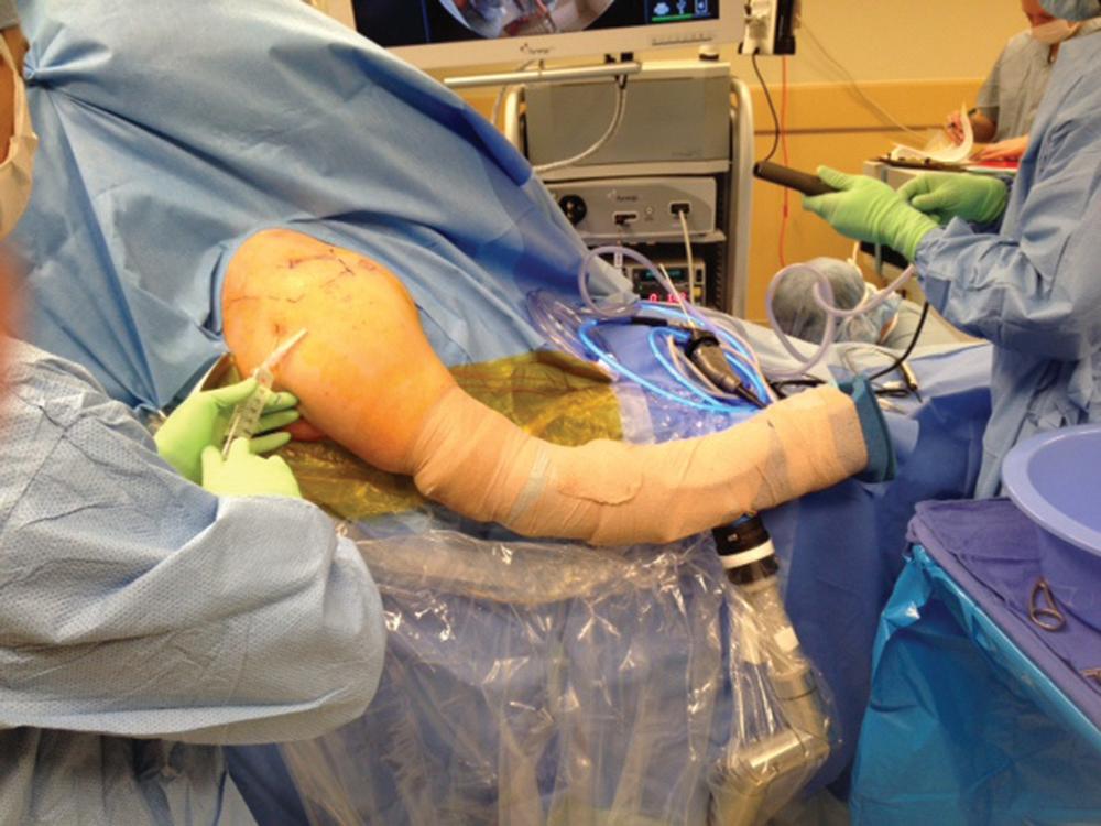
Patient Positioning
■ The patient is placed in a supine position on the operating table, with the knee at or below the break of the bed.
■ A tourniquet is placed high on the thigh, even if inflation is not planned, so that one is prepared in the case of unexpected bleeding; padding around the thigh before placement of the tourniquet is advised. The tourniquet is typically set to 250 to 300 mm Hg.
■ A plastic drape (sticky-u) is then placed around the tourniquet to create a barrier between the preparation solution and the tourniquet. CASE
MINIMUM REQUIREMENTS
• N = 30 (knee arthroscopy)
COMMONLY USED CPT CODES
• CPT Code: 29850—Arthroscopically aided treatment of intercondylar spine(s) and/or tuberosity fracture(s) of the knee, with or without manipulation; without internal or external fixation (includes arthroscopy)
• CPT Code: 29851—Arthroscopically aided treatment of intercondylar spine(s) and/or tuberosity fracture(s) of the knee, with or without manipulation; with internal or external fixation (includes arthroscopy)
• CPT Code: 29855—Arthroscopically aided treatment of tibial fracture, proximal (plateau); unicondylar, includes internal fixation, when performed (includes arthroscopy)
• CPT Code: 29856—Arthroscopically aided treatment of tibial fracture, proximal (plateau); bicondylar, includes internal fixation, when performed (includes arthroscopy)
• CPT Code: 29860—Arthroscopy, hip, diagnostic with or without synovial biopsy (separate procedure)
• CPT Code: 29866—Arthroscopy, knee, surgical; osteochondral autograft(s) (e.g., mosaicplasty; includes harvesting of the autograft[s])
• CPT Code: 29868—Arthroscopy, knee, surgical; meniscal transplantation (includes arthrotomy for meniscal insertion), medial or lateral
• CPT Code: 29870—Arthroscopy, knee, diagnostic, with or without synovial biopsy (separate procedure)
• CPT Code: 29871—Arthroscopy, knee, surgical; for infection, lavage and drainage
• CPT Code: 29873—Arthroscopy, knee, surgical; with lateral release
• CPT Code: 29874—Arthroscopy, knee, surgical; for removal of loose body or foreign body (e.g., osteochondritis dissecans fragmentation, chondral fragmentation)
• CPT Code: 29875—Arthroscopy, knee, surgical; synovectomy, limited (e.g., plica or shelf resection; separate procedure)
• CPT Code: 29876—Arthroscopy, knee, surgical; synovectomy, major, two or more compartments (e.g., medial or lateral)
• CPT Code: 29879—Arthroscopy, knee, surgical; abrasion arthroplasty (includes chondroplasty where necessary) or multiple drilling or microfracture
• CPT Code: 29880—Arthroscopy, knee, surgical; with meniscectomy (medial and lateral, including any meniscal shaving)
• CPT Code: 29881—Arthroscopy, knee, surgical; with meniscectomy (medial or lateral, including any meniscal shaving)
• CPT Code: 29882—Arthroscopy, knee, surgical; with meniscus repair (medial or lateral)
• CPT Code: 29883—Arthroscopy, knee, surgical; with meniscus repair (medial and lateral)
• CPT Code: 29884—Arthroscopy, knee, surgical; with lysis of adhesions, with or without manipulation (separate procedure)
• CPT Code: 29885—Arthroscopy, knee, surgical; drilling for osteochondritis dissecans with bone grafting, with or without internal fixation (including débridement of base of lesion)
■ A lateral leg post is placed on the outside of the operating table at the level of the midthigh and is positioned so a valgus stress can be applied to allow improved access to the medial compartment. The post should allow the surgeon to stand between the bed and the patient’s ankle (as the thigh is pressed against the leg post); often surgeons may need to use their hip against the patient’s leg if no assistance is available.
• Alternatively, a circumferential leg holder can be used, with placement in the same position along the thigh as the leg post. This leg holder is typically placed at the level of the tourniquet.
■ An examination of the knee with anesthesia should be performed after appropriate patient positioning, and various motions, including varus/valgus stress, should be performed to confirm that the position is adequate to permit a thorough examination of the knee.
■ A time-out should be performed to ensure patient safety and to confirm the procedure to be performed.
Prepping and Draping
■ Skin preparation is performed per surgeon/institution preference; the authors typically use alcohol followed by a chlorhexidine preparation solution while the assistant holds the foot in sterile fashion.
■ The extremity is then draped in layers, as follows:
• Down sheet under the operative leg, over the contralateral leg
• Sticky-u drape with tails aimed proximally around the thigh, just distal to the plastic drape applied before prepping
• Impervious stockinette applied over the foot to the midcalf, followed by Coban wrapping (3M, Minneapolis, MN) around the stockinette
• Arthroscopy extremity drape over the leg to the level of the midthigh, creating the final sterile field; this drape typically has a hole in the center that creates a seal
• The arthroscopy extremity drape is used by anesthesia to create a barrier to the surgical field.
• Before draping, a mayo stand can be placed near the head of the bed over the patient’s torso; after draping, this can be used to hold some of the arthroscopic equipment that is needed during the case.
Landmarks and Portal Placement
■ Helpful landmarks are the patella, patellar tendon, and femoral condyles.
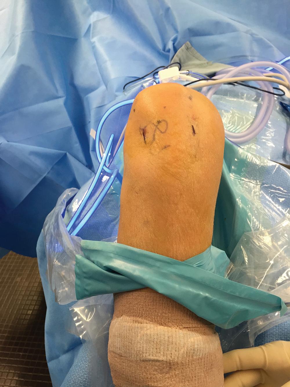
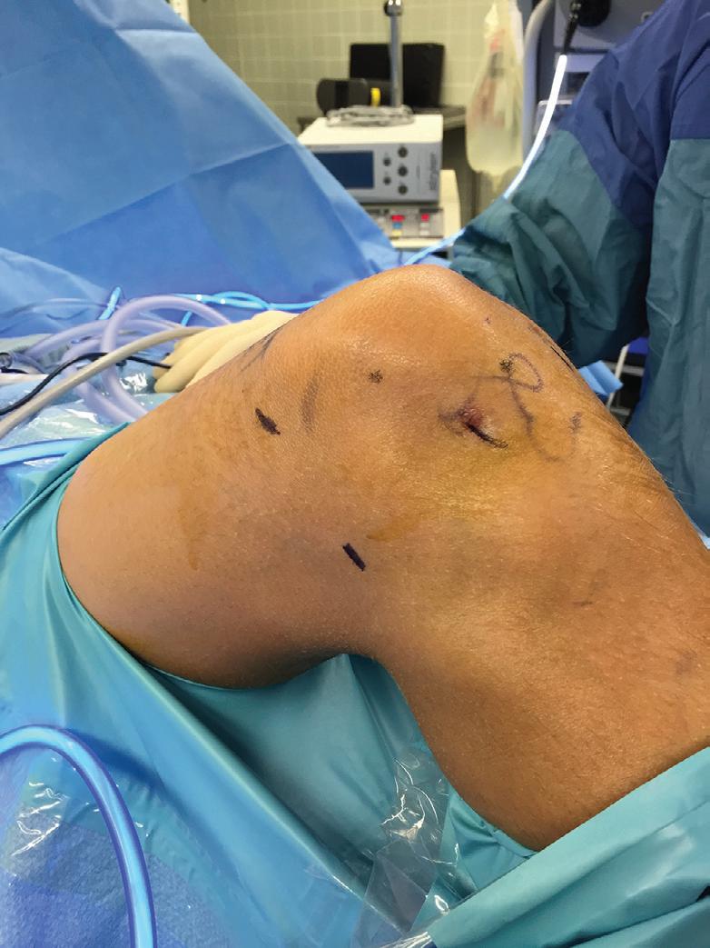
■ Standard portals used for diagnostic arthroscopy include the anterolateral (AL), anteromedial (AM), superomedial (SM), and superolateral (SL) portals (Fig. 1-1).
• With the knee flexed to 90 degrees, the landmarks become more visible.
■ The AL and AM portals are primarily used for diagnostic knee arthroscopy; the SM and SL portals are often but not always used.
■ The AL and AM portals are located in the “soft spot” on either side of the inferior pole of the patella.
• AL portal: Between the lateral femoral condyle and lateral proximal tibia (AL); primary viewing portal
• AM portal: Between the medial femoral condyle and medial proximal tibia (AM); primary working portal
■ SM and SL portals are made approximately 4 cm proximal to the medial and lateral poles of the patella, respectively.
• The SM and SL portals are often used for water flow; although these portals are not always created, they can be helpful in cases that involve extensive synovectomies and procedures within the patellofemoral joint.
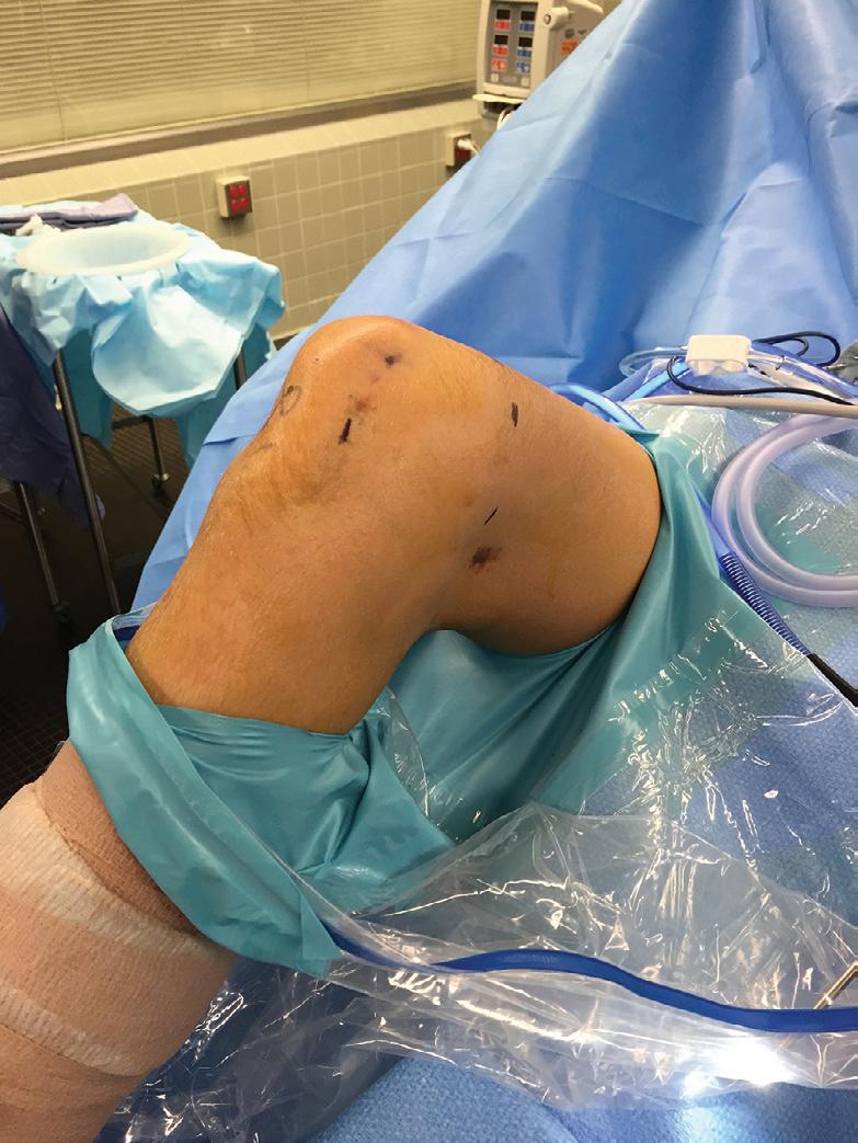
■ Additional portals: The posteromedial (PM) and posterolateral (PL) portals also occasionally are used in diagnostic knee arthroscopy, although these portals tend
Figure 1-1 Right knee indicates locations for anteromedial and anterolateral portals before a diagnostic knee arthroscopy.
to be used more often for procedure-specific arthroscopies, such as posterior cruciate ligament (PCL) reconstruction (Fig. 1-2).
• The PM and PL portals are made with direct arthroscopic visualization, typically with the knee flexed to 90 degrees.
• The PM portal is created 1 cm proximal to the joint line, at the posteromedial margin of the medial femoral condyle.
• This portal is helpful for visualization of the PCL and the posterior horn of the medial meniscus.
• The PL portal is created approximately 1 cm proximal to the joint line and 1 cm posterior to the lateral femoral condyle. Care must be taken to avoid injuring the biceps femoris muscle and the common peroneal nerve during creation of the PL portal.
• This portal is helpful for visualization of the PCL and the posterior horn of the lateral meniscus.
■ Other portals occasionally used in diagnostic knee arthroscopy include the transpatellar portal, the proximal superomedial portal, and accessory (far) medial/lateral portals.
Diagnostic Arthroscopic Examination
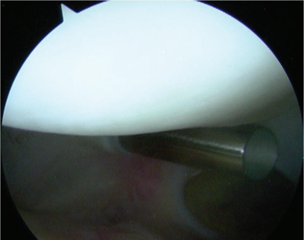
■ If an SM or SL portal is to be used as an outflow, this portal is created first (Fig. 1-3).
■ With the knee extended, a #11 scalpel is used to create a 5-mm incision in the SM or SL position, as described previously.
■ Next, an outflow cannula with a blunt trocar is inserted into the suprapatellar pouch through the portal.
COMMONLY USED ICD9 CODES
• 715.16—Primary localized osteoarthrosis, lower leg
• 715.26—Secondary localized osteoarthrosis, lower leg
• 715.36—Localized osteoarthrosis, not specified whether primary or secondary, lower leg
• 715.96—Osteoarthrosis, unspecified whether generalized or localized, lower leg
• 717.83—Old disruption of anterior cruciate ligament
• 844.2—Sprain and strains of knee and leg; cruciate ligament of knee
• 836.0—Medial meniscus/cartilage tear, includes bucket handle
• 836.1—Lateral meniscus/cartilage tear, includes bucket handle
• 836.2—Tear of meniscus/cartilage (semilunar), not specified as medial or lateral
• 717.0—Old bucket handle tear of medial meniscus
• 717.1—Derangement of anterior horn of medial meniscus
• 717.2—Derangement of posterior horn of medial meniscus
• 717.3—Other and unspecified derangement of medial meniscus
• 717.40—Derangement of lateral meniscus, unspecified
• 717.41—Bucket handle tear of lateral meniscus
• 717.42—Derangement of anterior horn of lateral meniscus
• 717.43—Derangement of posterior horn of lateral meniscus
• 717.49—Other derangement of lateral meniscus
• 717.5—Derangement of meniscus, not elsewhere classified
COMMONLY USED ICD10 CODES
• M17.0—Bilateral primary osteoarthritis of knee
• M17.1—Unilateral primary osteoarthritis of knee
• M17.2—Bilateral posttraumatic osteoarthritis of knee
• M17.3—Unilateral posttraumatic osteoarthritis of knee
• M17.4—Other bilateral secondary osteoarthritis of knee
• M17.5—Other unilateral secondary osteoarthritis of knee
• M17.9—Osteoarthritis of knee, unspecified
• M23.5—Chronic instability of knee
• M23.61—Other spontaneous disruption of anterior cruciate ligament of knee
• S83.5—Sprain of cruciate ligament of knee
• M23.20—Derangement of unspecified meniscus due to old tear or injury
• M23.21—Derangement of anterior horn of medial meniscus due to old tear or injury
• M23.22—Derangement of posterior horn of medial meniscus due to old tear or injury
• M23.23—Derangement of other medial meniscus due to old tear or injury
• M23.24—Derangement of anterior horn of lateral meniscus due to old tear or injury
• M23.25—Derangement of posterior horn of lateral meniscus due to old tear or injury
• M23.26—Derangement of other lateral meniscus due to old tear or injury
■ Once inserted, the cannula can be swept proximally and distally to release any attached synovium.
■ Once the trocar is removed, joint fluid typically is expressed, which confirms the intraarticular position.
■ Next, with the knee in 90 degrees of flexion, the AL portal is established, first with a #11 scalpel to make a 5-mm incision in the soft spot lateral to the inferior pole of the patella, as described previously.
• Vertical, horizontal, or oblique (along Langer’s lines) incisions can be used depending on surgeon preference.
• With vertical incisions, the blade should be directed cephalad to avoid injury to the meniscus.
■ After incision, a straight hemostat is inserted through the incision to open up the capsule, with the jaws opening both superior-inferior and medial-lateral.
■ Next, the arthroscope cannula with a blunt trocar is inserted through the portal and aimed toward the intercondylar notch.
■ The knee is then extended fully, and the trocar is advanced under the patella into the suprapatellar pouch.
■ A rotating motion of the hand can be used to gently advance the cannula into the pouch; this motion reduces the risk of iatrogenic cartilage damage.
■ The inflow tubing is then attached to the cannula, the trocar is removed from the cannula, and the 30-degree arthroscope is inserted into the cannula and locked into place.
■ At this point, the surgeon should ensure the outflow portal is truly in the joint (as opposed to stuck in the synovium) and reinsert if necessary.
■ Finally, the surgeon should assess the camera’s focus and adjust as appropriate. In the suprapatellar pouch, one can assess for loose bodies, a suprapatellar plica, or synovial hypertrophy.
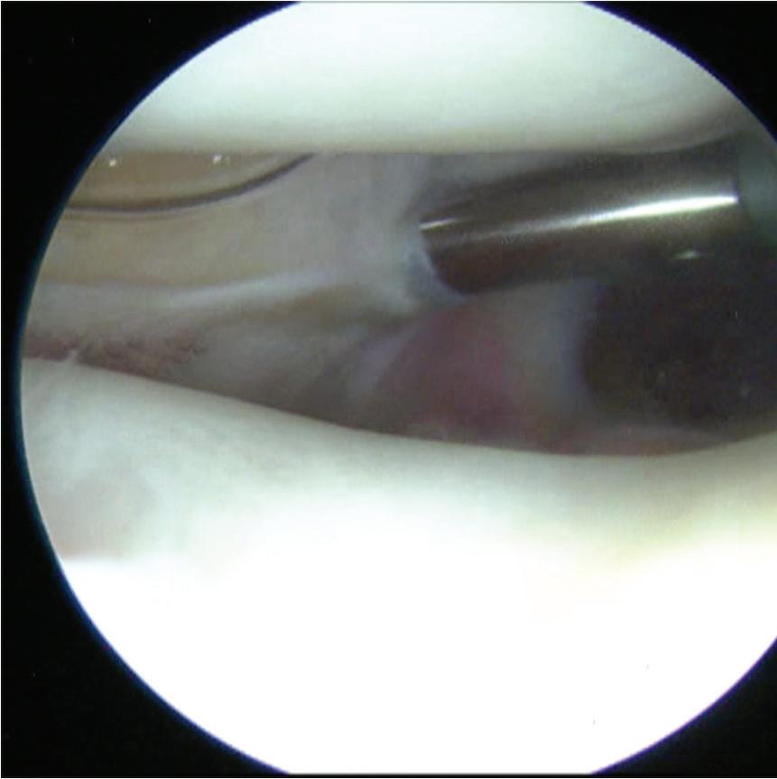
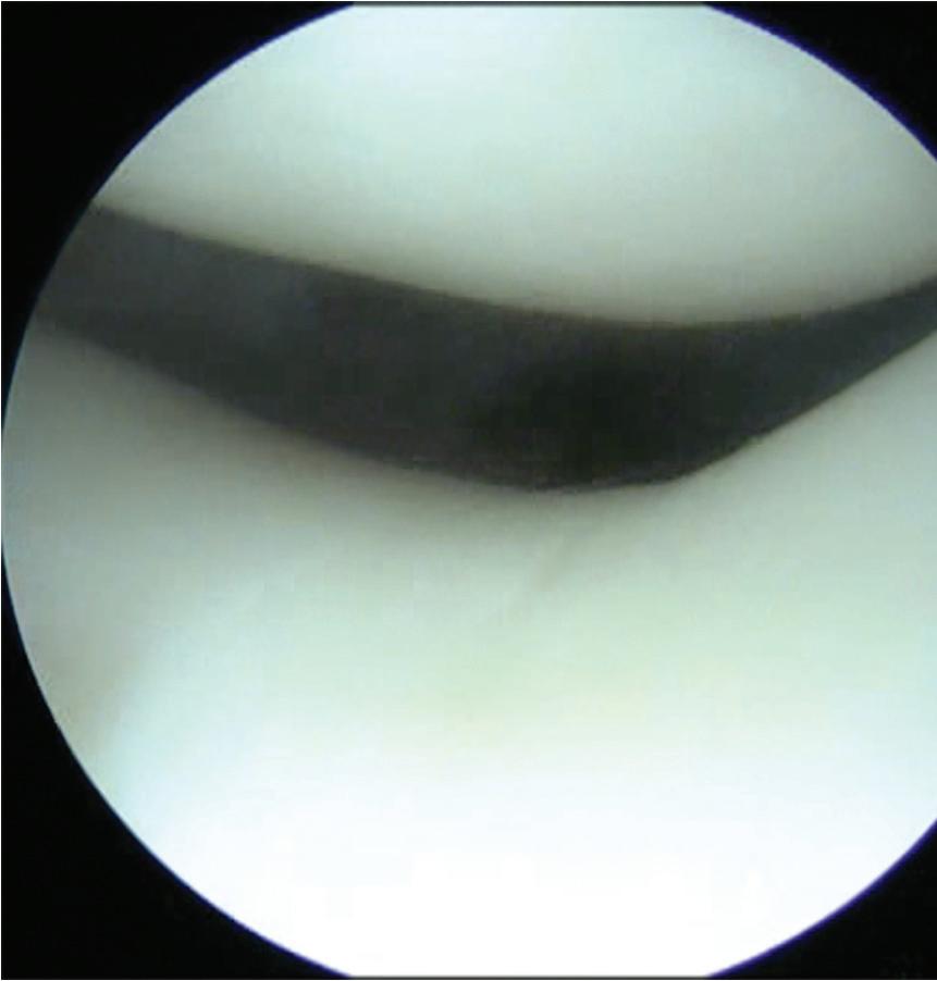
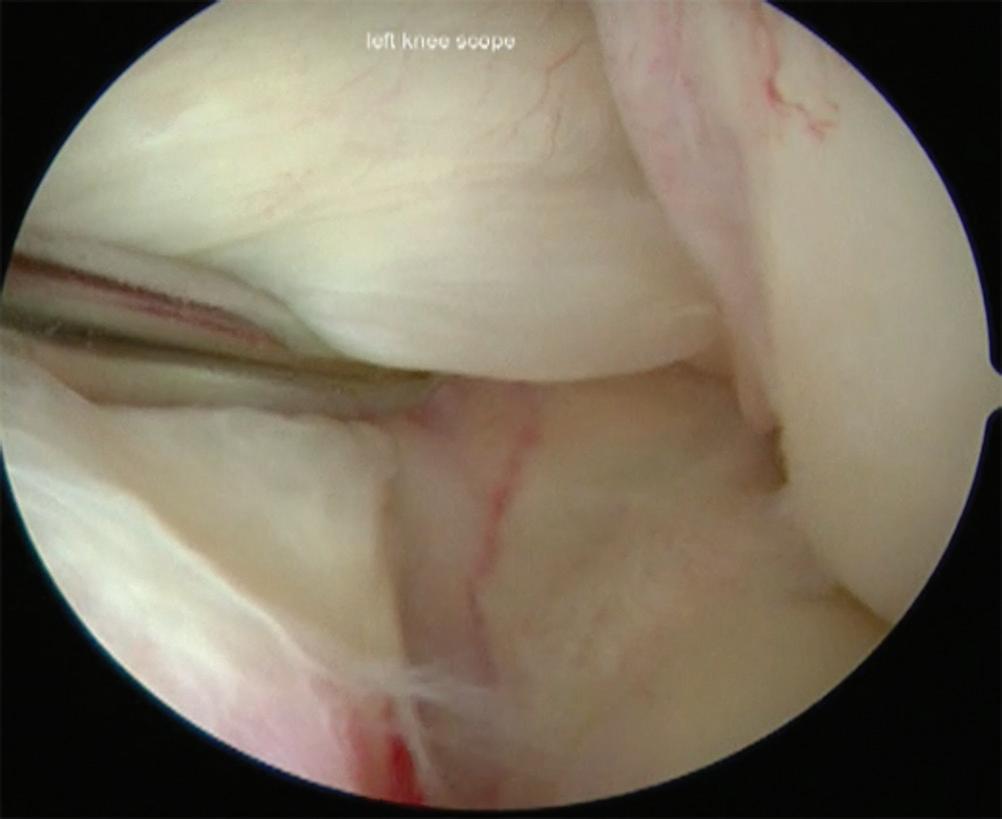
■ The arthroscope is then slightly withdrawn, and the patellofemoral joint is visualized (Figs. 1-4 and 1-5).
■ The eyes should be oriented superiorly and then rotated medially and laterally to assess the articular cartilage of the medial and lateral facets of the patella.
■ The arthroscope can then be directed laterally, with the camera aiming 30 degrees offset and slightly withdrawn to assess the relationship of the patella in the trochlear groove. The assistant should slowly flex the knee from extension to allow visualization of the entire trochlear groove.
■ The superior-most aspect of the femoral condyles is visible at this point.
■ The knee is then brought back into full extension, and the arthroscope is driven inward past the patella and directed laterally to enter the lateral gutter. The eyes should be directed medially.
■ The surgeon raises the hand and slightly withdraws the arthroscope to access the gutter.
■ The arthroscope passes over synovial folds in the gutter and continues to move inferiorly until the popliteus tendon is visualized in the popliteus hiatus.
■ Once the synovial folds are identified, the surgeon should raise the camera to visualize the popliteal hiatus.
■ Femoral condyle osteophytes or a tight lateral retinaculum can make this visualization difficult.
■ The examiner can “tap” the posterolateral aspect of the knee from the outside to visualize any loose bodies.
■ Assessment of the PCL in instability cases is recommended.
■ With the knee still in extension, the arthroscope is brought back to the suprapatellar pouch and then directed medially to enter the medial gutter.
■ The eyes should be directed inferiorly.
■ The surgeon raises the hand and slightly withdraws the arthroscope to enter the gutter.
■ Synovial folds again are visualized, and a plica may be seen.
■ Next, the arthroscope enters the medial compartment.
superomedially.
■ From the medial gutter, the arthroscope is slightly withdrawn and moved laterally as the knee is placed into flexion with approximately 10 degrees of external rotation.
■ A valgus force is applied to the leg, and the camera is directed posterior to visualize the medial compartment from within the notch.
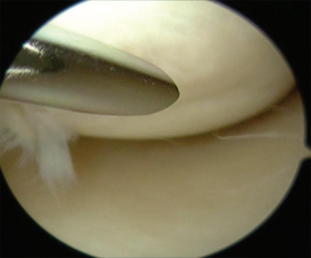
■ At this point, an 18-gauge spinal needle is placed into the portal site for the AM portal and is visualized arthroscopically (Fig. 1-6).
Figure 1-2 Right knee indicates locations for (A) posterolateral and (B) posteromedial portals before a diagnostic knee arthroscopy.
Figure 1-3 Arthroscopic photograph of the left knee shows the patellofemoral joint with the outflow cannula placed
Figure 1-4 Arthroscopic photograph of the left knee shows the patellofemoral joint in extension.
■ The AM portal is then established with a #11 scalpel to make a 5-mm incision, again orientated vertically, horizontally, or obliquely.
■ For known lateral meniscus tears, placement of the portal in a more superior position than for a medial meniscus repair can be helpful.
■ After creation of the AM portal, a probe is inserted through it into the medial compartment (Fig. 1-7).
■ To facilitate this, the surgeon should lift the hand and aim the probe toward the floor to reach the posterior horn of the medial meniscus.
■ If the probe does not pass easily into the medial compartment, the knee is brought into flexion and the arthroscope is used to look into the notch and triangulate the location of the probe. Both hands should be at the same vertical level.
■ Remember that the eyes of the camera are aimed 30 degrees from the trajectory of the arthroscope.
■ Once the probe is visualized, the maneuvers mentioned previously are used to reenter the medial compartment.
■ The medial meniscus should be probed along both the superior and the inferior surfaces to assess for tears.
■ Placement of the knee into full extension with a valgus force and raising of the hand holding the arthroscope superiorly while pushing inward allows for improved visualization of the posterior horn.
■ The eyes can be rotated while in the medial compartment to visualize and inspect the entire meniscus.
■ The posterior horn is best visualized looking inward to the notch.
■ The eyes should be rotated inferiorly to assess the status of the tibial plateau articular cartilage. The medial femoral condyle is assessed by moving the arthroscope superiorly while flexing the knee from extension.
■ Next, the intercondylar notch is visualized.
■ The knee is brought into 90 degrees of flexion with the leg hanging off the table.
■ The camera is directed from the medial compartment into the notch.
■ The entire arch of the notch can be visualized by sweeping the camera superiorly and laterally.
■ Once on the lateral side of the notch, the arthroscope can be withdrawn slightly and the anterior cruciate ligament (ACL) is visible.
Figure 1-5 Arthroscopic photograph of the left knee shows the patellofemoral joint in slight flexion.
Figure 1-6 Arthroscopic photograph of the left knee shows needle localization for establishing the anteromedial portal.
■ The probe can be inserted via the AM portal at this time and used to assess the ACL.
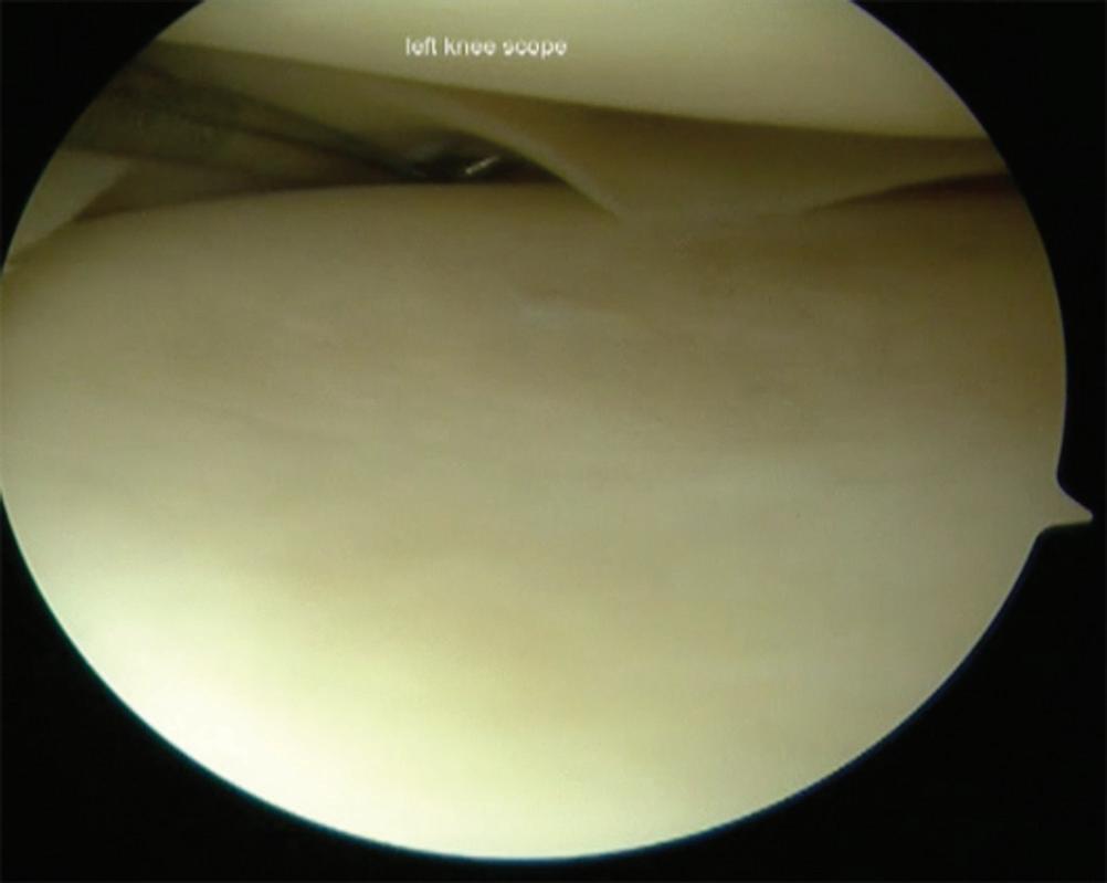
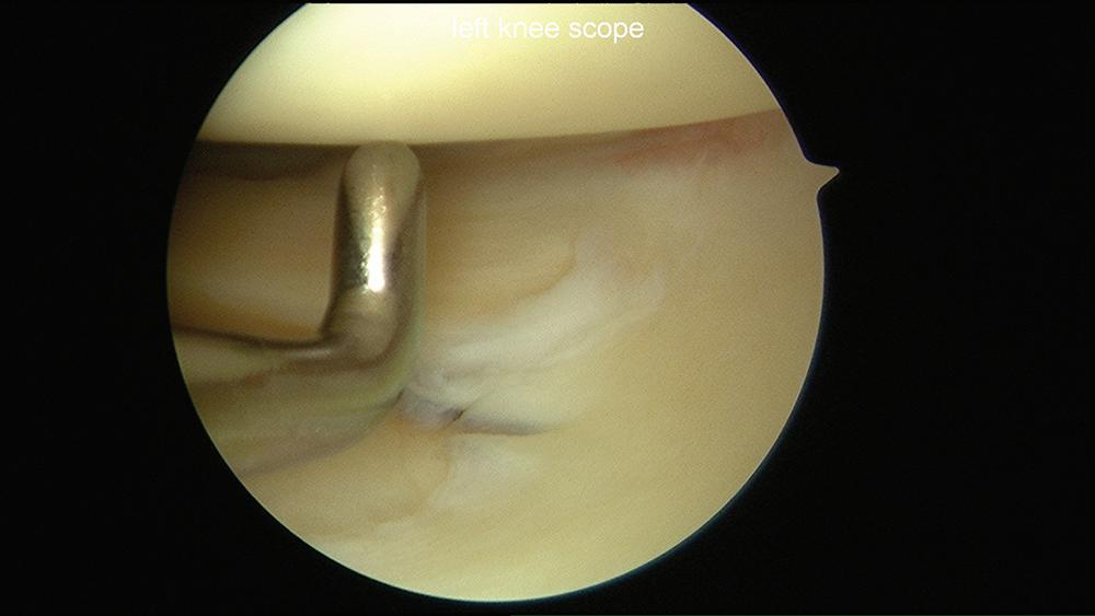
■ The attachment of the ACL on the lateral femoral condyle should be intact (Fig. 1-8).
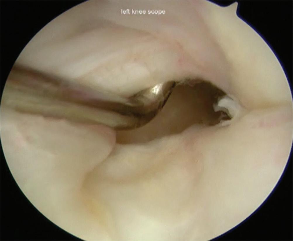
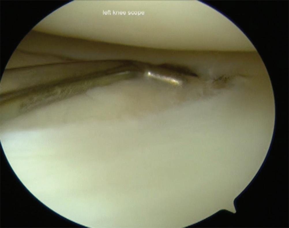
■ The probe can be used to retract the ACL laterally for visualization of the PCL (Fig. 1-9).
■ If visualization of the notch is difficult because of what may appear to be the retropatellar fat pad, this can be débrided with the shaver.
■ Next, the lateral compartment is visualized.
■ To move from the notch to the lateral compartment, the arthroscope is parked at the level of the inferior aspect of the lateral femoral condyle and the probe is placed in the “parking spot” triangle formed by the lateral border of the ACL, the medial border of the lateral femoral condyle, and the anterior horn of the lateral meniscus.
Figure 1-7 Arthroscopic photograph of the left knee shows assessment of the medial meniscus.
Figure 1-8 Arthroscopic photograph of the left knee shows assessment of the anterior cruciate ligament.
Figure 1-9 Arthroscopic photograph of the left knee shows assessment of the anterior cruciate ligament under mild tension with the probe.
■ The knee is brought into the figure-four position with flexion and application of a varus load with internal rotation.
■ The foot of the operative leg is rested on the anterior tibia of the contralateral leg.
■ As the leg is brought up into the figure-four position, the hand holding the arthroscope should supinate to rotate approximately 90 degrees while aiming posterior with the camera.
■ Thus, the correct visual orientation of the lateral compartment is maintained. The entire lateral meniscus should be probed and inspected, especially the posterior horn where tears are often missed (Figs. 1-10 and 1-11).
■ The hand holding the arthroscope should be raised toward the ceiling and pushed posterior to facilitate adequate visualization. Gentle increases in varus stress help open up this area.
■ The popliteus hiatus should be well visualized.
■ The posterior horn of the lateral meniscus is naturally more lax than the posterior horn of the medial meniscus.
■ The camera should be gently moved laterally, with the eyes oriented laterally to view the midbody of the meniscus followed by the anterior horn.
■ To truly visualize the anterior horn, the camera is slowly and gently withdrawn.
■ The anterior horn is sometimes better visualized with the arthroscope through the AM portal.
■ The tibial plateau and lateral femoral condyle articular surfaces are subsequently assessed.
Posterior Diagnostic Knee Arthroscopy (Video 1-1)
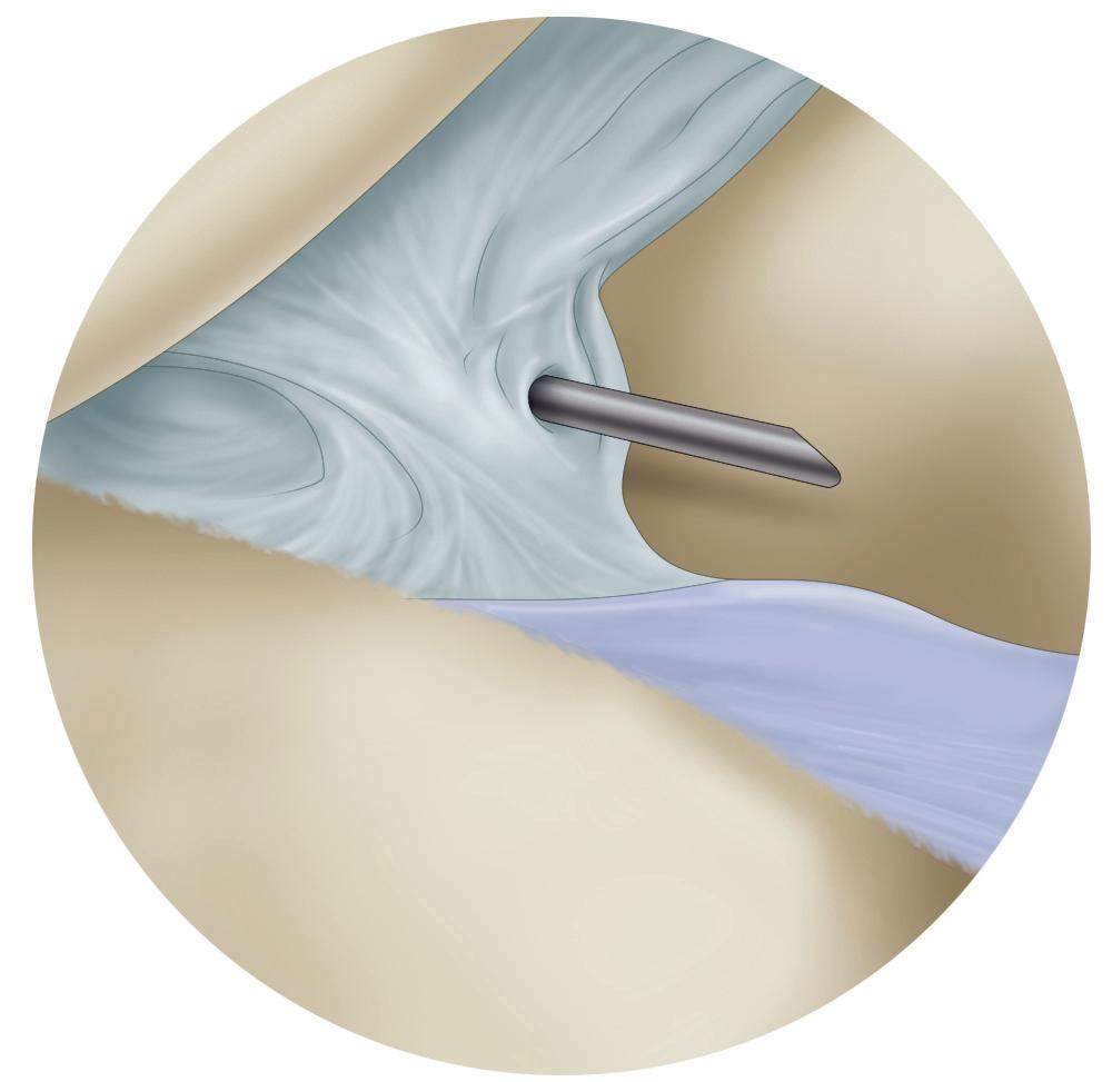
Posterior Compartments (Fig. 1-12)
■ Although many authors agree that posterior knee arthroscopy should be performed as part of most, if not all, diagnostic arthroscopic procedures, visualization of the posterior compartments of the knee is especially helpful in evaluation for loose bodies and in repair of meniscus root tears.
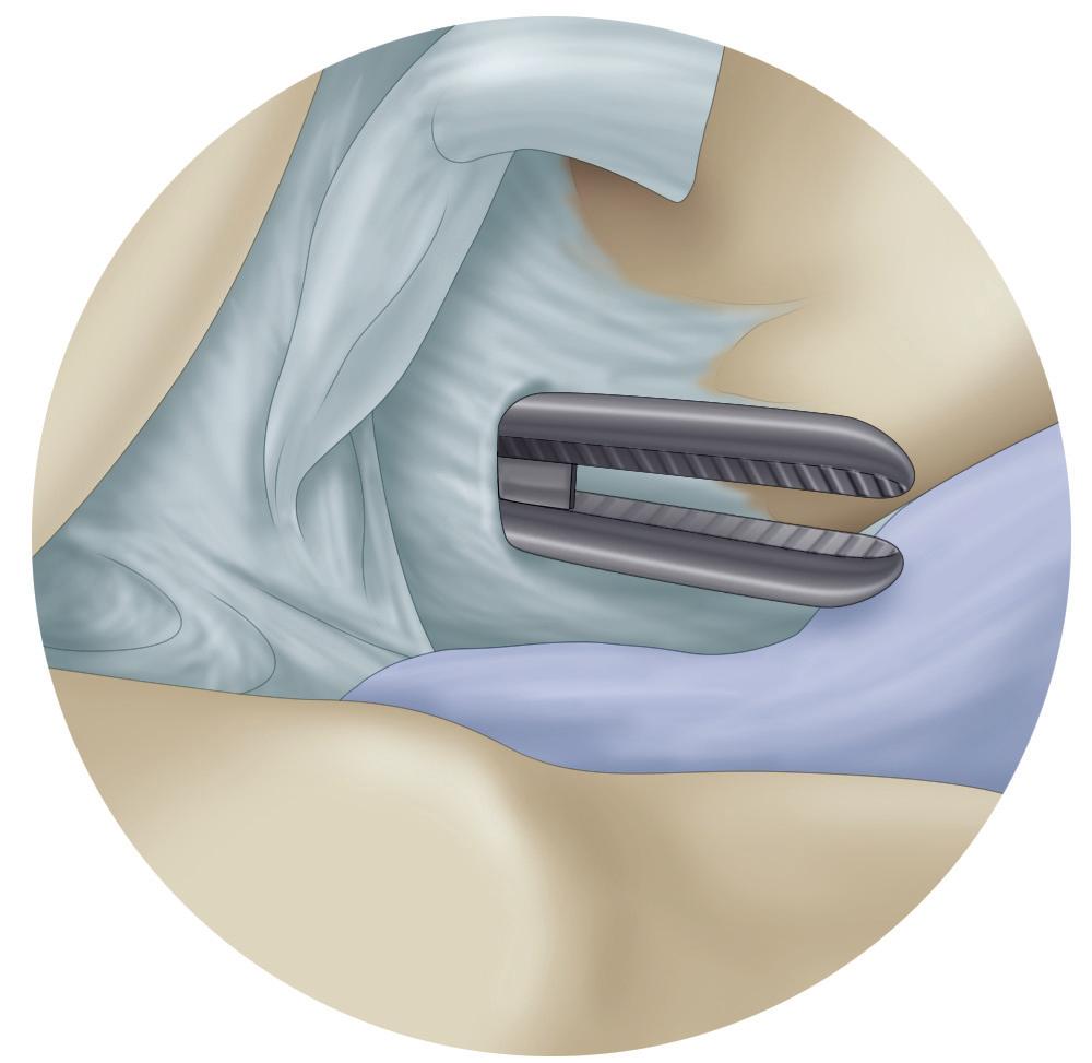
■ For access to the posteromedial and posterolateral compartments of the knee, the modified Gillquist maneuver is typically performed.
• This maneuver is referred to as a contralateral drive-through maneuver.
■ For visualization of the posteromedial compartment, the knee is flexed to 90 degrees and a blunt trocar is placed through the AL portal toward the anterolateral wall of the medial femoral condyle.
Figure 1-10 Arthroscopic photograph of the left knee shows assessment of the lateral compartment.
Figure 1-11 Arthroscopic photograph of the left knee shows assessment of the lateral meniscus.
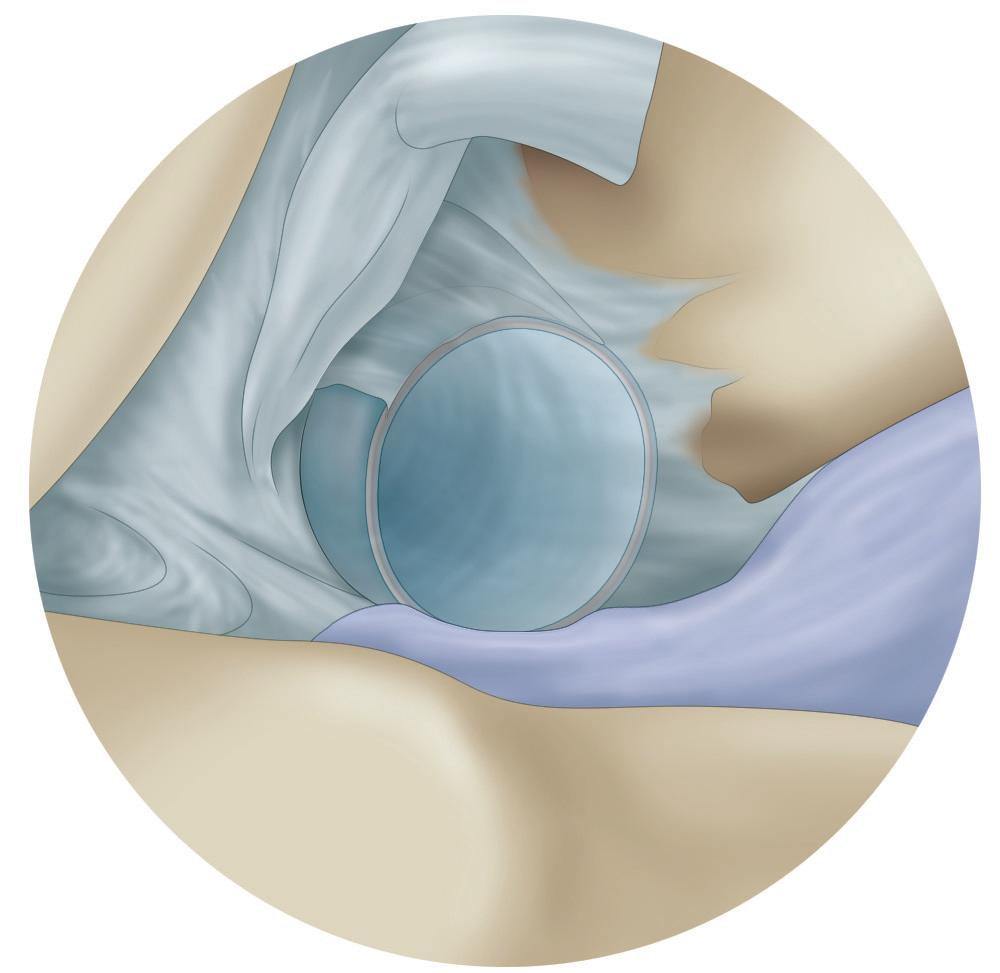
Figure 1-12 Schematics show establishment of the posteromedial portal, including (A) needle localization of the portal site with a spinal needle with direct visualization, (B) blunt dissection of the portal through the capsule (in this case with a hemostat), and (C) insertion of a plastic cannula to complete portal establishment.
■ The obturator is slowly advanced posteriorly while the knee is slowly extended until it “pops” through the interval between the medial femoral condyle and the PCL; a valgus stress is applied to help facilitate access.
■ Care should be taken to avoid injury to either the PCL or the medial wall of the intercondylar notch.
■ The arthroscope is advanced over the trocar. The same technique is used to access the posterolateral compartment, with the trocar inserted into the AM portal and gently pushed and advanced through the interval between the medial aspect of the lateral femoral condyle and the ACL; a varus stress is applied to help facilitate access. Pending surgeon comfort, the arthroscope can be used directly instead of the blunt trocar. Often, the use of a 70-degree arthroscope is helpful for visualization of the posterior compartments of the knee.
■ The authors have also found that an ipsilateral drive-through maneuver can be helpful for accessing the posterior compartments.
■ When this maneuver is performed, the arthroscope is placed from the AL portal and slid in the interval between the ACL origin and the lateral wall of the intercondylar notch; conversely the AL portal may be used to slide into the posteromedial portal between the PCL and the medial wall of the intercondylar notch.
■ Dependent on the visualization and relative joint tightness, varying degrees of knee flexion from 70 degrees (i.e., figure 2-4 position) to 30 degrees may facilitate this maneuver.
■ Although the contralateral drive-through maneuver is generally easier to perform, on occasion a larger loose body blocks visualization in either the medial or lateral compartment, which makes visualization for creation of an accessory PM or PL portal difficult.
■ In general, transitioning into the posterior compartments may be necessary to access for meniscocapsular tears, meniscal root tear repairs, loose bodies, visualization of the posterior cruciate tibial footprint during PCL reconstruction, synovectomy, and in unusual situations, posterior capsular releases or Baker’s cyst decompression.
BRIEF SUMMARY OF SURGICAL STEPS
• Suprapatellar pouch
• Patellofemoral joint
• Trochlear groove
• Medial gutter
• Lateral gutter
• Medial compartment
• Intercondylar notch
• Cruciate ligaments
• Lateral compartment
• Posterior compartments
TECHNICAL PEARLS
• Suprapatellar pouch → eyes at 12 o’clock to identify the proximal patellar pole when retracting
• Lateral gutter → eyes aimed medially, raising scope up when synovial folds visualized
• Medial gutter → eyes at 6 o’clock or aiming medially
• Medial compartment → eyes aimed laterally at the notch; in a tight knee, eyes may need to look up as well
• Placement of the scope on the anterior horn of the meniscus medially may provide a second way to visualize the posterior horn of the meniscus
• Intercondylar notch → the anterior cruciate ligament femoral insertion is best visualized with the eyes placed at 10 or 2 o’clock
• Lateral compartment → eyes at 12 o’clock to visualize the posterior horn of the meniscus, rotating laterally to inspect the midbody and anterior horn
• Portals placed too inferiorly risk damage to the meniscus and prohibition of adequate visualization of the medial joint
• Aggressive débridement of fat pad may cause bleeding and an increase in postoperative pain
• Significant valgus stress to visualize the medial compartment may risk injury to the medial collateral ligament
• Aggressive insertion of trocar, scope, or probe may cause iatrogenic injury to articular cartilage
• A stiff knee may make entering the gutters difficult; starting in the patellofemoral joint and entering the compartments via the intercondylar notch is helpful in these cases
• Be careful with radiofrequency near the gutters; a blister can be caused by being too close to the skin
POSTOPERATIVE PROTOCOL
Weeks 1-2: Weight bearing as tolerated without assistance by 48 hours after surgery
Range of motion (ROM): Progress through passive, active, and resisted ROM as tolerated (goal: full extension by 2 weeks, 130 degrees of flexion by 6 weeks)
Patellar mobilization daily
Strengthening: Quad sets, straight leg raises (SLR), heel slides, etc.
No restrictions to ankle and hip strengthening
Modalities: Electric stimulation, ultrasound, heat before and after, ice before and after
Weeks 2-6: ROM: Continue with daily ROM exercises (goal: increase ROM as tolerated)
Strengthening: Increase closed chain activities to full motion arc; add pulley weights, theraband, etc.; progress strengthening activities (wall sits, lunges, balance ball, leg curls, leg press, plyometrics, squats, core strengthening)
Continue stationary bike and biking outdoors for ROM, strengthening, and cardiovascular
Modalities: Electric stimulation, ultrasound, heat before and after, ice before and after
POSTOPERATIVE CLINIC VISIT PROTOCOL
7-10 days: First postoperative visit for suture removal and ROM check 4-6 weeks: Second postoperative visit for gait, ROM, and strength check
8-10 weeks: Final postoperative visit
SUGGESTED READINGS
1. Frank RM, McCormick FM, Harris JD, et al. Diagnostic knee arthroscopy: surgical technique. <http:// orthoportal.aaos.org/oko/article.aspx?article=OKO_SPO079#abstract>; 2014. Accessed 16.02.15.
5. Ward BD, Lubowitz JH. Basic knee arthroscopy part 4: chondroplasty, meniscectomy, and cruciate ligament evaluation. Arthrosc Tech. 2013;2(4):e507-e508. doi:10.1016/j.eats.2013.07.011.
6. Jackson RW. Arthroscopic surgery. J Bone Joint Surg Am. 1983;65(3):416-420.
7. Kramer DE, Bahk MS, Cascio BM, Cosgarea AJ. Posterior knee arthroscopy: anatomy, technique, application. J Bone Joint Surg Am. 2006;88(suppl 4):110-121. doi:10.2106/JBJS.F.00607.
8. Morin WD, Steadman JR. Arthroscopic assessment of the posterior compartments of the knee via the intercondylar notch: the arthroscopist’s field of view. Arthroscopy. 1993;9(3):284-290.
CHAPTER
DIAGNOSTIC SHOULDER ARTHROSCOPY
SURGICAL
TECHNIQUE
Rachel M. Frank | Brian J. Cole
CASE MINIMUM REQUIREMENTS
• N = 20 (shoulder arthroscopy)
COMMONLY USED CPT CODES
• CPT Code: 29805—Arthroscopy, shoulder, diagnostic, with or without synovial biopsy (separate procedure)
• CPT Code: 29825—Arthroscopy, shoulder, surgical; with lysis and resection of adhesions, with or without manipulation
• CPT Code: 29826—Arthroscopy, shoulder, surgical; decompression of subacromial space with partial acromioplasty, with or without coracoacromial release
The ability to perform a basic diagnostic shoulder arthroscopy is a critical skill for orthopaedic surgeons. With few exceptions, shoulder arthroscopy is likely to be performed multiple times per year, regardless of the field in which an orthopaedic surgeon ultimately decides to specialize. In many instances, especially for surgeons who specialize in shoulder and elbow surgery or sports medicine or practice general orthopaedic surgery, shoulder arthroscopy remains among the most common procedures performed. The surgical skills necessary for thorough, accurate, and efficient shoulder arthroscopy are typically developed early in residency training. With limitations in work hours, combined with the 2013 Accreditation Council for Graduate Medical Education (ACGME) implementation of skills training requirements for junior residents, development of excellent habits during initial training sessions is now, more than ever, imperative to build a foundation on which to expand one’s ability to treat different shoulder pathologies arthroscopically. The purpose of this chapter is to provide up-to-date technical pearls for performing a thorough, accurate, and efficient diagnostic shoulder arthroscopy. In this chapter, the authors present the basic techniques for performing diagnostic shoulder arthroscopy in both the beach chair (BC) and lateral decubitus (LD) positions (Video 2-1). With appropriate set-up and positioning, both techniques are reliable with low complication rates. The BC position offers the advantage of easy conversion to open techniques, and the LD position may allow for lower suture anchor position on the glenoid. Of note, many different techniques are used to effectively navigate through the shoulder, and the technique presented here represents just one of these techniques. As such, the authors wish to emphasize that the reader understand the importance of learning and developing a specific routine for performing a diagnostic shoulder arthroscopy in order to perform the procedure in a routine fashion for every single shoulder.
SURGICAL TECHNIQUE
Room Set-Up
■ Ensure that all appropriate equipment is in the room.
■ Ensure that all implants and instruments are available and sterile.
■ Confirm that the monitors are ergonomically positioned.
■ Confirm that the video monitor, pump, and shaver systems are functional.
■ The video monitor should be placed opposite the surgeon at head level.
Patient Positioning
Beach Chair
■ A leg pad is placed securely against the patient’s buttocks to ensure that the buttocks and back are firmly against the beach chair; this placement prevents sciatic, lower back, and pelvic injuries related to positioning.
■ Place a facemask over the patient, with care taken to not obstruct the airway; protect the eyes, ears, and nose at all times.
• If a facemask is not used, tape the patient’s head into place in a neutral position with a towel over the forehead.
■ A team effort then moves the patient from the supine to the beach chair position on the operating table (head of bed is elevated approximately 60 degrees). Confirm that the airway and facemask remain in a secured position, and confirm that the leg pad is firmly against the patient’s buttocks and that the patient’s back is firmly against the operating table.
■ The upper portion of the operating table can then be adjusted to improve exposure of the posterior aspect of the shoulder, typically with sliding the back of the table toward the contralateral shoulder while shifting the patient’s torso toward the operative side. Take care to confirm that the patient’s head and neck remain in a neutral position.
■ Folded towels can be placed behind the medial border of the ipsilateral scapula to stabilize it on the operating table.
■ Take the shoulder through the range of motion (ROM) that is necessary for the intended operative procedure.
■ Ensure that the patient’s knees and elbows are appropriately padded.
■ Turn the table 45 to 90 degrees to improve access to the patient for both the anesthesia team and the surgical team (Fig. 2-1).
Lateral Decubitus
■ Ensure that the bean bag is on the operating table before attempting the transfer. A sheet should be under and on top of the bean bag.
■ Transfer the patient to the operating table.
■ A team effort is used to roll the patient into the lateral decubitus position on the bean bag, with the operative extremity up.
■ Place the axillary roll under the patient, approximately 2 to 3 fingerbreadths distal to the axilla against the rib cage. This placement minimzes the pressure on the brachial plexus during the case.
■ Position the bean bag as desired to ensure optimal exposure and access to the shoulder and all necessary portals.