MICROBIOLOGY


For 1st year


1. Bacterial structure……………..…………………………………………………………………………………..6 2. Bacterial growth…………….. ……………………………………………………………………………………21 3. Bacteriophage……………….……………………………………………………………………………………..27 4. Bacterial genetics……………………………………………………….……………………………………….33 5. Recombinant DNA technology …………………………………..……………………………………….41 6. Anti-microbial chemotherapy …………………………………..………………………………………..61 7. Bacterial infections……………..……………………………………………………………………………….70 8. Viral structure……………... ………………………………………………………………………………………76 9. Viral classification……………….……………………………………………………………………………....81 10.Viral genetics…………..………………………………………………….……………………………………….86 11. Viral interactions …………………………………..……………………………………………………………90 12. Viral replication…………………………………..………………………………………………………………95 13. Viral infections……………..…………………………………………………………………………………….101 14. Fungal structure…………….. …………………………………………………………………………………110 15. Fungal reproduction ……………….…………………………………………………………………………113 16. Fungal diseases……………………………………………………….………………………………………...117 1. Immunity……………..………………………………………………………………………………………………..127 2. Immunogens…………….. ……………………………………………………………………………………..…136 3. Acquired immunity ……………….…………………………………………………………………………….142 4. Cell mediated immunity……………………………………………………….…………………………….149 5. Antigen presenting cells …………………………………..……………………………………………….154 6. Cytokines …………………………………..………………………………………………………………………..161 Content
7. Humoral immunity……………..………………………………………………………………………………..168 8. The complement system……………... ……………………………………………………………………186 9. Tumor immunology……………….…………………………………………………………………………….193 10.Immunization………..………………………………………………….…………………………………….….199 11. Hypersensitivity …………………………………..……………………………………………………………203 12. MHC…………………………………..………………………………………………………………………………..217 13. Transplantation……………..………………………………………………………………………………….221 14Tolerance and autoimmune diseases…………….. ………………………………………………228 15. Immunodeficiency ……………….…………………………………………………………………………..234 16. Basics of infection control……………………………………………………….……………………….241
Introduction

Outlines
✔ An introduction to medical microbiology
✔ Comparison between Prokaryotes and Eukaryotes

✔ Comparison between Viruses, Bacteria and Fungi
Microbiology
Microbiology (from Greek means MICRO = small, BIO = living, and LOGY = to study). So, this science deals with the study of living microorganisms, which are microscopic in size (cannot seen by naked eye) and simple in structure.
Microbiology has become an umbrella term including many sub fields of study. These include:
• Bacteriology: The study of bacteria

• Mycology: The study of fungi
• Parasitology: The study of parasites
• Virology: The study of viruses
Medical Microbiology
Is the study of microorganisms causing infectious diseases of humans and the human reactions to such infections.

In other words, it deals with etiology, pathogenesis, laboratory diagnosis, specific treatment and control of infection (immunization).

1
All living organisms can be divided into:
• Prokaryotes mean (Pro= primitive or premature, karyon= nucleus) as bacteria
• Eukaryotes mean (Eu= true, karyon= nucleus) as fungi, algae, protozoa, helminths, human and animal cells.
Microorganisms that cause human infectious diseases include 5 major groups of organisms: bacteria, fungi, protozoa, helminths and viruses.
Average size Nucleus Size of Ribosome Mitochondria Cell membrane sterols Cell wall containing peptidoglycan.
Mitotic division Chromosome number Eukaryotes Prokaryotes 0.2-2 mm in diameter 10-100mm in diameter Yes No Yes 80S Present Enclosed by a nuclear membrane. More than one Fungi and protozoa Yes No 70S Absent No enclosing nuclear membrane One No Bacteria 2
Examples
Bacteria
Bacteria are widely distributed in nature, and according to their habitat and way of living they are described as:

1) Saprophytic bacteria: Live on inanimate material; in the soil, in water, in dust, in the air, on clothes or on dead bodies and decaying organic matter.
2) Parasitic bacteria: Live on or in the body of living creatures. Parasitic bacteria are classified into:
a-Pathogenic bacteria: Cause disease in man or animals or plants.
b-Commensal bacteria: Live on or in the body without exerting a harmful effect.
Some of these commensals may be potentially pathogenic and may cause disease if the body resistance is lowered by any means.
Viruses
Viruses are quite distinct from other organisms as they have no cell structure. A viral particle consists of a nucleic acid molecule, either DNA or RNA, enclosed in a protein coat or capsid. They are obligate intracellular parasites; they require the biological machinery of a host cell for reproduction and survival.
3
Taxonomy
Taxonomy is the classification of organisms in an ordered system that indicates natural relationships. The organisms are divided into related groups based on similar characteristic and include species as the smallest and most definitive level of division.
Size
Nucleus Ribosome Mitochondria Nature of outer surface Motility Replication Viruses Bacteria Fungi Absent 70S 80S None Prokaryotic Eukaryotic DNA or RNA Both Both 0.02- 0.2 0.2-5 3-10 Absent Absent Present Protein capsid & lipoprotein envelope Rigid wall containing peptidoglycan Rigid wall containing chitin None Some None Special process of biosynthesis Binary fission Yeast by budding, moulds by mitosis.
(µ) Nucleic acid
4
Microbiological nomenclature
• In microbiology the binominal system of nomenclature is accepted where each species has a generic and a specific name. The generic name is written with a capital letter, and the specific name – with a small letter.

• For example: The anthrax bacillus – Bacillusanthracis-The tetanus bacillus – Clostridiumtetani .
• Viruses are not named so.
5
Bacterial structure 1
Outlines
✔ Bacterial Cell Wall and cell wall deficient bacteria

✔ Cytoplasm and Cytoplasmic membrane functions
✔ Structures outside the cell wall
Bacterial Structure
1-Size of the Bacterial Cell:
The majority of bacteria fall within the general dimensions of 0.2 to 8 µm

2-Shape of the Bacterial Cell:
They are unicellular structures which occur as cocci, bacilli and spirals. Some bacteria are variable in shape and are said to be pleomorphic. The shape of a bacterium is determined by its rigid cell wall (except in spirochetes it is determined by the axial filament)
3-Arrangement of the Bacterial Cell:
When the bacterial cell divides, the two daughter cells may at once separate, or they may remain attached to one another by the cell membrane.

It may occur in clusters (e.g., Staph) some in pairs (diplococci) e.g. (Pneumococci) and others occurring in chains e.g. (Streptococci). Sometimes the two daughter cells may be parallel to each other or at angle as in the diphtheria bacillus.
6
Bacterial cell wall
The cell wall is the outermost component of all bacteria. It is a multilayered structure located external to the cytoplasmic membrane. It is composed of a layer of peptidoglycan to which the cell owes its rigidity.

Gram positive bacteria
A thick "peptidoglycan" layer (40 sheets) forms about 50% of the cell wall material. It is responsible for the rigidity of the cell wall and for maintaining the shape of the bacterial cell.
Teichoic acid also forms a major surface component; it may be on both sides of the peptidoglycan layer. Some of the polymers penetrate the peptidoglycan layer and are linked to the lipid in the cytoplasmic membrane to form lipoteichoic acid. It may mediate adherence of some organisms to mucosal cells.
Gram negative bacteria
There is an outer layer of lipopolysaccharides (LPS) which is extremely toxic to the human body and is called the endotoxin. It is released only when the bacterial cells are lysed.
The toxicity is associated with the lipid fraction which is called lipid A. The polysaccharide represents a major surface antigen called somatic (O) antigen which is immunogenic.
A thin layer of peptidoglycan (2 sheets) which forms only 5-10% of the cell wall material is located inside the outer layer of LPS. Lipoprotein molecules cross link the outer membrane and peptidoglycan layers.
The space between the outer and inner layer (cytoplasmic membrane) is called the periplasmic space and is filled with a gel-like solution of proteins. It is the site, in some species, of the enzymes called beta lactamases that degrade penicillins and other β-lactam drugs.
7


8
Cell wall function

1- It is a rigid structure that maintains shape of bacteria.
2- Protects the cytoplasmic membrane from bursting in hypotonic solutions (i.e., it is osmotically insensitive).
3- Play a role in cell division by the formation of transverse septum
4- Determine the reaction to Gram stain:
In G -ve bacteria:
In G +ve bacteria:
Cell wall contains more lipids that dissolve on addition of alcohol as decolorizing agent so increasing pore size that allows exit of methyl violet and entry of carbolfuchsin so G -ve is pink
Cell wall is rich in proteins that shrink on addition of alcohol decreasing pore size so prevent exit of methyl violet iodine complex and entry of carbolfuchsin so G +ve is violet.
5- Teichoic acid: Is highly immunogenic it induces the production of tumor necrosis factor (TNF-α) and interleukin 1 (IL-1) by macrophages.
It participates in toxic shock produced by some Gram-positive bacteria.
6- Since peptidoglycan is present in bacteria but not in human cell it is a good target for antimicrobial drugs, such as penicillins and cephalosporins that inhibit its synthesis
9
7- Lipopolysaccharides (LPS) of gram -ve organisms:
Lipid fraction
Which is called lipid A is extremely toxic to the body and is called the endotoxin. It is responsible for the fever, hypotension and shock i.e. endotoxic shock caused by G-ve organisms.
Polysaccharide fraction
Represents a major surface antigen called somatic (O) antigen which is immunogenic and used for serotyping.
Cell wall deficient bacteria
There are forms of bacteria which are treated to remove the cell wall and to reproduce new generation of organisms lacking the cell wall. The process can be induced by:
• Destruction of the cell wall by treating the organism with an enzyme i.e., lysozyme which has a selective action on the bacterial cell wall.
• Cultivation of the organism with penicillin which inhibits the synthesis of cell-wall substance without interfering with the growth.
A) Protoplast, spheroplast and L. forms:
A) Protoplast, spheroplast and L. forms
10
B) Mycoplasma:
• They may be produced spontaneously.
• If such treated cells are liberated from Gram positive cells, they are called protoplasts (they must be placed in isotonic solution to maintain the spherical configuration and guard against cell lyses and death because they entirely lack cell wall).
• If such treated cells are liberated from Gram negative cells, they are called spheroplasts which unlike protoplasts are osmotically insensitive because they have intact cell wall lacking peptidoglycan only.
• If these cell wall deficient cells are allowed to grow and divide, they are called L forms. L-forms may be formed during active infection under the effect of antibiotics and can cause chronic infection, their reversion to parent form can cause relapse of infection.
B) Mycoplasma:
Are the only bacterial species deficient in cell wall hence, they are pleomorphic and resistant to penicillin which acts on the bacterial cell wall and cannot be stained with gram stain.
Cytoplasmic Membrane

It's a semi-permeable , composed of phospholipids and protein. It differs from eukaryotic cells in the absence of sterols (except mycoplasma that lack cell wall, so cholesterol provides rigidity to its cell membrane.
11
Cytoplasmic membrane functions
I. Selective permeability to different molecules and active transport of ions (H+, Na+, K+, etc..) and nutrients to achieve osmotic balance and a pool of nutrients.
II. Energy generating functions, through electron transport and oxidative phosphorylation i.e., site of respiration.
III. It provides enzymes and carrier molecules that function in DNA, cell wall, and lipid membrane synthesis.

IV. Excretion of hydrolytic exoenzymes which degrade the different nutrients into subunits small enough to penetrate the cytoplasmic membrane
V. Excretion of pathogenicity proteins, e.g., IgA protease and some exotoxins.
VI. Chemotactic systems: It bears receptors and other proteins which have roles in chemotaxis of bacteria towards nutrients.

Mesosomes Function
They are invaginations of the cytoplasmic membrane
1-They play a role in respiration
12
2- They play a role in cell division where they function as:
a) The origin of the transverse septum that divides the cell in half
Cytoplasm
b) The binding site of the cell DNA which duplicates to provide the genetic material of each daughter cell.
Soft gel like structure that contains:
a) Ribosomes
a) Ribosomes:
b) Intracytoplasmic inclusions
c) Plasmids
d) Bacterial Nucleoid

- Ribosomes are present as tightly packed spherical particles in the cytoplasm.
- Clusters of ribosomes are called polysomes.
- They are composed of 40% protein and 60% RNA.
- Bacterial ribosomes are 70S (S→Sved berg i.e., sedimentation unit) in size, with 30S and 50S subunits.
- They are the site of protein synthesis.
b) Intracytoplasmic inclusions:
It represents accumulation of food reserve e.g., Volutin granules rich in meta polyphosphate found in the genus Corynebacterium.
13
c) Plasmids:
These are extra chromosomal double stranded circular DNA molecules. that are capable of replication independent of bacterial chromosome.
d) Bacterial Nucleoid:
The area of the cytoplasm where DNA is contained in masses.

- It is a single circular packed bundle of double stranded DNA molecule (chromosome)
There is no nuclear membrane, no nucleolus.
- No histones
- It carries the genetic information to the daughter cells, and it duplicates before cell division.
Structures outside the cell wall
1- Capsules
2) Flagella
14
3) Fimbriae (Pilli)
1- Capsules

Definition
It is a thick gelatinous layer outside the cell wall of some bacterial species.
Site of formation
May be formed in vivo.
e.g., B.anthracis
Chemical composition

Capsules differ in their chemical structures as well as development. The capsular material is responsible for the mucoid appearance of the colonies. Capsular material may be Polysaccharides as pneumococcior may be polypeptide as B.anthracis
May be formed both in vivo and in vitro.
e.g., klebsiella Pneumoniae.
NOTE
Capsules are not stained with gram stain and appear as unstained halos around the organism.
15
Function
1- The capsule is an important virulence factor that protects the organism against phagocytosis by 2 different ways:
a) The slimy capsules make it difficult for phagocytes to hold firmly on the bacterial surface.
b) Some of the bacterial cell wall components e.g., complement receptors are masked by the capsule.
2- The capsular polysaccharides are used as immunogens in certain vaccines.
3- Also, it is used in identification of organism (serologic typing) due to the antigenic variations in the sugar components of the polysaccharide.
- Is a loose polysaccharide network of fibrils.
- It allows the bacteria to adhere firmly to various structures e.g., skin, heart valves.
- It mediates adherence of strept.mutansto the teeth leading to formation of dental plaque which is the precursor
Definition
These are long, delicate, processes attached to the bacterial cell wall and cytoplasmic membrane by a basal body. These are the organs of motility (except some organisms like spirochetes that move without flagella by axial filaments). They move the bacteria towards nutrients and other attractants(chemotaxis).
Glycocalyx
2-Flagella
16
Structure
These projections can be seen by special methods as electron microscope.
They are composed of protein (flagellin) that is antigenic and differ in different bacterial species.
They differ in their antigenic structure from the body of the bacterial cell. Antisera against flagellar (H) antigen can be used for serotyping of flagellated species.
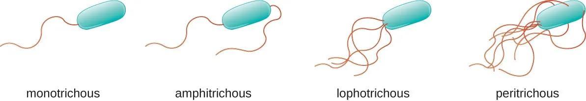
A- Monotrichous; one flagellum
Arrangement of flagella

C- Amphitrichous; both ends.
Function
B- Lophotrichous; tuft at one end
D- Peritrichous. all around bacteria
1- Organ of locomotion: chemotaxis→ movement of bacteria towards nutritives.
2- Antigenic: H antigen is used for typing and diagnosis.
3- Penetrating through viscid mucus secretions and epithelial barriers and spreading throughout body fluids and tissues.
17
Definition
These are very thin and short thread-like structure found on the surface of certain bacterial cells. They are present in Gram-negative bacilli and responsible for adhesion and attachment to mucosa. It acts as a virulence factor of bacteria It's composed of subunits of a protein, pilin.
These are two types:
Also known as colonization antigens.
These mediate adherence of bacteria to specific receptors on human cell surface, which is a necessary step in initiation of infection of some organisms (e.g., mutants of N. gonorrhoeae that do not have Pilli are nonpathogenic).
They play a role in the transfer of part of the genetic material from one cell (F+ donor) to another (F- recipient) during conjugation.
Bacterial Endospores
Definition
They are small oval or spherical cells which are highly resistant to unfavorable conditions as lack of nutrients, heat, dryness, etc...
3-Fimbriae (Pilli)
I) Ordinary Pilli:
ii) Sex Pilli or F-Pilli:
18
Spore formation is a characteristic feature of some gram-positive bacteria as the aerobic genus Bacillus e.g., B.anthracisand anaerobic genus clostridium e.g., clostridium Tetani.
Spores are considered to be a dormant or resting phase (no metabolic activity) of bacterial cell that can remain dormant for years.
Sporulation
The development of the spore passes through several stages of development.
1- Development of an ingrowth of the cytoplasmic membrane cutting off a portion of the cell's cytoplasm and including the nuclear material, ribosomes, glycolytic enzymes and little water (spore core).
2- A thick cortex of peptidoglycan and a tough keratin-like spore coat are formed around.
3- Outer spore membrane which contains calcium and Dipicolinic acid.
Positions of spores

Position of spores in relation to the body of the bacillus is variable and characteristic.
for species e.g.
* It may be spherical, terminal and projecting as in cl. Tetani.
* It may be oval, sub terminal and non-projecting as in cl. perfringens.
* It may be oval, central and non-projecting as in B. anthracis
19
Causes of marked resistance of spores:
1-Thick spore cortex and tough coat.
2-Their dehydrated state.
3-Their very low metabolic and enzymatic activity.
4- The bacterial spores contain large amount of Dipicolinic acid which is absent in vegetative cells.
Also, calcium is present. They form a complex material which is present in the outer spore membrane.
This complex material is impermeable and acts for the protection of the spore from the unfavorable physical and chemical conditions.
Consist of 3 main steps:
1- Activation: On exposure to water and appropriate nutrition.
2-Initiation: Calcium dipicolinate is released and spore coat and cortex are degraded by glycolytic enzymes.
3- Outgrowth of new vegetative form that contains the spore protoplast.
Medical importance of spores
Due to their extraordinary resistance to heat and chemicals. Sterilization by autoclave at 121oc for 20-30 minutes is required to eliminate them and also due to their use as

Germination
20
BACTERIAL GROWTH 2
Outlines
✔ Bacterial multiplication
✔ Binary fission
✔ Bacterial growth requirements

✔ Bacterial growth curve
✔ Bacterial metabolism
Bacterial multiplication: Bacteria reproduce by binary fission each cell divide into 2 daughter cells identical to the parent cell.

The doubling time (generation time): The time required for bacteria to double its number. It ranges from 20 minutes for E. coli to 24 hours for M. tuberclousis.
Binary fission: It is the process that bacteria (prokaryotes) use to replicate. Bacterial cell increases in size and elongate. DNA replicates by forming 2 sets of chromosomes which attached by mesosomes to cytoplasmic membrane. Protoplasm divides by newly formed cell membrane and cell wall which separate the 2 daughter cells. Cells may remain attached after division giving the organism characteristic arrangement; pairs or chains.
21
Requirement of bacterial growth

For bacteria to grow and multiply certain requirements must be present which are:


1. Bacterial nutrition
Bacteria like all cells require nutrients for maintenance of their metabolism & for cell division. Bacteria vary in their nutritional requirements; two nutritional groups can be distinguished:
Autotrophs : Utilize simple inorganic substances; these are free living of no medical importance.
Heterotrophs : Can break down organic matter as proteins & sugars into simpler chemical substances. All bacteria of medical importance are heterotrophs. (Parasitic bacteria).
2.Moisture
Water is essential for bacterial growth acts as vehicle for food transmission. Drying affect some types of bacteria while others resist drying for different periods. Spores resist dryness while delicate bacteria as gonococcus resist dryness only for few hours.
22
3. Temperature
Bacteria have different optimum temperature requirements in which they grow best.
Bacteria are classified according to their optimal temperature requirements into:
Psychrophiles : Grow best in cold temperatures between 0 20 °C.
Mesophiles : Grow best in temperatures between 20 40 °C.
Thermophiles : Grow best in temperatures between 40 90 °C.
Extreme thermophiles : Grow best in temperatures above 90 °C.
Most bacteria are mesophiles especially pathogens that require temperature around 37 °C. Cold temperature or temperature below the minimum requirement will stop or retard the growth of the bacteria, so cooling is used for this purpose as a method for preserving bacterial strains. When bacteria later exposed to a favorable temperature for their growth multiplication will start again.
4. Acidity or Alkalinity (pH)

Most pathogenic species of bacteria can grow at a narrow range of pH 7.2-7.6.
However, few species as V. cholera can grow at alkaline pH 8 and lactobacilli prefer acid pH 4.
23
5. Gaseous requirements
Oxygen is required for aerobic respiration and energy production. Bacteria are classified according to their oxygen requirements into:

Obligate aerobes grow only in presence of oxygen. M. tuberculosis.
Microaerophilic grow in low level of oxygen (5% oxygen). (No growth in absence of oxygen, high concentration of oxygen is toxic also) e.g.
Campylobacter jejuni
Obligate anaerobes grow only in absence of oxygen (oxygen is toxic). They lack enzymes that destruct h2o2 and o3 (catalase - Superoxide dismutase).
Facultative anaerobes grow in presence or absence of oxygen.
Aerotolerant anaerobes grow in absence of oxygen but are not affected if oxygen is present e.g. cl. Perfringens.
Carbon dioxide (CO2): The CO2 present in air is sufficient to most bacteria. Some bacteria require higher concentration of CO2 and called capnophilicbacteria e.g.Niesseriaspp and Brucellaabortus.
24
Bacterial growth curve
If bacteria inoculated into liquid nutrient medium, bacteria are counted at frequent intervals and results are blotted. A characteristic growth curve with four phases results:
Lag phase: No cell division; bacteria adapt to new environment.


Logarithmic phase: Rapid cell division, increase number of bacteria by time.

Stationary phase: Nutrients exhausted, toxic products accumulate, rate of growth decreases. Number of dying cells = newly formed cells →number of living bacteria remains constant
Decline phase: As nutrients exhausted, toxic products accumulate ,death rate exceeds multiplication rate → number of living bacteria decreases steadily.

1
2 3 4 25
Bacterial metabolism
1- Catabolism


Bacteria secrete enzymes e.g. lipases, nucleases and proteinases to break down extracellular material into simple molecules that can be transported into the cell. They oxidized to yield energy
2- Anabolism
The degradation products and energy are used to build up new molecules.
3- Respiration and energy production
Aerobic respiration: It occurs in aerobic organisms which use oxygen as a terminal electron acceptor. Superoxide and hydrogen peroxide are released as a result. They are highly toxic. Enzymes superoxide dismutase and catalase remove them to prevent cell damage.
Anerobic respiration: Anaerobic organisms use other electron acceptors as nitrate, sulfate or CO2. These inorganic compounds have a lower reduction potential than oxygen, meaning that respiration is less efficient in these organisms and leads to slower growth rates than aerobes.
Fermentation: Occur in absence of oxygen and in absence of inorganic compounds. Occur in facultative anaerobes and release the least energy with production of acid or alcohol.
26
BACTERIOPHAGES 3


Outlines
✔ Definition of Bacteriophages & History
✔ Structure of Bacteriophages
✔ Replication of phage: (1) Lytic phage cycle (2) Temperate phage cycle

Definition of Bacteriophage
A bacteriophage also known informally as a phage is a virus that infects and replicates within Bacteria and Archaea.

•The term was derived from "bacteria" and the Greek word (phagein), "to devour".
Bacteriophages are composed of proteins that encapsulate a DNA or RNA genome, and have relatively simple structures.
• Their genomes may encode as few as four genes and as many as hundreds of genes.
• Phages replicate within the bacterium following the injection of their genome into its cytoplasm.
• Bacteriophages are ubiquitous viruses, found wherever bacteria exist. It is estimated there are more than 1031 bacteriophages on the planet, more than every other organism on Earth, including bacteria, combined.
One of the densest natural sources for phages and other viruses is seawater, where up virions per millilitre have been found, and up to 70% of marine bacteria an alternative to antibiotics in the former Soviet Union and Central
27
Europe as well as in France. They are seen as a possible therapy against multidrug-resistant strains of many bacteria (phage therapy)

History
• In 1896, Ernest Hanbury Hankin reported that something in the waters of the Ganges and Yamuna rivers in India had marked antibacterial action against Cholera and could pass through a very fine porcelain filter. In 1915, British Bacteriologist Frederick Twort discovered a small agent that infected and killed bacteria.
• French-Canadian microbiologist Félix d'Hérelle, working at the Pasteur Institute in Paris, announced on 1917, discovered that it is a virus and named it a bacteriophage or bacteria-eater (from the Greek phagein meaning "to devour").
Structure of the phage


Bacteriophage consists of a head and tail


the head is composed of a protein coat (capsid) containing the nucleic acid which is DNA and less commonly RNA

The head may take different shapes. It is usually hexagonal. tail is composed of a hollow core surrounded by contractile sheath and a terminal base plate To which are attached tail fibers. The phage tail is the organ of attachment to host cell.
* *
Temperate phage cycle Lytic phage cycle 1 2 28
Replication of phage
1- Lytic phage cycle
• Bacteriophages may have a lytic cycle or a lysogenic cycle, and a few viruses are capable of carrying out both. With lytic phages such as the T4 phage, bacterial cells are broken open (lysed) and destroyed after immediate replication of the virion.
•As soon as the cell is destroyed, the phage progeny can find new hosts to infect. Lytic phages are more suitable for phage therapy. Some lytic phages undergo a phenomenon known as lysis inhibition, where completed phage progeny will not immediately lyse out of the cell if extracellular phage concentrations are high. This mechanism is not identical to that of temperate phage going dormant and is usually temporary.

Steps of lytic cycle


Attachment and penetration 1

• To enter a host cell, bacteriophages attach to specific receptors on the surface of bacteria, including lipopolysaccharides, teichoic acids, proteins, or even flagella. This specificity means a bacteriophage can infect only certain bacteria bearing receptors to which they can bind, which in turn determines the phage's host range. Host growth conditions also influence the ability of the phage to attach and invade them..
29
• Bacteriophages use a hypodermic syringe-like motion to inject their genetic material into the cell. After making contact with the appropriate receptor, the tail fibers flex to bring the base plate closer to the surface of the cell; this is known as reversible binding. Once attached completely, irreversible binding is initiated and the tail contracts, possibly with the help of ATP present in the tail, injecting genetic material through the bacterial membrane. The injection is done through a sort of bending motion in the shaft by going to the side, contracting closer to the cell and pushing back up.


2
Synthesis of proteins and nucleic acid


• Within minutes, bacterial ribosomes start translating viral mRNA into protein. For RNA-based phages, RNA replicase is synthesized early in the process. Proteins modify the bacterial RNA polymerase so it preferentially transcribes viral mRNA. The host’s normal synthesis of proteins and nucleic acids is disrupted, and it is forced to manufacture viral products instead.
3
Virion assembly
• Phages may be released via cell lysis, by extrusion, or, in a few cases, by budding. Lysis, by tailed phages, is achieved by an enzyme called endolysin, which attacks and breaks down the cell wall peptidoglycan. Released virions are described as free, and, unless defective, are capable of infecting a new bacterium.

4
Release of virions
• The base plates are assembled first, with the tails being built upon them afterward. The head capsids, constructed separately, will spontaneously assemble with the tails. The DNA is packed efficiently within the heads. The whole process takes about 15 minutes
30
2-Temperate phage cycle
• In contrast, the lysogenic cycle does not result in immediate lysing of the host cell.
Those phages able to undergo lysogeny are known as temperate phages. Their viral genome will integrate with host DNA and replicate along with it relatively harmlessly, or may even become established as a plasmid. The endogenous phages (known as prophages) become active. At this point they initiate the lytic cycle, resulting in lysis of the host cell.


As the lysogenic cycle allows the host cell to continue to survive and reproduce, the virus is replicated in all of the cell’s offspring.
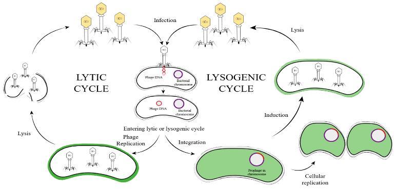
An example of a bacteriophage known to follow the lysogenic cycle and the lytic cycle is the phage lambda of E. coli.
The continuous presence of the prophage in the lysogenic bacterium gives it certain characters:
1- It will become immune to infection by another phage.
2- It may acquire new properties, such as production of exotoxin in diphtheria bacilli, Cl. botulinum, Strept. pyogenes erythrogenic toxin and Staph. aureus enterotoxin.
If the organism loses the prophage, it will become non toxigenic.
Acquisition of new character coded for by the prophage is called (lysogenic conversion).
Outcome of the temperate cycle
1 2
The cell may continue carrying the prophage indefinitely passing it to daughter cells

* * * * *
2- The prophage may be induced to separate from the bacterial chromosome 9by UV, cold, or alkylating agents) and start a lytic cycle. 31
Rarely an excisional error may occur during separation of the prophage from the bacterial chromosome. In this error the prophge is induced carrying with it part of the bacterial chromosaome. When the phage infects another bacterium it will transmit to it anew character, this is called specialized transduction.
Practical uses of bacteriophage: Bacteriophage
Phage therapy
Therapeutic uses of bacteriophages to treat bacterial infections in animals, plants and humans are under trials. When used to treat humans the term biocontrol is used. They are used to treat bacterial infections that don’t respond to conventional antibiotics mainly those in which bacterial biofilms are involved. Phages are more specific in their action on bacteria than many drugs, and are expected to have minimal or no side effects.
Phage typing
Since bacteria differ in their susceptibility to different phages, phages are used to identify and type bacteria according to the pattern of lysis.


This is very useful in epidemiologic tracing of sources of outbreaks such as wound infections and food poisoning.
Cloning vectors:
Bacteriophages are used as cloning vectors in recombinant DNA technology. A fragment of DNA (foreign gene) is carried on the phage DNA.
When the phage infects a bacterial cell its DNA carrying the foreign gene is incorporated into the bacterial chromosome and the gene is replicated in each cell division.
3
32
Bacterial genetics 4
Outlines
✔ Bacterial genetic material
✔ prokaryotic &eukaryotic cells
✔ Plasmids
✔ Transposable genetic elements
• All properties of the bacterial cell (e.g., growth, virulence, pathogenicity, and
• antibiotic resistance) are determined by genetic information contained within its genome.
• This genetic information is inherited from the parents to daughter cells, so they usually have the same properties.
Bacterial chromosome

• It is a single, circular , double stranded DNA , Bacterial chromosome is approximately carry 4000 genes (5 million nucleotide base pairs) and 1mm long

• it is not surrounded by a membrane and tightly packed in bacterial cell an area called nucleoid or nuclear body.
• It carries genes of bacterial growth and multiplication
33
DNA Replication
1- begins with unwinding of the double helix at a defined point (origin of replication).
2-DNA synthesis occurs in both directions around the circular chromosome. This occurs using the specific DNA polymerase and the two parental strands (as templates).


3- The localized area where the DNA is unwind and DNA synthesis is occurring is referred to as Replication fork

4-The DNA polymerase enzyme incorporates the appropriate nucleotides at each position according to the base pairing rule (A-T & G-C).
Protein Synthesis
1-Transcription

Transcription is the process of forming a working copy (mRNA) from the genome using the RNA polymerase enzyme to be used during the routine use of the cell.


2-Translation
Translation is a process by which the genetic code (in the form of mRNA is converted into a sequence of amino acids (protein) using tRNA molecule in the ribosomes.

34
Plasmids
Definition →They are extra-chromosomal double stranded circular DNA which :
-smaller -Dispensable -Single or multiple -Inherited by daughter cell
-Replicate independent of bacterial chromosome
Types of plasmids

• Conjugative(transmissible)
• Non conjugative ( non –transmissible)
1-Conjugative (transmissible) plasmids
Conjugative (transmissible) plasmids are large plasmids that present as one or two copies per bacterial cell.
They carry the genetic informations that are required for the conjugal transfer.
Examples:
→ F factor which promotes conjugation and transfer of the plasmid DNA from one bacterial cell to another.
→ R factors which mediate drug resistance to antibacterial drugs. Genes for multiple drug resistance may be located on the same plasmid.
2-Non-conjugative (non transmissible) plasmids
Non-conjugativeplasmids are small plasmids that lack the ability for their own transfer, but can be transferred by a conjugative plasmid. They present as multiple copies per cell
35
Functions of Plasmids
1- carry the basic genetic information necessary for self-replication and segregation into daughter cells during cell division
2- Large plasmids such as conjugative fertility factor (F) are able to mediate their own transfer from one cell to another by conjugation. They may also mediate the transfer of other plasmids or chromosomal DNA
3- Plasmids can carry additional genetic information responsible for new phenotypic propertie as:
→Antibiotic resistance (R factor).
→Virulence factor, (e.g. neurotoxin of Cl. tetani).


→Production of bacteriocines (e.g. Col factor in E. coli).
→Production of enzymes responsible for metabolizing some substance
4- Plasmids are widely used in laboratory, as a cloning vector to introduce a specific gene into a certain cell.

Bacterial Variations
include:

• Variation in morphological characters (e.g. spores and capsules formation)
bacterial cell (donor, F+ bacteria) makes contact with another bacterial cell (recipient, or F- bacteria), and the plasmid is transferred directly from the donor cell into a recipient cell

• Variation in the cultural properties e.g., colonial variation between the smooth (Sform) and rough (R-form).
• Variation in the metabolic requirement and enzymatic functions.
• Variation in the biological properties, pathogenicity and virulence.
Plasmid Transfer by Conjugation
36
Variations of microorganisms are divided into two groups

1- Phenotypic variations (Non-hereditary variations):
Phenotypic variation occurs in response to environmental changes. The environmental factors lead to suppression, or activation of the gene controlling process which could not take place under the previous environmental condition, the phenotypic variation is reversible, and being dependent on environmental conditions
2- Genotypic (hereditary) variations :
Genotypic variation occurs in response to changes in the genetic structure. This variation is irreversible (i.e., heritable). A change in the bacterial genome may be caused either as a result of mutation in the cell's own DNA, or from the acquisition of additional DNA from an external source [gene transfer].
Definition
any change in the DNA base sequence. It may produce no observable effect on structure or function of the encoded protein. In few cases, it results in effective changes (altered enzyme action, or a nonfunctional protein may be produced).

Types
Point Mutation
Multi-site Mutation (Null mutation)


Mutation
37
Point Mutation:
A) Substitution of one nucleotide by another
B) Frame shift mutation
Multi-site Mutation (Null mutation):


It includes extensive chromosomal rearrangement, multiple inversion, duplication and deletion. It results in change in the chromosomal structure extending too many thousands of bases → resulting in complete destruction of genes' functions.
Causes
1- Spontaneous Mutation

It occurs in nature during replication of DNA, but it is immediately corrected by the repairing mechanism of the polymerase enzyme
2- Induced Mutations:
→ Physical agents: a) Heat b) Ultraviolet light c) Ionizing radiation
→ Chemical agents: a) Nucleotide base analogs (5-bromo-uracil)
b) Ethedium bromide
c) Acridine derivatives
Gene Transfer

The exchange of genetic materials between bacterial cells may occur by one of three mechanisms:
a. Transformation
b. Conjugation
c. Transduction
a. Transformation :
Transformation is the process of uptaking and incorporating a free genetic fragment (exogenous DNA). It occurs in some bacterial species e.g.

38
Pneumococci, H. influenza and certain Bacillus species
b.Conjugation
Conjugation is a process in which one cell (the donor) makes contact with another cell (the recipient), and the DNA is transferred directly from the donor cell into the recipient cell.
Mechanism of Plasmid Transfer
1) Contactstage
Plasmids capable of mediating conjugation, carry genes coding for the production of a protein appendage (the sex pilus) on the surface of the donor cell (F+ cell). The sex pilus attaches to the surface of a recipient cell (F-cell) and hold the two cells together.
2) Mobilization stage
One strand of the circular DNA of fertility factor plasmid is nicked open at a specific site, and its free end passes into the recipient cell.
3)
Replication
The DNA is replicated during the transfer process. The DNA strand (which stay in the donor cell) is replicated, and the transferred single strand is replicated and re-circulized so that each cell receives a copy.

c. transduction
Transduction is the transfer of bacterial DNA between bacterial cells by means of bacteriophage. The transducing phages all contain double stranded DNA. Transduction may be generalized or specialized
a.
Generalized Transduction:
In lytic cycle of bacteiophage replication, a segment of bacterial DNA (chromosome or plasmid) is contained within the phage capsid instead of the phage genome (Error of assembly). The bacterial DNA can be transduced to another bacterial cell on infection with such phage.

39
b. Specialized transduction
In lysogenic cycle of bacteiophage replication, the DNA of latent phage (prophage) can be inserted into the bacterial chromosome, and replicates as a part of it. The lysogenic state is not permanent. After many generations, the phage can revert to its virulent and become excised again from the chromosome. Sometime, the excision is not exact (Error in excision), and the phage may pickup some of the chromosomal DNA adjacent to its insertion site. When such phage infects another cell, it integrates into the bacterial chromosome (at a specific site).
Transposable genetic elements
Transposons are segments of DNA able to move from one position to another in the genome, or from the chromosomal DNA to a plasmid, or the reverse. They carry the genetic information necessary for their own transfer through transposase and integrase enzymes. Transposons play an important role in collecting antibiotic resistant determinants in adjacent genes, and their transfer from a plasmid to a chromosomal location, leading to the development of multiresistant bacterial strains
Types of Transposons:
1) Insertion sequences are the simplest form of transposons (150-1500 base pairs). They carry only the genetic information necessary for their own transfer. They can be detected if their insertion leads to interruption or inactivation of genes, or turn on the expression of adjacent genes.


2) Complex transposons: These forms of transposons are carried by conjugative plasmids. In addition to genes encoding for their transposition, they carry a gene that encode for special characters (e.g., antibiotic resistance or virulence factor).
3) Integrons: These forms carry multiple genes (Gene cassette) that code for antibiotic resistance in bacteria, and spread in antibiotic resistant strains as a block
4) : These forms are present in virulent bacteria and carry multiple genes that code for virulence factors as adhesins, invasins and enzymes especially in gram –ve bacteria (E. coli and H. pylori).
40
Recombinant DNA Technology 5
Outlines
✔ What is Recombinant DNA Technology?
✔ Steps of Recombinant DNA Technology.
✔ Vectors and cloning Vehicles
✔ Diagnostic molecular biology methods (Nucleic acid probes and PCR).
✔ Applications of PCR
Recombinant DNA technology is one of the recent advances in biotechnology, which was developed by two scientists named Boyer and Cohen in 1973.


Is a form of artificial DNA that is created by combining two or more sequences that would not normally occur together through the process of gene splicing .

41
Is a technology which allows DNA to be produced via artificial means. The procedure has been used to change DNA in living organisms and may have even more practical uses in the future.
Recombinant DNA Technology


What is Recombinant DNA Technology?

✓ Recombinant DNA technology is a technology which allows DNA to be produced via artificial means .
✓ The procedure has been used to change DNA in living organisms and may have even more practical uses in the future .
✓ It is an area of medical science that is just beginning to be researched in a concerted effort .
✓ Recombinant DNA technology works by taking DNA from two different sources and combining that DNA into a single molecule. That alone, however, will not do much.
NOTE
Recombinant DNA technology only becomes useful when that artificially created DNA is reproduced. This is known as DNA cloning
The basic procedures of recombinant DNA technology
✓ DNA molecules that are constructed with DNA from different sources are called recombinant DNA molecules
✓ Recombinant DNA molecules are created in nature more often than in the laboratory; For example: every time a bacteria phage or eukaryotic virus infects its host cell and integrates its DNA into the host genome, a recombinant is created.
42
STEP 1
Basic steps are common to most recombinant DNA experiments :
Isolation and purification of DNA
Both vector and target DNA molecules can be prepared by a variety of routine methods, which are not discussed here. In some cases, the target DNA is synthesized in vitro.


STEP 2
Cleavage of DNA at particular sequences
as we will see, cleaving DNA to generate fragments of defined length, or with specific endpoints, is crucial to recombinant DNA technology. The DNA fragment of interest is called insert DNA. In the laboratory, DNA is usually cleaved by treating it with commercially produced nucleases and restriction endonucleases.
STEP 3
A recombinant DNA molecule is usually formed by cleaving the DNA of interest to yield inserts DNA and then ligating the insert DNA to vector DNA (recombinant DNA or chimeric DNA). DNA fragments are typically joined using DNA ligase (also commercially produced).
STEP 4
Introduction of recombinant DNA into compatible host cells
In order to be propagated, the recombinant DNA molecule (insert DNA joined to vector DNA) must be introduced into a compatible host cell where it can replicate. The direct uptake of foreign DNA by a host cell is called genetic transformation (or transformation). Recombinant DNA can also be packaged into virus particles and transferred to host cells by transfection.
43
STEP 5
Replication and expression of recombinant DNA in host cells: Cloning vectors allow insert DNA to be replicated and, in some cases, expressed in a host cell. The ability to clone and express DNA efficiently depends on the choice of appropriate vectors and hosts.
Vectors - Cloning Vehicles
• Cloning vectors can be plasmids, bacteriophage, viruses, or even small artificial chromosomes.
• Most vectors contain sequences that allow them to be replicated autonomously within a compatible host cell, whereas a minority carries sequences that facilitate integration into the host genome.
1. Plasmid vectors.

2. Bacteriophage vectors.

3. Virus vectors.
4. Shuttle Vectors (can replicate in either prokaryotic or eukaryotic cells).
5. Yeast Artificial Chromosomes as vectors.
• All cloning vectors have in common at least one unique cloning site, a sequence that can be cut by a restriction endonuclease to allow site-specific insertion of foreign DNA.
• The most useful vectors have several restriction sites grouped together in a multiple cloning site (MCS) called a polylinker.
44
Criteria of a good cloning vector:
1. Be as small as possible.
2. Be well characterized regarding: gene location and restriction endonuclease cleavage site.
3. Be capable of autonomous replication within the host.
4. Possess non-essential regions within which the target DNA can be inserted.
5. Carry a selectable marker (antibiotic resistance gene) so that cells transformed by the vector can be distinguished from non-transformed cells
6. Contain single cleavage site for restriction endonuclease.

7. Display limited host range in order to reduce the biohazards associated with the recombinant molecule.

Plasmids are circular, double-stranded DNA (dsDNA) molecules that are separate from a cell’s chromosomal DNA.

These extra chromosomal DNAs, which occur naturally in bacteria and in lower eukaryotic cells (e.g., yeast), exist in a parasitic or symbiotic relationship with their host cell.
Plasmids can replicate autonomously within a host, and they frequently carry genes conferring resistance to antibiotics such as tetracycline, ampicillin, or kanamycin.
The expression of these marker genes can be used to distinguish between host cells that carry the vectors and those that do not.
45
Plasmid Vectors
Restriction Enzymes
Restriction enzymes: are endonucleases produced by bacteria that typically recognize specific 4 to 8bp sequences, called restriction sites, and then cleave both DNA strands at this site

Restriction sites:
commonly are short palindromic sequences; that is, the restriction-site sequence is the same on each DNA strand when read in the 5′ → 3′ direction .

✓ Each enzyme recognizes and cleaves a specific double-stranded DNA sequence that is 4–7 bp long.
✓ These DNA cuts result in blunt ends or overlapping (sticky) ends, depending on the mechanism used by the enzyme .
✓ Sticky ends are particularly useful in constructing hybrid or chimeric DNA molecules.
NOTE
Restriction enzymes are named after the bacterium from which they are isolated: For example, Eco RI is from Escherichia coli, and Bam HI is from Bacillus amyloliquefaciens. The first three letters in the restriction enzyme name consist of the first letter of the genus (E) and the first two letters of the species (co). These may be followed by a strain designation (R) and a roman numeral (I) to indicate the order of discovery (e.g. Eco RI and Eco RII).

46
Identification of Host Cells Containing Recombinant DNA
Once a cloning vector and insert DNA have been joined in vitro, the recombinant DNA molecule can be introduced into a host cell, most often a bacterial cell such as E. coli.
In general, transformation is not a very efficient way of getting DNA into a cell because only a very small percentage of cells take up recombinant DNA.
Consequently, those cells that have been successfully transformed must be distinguished from the vast majority of untransformed cells.
Identification of host cells containing recombinant DNA requires genetic selection or screening or both.
✓ In a selection: cells are grown under conditions in which only transformed cells can survive; all the other cells die.
✓ In a screen: transformed cells have to be individually tested for the presence of the desired recombinant DNA.
✓ Normally, a number of colonies of cells are first selected and then screened for colonies carrying the desired insert
47
