CAMERA SOFTWARE OPERATIONS COOPERATIVE PATIENTS
HOW TO GET A COOPERATIVE PATIENT 1. Explain the process of conducting a retinal screen. This will increase understanding and cooperativeness (i.e. What is being photographed, and why they are being photographed.). 2. Show the patient where to place their head and chin on the retinal camera. 3. Tell the patient to expect bright flashing lights when retina is photographed. 4. Assure patient that the procedure is quick and easy, but very important. In the course of taking images, one may encounter several challenges that interfere with the ability to take good quality images. The challenges and their solutions are divided into two catergories below, Photographic or Camera Challenges: PHOTOGRAPHIC CHALLENGES
CHALLENGE
SOLUTION
Fixation
Instruct patient to focus on camera’s interior fixation point, normally a flashing light.
Cataracts
Dilate patient
Synechia
Dilate patient
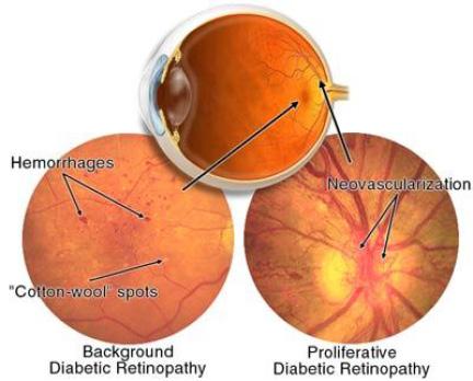
Hemorrhages
Refer patient to an ophthalmologist. Hemorrhages are often a warning sign for a greater complication.
Corneal Dryness
Patient’s eyes quickly go out of focus. Have patient blink a few times and that should resolve the issue.
No natural dilation
Dilate patient
Shadow at the top of image
The camera is positioned too high or patient’s eyelid is encroaching the pupil. Lower the optical head or raise the patient’s eyelid manually andre-photograph.
Shadow at the bottom of image
The camera is positioned too low. Raise the optical head and rephotograph.
Shadow at side
Diopter lens is not switched to the proper position, or when photographing the optic disk at the center of the display, working dots of fundus were not located off toward yellow dots. Make adjustments and re-photograph.
Circular or linear white blur
Eyelash in optical path, instruct patient to open eyes wider and re-photograph.
White reflection at left/right
Optical head too far to the left/right. Make adjustments and rephotograph.
White flare in periphery
Optical head too close to subject eye. Make adjustments and re-photograph.
Out of focus in general
Too much or too little tear on cornea surface. Instruct the patient to blink a few times and re-photograph.
Partially blue
Objective lens is dirty. Clean the lens and re-photograph.
13 / DI A B ET I C R ET I N O PAT H Y TOOLK I T