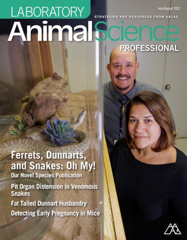
Ferrets, Dunnarts, and Snakes:

Oh My!
Our Novel Species Publication
Pit Organ Distension in Venomous Snakes
Fat Tailed Dunnart Husbandry
Detecting Early Pregnancy in Mice
July/August 2023
Oh My!
Our Novel Species Publication
Pit Organ Distension in Venomous Snakes
Fat Tailed Dunnart Husbandry
Detecting Early Pregnancy in Mice
We are an alliance of proven suppliers to the industry We are spread out across the country and globe to serve our local markets with LabDiet® and other supplies; and in times of troubles...service yours!
• We are positioned to provide feed, bedding, and more like no other supply-chain in the industry


• We have clean, organized, and safe facility environments.
• We are skilled professionals, trained and focused on your research standards.


Solving inappetence in Prairie Rattlers

Holding On
How to keep your employees happy and engaged
When the Working Day Is Done
See how mice "just want to have fun!"

Ferrets: They Eat the Meat
More surprising facts about these cute animals
Keeping Tabs
Using a data capture system to maintain medical records

Green Lab Practices
A conversation with Michael Saad and Tina Woolston
Financial Empowerment
Pushing for technological advancements
On


Our flexible gavage tubes have a soft tip to minimize trauma, and they are disposable to eliminate cross contamination and the hassle of cleaning. Available in a range of sizes for mice, rats and other rodents.


Being the AALAS President brings me great joy, especially when I can participate in the many incredible opportunities our organization offers. We kicked off the summer with a very enthusiastic and educational ILAM experience in AALAS’ hometown of Memphis. With 49 Level 1 and 51 Level 2 students, our week was 100% fun! Our opening speaker, Betsy Charles, got the crowd buzzing, and that energy carried us through the week, all the way to the awards ceremony. Once again, congratulations to our 2023 ILAM graduates and to the 2024 ILAM class president, Mark Snead!
For those of you new to the field, ILAM – the Institute for Laboratory Animal Management – is an educational program developed to provide instruction in management concepts for laboratory animal science. The certifications achieved during ILAM help propel new graduates to take on management roles, continuing to make an impact in the field. Registration is not until December, but I highly encourage you to research this opportunity and determine if it’s right for you.
Another exciting way to get involved is through the AALAS Foundation. This organization supports educational outreach on the essential role of responsible laboratory animal care and use in science to advance human and animal health. The newest addition, the Ann Turner CARE Academy, equips people in our community with the skills and resources to educate the public on laboratory animal science.
By the time you read this, we will have elected our new Vice President-Elect and several trustee seats. I’m excited to have the opportunity to work with the new board, their commitment to AALAS is unparalleled!
As we look to the remainder of 2023, it will come as no surprise that our biggest event of the year - the National Meeting - will take place Oct. 22-26 in Salt Lake City. If you’ve never attended a National Meeting, I welcome you to join me in the mountains of Salt Lake City for the 74th annual event!
The National AALAS Program Committee led by Jenny Wood has completed the education program, and it’s packed with all of the expansive training, education, and information sharing that has made the AALAS National meeting the preeminent event for our profession for the last 74 years. In addition, Perry Spires and the EAC have coordinated a wonderful list of vendors for our exhibit hall, where you’ll learn all about the latest products and services our field has to offer. This year we have also added a new program called Exhibitor Teach & Chat (ETC) where vendors will host 20-minute sessions about their products and services. The response from exhibiting companies has been overwhelming, and we hope you find time to take part in the sessions during your time in Salt Lake City!
There are so many opportunities to plug in and get involved with our amazing organization, and I encourage you to do some digging, figure out what’s best for you, and get started!
Staff Publisher Thomas L. Joseph
Associate Publisher Chris Lyons
Managing Editor Matt Coffey
Associate Editor Morgan McCloud
Ad Sales Heather Lampi

Design/Production Zara Garza
Editorial Advisory Board
Leslie Birke Louisiana State Univ
Andrew Burich Benaroya Research Institute
Bob Dauchy Tulane Univ School of Medicine
David DeOrnellis Champions Oncology
Penny Devlin Pennsylvania State Univ College of Med
Sonia Doss Duke Univ Medical Center
Kelly Ethun Emory University
Glenn Jackson Cornell University
Richard Marble Alpha Genesis Inc
Elizabeth Nunamaker Charles River Laboratories
Sara Oglesby Abbvie
Jane Olin Edwards Life Sciences
Karuna Patil Seattle Children's Research Institute
Amy Pierce Tulane Univ School of Medicine
Stacy Pritt UT Southwestern Medical Center
Robin Tucker Georgetown Univ
Mission Statement
Laboratory Animal Science Professional (LAS Pro) is the official magazine for American Association for Laboratory Animal Science members. LAS Pro provides a wide range of useful resources and knowledge to the association’s 15,000 laboratory animal science professionals who are involved in advancing responsible laboratory animal care and use to benefit people and animals. All signed articles, including, committee reports, news, and commentary, reflect the individual views of the authors and are not official views of AALAS.
Authorization to photocopy portions for personal or internal use is granted by the American Association for Laboratory Animal Science. Photocopying for purposes of resale or outside distribution is prohibited unless written approval is obtained from the AALAS Director of Communications.

Copyright 2023 by the American Association for Laboratory Animal Science.
Laboratory Animal Science Professional (USPS 010-730) is published bimonthly by the American Association for Laboratory Animal Science, 9190 Crestwyn Hills Drive, Memphis, TN 38125. Periodicals Postage paid at Memphis, TN 38101 and additional mailing offices.

POSTMASTER: Send address changes to AALAS, 9190 Crestwyn Hills Drive, Memphis, TN 38125-8538.
American Association for Laboratory Animal Science 9190 Crestwyn Hills Drive Memphis, TN 38125-8538
Phone: 901-754-8620
Fax: 901-753-0046
E-mail: info@aalas.org
Web: www.aalas.org
The first ever Ann Turner CARE Academy meeting took place at our office in Memphis! Participants heard from Larry Shelton and Laura Breese on ways to speak confidently to the general public about the important work happening in the laboratory animal science community.


ILAM took place in May here in Memphis, Tennessee. There was a lot of learning and plenty of laughs! Congratulations to 2024 Class President, Mark Snead!


The Association of Primate Veterinarians (APV) visited our office to hold their annual meeting. While here, they collaborated on setting the organization's goals for next year.


Shayna Burg obtained her LAT recently! “Achieving my LAT means having confidence in my research position while knowing that I'm providing the most humane care to the animals that have helped millions.”


Ica Hyatt earned her LAT as well! “Earning my LAT certification is such an awesome accomplishment for me. I love learning about the best ways to care for animals in research and their great sacrifices for science. I pride myself in advocating for animals and their welfare to ensure their days spent in lab care, whether it’s short-term or long-term, are the best days of their lives. This certification provides me with the knowledge to do that and to keep improving. Onto the LATg for me!”
Zoe Gilchrest obtained her LAT! “As a newly certified Laboratory Animal Technician, I am excited to utilize my knowledge and skills to my daily work at Pfizer. Acquiring my LAT certification has made me more confident within my role in Comparative Medicine. Soon, I will begin to prep from my LATG exam! Lastly, I look forward to furthering my Laboratory Animal knowledge every day through hands-on experiences and reading.”

Hello, my name is Matt Coffey and I have worked in the public/media relations arena for almost 20 years. I have held positions as a magazine editor, public relations manager, marketing manager and have even held positions in newspapers and television. My previous positions also include nearly 10 years in the Communications department at a nonprofit organization, so I am very familiar with the nonprofit world. I grew up outside Louisville, KY, and went to college at the University of Tennessee, Knoxville (Go Vols!) where I was an English major with a creative writing concentration. Currently, I live in Memphis with my wife, two sons, two dogs and a cat. Should you have any questions or if I can ever be of service, please don’t hesitate to reach out anytime.
I am Amanda Pinto, the new Administrative Assistant for AALAS. I originally attended Indiana University before transferring down to the University of Memphis where I completed my Bachelor of Arts in Sociology. I worked as a service advisor for an autobody shop before joining AALAS in March. My primary role here is to provide support for ACLAM and the various committees within. I also assist with administrative needs for the AALAS Foundation and ASLAP. Outside of work, I enjoy reading, crafting, spending time with my animals (3 cats, 2 dogs), and geeking out with my husband and daughter at Comic Cons.
Nicole Elizabeth Duffee was born in Quincy, Massachusetts, on the 12th of December 1951 to Malcolm and Denise Duffee. She was one of two children. She graduated from Vassar in 1973 and obtained her DVM and PhD from the University of Illinois Urbana-Champaign. Nicole was passionate about continued education in the laboratory animal science field, and one of her most significant accomplishments was obtaining the AALAS (American Association for Laboratory Animal Science) Learning Library with her friend and colleague, Mike Fallon. She was a founding member of LAWTE (Laboratory Animal Welfare Training Exchange) and previously acted as president. Nicole worked at the University of Oregon and then Washington University in St. Louis, MO for ten years as a training coordinator before taking her expertise to AALAS as the Director of Education and Scientific Affairs for twenty-one years. During her career, Nicole received many awards for her work, including the George Collins Education & Training Award at
the 1999 AALAS National Meeting. An avid animal lover, Nicole owned many pets throughout her life, including cats, dogs, and rats.
Nicole passed away from pneumonia complications on June 11, 2023, in Memphis, Tennessee at the age of 71. She is preceded in death by her father, Malcolm, her brother, Glenn Duffee, and her daughter, Catherine “Katya” Duffee. She is survived by her mother, Denise, her nieces Lauren (Blair) Gisel, Regan (Dan) Levine, and a host of many friends and loved ones.

In lieu of flowers, please consider donating to the following organizations: The AALAS Foundation: https://www.aalasfoundation. org/
AALAS Grants for Laboratory Animal Science https:// www.aalas.org/glas
Youth Villages: https://youthvillages.org/ Bethany Christian Services: https://bethany.org/


Best binge-watching TV series?
I don’t watch TV, but if I need something, my go to is The Office.
What are your favorite hobbies?
Anything outside and on in or around the water!
Where is your dream vacation spot?
Greece!
What is your favorite dessert?
Pineapple upside down cake. Yum!
Facility/Employer: Tecniplast USA
Job Title: Housing Business Manager
How did you get in this field? I was a zookeeper and animal trainer. I saw an opening for a technician caring for animals being used in research and thought I’d check it out for a year or so. Been here 15 years!
Who were your mentors? Ooooh, so many to mention but narrowing to two most notable would be my former director and now dear friend, Dr. Sylvia Gografe for believing in me and being the best example of a strong leader a girl could ask for. Next would be Lisa Secrest, for always encouraging me outside of my comfort zone and putting me in the game!
What are your current interests in animal science? I love this industry. My main passion has always been teaching and training. To see that “aha” moment on someone’s face anytime I have the chance to connect the why with the what is very exciting to me.

Where do you see yourself in 5 years? Right where I’m at! I love my job! I’m very fortunate to have acquired a role that really feels designed exactly for me and what I enjoy doing. While there are crazy days, every day is awesome!
What is your favorite part of your job? Being a part of an innovative company means you get to be the first to see some really cool, new things. My role goes a step further, and I actually get to be a part of the discussion and development of those advancements/ new products, and that aspect is so fun and exciting to me!
What advice do you have for others just beginning their animal science career? Do all the things. The trainings, the certifications, the conferences. Participate in your local branch things, join the board! Talk to your vendors –
some of them used to be you, and they have the best advice! This can be your forever home and a very fulfilling and fruitful career!
What is the most rewarding aspect of your career? Being a small part of the bigger picture. No one on earth is untouched by the discoveries which have been made and are being made in Biomedical Research. And we helped! That is so amazing!
What is something unexpectedly interesting about your career? Going into all the facilities I’ve been in. It’s well over a hundred by now. So interesting how we are all different, yet eerily the same!
What companion animals do you have? If you have none, then what kind of pet would you like to have? I have the most awesome, bestest boy, Maverick. My 4-year-old Dalmatian.


Laboratory animal veterinary care units may have unique clinical responsibility for university-managed collections. The Integrative Biology (IB) Department at Oklahoma State University (OSU) has a collection of 7 native venomous snake species, all of which are pit vipers, family Viperidae, subfamily Crotalinae. This subfamily is distinguished by bilateral pit organs between the eyes and nostrils that enable infrared detection of prey. The snakes are part of an IACUC-approved educational display protocol to enhance public awareness of demographic information and characteristics of venomous species. Though OSU’s College of Veterinary Medicine has a Zoo, Exotics, and Wildlife division, the Veterinary Medical Teaching Hospital has a policy against providing care for venomous species. Clinical responsibility for the collection, therefore, falls to the Animal Resources (AR) unit under the direction of the University Attending Veterinarian.

The snakes are individually housed in acrylic aquaria within a wood and glass exhibition case on the ground floor of the IB Department. Heat and light are provided by overhead incandescent fixtures set to a natural diel cycle. Feeding of thawed mice occurs every two weeks during summer months and less frequently during winter when snake metabolism naturally slows. Each snake has a large dish with clean water; sand, bark, rocks, and eco-earth substrate are provided on a species-appropriate basis. Snakes do not urinate, but excrete pasty uric acid mixed in fecal pellets. Because of the minimal waste, in addition to the goal of minimizing handling of venomous snakes, cage cleaning is therefore provided once defecation occurs, usually one week after feeding. The departmental Venomous Snake Safety Committee maintains a list of authorized users who are specially trained to catch, handle, and transport snakes. All hands-on procedures subsequently described were facilitated through coordination with one of the authorized users.
An approximately eight years old, 650g Prairie Rattlesnake (Crotalus viridis) from this collection was reported for clinical signs of inappetence and reluctance to strike at offerings during feeding. C. viridis is one of the 5 rattlesnakes native to western Oklahoma; its range includes much of the Plains east of the Rocky Mountains, heralding the nickname The Great Plains Snake. Its venom is hemotoxic with neurotoxic components. Safe handling is the utmost consideration when developing a clinical care plan for venomous species. The IB Department maintains Standard Operating Procedures for training, husbandry, handling, and snakebites. At a minimum, individuals must work in teams of two. The physical exam is aided by snake hooks and clear restraint tubes appropriate for the circumference of the snake (Image 1). The snake is removed from its enclosure with the snake hook and placed into a secondary container or a secured corner of a room. If transport is necessary, both a snake bag and a secondary sealed container are utilized. Upon removal, it is then directed with the hook to crawl up into the acrylic tube. Once the cranial 2/3 of the snake is safely restrained, its caudal portion can be held where it enters the tube.
Once restraint of the Prairie Rattlesnake was achieved for examination, left facial swelling was noted between the pit organ and left eye. Additionally, a space-occupying lesion was noted within the outer cavity of the pit organ, including slight protrusion precluding complete visualization. Involvement of the left eye, including progressive ocular opacity visible on cage side examination, was also noted during the monitoring phase over the course of approximately one week. Expert consultation via discussion with Dr. Dale DeNardo, Attending Veterinarian at Arizona State University, resulted in various surgical correction

plans as well as a post-operative treatment plan. Upon containment via a clear acrylic restraint tube, the snake was induced with 5% isoflurane delivered via vaporizer utilizing a nose cone on the rostral end of the restraint tube. Lengthy induction duration was necessary and expected given species-specific metabolism as well as breath-holding with the introduction of unfamiliar scents. Depth of anesthesia was confirmed first via visual cues, including lack of movement, muscular relaxation, and lack of tongue-flick. Before more invasive manipulation, additional assessment of complete anesthesia was confirmed via lack of response on a tail pinch. Resumption of tongue-flick is often the first indicator of recovery and was monitored closely during manipulation. During anesthesia and manipulation, the snake was maintained on a stainless-steel surgical table without any supplemental heat to maintain slowed metabolism and encourage sustained depth of anesthesia. Supplemental administration of inhalant anesthesia was also provided via nose cone intermittently throughout the procedure.
Once fully anesthetized, the snake was partially removed from the restraint tube onto a sterile field, exposing the head and a portion of the rostral quarter of the body to facilitate access to the areas of interest (Image 2). The pit organ and surrounding area were sterilely prepped using alternating circular scrubs of dilute chlorhexidine and sterile water. Close examination revealed a fluid-filled sac occupying most of the pit organ’s external opening, including protrusion externally beyond the lip of the pit. Blunt dissection was performed to drain purulent material from the sac, followed by excision of the sac from the pit organ opening. Dilute chlorhexidine scrub was used for flushing the pit organ several times following removal. A minimally invasive approach without skin incision was utilized successfully to completely remove the purulent material and associated sac. Before recovery, the snake received enrofloxacin (22.7mg/mL with a 1:9 sterile saline dilution; 10mg/kg IM) given in the rostral third of the epaxial muscles. Intramuscular placement in the rostral third is important, as reptiles have a renal portal system that pumps blood from the caudal half of the body through the kidney before returning to the heart and general circulation. Providing diluted enrofloxacin offsets pain associated with IM injection and the development of sterile abscesses. Post-operative enrofloxacin was continued every
48 hours for a total of 5 doses alternating sides of the injection each time. Though there is limited literature on the pharmacokinetics of antimicrobials, every other day dosing is standard practice due to the snake’s relatively slow metabolism.
Post-procedural recovery was facilitated by utilizing a supplemental heat source to stimulate metabolism. The snake was maintained in a substrate-free enclosure for 24 hours following the procedure to prevent any immediate issues from substrate introduction into the pit organ. The snake was then returned to its home enclosure and maintained normally during the recovery period. Given the findings on the anesthetized exam, we suspected that the left eye opacity was likely purulent material that entered the subspectacular space through the nasolacrimal duct. Approximately 3 weeks following the procedure, the snake underwent a full shed, after which the previous local inflammation and left eye opacity were noted to be completely resolved.
Approximately 7 months after surgical correction, a slight increase in pit organ distension was observed by the clinical team. Caretakers reported no changes in appetite, activity, or body condition; since minimal handling is a central tenant of venomous snake care and performance factors were consistent, the animal was monitored via cage side checks by AR on a weekly-to-biweekly schedule. The left spectacle intermittently appeared opaque, but such physical changes can be associated with the buildup of lymph-like fluid before a shed. Fourteen months after surgical correction, it was determined that the pit distension had suddenly increased (Image 3), internal swelling was apparent (Image 4), and the snake was exhibiting signs of discomfort, such as rubbing the left facial region on rocks within its enclosure (Image 5). A course of medical management, including enrofloxacin dosed as above and meloxicam (5mg/mL; 0.3mg/kg IM for 3 days), was initiated. Upon first treatment, the snake bit the handling tube and released purulent discharge (Image 6) from the pit organ.

Swelling and discomfort signs subsided during medical management; behavior and appetite returned to normal. The left facial region, however, displayed periocular granulation tissue for several weeks (Image 7). Reptilian wounds are generally slower to heal compared to mammalian counterparts; normal granulation tissue can appear gray, brown, and tan and persist until shedding. A

complete shed 3 months after medical management returned the snake’s appearance to normal (Image 8). This case highlights the patience and conservative approach necessary when working with venomous species; a successful outcome was achieved over a long clinical course through expert consultation, surgical management, medical management, attentive monitoring, and regular communication between collection caretakers and AR’s veterinary team.


Acknowledgments:
Special thanks to Dr. George Brusch IV and Jay Walton for handling assistance, Dr. Dale DeNardo for expert species-specific discussion, Dr. Joshua Place for assistance with medication administration, and fellow veterinary staff members Emily Godollei-Jacob and Katie Shrum-Hammer for clinical monitoring.
Corey Sage, BSAG, RVT, is the Registered Veterinary Technician of Animal Resources at Oklahoma State University in Stillwater, OK.
Mary Walker is the Clinical Veterinarian of Animal Resources at Oklahoma State University in Stillwater, OK.
Asheley Wathen is the University Attending Veterinarian and Director of Animal Resources at Oklahoma State University in Stillwater, OK.

Husbandry staff retention and development are vital to the successful operation of an animal research facility. High levels of employee turnover can negatively affect the morale of the team and service continuity for researchers, as well as increase the workload for the remaining employees. Critical turning points that put additional stress on an animal facility, such as the COVID-19 pandemic, can magnify the situation and further increase the sense of “burnout” amongst remaining staff.
In order to increase the likelihood of employee retention and encourage husbandry staff to pursue internal promotions, our leadership team has focused on seven key points to meet these goals:
1. providing opportunities for continuing education.
2. focusing on effective communication with the staff.
3. ensuring adequate recognition of employee contributions to the team.
4. creating a psychologically safe culture where coaching and mentoring are used to minimize the need for formal disciplinary action.
5. creating a competitive salary.
6. implementing “stay” interviews and,
7. having fun with the team.
Implementing these strategies has resulted in a substantial increase in promotions and decreased resignations, having a significant operational impact.
Historically, our institution has required AALAS certification at the ALAT level for all personnel holding an “Animal Technician” title within a specific timeframe after the hire date. Many of our Animal Technicians have little to no laboratory animal experience when they start their role, so in-house AALAS certification courses and individual tutoring (if needed) were provided to increase the likelihood of passing the exam. However, based on employee exit interview feedback, our institution decided to forego the ALAT certification for this particular job title. In general, we learned that employees felt stressed between learning a new job and having the certification requirement. Certification is now only required at the Senior Technician level or above. If an employee does obtain a certification that is not a requirement for their role, they receive a base pay increase to reward them for their accomplishment.
Another way we promoted staff education and developed a better connection to our institution’s mission was to create a monthly virtual presentation with one of our research staff members. We invited faculty or staff members from various research labs to present their work with animal models and how our team, the Department of Veterinary Medicine & Surgery (DVMS), played a role in the success of their research projects. All our technicians have access to a laptop in their housing rooms, as well as being assigned an iPad for their daily duties, so they were able to utilize the technology in their work areas to view these virtual presentations. We also solicited their feedback on the
researcher or lab they wanted to know more about. Based on those recommendations or suggestions, our leadership staff would invite that person to give one of the next monthly presentations.
Finally, to expand the knowledge of our husbandry leadership team, and help them develop new leadership skills, we implemented a leadership book club. From a list of management/leadership skills that are embraced by our institution, we designated one topic per month to focus on. We are fortunate that our institution provides a lot of free e-books on various leadership topics, so, based on the topic to be discussed that month, we chose several books that covered that topic and then took a vote on which one to read that month. Some months we also viewed short videos, took assessments, read management articles, and discussed the topic.
One of the biggest struggles we encountered with social distancing requirements and less personal interaction was the ability to keep everyone informed of institutional and departmental information and receive feedback from staff about their questions and concerns. To improve communication and psychological safety, our department implemented an electronic comment system called the “Think Tank.” Any employee in the department can anonymously submit a question or concern electronically to the Think Tank for a response from departmental leadership. Based on the topic of the question, it is routed to the appropriate leadership member to address. Questions and answers are then posted for the entire department to review. Anyone in the department may also submit a one-to-five-star rating of how well the question was answered.
Another communication tool that we implemented during the pandemic was a monthly electronic staff newsletter. Our institution implemented restrictions on in-person meetings at the start of the pandemic, so this was a great way to ensure that everyone was able to receive the same information. In the newsletters, we featured updates and announcements, reminders on proper procedures, resources for employees such as benefits information, diversity celebrations, staffto-staff “kudos,” and employee spotlights. Employees who recently obtained certifications or promotions were also congratulated, as well as employees who were celebrating birthdays or work anniversaries that month. Once restrictions were lifted and we could, again, have monthly in-person staff meetings, we shifted the facility reminders and updates to the meeting, and utilized the newsletter as a monthly celebratory tool. The newsletter is now focused on praise, announcements of accomplishments, and hyperlinks to activities, as well as an employee spotlight. The newsletter was so successful that we changed it from a single campus newsletter to a departmental-wide newsletter with institutional and departmental information.
MD Anderson has several institution-wide methods for employee recognition. Any employee can submit an electronic “Awesome Job Award” for a fellow employee who has demonstrated one of our institution’s 5 Core Values (i.e., Caring, Integrity, Discovery, Safety &
Stewardship) or 5 Service Excellence Standards (i.e., Safety, Courtesy, Accountability, Efficiency & Innovation). The recipients are automatically entered into a lottery for prize drawings throughout the year. All recipients receive a certificate they can print and hang in their work area. The employee’s immediate supervisor is also notified of their accomplishment.
Our institution also gives each people leader a certain number of performance reward “points” they can award to their direct reports. These points are then redeemable for gift cards, merchandise, or more by the employee. We also have recognition programs where a leadership staff member (e.g., supervisor) can grant a direct report “Recognition Leave” in appreciation of outstanding work performance.

In addition to these institution-wide initiatives, our husbandry team has implemented a “kudos” and rewards system. Employees can give “kudos” to fellow employees for help or assistance they have shown them above and beyond their assignments. These “kudos” are tallied by our Team Leads. One of the awardees is randomly selected each month as the “Grand Prize” winner, with several other awardees selected as secondary prize winners. Prizes can include snacks, candy, soda, or additional items such as a cup or a notebook.
Based on the results of an employee-wide survey, we noted that psychological safety declined after the onset of the Covid-19 pandemic. Our institution defined psychological safety as the feeling that any employee could bring up a question or concern without fear of rebuke or retaliation or that the employee was given an opportunity to correct their mistakes without fear of automatic disciplinary action. One of the ways the institution combatted this lack of psychological safety was to implement a “Just Culture” algorithm based on James Reason’s
decision tree.3 This algorithm focuses on differentiating willful misconduct, which would result in corrective action, from an honest mistake of an otherwise reliable employee, which can be addressed by coaching. This algorithm is published on our internal website for all employees to see. All institutional leaders were required to review it and follow the recommendations.
Working in the Houston metro area, our staff have many job opportunities due to the number of laboratory animal facilities near our institution. When any of the surrounding institutions raise their salary, some of our employees will apply for a position at that institution due to the higher salary. As a result, our department leadership worked with our HR compensation team to increase salaries for the animal care staff.
Unlike exit interviews, stay interviews are a tool for better understanding why an employee chooses to continue working for an organization and what the management can do to strengthen the employee’s engagement and retention.1 These interviews can uncover issues that lead to turnover and present opportunities to make positive changes. They also show the team that their leadership genuinely cares about them and wants to make the workplace more satisfying for the team. We conduct these interviews annually. The goal is to have a casual tone and ideally not take more than about 30 minutes each. By conducting stay interviews with our team, we were able to uncover what made employees satisfied with their jobs, as well as areas for improvement.
“A fun break can reenergize your employees and ready them for the next concentrated effort.”2
It is common for us to spend more time with our coworkers than with our family, so why not make that time enjoyable? Our department has made it a goal to have fun, team-building activities at least once per quarter where every department member can get together and socialize. Some examples of activities that our team has enjoyed are International Laboratory Animal Technician Week activities (e.g., movie day, field day), a Spring employee appreciation picnic, a Fall/ Halloween costume contest and movie day with snacks, Winter holiday parties with karaoke, and an ugly sweater contest and more.
Based on the employee survey results, the team appreciates all seven key points. However, the most appreciated initiatives were the salary increases and the DVMS Think Tank. Focusing on these critical points has had a significant operational impact on our team. Specifically, our promotions have significantly increased, and our resignations and terminations have decreased.
Most of us will undoubtedly face staffing challenges at some point in our careers. It’s essential to provide an environment for your employees where they feel appreciated and heard. Even if you cannot make monetary changes to salary or benefits, a lot of employees will want to stay with an employer if they truly feel part of the team. It’s essential to keep a “pulse” on your staff and see where, in your power, you can make the working environment better for them.
Adrienne Duran, LVT, BS, MBA, CMAR, is the Associate Director at UT MD Anderson Cancer Center in Houston, TX.
Jennifer Mitchell, VMD, DACLAM, is an Associate Professor at UT MD Anderson Cancer Center in Houston, TX.
1. Finnegan RP. 2015. How to conduct stay interviews. SAM Advanced Management Journal, 80(2), 49.
2. Kaye B, Jordan-Evans S. 2014. Love’em or lose’em: Getting good people to stay. Berrett-Koehler Publishers.
3. Reason J. (2016). Managing the risks of organizational accidents. Routledge.


 By Caroline Krall, Lydia M Hopper, Eric K Hutchinson
Refining laboratory rodent care remains increasingly essential to ensure optimum welfare and improve research validity. Indeed, not only is consideration of refinement techniques universally mandated by regulations covering animal use (e.g. the Animal Welfare Act [7 USC §21312159] and Regulations [9 CFR §1-4], Directive 2010/63/ EU, and the Animals [Scientific Procedures] Act 1986), but additionally a trend for maximizing psychological well-being and cumulative life experiences emerged recently in legislative discussions.4, 8, 10-12, 14 Practical mechanisms that can be implemented by animal facilities to enhance refinement include the use of non-aversive handling, non-invasive alternative procedures, and additional environmental enrichment. Thus, we sought to collate these techniques by developing a protocol that utilizes positive reinforcement training (PRT) whilst in a complex playpen to teach mice a refined task, specifically oral vehicle administration, to serve as an alternative to gavage.
Standard “shoebox” housing is often barren and precludes fulfilling the innate tendencies of rodents; thus, providing intermittent access to a complex playpen offers a practical solution to improve well-being.2, 9 Playpens are composed of a large enclosure containing enrichment items that promote the expression of natural behaviors. Moreover, as the exact contents are customizable, the required materials can be easily found within animal facilities and autoclaved to maintain health and biosecurity standards. For our mouse playpens, we used a polycarbonate static rat cage, a Mouse Igloo (Bio-Serv) for shelter, and the following unique items (Fig. 1):
Item
Deep cellulose or corncob bedding (~10 cm) (Teklad)
Facial tissue (Kleenex) & Nestlet (Ancare)
Hol-ee ball (JW Pet)
Paper tube
Sunflower seeds, chocolate chips, LabDiet
Burrowing
Complex nest building
Climbing
Gnawing
Foraging
PRT involves providing a reward when an animal performs a desired behavior in order to reinforce its expression. Beyond proving a faster, more effective, and human training tool than punishment, PRT upholds significant advantages as it can also be used to desensitize animals to procedures, proffering a sense of autonomy by yielding the ability to make choices, relives boredom, and increases the human-animal bond.3,5 Although most often used with larger lab animal species such as non-human primates or pigs, the principles of PRT can be applied to any animal, even zebrafish. We utilized PRT to habituate mice to each item or step involved in oral administration training and offer the following training tips:
• Understand the individual temperaments.
• Personally tailor the frequency and duration of sessions.
• Only train when an animal makes the choice to engage, and never force it.
• Use highly valuable rewards and cater the reinforcer’s strength to the task’s difficulty. From personal experience, mice prefer Nutella > chocolate chips > peanut butter > seeds.
• Be patient, as stress will impede learning.
We aimed to assess whether performing PRT on mice in a playpen would enable faster acquisition on oral vehicle administration as compared to training in the home cage. All mice were housed in an AAALAC-accredited facility under approval by the Johns Hopkins University Animal Care and Use Committee. In brief, two groups (n=3) of young, male Swiss-Webster and C57BL/6 mice were acclimated to non-aversive handling using the cupping method. We next initiated PRT using 0.1 mL of Nesquik, which was accomplished over the course of a week with daily sessions (Fig. 2). Following, up to ten daily sessions of 5 minutes were conducted whilst cupping the individual in one’s hand by the home cage. The frequency of sessions required for the mouse to consume the entire vehicle was recorded.

Following, the mice were provided with access to a playpen as described above for 2h twice weekly. By cage-side observation, mice preferentially utilized the sunflower seeds, chocolate chips, deep bedding, facial tissue, and Hol-ee ball, likely as these items enabled the expression of behaviors otherwise inhibited by standard housing. Conversely, the paper tube and Nestlet generated minimal use. Given the high use of enrichment and enhanced behavioral repertoire, we were able to infer a positive affect as the playpen likely provided excitement and appeased innate tendencies. After one week of playpen access, we repeated the oral vehicle administration sessions as in the home cage.

We found that all of the mice consumed the entire vehicle when it was conducted within the playpen on the first session, whereas only one mouse successfully learned in the home cage prior to playpen access despite ten attempts (Fig. 3). Thus, its use facilitated both faster and unanimous acquisition of a novel task. Moreover, approximately six months following initial training and without sessions in the interim, when offered the syringe in the playpen, mice successfully consumed, thus indicating the learning remains resistant to extinction.
Training mice on a novel task in a playpen rather than by hand produced an increased success rate and faster learning acquisition. Given the benefit of the playpen, it remains likely that playpen access induced a positive affective bias, which enabled an improved ability to learn, as the impact of a positive emotional state upon neurocognitive processes (e.g. learning and memory retention) is well-known.1, 6-7 Additionally, the accelerated PRT precludes the commonly cited impediment against laboratory implementation for non-invasive techniques, namely time, and may aid in more general adoption by the scientific community. Moreover, the same training technique is adaptable to many common procedures; for example, teaching oral administration is highly useful for transitioning from injectable to oral pre- and post-operative drugs. PRT can desensitize animals, thus decreasing associated stress to ear notching, injections, and restraint. Overall, the utilization of

playpens as a training tool, along with PRT on non-invasive techniques, offers a multifactorial method to refine laboratory animal care and significantly improve their well-being.


Caroline Krall, Lydia M. Hopper, and Eric Hutchison work in the Department of Molecular and Comparative Pathobiology, Johns Hopkins University School of Medicine in Baltimore, MD.
Caroline Krall also works at the Center for Alternatives to Animal Testing, Johns Hopkins University Bloomberg School of Public Health in Baltimore, MD.
1. Alexander R, Aragón OR, Bookwala J, Cherbuin N, Gatt JM, Kahrilas IJ, Kästner N, Lawrence A, Lowe L, Morrison RG, Mueller SC. 2021. The neuroscience of positive emotions and affect: implications for cultivating happiness and wellbeing. Neurosci Biobehav Rev 121: 220-249.
2. Cait J, Cait A, Scott RW, Winder CB, Mason GJ. 2022. Conventional laboratory housing increases morbidity and mortality in research rodents: results of a meta-analysis. BMC Biology 20: 1-22.
3. Coleman K & Maier A. 2010. The use of positive reinforcement training to reduce stereotypic behavior in rhesus macaques. App Anim Behav Sci 124: 142-148.
4. European Parliament, Council of the European Union. 2010. Directive 2010/63/EU of the European Parliament and of the Council of 22 September 2010 on the Protection of Animals Used for Scientific Purposes.
5. Guerrero-Martin SM, Brill S, Carlson BW, Graham ML, Hopper LM, Metcalf Pate K. 2022. Development and implementation of a positive reinforcement training program for nonhuman primates in biomedical research. LAS Pro 10: 48-52.
6. Hinchcliffe JK, Jackson MG, Robinson ES. 2022. The use of ball pits and playpens in laboratory Lister Hooded male rats induces ultrasonic vocalisations indicating a more positive affective state and can reduce the welfare impacts of aversive procedures. Laboratory Animals 56: 370-379.
7. Lagisz M, Zidar J, Nakagawa S, Neville V, Sorato E, Paul ES, Bateson M, Mendl M, Løvlie H. 2020. Optimism, pessimism and judgement bias in animals: a systematic review and meta-analysis. Neurosci Biobehav Rev 118: 3-17.
8. Parliament of the United Kingdom. 2013. Animals [Scientific Procedures] Act 1986 as Revised.
9. Ratuski AS, Makowska J, Dvorak KR, Weary DM. 2021. Using approach latency and anticipatory behavior to assess whether voluntary playpen access is rewarding to laboratory mice. Scientific Reports 11: 18683.
10. RSPCA, LASA, LAVA, IAT. [Internet]. Focus on severity suffering. Cited 23 March 2023. Available at: https://focusonseveresuffering.co.uk/
11. Smith D, Anderson D, Degryse AD, Bol C, Criado A, Ferrara A, Franco NH, Gyertyan I, Orellana JM, Ostergaard G, Varga O. 2018. Classification and reporting of severity experienced by animals used in scientific procedures: FELASA/ECLAM/ESLAV Working Group report. Laboratory Animals 47: 5-57.
12. Stevens C, Hawkins PE, Smulders T, Maclean AI, Lewejohann LA, Jirkof PA, Boxall J, Murphy H, Moody CM, Turner PV, Makowska J. 2021. Report of the 2020 RSPCA/UFAW rodent and rabbit welfare meeting - cumulative experiences. Anim Technol Welfare 20: 21-33.
13. United States Department of Agriculture. 2022. Animal Welfare Act as Amended. 7 USC §2131–2159.
14. United States Department of Agriculture. 2022. Animal Welfare Regulations. 9 CFR §1-4


Ferret anatomy and physiology have some major differences from dogs and cats, the more established carnivore laboratory animals. In addition to the sable and albino breeds, there are a large number of variations, including a pattern with congenital deafness. Behaviors, nutrition, and clinical techniques including restraint, injections, other administrations, and grooming, are all very different than done in many lab species. Preventive health care programs including being sure that the humans working with them are immunized against influenza and COVID.
The domesticated ferret, Mustela putorius furo, is the only domesticated member of the Mustelidae. They are an inbred, manmade species dating back at least 2000 years, where it is thought that the Egyptians derived them from the European polecat, Mustela putorius, to hunt rabbits and rodents. They are all one blood type worldwide, with very little genetic variations despite the different coats and eye colors.
Intact females are jills, intact males are hobs, and baby ferrets are kits. Neutered females are sprites and males are gibs. Males are generally larger than the females. (Figure 1) The lifespan is 5-7 years. They have a very high spontaneous tumor rate. The gut transit time averages 1.75 hours, resulting in a normal soft stool. Ferrets are still illegal in many municipalities and a few states. 1,4,7

The basic ferret coat colors are sable (dark) and albino, which are characterized by their non-pigmented eyes. Coats vary seasonally and can even change colors. Many ferrets become lighter or whiter as they age. The most common coat colors are silver, champagne, mitt (white feet), roan, chocolate, dark-eyed white ferrets (DEW), and cinnamon. Noses can have pigmented patterns or be pink. All blaze and pandas are congenitally deaf (Wardenburg’s syndrome). (Figure 2) Other ferrets can be congenitally deaf as well. 4

The ferret anatomy demonstrates an elongated body with fairly short limbs. (Figure 3) The body is extremely flexible, enabling the ferret to turn in the diameter of its body. This is advantageous when being in a tunnel, flushing out a rabbit. The heart is located almost in the middle of the body. They have a J-shaped os penis, with the urethral opening behind the curve. The male urethra opens on the mid-ventral of the body,
similar to a dog. All ferrets will develop some degree of adrenal disease, beginning with neutering. The adrenal glands take over sex steroid production. Hyperadrenocorticism in ferrets is not Cushing’s disease. The clinical signs of it can be prevented using a hormonal implant, which prevents sex steroid production. Ultrasonography is used to demonstrate adrenal anatomy. The right adrenal glands lie between the abdominal aorta and caudal vena cava and often have a direct vascular connection with the vena cava. The dental formula for adult ferrets is I 3/3, C 1/1, PM 3/3: M1/2. Ferrets often break the tips off of the canine teeth by chewing on cage bars or even during play. The thyroid glands are located cranially in the neck, just distal to the larynx. They are small, straplike shaped organs on either side of the trachea.
The spleen is contractile and enlarges under any sedation or anesthesia. It can take up to 35% of the blood volume and will contract after the medication. The size prior to anesthesia should be noted if radiographs or other imaging is done, as it may not be true splenomegaly. The spleen tends to lose elasticity as the ferret ages. The spleen is the site of extramedullary hematopoiesis as the ferret ages, taking over for the bone marrow, which is gradually replaced by fat.
The pancreas is C-shaped, and flat, adhering to the duodenum and stomach. There are major lymph nodes associated with the lesser curvature of the stomach, the pyloric area, and surrounding the colon, as well as many in the mesentery.
The small intestine is only 5 times the length of the ferret body, compared to a cat’s (8-10X) body length. The entire gut is under vagal and sacral innervation. It is spontaneously active even under anesthesia, with atropine only slightly inhibiting it. The stomach spontaneously produces acids, proteolytic enzymes, and histamine. With the fast transit time, it is only necessary to fast a ferret 2-3 hours prior to surgery. Prior to surgeries, a ferret should be checked for hypoglycemia, as many ferrets develop insulin-secreting tumors. A blood

glucose of less than 60 mg/dl after 2 hours of fasting should be screened for insulinoma and other disease problems. Normal blood glucose ranges from 80-120 mg/dl, depending on when the ferret last had a meal. They can get gallstones, and the entire gut secretes cholecystokinin, and a greenish stool can indicate over secretion of bile, due to fats or irritation in the gut. There is no cecum, no ileocolic junction, and the jejunum and ileum cannot be distinguished grossly. Retroperistalsis (emesis), begins in the colon.
The normal heart rate is 200-400 beats per minute. Blood volume is considered to be 5-7$ of the body weight in kilograms. Blood pressure (systolic) in most non-sedated ferrets ranges from 140-164 mmHg. They do have large anal glands, although many ferrets are demusked at an early age. There is still a “ferret odor” as there are scent glands particularly around their mouths and ears. Keeping the ears clean, changing bedding, and removing feces/urine from litter boxes decreases the musky odors. 1,4,7
An incomplete list of uses includes:
• Emetic model for drug testing
• Animal model for influenza, SARS-COV-19 and other coronaviruses
• Neuroendocrine and developmental research
• Neurologic, ophthalmic and special senses research
• Dental structures and pathology
• Reproductive and genetic work
• Training for neonatal intubation
Ferrets sleep over 20 hours a day. They have very active but short periods of play. They like to sleep in pillowcases, hammocks, and sleep sacks, but many are piled on top of each
other. They are extremely social and, if housed separately, should have social contact. Caged ferrets need to be allowed to exercise daily, and commercial “playpens” that work well are available in a laboratory facility. Ferrets tend to defecate and urinate in corners, so litter boxes, papers, puppy pads, etc., should be placed in the corners of a room or cage. Ferrets bound and bounceas they run—they dance around, fall over, vocalize a soft giggling sound, and jump on objects and other ferrets. This is called the “Weasel War Dance” and it is part of play. They will wrestle with other ferrets, shake toys, steal toys, objects and deposit them in hiding places. It is necessary to have an enrichment program in place, that includes toys and daily exercise.6 Ferrets are fearless and very curious and readily explore novel items.
Ferrets have many vocalizations including chuckles, giggles, dooking, and a soft phaser sound as they blow air through their front teeth, usually a sign of irritation. They will squeal when in pain and can groan and scream during nightmares. Deaf ferrets will squeal in a very high pitch while playing because they don’t know they are making sounds.
Biting is not tolerated any more than you would from a dog or cat. Ferrets can be trained not to bite, with rules consistent with how you train a puppy. Ferrets often will lick prior to a nip or chomp. They will latch on, and if you try to manually pull their mouth off, they will clamp down harder. Isopropyl alcohol on cotton swabs or tongue depressor dabbed into the mouth if the ferret is clamped on will usually cause the ferret to release. If a ferret is latched on, immediately scruff the ferret
and hold it up; the natural reflex when scruffed is to yawn. At the same time, establish eye contact, and loudly say, “NO BITE.” If the ferret still isn’t paying attention, once detached, a brief hold of the scruffed ferret to a horizontal surface (like the floor) and repeating “NO BITE” with then immediate release back into its cage for time out works well. Some ferrets need this to happen a few times, but they do get the message you are displeased, and they are not supposed to bite. Have all personnel dealing with that particular ferret trained to do the same discipline. The ferret can also be praised when it is not biting! 2,4,5,6
Lab animal ferret cages are similar to rabbit cages, although the feed box must have a smaller opening or the ferret can escape. Litter boxes if provided should be anchored to a corner. Litter type may depend on the facility or project, but paper-based pellets or wood-stove pellets work well. The key with getting a ferret to use a litter box or specific latrine corner is accessibility. Ferrets have little time to find a suitable defecation spot when the urge hits. They like clean pans or corners, so feces and wet litter should be removed at least daily or even twice daily.
Ferrets are obligate carnivores, and because of the short gut and fast transit time, must have easily digestible food. It should be high in protein and fat, and under four percent fiber. They cannot absorb calories from carbohydrates. They cannot digest fruit, raisins, vegetables although they like sweet tastes. Sugars, fats and oils can stimulate acid reflux. Commercial laboratory kibbles will meet their needs. 3
Ferrets are highly susceptible to human influenza and COVID. It is recommended that staff be vaccinated, and that no staff exhibiting a respiratory illness should work with ferrets, as it may be difficult to tell a cold, influenza and even COVID apart.
Scruffing is the most common restraint. A ferret will go limp and yawn. (Figure 4) Another hold is using your hand like a harness: under the front legs, across the back, and with fingers on either side of the head. (Figure 5 A & B) A cross-body hold similar to that used for rabbits, with the ferret either sternally or on its back, is also comfortable. Some ferrets will need to be wrapped in a towel. Chemical sedation is used for many short procedures, blood draws, and imaging. 1,4,7
Subcutaneous injections including fluids are most easily done in the flank area, where the skin is thinner and looser than the typical scapula area. The ferret’s thickest skin is in that area, so using the flank SC is more appropriate. Intramuscular injec-

tions can be given in the anterior thigh muscle or the epaxial muscles. (Figure 6) Intravenous access includes the cephalic, jugular, and lateral saphenous veins. If repeated intravenous access is needed, consider placing a jugular catheter or cutaneous vascular access port. Intraosseous access can be in the tibia or femur as is done in other small mammals. Oral medications can be done directly, usually at the side of the mouth, or if the ferret is cooperative and likes the taste, directly off a spoon or dish. For intranasal administration, it is done by holding the ferret on its back, with the head secured against your body. (Figure 7) The ferret will sneeze! 1,4,7

Blood draws are easily done via the sternal notch site, under the sternum, into the cranial vena cava. Other sites include the tail vein, cephalic, saphenous and jugular. Catheters are usually placed in the cephalic veins. Ferrets normally do not bother bandages.4,7


The more often you bathe a ferret, the more oils will be secreted, and the more it will have an odor. If you do bathe a ferret, use a shampoo designed for ferrets, as it contains more oils than dog or cat shampoos. Normal ferret earwax is dark brown to black – the color does not signify the presence of ear mites. You can swab the small outer ear easily with a cotton swab. If the ferret scratches at that ear violently while you are swabbing it, do check for ear mites. The mites are sometimes visible on the swab or are easily identified microscopically. Ferrets do need all 20 nails trimmed regularly. This can be done by one person; put a dab of salmon oil on the stomach and hold the ferret in your lap. It will be busy licking the oil so you can trim all the nails. (Figure 8) There are small cat nail trimmers or even small human clippers that work. The nails are non-pigmented, so it is easy to see the blood vessels.
Ferrets require canine distemper immunization, and many localities require rabies vaccination. They should also have dental cleaning and prophylactic care just like with dogs and cats. Unlike dogs or cats, they frequently have reactions to the vaccines. It is advisable to pretreat with diphenhydramine (preferably by SC injection) 15 minutes or so before a vaccine. It is also advisable to administer the vaccines on different days, so that if the ferret has a reaction, it will be obvious which it
reacted to. The reaction is with histamine, and corticosteroids and epinephrine are not the first medications to be administered. Reactions can start anywhere from 5 minutes to several hours after vaccine administration. Signs include hypersalivation, erythema of nose, skin, feet, bottlebrush tail (piloerection), retching, pawing at the mouth, vomiting which may be hemorrhagic, coughing, sneezing, explosive diarrhea which may be hemorrhagic, and dyspnea.
The vaccine reaction protocol is listed below.4


• I.M. diphenhydramine 5 mg (50 mg/mL; 0.1 mL most ferrets). If under 800 grams, give 0.08 mL
• If retching, vomiting, give 0.1 mL metoclopramide I.M. (5 mg/mL)
• Famotidine 2.5 mg I.M. or S.C. (0.25 mL of 10 mg/mL)
• Oxygen if dyspnea. Watch for vomiting and keep from aspirating
• Crystalloid fluids 10-20 mL plus 1 mL 50% dextrose S.C.
• If reaction is continuing, put an additional 5 mg diphenhydramine in fluid pocket
• If hemorrhage give 0.5 mL vitamin K (half in I.M., half in S.C.)
• If still retching, bleeding, flushing after 10-15 minutes for above treatment to work, give 0.5 – 1 mg I.M. dexamethasone
• If still dyspneic, laryngeal swelling, respiratory problems: give dopram (1-5 mg/mL I.M.), epinephrine (0.02 mg/kg I.M., I.T., I.V., S.C.), aminophylline (5 mg/kg I.M., I.V.) or terbutaline (2.5-5 mg/kg P.O.)
• If necessary to stop reaction: I.V. dexamethasone, diphenhydramine, additional symptomatic medications
• I.V. or Intra-rectal diazepam if seizuring (1-2 mg/kg) 4
With ferrets becoming more commonly used as a carnivore model, the many anatomical, physiological, and behavioral needs in addition to the differences in procedures and techniques, make it necessary for today’s laboratory animal professionals to learn about these unique animals.
Special thanks to the Washington Ferret Rescue and Shelter, Shelter Director Vondelle McLaughlin for assisting with many of the photos and teaching materials and all the ferrets who helped to demonstrate all these nuances of the domestic ferret.
Cathy Johnson-Delaney, DVM is co-founder and Secretary, Board of Directors of the Washington Ferret Rescue and Shelter in Everett, WA and consultant, NW Zoological Supply, Everett, WA.
1. Fox JG, Marini RP, editors. 2014. Biology and diseases of the ferret, 3rd edition. Ames (IA): John Wiley & Sons, Inc.
2. Harris LM. 2015. Ferret wellness management and environmental enrichment.Vet Clin North Am Exot Anim Pract 18:233244.
3. Johnson-Delaney CA. 2014. Ferret nutrition. Vet Clin North Am Exot Anim Pract 17: 449-470.
4. Johnson-Delaney CA, editor. 2017. Ferret medicine and surgery. Boca Raton (FL): CRC Press.
5. Larrat S, Summa N. 2021. Ferret behavior medicine. Vet Clin North Am Exot Anim Pract 24:37-51.
6. Madrea N, Adams D, Offord S, Funk A. 2022. Novel ferret enrichment: Ferrets have a ball. Lab anim sci professional Nov: 40-42
7. Matchett CA, Marr R, Berard FM, Cawthon AG, Swing SP. 2012. The laboratory ferret. A volume in the laboratory animal pocket reference series. Boca Raton (FL): CRC Press.
Our priority has been to ensure our clients accessibility to the highest level of expertise and customer service in the industry. We are dedicated to providing you non-biased high-quality results with fast turnaround times with unmatched customer service.
For a complete range of products and services, request our catalog or view it online at www.vrl.net


As part of a veterinary care program, medical records are critical for documenting and monitoring animal health. Timely relay of accurate information of animal health, behavior, and well-being is needed in order to appropriately address animal concerns. Therefore, a mechanism of direct and frequent communication between those observing the animals and the veterinary staff must be established. The disadvantages of paper-based notification and record systems, the standard in most facilities for many years, are apparent when compared to an electronic system. For example, paper-based notifications placed in designated locations may serve the purpose of notifying veterinary staff of non-urgent concerns but present a time gap between when the issue was observed and when veterinary staff is aware of a concern. In addition, only the staff who see the form (or are directly notified) would be aware of the situation unless further communication occurs. In addition, records filed in medical charts in a centralized location, especially in large facilities, make reviewing an animal’s history during an assessment extremely difficult and time-consuming. These disadvantages can lead to less timely veterinary care and poor communication between and within groups providing care to the animals. To address the myriad of issues of a paper-based system, an electronic system was designed as a replacement in a GLP-compliant contract research organization.
To address the first step in the process, notifying veterinary staff of an animal concern, an electronic “Sick Animal Notification” (SAN) form was made using Microsoft Forms (Figure 1). This form is available on all computers used for data collection and is easily accessible by anyone observing animals. The form captures all of the pertinent animal and study information, along with the observations of the technician submitting the form. When this form is submitted, an email is immediately sent to all members of the veterinary staff and the Study Director. In addition to the email notification, all information submitted in the SAN is automatically uploaded to a Microsoft Lists SharePoint site. This database is accessible by the veterinary staff and allows real-time visualization of all active cases on-site. This allows determination of which animals need to be seen and tracked and where they are in their treatment plan. The site can be filtered by parameters such as species, location within the facility, and study number.
Once the veterinary staff receives a notification, assigned staff members assess the animal. Rather than recording the animal’s assessment on paper, a Veterinary Assessment function was creat-

ed in an Electronic Data Capture System (EDCS), Pristima 7.5. This software is validated for GLP use and was already used for data recording in the facility. The Veterinary Assessment function in Pristima (Figure 2) provides prompts to reported health issues, a veterinary assessment, a treatment plan, and a date to recheck the animal if needed. If further consultation is needed with a veterinarian or Study Director, there are also options declaring that a consultation is required. The same form is filled out for any subsequent assessments of the animal. All information documented in the EDCS for an animal, including the initial observation from the technician, is available in the animal’s “Complete Animal History Report,” which can be generated within seconds. Therefore, all veterinary assessments and treatment plans are in a chosen animal’s history in the EDCS, allowing veterinary staff an electronic history anywhere with a network connection.

The electronic notification and records system has been in place for over one year. Veterinary staff are notified of issues more promptly, and the resulting actions taken are completed more quickly. Though a direct phone call is still required for urgent issues or emergencies, for issues deemed “non-urgent,” veterinary staff can use electronic notification to further assess the urgency of animal concerns in a timelier manner. The notification, along with the SharePoint Lists site, allows veterinary staff to better prioritize their day, resulting in faster care for animals that need it the most. In addition, the SharePoint Lists site has allowed veterinary staff to identify any trends regarding an individual or group of animals and to take actions to improve or prevent these issues. There is much more clarity about how many animals are being reported to veterinary staff and how many active cases are ongoing. This system has also greatly improved the ability to catch any duplicate notifications, decreasing the number of redundant assessments and treatments.
Moving animal veterinary assessments into the EDCS has made the records for all animals more accessible across the entire facility. Veterinary staff now have the ability to look at the whole medical and experimental history of a given animal and see the big picture of the situation. All entries also have a user’s full name associated with the veterinary assessment or observation, improving communication if questions arise about the information recorded. The combination of the notification system along with documenting records in the EDCS has resulted in animals being seen by veterinary staff in a timelier manner and has improved the facility’s data quality for treatments and veterinary monitoring. While the combination of methods is not as efficient as having all these capabilities under one piece of software, the transition to an electronic animal health system has been a net improvement for the facility. Realtime notifications and increased veterinary knowledge of ongoing animal concerns have resulted in overall improvement of animal welfare throughout the entire facility and enhances compliance with FDA and USDA-regulations.
Matt Johnson, RVT is a Senior Veterinary Technician for Altascieces Preclinical Columbia in Auxvasse, MO
Megan M Haney, DVM, PhD, DACLAM is the Director of Veterinary Services for Altasciences Preclinical Columbia in Auxvasse, MO.
Jody Davis, SharePoint Administrator for Altasciences Preclinical Columbia in Auxvasse, MO.
Keith Rettenmaier, BS, is the Pristima Administrator for Altasciences Preclinical Columbia in Auxvasse, MO.
Isaac King, Director of Resource Management for Altasciences Preclinical Columbia in Auxvasse, MO.

Article 3 of the Sustainable Vivarium Series: Laboratory practices to reduce waste and consumption
Michael Saad is a PhD candidate at the Tufts University Center for Cellular Agriculture (TUCCA) studying cultured meat. Like many of our laboratory animal professionals, his work helps replace the use of animals. Michael spoke with Dr. Corinna Beale, a Lab Animal Veterinarian and fellow sustainability advocate and shared his experiences on successful implementation of sustainable lab practices at Tufts University which can be translated and applied to our lab animal facilities. Tina Woolston, sustainability program director for Tufts University, joined the conversation to give additional insight into how the office of sustainability helps create the framework to support sustainable efforts like Michael’s across the University.
CB: I imagine when you arrived at Tufts University 2 years ago, the lab had many established practices and procedures already in place. As you set an intention to implement sustainability into the lab setting, where did you start?
MS: Before I arrived, there was a faculty member who was a huge advocate for sustainability. She single handedly set up a Styrofoam recycling program, but she wasn’t able to continue to expand her efforts on her own, especially because this was solely a passion project and not part of her official job. She and other pioneers who historically used our lab had laid the
groundwork, and my group was interested in building on her foundation.
When I arrived, there were five or six other lab members interested in sustainability, and we joined together to try and tackle a sustainability plan that could be scaled up and maintained. We called ourselves the “green team” and all worked in the same Science and Technology building on campus.
We focused on smaller, easier projects to start. The one that stood out at first was increasing access to recycling. Our lab consumables come in a lot of plastic film, and this plastic film isn’t unique to laboratory settings so there was likely a solu-
tion we could tap into already in place from other industries. We reached out to the Tufts’ Office of Sustainability, and they were able to provide us with bins to collect the plastic. The local grocery stores had free recycling programs for plastic film, and our Office of Sustainability already had a program to drop off this recycling on a regular basis. An easy solution to implementing this in our lab was to start putting out bins specifically for plastic film that could be added into the existing recycling program. The green team members volunteered to move the small plastic film recycling bins from the labs to the collection locations in our building.
CB: That makes a lot of sense, I never thought of tapping into local grocery store drop offs but using an existing recycling programs vs. trying to create your own from scratch makes a lot of sense. Tina, can you tell us more about how this program works?
TW: The office of sustainability reached out to the local grocery store who already had a plastic film recycling program. We asked if we could participate as a university. They were very open to the idea. Since the program was already in place it wasn’t much additional work on their part. Since we’re dropping off larger quantities, we call ahead and arrange a time to bring larger consolidated bags of plastic film directly to the loading dock and there is someone there to meet us when we arrive to make it easier on everyone.
CB: Michael, what are some other low hanging fruit (or should I say easy to culture sustainable meat product) practices that your “green team” implemented?
MS: Compost was another relatively easy practice that we implemented. We put compost collection bins in the offices, kitchens, and bathrooms (for paper towels). Again, this compost program was already in place at the University, so we just had to add the bins to participate and schedule lab members to regularly drop them off at the collection location behind our building. Reaching out to the office of sustainability helped us identify the existing programs to implement our own recycling as efficiently as possible. Additionally, a representative from the sustainability office came and did a recycling training with our lab members.
CB: That’s great that they were a helpful resource. I’ve heard that if you put anything that even looks like medical waste in a regular recycling bin, they will throw out the whole bin, is this true?
MS: Yes, this is true, and recycling programs vary depending on local infrastructure and programs so it’s important to learn the specific rules for your area before implementing anything. Most plastics are numbered, and this can be a helpful way to know specifically for your area what you can and cannot recycle. Our university already had a program to recycle lab plastics, like pipette tip boxes and media bottles. The Tufts Office of Sustainability sent our green team signs and best practices
for the program, which was helpful because, for example, the media bottles need to be triple rinsed.
CB: Tina, who do we work with for the lab waste recycling program? I imagine grocery stores aren’t able to offer free recycling for media bottles.
TW: At Tufts University we use Green Labs Recycling© (https://greenlabsrecycling.com/). This is a local group in Massachusetts that provides recycling services for lab products. The first green labs employee was actually a student from our Tufts Sustainability office. It just highlights the benefit of teaching students about sustainability as part of their education. You never know what you will inspire and how far they will take that knowledge after graduation. They often exceed our best expectations.
For pipet and media bottle recycling at Tufts University, we pay Green Labs Recycling© to pick up and recycle specific lab products (pipette tip boxes and media containers). People anywhere in the US can use https://search.earth911.com/ to look up where they can recycle different items. In MA use https:// recyclesmartma.org. It’s important to mention, occasionally we get people with good intentions trying to bring lab product plastic home to recycle it in their own trash. If the recycling facility receives lab products in your household recycling bin they could FAIL the entire residential load, along with all of your neighbor’s good recycling.
CB: Michael, what have been some of the positive and negative impacts of going “green” that we can anticipate as we implement similar practices into our lab animal programs?
MS: Our efforts to implement sustainable practices really gave us a sense of community. It’s really nice to share practices with other people passionate about being sustainable.
There were few negative impacts. Everyone adapted positively. People sometimes put things in the wrong bin, but they were receptive to feedback. We would remind people at staff meetings, or send out department emails. It probably helped that we were a smaller group so all the emails and reminders were directly relevant to the people we were addressing. At this point, about a year and a half into implementation, we don’t have much mis-placed recycling.
Successfully implementing a few recycling programs gave us the encouragement and momentum to scale up our efforts. We started using glass media bottles whenever possible. Instead of throwing out single-use plastic ones in the bio-waste bins, we re-use the glass ones by autoclaving them. We received a grant from Tufts Green Fund (go.tufts.edu/ greenfund), and our engineering department provided matching funds, to purchase a smaller, more efficient autoclave. For small autoclave needs, the new autoclave diverted runs from the large, bulky one we use primarily for bio-waste sterilization. Quantifiable impacts really hammer home the positive impacts. We just sent in our one-year report on the autoclave, and it was cool to see the cost impact from reduced energy and water consumption (Table 1).
CB: In the past, some of the efforts lead by others have faded over time as students and staff rotate out. How maintainable do you think the sustainable practices you’ve implemented are and how will they stand up to the test of time?
MS: Our green team meets once every 1-3 months to review logistics and brainstorm additions to our efforts. At one point we wrote up a semesterly newsletter to report our efforts, but this has fallen off recently. As staff rotate out, there will be new students taking over; we’ll see how that transition goes. We have created a culture and a team and are hopeful this will carry and grow the program into the future.
CB: Tina, I imagine there are other similar lab groups at Tufts implementing sustainable practices across our campuses. I’m sure you’ve seen some groups succeed and some groups fail. What are some of the key components to the groups that succeed?
TW: Yes, there are many groups across Tufts and at other local institutions implementing similar efforts. We came up with a summary of some of the practical practices our office has seen (Table 2). Because most of these practices require everyone’s participation to work, buy-in is really important. Letting groups take the lead on what is the easiest practice for them and supporting their efforts works. Prescribing a practice onto a group tends to get less compliance, since they were not
involved in the decision making. I’ve seen groups that create sustainable practice user guides and green lab certification checklists. As Michael mentioned earlier, benchmarks help validate and encourage everyone’s efforts. Newer technology allows tracking of energy, water, and gas expenditure by building or lab space and even offers real time displays of monthly totals for everyone to see. Unfortunately, a lot of these more advanced technologies are expensive to implement and impractical for older buildings or adjoined buildings where utilities and meters overlap or are absent. The International Institute for Sustainable Laboratories has a lot of resources (https://www.i2sl.org/), but you can also just search for “sustainable labs” online and find a lot of resources from other institutions.
TW: Accountability is another key tool in successful implementation of sustainable practices in a laboratory setting. We can build in accountability by including sustainability related tasks into job descriptions so we’re not relying solely on volunteers adding this into their workload. Creating a sustainability plan and having a dedicated manager to report and follow up on results creates accountability on an institutional level. Supervisors and managers should be asking their employees to set sustainable performance goals; this comes from the top down and as long as supervisors check in on progress, it leads to better, more consistent results. Ideally sustainability should be integrated into all departments and offices at an institution,
Shut off lights
Shut lights off when leaving offices/labs/procedure rooms or install automatic sensor on lights. Does not apply to set light cycle animal housing rooms.
Dependent on kW of the light bulbs. Example: a 60 watt light bulb will save 0.06kWh per hour or $0.006. The more lights, and longer they’re off the more savings will accumulate.5
Low or no $
Moderate effort to get full partition
Water aerator Install water aerators into faucets. Reduces the amount of water flowing out per second when the faucet is on. Also saves energy by reducing demand on water heaters. 4
Raise the freezer temperature
If using -80oF freezers, adjust to -70 oF if able to with what you are storing as most things can be stored at -70 oF.
Defrost freezers Defrost freezers on a regular basis and vacuum freezer coils.
Consolidate shipments
Chemical tracker
Place larger orders less often. Ask if vendors will take back packaging. May require identifying additional storage space.
Software program to track chemical use. Create a central supply for commonly used chemicals and supplies (similar to a pharmacy for drug dispensing in hospitals).
Shut fume-hood sash Train all staff to shut the sash on the fume hood
Reduces energy by 30-40%.1
Reduces the amount of energy required to keep freezers cold.3
Decreases packaging waste. If heating and cooling storage space, this should be factored into the sustainable impact.
Reduces chemical waste and energy consumption required to process the waste.
Dependent on hood type. Potential energy savings of > 40%.2 Check with EHS, sometimes the fume hood is the ventilation system for older buildings.
Low $
No additional effort once installed
No $
Moderate effort to determine if all samples can be stored at -70 oF
$ cost of labor only
Moderate effort to assign and schedule regularly
Take back programs often cost $$
Moderate effort to identify and implement programs or consolidate shipments
$-$$$
Significant effort to consolidate chemicals across departments and programs

No $
Moderate effort to get full partition
Share equipment
Identify redundant equipment. Relocate to a shared space. Implement a schedule for shared use.
Reduces direct energy for equipment use and reduces indirect HVAC energy for ongoing equipment support. Software programs like ilab© can significantly reduce efforts required for coordinating scheduled use of shared equipment.
No $
Significant effort to relocate and schedule shared use
Use re-usable
Switch out disposable products when reusable options are available.
Reduces overall waste. Energy and resource expenditure to clean reusable containers should be factored into the sustainable impact.
$-$$
Moderate effort to clean reusable products
Table 2: Sustainable lab practices that translate into a vivarium setting * Cost savings from sustainable practice implementation has not been factored into the relative implementation cost. Long-term savings from sustainable practices can significantly off-set implementation costs and lead to substantial savings.
tions build mandatory sustainable practices into required EHS trainings when new employees are on-boarded.
Students, faculty, and staff are increasingly more concerned about climate change. Climate anxiety is real and one way of dealing with it is empowering people to act in ways that impact their future. Laboratories, including animal facilities, as outlined in the previous article of this series, expend some of the highest amounts of energy and produce some of the largest amount of waste within institutions. We can leverage people that care and help alleviate some of their own climate anxiety by creating a way for them to take action as part of a sustainable practices team.
By taking the time to read this article, you’ve already taken the first step. Think of this as a buffet of options with low to moderate implementation costs and efforts. Here is the recipe:
1. Find out if your institution has a sustainability office or sustainability officer (manager, coordinator, etc.) and reach out to them to find out what’s already been done and what resources are already available.

2. Review your own facility and practices to find the easiest and highest impact implementable changes for your program. Collaborate with your Facilities and EH&S teams.
3. Identify your own “green team” of passionate people to help
4. Take action then track and share your results!
to share your stories of what worked and what failed. I’d love to share them in a future article or post. [ Corinna.Beale@ tufts.edu or www.linkedin.com/in/corinna-beale/ ]
Dr. Corinna Beale, DVM, SRS, DACLAM, is the Director of Preclinical Services for Comparative Medicine and Adjunct Faculty for the Cummings School of Veterinary Medicine and Research Instructor for the School of Medicine at Tufts University Middlesex County, MA.
REFERENCES:
1. “When to Turn Off Your Lights.” US Department of Energy, 1 Jun. 2023, www.energy.gov/energysaver/ when-turn-your-lights#:~:text=Calculating%20Energy%20 Savings&text=Every%20bulb%20has%20a%20watt,will%20be%20saving%200.04%20kWh.
2. “Water Sense Program Guidelines. Version 5.5” Environmental Protection Agency, 1 May 2020, www.epa.gov/ sites/default/files/2017-02/documents/ws-program-guidelines.pdf
3. “Be Good in the Hood.” My Green Labs, 1 Jun. 2023, www.mygreenlab.org/be-good-in-the-hood.html
4. “-70 is the New -80.” My Green Labs, 1 Jun. 2023, www. mygreenlab.org/-70-is-the-new--80.html
5. “Tips for Managing Energy Consumption in the Lab.” My Green Labs, 1 Jun. 2023, www.mygreenlab.org/blog-beaker/tips-for-managing-energy-consumption-in-the-lab


 By Gennifer Caesar, BS, ALAT


In the realm of laboratory animal research, there exists a crucial quest to find faster cures and improve animal welfare. It has been mentioned that sometimes research facilities can find themselves entangled in a web of repetitive work, limited resources, and not many chances to try new things. It is time to embark on a transformative journey, where a well-planned digital transformation (DX) can revolutionize the field, enhance efficiency, reduce animal usage, uplift animal welfare, and prove to be a financially prudent investment.


Imagine a laboratory animal research facility, where manual processes and archaic technologies reign supreme. Picture researchers and technicians, laboring tirelessly, investing valuable time in monotonous tasks, such as data entry and cumbersome paperwork. This monotonous work not only hinders their ability to focus on higher-value activities but also puts a strain on the welfare of the animals in their care. Days turn into weeks, weeks into months, and soon enough, their inefficiencies accumulate into an astronomical loss of productivity. However, a brave decision to embark on a transformative path holds the key to unlocking a brighter future.
By embracing automation and digital tools, the workforce can liberate themselves from mundane tasks and redirect their energy towards crucial research activities. For instance, implementing sophisticated data capture systems that directly interface with monitoring devices, such as electronic health records, animal monitoring sensors, and automated data analysis algorithms, can save valuable time each day.


Let’s delve into the financial viability of such a transformation. According to a study conducted by Deloitte, laboratories that embrace digital transformation can achieve up to a 30% increase in overall productivity.1 Consider a research facility with 20 technicians, each earning an average of $50,000 an-


nually. If the implementation of digital tools and automation can save each technician just one hour per day (20 technicians* 1hr/day* 365 working days/year), the accumulated time savings would be approximately 7,300 hours per year. With an average labor cost of $30 per hour (including benefits and overhead), this results in potential annual cost savings of $219,000 (7,300 working hours* $30/hr).
Another key cost-saving opportunity during a DX lies in ensuring interoperability and integration between different systems and technologies. Siloed solutions often lead to redundant workflows, data duplication, and increased maintenance costs. By investing in interoperable and integrated platforms, laboratories can streamline their operations, reduce data errors, and eliminate duplicate efforts, ultimately resulting in significant cost savings. A study published in the Harvard Business Review estimates that organizations can save up to 23% of their operating costs by implementing integrated and interoperable systems.3 For a laboratory with an annual budget of $2 million, this translates to potential savings of $460,000.
These freed-up resources can be reinvested in staff training, expanding the facility’s capabilities, or advancing initiatives aimed at improving animal welfare, ultimately propelling the facility closer to their goals of finding faster cures and enhancing animal well-being.
A DX can significantly reduce animal usage and enhance welfare through advanced monitoring systems and predictive analytics. By equipping research facilities with state-of-the-art sensors, artificial intelligence, and machine learning algorithms, it becomes possible to optimize experiments, minimize the number of animals required, and ensure better animal care protocols. This not only contributes to ethical research practices but also presents substantial financial benefits.
For example, consider a facility that spends $500,000 annually on animal procurement, maintenance, and veterinary care. By implementing a DX that reduces animal usage by 20%, the facility can save $100,000 each year.

In addition to enhancing efficiency and reducing animal usage, adopting cloud-based technologies offers remarkable cost-saving opportunities. Embracing cloud computing can significantly reduce upfront infrastructure costs, maintenance expenses, and facilitate seamless collaboration and data sharing.
According to Forbes, organizations can achieve substantial cost savings by migrating to the cloud.2 Let’s consider an example: a laboratory animal research facility spends $100,000 annually on maintaining physical servers, storage systems, and software licenses. By transitioning to cloud-based systems, they can reduce these costs by approximately 30%, resulting in annual savings of $30,000. Over a decade, this amounts to an

animal welfare, enhancing research capabilities, or investing in groundbreaking technologies.
A well-executed digital transformation in laboratory animal research facilities presents a win-win scenario. By freeing staff from repetitive work, reducing animal usage, and prioritizing animal welfare, these facilities can usher in a new era of progress and compassion. The financial viability is undeniable— through cost savings in labor, animal procurement, and care, research facilities can afford to invest in digital transformation technologies and create a positive impact for both humans and animals alike. Embrace the transformative power of digital technology, and let’s revolutionize laboratory animal research for a brighter future—faster cures, reduced animal usage, and enhanced welfare await.
SOURCES:
1. Deloitte. [Internet]. 2019. The digital transformation of clinical research. [Cited 5 June 2023]. Available at: https://www2. deloitte.com/us/en/pages/life-sciences-and-health-care/articles/ life-sciences-research-and-development.html
2. Forbes. [Internet]. 2020. The true cost savings of cloud computing. [Cited 5 June 2023]. Available at: https://www. forbes.com/sites/forbestechcouncil/2022/09/06/cost-savings-for-cloud-infrastructures-in-todays-market/?sh=6768fcf56b77
3. Ross JW, Beath, CM, Quaadgras A. (2013). Beyond the holy grail: achieving operational excellence in information systems. Harvard Business Review. 91(10), 114-121.
In this issue, we felt it was timely to provide a briefing of the changes to working with avian species within a research environment. The intent to include certain birds under USDA-coverage began almost two decades ago and the reality of how to adapt to and account for these regulatory changes has been finalized, as of February 2023.
The author of the column is a sought-after expert in bird health and avian medicine. Dr. Kristin Gardiner has been working with birds since 2010, when she began volunteering at Tri-State Bird Rescue and Research (TSBRR) as a 1st-year veterinary student. Upon completion of her veterinary medical education at Penn Vet in 2014, she completed a one-year Exotic Companion Animal Medical and Surgical Internship at Penn Vet, followed by a 3-year Laboratory Animal Medicine Residency. Dr. Gardiner currently serves on the Board of Directors and as a Relief Veterinarian for TSBRR. She also acts as a Consulting Avian Veterinarian for Columbia University.
Dr. Gardiner has highlighted resources and online recordings of discussions about how and why to include certain groups of birds in oversight by the UADA Animal and Plant Health Inspection Service (APHIS). We hope this will provide a high-level overview (a birds’-eye view!!) of how to approach the regulation of avian species that are not bred for use in research.
As members of the animal research community are likely aware, a series of newly published USDA regulations will affect the acquisition, husbandry, and scientific requirements for certain avian species used in research. Many organizations are wondering how these changes will impact the use of birds in their research programs, and this summary aims to provide insights and helpful resources.
To provide context, the Laboratory Animal Welfare Act (now called the Animal Welfare Act) passed in 1966 and gave the U.S. Secretary of Agriculture authority to enforce standards governing the humane handling, care, treatment, and transportation of certain animals by dealers, research facilities, exhibitors, operators of auction sales, carriers, and intermediate handlers. At the time of its initial passage, the AWA specifically defined the term animal as “live dogs, cats, monkeys (non-human primate mammals), guinea pigs, hamsters, and rabbits.”1
Stacy Pritt, DVM, MS, MBA, CPIA, CHRC, DACAW is the Associate Vice President of Research Support and Regulatory Management and Assistant Professor in Psychiatry at the University of Texas Southwestern Medical Center in Dallas, TX.
 F. Claire Hankenson, DVM, MS, DACLAM, is the Associate Vice Provost for Research and Attending Veterinarian and Executive Director, University Laboratory Animal Resources,

The 2002 Farm Security and Rural Investment Act amended the AWA and expanded this definition of animal to include “any live or dead dog, cat, nonhuman primate, guinea pig, hamster, rabbit, or any other warm-blooded animal, which is being used, or is intended for use for research, teaching, testing, experimentation, or exhibition purposes, or as a pet,” and specifically excluded “(1) birds, rats of the genus Rattus, and mice of the genus Mus, bred for use in research.”2 This amendment to the Act meant that those birds species not bred for research and not otherwise excluded under its provisions were, for the first time, explicitly subject to AWA regulation.
Despite this shift toward AWA inclusion of certain birds, performance standards were
not developed for enforcement of avian regulatory oversight for many years. In 2013, lawsuits were filed by the People for the Ethical Treatment of Animals (PETA), the American Anti-Vivisection Society (subsequently renamed Rise for Animals), and the Avian Welfare Coalition against the USDA for the failure to develop and enforce regulations for birds not bred for use in research. 5
In 2020, as a result of one of these lawsuits, the U.S. Court of Appeals for the D.C. Circuit determined that USDA Animal and Plant Health Inspection Service (APHIS) must issue regulatory standards applicable to birds not bred for use in research per the AWA and then further mandated a set deadline for the issuance of these standards. Listening sessions were conducted by the USDA, and held on September 29, October 7, and October 15, 2020, to obtain input from stakeholders. The first draft of the new regulations (the Proposed Rule) was published in the Federal Register on February 22, 2022. Transcripts of these listening sessions are available online at the following link: https://www.aphis.usda.gov/aphis/ ourfocus/animalwelfare/aw-news/bird-listening-sessions.
Stakeholder comments were collected between February 22 and May 25 of 2022, which were then collated and addressed. On February 21, 2023,
Under the Animal Welfare Act was published, with an effective date of March 23, 2023. According to the Final Rule, current AWA licensees and registrants must comply by August 21, 2023. For those facilities that do not currently have a license or registration but will now be required to maintain one under these new regulations, a new license
or registration must be obtained for compliance by February 21, 2024. The new regulations will be enforceable during USDA inspections on or after the aforementioned dates depending upon the registration or license status of your facility.
The Final Rule made some fundamental changes to the definitions of “bred for use in research” and “farm animal” and added definitions for “bird” and “poultry.” Bred for use in research was expanded to include “an animal that is bred in
captivity and used for research, teaching, testing, or experimentation purposes,” to clarify that the use of the captive-bred birds in a research capacity is what exempts them from USDA coverage, rather than the intended use for the bird at the time of breeding.3 This modified phrasing implies that captive-bred birds purchased from dealers, breeders, and fanciers for use in biomedical research would be exempt from USDA regulation, but wild-caught birds used for research, teaching, testing, or experimentation purposes would be covered and subject to additional oversight.


Wild-caught migratory birds, waterfowl, and their eggs are also regulated under the Migratory Bird Treaty Act (MBTA) by U.S. Fish and Wildlife Services (USFWS). However, the offspring of wild-caught birds will not be subject to AWA regulation, as offspring then fit the definition of being bred and raised in captivity (which excludes them based on the revised definitions above). Observational field studies are not covered under the AWA regulations, but any study that involves an invasive procedure or materially alters the behavior of the study subject would be regulated. Let’s review an example: Dr. Wing works with a colony of wild-caught cowbirds obtained under USFW permitting, canaries purchased from a retail bird vendor, and zebra finches obtained from another research institution – which of these would now be regulated? The cowbirds, based on their wild-caught status, are now subject to USDA regulation. Cowbirds are brood parasites and don’t rear their own young, but if this investigator also had a permit for wild-caught doves, then the captured individuals would be covered by the AWA, but any subsequent offspring generations produced via breeding in-house would not be. The canaries mentioned above would not be USDA-covered because they were purchased from a retail vendor (not a research breeder specifically, but captive-bred, so not subject to USDA coverage). The zebra finches in this case are purpose-bred and, therefore, not covered by the AWA.
The term bird is defined in the Final Rule as “any member of the class Aves, excluding eggs.”3 USDA regulation will not cover any research involving fertilized eggs that have not yet hatched (i.e. fully separated from the eggshell). This is consistent with PHS policy.4 The term poultry was newly defined in the AWA regulations to include the following species: “chickens, turkeys, swans, partridges, guinea fowl, and peafowl; ducks, geese, pigeons, and doves; grouse, pheasants, and quail.”3 Poultry are exempt from AWA regulations when used as farm animals (as food or fiber, or agricultural research for the improvement thereof), or when obtained from commercial production for research, teaching, and education
In terms of husbandry, veterinary care, and surgical requirements, the USDA acknowledges that the use of performance-based standards will generally be recommended. This is due to the vast disparity in the size, flight capability, diet, and spatial constraints for the numerous taxa of birds not bred for use in research. The USDA is applying the same surgical requirements for AWA-covered birds as for rodents, in that a dedicated surgical facility is not required, but aseptic technique must be used for all procedures.3 Operative procedures conducted at field sites must also be performed using aseptic
technique. Facilities and husbandry considerations such as room temperature, humidity, and cage sizing will be part of the full-time veterinarian’s records or must be part of the written program of veterinary care. Cage sizes and specifications must allow each bird to make normal postural and social adjustments and be approved by the Attending Veterinarian (AV); however, room for flight is not a requirement.3

Arranging for an AV for the facility is a requirement under the Program of Veterinary Care (PVC) for USDA-regulated birds, but APHIS acknowledges that a hands-on physical exam is not mandatory for all species and should only be performed at the discretion of the AV. Health monitoring can occur through scheduled visitation and records review. Facilities must develop, document, and follow a species-appropriate plan for environment enhancement adequate to promote the psychological well-being of birds.3 The AV will control the approval of the species-specific environmental enrichment program, with socially dependent birds required to be housed in social groups unless otherwise directed by the AV.
There are many nuances contained within the Final Rule that were beyond the scope of discussion in this summary article. For comprehensive commentary responses from USDA APHIS, see the Standards for Birds Not Bred for Use In Research as published in the Federal Register.3 The modifications to the AWA as they pertain to avian standards can be viewed in 9 CFR 3 Subpart G. Helpful video references for frequently asked questions can be found at the following: https://www. aphis.usda.gov/aphis/ourfocus/animalwelfare/new-bird-rule/ awa-standards-for-birds For additional questions regarding individual research programs, it is encouraged to reach out to Dr. Cody M. Yager, who is listed as the correspondence contact (cody.m.yager@usda.gov).
Kristin Gardiner VMD, MB, DACLAM is a clinical veterinarian and an Assistant Professor of Laboratory Animal Medicine at the University of Pennsylvania.


1. 89 Congress. Public Law No. 89-544 24 August 2016. Available at: https://www.congress.gov/bill/89th-congress/housebill/13881/text
2. 107 Congress. Farm Security and Rural Investment Act (P.L. 107-171). 1 May 2002. Available at: https://www. congress.gov/bill/107th-congress/house-bill/2646?q=%7B%22search%22%3A%5B%22Farm+Security+and+Rural+Investment+Act+of+2002%22%5D%7D&s=7&r=5.
3. Federal Register. (87 FR 9880-9913, Docket No. APHIS-2020-0068). Standards for Birds Not Bred for Use in Research Under the Animal Welfare Act. 21 February 2023. Available at: https://www.federalregister.gov/documents/2023/02/21/2023-03357/standards-for-birds-not-bredfor-use-in-research-under-the-animal-welfare-act
4. Office of Laboratory Animal Welfare. PSH Policy on Humane Care and Use of Laboratory Animals (4a-d). Available at: https:// olaw.nih.gov/faqs#/guidance/faqs?anchor=50288


5. United States Court of Appeals. American Anti-Vivisection Society and Avian Welfare Coalition v. USDA. 10 January 2020. Available at: https://www.cadc.uscourts.gov/internet/ opinions.nsf/80846063820C52F6852584EB005413E4/%24file/19-5015-1823484.pdf.

Neutral ■ Serum-Gel ■ Serum/Clotting Activator ■ EPTA ■ EDTA ■ Lithium-Heparin Citrate ■ Glucose ■ Plasma/Lithium-Heparin ■ Hematology/Potassium EDTA
Microvette® CB 300
0.3ml capillary collection tube and sample container in one unit


Microvette®
100/200 μl Volume
Provides all the benefits of a universally suitable capillary blood collection system

Microvette®
300/500 μl
Blood collection with collection rim
Screw Cap MicroTube, Neutral 0.5ml Screw cap micro tubes
0.5ml with skirted base

Microvette® APT Automated Processing Tubes
Automated sample processing from 250 μl sample volume. Leak-proof cap with pierceable membrane. K2 EDTA preparation for 250-500 μl capillary blood
Multivette® 600 μl
Capillary and venous blood collection in one system
Minivette® POCT
Precise dispensing of small whole blood volumes 20 μl, 30 μl, 50 μl
Prepared Micro Tubes with Attached* Push Cap or Screw Cap Closure


Skirted, conical base and label 1.1ml, 1.3ml, 0.5ml
*0.5ml Push Cap only
Scan to learn more about Prepared Micro Tubes
Request your Braintree Scientific catalog today!


A wide selection of Micro Tubes and preparations for accurate and efcient sample processing.
 By Jessica Powell, RVT;
Ensuring proper husbandry for all research animals is essential for excellent animal welfare. Non-native species, such as the fat-tailed dunnart (Sminthopsis crassicaudata; FTD), are no exception. FTDs are Australian carnivorous nocturnal marsupials, whose diet, environmental needs, and enrichment vary from typical research species. Due to the limited working knowledge on the husbandry and care of this species in a biomedical setting, information was compiled from multiple sources, including Hidden Vale Wildlife Center in Australia, to establish a baseline of husbandry and care for the FTDs in our facility. SOPs and guidance documents include pictures to aid with detailed-oriented tasks, such as food preparation. Utilizing multiple resources, and trial and error, we are now able to accommodate the FTDs physical and psychological needs, while also meeting the needs of the research program. All procedures were approved by Washington University’s (WashU) IACUC prior to initiation, and the program is AAALAC accred-
ited and compliant with all applicable federal regulations regarding animal welfare.
Adult FTDs weigh up to 22g with a total body length of 60-90mm. Their small size, quickness, and elite jumping abilities make FTDs difficult to handle. Due to these factors, the best way to handle FTDs is via manual restraint using the palm of the non-dominant hand, with fingers gently, but firmly, holding the body (Figure 1). Restraint by traditional scruffing is avoided, as it can be stressful for the animal and is technically difficult given their size and flighty nature. Unlike rodent species, such as mice and rats, FTDs should not be restrained or manipulated using their tails, as this can lead to degloving injuries. When possible, cardboard tubes and enrichment devices, such as plastic tunnels (Figure 2), are utilized as a less stressful capture and handling method to assist in cage changes and health checks. The immediate risk to personnel handling FTDs are bite injuries, with most biting out of fear. Luckily, their teeth are tiny
and lack traditional rodent incisors, so nitrile exam glove penetration is minimal.

FTDs are housed by sex with a maximum of five per cage, with the females typically more amendable to social housing. They are housed in the Opti-Rat Plus® caging system, which provides space for enrichment items, including running wheels, sand baths, shelters, and Bio-Serv ® Pyramid Feeders, which they use to express burrowing behavior (Figure 2). Similar to chinchillas, FTDs also participate in sand bathing to keep their fur clean and sleek, and every cage is provided with a sand


bath that is freely accessible. Dunnarts particularly enjoy the running wheel for enrichment, which is thought to mimic the long distances traveled for breeding and hunting, and is also a great way to assess their health.1,2
Wild FTDs are considered insectivores, with most feeding on invertebrates like worms, beetles, and spiders. 1,2 A small percentage of their diet may also consist of other protein sources such as young rodents, small lizards, and frogs.5,6 In captivity, FTDs are fed a range of foods to meet their protein requirements. Their main source of protein is comprised of a meatball containing Grade A raw ground beef, ground cat kibble, and additional vitamins. Depending on their life stage (adult, maternity, weanling), they are fed a specific quantity of meat, with crickets and mealworms as additional sources of enrichment. Other nutritional/enrichment items include scrambled eggs, apples, and husk-on sunflower seeds. Visual aids are used to assist care staff with appropriate portion sizes for the meatballs, apples, and egg pieces (Figure 3). Water provision is somewhat tricky with FTDs as they have shown no interest in using water bottles with sipper tubes. Due to the disinterest in water bottles, and the propensity of sipper tube leakage during cage movement, water bottle usage was discontinued, and open top pyramid dishes now provide daily water ad lib
Breeding FTDs can be challenging, as they have a lengthy weaning period, and the joeys (babies) remain with the dam for up to 72 days. In the wild, FTDs are seasonal breeders, with most breeding occurring during Australia’s summer, between July and February.1 Therefore, to facilitate year round breeding in captivity, FTDs are provided a “false winter,” which enables them to reset their breeding cycle. This “false winter” occurs every six months for three consecutive weeks, and it consists of a decrease in room temperature, a shortened light cycle, and a separation of all breeder pairs. Additionally, to maintain an outbred colony, the Robertson breeding system (Figure 4),3 a random mating scheme adopted from the founder colony, is employed.
Currently, pouch checks, used to identify joeys within the pouch, are the only accurate method to determine pregnancy in FTDs. They begin two weeks after pairing and continue twice weekly until a positive check is noted, or six weeks post-pairing. To pouch check, the dam’s rudimentary pouch is gently everted on her ventral abdomen to visualize any joeys (Figure 5). Pregnant dams are separated to prevent joey cannibalism by sires, as well as dam/sire aggression. Dams are singly housed in maternity cages and provided additional nutritional support and nesting material until weaning. Maternity cage handling is reduced to avoid additional stress on the dam. Due to their small size and hyperactive nature, upon weaning,
joeys are ear notched for identification utilizing a 2mm punch biopsy tool while under isoflurane anesthesia. The husbandry, enrichment, and breeding needs for a novel species such as the FTD can be difficult to navigate. Success of our current practices is evidenced by clinically healthy dunnarts who are successfully breeding, utilizing cage resources, and participating in research, such as behavioral assays. Through observation, documentation, and collaboration, we have managed to create a care program that supports the needs of both the colony and the research group, while also providing the best possible welfare for the FTDs. As we continue to gain knowledge of FTDs, our efforts to care for them evolve, and will hopefully continue to lead to the development of additional enrichment and husbandry practices to advance their welfare.

Acknowledgments: Many thanks to the team at Hidden Vale Wildlife Centre in conjunction with the University of Queensland for helping us in establishing our breeding colony and guidelines for care. A huge amount of gratitude and thanks to the dunnart care team here at Washington University in St. Louis. Without all of you, none of this would be possible.
Jessica Powell, RVT, is the Rodent Health Services Vet Tech Supervisor for the Division of Comparative Medicine at Washington University in St. Louis, MO.
Toi Collins, DVM, DACLAM, is a Clinical Veterinarian for the Division of Comparative Medicine at Washington University in St. Louis, MO.
Tracy Beaty, RVT, is a Veterinary Technician for the Division of Comparative Medicine at Washington University in St. Louis, MO.
Lynda Wilmott, PhD, is a Staff Scientist for Dr. Linda Richard’s lab at Washington University in St. Louis, MO. Nirah Shomer, DVM, PhD, DACLAM, is the Director of the Division of Comparative Medicine at Washington University in St. Louis, MO.
1. Cook L, Pask A. No bones about it, dunnarts crawl before growing a skeleton. Pursuit. 2021 Sep 6. https://pursuit.unimelb.edu.au/articles/no-bones-about-it-dunnarts-crawl-beforegrowing-a-skeleton.
2. Fat-Tailed Dunnart. Animalia.bio. https://animalia.bio/fattailed-dunnart.
3. Robertson A. The effect of non-random mating within inbred lines on the rate of inbreeding. Genetics Research. 1964;5(1):164–167. doi:10.1017/S0016672300001129
4. Suárez R, Paolino A, Kozulin P, Fenlon LR, Morcom LR, Englebright R, O’Hara PJ, Murray PJ, Richards LJ. 2017. Development of body, head and brain features in the Australian fat-tailed dunnart (Sminthopsis crassicaudata; Marsupialia: Dasyuridae); A postnatal model of forebrain formation. de Castro F, editor. PLOS ONE. 12(9):e0184450. doi:https://doi. org/10.1371/journal.pone.0184450.
5. Walker K. [Internet]. 2012. Australasia Society of Zoo Keeping Husbandry Guidelines Fat-Tailed Dunnart Sminthopsis crassicaudata. Available at: https://aszk.org.au/wp-content/uploads/2020/05/Mammals.-Fat-tailed-Dunnart-2012KW.pdf.
6. Warnecke L, Turner JM, Geiser F. Torpor and basking in a small arid zone marsupial. The Science of Nature. 2008;95(1):73–78. http://dx.doi.org/10.1007/s00114-0070293-4. doi:10.1007/s00114-007-0293-4.
Essential online training for investigators, technicians, veterinarians, managers, and IACUC members in the laboratory animal science field.
260+ courses on animal research and compliance – courses customizable
Translatable instantly to 100+ languages – website and courses

Specialty certificate programs on animal biosafety, transgenics, and research support
Training documentation and management
If your organization is an AALAS institutional or commercial member, you save even more! Learn more about AALAS membership: https://www.aalas.org/membership
 By: Camryn Davis; Julie Brown RVT, RALAT; Lee Matthews DVM; and Amanda Darbyshire DVM, DACLAM
Itchy, dry skin…on pigs? Yes! Yucatan minipigs are commonly used in biomedical research and are prone to dry skin (xerosis) accompanied by itchy skin (pruritus). At our institution, minipigs housed by the Purdue Laboratory Animal Program were oiled twice weekly with lanolin to mitigate this condition, however, this resulted in excessive debris along the dorsum sometimes accompanied by exfoliative lesions. We sought an alternative to lanolin that would help our pigs feel comfortable in their own skin! In this study, we evaluated the ability of glycerol and coconut oil to hydrate skin appropriately in comparison with lanolin or no treatment.
Moisturizers are categorized as occlusive, humectant, or emollient. Emollients like lanolin and coconut oil fill the spaces between desquamating corneocytes, also known as skin cells, to create a smooth surface by providing cohesion and flattening the curled edges of corneocytes. Humectants like glycerol pull water from the air and a deeper level of skin to the outer layer. Lanolin is also an occlusive agent that creates a physical barrier on the skin to prevent transepidermal water loss and locks in hydration. We hypothesized that glycerol may prove to hydrate the skin the most appropriately due to its humectant qualities.
For this study, we used 4, six-month-old female Yucatan minipigs that weighed between 23 and 28 kg. The pigs were randomly selected for application of either lanolin (Dr. Adorable Pure Organic Lanolin oil), coconut oil (Now Solutions Liquid Coconut oil), glycerol (Eisen-Golden Laboratories Glycerol 99.7%), or control. Thirty milliliters of each agent were applied twice a week using a 60 mL catheter tip syringe and massaged in with clean gloves. For four weeks each pig received treatment using a cross-over design with a 4-day washout

period. The control was rubbed with clean gloves only. The skin on their head, shoulder, and hips was analyzed prior to application, and 1, 24, and 48 hours after each application by a blinded observer to grade the skin on a 1-3 scale for visual appearance and tactile feel at each timepoint and location. A score of 1 was dry, ashy skin, a

score of 2 was ideally hydrated and supple, and a score of 3 was greasy, shiny, or oily as shown in Figure 1. Finally, skin biopsies were taken from each location on cadavers of a Yucatan and domestic pig used on another study to see if differences were apparent that may account for the dry skin since the pigs are housed in identical environments and dry skin is not seen in domestic pigs.
Results of visual and tactile scores at all time points broken down by location are shown in Figures 2 and 3, with application 1 at baseline and application 2 at 48 hours. Agents were applied on Monday and Wednesday of each week. On the following Monday of the new week scores were recorded to identify the new baseline.
No clinical significant difference was observed between body locations. Results from visual and tactile
scoring 24 and 48 hours after each application when all locations were combined showed that the ideal skin condition was achieved in the following order: glycerol (83.3% visual,75% tactile), coconut oil (79.2%,64.6%), control (33.3%,39.6%), followed by lanolin (29.2%,12.5%) as shown in Figure 4. Figure 5 shows an example of skin from each of the 3 visual scores. The overall trend was that glycerol was the ideal agent as shown in Figures 6 and 7, where green represents a high percent of score 2.
Finally, the only difference found in the biopsies between the pigs were the number of hair follicles that were present as shown by the arrows in Figure 8. We speculate that the lack of hair follicles can indicate a lack of sebaceous glands since hair follicles and sebaceous glands accompany each other. The lack of sebaceous glands could be attributed to less oil production resulting in dry skin.
Tactile and visual scores showed that glycerol stayed close to our ideal value of 2 on the visual and tactile scoring scale. While not significantly different from coconut oil, the overall trend was that glycerol was the ideal agent. Lanolin did not absorb well into the skin and resulted in greasy pigs days after application, which may contribute to the problems with exfoliative lesions we have seen in the past. Glycerol subjectively allowed for the longest reuse of the syringe applicator due to less friction and was the easiest to smooth onto the skin during application due to its low viscosity. While coconut oil is most cost effective ($0.91/oz), glycerol is more cost effective than lanolin ($1.17 /oz vs $1.62/oz).


A limitation of this study is the small sample size of pigs, which makes our study results low powered.
In conclusion, glycerol may be a superior agent for hydrating Yucatan minipig skin when applied twice a week. Not only did it have the highest frequency score of 2 visually and tactically, but it also has many benefits that include its ability to penetrate and hydrate the outer layer of skin, improve skin barrier function, and reduce

the thickness of scales and itching in skin conditions with xerosis. Our institution switched to glycerol after this study was completed and over one year out, we are still very happy with our Yucatan’s skin!
This study was approved by Purdue’s Institutional Animal Care and Use Committee. Purdue University is accredited by the AAALAC International.
Acknowledgments: The authors would like to acknowledge Purdue Biomedical Engineering and Dr. Sherry Harbin for use of their pigs, Boehringer Ingelheim and CVM summer research program for funding, and the Purdue Statistical Consulting Service.
Camryn Davis is a third-year veterinary student at Purdue University.
Julie Brown, RVT, ALAT is a veterinary nurse for the Laboratory Animal Program at Purdue University.
Lee Matthews, DVM is a Laboratory Animal Program veterinarian at Purdue University.
Amanda K Darbyshire, DVM, DACLAM is the Assistant Director of Clinical Medicine for the Laboratory Animal.
Program at Purdue University. She is also an Assistant Professor in the Department for Comparative Pathobiology at the Purdue College of Veterinary Medicine.
Although laboratory animal professionals are aware of failure that can occur in their daily work, transparent handling of failure and constructive discussion about reasons and consequences has not yet become common practice in laboratory animal facilities. CIRS-LAS (Critical Incident Reporting System in Laboratory Animal Science) is intended to be a first and simple step towards more transparency.2 CIRS-LAS is a web-based tool for reporting incidents anonymously, searching for reports about one’s own work area and sharing information about incidents, their reasons, and improvements (www.CIRS-LAS.org).
Is it always a concrete active human error that leads to the failure of an activity? The British psychologist answered the question with the “Swiss cheese model.”7 His model explains figurative, based on cheese slices lying one behind the other, that the holes in them represent weak points of an organization. According to this model, in addition to active human error, it is often various factors, including passive ones, such as organizational circumstances, that lead to the occurrence of damage in an unfavorable case.
The first reporting system for recording critical events originated in aeronautical engineering to improve pilot training.4 The evaluation of accidents and reported incidents in aviation led to the introduction of checklists, which are now standard in this and many other fields of work. In medicine, the first reporting system for critical incidents (CIRS) to improve the safety of patients was mentioned in anesthesiology in 1978.6 With the amendment of the Patient’s Rights Act in 2013 every German hospital was obliged to set up a CIRS for the risk analysis of critical incidents and the development of improvement measures.1 In addition to the local systems, nationwide CIR-Systems developed for a more extensive exchange about reasons and improvements.5,6 Thus, CIRS have become indispensable in medicine and have led to the establishment of constructive error management systems worldwide. It has been a long time since there has been more transparency in human medicine about critical incidents and failures, and people have had to deal with skepticism and rejection.
In laboratory animal research, employees are under special observation because of the high public interest and the overwhelming rejection of animal testing. People are afraid of being blamed or incriminated by colleagues, the management, or the public when talking about near misses, critical incidents, and possible failure. These critical incidents in animal research include unexpected side reactions of drugs with impact on the
animal or the unexpected higher strain of an animal. A near miss often does not result in the death of an animal, but provides an indication for risk factors. A failure may be a technical failure affecting the animal or an active human failure such as negligence.
The principal acceptance of a CIRS is achieved by the fact that the reports are anonymous, so that the reporting person does not have to fear any personal, disciplinary or legal consequences. One reason for the fears could be the lack of good failure management in the facility. That means it lacks open discussion of failure and even the acceptance of failure. Sharing problems in daily work with laboratory animals without assigning blame leads to constructive discussions. At best, refinement measures can be jointly developed for the welfare of animals and humans. To create this trust, facility management and leadership must also support and promote transparency. In this way, it can be demonstrated at all levels that it is possible to deal constructively with mistakes and learn from them.
Learning from mistakes and developing improvements are the best results we get from failure or near misses. Reporting a critical event in CIRS-LAS leads to the development of an increased risk awareness among the reporting person and the evaluation of the reported incidents results in an optimization of processes in daily work.
Anyone working with lab animals can report an incident or near miss in a very simple and quick way on www.cirs-las.org/ report_incident:
1. Being aware of a failure, a critical incident, or a near miss is the first step towards transparent and constructive work. Recognizing a mistake leads to self-reflection about possible reasons and the impact. If one recognizes the connection between an incident that occurred and the outcome of an animal experiment, then results can be better interpreted. At the same time, procedures can be adapted to improve the quality of the experimental results. In the case of animal husbandry, adjustments and changes contribute to improved animal welfare. Increased animal welfare can also make it easier to work with laboratory animals.
It does not matter whether you are part of the research group, a technical assistant in the department, or an external collaborator. The main point is to recognize an incident and to use the knowledge about reasons for further improvements. In addition, communicating about an incident helps others to avoid the same.
2. The incident can be reported anonymously via a web form to be filled in. The reporting person is asked for four essential items. The first query is the title and keywords for the subsequent search form. The second query includes information about the animal (e.g. species, age, sex). The third section includes information about the incident and its classification in a discipline. It also asks the reporting person for possible reasons. Providing background information about the context of an incident helps the reader to understand the circumstances. In this section, the reporting person can suggest improvements to avoid the incident in the future.
Regarding the impact on the animal, the severity level is asked. It is also necessary to assess whether the results of an experiment are usable and how often the incident occurs. If the reporting person sees factors that lead to the occurrence of the incident, they can be mentioned.
3. The project team checks the report for plausibility and anonymity. Furthermore, people working in the same field can comment the anonymous report and give recommendations, e.g. for further experiments.
4. The information provided about critical incidents, reasons for the occurrence, and improvement measures can help other people working in the same field of interest. They can inform themselves about risk factors before starting an experiment. Anybody can share experiences with lab animals and can get recommendations or can provide improvement measures.
Working with CIRS-LAS can be integrated into the daily routine with laboratory animals. The website www.CIRS-LAS. org is freely available worldwide. Anonymous reporting is possible without registration. The user-restricted area is secured by the registration process and only accessible to people working with laboratory animals. The registration provides a professional email address and the affiliation to an institution

working with animals. This makes CIRS-LAS a suitable tool for the safe and constructive handling of failures in lab animal science.
Anyone working with laboratory animals can help prevent further failures and the repetition of failed experiments by reporting an incident or searching the database before an experiment. This is consistent with the 3Rs principle reduction and refinement. For example, incident report about breeding of a genetically modified mouse line, resulted in a higher strain, read by another researcher, leads to reconsideration of the planned breeding concept. The experience of a researcher thus avoids the repetition of a failed breeding and reduces the number of animals. Developed refinement measures, improvement in breeding methods or environmental factors, leads to reduced strain of animals.
People who use CIRS-LAS work transparently and are willing to change a culture of blame into a culture of care – not only for the animals, but also for the staff working with the animals.3 This care is the result of good and transparent error management and can prevent future failure.
Sabine Bischoff, PhD, Veterinarian, is the Leader of the Animal Welfare Unit at Jena University Hospital of Friedrich Schiller University in Jena, Germany, and Project Leader of CIRS-LAS.
Astrid Enkelmann, PhD, Biologist, is a Research Assistant in the Animal Welfare Unit at Jena University Hospital of Friedrich Schiller University in Jena, Germany, and Research Assistant in the project CIRS-LAS.
1. Bartz HJ. Development of clinical risk management in German hospitals. Bundesgesundheitsblatt Gesundheitsforschung Gesundheitsschutz. 2022;65(3):293-301.
2. Bischoff S, Trietschel D, Enkelmann A, Schiffner R, Estrade P, Kobold M. Learning from negative results - critical incident reporting system in laboratory animal science (CIRS-LAS.de). J Anim Res Vet Sci. 2018;2:1-7.
3. Ferrara F, Hiebl B, Kunzmann P, Hutter F, Afkham F, LaFollette M, Gruber C. Culture of care in animal research – expanding the 3Rs to include people. Laboratory Animals. 2022:00236772221102238.
4. Flanagan JC. The critical incident technique. Psychological bulletin. 1954;51(4):327-58.
5. Hohenstein C, Hempel D, Schultheis K, Lotter O, Fleischmann T. Critical incident reporting in emergency medicine: results of the prehospital reports. Emergency Medicine Journal. 2014;31(5):415-8
6. Neuhaus C, Holzschuh M, Lichtenstern C, St Pierre M. Findings from 10 years of CIRS-AINS : An analysis of usepatterns and insights into new challenges. Der Anaesthesist. 2020;69(11):793-802.
7. Reason J. Human error: models and management. BMJ. 2000;320(7237):768-70.
Research involving mouse embryonic cells or perinatal development typically requires mice at defined developmental stages. A timed pregnant female is a mouse with a precisely known mating date. This permits either the harvest of embryos or fetuses at a desired gestational age, or production of litters born on a desired day. However, in-house pregnancy detection from plug confirmation has been found to be only 55% accurate. Using plug confirmation solely for pregnancy detection produces a large number of false positive females being euthanized and unable to use in further studies.
The purpose of this study was to refine our current weightbased algorithm to improve pregnancy detection and lower the false negative rate.The use of weights for pregnancy detection is an effective non-invasive method to reduce false positives from being produced for studies. Previously published data was not comprehensive and only based on a few inbred strains.1 We initially implemented a weight-based algorithm for transgenic colonies, however observed false negative rates as high as 26% for pregnancy detection on gestational days E7.5 and E8.5. Gestational days E7.5 and E8.5 were chosen for refinement as they had the highest error rates observed. Increasing accuracy at E7.5 and E8.5 allows for more flexibility in transportation and study planning.
We examined weight gain in a population of 394 transgenic mice from 21 different colonies on various backgrounds, between the time of plug detection and gestational ages of 7.5 and 8.5 days. Previously published data only looked at inbreds at E4-E12. We implemented a similar approach to our various transgenic lines from various strains and discovered that the false negative rates were high at E7.5 and E8.5. This refinement sought to improve our accuracy at that stage.The colony animals used in our study were divided into five groups from initial weight at the time of plug detection. The five groups included:
< 17g
17-19.75g
19.76-21.5g
21.6-25.5g > 25.5g
We were able to refine the weight gain scale thresholds to confirm pregnancy as follows:
This allowed us the ability to track trends within groups, as well as overall pregnancy determination for the population. The general strategy was to decrease the weight gain threshold as the initial weight gain increased, to account for natural weight gain being minimal and mostly due to pregnancy. The weight gain threshold was increased for the mice under 17 grams to account for a higher increase in natural weight gain regardless of pregnancy. The age range for these females was six weeks and above.

Graph 1: Scatter plot of percent weight gained from pregnant females in the 19.76 - 21.5g initial weight group at plug date. Each point represents the weight gain of an individual female. Plot is focused on females with 7-12% weight gain to highlight changes to the algorithm. By reducing the weight prediction algorithm threshold in this group, there was a -10.7% decrease of false negatives for this weight group.


9.9% fewer females than the old algorithm determined to be a false negative. An average of about 600 plugged females use our weight-based algorithm for the E7.5 and E8.5 stage every year. With the 9.9% decrease in false negatives, we are able to send around 60 more females for studies that would have otherwise been left in our breeding facility. This improvement requires fewer females produced as investigators are able to achieve their goals quicker.

Using a non-invasive method of pregnancy detection is beneficial not only in decreasing the amount of stress and handling, but also decreases the amount of time staff need to determine whether a mouse is pregnant. For this particular study, we focused on refining our current weight-based algorithm for the E7.5 and E8.5 harvest dates as they contained the highest error rates. With increased efficiency of sending pregnant females for experiments, investigators will complete their projects sooner. This will result in an overall decrease of animals produced in order to satisfy experiments. Operationally there is a reduction in husbandry work as well as samples produced for genotyping. Most importantly our study aligns with the 3Rs benefits of needing to produce less animals for the same project.
Fermin Gallardo-Chang, Bridget Hough, Marlea Lamoureux, and Meredith Dempsey hold the position of Scientist II at Genentech, Inc. and are part of the Colony Resources group.
Ben Grellman, Ben Torres hold the position of Colony Resources group supervisors at Genentech, Inc.
Graph 2: Scatter plot of percent weight gained from non-pregnant females in the 19.76 - 21.5g initial weight group at plug date. Each point represents the weight gain of an individual female. Plot is focused on females with 7-12% weight gain to highlight changes to the algorithm. Lowering the threshold in this weight group yielded 3 more false positives (+2.1%), but overall a total 8.5% more accurate for this group (n=140).
Based on our results and adjusted algorithm for E7.5-E8.5 plugged females, we were able to increase the total overall accuracy of determining pregnant vs. non-pregnant females by 9.4% in the E7.5 and E8.5 stage. While we observed a 0.5% increase in false positive females, we conversely observed
Juan Reyes holds the position of Scientist III at Genentech, Inc. and is part of the Molecular Biology group.
Alfred Wong, and Xin Rairdan hold the position of Principal Senior Scientific Managers at Genentech Inc.
Sumanth Kumar Putta holds the position of Senior Director, Animal Resources at Genentech Inc.
1. Heyne, Galen W., et al. “A simple and reliable method for early pregnancy detection in inbred mice.” Journal of the American Association for Laboratory Animal Science 54.4 (2015): 368-371.

The National Bio and Agro-Defense Facility (NBAF), located in Manhattan, Kansas, is a high and maximum-containment facility with the capability to house livestock at ABSL3-Ag and ABSL-4Ag. Along with the capability to house livestock, we will also house traditional laboratory animals such as rabbits and mice. NBAF will serve as a replacement for the aging Plum Island Animal Disease Center (PIADC) in New York.
The NBAF facilities are modern, state of the art, and have increased capabilities to support the vision of “a safer and more resilient America through a world-class science facility for large animal agricultural research, training, and diagnostics.” One such additional capability is the standup of a Biologics Development Module (BDM) for the pilot scale development of vaccines and other countermeasures, augmenting laboratory research and accelerating technology transfer to industry partners. NBAF’s location is within the Kansas City Animal Health Corridor, the largest concentration of animal health companies in the world.
Over 50 years ago, the principles of replacement, reduction, and refinement were first defined, and we know them by the “3Rs” of Russel and Burch fame1. We make conscious efforts to instill these principles, yet how often can we apply these principles to full capacity in the training of animal care staff?
Like all research institutions, NBAF’s success relies on its employees’ ability to demonstrate consistent proficiency when handling, caring for, and collecting samples from live animals. Proficiency in technical skills relates directly to research integrity and animal welfare. During the Covid-19 pandemic, USDA staff were limited on in-person activities, and no animal work was yet occurring at our facility. To maximize our time and training, we incorporated simulators and models into our program.
Rabbits are a species that may be housed at NBAF, revealing a need for us to incorporate a model into our training program. However, to our knowledge, and after some research, we could not find a full-size realistic animal simulator to replace live rabbits in training. We were able to find silicone rabbit ears from NPM SKIO (Fig. 1) and ordered some to use. The silicone ear has anatomically similar ear vessels to a live rabbit (Fig. 2). However, we quickly realized that although they were a great start, they lacked the realistic texture and performance of rabbit ears, and without having them attached to a body, their full potential for simulating our activities, specifically ear vein bleeds, was not reached. To improve these shortcomings, we fabricated a new model using commercially available products to enhance the effectiveness for training. We affectionately named our model “Reggie.”

We removed the ears from a purchased plush toy dog (Fig. 3) and replaced them with a set of costume rabbit ears (Fig. 4) that were then affixed to the head by hand sewing, hot glue, and craft wire. The legs were repositioned and sewn down to resemble the front and hindlimbs of a sitting rabbit (Fig. 5). Fabric straps (Fig. 5, A,
B, C) were added to multiple areas to secure purchased silicone rabbit ear with veins (Fig. 5), allowing for ease and accessibility for replacement. Four 3ml syringes were filled with synthetic blood to fill the vessels. Craft wire was also hand sewn on the inside of the ears to reinforce flexibility in a correct anatomical position. Trainees are able to manipulate the ear as they would in a live rabbit (Fig. 6 and 7). While restraining the model for blood collection, a second staff member would apply pressure to the filled syringe plungers to mimic the blood pressure of the artificial blood vessels as samples are drawn.
“Reggie” was available on three separate occasions for personnel to practice handling, restraint, IV blood collection, and IV injections as part of our clinical training lab. Twelve participants with little to no experience were offered an opportunity to build a solid foundation in handling rabbits. The participants felt that the simulator improved their overall confidence in these techniques. It also allows for increased ambidexterity using butterfly catheters, standard hypodermic needles, and syringes and learning how to manipulate all the pieces simultaneously. The lab created a lowrisk environment without live animals or pathogens present and enhanced the technical abilities and confidence of the staff.
After the simulator training, employees demonstrated their proficiency with live rabbits. Those who spent time practicing on the model appeared and felt more successful collecting blood from the live animal on the first needle stick. Personnel also grasped the overall process of rabbit handling more quickly. In addition, staff also manipulated syringes more efficiently with one hand.
NBAF will continue to use Reggie to train and instruct on proper rabbit venipuncture and restraint. While this model succeeded in its function, we know its limitations. On average, each vessel on the silicone ear is able to withstand up to 12 needle sticks until the integrity is damaged, and a new silicone ear is suggested. Models that can withstand excessive use are cost-effective for a training program. In some ways, our model is similar to the “Spongy Bunear2” developed by Dr Wendy Williams. Both of the designs encourage users to hone their needle skills without having to use a live animal. However, there are some major differences including the realistic feel of the blood vessels for venipuncture, the hollow tubing they used takes more force to adequately get a needle into the “vein.” The Spongy Bunear model also lacks the full rabbit head and body shape, therefore lacking the opportunity


to train on proper rabbit restraint and handling.
We also acknowledge that our initial animal model is not visually or weight comparable to a live rabbit. However, the benefits outweigh the disadvantages. This training increases staff proficiency, confidence, and skill levels without causing unnecessary pain and distress in research animals. Correct and safe animal handling and blood drawing techniques are paramount to accurate results and lowered animal stress responses. The current “Reggie” model offers innovative ideas for future versions of a more life-like rabbit replica. We are continuing to evolve our current model so that it can incorporate consistent blood pressure without human aid when drawing blood.
Maggie Struck Behnke DVM, DACLAM is the Attending Veterinarian and Unit Lead for Animal Resources Unit at NBAF.
Katie J Knapek DVM, MS, DACLAM is a Supervisory Veterinary Medical Officer at NBAF.
1. NC3Rs. Internet. 2022. 3Rs for the public. [Cited 20 January 2023]. Available at: www.nc3rs.org.uk/3rs-public
2. Center for Animal Resources and Education (CARE) Cornell University. [Internet]. Recipes for Crafting Your Own Purpose-Specific Training Tools for Non-Surgical Procedures. [Cited 8 June 2023]. Available at: https://ras.research.cornell.edu/ care/3t

1. Gunn D, Morton DB. 1995. Inventory of the behaviour of New Zealand white rabbits in laboratory cages. App An Behav Sci, 25: 277-292.
2. Humpenöder M, Corte GM, Pfutzner M, Weigard M, Merle R, Hohlbaum K, Erickson NA, Plendl J, Thone-Reineke C. 2021. Alternatives in Education-Evaluation of Rat Simulators in Laboratory Animal Training Courses from Participants’ Perspective. Animals, 11: 3462.

Acknowledgments: The authors wish to thank Gabrielle Peschel BS, RALAT, and all the staff members of Animal Resource Unit at the National Bio and Agro-Defense Facility for all of their contributions and efforts during the fabrication of the training model. Funding provided by NBAF-ARS-ARU. No federal endorsement implied on any of the products used or shown. Live animal rabbit training received IACUC approval.
3. Nie F, Xie H, Wang G, An Y. 2019. Risk Comparison of Filler Embolism Between Polymethyl Methacrylate (PMMA) and Hyaluronic Acid (HA). Aesthetic Plastic Surgery, 43:853-860.
4. NPM. [Internet]. 2022. The Silicon Rabbit Ear. [Cited 1 October 2022]. Available at: www.npm-siko.com/siko?lang=en
1 What is the scientific name for a Rhesus macaque:
3 In general, what type of cage is used to house groups of animals of the same species together?
5 Shielding, time, and distance are the key factors for personal protection against:
8 What is the date feed is made by a manufacturer called?
10 Which molecules join together to make up proteins?
13 What is the name of the bony plate that covers the gills of a fish?
14 What is the term for a bacteria that has formed a tough outer coat for protection against harsh environmental conditions?
15 What term refers to the environmental conditions within an animal's pen or cage?
19 What piece of equipment protects the user from chemical fumes?
20 What piece of equipment sterilizes instruments, equipment, and glass items using super heated steam and pressure:
22 A deficiency in can lead to anemia
23 How often should watering devices, such as sipper tubes and water valves, be checked to ensure they are in working order?
24 Respirator mask that protects against airborne particles:
DOWN
2 A document written by the PI and approved by the IACUC that describes and justifies the use of animals for project:
4 What is the term for microorganisms that cause disease?
6 What type of caging is the term "jump box" used to describe?
7 What federal mandate covers all vertebrates used or intended for use in research:
9 What piece of equipment burns material such as animal tissues, carcasses, and trash?
11 Taurine is an essential amino acid for what animal?
12 Nesting materials, toys, foraging devices, and food treats are examples of what type of enrichment?
16 What is the Latin term used to describe when an animal has feed available at all times?
17 Light from what spectrum as the least effect on animals' physiology if they are exposed to it during the dark period?
18 Diseases that can be transmitted from animals to humans are called:
21 This vitamin cannot be synthesized by NHPs or guinea pigs:

Are you a leader at your local branch level and looking for your next big opportunity to utilize and hone your leadership skills? Perhaps you’ve just always wanted to learn more about National AALAS committee and leadership participation – what are these roles and what do they entail?
The AALAS Emergent Leadership Forum (ELF), formerly known as Leadership Academy, may be just the ticket for you! ELF is a one-day annual workshop organized by the Leadership Development Subcommittee, a subcommittee of the AALAS Executive Committee. Attendees spend the day learning about a variety of AALAS and leadership-related topics including, but not limited to, association structure and governance, relevant legal policies, tips on how to be successful as a board or committee member, leadership styles and characteristics, as well as guidance in dealing with the media and public relations.
Registration is open to AALAS members at no cost who have an interest in getting involved and becoming leaders in the field, whether that be at a branch, district, or national level. It is a chance to learn more about the organization of AALAS, meet leadership, and network with other like-minded individuals.
The agenda for the day includes meals (breakfast and lunch), ice breaker and networking activities, and interactive talks from AALAS leadership. Speaker topics typically cover AALAS volunteering and the path to participating in branch and national leadership, how AALAS operates as an organization and the opportunities that are available for individual participation, identifying your leadership style, and branch resources. Attendees will have the opportunity to network with the AALAS Board of Trustees at lunch, and the AALAS Executive Committee participates in the program throughout the day.
Branch leaders and other AALAS members attend the Emergent Leadership Forum to gain valuable knowledge and experience to incorporate in leadership at their local branches and as they navigate a path to national leadership. Prior attendees shared their feedback of the program:
• “It was a great day that definitely left me feeling more prepared to take on a bigger leadership role in my branch and excited about the possibility of getting involved with AALAS on a district and national level. I'm excited to bring back potential ideas to implement in our branch.”
• “I wanted to take the opportunity to learn more about the AALAS organization and how to get more involved. I also wanted to learn more about what it means to be a leader within the organization.”
• “I love learning about the opportunities I have and the ways in which I can be a better leader.”
• “I thoroughly enjoyed my experience at the Leadership Academy. The flow of all events was great, and I would just say keep doing what you are doing-encouraging and educating us. Delighted to have been connected with people near and far in the various parts of the industry.”
• “I liked the tips on how to communicate. I really enjoy learning more about what I can do to be a better communicator within this field and about what I do.”
• “I have been an AALAS member for many years but learned


so much more about the organization by attending the AALAS Leadership Academy. I'm excited to share this information with my branch.”
This year, the ELF will take place at the National Meeting in Salt Lake City on Sunday, October 22. Registration for the ELF is limited and opens first for priority registration for branch leaders, followed by opportunities for the general membership to register if spots remain available. Please contact ELF@aalas.org if you have questions about the program or how to attend. Stay tuned for AALAS announcements when registration opens! If you are currently in branch leadership or are interested in assuming a leadership role in the future at the branch or national level, don’t miss out on this great opportunity to attend the Emergent Leadership Forum!
Gather your paintbrushes, put on your creative thinking cap, and register to enter the “Boot Up for Research” contest!

The “Boot Up for Research” contest challenges contestants to creatively paint/decorate a wooden 11” cowboy boot! The contest includes four categories: Individual, Corporate, Branch, and Institution/Organization. A winner will be selected from each category. Additionally, a Best of Show winner will be chosen by members of AALAS leadership. A Fan Favorite will be awarded to the entry whose photo receives the most likes in the “Boot Up for Research” contest photo gallery on the AALAS Foundation’s Facebook page.
All winners will be announced, and prizes awarded at the AALAS Foundation’s Appreciation Reception, held on October 25, 2023, at the AALAS National Meeting in Salt Lake City, Utah.
Go to https://tinyurl.com/Boot-Up-Research for more information and to register for the contest. The deadline to enter the contest is August 31, 2023.


The AALAS Foundation is excited to announce its hybrid Silent Auction at the National Meeting in Salt Lake City, Utah. All auction donations will be displayed at the Foundation Booth in Salt Lake City; however, all bidding will occur conveniently from your mobile device! Additionally, you do not have to attend the National Meeting in order to bid on an auction item.
Winning bidders attending the National Meeting may pick up their auction items at the conclusion of the auction. Winning bidders not attending the National Meeting will pay a shipping fee and their winning auction items will be shipped to them at a later date.
We are excited about this hybrid Silent Auction method and the ease it will make for placing bids and allowing all of our LAS community an opportunity to participate in the bidding!
We do still need your help to make this year’s Silent & Live auction a huge success – please consider donating an auction item!
Not sure what to donate? Items that always prove popular are animal-related gifts, event tickets, jewelry, gift baskets, gift certificates, hotel getaways, travel packages, sporting goods & memorabilia, unique items from your area, and the latest electronics.
Visit https://tinyurl.com/AF-2023-Auction to pledge your auction item donation and for more information.
Auction donors making pledges by August 15, 2023, will be recognized in the AALAS Final National Meeting Program.

The AALAS Foundation will be conducting its Appreciation Reception & Live Auction on Wednesday, October 25th, from 6:30-8:30 pm at the AALAS National Meeting in Salt Lake City, Utah.
The Appreciation Reception & Live Auction is always a fun and lively event. This year’s Appreciation Reception theme is “The Wild West” so plan on wearing your most fun and wacky Wild West clothes and join us for a casual and fun evening!
We are thankful for the following sponsors and supporters of this year’s Appreciation Reception & Live Auction:
$5,000 LEVEL SPONSORS
$1,000 LEVEL SUPPORTERS
We are still in need of Appreciation Reception sponsors. Please email vicki.campbell@aalas.org for sponsorship opportunity information.
The money raised through the AALAS Foundation’s annual contests and auctions helps support the AALAS Foundation and its mission to inform the public about the vital role of animals in research, the vast career opportunities available in biomedical research, and to educate the public about all the compassionate professionals working in the field of laboratory animal science.
The AALAS Foundation is also excited to announce that it is celebrating the Rat and its role in Mental Health research this year! A new “Celebrate the Rat” lapel pin has been designed. Anyone making a five-dollar or more in-person donation at the AALAS Foundation booth during the National Meeting in Salt Lake City, Utah, will receive a “Celebrate the Rat” Mental Health awareness lapel pin (while supplies last). Anyone making a ten dollar or more online donation to the AALAS Foundation between November 1, 2023, and December 31, 2023, will also receive a “Celebrate the Rat” lapel pin while supplies last.

We’re very thankful to Charles River Laboratories for sponsoring our 2023 “Celebrate the Rat” program.

Help the AALAS Foundation create awareness about the important work being done by LAS professionals!

Purchase a “Saddle Up for Research” shirt for yourself and/ or a colleague and be part of the 2023 grass roots effort to spread the word about how the amazing professionals working in LAS are helping advance medical progress!
The shirts are available in a variety of style options – unisex t-shirt, ladies V-neck t-shirt, long sleeves, sweatshirt, hoodie, and ¼ zip sweatshirt.
Go to https://tinyurl.com/Saddle-Up-Shirt and order your shirt today! Deadline to order is August 20, 2023.


Join us for the 74th AALAS National Meeting in Salt Lake City, Utah. Each fall since 1950, the American Association for Laboratory Animal Science has held its annual National Meeting. During the five days of the meeting, members and nonmembers come together to enjoy the workshops, lectures, poster sessions, and exhibits. The program is designed to have topics relevant to the entire membership. Exhibitors have an opportunity to interact with AALAS members from the academic community, research institutions, government organizations, and commercial companies.
The AALAS National Meeting is the largest gathering in the world of professionals concerned with the production, care, and use of laboratory animals.
 Written by Ashley Rennie

I have a long history of adopting shelter cats. I also have a soft spot for the house panthers (AKA: Black cats). About a year after my previous cat died of cancer, I started looking for a new addition to my family. I was in the beginning stages of seeing if one connected with me, when I decided to check out my local Animal Shelter. I originally came to visit a cat I saw on their website, but then I stumbled upon Sanders. Sanders “adopted” me in October 2019. He is full of attitude, curiosity, and affection. Known for his goofy faces, he is frequently seen staring out the window or watching “Bird TV” His favorite spot, however, is on my lap. He is definitely referred to as my cat, although he is clever enough to get treats out of all who come visit- even without letting them pet him. Sanders is also a model patient at the vet! In addition to being all black (they tend to be less desirable to adopt), Sanders is FIV positive. I am pleased to note that, at this time, no major health issues have arisen.


Over the last 55 years, providing you with innovative, reliable, and best-in-class solutions for your life-saving discoveries has been our driving force – all led by a simple question – how can we serve you? Now, in our next growth phase, we’re asking, how can we serve you, better? Our acquisition of BetterBuilt is just the beginning. The only provider in the USA that can offer housing, sterilization, and washing.


Learn more about our exciting announcement at www.AllentownInc.com and watch our special preview video.
