6 WHY SAMPLE MANAGEMENT SHOULD BE EVERY LABORATORY’S FIRST CONSIDERATION
Proper management of samples should be at the core of everything a laboratory does — so what software capabilities are needed to avoid the pitfalls?
14 FIRST PATIENT TREATED WITH PERSONALISED GENE EDITING
25 NEWLY CREATED ANTIVENOM PROTECTS AGAINST 19 DEADLY SNAKES
The antivenom has proven protective against the likes of the black mamba, king cobra and eastern brown snake in mouse trials.
27 PHOTOLUMINESCENT DETECTION OF GUNSHOT RESIDUE
14 28
In what is being described as a historic medical breakthrough, an infant diagnosed with a rare genetic disorder has been treated with a customised CRISPR gene editing therapy.
20 CONTACT LENS BREAKTHROUGH LETS HUMANS SEE INFRARED LIGHT
Newly created contact lenses enable infrared vision in both humans and mice by converting infrared light into visible light.
22 REPTILES ORIGINATED 35 MILLION YEARS EARLIER THAN WE THOUGHT
New findings overthrow the established evolutionary timeline of backboned land animals known as tetrapods.
As part of a new method for detecting gunshot residue, the lead particles found in the residue are converted into a light-emitting semiconductor.
28 ANTIBIOTICS HINDER VACCINE RESPONSE IN INFANTS
Giving antibiotics to newborns can reduce their immune response to vaccines, likely due to changes in gut bacteria.
New research simulates how molecules behave when excited by light — a process that classical computers struggle to model accurately or efficiently. 6
33 QUANTUM SIMULATION OF CHEMICAL DYNAMICS ACHIEVED
Baby blues
It’s fair to say that the last few months haven’t been a great time to be working in an IVF laboratory, with Monash IVF under the spotlight due to not one but two incorrect embryo implantations discovered of late (thankfully, one of these embryos was at least related to the patient, although their partner’s embryo was supposed to be used instead).
With no clear source for these mix-ups yet identified, outside of simple human error, it’s a bit of a scary time for those patients who are reliant on assisted reproductive technology — particularly as fertility rates continue to fall. With all Australian states and territories now set to undertake a review into the implementation of an independent verification body for fertility providers, this case clearly highlights the importance of quality assurance in the laboratory environment. Indeed, quality assurance is one of our features this issue, with the article on page 6 explaining why proper management of samples should be at the core of everything a laboratory does.
Another one of our features this issue is gene editing, which was in the news a few months ago as genetic engineering company Colossal Biosciences declared it had successfully brought the dire wolf back from extinction; bear in mind,
what this actually meant was that the company extracted DNA from ancient dire wolf fossils and made 20 changes in 14 genes of the grey wolf — determined to be the closest living relative of the dire wolf — resulting in the birth of baby grey wolves with dire-wolf-like characteristics. A significant breakthrough, to be sure, but one that has divided the scientific community, if only due to semantics.
More recently, the world has witnessed the first documented case of personalised gene editing, in an infant who was diagnosed with a rare genetic disorder shortly after birth but now appears to be thriving following treatment with customised CRISPR gene editing therapy. It’s a feel-good story that is sure to give you hope for the future treatment of rare diseases, and you can read all about it on page 14.
Other highlights this issue include the creation of contact lenses that enable infrared vision (page 20); a more sensitive method for detecting gunshot residue (page 27); and the quantum simulation of chemical dynamics with real molecules (page 33). And my apologies to any readers with ophiophobia, but I couldn’t resist the opportunity to put a lovely eastern brown snake on this issue’s front cover, in recognition of a new antivenom that is effective against 19 deadly snakes (page 25) — created with the help of a human donor who is hyper-immune to the effects of snake neurotoxins!
Circling back to the topic of quality assurance, Lab+Life Scientist and WF Media will be attending NATA’s Accreditation Matters conference at ICC Sydney on 30–31 July, which is set to cover all things accreditation, conformance, standards and more. I wasn’t able to be at last year’s event, but by all accounts I hear that the content was absolutely fascinating, so I look forward to remedying that this year by sitting in on a few conference sessions, networking with the other exhibitors and of course meeting our lovely readers. You may even be reading this magazine at Accreditation Matters right now, having picked up a copy from our stand, in which case: welcome and I hope you enjoy your stay!
Lauren Davis
Regards, Lauren Davis
Molecular Devices’ innovative technology and Bio-Strategy Part of DKSH, playing a key role in this latest facility.
The NSW Organoid Innovation Centre (NSWOIC), launched on March 26, aims to enhance access to stem cell and organoid cultures for researchers in both the academic and commercial sectors. Organoids, often described as “mini organs in a dish,” are clusters of cells derived from human stem cells that facilitate the development and testing of new medicines outside the human body.
The centre is a multi-institutional initiative, combining stem cell techniques with robotics and AI technology (Molecular Devices CellXpress.ai), to make organoid production and analysis widely available across Australia. Applications include drug discovery, personalised medicine, and disease modelling.
The NSW Government provided funding of $2.5 million across the three nodes: The University of Sydney, The Children’s Medical
A Southern Hemisphere First!
The future of cell culture just landed in Australia
Bio-Strategy’s expert engineering team, along with our premium partner Molecular Devices, successfully installed the first CellXpress.ai system in the Southern Hemisphere at the newly established NSW Organoid Innovation Centre (NSWOIC) in Sydney.
Research Institute (CMRI) at Westmead and The University of NSW, alongside significant co-contribution from each node.
The centre offers significant advantages over conventional drug discovery methods by utilizing patient-derived organoid cells, which provide more relevant testing environments compared to animal models.
The CMRI node at Westmead and the UNSW node will focus on stem cell and organoid production. In collaboration with The University of Sydney node, located within the refurbished Molecular Bioscience building, NSWOIC is poised to advance medical research and accelerate drug discovery.
CellXpress.ai
The future of cell culture backed by machine learning and data-driven science
Why sample management should be every laboratory’s first consideration
When considering the needs of a modern laboratory, it is common to first think of the core lab equipment and then software for experimental data capture and analysis. Clearly, these are necessary facets of a laboratory, but to overlook the importance of a foundational sample management software platform would be a mistake.
Too often, the tracking of samples is left to spreadsheets — yet the one currency common to all laboratories is samples.
Samples at the centre of laboratory operations
Samples can be defined as the materials being tested, analysed or studied in research or experiments. They are frequently unique or hard to replace, and may indeed be irreplaceable (as
opposed to other laboratory inventory such as reagents). They might include clinical materials obtained from study participants, drug candidates, stability or degradation samples in testing, compound libraries for screening, cell lines in cryogenic storage, samples obtained for QC or diagnostic tests — the list goes on.
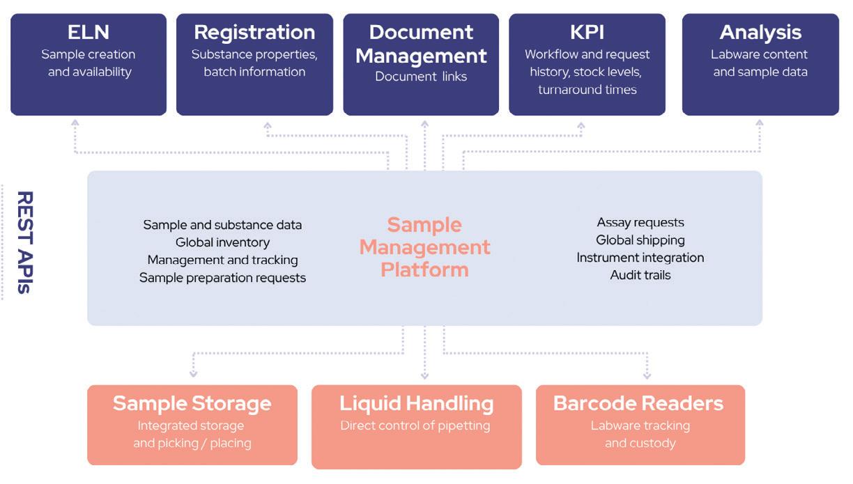
The key thing about samples is that they are directly linked to experimental or study outcomes and test results. Given this definition, it becomes clear that the proper management of such samples should be at the core of everything a laboratory does. Information about these samples is consumed by multiple upstream and downstream
processes, and Figure 1 shows the breadth of just some of the laboratory applications that rely on accurate sample information (see page 8).
Why good sample management matters Poor sample management can lead to significant issues in a laboratory setting. Ineffective sample tracking leads to misplacing or losing samples and inaccurate inventory, leading to wasted time finding samples for an experiment. Poor sample data integrity can cause inconsistent results, reducing the research reproducibility and credibility as well as increasing costs and lowering overall productivity. Ineffective sample storage management leads to
cluttered storage, sample degradation, wastage of labile samples, and increased energy needs due to inefficient use of space. Ultimately, the above could result in regulatory compliance failures that could even halt research activities and damage a laboratory’s reputation.
Given the importance of good sample management, what software capabilities are needed to avoid the pitfalls? Here is a quick guide to some of the top features that you should be looking for:
Audit trail
Every sample event should be tracked in a 21CFR11 compliant or equivalent audit trail —
including who, what and when. This ensures a detailed history of every sample for compliance or experimental reproducibility. It also allows laboratories to track down problems such as missing samples or liquid handler misfires and acts as a permanent record of a sample’s location history, such as which freezer it used to be stored in, when it was disposed, or where it was sent to.
Sample genealogy
Accurate tracking of sample lineage from tubeto-tube or well-to-well transfers between plates, including the lineage between materials when one substance is derived from another, is essential. This capability facilitates the repeatability of
experiments by ensuring that the same source sample is used. Maintaining clear records of splits and derivations is particularly important for human sample tracking compliance purposes.
Barcode management
Every sample is assigned a unique barcode from a managed range, and pre-barcoded samples are verified for uniqueness. Any sample in the lab can be identified unambiguously with a scan, and errors from manual labelling and misidentification are eliminated.
Storage management
This relates to the ability to model the hierarchy of sample storage — including freezers, incubators,
cupboards and specific locations within these. It reduces the need for manual searches, decreases equipment costs by freeing up storage space from outdated samples and improves overall sample organisation.
Data validation
Data validation prevents incorrect or conflicting data from being imported and ensures that all data are of the correct data type. Consistent and accurate data make it easier to locate samples using simple and consistent terminology.
Expiry date management
Tracking the date at which action should be taken on a sample — whether disposal, archiving or requalification — reduces the risk of using expired samples that might compromise compliance. It also makes it easy to perform regular housekeeping to dispose of expired samples efficiently.
Accurate tracking of sample amounts and concentrations
Knowing the inventory and quantity of samples available is essential, ideally updated automatically as samples are used. This makes it easier to confirm material availability for experiments and to quickly locate samples with both the amount and concentration required for specific assays.
Management of diverse container types
Supporting a variety of container types — such as tubes, vials, flasks, microtitre plates or tissue blocks/slides — ensures accurate recording for easier identification and more efficient storage utilisation. This capability also enables laboratories to leverage sample automation effectively, as automated stores and liquid handlers will expect specific container types. API and integration capabilities
Programmatic access to sample data and integration with other software platforms is increasingly critical. By linking foundational sample data with other applications, laboratories can easily associate experimental results and metadata with physical samples, ensuring accurate analyses.
Keeping pace with future developments
The increasing trend towards collaborative research provides further challenges for sample management. Samples may be sent out for testing or archival at remote sites, perhaps to be shipped onsite on demand. These sample movements need to be tracked, so consideration should be given to whether this is a future critical capability for your operations.
Artificial intelligence (AI) is already making its way into laboratories, and this trend is
Effective sample tracking is crucial for ensuring the success of research and development, enhancing operational efficiency and regulatory compliance.
accelerating. AI relies on high-quality data and, by providing access to accurate sample inventory data, could lead to game-changing process improvements, particularly when integrated with laboratory automation. Such process improvements might include:
• Enhanced experiment planning and scheduling — taking into account sample availability and equipment needs.
• Automation of stock monitoring and replenishment.
• Sample insights based on analytical results tied to those samples — eg, identifying suspect samples or differential results due to variation in assay plate layouts.
• Prediction of workflow times for different sample processing operations. This list could go on. A discussion of AI in laboratory operations deserves an article of its own. Suffice it to say that AI will transform the way sample data is used and analysed, leading to more efficient laboratory operations and research — but only if the sample data is accessible and of high quality.
In summary
Effective sample tracking is crucial for ensuring the success of research and development, enhancing operational efficiency and regulatory compliance.
It ensures the accuracy and reproducibility of results while preparing the lab for technological advances, such as AI, and can help streamline collaborative research efforts.
Whether a laboratory is managing millions of samples in 1536-well plates in HTS libraries, small numbers of clinical study samples or anything in between, all samples have value and the tracking of them is fundamental to the success of research, development or testing processes.
As life sciences R&D embraces increasingly diverse sample types, growing throughput and automation adoption, what might once have been possible to track manually soon becomes an impossibility. As a laboratory grows in size and scope to meet these challenges, having the right foundations of a sample management platform that supports scalability, integration with automation platforms and the management of diverse modalities enables this evolution.
In conclusion, a robust sample management platform forms the backbone of efficient, compliant and high-quality laboratory operations, accelerating research and improving experimental outcomes.
*Marcus Oxer is a Domain Solution Manager at Titian Software, provider of Mosaic — a sample management platform for life sciences.
Figure 1: Sample management at the centre of laboratory operations.
‘AI electronic nose’ used to digitally detect scents
A research team from the Daegu Gyeongbuk Institute of Science and Technology (DGIST) has developed a so-called ‘next-generation AI electronic nose’ that is capable of distinguishing scents like the human olfactory system does and then analysing them using artificial intelligence.
The technology, which converts scent molecules into electrical signals and trains AI models on their unique patterns, holds great promise for applications in personalised health care, the cosmetics industry, and environmental monitoring. It has been described in the journal ACS Nano
While conventional electronic noses (e-noses) have already been deployed in areas such as food safety and gas detection in industrial settings, they struggle to distinguish subtle differences between similar smells or analyse complex scent compositions. For instance, distinguishing among floral perfumes with similar notes or detecting the faint odour of fruit approaching spoilage remains challenging for current systems. This gap has driven demand for next-generation e-nose technologies with greater precision, sensitivity and adaptability.
The research team was inspired by the biological mechanism known as combinatorial coding, in which a single odorant molecule activates multiple olfactory receptors to create a unique pattern of neural signals. By mimicking this principle, the team engineered sensors that respond to scent molecules by generating distinct combinations of electrical signals. The AI system learns these complex signal patterns to accurately recognise and classify a wide variety of scents, resulting in a high-performance artificial olfaction platform that is said to surpass existing technologies.
The e-nose uses a laser to process a thin carbon-based material (graphene) and incorporates a cerium oxide nano catalyst to create a sensitive sensor array. This single-step laser fabrication method eliminates the need for complex manufacturing equipment and enables high-efficiency production of integrated sensor arrays.
In performance tests, the device successfully identified nine fragrances commonly used in perfumes and cosmetics, with over 95% accuracy. It could also estimate the concentration of each scent, making it suitable for fine-grained olfactory analysis.
The device is ultrathin, flexible and highly durable, making it ideal for wearable devices or bright patches attached to the skin or clothing. It can be bent more than 30,000 times around a 2.5 mm radius without any performance degradation.
“The core innovation of our research is the ability to integrate multiple scent-sensitive sensors with diverse properties, similar to those of the human nose, into a single unit through a one-step selective laser fabrication process,” said team leader Professor Hyuk-jun Kwon. “We are actively expanding development and commercialisation efforts to apply this technology to personal health care, environmental pollution detection and the fragrance industry.”
Early Parkinson’s detection with an RNA-based blood test
Diagnosis of neurodegenerative diseases is these days around the level that cancer diagnosis was 50 years ago: disease is identified when most of the relevant neurons have already died, and it is therefore too late to cure. To address this problem, a research team led by The Hebrew University of Jerusalem has developed a blood test that could enable early diagnosis of Parkinson’s disease (PD), paving the way for timely interventions and improved patient outcomes. Their work has been described in the journal Nature Aging
The test introduces a novel approach to detecting PD at its earliest stages through the analysis of transfer RNA fragments (tRFs): small RNA molecules with the potential to reveal significant changes in the body linked to neurodegeneration. The researchers identified two key biomarkers — an increase in PD-specific tRFs carrying a conserved sequence motif (RGTTCRA-tRFs) and a decrease in mitochondrial tRFs (MT-tRFs). By measuring the ratio between these biomarkers, the new test can distinguish pre-symptomatic Parkinson’s patients from healthy controls with an accuracy surpassing that of existing clinical diagnostic tools.
“This discovery represents a major advancement in our understanding of Parkinson’s disease and offers a simple, minimally invasive blood test as a tool for early diagnosis,” said Professor Hermona Soreq. “By focusing on tRFs, we’ve opened a new window into the molecular changes that occur in the earliest stages of the disease.”
The test employs a straightforward, dual qPCR assay, measuring the ratio between the repeated short motif and an exemplary mitochondrial sequence, making it cost-effective and accessible for use in a wide range of healthcare settings. In trials involving samples from multiple
international cohorts, including the Parkinson’s Progression Markers Initiative, the test achieved a diagnostic accuracy of 0.86, significantly outperforming traditional clinical scoring methods. Moreover, the study found that RGTTCRA-tRF levels decrease following deep brain stimulation, further linking these RNA fragments to both disease mechanisms and treatment responses.
Lead researcher Nimrod Madrer emphasised the importance of early detection, noting that Parkinson’s disease is often diagnosed only after significant brain damage has occurred. “This test has the potential to alleviate the uncertainty faced by patients and clinicians, offering a reliable and rapid method to identify the disease in its earliest stages,” he said.
Researchers from the University of Vienna and the Helmholtz Institute for Pharmaceutical Research Saarland (HIPS) have discovered a new type of glycopeptide antibiotic known as saarvienin A, found to have strong activity against highly resistant bacterial strains. Their work has been published in the journal Angewandte Chemie International Edition.
In the search for new antibiotic compounds, researchers have turned to the study of actinobacteria — microorganisms that are well-known for living in unusual environments and producing antibiotics such as vancomycin, rifamycin and chelocardin. Vienna’s Jaime Felipe Guerrero Garzón discovered strong antibiotic activity in extracts from a strain of Amycolatopsis isolated from a Chinese rare earth mine.
Martin Zehl, Head of the Mass Spectrometry Centre at the University of Vienna, found out that the antibiotic activity was associated with a potentially novel compound of the class glycopeptides. Using mass spectrometry and nuclear magnetic resonance (NMR) spectroscopy, the collaborating team at HIPS identified a completely new molecule: saarvienin A.
Saarvienin A’s special feature became clear early on: unlike established glycopeptides such as vancomycin, the new compound does not bind the typical bacterial target involved in cell wall synthesis. Instead, it probably acts through a different, as yet unresolved mechanism. Structural analysis revealed a distinctive architecture: a halogenated peptide core cyclised through an unusual ureido linkage, decorated with a chain of five sugar and aminosugar units — two of which are completely new to natural products.
“We were excited to find that saarvienin A doesn’t fit into any known category,” Guerrero Garzón said. “Its unique structure could pave the way for antibiotics that bacteria have never encountered before.”
Tests of the new molecule against bacteria focused on ‘ESKAPE pathogens’ — a notorious group of superbugs known to evade most current antibiotics. The compound showed remarkable activity against vancomycin-resistant Enterococcus and methicillin-resistant Staphylococcus aureus (MRSA), including 3 ESKAPE pathogens and 26 clinical isolates. It consistently outperformed vancomycin, even against strains already resistant to multiple other antibiotics.
With the biosynthetic genes for saarvienin A already identified and cloned, the researchers plan to use medicinal chemistry and biosynthetic engineering to optimise the molecule. A key goal is to reduce cytotoxicity while maintaining antibacterial activity.
“Discovering a new antibiotic is only the beginning,” said corresponding author Sergey B Zotchev, from the University of Vienna. “Now we face the fascinating challenge of refining it into a drug candidate suitable for clinical use.”
high performance steam traps
B – Bimetallic
M – Thermostatic
TD – Thermodynamic
CONALIFt Mechanical condensate pump
“All-in-One” Multi-valve system
AuSTrAlIAN AGeNT & STockIST
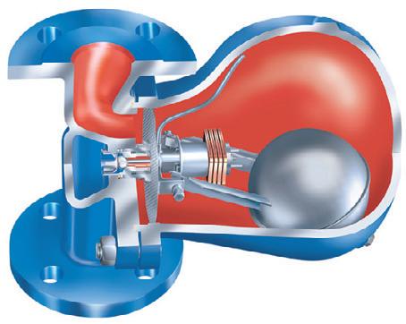
CONA SteAm trApS
CONA S Ball float steam trap
CONA
CONA p Pump trap
NMR used for structural identification at HIPS.
TGA approves donanemab for treatment of early Alzheimer’s
The Therapeutic Goods Administration (TGA) has granted marketing authorisation for Eli Lilly and Company’s Kisunla (donanemab), an injection to treat mild cognitive impairment and mild dementia due to Alzheimer’s disease. This makes it the first amyloid-targeting therapy for people with Alzheimer’s registered in Australia and the only amyloid plaque-targeting therapy with evidence to support stopping therapy when amyloid plaques are removed, Eli Lilly stated.
Amyloid is a protein produced naturally in the body that can clump together to create amyloid plaques. Donanemab works by inducing antibodies designed to attach to and remove these amyloid plaques from the brain. The drug is given as an intravenous infusion through the arm every four weeks for a maximum of 18 months, and may delay the progression of symptoms in people in the early stages of Alzheimer’s disease.
The registration of donanemab in Australia was based on the TRAILBLAZER-ALZ 2 Phase 3 and TRAILBLAZER-ALZ 6 clinical trial data. The TRAILBLAZER-ALZ 2 study demonstrated that donanemab significantly slowed cognitive and functional decline by up to 35% compared to placebo at 18 months and reduced the risk of progressing to the next clinical stage of disease by 39% over the same period.
“In our TRAILBLAZER-ALZ 2 Phase 3 study, results showed that Kisunla significantly slowed cognitive and functional decline
in patients with early symptomatic Alzheimer’s disease, which allowed them more time to do things that mattered most to them like remember information, make meals, manage finances and maintain independence,” said Ilya Yuffa, Executive Vice President and President of Lilly International, Eli Lilly and Company. “As our data showed, the earlier patients are identified, diagnosed and treated with Kisunla, the greater their response to treatment.”
It should be noted that donanemab comes with the possible side effects of brain swelling and bleeding, although the approved dosing schedule is based on TRAILBLAZER-ALZ 6, which demonstrated lower incidence of brain swelling at 24 weeks versus the original dosing schedule. Anyone who has two copies of an Alzheimer’s risk gene called ApoE4 is at a higher risk of brain swelling and bleeding, so patients considering the drug must have genetic testing to check for this gene.
MRI can reveal the ‘true’ age of your heart
Scientists have developed a way of uncovering the ‘true age’ of your heart, with new research showing how an MRI scan can reveal your heart’s functional age — and how unhealthy lifestyles can dramatically accelerate this figure. It is hoped that the team’s findings could transform how heart disease is diagnosed, catching problems before they become deadly.
Led by the University of East Anglia (UEA), the research team collaborated with hospitals in the UK, Spain and Singapore. They studied MRI scans from 557 people — 191 healthy individuals and 366 with conditions like high blood pressure, diabetes or obesity.
Using advanced imaging, the researchers measured things like the size and strength of the heart’s chambers. Then, they built a formula to calculate the heart’s ‘functional age’ and checked it against healthy hearts to make sure it was accurate.
“We found that an MRI scan can reveal your heart’s ‘functional age’ — how old it acts, not how old you are,” said lead researcher Dr Pankaj Garg, from UEA’s Norwich Medical School.
“In healthy people, we found that heart age was similar to chronological age. But for patients with things like diabetes, hypertension, obesity and atrial fibrillation, their functional heart age was significantly higher.
“For example, a 50-year-old with high blood pressure might have a heart that works like it’s 55.
“People with health issues like diabetes or obesity often have hearts that are aging faster than they should — sometimes by decades. So, this could help doctors step in early to stop heart disease in its tracks.”
Garg said the new technique is a “game changer” for keeping hearts healthier for longer, giving doctors a powerful tool to look inside the heart like never before and spot trouble early — before symptoms even start.
“By knowing your heart’s true age, patients could get advice or treatments to slow down the aging process, potentially preventing heart attacks or strokes,” he said.
“It could also be the wake-up call people need to take better care of themselves — whether that’s eating healthier, exercising more, or following their doctor’s advice. It’s about giving people a fighting chance against heart disease.”
The research has been published in European Heart Journal - Open
‘Pain-on-a-chip’ device identifies different chronic pain types
Researchers from the Monash Institute of Pharmaceutical Sciences (MIPS), in collaboration with Flinders University, have developed a so-called ‘pain-on-a-chip’ that uses live sensory nerves on a chip to help objectively diagnose chronic pain conditions. Their research has been published in the journal Biosensors and Bioelectronics
Chronic pain is globally prevalent and incredibly challenging to treat. Furthermore, clinical strategies for chronic pain management rely heavily on self-reporting, which is naturally subjective and particularly problematic for non-verbal patients. As such, new methods for detection of pain biomarkers are essential.
“Improving pain classification and identifying new treatments requires new strategies that objectively recognise specific pain conditions and minimise subjectivity,” said Professor Nicolas Voelcker from MIPS, a lead author on the new preclinical study.
“Our pain-on-a-chip concept has the potential to provide a biosensor platform for a minimally invasive and objective analysis method to discriminate between chronic pain subtypes.”
The team’s microfluidic biosensor works by distinguishing cells that initiate the sensation of pain; these cells, known as ‘nociceptors’, are associated with a number of pain conditions, including chronic pain. The research team used the device to differentiate between blood samples extracted from two different animal models of chronic pain — one focusing on fibromyalgia and the other on diabetic neuropathy.
The researchers were able to demonstrate the device’s ability to objectively distinguish the response of the nociceptor cells towards the two chronic pain subtypes. Co-lead author Associate Professor Nicholas Veldhuis, from MIPS, said their findings could pave the way for the development of an entirely new tool to determine chronic pain states based on blood sampling.
“In recent years, biosensor technology has emerged as a promising method for rapid, affordable and direct detection of biomarkers in disease; however, the technology has not yet made it into a clinical setting,” Veldhuis said. “Our research builds on recent developments which we hope to continue evolving and, ultimately, deliver a device that will improve the lives of those living with chronic pain conditions.”
Co-lead author Dr Dusan Matusica, from the Flinders Health and Medical Research Institute, concluded, “Our research lays the foundation for the development of an objective discriminatory tool for the determination of chronic pain states based on blood sampling. Such a diagnostic tool set is currently missing in both preclinical and clinical applications.”
First patient treated with personalised gene editing
In what is being described as a historic medical breakthrough, an infant diagnosed with a rare genetic disorder has been treated with a customised CRISPR gene editing therapy by a team at the Children’s Hospital of Philadelphia (CHOP) and Penn Medicine.
The story has been detailed in a study published in The New England Journal of Medicine and presented at the American Society of Gene & Cell Therapy’s Annual Meeting in New Orleans. It is believed that the findings could provide a pathway for gene-editing technology to be successfully adapted to treat individuals with rare diseases for whom no medical treatments are available.
The case revolves around an infant known as KJ, who was just two days old when a doctor at the Hospital of the University of Pennsylvania noticed he had become unusually lethargic, he wasn’t eating and he struggled to maintain his temperature. The doctor checked KJ’s blood ammonia level — which can be a marker for some metabolic diseases — and found it was extremely high. After being moved to
CHOP for tests and examinations, KJ was deemed to have a urea cycle disorder — a genetic condition caused by deficiencies in specific enzymes that leads to a toxic build-up of ammonia in the body.
During the normal breakdown of proteins in the body, ammonia is naturally produced. Typically, our bodies know to convert the ammonia to urea and then excrete that urea through urination. However, children with a urea cycle disorder lack an enzyme in the liver needed to convert ammonia to urea. Ammonia then builds up to a toxic level, which can cause organ damage, particularly in the brain and the liver. Episodes of increased ammonia can thus put patients at risk for ongoing, lifelong neurologic damage or even prove fatal.
After doctors placed KJ on dialysis to filter the ammonia out of his blood and stabilise his health, they performed genetic tests to figure out exactly what type of urea cycle disorder he had, as
determining which gene or enzyme was affected could alter the recommended treatment. They discovered that KJ had been born with a rare metabolic disease known as severe carbamoyl phosphate synthetase 1 (CPS1) deficiency, caused by the CPS1 gene.
The only known lifesaving treatment for CPS1 deficiency was a liver transplant, which is often better suited to children who are older, larger and in better health; treatments for younger, smaller patients are sadly lacking. Nonetheless, KJ was added to the National Transplant Waiting List with the hope he would be stable enough for a liver transplant when — or if — a suitable organ became available. In the meantime, KJ would spend the first several months of his life at CHOP, on a very restrictive diet, where he could receive constant support and monitoring around the clock.
Developing a custom treatment
Before KJ was even born, a team of scientists at CHOP and the University of Pennsylvania were researching how to use gene editing to create customised treatments for diseases like CPS1 deficiency. Leading the group were Dr Rebecca Ahrens-Nicklas, a paediatric geneticist and Director of CHOP’s Gene Therapy for Inherited
we programmed it to go to the site of the genetic variant that was causing the disease in KJ.”
“The drug was specifically designed for KJ and the genetic variants he has,” Ahrens-Nicklas added. “It’s personalised medicine.”
Just a few days after KJ’s first infusion, he showed signs of improvement. The colour in his cheeks returned; he could tolerate more protein in his diet without causing a toxic increase in ammonia; and clinicians were able to slowly decrease his medication. Over the next two months, KJ received two additional infusions
Metabolic Disorders Program, and Dr Kiran Musunuru, a cardiologist, geneticist and gene editor at Penn Medicine.
Ahrens-Nicklas and Musunuru first began collaborating to study the feasibility of creating customised gene editing therapies for individual patients in 2023, building upon many years of research into rare metabolic disorders, as well as the feasibility of gene editing to treat patients. CRISPRbased gene editing can already be used to precisely correct disease-causing variants in the human genome, but up until this point, gene editing tools have been built to target more common diseases that affect tens or hundreds of thousands of patients, such as the two diseases for which there currently are FDA-approved therapies: sickle cell disease and beta thalassemia. Furthermore, relatively few diseases benefit from a ‘one-size-fits-all’ gene editing approach since so many disease-causing variants exist, so many patients with rare genetic diseases have been left behind.
“We’ve been practising developing … personalised therapies for about two years now with the idea that someday we might be in a position where we could very rapidly try to figure out how to use gene editing to correct a patient’s broken gene that’s responsible for their disease,”
Musunuru said. During this time, the researchers learned a great deal about ways to correct faulty genes. With each potential genetic change they considered for gene editing therapy, the time it took to find a personalised solution shortened.
After years of preclinical research with similar disease-causing variants, Ahrens-Nicklas and Musunuru targeted KJ’s specific variant of CPS1. They approached KJ’s family about the possibility of participating in a gene-editing research study, and after just six months of research and successful results in the lab, the team had designed and manufactured a base editing therapy delivered via lipid nanoparticles to the liver in order to correct KJ’s faulty enzyme. In essence, base editing allows scientists to rewrite DNA one ‘letter’ at a time.
On 25 February 2025, between six and seven months of age, KJ received his first infusion of this experimental therapy, making him the first patient ever to receive a systemic personalised gene editing drug. The drug was delivered in an IV line and flowed into the bloodstream, travelling to the liver. KJ received the lowest possible dose of the therapy to allow his body time to adapt and minimise any risks.
“CRISPR, a gene editor, enters the nucleus of the cell,” Musunuru explained. “In this case,
KJ was only days old when he was diagnosed with a rare metabolic disorder.
at higher levels. He has not had any serious side effects to date and is now growing and thriving, even recovering from typical childhood illnesses like rhinovirus without ammonia building up in his body.
KJ continues to be monitored as an inpatient at CHOP — indeed, he will need careful monitoring for the rest of his life — and longer follow-up will be needed to fully evaluate the benefits of the therapy. But Ahrens-Nicklas believes the initial findings are quite promising, while KJ’s parents are looking forward to soon welcoming him home.
“Years and years of progress in gene editing and collaboration between researchers and clinicians made this moment possible, and while KJ is just one patient, we hope he is the first of many to benefit from a methodology that can be scaled to fit an individual patient’s needs,” Ahrens-Nicklas said.
“We want each and every patient to have the potential to experience the same results we saw in this first patient, and we hope that other academic investigators will replicate this method for many rare diseases and give many patients a fair shot at living a healthy life,” Musunuru added. “The promise of gene therapy that we’ve heard about for decades is coming to fruition, and it’s going to utterly transform the way we approach medicine.”
Dr Kiran Musunru (left) and Dr Rebecca Ahrens-Nicklas (right) led the group of researchers who developed a personalised treatment for baby KJ (centre).
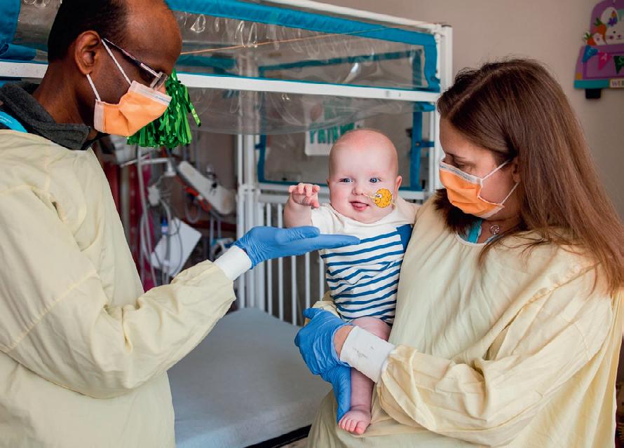
RSV immunisation program for babies slashes hospital stays
An Australian-first study demonstrating the effectiveness of immunisation against respiratory syncytial virus (RSV) for babies found it to be almost 90% effective in reducing hospitalisation rates, helping more than 500 families avoid a hospital stay. Their work has been published in the Journal of Infection
Affecting the airways and lungs, RSV is a life-threatening virus responsible for sending 3.6 million children to hospitals worldwide each year. It is especially dangerous for vulnerable young babies at high risk of potentially deadly complications such as severe bronchiolitis and pneumonia.
Last year, Western Australia introduced a free RSV immunisation program providing babies with a long-acting monoclonal antibody called nirsevimab. As noted by study author Dr Ushma Wadia, from the Wesfarmers Centre of Vaccines and Infectious Diseases, “More than 24,000 doses of nirsevimab were distributed throughout April–September last year, providing coverage to 85% of newborns and 66% of a ‘catch-up’ cohort of babies in the lead-up to their first winter season.”
Wadia and her fellow researchers set out to investigate RSV-related hospital admissions following the introduction of WA’s immunisation program. She explained, “Our team worked alongside WA Health to evaluate hospitalisation rates at Perth Children’s Hospital, Fiona Stanley Hospital and Joondalup Health Campus, and allowed us to become the first place in the Southern Hemisphere to successfully demonstrate the major impact of RSV immunisation in young babies.”
In addition to a reduction in hospital admission rates, the study also investigated the effect immunisation had on the severity of RSV cases. It found that nirsevimab recipients were 60% less likely to require oxygen or assistance with their breathing if admitted with RSV.
Professor Chris Blyth, a Perth Children’s Hospital physician and Head of the Wesfarmers Centre of Vaccines and Infectious Diseases, spoke about the global significance of the work.
“The data gained from the REVIVE Study aligns with outcomes from research conducted in the Northern Hemisphere and can now be used as evidence to inform vaccine policy throughout the world, including in lower-income countries where morbidity rates for RSV are at their highest,” Blyth said.
The success of WA’s nirsevimab program has also contributed to the rollout of a national, $174.5 million RSV immunisation program now underway for all pregnant women and newborn babies, which seeks to keep 10,000 Australian babies out of hospital each year.
Modifications in the placenta linked to psychiatric disorders
An international research team, led by the Immunogenetics Research Laboratory at the University of the Basque Country (UPV/EHU) and the Biobizkaia Health Research Institute, has identified associations between modifications in the placenta and the risk of developing schizophrenia, bipolar disorder and major depression disorder. The team’s findings, published in the journal Nature Communications, represent a significant advance in understanding the biological basis of neuropsychiatric disorders and open new lines of investigation for early detection, as well as for the development of more effective therapies.
Epigenetic modifications are chemical changes in DNA and its associated proteins that regulate gene activity without altering their sequence. One of the most studied modifications is DNA methylation, a process in which methyl groups — small molecules composed of one carbon and three hydrogen atoms — are added to specific regions of the DNA. This mechanism, essential for development, environmental adaptation and disease predisposition, is influenced by genetics and responds to factors such as diet, stress, and exposure to pollutants.
The study results indicate that schizophrenia, bipolar disorder and major depression disorder are the neuropsychiatric disorders most strongly linked to DNA methylation in the placenta, highlighting the placenta as a key element in neuropsychiatric development. Other conditions, such as attention deficit hyperactivity disorder (ADHD) or autism, show some potentially causal associations, although to a lesser extent, while no visible effects were found in other analysed pathologies.
“These findings reinforce the hypothesis that schizophrenia and other disorders have a neurodevelopmental origin and that the placenta plays a fundamental role in this process,” said study coordinator Dr Nora Fernandez-Jimenez, assistant professor at the UPV/EHU Faculty of Medicine and Nursing and a researcher at Biobizkaia.
The discovery that genetic risk may be linked to placental DNA methylation opens new avenues for preventing and treating psychiatric disorders. As noted by first author Dr Ariadna Cilleros-Portet, who completed her PhD at UPV/EHU, “If we could identify risk factors at the prenatal stage, we could intervene before symptoms appear, adjusting treatments or designing personalised preventive strategies.”
The study also underscores the importance of understanding where and when each genetic factor acts in pathology, since this could impact therapeutic decision-making. According to Fernandez-Jimenez, “Not all genes associated with a disorder should be treated directly; some may have acted in an earlier developmental stages and may not be actionable in adulthood.”
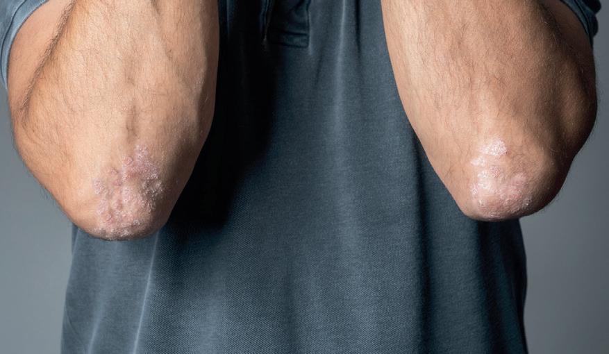
A targeted treatment option for psoriasis
Treatment options for psoriasis — one of the most common chronic inflammatory skin diseases — have previously focused on inhibiting pro-inflammatory immune cells. Now a new study led by the Medical University of Vienna has shown that it is possible to restore the function of certain anti-inflammatory immune cells in a targeted manner, paving the way for the development of a therapy that not only works more precisely but is also associated with fewer side effects.
The research team focused its investigations on the role of regulatory T cells (Treg cells) in chronic inflammatory skin diseases such as psoriasis. Treg cells are important components of the body’s immune system that specialise in preventing excessive immune responses and thus inflammation.
It is already known that Treg cells lose their regulatory function in chronic skin inflammation, causing the immune response to become
uncontrolled and the disease to progress. The researchers have now decoded the exact mechanism behind this for the first time, with their results published in the journal Immunity
“We were able to show that the loss of the anti-inflammatory function of regulatory T cells is caused by a malfunction of the cellular metabolism,” said study leader Georg Stary.
As the researchers’ analysis revealed, the enzyme SSAT plays a key role in the loss of function of T reg cells. SSAT is involved in the regulation of certain molecules (polyamines) that are important for the balance between anti-inflammatory and pro-inflammatory immune cells. If SSAT is produced in increased amounts in Treg cells, they lose their regulatory function and begin to produce pro-inflammatory messenger substances themselves. This fuels the excessive immune response typical of psoriasis.
With the key role of SSAT in the inflammatory process, the researchers have discovered a new starting point for therapy. In a mouse model with psoriasis-like skin inflammation, it was shown that inhibiting SSAT can restore the regulatory function of Treg cells and break the cycle of inflammation. Thus, the development of specific drugs that specifically inhibit SSAT could represent a promising alternative to existing treatment approaches, which are often associated with immunosuppression and increased susceptibility to infection.
“Since other chronic inflammatory diseases of the skin or other organs are also characterised by impaired immune regulation, our approach could be important beyond psoriasis,” Stary concluded.
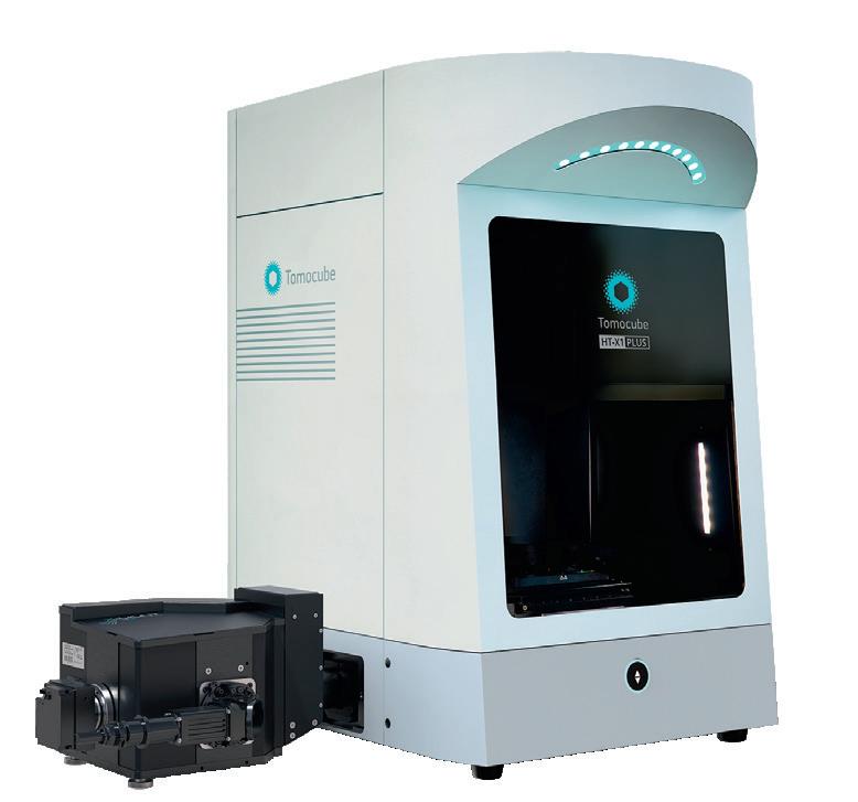
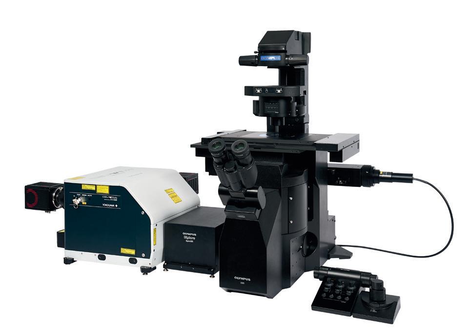
Combined holotomography and spinning disk confocal microscopy system
The HT-X1 Plus holotomography (HT) system from Tomocube has been integrated with the X-Light V2 spinning disk confocal microscope (SDCM) from CrestOptics to offer spinning-disk confocal holotomographic imaging. This next-generation multimodal imaging platform, now available from Tomocube, provides high resolution and sensitivity for 3D biophysical imaging applications where 3D holotomography is combined with fluorescence microscopy to allow researchers to correlate quantitative, label-free imaging with molecular-functional information, which should unlock a more advanced understanding of biological processes.
Holotomographic imaging involves illumination of a live cell with low-power visible light from multiple illumination angles, followed by measurement of the phase delay of the transmitted light. The technique uses the measured refractive index as imaging contrast, allowing visualisation of 3D living cells and tissues without labelling or staining. With its high spatial resolution capable of capturing subcellular organelles, users have increasingly sought confocal fluorescence capabilities to design multiplex imaging workflows that minimise photodamage. The HT-X1 Plus combines holotomographic imaging with CrestOptics’ spinning disk high-speed confocal technology within a fully integrated system, in order to enable the acquisition of label-free, highresolution 3D holotomography data and highly sensitive, precise 3D fluorescence data.
By combining these two imaging modalities, the HT-X1 Plus helps to facilitate deeper, more complete insights into molecular distribution within samples, without requiring invasive methods. Additionally, the integrated platform is designed to provide precision alignment of HT and fluorescence-based imaging datasets, allowing researchers to easily study molecular and phenotypic data in tandem and generate more powerful insights across biophysical imaging applications. SciTech Pty Ltd www.scitech.com.au
Genome-wide profiling of histone marks
The CUT&Tag-IT Express Assay Kit, from Active Motif, has been engineered to deliver high levels of sensitivity and reproducibility when it comes to CUT&Tag epigenomic profiling. The kit streamlines the CUT&Tag assay workflow by integrating simultaneous tagmentation and adapter incorporation into a single, efficient 0.2 mL reaction, utilising silica beads for DNA purification for a fast workflow.
Designed for low-input samples and optimised for minimal background noise, Active Motif’s assay generates robust, next-generation sequencing-ready libraries with ease, which should help to accelerate discoveries and reduce costs. Optimised for working with rare cell populations or complex tissue samples, its high performance and simplified operation should enable precision in gene regulation and chromatin mapping — even with minimal input material.
The CUT&Tag-IT Express Assay Kit provides consistent epigenetic mapping that is said to outperform traditional methods. It is designed to work with fresh or frozen cell samples, along with tissue samples processed with the Active Motif Tissue Prep for NGS Assays kit.
United Bioresearch Products Pty Ltd www.unitedbioresearch.com.au
Kinase screening library
Cayman Chemical’s Kinase Screening Library (96-Well) consists of two plates and contains approximately 160 carefully chosen selective and non-selective kinase inhibitors in a 96-well Matrix tube rack format as 10 mM stock solutions in DMSO.
This curated library includes inhibitors of a wide range of lipid, receptor and non-receptor tyrosine, serine/threonine and dual specificity kinases including those belonging to the ROCK, activin-like kinase (ALK), GSK3, PKC, PDGFR, VEGFR, Src, MAPK, CDK and PI3K families, among many others.
It offers expansive coverage, targeting more than 70 distinct kinases and kinase families, as well as numerous additional kinase isoforms and individual kinases within target families.
Sapphire Bioscience www.sapphirebioscience.com
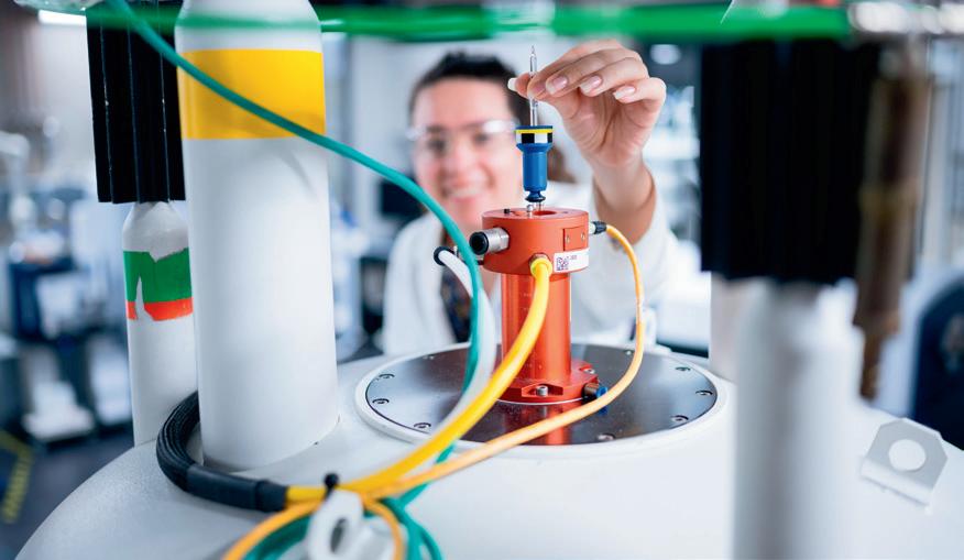
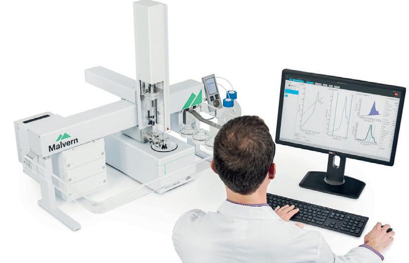
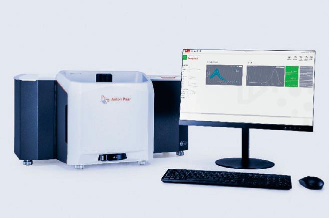
Microcalorimeter for vaccine development
As researchers develop vaccines for novel viruses and discover new ways to prevent disease, differential scanning calorimetry (DSC) is helping developers achieve what was once out of reach. By increasing the accuracy and speed of candidate characterisation and screening the most promising and stable candidates, DSC is accelerating development timelines and helping bring life-saving vaccines to people faster than ever before.
Traditionally, differential scanning fluorimetry (DSF) has been the go-to method for early-stage screening due to its highthroughput format and small sample requirements, but DSF comes with limitations. It relies on detecting protein unfolding through fluorescence, but only for proteins that contain fluorophores or can be tagged. Fluorescent dyes and buffer conditions can interfere with results, and DSF only reports unfolding events — not the full thermodynamic picture. As the biopharmaceutical landscape expands to include increasingly complex modalities such as antibody-drug conjugates, bispecifics, nucleotides and mRNA lipid nanoparticles, the limitations of DSF become more evident.
Malvern’s MicroCal PEAQ-DSC microcalorimeters offer a robust and versatile alternative for protein thermal stability screening. The product directly measures the heat absorbed during unfolding without the need for fluorescent dyes, offering label-free analysis and a complete thermodynamic profile — including melting temperature (Tm), enthalpy change (∆H) and heat capacity (Cp). The removal of fluorescent dyes makes PEAQ-DSC applicable to proteins, nucleic acids, lipids and their assemblies. In addition, because the product does not rely on optical, signal-based measurements, it is free from artefacts caused by quenching, protein aggregation or light scattering.
According to Malvern, PEAQ-DSC’s ability to generate precise thermostability profiles or thermal fingerprints enables reproducibility and reduces false positives. It also complements DSF and circular dichroism (CD) by acting as a benchmark, which should enable researchers to validate rapid assays with confidence. By identifying stable candidates early, defining optimal conditions and supporting QbD frameworks, the product helps to accelerate development timelines and support regulatory success. It thus provides the versatility needed to quickly and safely bring the next generation of vaccine candidates to market.
ATA Scientific Pty Ltd www.atascientific.com.au
Contact lens breakthrough lets humans see infrared light
A Chinese-led team of neuroscientists and materials scientists has created contact lenses that enable infrared vision in both humans and mice by converting infrared light into visible light — a breakthrough that could transform medical imaging and visual assistance technologies.
The team’s contact lens technology uses nanoparticles that absorb infrared light and convert it into wavelengths that are visible to mammalian eyes (eg, electromagnetic radiation in the 400–700 nm range). The nanoparticles specifically enable detection of near-infrared light, which is infrared light in the 800–1600 nm range, just beyond what humans can already see. The team previously showed that these nanoparticles enable infrared vision in mice when injected into the retina, but they wanted to design a less invasive option.
To create the contact lenses, the team combined the nanoparticles with flexible, nontoxic polymers that are used in standard soft contact lenses. After showing that the contact lenses were non-toxic, they tested their function in both humans and mice, with their results published in the journal Cell
They found that contact lens-wearing mice displayed behaviours suggesting that they could see infrared wavelengths. For example, when the mice were given the choice of a dark box and an infrared-illuminated box, contact-wearing mice chose the dark box whereas contact-less mice showed no preference. The mice also showed physiological signals of infrared vision: the pupils of contact-wearing mice constricted in
the presence of infrared light, and brain imaging revealed that infrared light caused their visual processing centres to light up. Meanwhile, in humans, the infrared contact lenses enabled participants to accurately detect flashing Morse code-like signals and to perceive the direction of incoming infrared light.
“It’s totally clear cut: without the contact lenses, the subject cannot see anything, but when they put them on, they can clearly see the flickering of the infrared light,” said senior author Tian Xue, a neuroscientist at the University of Science and Technology of China. “We also found that when the subject closes their eyes, they’re even better able to receive this flickering information, because near-infrared light penetrates the eyelid more effectively than visible light, so there is less interference from visible light.”
An additional tweak to the contact lenses allows users to differentiate between different spectra of infrared light by engineering the nanoparticles to colour-code different infrared wavelengths. For example, infrared wavelengths of 980 nm were converted to blue light, wavelengths of 808 nm were converted to green light, and wavelengths of 1532 nm were converted to red light. In addition to enabling wearers to perceive more detail within the infrared spectrum, these colour-coding nanoparticles could be modified to help colourblind people see wavelengths that they would otherwise be unable to detect.
“By converting red visible light into something
like green visible light, this technology could make the invisible visible for colourblind people,” Xue said.
Unlike infrared night vision goggles, the contact lenses do not require a power source, and they enable the wearer to perceive multiple infrared wavelengths. Furthermore, because they are transparent, users can see both infrared and visible light simultaneously.
“Our research opens up the potential for non-invasive wearable devices to give people super-vision,” Xue said. “There are many potential applications right away for this material. For example, flickering infrared light could be used to transmit information in security, rescue, encryption or anti-counterfeiting settings.”
Currently, the contact lenses are only able to detect infrared radiation projected from an LED light source, but the researchers are working to increase the nanoparticles’ sensitivity so that they can detect lower levels of infrared light. The contact lenses also have limited ability to capture fine details (due to their close proximity to the retina, which causes the converted light particles to scatter), so the team has developed a wearable glass system using the same nanoparticle technology, which enabled participants to perceive higher-resolution infrared information.
“In the future, by working together with materials scientists and optical experts, we hope to make a contact lens with more precise spatial resolution and higher sensitivity,” Xue said.
Advancing Precision and Reliability in Analytical Systems
Leveraging Valve Diagnostics for Optimal Performance
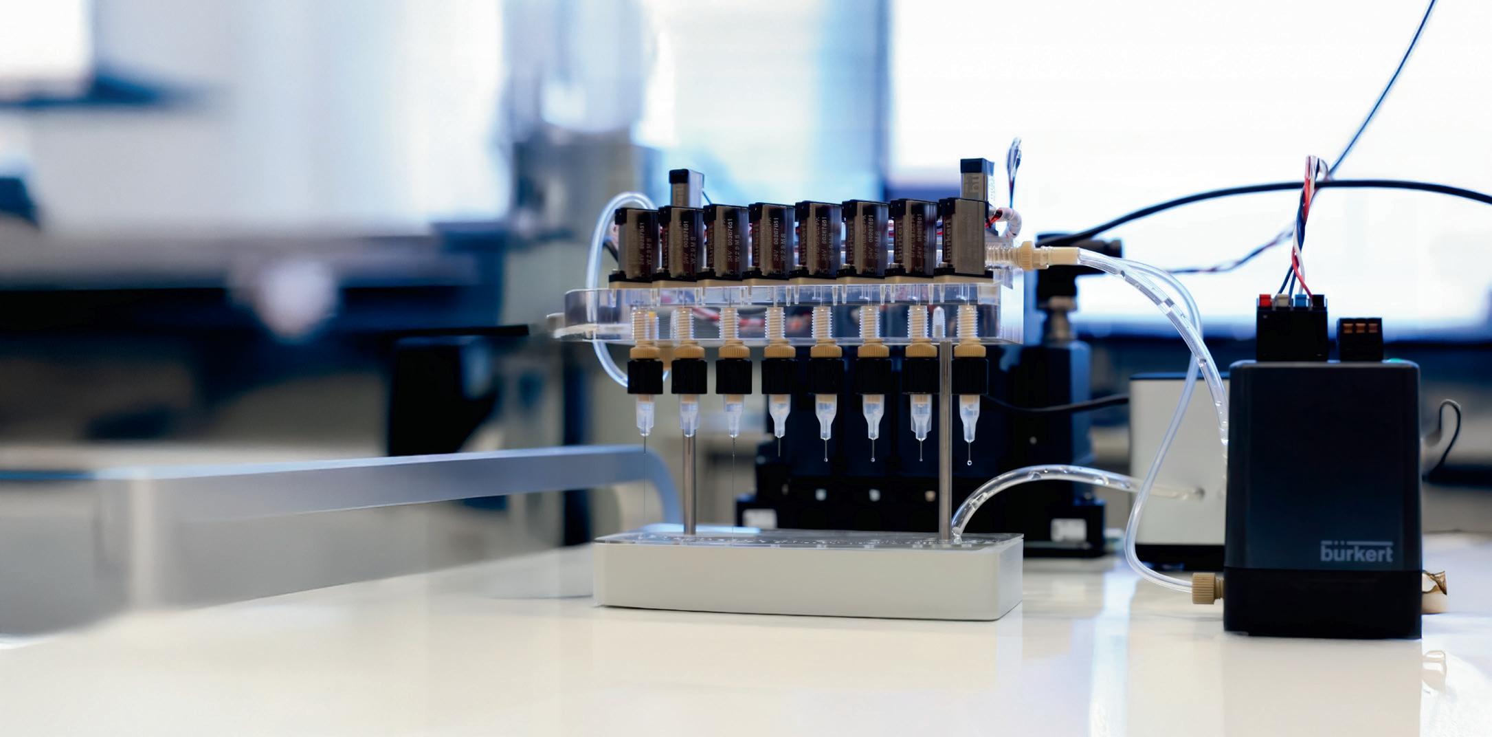
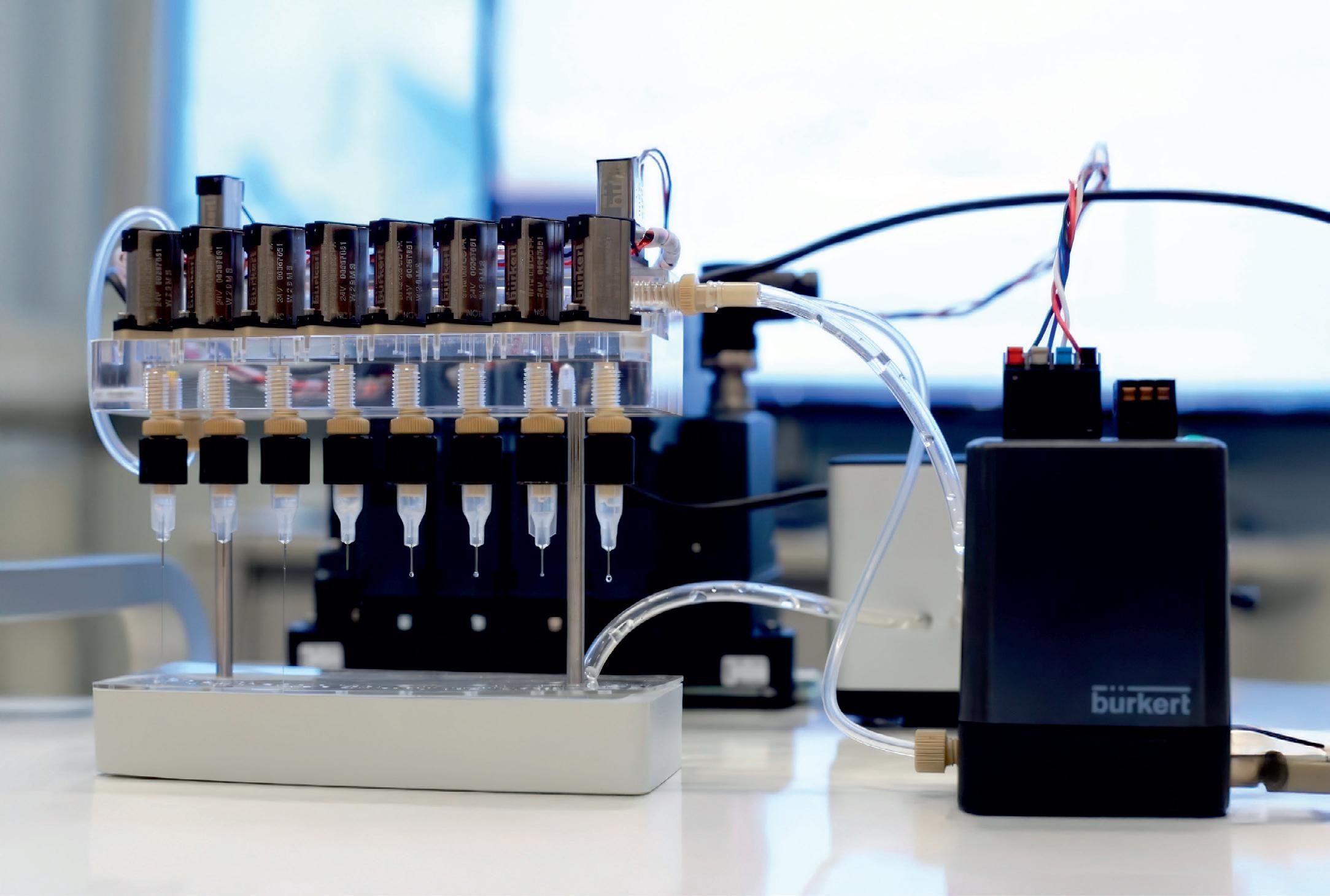
In demanding analytical environments, the ability to maintain precision, ensure reliability, and minimise downtime is paramount. The accuracy of diagnostic results, the efficiency of research processes, and the success of industrial applications all hinge on the seamless operation of fluid control systems. Bürkert Fluid Control Systems, a leading innovator in this field, understands these challenges and offers a sophisticated approach to optimising valve performance through integrated diagnostics.
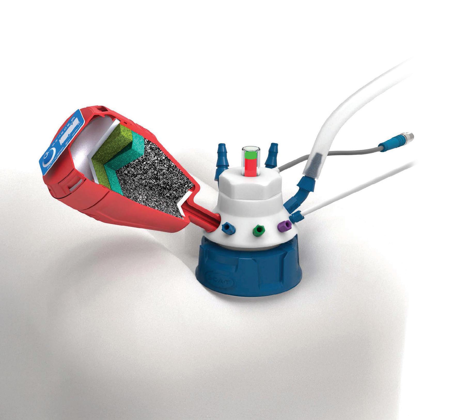
In laboratory devices, the accurate dosing of the right amount of reagents per sample is crucial to ensure high product quality and reliable analytical results. In this context, time-pressure dosing is the preferred method. The liquid is maintained at a constant pressure. The correct amount of reagents is dispensed by controlling the opening time of a dosing valve and the pressure applied to the liquid in the dosing tank.
A critical advancement is the integration of valve diagnostics, moving beyond simple monitoring to actively assessing and optimising valve function. This capability has significant implications for improving efficiency, reducing errors, and enhancing the reliability of analytical systems.
By integrating valve functionality and diagnostic capabilities, new devices can be more compactly designed. By diagnosing the switching behaviour, changes in other parameters such as temperature or pressure can also be deduced.
To reduce such errors, installing independent diagnostic devices is an option. One option for such a diagnostic device is a flowmeter positioned downstream of the dosing valve to monitor whether the valve operation was successful. Alternatively, a photoelectric sensor can be installed for monitoring purposes. However, independent diagnostic devices come with some drawbacks: they incur additional costs, require more space,
and significantly complicate the architecture of the end device.
Diagnostic Capabilities: A Deep Dive
A simpler and more elegant way to detect valve malfunctions is to use the dosing valve itself as a sensor. A valve with an electrodynamic actuator, which operates with a moving coil, is particularly well-suited to this application. Thanks to the innovative Whisper Valve actuator, induction can be directly measured, enabling early detection of valve malfunctions.
This approach enables early detection of valve malfunctions by leveraging electrodynamic actuators, where the actuator itself functions as a sensor. Monitoring the time-dependent current during operation unlocks valuable information about the valve’s parameters and its surrounding environment.
In the example of laboratory devices that dose reagents using time-pressure dosing in analysis processes, it is evident that the system can greatly benefit from intrinsic diagnostics using electrodynamic valves. It is possible to verify the proper functioning of the valve without requiring external sensors. Additionally, the pressure of the medium can be estimated, allowing for the verification of the correct functioning of the pressure controller or pump by using the valve as a sensor.
The hardware for this type of diagnostics is cost-effective compared to adding an additional sensor. The complete information extraction relies on an intelligent algorithm tailored to the specific valve type. To utilise this technology, a time-resolved current measurement and a microcontroller for analysis are required. If the application already has a microcontroller, the analysis can be performed there. No external sensors are needed on the valve.
Through real-time monitoring of the valve’s inrush current, valuable data about the actuator’s movement and the valve stroke is obtained. This data is invaluable for identifying issues that might otherwise go unnoticed.
The Advantages of Integrated Valve Diagnostics
• Increased Process Reliability: Integrated diagnostics ensure accurate dosing and fluid handling, leading to more consistent and reliable analytical results.
• Early Detection of Malfunctions: Identifying deviations from normal operation early on prevents erroneous results and costly sample losses.
• Reduced Downtime: Proactive diagnostics enable timely maintenance and repairs, minimising disruptions to critical analytical processes.
• Cost Savings: By eliminating the need for external sensors and streamlining maintenance, integrated diagnostics provides significant cost advantages.
Utilising the advanced ValveInsight concept, this approach transforms the role of valves in analytical processes from simple components to intelligent and adaptive elements within the overall system. This integration not only ensures peak performance and minimises errors, but also represents a forward-looking step toward smarter, more resilient analytical systems.
Read the full whitepaper here: https://www. burkert.com.au/en/landingpage/reliableanalytical-devices-with-valveinsight-diagnostics.
Burkert Fluid Control Systems www.burkert.com.au
Reptiles originated 35 million years earlier than we thought
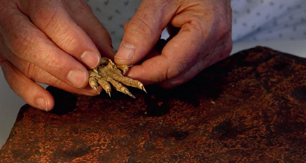
Newly discovered fossilised footprints with long toes and claws, found in a small rock slab in Victoria, are believed to have been made by a small goanna-like creature about 355 million years ago.
This is the oldest evidence in the world of reptile-like animals walking around on land — and it suggests they evolved 35 to 40 million years earlier than we thought, thus overthrowing the established evolutionary timeline of backboned land animals known as tetrapods. Published in the journal Nature, the study was led by Uppsala University and also involved Australian researchers.
Tetrapods evolved from a group of fish that left the sea about 390 million years ago, during the Devonian period. They became the ancestors of all modern backboned land animals, ie, amphibians
and amniotes — the latter of which includes mammals, reptiles and birds.
The oldest amniote fossils prior to this latest discovery were found in the Northern Hemisphere and come from the later part of the Carboniferous period, making them about 320 million years old. But the Australian rock slab, discovered in the Mansfield district of northern Victoria, shows that reptiles must have already existed 35 million years earlier, at the beginning of the Carboniferous, originating in the ancient southern supercontinent of Gondwana.
“Locals Craig Eury and John Eason (co-authors on the paper) found this slab covered in trackways and, at first, we thought they were early amphibian trackways, but one in the middle
has a hooked claw coming off the digits, like a reptile — an amniote, in fact,” said Professor John Long, from Flinders University.
“It was amazing how crystal clear the trackways are on the rock slab. It immediately excited us, and we sensed we were onto something big — even though we had no idea just how big it is.”
The Flinders palaeontology team working on the project included Dr Alice Clement, who scanned the fossil footprints to create digital models that were then analysed in detail, working closely with Uppsala University researchers led by Professor Per Erik Ahlberg.
“We study rocks and fossils of the Carboniferous and Devonian age with specific
interest to observe the very important fish–tetrapod transition,” Clement said.
“We’re trying to tease apart the details of how the bodies and lifestyles of these animals changed, as they moved from being fish that lived in water, to becoming tetrapods that moved about on land.”
Study co-author Dr Aaron Camens, who studies animal trackways from around Australia, produced heatmaps that explain details of the fossil footprints much more clearly. He noted, “A skeleton can tell us only so much about what an animal could do, but a trackway actually records its behaviour and tells us how this animal was moving.”
Because Long had been studying ancient fish fossils of this area since 1980, he had a clear idea that the rock deposits in the Mansfield district dated from the Carboniferous period, which started about 359 million years ago.
“This new fossilised trackway that we examined came from the early Carboniferous period, and it was significant for us to accurately identify its age — so we did this by comparing the different fish faunas that appear in these rocks with the same species and similar forms that occur in well-dated rocks from around the world, and that gave us a time constraint of about 10 million years,” he said.
The researchers received further support for the interpretation that reptiles emerged around this time from newly discovered fossil reptile tracks from Poland. They are not as old as the Australian rock slab, but substantially older than the previous oldest known examples.
Moving the origin of reptiles back in time changes the whole timeline of tetrapod evolution. Previously, scientists believed that the last common ancestor of amphibians and amniotes lived around 355 million years ago. But as the ancestor must be older than the oldest reptiles, that is now being called into question.
“All stem-tetrapod and stem-amniote lineages must have originated during the Devonian period — but tetrapod evolution proceeded much faster, and the Devonian tetrapod record is much less complete than we have believed,” Long said.
By combining the dating of fossils with the DNA of living descendants, the researchers are now trying to estimate when the last common ancestor of amphibians and amniotes might have lived. Their analysis shows that it was probably at the beginning of the latter part of the Devonian period, which was previously thought to be populated exclusively by primitive fish-like tetrapods and transitional forms such as Tiktaalik.
“This means that advanced tetrapods were already living at a time when it was previously thought that only primitive ‘four-legged fish’ were dragging themselves along the shores and just beginning to explore the land,” Ahlberg said.
Ahlberg added that the Australian rock slab is currently the only tetrapod fossil of earliest Carboniferous age we have from the whole of Gondwana, which comprised Africa, South America, Antarctica, Australia and India. “Who knows what other animals may have lived there?” he pondered.
An impression of what the amniote would look like from 350 million years ago.
Professor John Long compares the trackways with a modern iguana foot.
Image credit: Martin Ambrozik.
Image credit: Traci Klarenbeek.
Automated cell culturing system
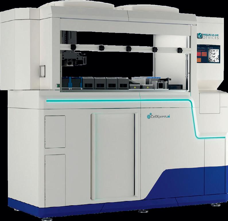
Molecular Devices’ CellXpress.ai is a cell culturing system with an integrated incubator, liquid handler, and image-based, deep-learning decision-making to automate the entire cell culture process. The product helps to scale up complex cell culture workflows, with actionable imaging and turnkey protocols for media exchange, monitoring and passaging.
Automated cell culture and image analysis workflows will run 24/7 — even when the lab is closed. The user can track the complete cell journey over time, with a unified software environment making it easy to develop traceable and reproducible cell cultures specific to the desired assay endpoint.
Users can answer critical questions quickly by easily identifying outliers at the well, plate or experiment level to help detect variability sources and make decisions. By removing these plates or wells from downstream processing early in the drug discovery process, reagents can be saved.
The use of standardised protocols and consistent automation speeds up the development process. Additionally, automated decision-making triggers events such as feeding, monitoring and passaging, and can notify users of assay milestones or required intervention.
The product is designed to improve productivity and optimise hands-on time with imagebased, deep-learning decision-making. This should help to remove variability, reduce human error, maintain sterility, and increase confidence in success with automated cell handling.
Users can solve complex image analysis problems utilising advanced artificial intelligence (AI) to transform images into results and data into decisions. User-friendly workflows help users get answers fast from 2D, 3D and time-lapse experiments.
Bio-Strategy - Part of DKSH Group www.bio-strategy.com
Vaisala’s HMT120 and HMT130 humidity transmitters are designed to enable accurate measurements with good long-term stability. These multi-purpose transmitters with exchangeable measurement probe are a suitable choice for cleanrooms and other demanding HVAC applications.
Features include: ±1.5%RH, ±0.1°C/±0.18°F accuracy; the option to be wall-mounted, duct-mounted with a kit, or outdoors with a shield; the probe can be attached direct to the transmitter or using a cable; analog output options 0–10 V/ 4–20 mA (loop powered); optional parameters (dew point/frost point, wet bulb, enthalpy, absolute humidity, mixing ratio); and traceable calibration (certificate included).
Humidity measurement with the Vaisala HUMICAP 180R humidity sensor enables state-ofthe-art precision, stability and environmental resistance, the company says. Temperature is measured with a Pt1000 sensor element, designed to be highly accurate. The high quality and stability of the measurements should enable precise control of HVAC systems, even in demanding conditions or sites.
The instruments are individually adjusted and delivered with a traceable (ISO 9001) calibration certificate. The humidity transmitter is easy to calibrate onsite using a Vaisala handheld meter or Vaisala Insight PC software. Vaisala Pty Ltd www.vaisala.com
Pipette tip system
The Axygen MultiRack tip system, available from Pacific Laboratory Products, has been engineered to fit most pipettors on the market. It is an interchangeable, complete system offered in bulk, racked, filter and reload packaging styles to meet the variety of needs in the lab setting.
The MultiRack tip system offers standard and extended-length tips, allowing volumes from 0.1 to 1250 µL (extended tips are offered in 10, 200, 300 and 1000 µL sizes). For racked and reload packaging styles, the tips are pre-loaded into easy-load, colourcoded deck inserts for easy volume identification. With a small benchtop footprint, stackable design and reusable rack, the system minimises environmental impact and lab space.
Axygen MultiRack tip system products are made in the USA and meet ISO standards. All tips are free of detectable RNase, DNase, DNA and pyrogens.
Pacific Laboratory Products www.pacificlab.com.au
Newly created antivenom
protects against 19 deadly snakes
US researchers claim to have developed the most broadly effective antivenom to date, which is protective against the likes of the black mamba, king cobra and eastern brown snake in mouse trials. Described in the journal Cell, the antivenom combines protective antibodies and a small molecule inhibitor, and opens a path toward a universal antiserum.
How we make antivenom has not changed much over the past century. Typically, it involves immunising horses or sheep with venom from single snake species and collecting the antibodies produced. While effective, this process could result in adverse reactions to the non-human antibodies, and treatments tend to be species and region-specific.
While exploring ways to improve this process, scientists stumbled upon Tim Friede, who is hyper-immune to the effects of snake neurotoxins. As explained by first author Jacob Glanville, who serves as CEO of Centivax, Friede agreed over a period of 18 years to undertake “hundreds of bites and self-immunisations with escalating doses from 16 species of very lethal snakes that would normally a kill a horse”.
By exposing himself to the venom of various snakes over several years, Friede generated antibodies that were effective against several snake neurotoxins at once. Glanville explained, “What was exciting about the donor was his once-in-alifetime unique immune history. Not only did he potentially create these broadly neutralising
antibodies; in this case, it could give rise to a broad-spectrum or universal antivenom.”
To build the antivenom, the team first created a testing panel with 19 deadly snakes across the elapid family — a group which contains roughly half of all venomous species. Next, researchers isolated target antibodies from Friede’s blood that reacted with neurotoxins found within the snake species tested. One by one, the antibodies were tested in mice envenomated from each species included in the panel. In this way, scientists could systematically build a cocktail comprising a minimum but sufficient number of components to render all the venoms ineffective.
The team formulated a mixture comprising three major components: two antibodies isolated from the donor and a small molecule. The first donor antibody, called LNX-D09, protected mice from a lethal dose of whole venom from six of the snake species present in the panel. To strengthen the antiserum further, the team added the small molecule varespladib, a known toxin inhibitor, which granted protection against an additional three species. Finally, they added a second antibody isolated from the donor, called SNX-B03, which extended protection across the full panel.
“By the time we reached three components, we had a dramatically unparalleled breadth of
full protection for 13 of the 19 species and then partial protection for the remaining that we looked at,” Glanville said. “We were looking down at our list and thought, ‘what’s that fourth agent’? And if we could neutralise that, do we get further protection?” Even without a fourth agent, their results suggest that the three-part cocktail could be effective against many other, if not most, elapid snakes not tested in this study.
With the antivenom cocktail proving effective in mouse models, the team now looks to test its efficacy out in the field, beginning by providing the antivenom to dogs brought into veterinary clinics for snake bites in Australia. Furthermore, they wish to develop an antivenom targeting the other major snake family: the vipers.
“We’re turning the crank now, setting up reagents to go through this iterative process of saying what’s the minimum sufficient cocktail to provide broad protection against venom from the viperids,” said lead author Professor Peter Kwong, from Columbia University’s Vagelos College of Physicians and Surgeons. “The final contemplated product would be a single, pan-antivenom cocktail or we potentially would make two: one that is for the elapids and another that is for the viperids, because some areas of the world only have one or the other.”
The other major goal is to approach philanthropic foundations, governments and pharmaceutical companies to support the manufacturing and clinical development of the broad-spectrum antivenom. “This is critical,” Glanville said, “because although there are millions of snake envenomations per year, the majority of those are in the developing world, disproportionately affecting rural communities.”
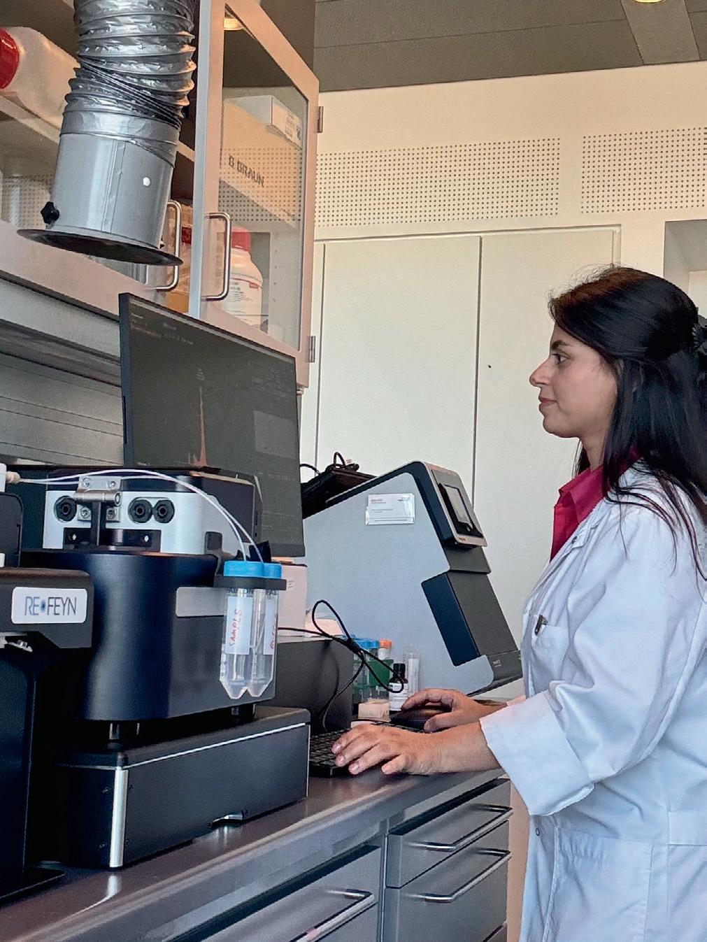
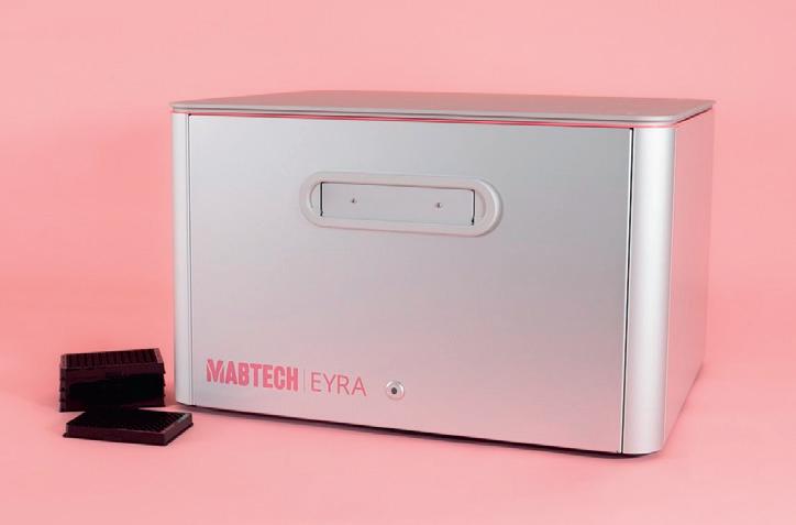
Refeyn installs its 500th mass photometry system
Refeyn, a pioneer in mass photometry technology, has announced the installation of the 500th system based on its novel single-particle bioanalytical technology within just seven years of its spinout from the University of Oxford. The company’s TwoMP mass photometer and MassFluidix HC system were installed for the Ploug Group at the University of Copenhagen’s Biotech Research & Innovation Centre, in order to further studies of proteins involved in diseaserelevant pathways.
Refeyn’s technology enables the label-free characterisation of single biomolecules and viral particles in solution in their native state. The Ploug Group is using mass photometry to study intravascular lipid metabolism, already citing it in a new research article investigating the regulation of lipoprotein lipase (LPL), a key enzyme for the lipolytic processing of triglyceride-rich lipoproteins in capillaries1
“We anticipate that the acquisition of mass photometry in our laboratory is going to be a game changer in our studies on purified proteins involved in intravascular lipolysis,” said Group Leader Dr Michael Ploug. “We’ve already shown by mass photometry that the LPL inhibitor ANGPTL3/8 is not an oligomeric complex composed of three ANGPTL3 protomers and one ANGPTL8 protomer as was reported previously, but is actually a trimeric complex composed of only two ANGPTL3 protomers and one ANGPTL8 protomer. This information is self-evidently important for future studies on the regulation of LPL activity in capillaries.”
Mass photometry now provides a valuable orthogonal tool to those already available to the group for its functional and structural protein studies, proving ideal for studying proteins in low concentrations where SEC-MALS is challenged. Ploug noted, “Since our proteins generally operate in the lower nanomolar range, we need to determine their oligomeric states at these low concentrations and we found that our studies using SEC-MALS often lead to misleading conclusions as it requires higher protein concentrations. We also chose the MassFluidix HC as an add-on to the TwoMP, as some of our biophysical analyses do need to be conducted at higher concentrations.
“Compared to SEC-MALS, mass photometry is fast and provides robust and reliable data. We’re also very much impressed by the software used to control the mass photometer and evaluate the data — it’s very intuitive and easy to operate.”
With over 1000 scientific citations highlighting the bioanalytical workflow capabilities of its mass photometry and macro mass photometry systems, Refeyn’s technology
is proving invaluable for a wide variety of scientific discovery, drug development and manufacturing applications. In addition to protein analysis, other published application examples include characterisation of bispecific antibody by-products (including aggregates)2, lipid nanoparticles (LNPs)3, direct measurement of mRNA length4, and also quantification of empty, partially filled and full adeno-associated virus (AAV) vector capsids5
1. Kumari, Larsen, Bondesen et al (2025). ANGPTL3/8 is an atypical unfoldase that regulates intravascular lipolysis by catalyzing unfolding of lipoprotein lipase. Proc Natl Acad Sci USA (2025) 122 (12).
2. Cramer, Franc, Heidenreich et al (2022). Characterization of high-molecular weight by-products in the production of a trivalent bispecific 2+1 heterodimeric antibody. mAbs (2022) 15 (1).
3. Wu, Zhang, Wu et al (2025). Characterization of Lipid Nanoparticles Using Macro Mass Photometry: Insights into Size and Mass. Analytica Chimica Acta (2025) 1351, 343944.
4. Schmudlach, Spear, Hua et al (2025). Mass photometry as a fast, facile characterization tool for direct measurement of mRNA length. Biol Methods Protoc (2025) 10 (1).
5. Wagner, Fuchsberger, Innthaler et al (2023). Quantification of Empty, Partially Filled and Full Adeno-Associated Virus Vectors Using Mass Photometry. Int J Mol Sci (2023) 24 (13).
NewSpec Pty Ltd www.newspec.com.au
Dr Anni Kumari with Refeyn’s TwoMP mass photometer and MassFluidix HC system, newly installed at the University of Copenhagen.
Photoluminescent detection of gunshot residue
Crime scene investigation may soon become more accurate and efficient thanks to a new method for detecting gunshot residue — one in which the lead particles found in the residue are converted into a light-emitting semiconductor.
The new method, published in the journal Forensic Science International, is understood to be faster, more sensitive and easier to use than current alternatives. Furthermore, forensic experts in the Amsterdam police force are already testing it in actual crime scene investigations.
Currently, when a weapon is fired, it leaves gunshot residue containing traces of lead in the surrounding environment, including clothing and skin. Obtaining an indication of gunshot residue at the crime scene helps to answer key questions about shooting incidents, so police will test for lead traces on possible bullet holes and any suspect or victims’ clothing and hands, sending all samples to the lab for analysis. However, the methods used there are often time-consuming, labour-intensive, and require expensive equipment.
A few years ago, the research group of Wim Noorduin at AMOLF in the Netherlands developed an easy-to-use lead detection method based on perovskite technology (perovskite being a promising material used in applications ranging
from solar cells to LEDs). In this method, a reagent converts lead-containing surfaces into a perovskite semiconductor. A UV lamp will make the newly formed semiconductor emit a bright green glow visible to the naked eye — making even small traces of lead easily detectable.
In 2021, Noorduin and Lukas Helmbrecht — a former PhD student in Noorduin’s group — established a startup company to develop this lead testing method into a practical lead detection kit: Lumetallix. In recent years, people worldwide have been investigating their surroundings using the Lumetallix test kit, reporting positive tests in all sorts of objects — from dinner plates, to beer glasses, to paint dust at construction sites. Helmbrecht has now developed an altered version of the Lumetallix reagent for forensic applications: one that reacts especially well with lead atoms in gunshot residue and produces a long-lasting green glow.
To validate the effectiveness of this method, the researchers conducted a series of controlled experiments, which saw PhD students Kendra Adelberg and Arno van der Weijden visit an Amsterdam shooting range. Adelberg explained,
“We used standard 9 mm full metal jacket bullets and fired them from two different pistols at cotton cloth targets placed at various distances. After applying the reagent, we visualised the gunshot residue patterns. The results revealed well-defined luminescent patterns that were clearly visible to the naked eye, even at extended distances.”
During their experiments, Adelberg and her colleagues made two other remarkable discoveries. First, unlike other methods, the lightemitting technique remains effective even after extensive washing of the shooter’s hands. This is important for forensic investigations, as suspects often try to tamper with or remove evidence of their involvement. Second, bystanders standing approximately two metres away from the shooter also tested positive for lead traces on their hands.
“'These findings provide valuable pieces of the puzzle when reconstructing a shooting incident,” Adelberg said. “But a positive test also needs to be carefully interpreted; it does not automatically mean that you fired a gun.”
The researchers believe their method will be especially beneficial to first responders, such as police officers, who can use it to rapidly screen potential suspects and witnesses to secure crucial evidence. Beyond forensic applications, the team is also exploring the potential of this lightemitting method to detect lead contamination in environmental samples such as water and soil. Since lead is toxic and harmful to the environment, this research could have broader implications for environmental monitoring and public health.
Antibiotics hinder vaccine response in infants
Australian researchers have found that giving antibiotics to newborns can reduce their immune response to vaccines, likely due to changes in gut bacteria.
Published in Nature, the team’s study followed 191 healthy babies from birth to 15 months, tracking how their immune systems responded to vaccines. The project brought together collaborators from a dozen institutions around Australia, co-led by Professor David Lynn, a program director at the South Australian Health and Medical Research Institute (SAHMRI) and Professor of Systems Immunology at Flinders University, and Professor Helen Marshall, Medical Director of the Vaccinology and Immunology Research Trials Unit (VIRTU) at the Women’s and Children’s Hospital.
According to Lynn, infants who received antibiotics in the first few weeks of life had significantly lower levels of antibodies against multiple different vaccine antigens at seven and 15 months.
“We’ve known for some time that gut bacteria play an important role in shaping the immune system, but this study provides strong evidence that early-life antibiotics can disrupt that process in a way that weakens vaccine responses,” he said.
“This raises important questions about how we use antibiotics in newborns and what we can do to reduce any unintended consequences.”
The research team found that babies given antibiotics had fewer beneficial gut bacteria, particularly Bifidobacterium , at the time of vaccination. This reduction was linked to weaker immune responses when researchers measured antibody levels six and 14 months later.
“What’s really interesting is that babies who had fewer Bifidobacterium in their gut before vaccination had lower antibody levels months later,” Lynn said. “This suggests that these gut bacteria play a key role in helping the immune system respond optimally to vaccines.”
The study went on to investigate the effects of Bifidobacterium on vaccine responses in germfree mice, mice with no microbiota that are kept in special sterile isolators, and demonstrated the critical role that these, but not other, bacteria had in supporting optimal immune responses to vaccination in these mice. SAHMRI has one of only three fully equipped germ-free mouse facilities in Australia that enables such work to be done.
The researchers found that babies whose mothers received antibiotics during labour did not have reduced vaccine responses. It’s believed this may be because direct antibiotic treatment in the first few weeks of life has a more persistent impact on gut bacteria than exposure during labour.
“This is an important distinction because it suggests that not all antibiotic exposure carries the same risks when it comes to the impact on the infant’s immune responses,” Marshall said.
The study highlights the need to consider the broader impact of antibiotic use in early life, beyond just concerns about antibiotic resistance, as well as the need to discern whether other factors play a role in shaping immune responses to vaccines. Future studies will look more closely at how antibiotics affect different types of vaccines, as some vaccines seem more dependent on gut bacteria than others.
"Antibiotics are essential, and we absolutely need to use them when necessary, but our research suggests we should be more mindful of their potential long-term effects on the immune system,” Lynn said.
“If we can find ways to mitigate those effects, such as through targeted probiotic use, we could improve vaccine responses and overall health in infants.”
The study has resulted in what is being described as a first-of-a-kind human trial, also being led by Lynn and Marshall, which will investigate if the infant immune response to vaccines can be improved by giving probiotics to babies treated with antibiotics in the first week of life.
As part of the trial, newborns will be randomised to receive either a daily probiotic or placebo for two weeks before they have their first immunisations. The probiotic is already used in preterm infants for protection against necrotising enterocolitis (NEC) and has been proven safe for consumption. Blood and stool samples will be collected to assess how the probiotics impact the infants’ microbiome and subsequent vaccine responsiveness.
“This will provide evidence as to whether this simple probiotic intervention can support optimal immune responses to vaccination in early life, and we’ll also be able to identify the molecular mechanisms governing the differences in vaccine efficacy,” Lynn said.
The trial could have broader implications for infants whose microbiome may be disrupted for other reasons, such as those born by C-section, or for those infants in low-income countries who
Vented tabletop workstation
Typical uses for HEMCO’s Model 24000 Vented Tabletop Workstation include histology, microprocessor, venting for hot plates, microscope stations, student workstations, sample weighing stations and handling pharmaceuticals. Constructed of chemicalresistant, lightweight advanced composites, it can be easily moved as procedures or workflows change.
The unit measures 61 cm wide, 38 cm deep and 61 cm high. Its base consists of an integral recessed work surface to contain spillage, and a 7.6 cm-diameter outlet collar is provided for duct connection. Fumes are vented through the integral fume side and rear walls and out the top.
HEMCO Corporation www.hemcocorp.com
incur repeated gastrointestinal tract infections and malnutrition which can similarly deleteriously impact the infant’s microbiome. Trial recruitment is set to start in the coming months.
“Our findings could also be relevant to longterm child health, given prior associations between antibiotic exposure and an increased propensity to develop conditions such as allergies, asthma and obesity,” Lynn said.
Hyperspectral imaging tech selected for homeland security
Living Optics, a hyperspectral imaging (HSI) company with defence technology applications, has been selected by Singapore Homeland Security’s HTX division for a strategic proof-of-concept initiative to demonstrate its advanced imaging capabilities for security and forensic use cases. This will enable the University of Oxford spinout to showcase its ready-to-deploy technology, demonstrating how its advanced imaging capabilities strengthen border security, support forensic scene analysis, and serve a wide range of homeland security applications.
At the core of the Living Optics platform is a compact, high-performance hyperspectral video camera that enables security teams to identify threats and anomalies that are invisible to traditional sensors. By analysing the unique spectral signatures of materials, the system can uncover hidden objects, track movements, detect camouflage and identify residual traces of activity. With edge computing and AI-powered analytics, the solution delivers actionable intelligence in real time, without requiring extensive technical training or complex set-up.
“This initiative underscores our technological maturity and readiness for immediate deployment in practical national security scenarios,” said Dr Steve Chappell, co-founder and CTO of Living Optics. “Our mission has always been to make hyperspectral imaging practical and impactful for frontline teams. The camera was designed so that non-specialist personnel can operate it effectively and make faster, smarter decisions. We are excited to demonstrate our technology’s versatility, readiness, and potential to shape next-generation security solutions across the region.”
Unlike conventional forensic tools, Living Optics’ HSI technology enables authorities to rapidly examine a scene without disturbing or contaminating it — replacing chemical sprays and invasive techniques with clean, non-contact scanning. The system will be used to detect signs of human presence as well as oil residues on water, providing key insights into illegal border crossings by boat or aircraft. The camera’s compact design also allows potential integration with drones and CCTV infrastructure for wide-area or tactical surveillance applications.
Living Optics’ technology has been validated in trials and research conducted with the UK Armed Forces, Singaporean defence accelerators and NATO-affiliated science teams. It has the potential to detect landmines, assess battlefield damage and spot wear and tear on equipment, giving security forces greater awareness while minimising risk to personnel. Designed for deployment in both urban and remote environments, the Living Optics camera is lightweight, rugged, and easy to use in the field or to integrate into robots, drones and vehicles.
The company’s imaging solution has broad applications across defence and homeland security, including surveillance enhancement, emergency response, and maintenance and inspection activities. It can detect burned or degraded materials in post-fire investigations, reveal signs of smuggling activity in hidden compartments, and even identify stress or fatigue in structural materials. These capabilities are transforming how military and security professionals gather and act on intelligence, making hyperspectral vision a mission-critical advantage.
Living Optics www.livingoptics.com
Viscometers
For laboratories whose viscometers are nearing the end of their operational life, it may be time for an upgrade to Brookfield’s innovative viscometer range.
The DVPlus Viscometer features a 5 ″ full-colour touch screen display. This model offers a total of 740 speeds, providing a range capacity that meets diverse testing needs. With automatic range calculation and an accuracy of ±1%, alongside a repeatability of ±0.2%, it is designed for precision and efficiency.
Another option is the DV2T Touch Screen Viscometer, which has a 5 ″ full-colour display that supports multiple languages. Its auto range feature cleverly reveals the maximum viscosity measured with any spindle/ speed combination, making it suitable for a wide range of applications. Also available is a magnetic coupling system, durable ball bearing suspension system, quick-action lab stand and RheocalcT software.
Finally, the company’s flagship model, the DVNext Rheometer, is a state-of-the-art instrument featuring digital levelling and a user-friendly start-up wizard. The inclusion of gel-timer functionality and a handheld scanner for barcode detection enhances its utility, while the magnetic coupling attachment replaces the EZ-Lock, for seamless use.
These innovative instruments have been designed to help laboratories maintain their standard of excellence and embrace the future of viscosity measurement.
Bio-Strategy - Part of DKSH Group www.bio-strategy.com
Food allergy test
Beckman Coulter Life Sciences introduces its next-generation Basophil Activation Test (BAT) to more effectively characterise food allergies without exposure to potentially harmful allergens.
Most food allergies and treatment efficacies are currently determined through an oral food challenge (OFC), exposing a patient to gradual amounts of foods to determine if they have a reaction, which could include anaphylaxis in some situations. The BAT is designed to eliminate that risk, testing for multiple allergens at once through a blood draw — saving hours of food testing and exposure to potentially harmful reactions. It thus removes patient obstacles and reduces ethical concerns associated with direct allergen exposure in vulnerable participants, unlocking an opportunity to expand food allergy drug development and research.
BATs are functional assays measuring basophil activation in response to specific allergens or other stimuli. By leveraging dry technology to overcome traditional BAT challenges, standardisation is enabled with conjugated antibodies and allergens pre-mixed and dried down together in a single tube with only four pipetting steps. Negative and positive controls can also be included, all without the need for compensation or centrifugation, and the test is compatible with any flow cytometer.
While BATs have been around for more than 30 years and provide valuable insights in the study of allergies, their adoption has been limited due to the perceived complexity and lack of reproducibility associated with the assay. The use of dry reagents helps to overcome these challenges. The next-generation test thus offers high levels of flexibility, with customisable dried down allergens, antibodies and concentrations available through Custom Design Service from Beckman Coulter Life Sciences.
For research use only. Not for use in diagnostic procedures.
Beckman Coulter Australia www.beckman.com.au
Particle sizer
Anton Paar has launched the Litesizer DIF
500 — a laser diffraction particle size analyser for delivering particle size measurements in high-tech laboratories and challenging industrial environments, including cement plants, mining sites, powder manufacturers and more.
Featuring a wide measurement range of 0.01 to 3500 µm, the product has been designed to deliver next-generation particle sizing with safety and robustness in mind. Its IP41-rated optical bench is isolated from dust and direct vibrations, while its solid-state lasers come with a 10-year warranty.
The user can start a measurement in three clicks with Kalliope software. Quick-click connection enables easy switching between wet and dry dispersion units, so there are no hoses or cables to worry about.
The company’s factory-trained technical support specialists and service engineers are based in Australia and New Zealand. Anton Paar Australia Pty Ltd www.anton-paar.com/au-en
Spinning disk confocal microscope
Evident has released the IXplore IX85 SpinSR spinning disk confocal microscope. Built on the company’s stable IXplore IX85 automated inverted microscope system, the device is designed to offer fast imaging that is precise and gentle on live cells, thus expanding the possibilities of what researchers can capture and discover.
Powered by Yokogawa CSU-W1 spinning disk technology, the superresolution microscope enables users to capture ultra-small cellular dynamics in high detail. Designed for prolonged cell viability in time-lapse experiments, the system combines fast image acquisition, advanced algorithms and XY resolution down to 120 nm to accelerate 3D super-resolution imaging. Equipped with a real-time controller for precisely coordinated imaging, it delivers fast, gentle live-cell imaging at the nanoscale.
The microscope’s TruSight SR super-resolution algorithm enhances image resolution down to 120 nm with one click. It automatically optimises the image processing filter based on the signal-to-noise ratio, enabling appropriate processing for live-cell imaging even in dim fluorescence conditions.
TruSight SR also preserves the original image’s intensity levels, enabling users to analyse and compare the brightness of samples in super-resolution images for quantitative analysis. Its 3D algorithm is designed to support organoid research by delivering clear and precise super-resolution results, even deep inside samples.
The microscope can be paired with Evident’s silicone gel pad objective (LUPLAPO25XS) for 3D volumetric reconstruction and deeper focus into samples.
Evident Australia Pty Ltd www.evidentscientific.com
Hybridisation capture kit
Integrated DNA Technologies (IDT) has unveiled its xGen Hybridization and Wash v3 Kit — a target enrichment solution with a user-friendly workflow that provides next-generation sequencing (NGS) cancer researchers with an optimised approach for identifying variants and mutations, including rare variants, from low-input samples.
The kit features a high-performing, high-throughput workflow that is designed to eliminate heated buffers and reduces hands-on time by 20%. The one-hour hybridisation step delivers high-quality results and enables users to get from sample to sequencer in a single workday. The kit also supports library inputs as low as 100 ng to improve compatibility for challenging oncology research samples, and pairs effectively with xGen Predesigned or Custom Hyb Panels and xGen Blocking Oligos for a complete, automation-friendly, high-quality target enrichment solution.
According to IDT, hybridisation capture approaches are key to advancing minimal residual disease (MRD) identification in cancer research. The benefits include automation-friendly workflows that allow for high-throughput processing of samples, flexibility and customisation of panels based on various research needs, and the ability to capture low-input samples, including FFPE and cfDNA. IDT’s technology helps researchers overcome their pain points while generating good performance with library input and hybridisation time flexibility. Integrated DNA Technologies www.idtdna.com
Multiplex platform
The Mabtech EYRA, developed by life science tools company Mabtech in collaboration with research and technology specialist Qamcom, is a no-contact, automation-ready multiplex platform based on a purpose-built confocal microscope. Suitable for use with Mabtech’s EYRAplex analyte kits, the platform is a powerful tool developed for researchers in the global arena driving innovation in immunology and antibody-based vaccine and drug development.
The product’s intuitive user interface and automated data acquisition and analysis software are designed to streamline efficiency, workflow and reproducibility of data, reducing the time to analyse results and offering ease of use. Standard solutions using flow cytometry devices typically require extensive, time-consuming manipulation, preparation, clean-up and shutdown; by contrast, the no-contact EYRA read-out machine provides a ‘push start and walk away’ solution, with no star-up, shutdown, calibration procedures or waste.
Developed for use cases across the drug development value chain, the product provides a one-stop shop for research teams in a user-friendly, complete platform including advanced software, a benchmark reader instrument, and robust analysis kits to enable fast, precise results. The image-based reader can analyse 40 analytes simultaneously at picogram levels in a single well, using high-quality image scanning for results in only 12 min per 96-well plate. It can run 24/7, with automatic output of publication-ready data.
Millennium Science Pty Ltd www.mscience.com.au
Quantum simulation of chemical dynamics achieved
Understanding in real time how atoms interact to form new compounds or interact with light has long been expected as a potential application of quantum technology. Now researchers at The University of Sydney have performed a quantum simulation of chemical dynamics with real molecules, with their results published in the Journal of the American Chemical Society.
Until now, quantum computers have been limited to calculating static properties of molecules — such as their energies — leaving the dynamic, time-evolving processes largely inaccessible given their complexity. However, the new research pushes the frontier by simulating how molecules behave when excited by light — a process involving ultrafast electronic and vibrational changes that classical computers struggle to model accurately or efficiently. Quantum chemist Professor Ivan Kassal, from The University of Sydney Nano Institute and School of Chemistry, compares this to hiking up a mountain.
“It is one thing to understand your starting point, your end point, and how high you’ll need to climb. But this doesn’t help you understand the path you will take,” he said.
“Our new approach allows us to simulate the full dynamics of an interaction between light and
chemical bonds. It’s like understanding the position and energy of the mountain hiker at any time point of their journey through the mountains.”
The new work leverages a novel, highly resource-efficient encoding scheme implemented on a trapped-ion quantum computer in the Sydney Nanoscience Hub. It also builds on an earlier study from 2023, where the research team simulated abstract generic quantum dynamics by slowing the process down a factor of 100 billion times.
“We have taken that study and applied its approach to the dynamics of three different molecules after they’ve absorbed light,” said Physics Horizon Fellow Dr Tingrei Tan.
“It is possible to simulate the interactions for these particular molecules using classical supercomputers. But more complex molecules will be beyond their capabilities. Quantum tech will be able to simulate such complexity that is beyond all classical capability.”
Imagine witnessing a molecule absorb a photon, vibrate and undergo rapid electronic transition — all encoded in a quantum simulation that unfolds over a staggering time-dilation factor of 100 billion (1011). This means the quantum simulation runs on an accessible timescale of milliseconds, while faithfully reproducing the ultrafast chemical events occurring in femtoseconds (10–15).
Unlike the earlier work of this team and other research efforts that only modelled abstract dynamical systems, this study simulates real molecules — demonstrating the method’s capacity to mimic actual chemical processes. The simulation was of light interacting with allene (C3H4), butatriene (C4H4) and pyrazine (C4N2H4).
What makes this achievement particularly groundbreaking is the efficiency of the approach. The team employed an analog quantum simulation method using just a single trapped ion — a fraction of the hardware resources needed by traditional digital quantum computers.
“Performing the same simulation using a more conventional approach in quantum computing would require 11 perfect qubits and 300,000 flawless entangling gates,” Kassal said. “Our approach is about a million times more resource-efficient, enabling complex chemical dynamics to be studied with far fewer resources than previously thought possible.”
This development holds great promise for simulating chemical reactions and chemical dynamics in any situation where light is involved: from photosynthesis and DNA damage caused by UV radiation to advanced medical therapies like photodynamic therapy for cancers or skin disorders and sunscreens.
“In all these cases, the ultrafast photo-induced dynamics are poorly understood,” Tan said. “Having accurate simulation tools will accelerate the discovery of new materials, drugs or other photoactive molecules.”
Quantum simulation of real, complex molecules thus appears to be within practical reach — which could dramatically accelerate scientific discovery in chemistry and beyond. By harnessing the power of quantum mechanics in a resource-efficient way, the Sydney team has opened a pathway towards understanding the ultrafast chemical processes that underpin vital biological functions, energy solutions and materials innovation.
Inside the trapped-ion quantum computer at The University of Sydney Nano Institute.
event horizon
ASM National Meeting 2025 July 14–17, Adelaide www.theasmmeeting.org.au
National Science Week 2025 August 9–17, Australia-wide www.scienceweek.net.au
AIFST25 Convention
August 12–13, Melbourne www.aifst.asn.au/AIFST-2025-Convention
Beaker Street Festival 2025 August 12–19, Hobart www.beakerstreet.com.au
73rd CSANZ Annual Scientific Meeting August 14–17, Brisbane www.csanzasm.com
Falling Walls Lab Australia Finale September 1, Canberra www.science.org.au/news-and-events/events/ falling-walls-lab-australia-2025
Science at the Shine Dome 2025 September 2–4, Canberra www.science.org.au/news-and-events/events/ science-shine-dome/science-at-the-shine-dome-2025
ASCIA 2025 Conference September 2–5, Brisbane and online ascia2025.com
BioProcessing Network 2025 Conference September 15–17, Melbourne www.bioprocessingnetwork.org.au/conference
IVC-23: 23rd International Vacuum Congress September 15–19, Sydney ivc23.org
ASC’s 52nd Annual Scientific Meeting September 26–28, Perth www.cytology.com.au/Web/Web/Events/AnnualScientific-Meeting.aspx
Accreditation Matters 2025 July 30–31, Sydney
Accreditation Matters 2025 is Australia’s biggest conference on accreditation and conformity assessment. Presented by NATA, this two-day, in-person conference event will be attended by industry leaders, government stakeholders, quality professionals, technical experts, and staff from Australia’s leading conformity assessment bodies and testing laboratories. Join more than 500 industry leaders, technical experts and quality professionals from key organisations across Australia’s technical infrastructure for a highly engaging, diverse and informative conference program packed with expert-led discussions, interactive workshops, and real-world case studies across critical areas of testing and accreditation. Registration also includes a ticket to the NATA Celebration Dinner. www.accreditationmatters.com.au/am2025
ASBMB 2025 September 29–October 1, Brisbane www.asbmb2025.com.au
Earth Science Week 2025 October 12–18, Australia-wide www.ga.gov.au/about/earth-science-week
AACB 62nd Annual Scientific Conference October 13–16, Auckland aacb.eventsair.com/aacb-62nd-annual-scientificconference
Australasian Radiation Protection Society (ARPS) 2025 Conference October 19–22, Melbourne arpsconference.com.au
AusBiotechInvest 2025 October 21, Melbourne www.ausbiotechinvestment.com.au/flagshipevents-ausbiotechinvest
AusBiotech International Conference 2025 October 21–24, Melbourne www.ausbiotechnc.org
Blood 2025 and ISBT Perth 2025 October 26–29, Perth www.blood-isbt-2025.com
2025 AAMRI Convention and Dinner November 4–5, Canberra aamri.org.au/news-events/events/2025-aamriconvention
Acoustics 2025 — Sounds of the Sunset Coast November 12–14, Busselton www.acoustics.org.au/events/acoustics-2025/
CYTO-Connect Perth November 27–29, Perth cytoconnectperth2025.com.au
AIP Summer Meeting 2025 December 1–5, Wollongong aip-summer-meeting.com
Westwick-Farrow Media
A.B.N. 22 152 305 336 www.wfmedia.com.au
Head Office
Unit 5, 6-8 Byfield Street, (Locked Bag 2226) North Ryde BC NSW 1670, AUSTRALIA Ph: +61 2 9168 2500
Editor Lauren Davis LLS@wfmedia.com.au
Publishing Director/MD Janice Williams
Art Director/Production Manager Linda Klobusiak
Art/Production Marija Tutkovska
Circulation
Alex Dalland circulation@wfmedia.com.au
Copy Control Ashna Mehta copy@wfmedia.com.au
Advertising Sales
Sales Manager: Andrew Jackson Ph: 0400 604 646 ajackson@wfmedia.com.au
Tim Thompson Ph: 0421 623 958 tthompson@wfmedia.com.au
and bound by
Print Post Approved PP100008671 ISSN No. 2203-773X
If you have any queries regarding our privacy policy please email privacy@wfmedia.com.au All material published in this magazine is published in good faith and every care is taken