Testing Natural Extracts Food

Testing Natural Extracts Food
The standard for pe rformance and eco-friendliness


The L-250 embodies BUCHI’s commitment to enhancing the sustainability of laborato ry processes worldwide. The innovative cooling technology manages to reduce the environmental footprint of the instrument without compromising the commitment to qualit y and reliabilit y.

EcoStream™ Innovation
· Achieve a condenser temperature of -85 °C with the ground-breaking compressor design
· Low global warming potential (GWP) of 4 with natural coolants reducing the environmental impac t.
Embrace e ciency and elevate control
· Featuring In nite-Control™ technology
· Live graphic of process parameters on instrument display.
· Easy installation on a bench, trolley, or fume hood
Cer ti ed sustainability for your lab
The Lyovapor™ L-250 has been awarded the AC T Label, a certi cation granted by My Green Lab indicating the instrument’s environmental impac t across its lifecycle

For here am I sitting in a tin can…
At the time of writing, astronauts Butch Wilmore and Suni Williams have recently spoken out following their return to Earth, nine months after being stranded on the International Space Station (ISS). Their story gripped the entire world, culminating with a triumphant return in mid-March — yet the two astronauts appear to bear no ill will towards the organisations involved in their ordeal, even suggesting that they would be happy to go up again.
But what does nine months living on a space station do to the human body? There have of course been several studies conducted over the years on the impact of low-gravity conditions on human health — and there will be more to come — but I was particularly intrigued by recent research led by the University of California, San Diego suggesting the excessively sterile nature of the ISS could bring its own issues. Indeed, astronauts often experience immune dysfunction, skin rashes and other inflammatory conditions while travelling in space.
Having swabbed 803 different surfaces on the ISS and identified which bacterial species and chemicals were present in each sample, researchers found that ISS microbial communities were less diverse than most of the samples from Earth and were more similar to samples from industrialised, isolated environments, such as hospitals and closed habitats, and homes in urbanised areas.
Overall, human skin was the main source of microbes throughout the ISS, while chemicals from cleaning products and disinfectants were ubiquitous throughout the station.
With the ISS surfaces lacking in free-living environmental microbes that are usually found in soil and water, the researchers suggested that intentionally incorporating these microbes and the substrates they live in into the ISS could improve astronaut health, similarly to how gardening benefits the immune system on Earth. As noted by study co-author Rob Knight, “There’s a big difference between exposure to healthy soil from gardening versus stewing in our own filth, which is kind of what happens if we’re in a strictly enclosed environment with no ongoing input of those healthy sources of microbes from the outside.”
Speaking of microbes, this issue of Lab+Life Scientist touches on the connection between gut microbes (or lack thereof) and anxiety-related behaviour, in our article on page 20. On page 22, we take an extended look at how genomics research has until recently excluded people from non-European ancestries, and how their inclusion has resulted in some significant new discoveries. And on page 30, we reveal the burden of hot weather on the human heart, which is rather concerning given the results of the latest State of the Global Climate report. Of course, you could always get yourself a Total Artificial Heart if yours is at risk of giving out (see page 15), but if that’s not an option, here are a few tips that I’ve gleaned from recent research:
• Don’t consume sweetened beverages like fizzy drinks. Swedish research has found that, although higher sugar intake raises your risk of certain cardiovascular diseases, consuming sweet drinks is worse for your health than any other form of sugar, increasing the risk of ischemic stroke, heart failure, atrial fibrillation and abdominal aortic aneurysm.
• Don’t live near an airport. UK research has found that those who live in areas with higher than recommended aircraft noise levels have stiffer and thicker heart muscles that contract and expand less easily and are less efficient at pumping blood around the body.
• Don’t be male. More Swedish research has found that testosterone worsens the damage caused by a heart attack by increasing the number of white blood cells released from the bone marrow, which may explain why damage to the heart muscle after a heart attack is more extensive in men than women.
Happy reading!

Dr David Flitsch, Head of Application Support, Kuhner Shaker

The need for advanced monitoring in shake flasks
Shake flasks have been a cornerstone of biotechnological research for nearly a century and remain one of the most commonly used cultivation systems for early-stage bioprocess development today. Despite the emergence of more sophisticated bioreactor systems, shake flasks continue to play a crucial role in microbial, fungal and mammalian cell culture research, as they offer cost efficiency, ease of handling and high experimental throughput. A recent review highlighted that the number of scientific publications mentioning shake flasks has been steadily increasing, with more than 20,000 papers per year referencing their use in screening and process development studies (Brauneck et al, 2025).
However, while shake flasks are widely used, early-stage process development at this scale often suffers from a lack of process insights. Many processes are still operated as black boxes, where key parameters such as oxygen transfer, carbon dioxide evolution and metabolic activity remain unknown due to limited monitoring capabilities. This can lead to unforeseen challenges in laterscale bioreactor experiments, where process conditions change dramatically. For instance, oxygen limitation in shake flasks — caused by inadequate gas exchange or incorrect shaking
conditions — can falsely select for strains or conditions that perform well under oxygen stress but fail under well-aerated industrial conditions (Brauneck et al, 2025).
To ensure successful bioprocess development, it is essential to obtain quantitative process knowledge even at the small scale. Understanding oxygen transfer rates (OTR), carbon dioxide transfer rates (CTR) and respiration quotient (RQ) can provide critical insights into cellular metabolism, helping researchers identify optimal growth conditions, metabolic shifts and potential bottlenecks early in the development pipeline.
This review provides an overview of shake flask off-gas analysis and focuses on applications across biotechnology.
One approach for off-gas analysis for shake flask scale is the Kuhner TOM system. It utilises a bypass-based design, which allows continuous measurement of gas exchange without interfering with the shaking motion of the culture flasks. Unlike direct sensor placement inside or on top of the cultivation vessel, this design ensures stable measurement conditions while maintaining the natural gas transfer dynamics of shake flask systems (Schulte et al, 2025).
Applications and case studies
Mammalian cell culture and biopharmaceutical applications
1. CHO cell cultivation and scale-down modelling
CHO cells are widely used for biopharmaceutical production, and process conditions at small scales must match those at production scale. Ihling et al (2023) used shake flask off-gas analysis to refine a scale-down model where OTR-based monitoring ensured oxygenation conditions in microtitre plates resembled those in larger shake flasks, improving process comparability. Additionally, Neuss et al (2024) investigated the impact of hydromechanical stress on CHO metabolism, using shake flask off-gas analysis to correlate oxygen uptake with productivity, helping define optimal agitation conditions for cell growth and monoclonal antibody production (Ihling et al, 2023; Neuss et al, 2024).
2. Assessing metabolic load in monoclonal antibody production using shake flask offgas analysis
Monoclonal antibody (mAb) production in CHO cells imposes a metabolic burden, often reducing growth rates and productivity. Reyes et al (2025) used shake flask off-gas analysis to monitor metabolic responses during recombinant protein expression. Induced CHO cells exhibited slower growth, likely due to the energy demand of protein synthesis. Off-gas analysis detected metabolic shifts
Synthetic biology and metabolic engineering
9. Cannabinoid precursor production in amoebae
Scaling up alternative hosts for bioproduct synthesis is a major challenge. Kufs et al (2022) demonstrated how shake flask off-gas analysis can assist in scaling up Dictyostelium discoideum fermentations for olivetolic acid production, a precursor for cannabinoids. By monitoring hydromechanical stress and oxygen supply, researchers successfully scaled the process from shake flasks to a 300 L stirred tank reactor without yield reduction from shear stress (Kufs et al, 2022).
10. Controlling metabolic fluxes with optogenetics
Optogenetics is emerging as a tool to dynamically regulate metabolic pathways. Chen et al (2021) engineered a photo-switchable isocitrate dehydrogenase (IDH) in Saccharomyces cerevisiae, allowing researchers to modulate the citric acid cycle with light. Shake flask off-gas analysis was crucial for evaluating metabolic responses to optogenetic activation, providing insights into real-time metabolic control strategies (Chen et al, 2021).
Biopolymers and biosurfactants for industrial applications
11. Alginate production in Azotobacter vinelandii
Alginate, a biopolymer with applications in wound dressings and food additives, is sensitive to medium composition. A study by Sparviero et al (2023) used shake flask off-gas analysis to track respiration dynamics and oxygen uptake rates, revealing that different yeast extracts significantly influence alginate production efficiency. This highlights the importance of media selection and process control in biopolymer fermentations (Sparviero et al, 2023).
12. Foam-free biosurfactant production in Pseudomonas putida
Biosurfactant production, such as rhamnolipids, often suffers from excessive foaming, complicating bioreactor operations. Weiser et al (2022) used shake flask off-gas analysis to develop a pressurised, foam-free bioprocess for the production of 3-(3-hydroxyalkanoyloxy) alkanoic acid (HAA), a rhamnolipid precursor. The study showed that headspace aeration combined with overpressure allowed efficient oxygen transfer without excessive foam formation, making large-scale production more feasible (Weiser et al, 2022).
13. Biosurfactant production in marine bacteria
Biosurfactants play a role in bioremediation and oil spill clean-up. Cui et al (2022) investigated Alcanivorax borkumensis, a marine bacterium that produces glycine-glucolipid biosurfactants to facilitate alkane degradation. Using shake flask off-gas analysis, they tracked oxygen consumption and metabolic shifts between different carbon sources, identifying optimal conditions for biosurfactant production and hydrocarbon breakdown (Cui et al, 2022).
Environmental and sustainable bioprocesses
14. Biocalcification and carbon sequestration studies
Microbial-induced calcite precipitation (MICP) is an innovative biotechnology with applications in carbon sequestration, self-healing concrete and soil stabilisation. Lapierre et al (2020) used shake flask off-gas analysis to monitor oxygen uptake in Sporosarcina pasteurii, a bacterium known for its ability to precipitate calcium carbonate. By tracking real-time respiration activity, researchers were able to fine-tune nutrient formulations and aeration conditions, leading to improved bacterial growth and enhanced calcite precipitation. These optimisations helped increase process efficiency, making MICP a more viable and scalable solution for improving construction materials.
A key advantage of shake flask off-gas is its ability to provide continuous insights into respiration dynamics, reducing the need for frequent manual sampling and allowing for early detection of metabolic shifts. It has proven to be a versatile and valuable tool across multiple fields of biotechnology, enabling precise realtime monitoring of metabolic activity in both mammalian and microbial systems.
The broad range of applications covered in this article — from biopharmaceutical development and synthetic biology to biopolymer production and environmental sustainability — demonstrates the adaptability of shake flask off-gas for process optimisation in diverse biotechnological workflows. To learn more, contact Capella Science on (02) 9575 7512 or sales@capellascience.com.au.
1. Brauneck, G., Engel, D., Grebe, L. A., Hoffmann, M., Lichtenberg, P. G., Neuß, A., Mann, M., & Magnus, J. B. (2025). Pitfalls in early bioprocess development using shake flask cultivations. Engineering in Life Sciences, 25:e70001.
2. Schulte, A., Brockmann, J., Müller, N., Anderlei, T., & Büchs, J. (2025). A new approach to off-gas analysis for shaken bioreactors showing high CTR and RQ accuracy. Journal of Biological Engineering, 19:11.
3. Ihling, N., Berg, C., Paul, R., Munkler, L. P., Mäkinen, M. E.-L., & Chotteau, V. (2023). Scale-down of CHO cell cultivation from shake flasks based on oxygen mass transfer. Biotechnology Journal, 2300053.
4. Neuss, A., Borges, J. S. T., von Vegesack, N., & Büchs, J. (2024). Impact of hydromechanical stress on CHO cells’ metabolism and productivity: Insights from shake flask cultivations with online monitoring of the respiration activity. New Biotechnology, 84, 96–104.
5. Reyes, S.-J., Lemire, L., Durocher, Y., Voyer, R., Henry, O., & Pham, P. L. (2025). Investigating the metabolic load of monoclonal antibody production conveyed to an inducible CHO cell line using a transfer-rate online monitoring system. Journal of Biotechnology, 399, 47–62.
6. Ihling, N., Munkler, L. P., Paul, R., Berg, C., Reichenbächer, B., Kadisch, M., Lang, D., & Büchs, J. (2022). Non-invasive and time-resolved measurement of the respiration activity of Chinese hamster ovary cells enables prediction of key culture parameters in shake flasks. Biotechnology Journal, 17:2100677.
7. Ihling, N., Munkler, L. P., Paul, R., Lang, D., & Büchs, J. (2022). Introducing oxygen transfer rate measurements as a novel method for time-resolved cytotoxicity assessment in shake flasks. Environmental Sciences Europe, 34:97.
8. Schlembach, I., Hosseinpour Tehrani, H., Blank, L. M., Büchs, J., Wierckx, N., Regestein, L., & Rosenbaum, M. A. (2020). Consolidated bioprocessing of cellulose to itaconic acid by a co-culture of Trichoderma reesei and Ustilago maydis Biotechnology for Biofuels, 13:207.
9. Peña, L., Schlembach, I., Lackner, G., & Regestein, L. (2020). Impact of oxygen supply and scale-up on Mycobacterium smegmatis cultivation and mycofactocin formation. Frontiers in Bioengineering and Biotechnology, 8:593781.
10. Janevska, S., Weiser, S., Huang, Y., Lin, J., Hoefgen, S., Jojić, K., Barber, A. E., Schäfer, T., Fricke, J., Hoffmeister, D., Regestein, L., & Valiante, V. (2024). Optimized psilocybin production in tryptophan catabolismrepressed fungi. Microbial Biotechnology, 17:e70039.
11. Schlosser, N., Espino-Martínez, J., Kloss, F., Meyer, F., Bardl, B., Rosenbaum, M. A., & Regestein, L. (2021). Host nutrition-based approach for biotechnological production of the antifungal cyclic lipopeptide jagaricin. Journal of Biotechnology, 336, 1–9.
12. Kufs, J. E., Reimer, C., Steyer, E., Valiante, V., Hillmann, F., & Regestein, L. (2022). Scale-up of an amoeba-based process for the production of the cannabinoid precursor olivetolic acid. Microbial Cell Factories, 21(217).
13. Chen, H., Mulder, L., Wijma, H. J., Wabeke, R., Vila Cha Losa, J. P., Rovetta, M., & Heinemann, M. (2021). A photo-switchable yeast isocitrate dehydrogenase to control metabolic flux through the citric acid cycle. bioRxiv
14. Sparviero, S., Dicke, M. D., Rosch, T. M., Castillo, T., Salgado-Lugo, H., Galindo, E., Peña, C., & Büchs, J. (2023). Yeast extracts and supplementation of amino acids impact alginate production by Azotobacter vinelandii. Microbial Cell Factories, 22(99).
15. Weiser, S., Tiso, T., Willing, K., Bardl, B., Eichhorn, L., Blank, L. M., & Regestein, L. (2022). Foam-free production of the rhamnolipid precursor HAA by Pseudomonas putida. Discover Chemical Engineering, 2(8).
16. Cui, J., Hölzl, G., Karmainski, T., Tiso, T., Kubicki, S., Thies, S., Blank, L. M., Jaeger, K.-E., & Dörmann, P. (2022). The glycine-glucolipid of Alcanivorax borkumensis is resident to the bacterial cell wall. Applied and Environmental Microbiology, 88(16), e01126-22.
17. Lapierre, F. M., Schmid, J., Ederer, B., Ihling, N., Büchs, J., & Huber, R. (2020). Revealing nutritional requirements of MICP-relevant Sporosarcina pasteurii DSM33 for growth improvement in chemically defined and complex media. Scientific Reports, 10:22448.
Researchers from Technical University of Munich (TUM), LMU Hospital Munich and Helmholtz Munich have discovered that chronic intermittent fasting disrupts the development of insulin-producing beta cells in young mice, raising concerns about potential risks for humans — especially teenagers.
As noted by TUM Professor Alexander Bartelt, “Intermittent fasting is known to have benefits, including boosting metabolism and helping with weight loss and heart disease — but until now, its potential side effects weren’t well understood.” This led Bartelt and his colleagues to conduct the new study, which has been published in the journal Cell Reports
The researchers studied three groups of mice: adolescent, adult and older animals. The mice remained without food for one day and were fed normally on two days. After 10 weeks, insulin sensitivity improved in both the adult and older mice, meaning that their metabolism responded better to insulin produced by the pancreas. This is key to regulating blood sugar levels and preventing conditions like type 2 diabetes.
However, the adolescent mice showed a troubling decline in their beta cell function, the insulin-producing cells of the pancreas. Insufficient insulin production is linked to diabetes and disrupted metabolism.
“Intermittent fasting is usually thought to benefit beta cells, so we were surprised to find that young mice produced less insulin after the extended fasting,” said Leonardo Matta from Helmholtz Munich, one of the study’s lead authors.

The researchers used the latest single-cell sequencing to uncover the cause of the beta cell impairment. By examining the blueprint of the pancreas, the team found that the beta cells in the younger mice failed to mature properly.
“At some point, the cells in the adolescent mice stopped developing and produced less insulin,” said Peter Weber from Helmholtz Munich, also a lead author. Older mice, whose beta cells were already mature before the fasting began, remained unaffected.
The team compared their mouse findings to data from human tissues. They found that patients with type 1 diabetes, where beta cells are destroyed by an autoimmune response, showed similar signs of impaired cell
maturation. This suggests that the findings from the mouse study could also be relevant to humans.
“Our study confirms that intermittent fasting is beneficial for adults, but it might come with risks for children and teenagers,” said Stephan Herzig, a professor at TUM and Director of the Institute for Diabetes and Cancer at Helmholtz Munich.
“The next step is digging deeper into the molecular mechanisms underlying these observations. If we better understand how to promote healthy beta cell development, it will open new avenues for treating diabetes by restoring insulin production.”
Researchers at Tohoku University have led the development of a titanium-aluminium (Ti-Al) superelastic alloy that is not only lightweight but also strong, offering the capability to function across a broad temperature range — from as low as -269°C, the temperature of liquid helium, to 127°C, which is above the boiling point of water. Their work has been reported in the journal Nature
Currently, most shape-memory alloys — materials capable of regaining their original shape after force is removed — are limited to specific temperature ranges. The Ti-Al-based alloy overcomes this limitation, offering applicability in fields that require materials with exceptional strength and flexibility.
The research team employed advanced techniques such as rational alloy design and precise microstructure control. By using phase diagrams, the researchers were able to select alloy components and their proportions. Additionally, they optimised processing and heat treatment methods to achieve the desired material properties.
“This alloy is the first of its kind to maintain superelasticity at such an extreme range of temperatures while remaining lightweight and strong, which opens up a variety of practical applications that were not possible before,” said Sheng Xu, an Assistant Professor at Tohoku University’s Frontier Research Institute for Interdisciplinary Sciences.
“The alloy’s properties make it ideal for future space missions, such as creating superelastic tires for lunar rovers to navigate the extreme temperature fluctuations on the Moon’s surface.”

The alloy’s flexibility at extremely low temperatures makes it a promising material for use in various industries, including hydrogen, space exploration and more. It can also be used in everyday applications requiring flexibility, such as medical devices like stents. Furthermore, the implications of the study extend beyond immediate practical applications.
“This discovery not only sets a new standard for superelastic materials but also introduces new principles for material design, which will undoubtedly inspire further breakthroughs in materials science,” Xu said.

Researchers from Monash University and Griffith University have developed a generative AI tool that mimics scientists to support and speed up the process of scientific discoveries.
Named LLM4SD (Large Language Model 4 Scientific Discovery) and described in Nature Machine Intelligence, the new AI system is an interactive LLM tool which can carry out the basic steps of scientific research — ie, retrieve useful information from literature and develop hypotheses from data analysis. When asked, the system is also able to provide insights to explain its results — a feature that is not available for many current scientific validation tools.
“Just like ChatGPT writes essays or solves math problems, our LLM4SD tool reads decades of scientific literature and analyses lab data to predict how molecules behave, answering questions like, ‘Can this drug cross the brain’s protective barrier?’ or ‘Will this compound dissolve in water?’” said lead author Yizhen Zheng, a PhD candidate at Monash University.
“Apart from outperforming current validation tools that operate like a ’black box’, this system can explain its analysis process, predictions and results using simple rules, which can help scientists trust and act on its insights.”
LLM4SD was tested with 58 separate research tasks relating to molecular properties across four different scientific domains: physiology, physical chemistry, biophysics and quantum mechanics. It outperformed state-of-the-art scientific tools that are currently used to carry out these tasks; for example, it boosted accuracy by up to 48% in predicting quantum properties critical for materials design.
“Rather than replacing traditional machine learning models, LLM4SD enhances them by synthesising knowledge and generating interpretable explanations,” said lead co-author Jiaxin Ju, a PhD candidate at Griffith University.
“This approach ensures that AI-driven predictions remain reliable and accessible to researchers across different scientific disciplines,” added lead co-author Huan Yee Koh, a PhD candidate at Monash.
Co-author Professor Geoff Webb, also from Monash, said that LLMs can accurately mimic the key scientific discovery skills of synthesising knowledge from the literature and developing hypotheses by interpreting data.
“We are already fully immersed in the age of generative AI and we need to start harnessing this as much as possible to advance science, while ensuring we are developing it ethically,” Webb said.
“This tool has the potential to make the drug discovery process easier, faster and more accurate, and become a supercharged research support for scientists in every field all across the world.”
The open-source tool is freely available on GitHub.

Researchers from the University of Turku, Åbo Akademi University and the University of Wisconsin–Madison have demonstrated that individuals who had childhood epilepsy have an increased accumulation of brain amyloid later in life, potentially predisposing them to late-onset brain amyloid disorders such as Alzheimer’s disease.
The team’s study, which is published in the journal Neurology, is based on a globally unique population-based cohort collected by Professor Emeritus Matti Sillanpää. The cohort has monitored the health and social prognosis of individuals who had childhood epilepsy since the early 1960s and their matched controls since 1992. The follow-up is conducted progressively at regular intervals in collaboration with domestic and international researchers (TACOE project).
In the previous timepoint of the study (2013–2016), after 50 years of follow-up, researchers observed that individuals with childhood epilepsy had more amyloid plaques in their brains than the controls. The accumulation of beta-amyloid protein in the brain is considered an early pathological brain change in Alzheimer’s disease, although the exact cause of amyloid accumulation is unknown.
“The finding was the first of its kind globally, and it was unclear at that point whether the abnormal amyloid accumulation in the brain would continue, further predisposing these individuals to the development of memory disorders. This motivated our current study,” explained Professor Juho Joutsa from the University of Turku.
The recent study was conducted approximately seven years after the previous timepoint, with the participants being 60–65 years old. The study included 82% of the previous participants, a total of 36 individuals with childhood-onset epilepsy, and 35 controls.
In the patient group, abnormal amyloid accumulation was observed in nearly one-third of the participants, whereas only 11% of the controls showed this accumulation. During the seven-year follow-up, the patient group accumulated more amyloid in the brain than the controls. The patient group also performed worse in cognitive tests compared to the controls, but this was not linked to the amount of amyloid plaques.
“This suggests that the amyloid accumulation in the brain has not yet led to memory disorders,” Joutsa said.
The cohort has already provided extensive information about childhood epilepsy and its prognosis over decades, with the latest study results adding new information about the very long-term effects of childhood epilepsy on the brain — and the follow-up continues.
“This study is also an excellent example of what can be achieved scientifically through long-term commitment of both participants and researchers, as well as collaboration across disciplines and generations of researchers,” Joutsa said.


People who take over-the-counter pain relievers after a concussion may recover faster than those who do not take pain relievers, according to a preliminary study presented at the American Academy of Neurology’s 77th Annual Meeting.
The cohort study was conducted by the NCAA and US Department of Defense CARE Consortium and looked at NCAA (National Collegiate Athletic Association) athletes and military cadets who had concussions. A total of 813 people took over-thecounter pain relievers such as acetaminophen or ibuprofen and other non-steroidal anti-inflammatory drugs after their concussion and 848 people did not take any pain relievers.
Researchers looked at the amount of time it took the athletes to be cleared to return to activities with no restrictions at both 50% recovery and 90% recovery, meaning when 50% of the athletes in the study recovered and then later when 90% recovered.
People who took the pain relievers were 20% more likely to have a faster time before they were cleared to return to activities with no restrictions than those who did not take pain relievers. Those who took the medications were cleared at 50% recovery an average of two days faster, and at 90% recovery an average of seven days faster than those who took no medication.
People who took pain relievers were also about 15% more likely to return to having no symptoms more quickly than those who did not take pain relievers. At 50% recovery, those taking the medications had no symptoms one day sooner than those not taking the medications. At 90% recovery, they had no symptoms three days sooner. Those who took pain relievers also had lower scores on tests of how severe their symptoms were overall and how severe their headaches were.
The researchers also found that the earlier people took the pain relievers after the injury, the faster they recovered. For instance, at 50% recovery, those who started using pain relievers on the first day of their injury returned to play and had resolution of symptoms approximately eight days faster than those who started taking them after five or more days.
There was no difference between the type of pain reliever taken and how quickly people recovered.
“These results are exciting as there are limited treatment options for concussion, and over-the-counter pain relievers are readily available and inexpensive,” said study author Dr Kyle Arnold, from the University of Washington. “If these results can be confirmed by a controlled study, they could guide us to possible treatment options for people after a concussion.”
The new method, published in the journal , extends this technique to the realm of microscopic detail.
“The quantum sensors used make it possible to convert magnetic resonance signals into optical signals. These signals are captured by a camera and displayed as images,” said Dominik Bucher, Professor for
Quantum Sensing at the TUM School of Natural Sciences and researcher at the Cluster of Excellence Munich Center for Quantum Science and Technology (MCQST).
At the heart of the new microscope is a tiny diamond chip. This diamond, specially prepared at the atomic level, serves as a highly sensitive quantum sensor for MRI magnetic fields. When irradiated with laser light, it generates a fluorescent signal containing the MRI signal’s information. This signal is recorded with a high-speed camera and enables imaging with a resolution down to 10 millionths of a metre — so fine that even the structures of individual cells can be made visible in the future.
The potential applications of nuclear spin microscopy are many and varied. In cancer research, individual cells could be examined in detail to gain new insights into tumour growth and spread. In pharmaceutical research, the technology could be used to efficiently test and optimise active ingredients at a molecular level. It also offers excellent potential in materials science, such as analysing the chemical composition of thinfilm materials or catalysts.
The team has applied for a patent for its development and is already planning to develop the technology further to make it even faster and more precise. In the long term, it could become a standard tool in medical diagnostics and research.
“The fusion of quantum physics and imaging opens up completely new possibilities for understanding the world at the molecular level,” said first author Karl D Briegel.


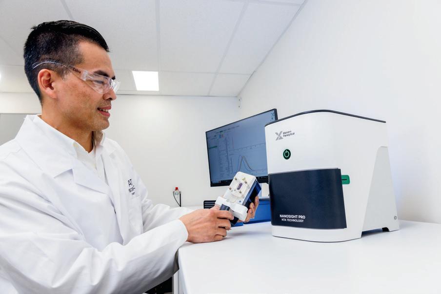
Once considered mere cellular debris, extracellular vesicles (EVs) are now recognised as crucial mediators of intercellular communication, offering exciting possibilities in disease diagnostics and therapeutics. These membrane-bound nanoparticles carry proteins, lipids and nucleic acids (RNA and DNA), making them valuable biomarkers for conditions such as cancer, cardiovascular disease and neurodegenerative disorders. Understanding EV size, heterogeneity and concentration is key to deciphering their biological function and diagnostic potential.
Accurate EV characterisation, however, remains a challenge due to their small size and heterogenous nature. Size and size distribution analysis helps determine their origin and purification efficiency, while changes in concentration may indicate altered cellular activity. The ability to detect and quantify RNA within EVs also provides critical insights into the success of encapsulation in therapeutic applications. While the potential of EVs is immense, realising this potential hinges on our ability to understand and characterise these nanoparticles.
The NanoSight Pro is increasingly being used in EV research due to its ability to track individual particles for high resolution size, size distribution and concentration measurements. Its fluorescence capabilities further enhance EV analysis, allowing researchers to distinguish subpopulations, assess purification efficiency and confirm biomarker presence. The system uses a laser to illuminate particles and capture light scattering through an optical microscope and integrated digital camera. Machine learning algorithms track Brownian motion to determine particle size, while concentration is derived from particle counts.
With NanoSight Pro, researchers can: confirm and quantify EVs using fluorescent membrane dyes, which incorporate into the lipid bilayer to verify their presence; detect tetraspanins — surface proteins essential for EV biogenesis and cargo sorting, serving as accessible biomarkers; characterise RNA cargo without disrupting small EVs, offering insights into EVs’ diagnostic potential and encapsulation success for therapeutics; and perform advanced fluorescence analysis, leveraging multiple laser wavelengths and long-pass filters to work with a variety of EV-targeting labels. The product provides detailed, reproducible EV characterisation for use in precision medicine.
ATA Scientific Pty Ltd www.atascientific.com.au
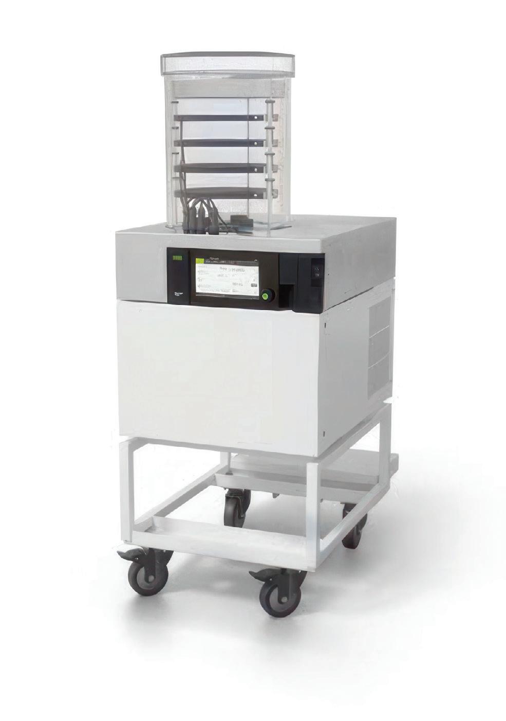
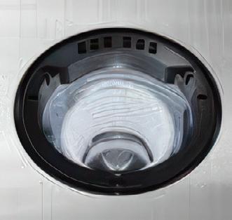
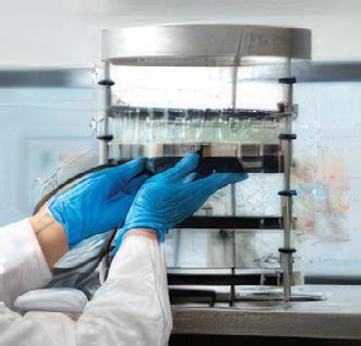
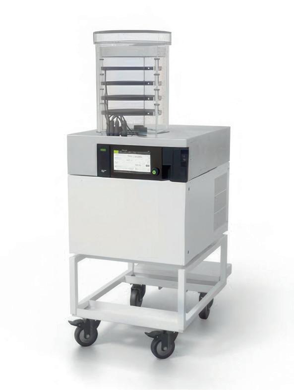
BUCHI’s Lyovapor L-250 freeze dryer has been designed to enable sustainable laboratory practices, introducing EcoStream technology to reduce the environmental footprint of the instrument without compromising on efficiency and reliability.
The compressor design uses natural coolants and achieves an ice condenser temperature of -85°C. The coolant gas used has a low global warming potential (GWP) of just 4, helping to make the instrument environmentally sustainable. Heat output and noise emissions have also been reduced, contributing to a comfortable working environment.
The product comes in basic or pro configurations with touchscreen controls and a wide range of drying chambers for basic and advanced processes. It features a total capacity of 5 kg and a condensing capacity of 4 kg/24 h for aqueous or organic mixtures. This allows for a high degree of flexibility and contributes to a fast, efficient freezedrying process that is focused on sample quality.
Like all instruments in the Lyovapor line, the L-250 provides a stable condenser temperature that allows for complete solvent collection, even for large sample batches. For delicate samples, the instrument features a sample protective state that means such products are treated with care by activating if the sample temperature rises above the set collapse temperature.
In addition to the intelligent and innovative cooling technology, the product features Infinite-Control technology that makes it easy to operate. It can be tailored to the needs of working environments from small research facilities to large-scale industrial laboratories, enabling sustainable innovation. Bio-Strategy - Part of DKSH Group www.bio-strategy.com
Australia’s first implant of a BiVACOR Total Artificial Heart has been announced as an unmitigated clinical success, after the recipient of the implant was discharged from hospital in February and received a donor transplant in March.
The titanium-based Total Artificial Heart is a blood pump that has been designed as a bridge to keep patients alive until a donor heart transplant becomes available, although the long-term ambition is for implant recipients to be able to live with the device without needing a transplant. The world’s first BiVACOR Total Artificial Heart implant occurred on 9 July 2024 at The Texas Heart Institute at Baylor St Luke’s Medical Center in the Texas Medical Center, as part of an early feasibility study, with a suitable donor heart identified eight days later. Since that operation, four more implants have taken place in the US. The Australian implant of the Total Artificial Heart is the first to take place outside the US and the sixth in the world.
The Australian implant — the first in a series of procedures planned as part of the Monash University-led Artificial Heart Frontiers Program, which benefited from a $50 million grant from the Australian Government’s Medical Research Future Fund — took place on 22 November 2024 at St Vincent’s Hospital Sydney in a six-hour procedure led by renowned cardiothoracic and transplant surgeon Dr Paul Jansz. After a few weeks in the ICU, the patient was kept under observation in the ward by clinicians including heart failure and transplant cardiologist Professor Chris Hayward.
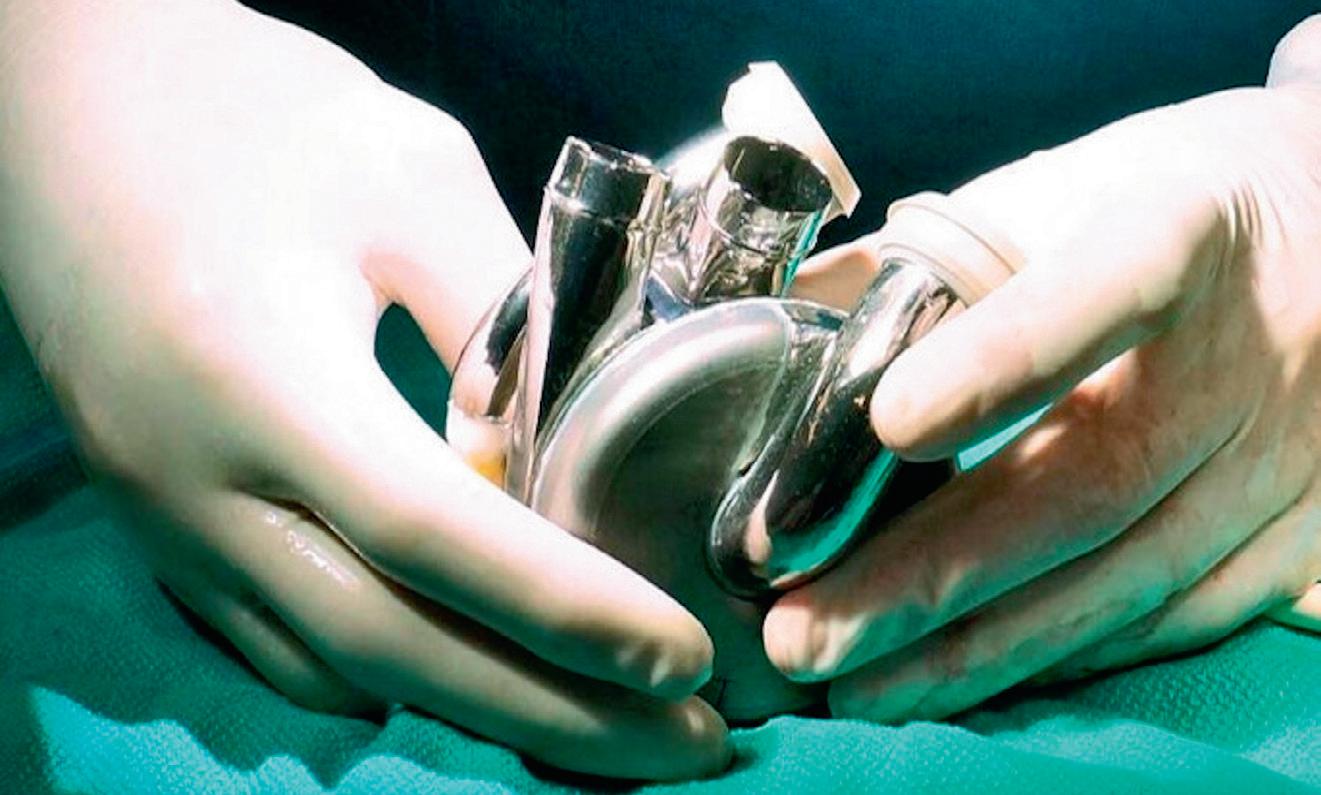
The patient — a man in his 40s who was experiencing severe heart failure — ultimately became the first in the world to be discharged from hospital with the BiVACOR Total Artificial Heart in early February 2025. He finally received a donor heart transplant on 6 March 2025 — a record 105 days after obtaining the Total Artificial Heart.
“It is incredibly rewarding to see our device deliver extended support to the first Australian patient,” said BiVACOR founder Dr Daniel Timms, the inventor of the Total Artificial Heart.
“The unique design and features of the BiVACOR Total Artificial Heart translate into an unmatched safety profile, and it’s exhilarating to see decades of work come to fruition.
“Being able to bring Australia along this journey and be part of the first clinical trials is immensely important to me and something that I set out to do from the very beginning,” he continued.
“The entire BiVACOR team is deeply grateful to the patient and his family for placing their trust in our Total Artificial Heart. Their bravery will pave the way for countless more patients to receive this lifesaving technology.”
Hayward said the BiVACOR Total Artificial Heart would transform heart failure treatment internationally, stating, “The BiVACOR Total
Artificial Heart ushers in a whole new ball game for heart transplants, both in Australia and internationally. Within the next decade we will see the artificial heart becoming the alternative for patients who are unable to wait for a donor heart or when a donor heart is simply not available.”
Monash University’s Vice Chancellor and President, Professor Sharon Pickering, said the Artificial Heart Frontiers Program is an example of the transformative innovation our nation can achieve when universities, industry and government come together.
“Over the next three years and beyond, the AHFP Consortium’s expert engineers, clinicians and researchers will accelerate this Australiangrown, world-leading research and development program to further develop the TAH and related game-changing mechanical circulatory support devices that will deliver substantial global health and economic benefits through urgently needed solutions for advanced forms of heart failure, for which there are limited or no available treatments.
“Monash’s world-leading cardiac and engineering research, and our commitment to working with partners to address global challenges, has been central to the opportunity now before us of developing an Australian-based cardiac device industry.”
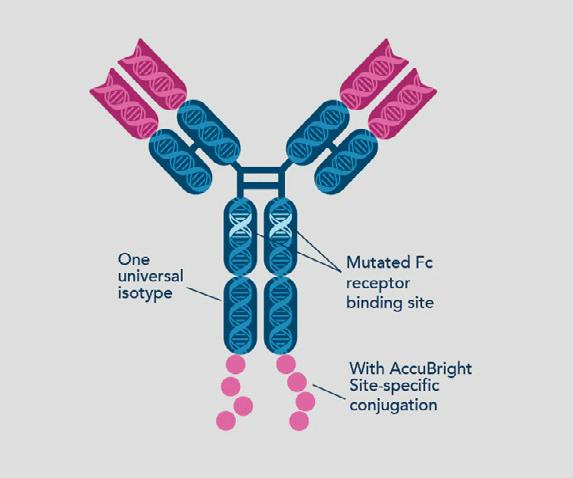
Proteintech has designed recombinant antibodies featuring the company’s FcZero backbone, engineered to silence the Fc domain for clean flow cytometry data.
Non-specific binding of antibodies to Fc receptors in a sample can cause background signal in data. The FcZero-rAb backbone features a mutation in the Fc receptor binding region to eliminate Fc receptor binding. The resulting data is cleaner than using a conventional antibody and Fc receptor block, Proteintech says.
The FcZero recombinant antibody backbone features AccuBright site-specific conjugation technology, designed to allow precise control of the location of the fluorophores on each IgG backbone. This means that fluorophores do not block the antigen-binding region of the antibody. AccuBright also allows for good control over the degree of labelling (DOL), or number of fluorophores conjugated to each antibody, so that signal intensity is consistent from lot to lot.
Popular gold standard clones for flow cytometry are now available with an FcZero-rAb backbone. The antigen-binding regions of these classic clones are combined with a Mouse IgG2a backbone that includes the FcZero mutation to the Fc receptor binding region and AccuBright site-specific conjugation. These antibodies have well-established specificity, plus the high purity and zero non-specific background from the Fc receptor binding.
FcZero-rAb has all the advantages of Proteintech’s Uni-rAb recombinant antibodies, including good specificity, high affinity, lot-to-lot consistency and high purity. It is available in carrier-free formulations.
United Bioresearch Products Pty Ltd www.unitedbioresearch.com.au
Mercodia’s C-peptide ELISA is a highly specific tool that is calibrated against the International Reference Reagent for human C-peptide, IRR84/510. It is a highquality immunoassay for specific quantification of human C-peptide in serum, plasma or urine. The specificity of the utilised antibodies enables measurement of human C-peptide without interference from insulin or proinsulin, and measurement of human C-peptide in mouse and rat models.
Each ELISA kit contains reagents for 96 wells, sufficient for 42 samples and one calibrator curve in duplicate. The microplate strip format (12 strips x 8 wells) is suitable when only a limited number of samples need to be analysed. The kit allows for a sample volume of 25 µL, has an assay range of 100–4000 pmol/L with a detection limit of 25 pmol/L, and has a total incubation time of 2 h 15 min. A Diabetes Antigen Control is available to be used as a two-level control for the kit.
Sapphire Bioscience www.sapphirebioscience.com
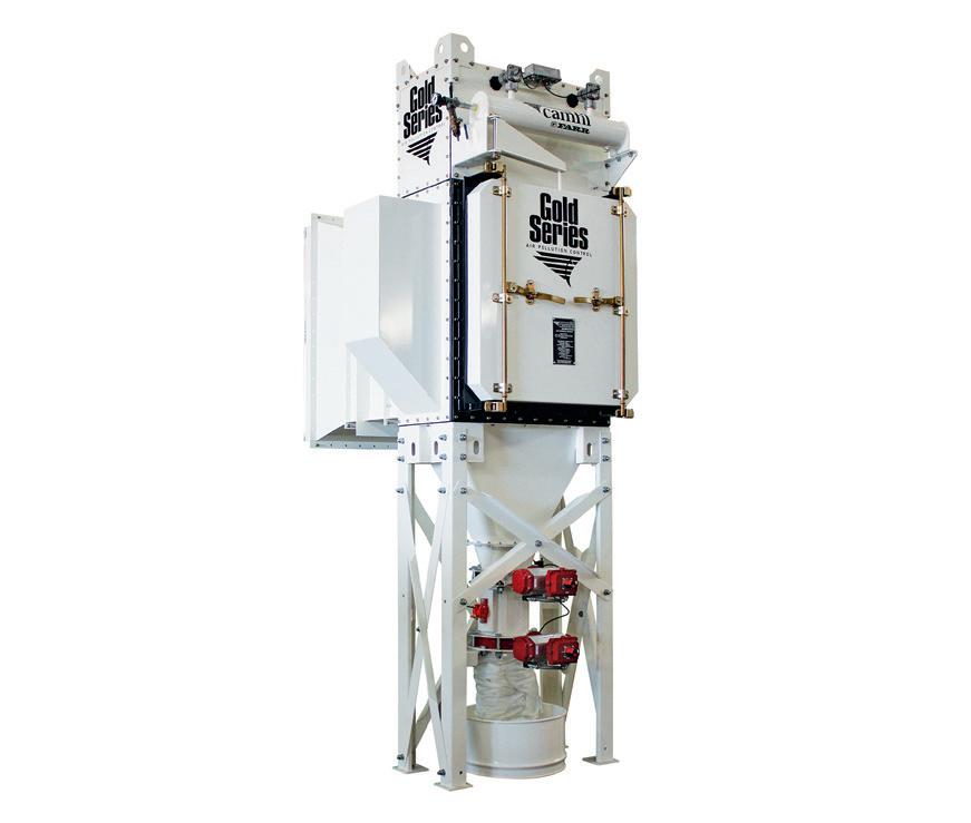
Camfil’s Gold Series Camtain dry dust collector is expertly designed for the pharmaceutical and chemical industries, where effective containment of harmful substances is critical. This modular extraction system efficiently manages medium to high air volumes and complies with stringent ATEX AND NFPA standards, for safety in explosive environments.
Key features include a bag-in/bagout system for safe filter changes, minimising operator exposure during maintenance. Furthermore, the continuous liner system facilitates secure dust discharge, enhancing operational safety.
Equipped with HemiPleat filter technology, the Camtain achieves good filtration efficiency, capturing 99.5% of particles at the most penetrating particle size (MPPS) and exceeding 95% at ePM1. The specially treated filter media repels fine particulates, resulting in reduced pressure drops and extended filter life.
Suitable for industries prioritising health and safety, the Gold Series Camtain not only protects operators but also contributes to regulatory compliance, making it a useful investment for any facility handling hazardous materials.
Camfil Australia Pty Ltd www.camfil.com.au

BIOVIA
For years, computational methods for small molecule drug design have offered numerous algorithms and methodologies to help generate new ideas and guide the iterative process of lead design and optimisation. For a particular drug target, these methods help to identify highquality candidates that may eventually advance to clinical development with fewer experiments and less time in the lab. We have seen great advancements over the years and most recently, AI and machine learning algorithms have become popular in allowing researchers to rapidly explore
more ideas in the chemical space and propose novel structures that a medicinal chemist may not have considered trying out when looking for new drugs.
Traditionally, the computational design tools for biotherapeutics required more expertise and they were application-specific compared to the tools that exist for small molecule therapeutics. For designing certain types of biological therapies, such as monoclonal antibodies, there were also methods such as affinity maturation, humanisation and immunogenicity prediction algorithms.
However, to help answer directly what variation of our biotherapeutic we should make and test next, two recent AI methods, RFdiffusion
The question of what we should make next has challenged the world of drug discovery for decades.
and ProteinMPNN, have totally changed the nature of biotherapeutics discovery. These tools have the potential to change the way we design biotherapeutics by helping to identify novel candidates that the computational and molecular biologists may not have considered.
Generating proteins with AI: RFdiffusion and ProteinMPNN
RFdiffusion is a cutting-edge generative AI algorithm that can ‘diffuse’ a collection of amino acids into a protein structure. The diffusion process starts with a random, noisy collection of atoms and, through a series of controlled refinements, the algorithm adjusts the structure to reduce the noise and move
closer to a biologically realistic and functional protein structure. One common analogy for the diffusion process is developing a photo from a blurry image; iterative processing steps can take an initial grainy image and refine the detail and clarity to produce a final clear picture.
RFdiffusion can be utilised to overcome several different biotherapeutic design challenges, such as engineering a biologic that can bind to a viral protein to neutralise the virus, or to generate enzyme therapeutics that may break down a specific substrate to treat metabolic disorders. Beyond biotherapeutics, the algorithm also has potential to help design proteins for industrial and biotechnological applications such as making enzymes that catalyse specific chemical reactions or proteins that suit very specific conditions including low or high temperature or pH.
ProteinMPNN, on the other hand, is a stateof-the-art neural network that can predict one or more probable protein sequences given a protein structure. This algorithm has been published with success in one of the most critical aspects of protein sequence design — generating sequences that fold into a stable protein/peptide with propensity to crystallise, facilitating the structure determination of these proteins.
One of the strengths of ProteinMPNN is its ability to generate multiple sequence variants, which is invaluable as different variants provide more options to test and identify candidates with the best performance in terms of efficacy, safety and manufacturability. Just as significantly, these variants also provide alternative leads when candidates encounter unforeseen issues in protein optimisation during protein expression, or ADMET challenges such as solubility and immunogenicity.
ProteinMPNN can be used in conjunction with RFdiffusion to generate new protein designs such as new enzymes or antibodies that can be further evaluated for desired properties such as stability, activity, affinity and specificity. Together, they significantly expand the biological space that can be explored in silico before biologists need to commit to expensive and time-consuming physical experimentation, opening up exciting avenues for more intelligent, model- and data-driven workflows driving innovation in biotherapeutic design.
Generating proteins with RFdiffusion and ProteinMPNN
Solutions are being made available to provide researchers easy access to RFdiffusion workflows.
For example, BIOVIA offers a protocol that provides access to motif scaffolding, enabling users to start with a specific part of an existing protein (the motif) and design a complete new protein scaffold that incorporates this motif. This approach allows precise control over the functional regions of the protein, as well as control over the protein scaffold design, via different model weights that suit particular proteins and complexes.
BIOVIA also offers another protocol which allows users access to not only ProteinMPNN, where they can easily define sequence residues for design, but also LigandMPNN and SolubleMPNN models. LigandMPNN can consider protein, small-molecule, nucleic acid and metal ion ligands as additional context for designing sequences, while SolubleMPNN can be used when
protein solubility is part of the design criteria of researchers. Ultimately, users can determine the degree of sequence diversity and confidence desired, and have the ability to control the bias of particular amino acids.
Such tools are examples of how we can expand the ever-growing arsenal of powerful AI tools for molecular modellers and biologists to help answer the question of ‘what to make and test next’ and accelerate the rational design of biologics. In combination with existing physics-based methods already available, researchers can rapidly explore many more possibilities in silico before arriving at the final handful of candidates that are ready to become a successful commercial biotherapeutic or a biological to be used in agriculture, food and beverage, or environmental industries.

Maintaining the viability and functionality of live cell cultures during transport has long been a challenge for biomedical researchers. Traditional transport methods expose cells to at least one freeze-thaw cycle, which is known to cause cell death, reduction in cell proliferative capacity and altered gene expression. Cells are vulnerable to metabolic stress and toxicity from cryoprotectants used in preservation, while temperature fluctuations and mechanical stress can further compromise cell integrity. The Cellbox portable CO2 incubator addresses these challenges by maintaining stable conditions for live cell transport.
The product provides an environment with controlled CO2 levels and temperature, mimicking standard laboratory incubator conditions; this means that cells should remain healthy and viable throughout transit, whether for collaborative research, clinical trials or off-site experiments. Its compact, lightweight design allows for easy handling and secure transportation.
Using battery-powered operation, Cellbox offers up to 48 h of transport time (at a preset temperature of 37°C). The userfriendly interface provides real-time monitoring of CO2 levels and temperature, offering peace of mind during critical shipments.
In a study conducted by UPM Biomedicals, Cellbox Solutions and the Max Plank Institute, midbrain organoids were transported under laboratory incubation conditions (37°C and 5% CO2) for more than 8 h by road in the Cellbox live cell shipper. The results confirmed Cellbox offers seamless transport of delicate cellular models, such as tumour spheroids or organoids, without disruption to their microenvironment. This capability is particularly valuable for preserving advanced models used in drug discovery and disease research.
Available in Ground or Flight models, in the form of the original Cellbox Shipper or the compact Cellbox Go, the portable incubator allows worry-free mobility between labs while safeguarding the integrity of sensitive biological materials essential for bio assays, cell manufacturing, cell storage and 3D cell culture processes.
ATA Scientific Pty Ltd www.atascientific.com.au


Scientists from Duke-NUS Medical School and Singapore’s National Neuroscience Institute have discovered a crucial connection between gut microbes (or lack thereof) and anxiety-related behaviour. Their findings, published in EMBO Molecular Medicine, open up exciting possibilities for new probioticbased therapies to improve mental health.
With the prevalence of mental health disorders having risen over the years, the research team set out to study the role microbes play in anxious behaviour. In preclinical studies, they observed that in a germ-free environment, mice which were not exposed to live microbes showed significantly more anxiety-related behaviour than those with typical resident live microbes.
Further investigation revealed that the increased anxiety was associated with heightened activity in a brain region involved in processing emotions such as fear and anxiety, the basolateral amygdala (BLA). This was further identified to be related to specialised proteins within brain cells known as the calcium-activated potassium (SK2) channels, associated with anxiety behaviour. In conditions when the body and brain are exposed to live microbe metabolites, the SK2 channels act like a clutch, thus preventing neurons from becoming overly excited and firing too frequently.
“Our findings reveal the specific and intricate neural process that links microbes to
mental health,” said co-lead author Associate Professor Shawn Je, from Duke-NUS. “Those without any live microbes showed higher levels of anxious behaviour than those with live bacteria. Essentially, the lack of these microbes disrupted the way their brains functioned, particularly in areas that control fear and anxiety, leading to anxious behaviour.”
To better understand the role of microbes in this process, the researchers introduced live microbes into germ-free mice. This reduced the elevated neuronal activity in the basolateral amygdala and thus SK2 channel activity. As a result, the mice showed significantly less anxietyrelated behaviour —their emotional responses became like those exposed to microbes.
The researchers also tried treatment with indoles — microbial metabolites produced by certain microbes. When the germ-free mice were given indoles, they showed reduced activity in the basolateral amygdala and displayed less anxietyrelated behaviour. This demonstrated that our indigenous microbes produce metabolites, which suggest a direct link between our microbiota and maintaining mental balance.
“Birth is associated with exposure to breast milk, known to contain microbes that can produce
… indoles,” said co-lead author Professor Sven Pettersson, from National Neuroscience Institute. “Indoles are known to be secreted in plants when they are exposed to stress or malnutrition (drought) and in this paper we report a similar mechanism in which indoles can regulate anxiety levels in mammals. That is, different levels of circulating microbial plasma indoles in the blood may reflect different sensitivity and vulnerability to stressful situations and therefore variable risk of experiencing anxiety-related situations.”
The team now hopes to explore clinical trials to determine whether indole-based probiotics or supplements can be effectively used in humans as a natural anxiety treatment. If successful, this could mark the beginning of a new era in mental health care — one where gut microbes help to keep our minds at ease.
“Our findings underscore the deep evolutionary links between microbes, nutrition and brain function,” said Professor Patrick Tan, Senior Vice-Dean for Research at Duke-NUS. “This has huge potential for people suffering from stress-related conditions, such as sleep disorders or those unable to tolerate standard psychiatric medications. It’s a reminder that mental health is not just in the brain — it’s in the gut too.”
he really
Spider-Man’s powers may be the stuff of comic book legend, but in reality, it’s not a bite that gives spiders their edge — it’s their venom. While Marvel’s Venom turns this into a dark and dangerous symbiote, real-world scientists are harnessing the power of spider venom for something far more extraordinary: lifesaving medical treatments. Certain venom-derived peptides are showing promise in treating epilepsy, chronic pain, and even preventing brain damage from strokes and heart attacks.
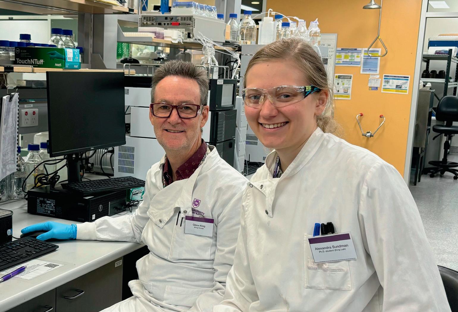
Few scientists embody this frontier of venom research like Professor Glenn King from The University of Queensland’s Institute for Molecular Bioscience (UQ IMB)1. With decades of research in this field, he is reshaping our understanding of nature’s most potent molecules and transforming them into medical breakthroughs. Just like his Marvel counterpart, Glenn doesn’t shy away from a challenge.
At the Lorne Proteins Conference 50th anniversary2, ATA Scientific reconnected with long-time friends, including Prof. Glenn King and his student Alexandra Sundman from IMB UQ. Both tried their luck with the ATA Scientific Lorne Proteins “SPIN TO WIN” Young Scientist competition3 and won. Well, Glenn won, but true to form, he presented the proceeds to Alexandra, with the remark, the certificate was going up with the PM’s Award! His now famous “Happy Dance” sealed the moment, not as Spiderman — but Venom!
Glenn’s association with Lorne dates back 30 years, when he was exploring spider venoms as natural insecticides at the University of Sydney. His reasoning was simple: “Spiders are the best insect killers on the planet — they’ve been perfecting it for 400 million years” 4 But Glenn realised spider venoms could do more than kill pests — they could contribute enormously to medicine. His team is harnessing these potent peptides to develop painkillers, epilepsy treatments, and neuroprotective drugs for stroke victims, while also creating ecofriendly insecticides. These are designed to protect crops and combat disease-spreading pests which destroy approximately 15 per cent of the world’s food supply and spread pernicious diseases such as dengue and malaria5. Tackling even one of these challenges would be a lifetime achievement — but Glenn is determined to solve them all.
A quick search for Glenn reveals not just his TEDx talk6 and university profile but also his global outreach — engaging with groups like Genetic Epilepsy Team Australia and Tory Robinson UK with Epilepsy sparks, even appearing on Spotify. The impact of his research is evident in the heartfelt comments from those it inspires. Glenn has received the 2023 Prime Minister’s Award, was named a Fellow of the Australian Academy of Science in 2024 and he is now the Chief Scientific Officer for Infensa Bioscience, developing spider venom
1. “The University of Queensland,” [Online]. Available: https://imb.uq.edu.au/. [Accessed 27 February 2025].
2. L. P. 2025, “lorneproteins,” 9 February 2025. [Online]. Available: https://www.lorneproteins.org/. [Accessed 27 February 2025].
3. ATA Scienti c, “ATA Scienti c Our Previous Award Winners,” [Online]. Available: https://www.atascienti c.com.au/awards-eventstraining/previous-award-winners/. [Accessed 27 February 2025].
4. Australian Academy of Science, “Professor Glenn King – 2024 Academy Fellow,” [Online]. Available: https://youtu.be/ YmeE5gzEOBU. [Accessed 27 February 2025].
5. BUGS and Drugs. [Online]. Available: https://about.uq.edu.au/experts/1593. [Accessed 27 February 2025].
6. T. Talks, “Deadly cures: how venomous animals could save your life | Glenn King | TEDxUQ,” [Online]. Available: https://youtu.be/ DybEzz6A6z4?si=HXb2zHranTD6wNmW. [Accessed 27 February 2025].
7. RedShiftBio, “RedShiftBio,” [Online]. Available: https://www.redshiftbio.com/products/aurora-tx. [Accessed 27 February 2025].
therapeutics for Australian-based clinical trials — just a few among his many accolades. Glenn thrives on collaboration, driving groundbreaking science while embracing new technologies to answer complex questions. Our first encounter was over a Spectropolarimeter for protein secondary structure analysis — now, with the RedShiftBio Aurora TX7, we have a powerful tool to elucidate the Higher Order Structures of proteins, detect subtle changes and quantitate this using Microfluidic Modulation Spectroscopy (MMS).
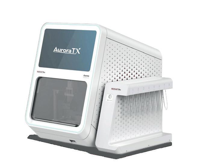
Understanding Higher-Order Structures (HOS) is key to developing next-generation therapeutics, but traditional methods often fall short in sensitivity and resolution. The Aurora TX overcomes these limitations with Microfluidic Modulation Spectroscopy (MMS), providing unmatched accuracy in protein analysis.
For more detailed information, contact ATA Scientific Pty Ltd Ph: +61 2 9541 3500 enquiries@atascientific.com.au
ATA Scientific Pty Ltd www.atascientific.com.au

Recent studies have highlighted the current diversity gap in genomics research, which poses something of a risk for medical misdiagnosis in people from non-European ancestries.

‘Rare’ gene variant found to be common
Between the DNA of any two people, there are millions of letter differences, or genetic variants. These variants determine our looks and how our body functions, and they can also influence our risk of disease, including some of the most common — heart disease, diabetes and some cancers. But while scientific advances in genomics have been transformative for our ability to diagnose and treat disease, most genetic studies have focused on people of European descent.
As part of a study published in the European Heart Journal, Associate Professor Jodie Ingles’ team at the Garvan Institute of Medical Research investigated a genetic variant they found in two individuals of Pacific Island ancestry with cardiomyopathies, who underwent research-based whole genome sequencing at a Sydney clinic. Cardiomyopathies are potentially fatal inherited heart conditions that make it harder for the heart to pump blood around the body, and affect up to one in 500 people.
The researchers found that the variant in the cardiac troponin T gene, which plays a role in regulating heart muscle contractions, is extremely rare in large international population genomic databases. According to first author Dr Alexandra Butters, “This meant that among seemingly healthy people, the variant is not often present. This can indicate it is the cause of an inherited disease, and indeed numerous clinical testing labs had already flagged this as a potential causative variant.”
Butters continued, “We became suspicious about this variant being the cause of the patients’ cardiomyopathy, and when we investigated this further with colleagues who had more genomic reference data of seemingly healthy individuals from various Oceanian regions (comprising Australasia, Melanesia, Micronesia and Polynesia), we found this variant to be really common — 3 to 4% of the population.”
“This is far too common to be the cause of an inherited cardiomyopathy and indicates this is a benign genetic variant,” Butters said. Indeed, when the researchers investigated the variant further, they found it to be also present in Neanderthal DNA, which speaks to how long this variant has been in the population and supports it as unlikely to be causing disease.
According to Butters, “The variant was listed as rare and potentially pathogenic because the datasets we use to understand common and rare genetic variation in healthy people contain predominantly data from people of European background. This means we don’t necessarily know what is common and rare across historically underrepresented ancestry groups, such as those from our regions. The danger of considering this as a cause of disease is that we could then incorrectly use it to tell family members they are or are not at risk of developing disease. That has potential for harm.”
Ingles added, “The reality in the clinic today is that if I’m seeing a family of non-European ancestry, we are less likely to be able to make a genetic
It is … important to study depression in diverse samples because some of the findings might be ancestry specific.

The study authors said they have made major advances identifying genes that are linked to risk of depression, both for newly identified links and by strengthening prior evidence, and showcasing some genes with potential implications for drug development, such as NDUFAF3. The protein that NDUFAF3 encodes has been implicated previously in mood instability, and it is targeted by metformin, the first-line drug for treating type 2 diabetes. Animal studies of metformin have suggested a possible link with reduced depression and anxiety, so this latest finding further suggests that additional research into metformin and depression may be warranted.
Other genes identified in the study may have biologically plausible links with depression, such as a gene linked to a neurotransmitter involved in goal-directed behaviour, and genes encoding a type of protein previously linked with multiple neurological conditions.
Surprisingly, the researchers found less overlap in the genetic hits for depression across ancestry groups than expected, at about 30%, which is less overlap than previously found for other traits and diseases. Therefore, it is even more important to study depression in diverse samples because some of the findings might be ancestry specific.
More genome regions found to increase bipolar risk
In addition, an international team of psychiatric genetics researchers has recently identified 298 regions of the genome containing DNA variations that increase risk for bipolar disorder, in the first large multi-ancestry genomic analysis of the disorder to include data from people of European, East Asian, African American and Latino ancestries. Their work has been published in the journal Nature
Bipolar disorder is an often lifelong mood disorder that impairs quality of life and functional ability, and is associated with suicidality. Bipolar disorder type 1 is characterised by episodes of both mania and depression, while bipolar disorder type 2 includes episodes of hypomania (a less severe form of mania) and depression.
To help elucidate bipolar disorder’s underlying biology, scientists from the international Psychiatric Genomics Consortium conducted a genomewide association study, scanning the DNA of 2.9 million study participants from cohorts worldwide to identify genetic markers that were more common in those with the condition. This involved scanning more than 6.7 million common variations in the DNA sequences among the study participants, more than 158,000 of whom experience bipolar disorder.
As well as identifying the 298 genome regions that increase risk — a more than fourfold increase over the number previously identified — the researchers also found a new region associated with an increased risk within the East Asian samples. Cross-referencing a range of methods, including fine-mapping and other variant-to-gene-mapping approaches, the team identified 36 genes suspected to be relevant to bipolar disorder.
“It is well established that bipolar disorder has a substantial genetic basis, so identifying DNA variations that increase risk is of paramount importance to understanding the condition’s genetic architecture,” said Assistant Professor Niamh Mullins from the Icahn School of Medicine at Mount Sinai, a senior author on the paper.
“In addition to identifying 298 regions of the genome that contain variations that increase risk for bipolar disorder, the 36 key genes we identified as being linked to the condition can now be followed up in a range of experiments to uncover the biological mechanisms through which each relates to the disorder.
“The newly identified genes may also be used in experiments to explore new drug targets and drug development for bipolar disorder.”
The study team also found differences in the genetic characteristics of bipolar disorder between clinical (patients recruited from hospital inpatient or outpatient units), community-based (participants in general population biobanks) and self-reporting (participants in online personal health surveys) participants. These genetic differences are likely to be driven by a higher prevalence of bipolar subtype 1 in the clinical samples versus a higher prevalence of bipolar subtype 2 in the self-reporting samples, which highlights the need for researchers to be mindful of the data-gathering strategies used within their studies of the condition.
According to the research team, the genetic signal of bipolar disorder is related to specific brain cell types, including GABAergic interneurons and medium spiny neurons, in the prefrontal cortex and hippocampus. They also found that cells in the intestine and pancreas are involved, although more research is necessary to further understand this biology.
Senior author Professor Ole Andreassen, from Oslo’s Institute of Clinical Medicine, concluded, “Our research paves the way for the development of improved treatments, earlier interventions and precision medicine approaches that will support clinicians in their decision-making to enable them to manage the condition in the most effective way for each patient.”
As part of a co-exclusive distribution agreement, Beckman Coulter Life Sciences will market and distribute Rarity Bioscience’s superRCA technology assays. The assays are claimed to accelerate the detection of mutations by using flow cytometry with logarithmically higher sensitivity compared to the current gold standard digital PCR (dPCR) method. While traditional flow cytometry research analyses phenotypes, the platform combines a molecular assay based on rolling circle amplification and padlock probes with a flow cytometry readout for high sensitivity and specific multiplex detection of sequence variants, including single point DNA mutations. The assay offers high throughput, resulting in fast turnaround without the need for additional hardware, lowering the barriers to molecular research assays.
Beckman Coulter Australia www.beckman.com.au


Proteomics International Laboratories has launched its first-inclass predictive test for diabetic kidney disease, PromarkerD, in Australia. By enabling proactive management, the company aims to reduce the personal and financial costs associated with what is a life-threatening condition.
The product is a clinically validated blood test that can predict the risk of developing chronic kidney disease in type 2 diabetes patients up to four years before clinical symptoms appear. This early warning provides healthcare professionals with a critical window to intervene, which should enable better patient management and reduce the risk of severe kidney complications, including kidney failure and the need for dialysis or transplant.
The test will initially be available through referral by a healthcare professional in Western Australia and the Northern Territory, before it is made available nationwide.
Proteomics International Pty Ltd www.proteomics.com.au/
Bio-Rad Laboratories has announced its Nuvia wPrime 2A Media, a scalable weak anion exchange and hydrophobic interaction (AEX-HIC) mixed-mode chromatography resin for small- to large-scale biomolecule purification. The resin has been used to remove host cell proteins, high and low molecular weight aggregates and impurities, and DNA from monoclonal and bispecific antibodies, as well as adeno-associated virus.
Built upon Bio-Rad’s Nuvia bead technology, the product features a polyacrylamide (acrylamide polymer) base bead that can be used for laboratory- and small-scale purification workflows, as well as for pilot-scale bioproduction and manufacturing-scale downstream processing. Nuvia resins are designed to have minimal nonspecific binding reactions, due to the hydrophilic nature of the bead polymer, and maintain a constant dynamic binding capacity over a wide range of flow rates. The bead allows for fast mass transfer, is chemically and mechanically stable over a wide range of conditions, and is easy to pack.
Developed in collaboration with Bio-Rad’s partners in industrial biotherapeutic production, the product provides a wide purification design space. The charge state of the resin’s functional ligand can be modulated by the pH of the buffer, enabling the purification of otherwise difficult-toseparate biomolecules from other impurities. Bio-Rad Laboratories Pty Ltd www.bio-rad.com

For as long as scientists have studied Mars, they have wondered what gives the planet its distinctive reddish hue. Now a study led by researchers from Brown University and the University of Bern has suggested that a water-rich iron mineral known as ferrihydrite may be the main culprit behind Mars’s reddish dust, countering the prevailing theory that a dry, rust-like mineral called hematite is the reason for the planet’s colour.
Ferrihydrite is an iron oxide mineral that forms in water-rich environments. On Earth, it is commonly associated with processes like the weathering of volcanic rocks and ash. Until now, its role in Mars’s surface composition was not well understood, but the new study — published in the journal Nature Communications — suggests that it could be an important part of the dust that blankets the planet’s surface.
“From our analysis, we believe ferrihydrite is everywhere in the dust, and also probably in the rock formations as well,” said Adomas Valantinas, a postdoctoral fellow at Brown who started the work as a PhD student at the University of Bern. “We’re not the first to consider ferrihydrite as the reason for why Mars is red, but it has never been proven the way we proved it now using observational data and novel laboratory methods to essentially make a Martian dust in the lab.”
The researchers analysed data from multiple Mars missions, combining orbital observations from NASA’s Mars Reconnaissance Orbiter and the European Space Agency’s Mars Express and Trace Gas Orbiter with ground-level

measurements from rovers like Curiosity, Pathfinder and Opportunity. Instruments on the orbiters and rovers provided detailed spectral data of the planet’s dusty surface; these findings were then compared to laboratory experiments, where the team tested how light interacts with ferrihydrite particles and other minerals under simulated Martian conditions.
“Martian dust is very small in size, so to conduct realistic and accurate measurements we simulated the particle sizes of our mixtures to fit the ones on Mars,” Valantinas said. “We used an advanced grinder machine, which reduced the size of our ferrihydrite and basalt to submicron sizes. The final size was 1/100th of a human hair, and the reflected light spectra of these mixtures provide a good match to the observations from orbit and red surface on Mars.”
The finding offers a clue to Mars’s wetter and potentially more habitable past, because unlike hematite, which typically forms under warmer, drier conditions, ferrihydrite forms in the presence of cool water. This suggests that Mars may have had an environment capable of sustaining liquid water — an essential ingredient for life — and that it transitioned from a wet to a dry environment billions of years ago.
“What we want to understand is the ancient Martian climate and the chemical processes on
Mars — not only ancient, but also present,” Valantinas said. “Then there’s the habitability question: was there ever life? To understand that, you need to understand the conditions that were present during the time of this mineral formation.
“What we know from this study is the evidence points to ferrihydrite forming, and for that to happen there must have been conditions where oxygen, from air or other sources, and water could react with iron. Those conditions were very different from today’s dry, cold environment. As Martian winds spread this dust everywhere, it created the planet’s iconic red appearance.”
As exciting as the new findings are, the researchers are aware that they can’t be fully confirmed until samples from Mars are brought back to Earth, leaving certainty about the mystery of the Red Planet’s past just out of reach.
“The study is a door-opening opportunity,” said Brown planetary scientist Jack Mustard, a senior author on the study. “It gives us a better chance to apply principles of mineral formation and conditions to tap back in time. What’s even more important though is the return of the samples from Mars that are being collected right now by the Perseverance rover. When we get those back, we can actually check and see if this is right.”
The pco.edge 9.4 bi CLHS, from Excelitas Technologies, is a suitable camera for advanced imaging applications in microscopy, life sciences and physical sciences, such as lightsheet fluorescence microscopy (LSFM/SPIM), structured illumination microscopy (SIM), confocal spinning disk microscopy, and dual-image acquisition for spectral analysis, HDR imaging and polarisation studies.
The camera combines high image quality, photon-counting sensitivity, and a large image circle to deliver ultralow readout noise, high resolution and good modulation transfer function (MTF), as it incorporates microlenses and a full pixel height deep trench isolation for crosstalk suppression.
Further, the camera provides a large image circle of 21.6 mm by using a high-resolution 9.4 MP image sensor with a square pixel size of 4.6 µm optimised for lower magnifications and detailed imaging. It is equipped with CLHS Fiber Optic Link channels which are all aligned in one compact plug and capable of transmitting up to 4.9 GBps.

Key specifications and features include good photon counting performance enabled by readout noise <0.3 e¯ and a high quantum efficiency of up to 85%. Dual image recording is possible as the camera captures two square images side by side, supporting image splitting for spectral, HDR or polarisationbased applications.
SciTech Pty Ltd www.scitech.com.au
Pacific Laboratory Products believes that the growth of genomic profiling will be further accelerated by the combination of automation and artificial intelligence (AI). The company’s Axygen hardshell PCR microplates, released by Corning, have an industry standard footprint enabling compatibility with automated liquid handlers and most thermal cyclers — including BioRad, MJ, Eppendorf and ABI real-time and end-point PCR thermocyclers.
Each microplate is engineered to stay warp-free, provide precise positioning for automation, and deliver pressure resistance without distortion. The Axygen brand portfolio provides users with automation-compatible, high-efficiency PCR consumable products that can be used from sample preparation to gene expression analysis to storage.
The standard ANSI/SLAS microplate footprint is available in 96- and 384-well formats. The polycarbonate frame reduces plate distortion at both high and low temperatures, while the thin-walled polypropylene wells allow rapid and efficient heat transfer during PCR. The raised rim design helps to facilitate secure sealing.
Pacific Laboratory Products www.pacificlab.com.au
Multi-component, multi-range portable FTIR gas analyser
Measure 1000’s of gases with single unit
Software offers no-limit on number of gas measurements at once, using powerful PLS algorithms
Data can be downloaded and re-analysed offline for new gases
Built in O2 sensor, heated inlet filter and sampling control


• Siloxane Measurements

Tracking targeted drug delivery is often a challenge due to limitations in current imaging techniques. Now, Japanese researchers have developed a technique that enables direct and highly sensitive tracking of gold nanoparticles (AuNPs) inside the body, which could revolutionise cancer drug delivery.
Gold nanoparticles (AuNPs) are tiny gold particles of 1–100 nm and have unique chemical and biological properties. Due to their potential to accumulate in tumours, they have emerged as promising drug carriers for cancer therapy and targeted drug delivery. However, tracking the movement of these nanoparticles in the body has been a major challenge. Traditional imaging methods often involve tracers like fluorescent dyes and radioisotopes, which give limited visualisation and inaccurate results due to detachment from AuNPs.
Researchers from Waseda University have now introduced a new imaging technique that
uses neutron activation to transform stable gold into a radioisotope of gold and enables long-term tracking of the AuNPs within the body. The study was led by Nanase Koshikawa and Professor Jun Kataoka from Waseda University, in collaboration with Osaka University and Kyoto University. Their findings were published in the journal Applied Physics Letters
“Traditional imaging methods involve external tracers, which may detach during circulation,” Koshikawa said. “To overcome this limitation, we directly altered the AuNPs, making them detectable via X-rays and gamma rays without the use of external tracers.”
For activation of the AuNPs, the researchers irradiated the stable gold nanoparticles with neutrons, converting the stable (197Au) to radioactive
(198Au). The radioactive 198Au emits gamma rays, which are detectable from outside the body.
“Activation of atoms through particle irradiation is a technique that directly alters the material,” Kataoka explained. “The altered elements are sometimes unstable and emit X-rays and gamma rays that make the material visible from outside the body. This does not change the atomic number, and thus the chemical properties of the element are preserved.”
The researchers further confirmed the tracking of these radioactive AuNPs by injecting them into tumour-bearing mice and visualising them using a special imaging system.
Additionally, the study demonstrated their imaging technique for drug delivery of 211At, a radio-therapeutic drug used in targeted cancer therapy. 211At emits alpha particles and X-rays, which are detectable for a shorter duration due to a shorter half-life. The researchers labelled 211At with the radioactive AuNPs, forming 211At-labeled (198Au) AuNPs. This approach provided long-term imaging of the drug due to the longer half-life (2.7 days) of 198Au, overcoming the limitations of the short half-life of 211At.
“211At has a half-life of only 7.2 hours, and hence its emitted X-rays disappear within two days, but with the (198Au) AuNPs labelling, we were able to track the drug’s distribution for up to five days using gamma rays from 198Au, which has a longer half-life of 2.7 days,” said co-author Atsushi Toyoshima, from the Institute for Radiation Sciences at Osaka University.
The study could lead to major advancements in drug delivery systems, with the direct tracking of AuNPs inside the body potentially paving the way for more effective cancer treatments with precise monitoring of drug distribution. The study could also open new possibilities for realtime pharmacokinetic studies, ensuring improved drug safety and efficacy.
“AuNPs are being actively researched for medical applications,” said co-author Hiroki Kato, from the Institute for Radiation Sciences. “We developed a simple and scalable technique for tracking AuNPs that could significantly advance nanomedicine while driving the optimisation of gold-based nanomaterials.”
Co-author Assistant Professor Yuichiro Kadonaga, also from the Institute for Radiation Sciences, added, “We plan to enhance the imaging resolution and extend this technique to various nanoparticle-based systems. By further refining neutron activation imaging, we aim to make drug monitoring a clinical reality, potentially revolutionising the field of imaging technologies.”
With the JENOPTIK GRYPHAX series of microscopy cameras, users can upgrade their old camera to the latest generation — such as the NAOS, KAPELLA, PROKYON, RIGEL or POLARIS — and continue using their microscope instead of replacing the complete system and accessories. Suitable applications for the series include bright-field, dark-field, differential interference contrast (DIC), polarisation and fluorescence microscopy.
The JENOPTIK GRYPHAX series is specifically designed for microscopy imaging, with cameras that provide highquality, low-noise images with reproducible and true colour. Short exposure times and high image refresh rates make the cameras time-efficient and convenient to use.
The NAOS colour camera offers 20 MP and 30 fps; the PROKYON offers 20 MP and 60 fps; and the KAPELLA offers 2.3 MP and 60 fps. The mono versions are the RIGEL (2.3 MP, 60 fps) and the POLARIS (3.2 MP, 22 fps). The cameras are made in Germany with a 24-month warranty.
The user can position and focus their camera quickly and easily, even on moving objects. The USB cameras can even deliver live images at video speed in high quality due to the state-of-the-art, back-illuminated CMOS sensor technology used in the cameras. The user-friendly and intuitive microscopy imaging software is part of every camera package (including future updates) and runs on Windows/Mac/Linux. In addition, there are many other third-party software options for microscopy imaging as well as a free SDK.
SciTech Pty Ltd www.scitech.com.au

HEMCO’s C-Frame Tables are designed and built to support lab instrumentation, small hoods and workstation procedures.
Featuring a welded steel tube construction, the table has a chemical-resistant epoxy powder coat finish. It is offered in either a fixed or mobile model with locking casters.
The ergonomic C-frame design allows for convenient sitting with full knee space. The factory-installed countertop is made of chemical-resistant phenolic resin.
The table is manufactured in three sizes, including 0.76, 0.91 and 1.21 m-wide models in either 0.76 or 0.91 m heights. All tables are 0.61 m deep.
HEMCO Corporation www.hemcocorp.com

Largest in-house manufactured human antibody portfolio to meet all your needs. We offer all types and formats of antibodies to help you reach your goal.
— and it's going to get worse

Hot weather is responsible for an average of almost 50,000 years of healthy life lost to cardiovascular disease every year among people in Australia, according to a study published in the European Heart Journal. And the news gets worse, with the study suggesting this figure could double, or even triple, by the middle of the century if we continue with the current trend of greenhouse gas emissions.
The World Meteorological Organization’s State of the Global Climate report recently confirmed that 2024 was likely the first calendar year to be more than 1.5°C above the pre-industrial era, with a global mean near-surface temperature of 1.55°C above the 1850–1900 average. Furthermore, each of the past 10 years was individually the 10 warmest years on record, and each of the past eight years has set a new record for ocean heat content. But while a hotter climate can increase the risk of dangerous weather events such as bushfires and floods, the heat itself can also be a serious burden on our health.
“When the weather is hot, our hearts have to work harder to help us cool down,” said study leader Professor Peng Bi, from The University of Adelaide. “This added pressure can be dangerous, especially for people with cardiovascular disease.
“Many of us have experienced how a warming climate can make us feel unwell, particularly during longer periods of extreme heat. However, it’s still not clear exactly how many people are living with serious heart disease or dying early because of higher temperatures, and we need to understand how this burden will increase in the future.”
To calculate the current impact of high temperatures, Bi and his fellow study authors used data from the Australian Burden of Disease Database on illness or death caused by cardiovascular disease between 2003 and 2018. Then they used a statistical model to calculate how much cardiovascular disease or death can be attributed to hot weather in different parts of Australia and the country as a whole.
Using a measure called disability-adjusted life years (DALYs), which quantifies the number of years of healthy life lost through either illness or death, the researchers found that there was an average of 49,483
years of healthy life lost annually to cardiovascular disease caused by hot weather, equating to around 7.3% of the total burden due to illness and death from cardiovascular disease. Most of these years were lost due to death, rather than illness.
The researchers then used their model to look at the likely impact of climate change driven by greenhouse gas emissions in the future. They used two of the climate change scenarios outlined by the Intergovernmental Panel on Climate Change: a scenario where emissions stabilise (Representative Concentration Pathway 4.5 or RCP4.5) and a scenario with continually rising emissions (RCP8.5). They also looked at the impact of population growth and how people might adapt to cope with higher temperatures.
The model shows that by 2030, the number of DALYs lost due to cardiovascular disease caused by hot weather will increase by 83.5%, reaching 90,779.7 DALYs, under the RCP4.5 scenario. By 2050, this number is expected to rise further to 139,828.9 DALYs, a 182.6% increase. Under the more severe RCP8.5 scenario, the DALYs are projected to increase by 92.7% to 95,343.0 DALYs in 2030 and by 225.6% to 161,095.1 in 2050.
“This study combines several key factors — climate change, population shifts and adaptation strategies — to give a full picture of the disease burden across Australia,” Bi said.

By 2030, the number of DALYs lost due to cardiovascular disease caused by hot weather will increase by 83.5%.
“Predicting future disease burden always comes with some uncertainty, and our models rely on assumptions that may not capture every real-life detail. However, despite these uncertainties, the comprehensive nature of our approach makes the study especially valuable for planning future climate change adaptation and mitigation strategies.”
Bi noted that the study’s findings are likely relevant in many parts of the world — not just Australia — although the specific risks may vary depending on local climates and population demographics. The good news is that the model
also shows that it would be possible to lower the impact of high temperature on cardiovascular disease with strategies that help people adapt to hotter weather.
“Our research … highlights the importance of taking precautions during hot weather, such as staying hydrated, finding cool environments and seeking medical help when needed,” he said.
“Our findings also call for urgent investment in adaptation and mitigation strategies, including urban cooling plans, public health campaigns and improved emergency responses during hot weather.”
Optiscan Imaging has announced InForm, a microscopic medical imaging device designed to transform pathology by delivering real-time digital insights across the full pathology workflow at point of contact with a tissue sample. Utilising proprietary technology, the device is crafted for pathologists and pathology technicians to obtain immediate insights to improve diagnostics across laboratory medicine, pathology practice and point-of-care practices to enhance real-time decision-making and redefine diagnostic workflows.
Traditionally, tissue pathology has been a slow and fragmented process, dependent on analog workflows, multiple time-intensive procedures, resource-intense slide preparation, and delayed interpretation and result generation. Optiscan’s imaging technology is designed to transform this approach by delivering point-of-care digital pathology insights, streamlining workflows, and enhancing both diagnostic efficiency and precision.
The InForm device has been designed for ease of use across the full spectrum of anatomical pathology applications: from fresh single sample diagnoses in the operating room (OR), to specimen margin assessment adjacent to the OR, frozen section biopsy replacement for more immediate assessments during surgery, and routine laboratory assessments on fresh or fixed tissue generating digital microscopic images. The latter images are comparable to those obtained with benchtop white light microscope using traditional histopathology methodologies, but without any time-consuming or complicated tissue processing.
The device has a spatial resolution of 0.55, offering up to 1000x real magnification at point of care, compared to the traditional 40-100x magnification on conventional light microscopes. It produces digital images instantly, and does not use any consumables beyond the topical dye preferred by the pathologist assessing the tissue.
The product is DICOM-compliant and PACS-enabled, allowing it to be connected to hospital or laboratory archiving systems in a fashion similar to that of radiology platforms such as CT or MRI. This should enable the seamless adoption and integration of digital pathology into the wider healthcare space. Additionally, the device is designed to integrate with Optiscan’s cloud-based telepathology streaming platform, which allows remote consultation in real time anywhere in the world, and which is expected to be revealed in mid-2025.
Optiscan Pty Ltd
www.optiscan.com/

A research team led by QIMR Berghofer and Emory University has shown that a potential new targeted therapy for childhood brain cancer is effective in infiltrating and killing tumour cells in preclinical models. The researchers said their findings, which have been published in the journal Nature Communications, are potentially transformative for the treatment of the most common childhood brain cancer (medulloblastoma) and could apply to other brain cancers as well.
Brain cancer is the second leading cause of death in children in the developed world. For the children who survive, standard treatments have long-term impacts on their development and quality of life, particularly in small children and infants. When medulloblastoma recurs, the prognosis is usually extremely poor.
“Current treatments, including radiation and chemotherapy, often eliminate most of the tumour, but sometimes fail to eliminate cancer stem cells,” said Emory University Professor Timothy Gershon. “These cancer stem cells can regrow the tumour after treatment, causing fatal recurrence.”
The research teams collaborated with US biopharmaceutical company Curtana Pharmaceuticals, which has developed an experimental drug called CT-179. The researchers found that the drug effectively targets the protein OLIG2, which is a known stem cell marker crucial in the initiation and recurrence of brain cancers.
“We show that CT-179 treatment specifically disrupts cancer stem cells,” said Gershon, who is also a paediatric neurologist at Children’s Healthcare of Atlanta and Director of the Children’s Center for Neurosciences Research.
“Combining CT-179 with treatments such as radiation therapy treats the whole tumour more effectively, including both stem cells and tumour cells that are not stem cells. Adding CT-179 to combinations of treatments may bring new efficacy to brain tumour therapy.”
Professor Bryan Day, who leads QIMR Berghofer’s Sid Faithfull Brain Cancer Laboratory and is Co-Director of the Children’s Brain Cancer Centre, described the findings as a breakthrough, made more significant because they result from independent studies.
“Children with brain cancer urgently need more effective and less toxic treatments,” Day said. “Our study demonstrated that the drug CT-179, used in combination with standard radiation therapy, can cross the blood–brain barrier and penetrate the tumour. It prolonged survival in a range of preclinical medulloblastoma models, delayed recurrence of the disease, and increased the effectiveness of radiotherapy.”
QIMR Berghofer postdoctoral researcher Dr Yuchen (Michelle) Li, who was joint first author on the study, noted that the blood–brain barrier is a big obstacle in terms of treating brain cancer.
“As a small molecule drug, CT-179 can penetrate the blood–brain barrier and could also be taken orally, making it easier to administer to young patients,” Li said.
“In our experimental models in the lab, the drug ‘hangs around’ in the brain. When used in combination with radiotherapy, the hope is that overall treatment is more effective and less toxic, which reduces the long-term therapy-induced side effects that have such an impact on patients.”
The team’s findings complement the results of another study, published concurrently in Nature Communications, led by Professor Peter Dirks from the University of Toronto. That study identified the OLIG2 protein as a key regulator of the tumour’s growth transitions and presented a novel therapeutic angle, highlighting a shift from general tumour treatment to precise interventions targeting tumour-initiating cells.
“Our study demonstrated that the OLIG2 protein is a critical driver of the complex early stages of medulloblastoma tumor formation, making it a highly promising treatment target,” said Dirks, who is also Neurosurgeon-In-Chief and a Senior Scientist at The Hospital for Sick Children.
“We showed that inhibiting the OLIG2 protein with the CT-179 drug prevented cancer stem cells from changing to a proliferative state, effectively blocking the growth and recurrence of tumours. This could have potentially profound implications for treatment in the future.”
With both studies now published, Day said the next step is to undertake clinical trials.
“We’ve been working hard toward this goal with our collaborators, particularly in the United States and here in Australia, and we are now seeking funding to advance to first-in-human clinical testing of CT-179 in patients with brain cancer,” he concluded.
14th ISO/TC 276 Biotechnology Plenary and Working Group Meetings
May 31–June 7, Cairns
Science Industry Australia (SIA) and Standards Australia are hosting the 14th ISO/ TC 276 Biotechnology Plenary and Working Group Meetings in Cairns. This is the first time that the event will be held outside the Northern Hemisphere. The meeting will comprise three days of plenary sessions and five days of concurrent Working Group sessions, bringing together expert delegates from 42 countries covering six continents. Delegates will actively participate in productive discussions and exchange ideas in a collaborative environment, while formulating consensus-based, market-relevant international standards that support innovation and provide solutions to global challenges.

The ISO/TC 276 Committee is responsible for 38 ISO standards, 26 of which are currently under development or revision. It examines standardisation needs and develops international standards in the field of biotechnology processes, and has recently expanded to include nucleic acid- and protein-based biodevices, as well as organoid and organ-on-chip technologies. The aim of the committee is to utilise biotechnology to enable a more globally sustainable future. adminSIA@scienceindustry.com.au
AusMedtechInvest 2025
May 6, Sydney www.ausbiotechinvestment.com.au/aboutausmedtechinvest
AusMedtech 2025
May 6–8, Sydney www.ausmedtech.com.au
AUS-oMicS 2025
May 18–21, Cairns www.ausomics.com
Lab Leadership 2025
June 5–6, Melbourne www.marcusevans.com/conferences/lab-leadership
ANZAN Annual Scientific Meeting
June 10–13, Brisbane and online www.anzanasm.com.au
15th International NanoMedicine Conference
June 16–18, Sydney www.oznanomed.org
FoodTech Qld
June 19–20, Gold Coast foodtechqld.com.au
ASERA56 Conference
July 1–4, Melbourne www.asera.org.au/2025-conference/
ASM National Meeting 2025
July 14–17, Adelaide www.theasmmeeting.org.au
Accreditation Matters 2025
July 30–31, Sydney www.accreditationmatters.com.au/am2025
National Science Week 2025
August 9–17, Australia-wide www.scienceweek.net.au
AIFST25 Convention
August 12–13, Melbourne www.aifst.asn.au/AIFST-2025-Convention
Beaker Street Festival 2025 August 12–19, Hobart www.beakerstreet.com.au
73rd CSANZ Annual Scientific Meeting August 14–17, Brisbane www.csanzasm.com
Science at the Shine Dome 2025 September 2–4, Canberra www.science.org.au/news-and-events/events/ science-shine-dome/science-at-the-shine-dome-2025
ASCIA 2025 Conference September 2–5, Brisbane and online ascia2025.com
IVC-23: 23rd International Vacuum Congress September 15–19, Sydney ivc23.org
Australian Society of Cytology Annual Scientific Meeting September 26–28, Perth www.cytology.com.au/Web/Web/Events/Event_ Display.aspx?EventKey=52ASM
Australasian Radiation Protection Society (ARPS) 2025 Conference October 19–22, Melbourne arpsconference.com.au
AusBiotechInvest 2025 October 21, Melbourne www.ausbiotechinvestment.com.au/flagshipevents-ausbiotechinvest
AusBiotech 2025 October 21–24, Melbourne www.ausbiotech.org/conference-information
Acoustics 2025 — Sounds of the Sunset Coast November 12–14, Busselton www.acoustics.org.au/events/acoustics-2025
CYTO-Connect Perth November 27–29, Perth cytoconnectperth2025.com.au
Westwick-Farrow Media
A.B.N. 22 152 305 336 www.wfmedia.com.au
Head Office
Unit 5, 6-8 Byfield Street, (Locked Bag 2226) North Ryde BC NSW 1670, AUSTRALIA Ph: +61 2 9168 2500
Editor Lauren Davis LLS@wfmedia.com.au
Publishing Director/MD Janice Williams
Art Director/Production Manager Linda Klobusiak
Art/Production Marija Tutkovska
Circulation Alex Dalland circulation@wfmedia.com.au
Copy Control Ashna Mehta copy@wfmedia.com.au
Advertising Sales
Sales Manager: Andrew Jackson Ph: 0400 604 646 ajackson@wfmedia.com.au
Kerrie Robinson Ph:0400 886 311 krobinson@wfmedia.com.au
Tim Thompson Ph: 0421 623 958 tthompson@wfmedia.com.au
If you have any queries regarding our privacy policy please email privacy@wfmedia.com.au