
Setting the Pace
SPRING 2024
A MAGAZINE FOR FRIENDS OF THE UNIVERSITY OF WISCONSIN SCHOOL OF VETERINARY MEDICINE On Call
Revolutionizing racehorse safety

To honor his career and achievements as dean, the Dean Mark D. Markel Strategic Impact Fund will support new and innovative initiatives and strategies at the School of Veterinary Medicine.

This permanent endowment will focus on ideas that have the largest potential to improve veterinary medical education and One Health, an integrated, unifying approach that aims to sustainably balance and optimize the health of people, animals and the environment.
Make a gift today. www.supportuw.org/giveto/MarkelFund
Questions? Please contact:
Pat Bowdish | 608-332-4750 | pat.bowdish@supportuw.org
Heidi Kramer | 608-327-9136 | heidi.kramer@supportuw.org


Science and ingenuity come together at the SVM to develop a new prerace screening protocol that is not only improving the health and welfare of Thoroughbred racehorses, but may also help solve one of the horseracing industry’s most complex problems.

SVM researchers have uncovered new information about orofacial development in mice that could one day help reduce the risk of cleft lip and palate in humans – common birth defects that impact more than 175,000 newborns around the world each year.

It is an exciting time here at the University of Wisconsin School of Veterinary Medicine. We truly are facing a new era at the school. In February, four well qualified candidates were interviewed for the opportunity to serve as the next Dean of the SVM. As you’ll see in the story on page 5, UW-Madison Provost Charles Isbell announced last month that Jonathan Levine will be the fourth Dean in the school’s 41 year history. I want to thank Dr. Kristen Bernard and the entire search committee for their hard work in successfully identifying the future leader of the UW-Madison School of Veterinary Medicine.

We are all excited to announce the opening of our new North Building. We currently anticipate that our new hospital will be open and fully functional in May, along with our second-floor research laboratory spaces, Badger Market, a clinical pathology laboratory and rooftop terrace, all available for our faculty, staff, students and clients and their animals. We will continue to completely remodel our current South building small animal hospital this coming year and anticipate completing the entire project by the end of 2025. We also look forward to the opening of our large animal arena sometime this summer, enabling us to better serve our large animal clients in the coming decades.
We continue to actively work on the school’s new curriculum, with plans to vote on and hopefully approve the curriculum for our students’ first year this month. The goal is to begin the implementation of our new curriculum in the fall of 2025. Our curriculum is focused on competency-based veterinary education, with enhanced clinical skills and professional skills training. Although I’m firmly convinced that we currently provide an outstanding education to our students, I do believe that our new curriculum will even better prepare our veterinary medical students to be the future leaders of this amazing profession.
As the end of my deanship approaches this August, I reflect on all that we’ve achieved over the last decade, and I’m truly thankful to our faculty, staff and students for all that they do to make the School of Veterinary Medicine an amazing place to learn, discover and provide exceptional veterinary medical care. We wouldn’t be able to do any of this without the support of friends and committed donors who have paved the way for our successes. For that, I thank each and every one of you. Have an amazing spring!
Mark D. Markel, Dean @uwvetmeddean
Administration
Mark D. Markel , Dean
Richard Barajas, Assistant Dean for Diversity, Equity and Inclusion
Fariba Kiani , Chief Financial Officer
Lynn Maki , Associate Dean for Student Academic Affairs
Nancy Parkinson , Assistant Dean for Human Resources
Peggy Schmidt, Associate Dean for Professional Programs
Chris Snyder , Associate Dean for Clinical Affairs and Director, UW Veterinary Care
M. Suresh , Associate Dean for Research and Graduate Training
Kristi V. Thorson , Associate Dean for Advancement and Administration and Chief of Staff
Lauren Trepanier , Assistant Dean for Clinical and Translational Research
Editorial
Editor/Writer : Gian Galassi
Contributing Writers: Alicia Artus
Photography : Seth Moffitt, John Maniaci
Design : Kelly Bird
Connect with Us
Please send your feedback and
www.vetmed.wisc.edu
www.uwveterinarycare.wisc.edu
facebook.com/uwvetmed
facebook.com/uwveterinarycare
twitter.com/uwvetmed
twitter.com/uwvetmeddean
youtube.com/uwvetmed
instagram.com/uwvetmed
linkedin.com/school/uwvetmed

This expert response comes from Dr. Amy Nichelason , clinical assistant professor of primary care services.
Question: As a dog owner who likes to spend time on or near the water during the summer, what do I need to know about the threat of blue-green algae?
Answer: Much like humans, dogs are drawn to water during the hot summer months, but they don’t think twice before jumping into a lake for a swim or a drink on a sweltering day. As the temperatures rise, however, so too does the number of dogs who end up in the emergency room after ingesting dangerous levels of blue-green algae – a toxic cyanobacteria that can result in severe neurologic damage, liver damage, or death. These toxic algal blooms, which are most often found on stable, slow-moving, or stagnant water during warmer summer months, typically appear as pea-green slime on the water’s surface, but they can also appear as green, brown, red, pink, blue, and sometimes even foamy. Algal blooms can also emit a foul odor that is often described as swampy or fishy. However, there is no way to visually confirm if an algal bloom is toxic or not. If you see one, you should avoid it and alert public health authorities.
The good news is there are some simple rules you can follow that will go a long way in keeping you and your pet happy, healthy and safe this upcoming summer:
• Check with the local public health department about blue-green algae outbreaks in the area prior to heading out for a day of swimming and/or boating with your dog.
• Never allow your dog to drink, swim or paddle in stagnant ponds, lakes or other bodies of water that have obvious signs of blue-green algae. Dogs can get fatally sick from just licking the toxic bacteria off their fur or paws.
• If you suspect your dog was exposed, immediately wash your pet off with clean water and contact your veterinarian for advice.
If your dog is showing any of the following symptoms after suspected exposure, you should immediately go to a local ER veterinarian: diarrhea or vomiting; drooling; weakness or tremors, disorientation/confusion; collapse/ unconsciousness; seizures; or breathing difficulties.
There is no antidote, so prevention is key. Symptoms could arise anywhere from 15 minutes to several days after exposure. Early intervention is critical; if caught early your veterinarian may be able to flush the toxins from your dog’s system before they become severely ill. However, exposure can be fatal or result in long term complications.


As this magazine was going to print, UW-Madison Provost Charles Isbell announced that Jonathan Levine has been selected as the next leader of the University of Wisconsin School of Veterinary Medicine.
Dr. Levine is a professor of veterinary neurology and the head of small animal clinical sciences at Texas A&M University where he has also served as interim director for the Texas A&M Small Animal Hospital.
He earned his undergraduate degree and doctor of veterinary medicine degree from Cornell University before completing a small animal rotating internship at Colorado State University. He completed residency in neurology at both Texas A&M and the University of Missouri.
Dr. Levine will step into the role on August 1, 2024, replacing Dean Mark Markel who announced last summer that he will return to the faculty as a professor of large animal surgery.
Have a question for our veterinary medical experts?
Send it to oncall@vetmed.wisc.edu
For health issues concerns needing immediate attention, please contact your veterinarian directly.
More information about Dr. Levine and his vision for the SVM will be shared in future issues of this magazine.
Knowing what dogs like to watch could help veterinarians assess their vision

Ever wonder what kind of TV shows your dog might choose to watch if they could work the remote control? New research from the University of Wisconsin School of Veterinary Medicine (SVM) has some answers, but the study was more interested in solving a longstanding problem in veterinary medicine than turning canine companions into couch potatoes.
According to Freya Mowat, veterinary ophthalmologist and associate professor in the SVM’s department of surgical sciences, the goal of the study was to determine factors that influence a dogs’ interest in interacting with video content and to see if age or vision were related to this behavior. Ultimately, they hope to develop more sensitive ways for veterinarians to assess vision in dogs− something that has been sorely lacking in veterinary medicine.
“The method we currently use to assess vision in dogs is a very low bar. In humans it would be equivalent to saying yes or no if a person was blind,” says Mowat. “We need more sensitive ways to assess vision in dogs, using a dog eye chart equivalent. We speculate that videos have the potential for sustaining a dog’s attention long enough to assess visual function, but we didn’t know what type of content is most engaging and appealing to dogs.”
Published recently in the journal Applied Animal Behaviour Science, the study found that dogs are most engaged when watching videos that feature other animals, and content about their own species is the most popular. But if a National Geographic documentary about canine evolution seems too highbrow for your four-legged friend, Scooby Doo might be a perfectly acceptable option as well.
To better understand the type of content dogs might be most attracted to on screen, Mowat created a web-based questionnaire for dog owners about the TV-watching habits of their canine companions and then made it available to people around the globe.
“We need more sensitive ways to assess vision in dogs, using a dog eye chart equivalent. We speculate that videos have the potential for sustaining a dog’s attention long enough to assess visual function, but we didn’t know what type of content is most engaging and appealing to dogs.”

Participants were asked to answer questions about the types of screens in their home, how their dogs interacted with screens, the kinds of content their dogs interacted with the most, as well as information about their dog’s age, sex, breed, and where they live. They were also asked to describe the behaviors their dogs exhibited when watching screen-based content. Most commonly emotions were described as “active” (running, jumping, tracking etc.) compared with “passive” (lying, sitting etc.). Vocalization (barking, whining, growling) was a commonly described behavior.
Dog owners were also given the option of showing their dog four short videos that featured subjects of possible interest, including a panther, a dog, a bird and traffic moving along a road. They were then asked to rate their dog’s interest in each video and how closely the dog tracked the moving objects on the screen.
Mowat received 1,600 responses from dog owners across the world, including the US, Canada, United Kingdom, European Union and Australasia. Of those respondents, 1,246 ultimately completed the study. Mowat says she plans to build on the results of this study by focusing future research on the development and
optimization of video-based methods that can not only assess changes in visual attention as dogs age, but also answer questions that could help our four-legged friends age as gracefully as possible.
“We know that poor vision negatively impacts quality of life in older people, but the effect of aging and vision changes in dogs is largely unknown because we can’t accurately assess it,” she says. “Like people, dogs are living longer and we want to make sure we support a healthier life for them as well.”
Another future goal for Mowat is to compare how a dogs’ vision ages compared with the human or humans they share a home with.
“Dogs have a much shorter lifespan than their owner, of course, and if there are emerging environmental or lifestyle factors that influence visual aging, it might well show up in our dogs decades before it shows up in us,” she explains. “Our dogs could be our sentinels—the canine in the proverbial coal mine.”
Interesting highlights from the study:
• Age and vision were related to how much a dog interacted with a screen.
• Sporting and herding dog breeds appear to watch all content more than other breeds.
• Video content featuring animals was the most popular, with other dogs being by far the most engaging subjects to watch.
• Humans do not appear to be very appealing for dogs to watch, ranking 9th out of 17 predetermined categories.
• Cartoons were engaging for more than 10% of dogs.
• Movement on screens was a strong motivator for screen attention.
 PHOTOGRAPHY: MEDIA SOLUTIONS
Freya Mowat, veterinary ophthalmologist, and associate professor in the SVM department of surgical sciences.
PHOTOGRAPHY: MEDIA SOLUTIONS
Freya Mowat, veterinary ophthalmologist, and associate professor in the SVM department of surgical sciences.


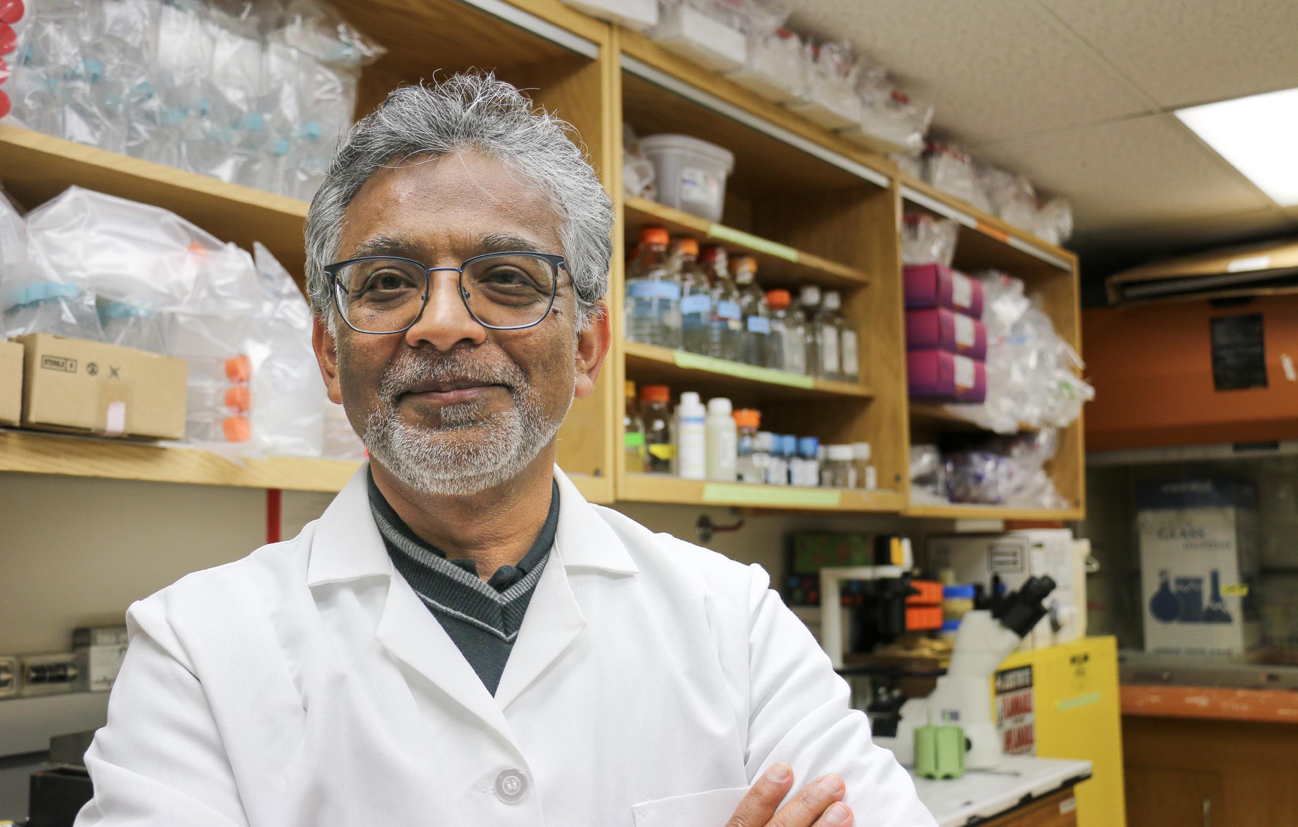
As new research continues to improve the care of humans who develop cancer, SVM veterinary oncologist Dr. David Vail is among those who investigate how these discoveries can also help our four-legged friends.
Cancer is the number one cause of death for adult dogs and cats, in part because our companion animals are living longer than in the past, and cancer risk rises with age.
“We see it all the time, and for some forms of cancer we don’t have a good standard of care,” says Vail, who holds the Barbara A. Suran Chair in Comparative Oncology at the UW School of Veterinary Medicine and is a researcher with the UW Carbone Cancer Center. “At the end of the day I’m a veterinarian, and so I want to help my patients as well.”
Comparative oncology seeks to investigate cancer diagnostics, treatments and prevention in all species in hopes that new technologies and treatments can be developed faster and translated into both the veterinary care of companion animals as well as preliminary data that can inform human clinical trials.

The most common malignancies in dogs and cats include skin cancer, lymphomas and sarcomas. Because comparative oncology studies animals who naturally develop cancer, Vail looks at the authentic conditions of malignant cell development and spread, as well as the natural state of these animals’ immune systems.
Cancer spreads because it develops a way to circumvent the body’s natural immune response to abnormal cells. Immunotherapy, which jumpstarts the body’s natural immune response to attack or prevent cancer, has become a key tool for human cancer care. Vail, along with fellow cancer researchers at UW Carbone, runs clinical trials in companion species using new immunotherapies with the hope of improving care in both veterinary and human patients.
“Unfortunately, just like the rest of our body as we get older, not only do joints get tired and our organ systems get tired, our immune system gets tired as well and stops doing what it was set up to do,” Vail said. “And so a lot of our research is trying to make an old immune system young again.”
According to Vail, there are cancer types that are remarkably similar between humans and dogs. One example is how bone cancer, which is most common in children and young adults, can be almost indistinguishable from canine bone cancer at a microscopic level.
There are also certain cancer-driving mutations at the cellular level that are shared between different types of dog and human cancers. One example is a mutation of the c-Kit gene, which is a driver mutation for gastrointestinal stromal tumors in humans as well as for mast cell tumors in dogs.
“So even though, histologically, the tumors are totally different, the target is the same, and so the drugs that were developed for people that target that mutation were actually co-developed and preclinically investigated in dogs with mast cell tumors,” Vail said. “So even though it’s a totally different tumor type, the target was the same, so we informed the human clinical trials for drugs that are now available to help people.”
Vail collaborates with several UW Carbone human cancer researchers, including Drs. Paul Sondel, Zachary Morris, Jamey Weichert and Mark Albertini. In addition to treatment research, Vail is also focused on improving methods of early detection and cancer prevention.
Vail has 30 years of experience in comparative oncology—he started working in the field during his master’s degree study at Colorado State University. At the time, it was a more
John Svaren , professor of Comparative Biosciences, was named the interim Associate Vice Chancellor for Research in the Biological Sciences and appointed as the inaugural recipient of the Boespflug Family Professorship in Myelin Biology.
Chad Vezina , associate professor in the Department of Comparative Biosciences, was elected by his peers to serve as department chair, effective April 1, 2024. He succeeds Jyoti Watters , who is retiring from the SVM later this year.
Meghan Hoel and Brianna Lynch , fourth-year DVM students, received the 2023-24 Jeff and Sara Wiesner Shelter Medicine Scholarships. As Wiesner Scholars, they engaged in externships at various animal shelter facilities nationwide where they acquired essential hands-on, real-world experience in shelter medicine.
novel field of science that few academic centers offered. It has since grown exponentially, in terms of available funding and the number of research programs around the country. Vail said the demonstrated value of that research, benefiting both humans and animals, as well as people’s attitudes towards pets has helped fuel that growth.
“This comparative approach is truly a bidirectional approach, and everybody has the potential to win,” Vail said. “This whole approach brings many, many super-talented people together to try and solve this common underlying problem of a hundred different diseases that we call cancer.”
— Alicia Artus
The National Academy of Inventors named Yoshihiro Kawaoka , professor of virology, department of pathobiological sciences, to its 2023 class of fellows. Kawaoka holds an impressive portfolio of 52 U.S. and 171 international patents related to virology and infectious diseases. He joins seventeen other inventors from the university who have previously received this honor.
O.J. Ginther , SVM professor emeritus, received first place among 173,000 scientists worldwide by ScholarGPS for his research contributions to the field of animal reproduction. During his 61 years at UW as a graduate student, faculty member, and emeritus professor, Ginther and his graduate students and post-doctorates produced 700 manuscripts on the topic of reproductive biology.


WHILE WATCHING AUSTRALIA’S FAMED MELBOURNE CUP, SVM PROFESSOR PETER MUIR CAN’T HELP BUT FEEL LIKE HE HAS A HORSE IN THE RACE. IN SOME WAYS YOU COULD SAY HE HAS MANY.
Dubbed “the race that stops a nation,” the Melbourne Cup has long been the most popular Thoroughbred horse race in Australia, but a troubling number of highly publicized racehorse deaths before 2021 resulted in public outcry and increased scrutiny of Racing Victoria, the governing racing authority that manages the Melbourne Cup and its Spring Carnival of races. In response, Racing Victoria introduced a bold new pre-race screening protocol in 2021 designed to improve the health and welfare of the racehorses. Among the safety measures they implemented were mandatory pre-race scans of racehorses ahead of the Melbourne Cup using a standing CT machine, an advanced imaging modality first imagined by Muir in 2005 while he was doing research on equine stress fractures in his lab at the UW School of Veterinary Medicine. The objective of the new protocol is to better identify horses that have an increased imminent risk of experiencing catastrophic injury or death from stress fracture if they are allowed to race. So far, the program seems to be working. In the three years since Racing Victoria implemented mandatory pre-race standing CT scans, there has not been a single catastrophic equine injury or death during the Melbourne Cup race.

While improved equine safety at the Cup has been validating for Muir, he knows there’s still a lot of work to do before the technology becomes broadly accepted by the racing industry worldwide. But with each new scientific study that he and his colleagues publish about the benefits of using standing CT imaging as a pre-race screening tool, the closer he gets to proving that the beneficial impact of standing CT imaging, which once felt like little more than a longshot, is now a legitimate frontrunner to solve one of horseracing’s most vexing problems.
Muir’s initial idea for the standing CT machine, more formally known as a standing helical computed tomography scanner, evolved for years before finally becoming a reality. As an orthopaedic surgeon and internationally recognized scholar on musculoskeletal injuries in racing greyhounds and horses, Muir first realized back in 2005 that standing CT equipment would be a transformative advance in equine veterinary medicine and, if designed appropriately, would be cost efficient, effective, safer to use, and diagnostically more powerful than what was currently available at the time. Based on his stress fracture injury research, he also knew that if this new modality could be used for detection of early injuries that might lead to catastrophic equine injury and death, it would be highly impactful to the sport of Thoroughbred horse racing as well.
“I had just published my first research paper on equine stress fractures when I started thinking about what might be possible with a more robust diagnostic imaging tool,” recalls Muir, who today co-directs the Comparative Orthopaedic Research Laboratory at the School of Veterinary Medicine. “It was obvious that what the industry needed was a technology that eliminated the barriers that made imaging of large animals so complicated, which meant it needed to be less risky for patients and providers, and far more accessible and cost effective.”
What he imagined was a CT machine in which horses could be scanned while they were under light sedation and standing upright, which for the first time would allow veterinarians to obtain accurate imaging of the structural problems in horses’ legs while they were bearing the full weight of their body, something the best technology couldn’t do.
As with most medical innovations, it took some time for Muir’s idea to evolve from a promising concept to a viable possibility. To bridge that gap, Muir collaborated with Professor Rock Mackie in 2013, then director of medical engineering at UW’s Morgridge Institute for Research and co-founder of TomoTherapy®, the radiation-based cancer treatment machine that’s now commonly used in human medicine. Mackie’s medical physics expertise and entrepreneurial spirit proved invaluable to the project, as did the input and expertise from current SVM Dean and equine orthopaedic surgeon Mark D. Markel and UW adjunct Professor of Medical Physics David Ergun. Over the next several years, the four of them further refined the concept of the standing CT technology and worked with the Wisconsin Alumni Research Foundation (WARF) to help bring their product to market.
In 2015, the company Asto CT was born, and Muir’s brainchild, now more than 10 years old, finally got a name: Equina®. At the time, it was the world’s only standing helical CT scanner on the market that could vertically scan the lower legs of a standing, sedated horse as well as move horizontally to scan the head and neck, three areas of the body where CT is particularly beneficial to large animal veterinarians. Since then, the Equina® standing CT system has proven very useful for the non-racing equine community, filling a longstanding, unmet need in the diagnosis and treatment of conditions facing other large animals as well, including head and neck tumors and diseases of the feet, teeth and sinuses.
But for the Equina® standing CT System to fulfill the original mission of improving the health and safety of racehorses, Muir has been doing what he does best: letting science take the lead in demonstrating how the technology benefits not only animal welfare but the welfare of the sport’s various stakeholders as well.

Like a jockey who initially keeps their horse from running at top speed, Muir is not yet ready to definitively say the Equina® standing CT System is or should be the leading tool in pre-race injury risk assessment. Before he can, Muir and fellow researchers need to answer one of the racing industries most pressing questions: can standing CT technology reliably predict which horses should be scratched from a race due to a high risk of catastrophic injury and which ones are healthy enough to run?
“Any effective screening test designed to prevent acute, potentially catastrophic injuries should be highly sensitive (produce few false negatives) at identifying at-risk horses so that racetrack veterinarians can be confident when they clear a horse to race” says Muir. “But test specificity (few false positives) is equally important, because the Thoroughbred racehorse industry will not accept a screening test that allows many healthy horses to be removed from racing if they ultimately don’t need to be.”
To address the issue, Muir designed a study a couple years back to determine the sensitivity, specificity, and reliability of using standing CT imaging to assess the risk of condylar stress fracture, an orthopaedic injury often associated with race-related injury and death. The goal was to compare the diagnostic metrics of standing CT technology to the use of digital radiography, the most commonly used imaging modality to assess injury risk in racehorses today.
The study itself was relatively straightforward. Muir and his team recruited four “observer” veterinarians with extensive experience with equine orthopaedic imaging to examine a blinded set of digital radiographs and standing CT images of the lower legs of 31 Thoroughbred horses that died or had to be euthanized during training or racing activities. Based on the images they examined, each veterinarian was then asked to assign a risk assessment grade to indicate how likely the changes they saw in the horses’ fetlock (the part of the leg that most closely resembles a human ankle) would result in elevated imminent risk of catastrophic stress fracture. Their diagnostic predictions were compared to a reference assessment to determine sensitivity and specificity, and then compared to each other’s observations to determine reliability and repeatability of their predictions—important metrics for determining how useful the imaging methods would be for pre-race screening. Some of the most salient results of the study, which were published last fall on the preprint server BioRxiv, demonstrated that sensitivity of risk assessment was better with standing CT technology than with digital radiography, particularly for horses with elevated risk of injury. However, the study also made it clear that although standing CT technology makes it easier for veterinarians to find problematic bone lesions, there remains uncertainty in veterinarians’ ability to determine from those images which horses’ injuries have reached a critical threshold of mechanical compromise. And that A


A) Photograph of the joint surface after removal of the articular cartilage shows parasagittal fatigue cracks (red arrows). B) Standing CT imaging has a higher sensitivity for capturing the parasagittal fatigue damage (red arrows) and the surrounding dense bone (orange arrows) than C) digital radiography.
Diagnostic imaging and virtual mechanical testing of the cannon bone of a Thoroughbred racehorse with a high degree of fatigue damage. B C D E F


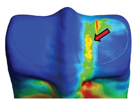
D) The standing CT images are used to make a 3D model of the cannon bone with E) segmentation of sites of fatigue damage and the surrounding adaptive response shown with red and orange arrows respectively. The surrounding bone becomes more dense (orange arrows) in response to race training. F) Virtual mechanical testing of one of the condyles shows concerning parasagittal groove strain concentration (red arrow), suggesting elevated risk of serious injury during racing because of mechanical compromise to the bone.
uncertainty does not yet provide an acceptable level of accurate risk assessment that’s required for an optimal prerace screening tool.
“The connection between structural change in the bone and mechanical compromise is one of the most important gaps in knowledge right now,” says Muir. “And it’s a gap that has to be filled so veterinarians tasked with determining the fitness of Thoroughbred horses to race can make a more appropriate risk assessment of how compromised the bones really are, especially as more research on the topic gets published.”
Muir and his team are currently addressing this gap in knowledge through research that will develop a validated 3D finite element computer model of the equine cannon bone built from CT imaging. Virtual mechanical testing can then be performed using computer software to look for mechanical compromise in the bones from individual horses. With more work, this assessment approach will enable veterinarians to optimally and objectively analyze CT scans in a manner that would be suitable for clinical implementation. This work is being done in collaboration with Corinne Henak from the UW Department of Mechanical Engineering.

It’s a complex orthopaedic problem that the racing industry and horse owners will likely continue to struggle with for years to come: finding the right balance between identifying the small subset of horses that have concerning lesions from the horses that have a subchondral bone injury lesion, but are ultimately doing ok. Muir says that even the most sophisticated technology will likely never completely resolve the issue, which further underscores a major ethical paradox facing racetrack authorities and racehorse owners.
“Nobody wants any of these horses to suffer injuries or die, of course, but they also want them to race and win a million dollars.”
So what can be done? For one thing, Muir says more peer reviewed science is crucial, as is making more information available to help guide clinicians to better identify horses whose bone injury puts them at imminent risk of catastrophic injury. To aid in that effort, Muir and his research team publicly released the blinded image set they used in their study so that other clinicians and researchers around the globe can use them to either train their veterinarians or conduct additional studies on the topic. After all, science at its best is a team sport, and Muir believes strongly in not only learning from but empowering researchers around the globe to work collaboratively toward improved equine safety.
“I see this not only as the basic advancement of knowledge, but also an example of the Wisconsin Idea in action, which strives to make societal progress at the broadest level.”
A recent review of racehorse deaths by the Horseracing Integrity and Safety Authority (HISA) found that the rate of equine deaths at racetracks in their jurisdiction is at a 10-year low, but intense media coverage of race-related deaths has dominated the headlines of late, overshadowing their otherwise optimistic news. In 2022, the incidence of catastrophic injury was 1.25 fatalities per 1,000 race starts in the United States, which represents a loss of hundreds of horses. That number was slightly better in 2023, but it included the highly publicized death of 12 horses at Churchill Downs, including seven in the run-up to last May’s Kentucky Derby, and 13 at Saratoga Race Racetrack, home of the Belmont Stakes, the third leg of racing’s triple crown. Many of the deaths were the result of musculoskeletal injuries.
Taking a somewhat similar approach to what Racing Victoria did when facing comparable scrutiny, racing authorities in the US last year strengthened their own equine safety and race policies, none of which, however, include the use of standing CT technology or mandatory scans for finalists.
“If finalists in the Kentucky Derby and other major races had pre-race standing CT scans assessed by a panel of skilled reviewers, would that have had an impact on what happened last year?” asks Muir. “Based on extrapolations from what we’ve learned so far from the Melbourne Cup, I think it would.”
As the industry continues to endure public scrutiny regarding equine health and welfare, particularly when a cluster of deaths occur, racing advocates and veterinarians alike are starting to question whether it makes sense anymore to delay mandatory pre-race screenings, using standing CT or similar technologies, until more comprehensive evidence exists.

“If you are a racing authority or racetrack owner, I think it would be imprudent to ignore the increasing scrutiny and pressure that comes from negative public perceptions of the sport,” says Muir. “Our existing knowledge has evolved quickly over the last few years, and although we still have a way to go, I think a lot of people are really starting to take notice what’s been happening at the Melbourne Cup.”
Like any good scientist, Muir champions the role of science and the value that additional research will bring to the debate, but now believes that perhaps the most pragmatic and efficient path forward would be for the racing industry to use all available data to them and implement pre-race screening sooner than later, while working to fill the gaps in knowledge with ongoing and robust peer-reviewed science.
Toward that end, Muir was recently awarded grants from the Hong Kong Jockey Club Equine Welfare Research Foundation and the Grayson-Jockey Club Research Foundation to extend his research program on the use of standing CT and computer modeling analysis to predict risk of fetlock stress fracture. Going forward, Muir and his team will also focus on stress fracture of the proximal sesamoid bone, another common cause of fatal injury in Thoroughbred racehorses. This new research will advance understanding of the relationship between specific structural changes in the proximal sesamoid bone
and imminent risk of stress fracture, further improving longitudinal monitoring of horses in training and reducing catastrophic injury during races.
For Muir, the improved health and welfare of racehorses is the ultimate victory, and he’s confident that the impact of his research on the issue, along with that of his academic collaborators, colleagues, and fellow researchers, will only continue to grow as does the mounting body of scientific evidence. There’s no doubt the Melbourne Cup’s achievement of zero equine fatalities since 2021 represents a turning point in the world of horse racing, one that Muir and others hope is an inspiring example for the rest of the global horse racing community to follow.
Until then, Muir can’t help but feel like the finish line is in view.
“Bringing innovation to bear on issues impacting the world in various way is, of course, just one of the things that sets this university apart from its peers,” says Muir. “And that’s certainly been true from my point of view. I don’t think this interdisciplinary project, or the subsequent benefit to animal welfare, would be a reality right now had I worked at any other university or veterinary school”.
Cleft lip and palate are the most common craniofacial birth defects in humans, affecting more than 175,000 newborns around the world each year. Yet despite decades of research, it’s still not known what causes most cases or what can be done to prevent them. But a recent study from the University of Wisconsin School of Veterinary Medicine (SVM) has uncovered new information about orofacial development in mice that researchers believe could one day help reduce the risk of these birth defects in humans.
Published earlier this year in the Proceedings of the National Academy of Sciences (PNAS) the study provides the first direct evidence of a mechanism called DNA methylation being required for craniofacial development. DNA methylation is a process where a group of molecules are added to DNA that change the expression of genes without actually altering the DNA sequence. It’s also affected by various environmental factors. The researchers discovered that disruption to DNA methylation interferes with development of the lip and palate and causes these birth defects in mice.
Led by Robert Lipinski, associate professor of comparative biosciences at the SVM, the research is an important step toward developing preventive strategies that could one day lessen the risk of cleft lip and palate, known collectively as orofacial clefts (OFCs), in both animals and humans.
“We knew from past research that genetics and the environment interact to cause these types of birth defects, but our understanding of the environmental component lagged far behind that of genetics.” says Lipinski. “Unlike genetics, we don’t have a permanent record of the prenatal environment that can be examined retrospectively, but connecting OFCs to DNA methylation helps narrow our focus on the particular environmental influences that modify the risk for these types of birth defects.”
His team’s work confirmed the essential role of DNA methylation in regulating orofacial development during embryonic development and demonstrates how disruptions to that process alter the ability of stem cells to form the connective tissue of craniofacial bone and cartilage, resulting in OFCs.


Lipinski and his team arrived at these results by first genetically manipulating DNA methylation in two separate groups of mouse embryos. The experiments resulted in seemingly contradictory results, with OFCs developing in one group of mice, but not the other. To understand why there was a difference between the groups, the team conducted another round of experiments in which they inhibited DNA methylation in mouse embryos at different stages of development. The timing of when DNA methylation occurs was critical to the development of orofacial clefts.
They found that exposure on the 10th gestational day resulted in OFCs but administering the same inhibitor just 48 hours later resulted in normal orofacial development.
Identifying this narrow window of gestational sensitivity is important, Lipinski says, because it not only helps
“...having a better understanding of how orofacial development is regulated by environmentally sensitive mechanisms could directly inform birth defect prevention strategies.”
narrow the focus of the next stage of his team’s research but it will also help design future public education initiatives once more is known about the modifiable environmental and behavioral risk factors that impact OFC risk in humans. The 10th gestational day in mouse embryos corresponds with the beginning of the 5th week of embryonic development in humans–a stage at which many pregnancies may not yet be recognized.
“We know DNA methylation can be influenced by a variety of environmental factors, including maternal stress, diet, and exposure to drugs, toxins and environmental pollutants, and having a better understanding of how orofacial development is regulated by environmentally sensitive mechanisms could directly inform birth defect prevention strategies,” says Chris Jabbarpour, research specialist, Lipinski Lab manager, and one of the coauthor’s of the study. “This next phase of our team’s research is focused on identifying specific factors that influence DNA methylation during orofacial development and which could therefore alter OFC risk.”
Lipinski and his team are uniquely positioned to pursue this next stage of research because of another important outcome of the study: a new in vitro model the team developed. The model will allow them to rapidly screen thousands of dietary and environmental factors in a laboratory dish before testing the impact of specific factors on cleft susceptibility in mouse models.
The results in cell and animal models will help the researchers more quickly and accurately identify factors likely to be of consequence to human development.
Orofacial clefts of the upper lip and palate affect approximately 1 in 700 newborns, and individuals with OFCs navigate feeding difficulties as infants that require multiple surgeries, dental procedures, and speech therapy during childhood and adolescence. Studies have shown higher mortality rates at all stages of life for individuals with OFCs.
— Gian Galassi


New research shows mRNA vaccines recruit “trained assassin” cells to combat COVID-19 in lungs
The efficacy of mRNA vaccines in reducing disease severity and hospitalization from COVID-19 is well established. Now, new research from the University of Wisconsin School of Veterinary Medicine (SVM) advances our understanding of how these vaccines protect the lungs following breakthrough infections from emerging variants of SARS-CoV-2, the virus that causes COVID-19.
The study is the first to directly demonstrate the role of memory CD8 T cells in mRNA vaccine-induced immunity to COVID-19. Memory CD8 T cells are a
specialized type of white blood cell that rapidly respond when re-exposure to a pathogen occurs. They are often referred to as “trained assassins” because they control viral infections by targeting and then destroying virallyinfected cells. This study, conducted in mice, shows that memory CD8 T cells were necessary and sufficient in controlling SARS-CoV-2, independent of antibodies. Researchers demonstrated this by showing how the protection afforded by mRNA vaccines was lost when memory T cells were depleted prior to SARS-CoV-2 infection.
It’s widely accepted that CD8 T cells provide a more robust form of protection because the viral fragment they target to kill infected cells does not change considerably with each new viral variant. Antibodies on the other hand, typically lose their ability to prevent infection because the part of the virus they target changes with each new mutation.
Marulasiddappa Suresh, the John E. Butler Professor in Comparative and Mucosal Immunology in the SVM Department of Pathobiological Sciences, says this study sheds new light on the protective mechanisms mRNA vaccines use to lessen severe disease following breakthrough infections. It also raises important new questions about the role of memory T cells in limiting the spread of the virus, the frequency with which we get vaccinated, and the most effective methods for vaccine delivery.
“The key finding of our research shows that memory T cells play an essential role in mediating SARS-CoV-2 viral control in lungs, independent of antibodies.” says Suresh, who was also the study’s principal investigator. “We hope this new understanding of vaccine-induced immunity will inform the development of new vaccines and treatment strategies that more effectively combat the emergence of global variants and limit the impact they’ll have on our health in the future.”
While previous studies have documented a strong correlation between vaccine-induced T cells and more positive outcomes following infection with SARS-CoV-2, the ability to study these protective mechanisms in detail is not possible in humans. As a result, researchers administered various doses of the Pfizer BioNTech COVID-19 mRNA vaccine to a specialized mouse model in order to study the defining characteristics of T cell responses induced by the vaccine. Their results showed the T cell response to mRNA vaccine in the peripheral blood is largely similar between mice and humans. They also found that T cells actively sought out the virus in the respiratory tract—airways, lung vasculature, and mediastinal lymph nodes—to effectively reduce the burden of SARS-CoV-2 in the lungs.
Other key findings show that intramuscular immunization produced unexpectedly high frequencies and numbers of memory T cells in the airways of the respiratory tract — the main portal of entry for SARSCoV-2. According to Suresh, future research on this topic will need to assess the biological significance of nasal and airway resident memory T cells in protection against emerging variants of SARS-CoV-2 and whether individuals who recover from breakthrough SARS-CoV-2 infections will require further vaccinations.

“We hope this new understanding of vaccine-induced immunity will inform the development of new vaccines and treatment strategies that more effectively combat the emergence of global variants and limit the impact they’ll have on our health in the future.”
“It’s still unclear if the combination of vaccineinduced immunity and infection-induced immunity is sufficient to provide broad mutation-resistant immunity to future SARS-CoV-2 variants,” he says.
Other members of the research team from the UW School of Veterinary Medicine include Brock KingstadBakke, Thomas Cleven, Hailey Bussan, Hongtae Park, Peter Halfmann and Yoshihiro Kawaoka from the Department of Pathobiological Sciences; and Jay Mishra and Sathish Kumar from the Department of Comparative Biosciences. The study was published last fall in the journal JCI Insight .
Alumnus promotes entrepreneurial vision, courage, and urgency as keys to future success
Since graduating from the SVM, Scott Spaulding, DVM ‘91, has recognized the importance and value in advocating for and representing the veterinary medicine industry. During his accomplished career, he has been a practicing equine doctor, president/CEO of Badger Veterinary Hospital, and co-founder of Vet24seven, veterinary medicine’s first telehealth platform. As a lifelong entrepreneur and inspirational leader, he is passionate about sharing his experiences and expertise with the next generation of veterinarians, leaders, and entrepreneurs.
How did your SVM education contribute to or inspire your success after graduation?
Entering veterinary school, I was well aware the veterinary degree was a fabulous degree. At the time, I assumed it set one up to be a successful clinician, a practitioner. I didn’t appreciate the well-rounded degree it truly is. Veterinary school instilled tenacity, perseverance, and accentuated my problem-solving ability. It taught me teamwork and the ability to research complex problems that have multitudes of solutions. I honed my ability to professionally communicate with colleagues and to convey complex messages to animal owners, often under circumstances of extreme duress. I never imagined the doors that my veterinary medical education and veterinary degree would open.
Who influenced your time at the SVM the most and why?
Barney Easterday was a great inspiration to me. He was so proud of being a veterinarian and of his students. He and a small group of people actually founded and built the University of Wisconsin School of Veterinary Medicine, from the ground up. His passion, vision, courage, and tenacity inspires me to this day. Sue Hyland, the school’s first associate dean for academic affairs. Sue instilled incredible empathy in all the students she advised and she remains a close friend of mine She has incredible listening skills and is someone that can see the truth so easily. Sheila McGuirk is an amazing clinician and an incredible communicator. Sheila was an incredible influence on every day that I was a practicing veterinarian. I learned how to perform a thorough physical exam, interpret clinical laboratory results, formulate a list of differential diagnoses, compile and implement a treatment plan and to communicate with animal owners professionally and empathetically. Peter MacWilliams and John Dahl
introduced me to and encouraged me to become involved in organized veterinary medicine. They instilled in me the value of growing my professional network and giving back to the profession of veterinary medicine.
Why is entrepreneurship important for the future of veterinary medicine?
When I was part of the inaugural group that founded the AVMA’s Economics Division in 2012, one of our first

to chart the course moving forward to satisfy the demand for veterinary services. If we choose not to abdicate this space to others, we will need leaders with strong visions for the future, the ability to recognize those visions and elucidate the path forward. We need leaders to act boldly and who have the courage to take action with urgency. Those entrepreneurial-minded individuals with these characteristics can enjoy financial reward beyond their wildest imaginations.
What hard-earned truth did you learn during your career that you wish you’d known sooner?
My career has been a journey, sometimes the path forward isn’t entirely clear. One needs to believe in themselves and always find the courage to keep moving forward. Things happen for a reason, and sometimes the reason becomes apparent and other times it doesn’t. Rely on your intuition to help you through challenging times. Your “gut” seldom fails you.
What are the three most valuable traits you think are necessary to make an impact in this field?
The ability to recognize your vision, to find the courage to take action to realize your vision, and the ability to act with urgency. Don’t delay until tomorrow. Learn to make that call today!
What are the most exciting things that will define the future of veterinary medicine?
At this point in my career, the most exciting thing I see today is the application of augmented intelligence to clinical practice. I don’t think most of us can even imagine the impact this will have on clinical practice. Another thing that excites me is the continual need for strong leadership and great communication skills. These are basic skills that have set great entrepreneurs apart for decades. The exciting part today is the fabulous tools and extraordinary people available. Learn how to leverage them to your ability.

Dear alumni — You may have heard that the campus is celebrating its 175th anniversary – also known as its demi-semi-sept-centennial (try saying that 5 times fast!). The School of Veterinary Medicine (SVM) is a relative newcomer on campus, though its history actually began in 1911 when the Department of Veterinary Science was established in what is now known as the College of Agricultural and Life Sciences (CALS). That award-winning research training program eventually became the comparative biomedical sciences graduate program that’s housed in the SVM. In 1979, Wisconsin Governor Lee Dreyfus signed into law the establishment of the school, which opened its doors and welcomed its first class in 1983.
Throughout its history – from a veterinary science department to a school of veterinary medicine – it has been a leader in teaching, research, clinical care and outreach, with its programs consistently ranking among the top of its peers. And the Wisconsin Idea - the belief that the university’s work should extend beyond the boundaries of the campus to improve the lives for all –has been at the forefront of its mission.
As we look ahead to the next milestone (bicentennial – much easier to say!), I have no doubt that the school will continue to be a leader and will play a significant role in improving both animal and human health. While I expect the world may look quite different 25 years from now, the school is uniquely poised to meet the challenges and opportunities ahead. We are developing a new curriculum that will meet the changing demands of the profession as well as how our students learn. And
1. The Motivation Manifesto: 9 Declarations to Claim Your Personal Power by Brendon Burchard
2. The E-Myth Enterprise: How to Turn A Great Idea Into a Thriving Business by Michael Gerber
3. Slight Edge: Turning Simple Disciplines into Massive Success and Happiness by Jeff Olson


The entire SVM community was saddened by the loss of SVM alumnus Galen Heyne DVM ‘19, who died unexpectedly in February. Throughout his time on the UW-Madison campus, Galen worked with many faculty, staff, and other students and left an indelible mark on our entire SVM family and the greater Madison veterinary medical community. He will be missed.
Scott Spaulding’s 3 favorite books about entrepreneurshipWhen Nicolette Kametas-Dykes was being treated for a rare brain infection and benign brain tumor last spring, her beloved dog Coco never left her side, providing constant companionship during one of the darkest times in her life. So when Coco received her own diagnosis of Oral Squamous Cell carcinoma just a few months later, Nicolette and her husband, Rob, knew they were going to do everything they could to return the favor.
Little did they know how challenging that would be. For one thing, all the veterinary clinics near their home outside Chicago wanted approximately $10,000 before they’d even consider treating Coco’s cancer. That was a steep price to pay, especially since the cost of treating Nicolette’s own illness–the symptoms of which also cost her her job as an ultrasound technician—had severely strained the young couple’s finances. Undeterred, Nicolette kept searching for possible options for Coco’s care, and was repeatedly met with the same insurmountable financial obstacles. It wasn’t until she contacted UW Veterinary Care in Madison, WI, where she finally found a glimmer of hope.
“For most people, pets are more than animals, they are members of the family. ”
“We initially thought Coco might qualify for a clinical study that UW was doing for her type of cancer, but she ultimately didn’t meet the requirements,” Kametas-Dykes recalls. “But everybody we talked to at UW was so helpful, and they told us that if we were willing and able to make the trip to Madison that they’d try to help Coco if they could.”
Nicolette and Rob didn’t think twice. After making the three-hour trip north, they met with UW radiation oncology resident Dr. Claire Faletti, who presented them with a possible care plan for Coco: radiation treatment, once a week, for

a month. What they still didn’t have, however, were the finances to pay for it. That’s when Dr. Faletti encouraged them to apply for financial assistance from the Petco Love Cancer Treatment Fund, which helps clients who don’t have the financial resources to cover the costs of oncology care. They applied and were granted $4,000 for Coco’s care, which covered nearly the entire amount of her month-long radiation treatment plan. Nicolette says the funding could not have come soon enough.
“The Petco funding meant everything to us, especially since we were told Coco would likely only live another month without treatment” says Kametas-Dykes. “We literally had no options left for Coco before coming to UW, and everybody up there was so kind, so caring, and so willing to help us when nobody else would even try. We are so grateful to Petco Love and to all the people at UW who helped Coco and us through this.”
Dr. Faletti says that without the Petco funds, the progression of Coco’s disease would have required Nicolette and Rob to make the hardest decision owners can make. Instead, Coco is not only doing amazing but is once again by Nicolette’s
side as she continues her own treatment and recovery.
“Like most vets, I got into this field because I love animals, but I have come to realize that the bonds we form with wonderful owners like Nicolette, and pets like Coco, is an amazing “perk” that truly makes this job so great,” says Faletti. “I cannot overstate the value of being able to offer Petco funds to owners who otherwise wouldn’t be able to help their pets. For most people, pets are more than animals, they are members of the family. And at the end of the day, we all want to be able to help our family members.”


• Improved and expanded emergency & critical care unit
• New immediate card ward
• More exam rooms
• Additional surgery suites
• Expanded waiting area with dedicated spaces for cats
• Enhanced diagnostic imaging
• New state of the art treatment capabilities
• Small animal hospital area doubled
• Student collaboration and instruction areas
• Infectious disease research lab space tripled
• And more!
