Chapter 1
REVIEW EXERCISES
1. Complete the blanks in the following paragraphs using each word from the list below only once except for the ones marked with two asterisks ** which are used twice. The number of blanks indicates the number of letters of the word for each blank.
a. anterior** s. medial
b. anteroinferior t. palmar
c. anterolateral u. plantar
d. anteromedial v. posterior**
e. anteroposterior w. posteroinferior
f. anterosuperior x. posterolateral
g. bilateral y. posteromedial
h. caudal z. posterosuperior
i. cephalic aa. prone
j. contralateral bb. proximal
k. deep cc. superficial
l. distal dd. superior
m. dorsal ee. superolateral**
n. inferior ff. superomedial
o. inferolateral gg. supine
p. inferomedial hh. ventral
q. ipsilateral ii. volar
r. lateral
When Jacob greeted Stephanie at the beach he reached out with the P A L M A R surface of his hand to grasp the V O L A R surface of her hand for a handshake. As the A N T E R I O R aspect of their bodies faced each other, Jacob noticed that the hair located on the most S U P E R I O R part of Stephanie’s head appeared to be a different color than he remembered. He then asked her to turn around so that he could see it from a P O S T E R I O R view. As she did so, it became obvious to him that she had blonde streaks running from her C E P H A L I C region in an I N F E R I O R direction all the way down to her C A U D A L region.
Stephanie then asked Jacob if the sunburn on the S U P E R O L A T E R A L portions of his shoulders was due to the exposure that his tank-top shirt provided. He replied yes but that it was only a S U P E R F I C I A L burn and did not go too D E E P. He then said, “I wished I would have had my shirt off so that I would have gotten some more sun on the S U P E R O M E D I A L portion of my shoulders up to my neck.”
Stephanie said that she recently got sunburned on her back while lying P R O N E at the beach. She then flipped her hair around the L A T E R A L side of her neck toward the V E N T R A L portion of her trunk to expose her D O R S A L region. Jacob remarked, “Wow, instead of the bikini you have on today with straps over your shoulders running from your A N T E R O S U P E R I O R chest to your P O S T E R O S U P E R I O R shoulders, you must have been wearing one with crossing straps as I see you have tan lines running in an I N F E R O M E D I A L direction to your M E D I A L low back from the S U P E R O L A T E R A L aspect of your P O S T E R I O R shoulders. You should have spent more time lying S U P I N E.” She replied “Well, I
did lie partially on my back and my right side for a while. See where the A N T E R O M E D I A L portion of my right thigh and the A N T E R O L A T E R A L portion of my left thigh are tanned just right, but unfortunately in that position the P O S T E R O L A T E R A L right thigh and P O S T E R O M E D I A L thigh received relatively little exposure”. Jacob commented “Yep, when you lie on one side most of the time, you get all the sun on the C O N T R A L A T E R A L side and none on the I P S I L A T E R A L side. It looks like you must have had a towel covering your feet and ankles since your D I S T A L lower extremities are not nearly as tan as your P R O X I M A L lower extremities.” Stephanie replied, “You are correct. I kept the bottom of my lower legs covered almost all of the time while lying on both sides so that the sensitive skin on my A N T E R O I N F E R I O R and P O S T E R O I N F E R I O R shins would not burn. But, I did get a good B I L A T E R A L tan on my A N T E R I O R trunk, except for the I N F E R O L A T E R A L aspect of my right elbow I was resting on.” As Jacob slipped his sandals on to protect the P L A N T A R aspect of his feet from the hot sand, he said, “Well, nice to see you. I have to go by the doctor’s office and get an A N T E R O P O S T E R I O R chest X-ray to make sure my pneumonia has cleared up.”
2. Joint movement terminology chart
The specific body area joint movement terms arise from the basic motions in the three specific planes: flexion/extension in the sagittal plane, abduction/adduction in the frontal plane, and rotation in the transverse plane. With this in mind, complete the joint movement terminology chart by writing the basic motion in the right column for each specific motion listed in the left column by using either flexion, extension, abduction, adduction, or rotation (external or internal).
Specific motion Basic motion
Eversion abduction
Inversion adduction
Dorsal flexion flexion
Plantar flexion extension
Pronation (radioulnar) internal rotation
Supination (radioulnar) external rotation
Lateral flexion abduction
Reduction abduction
Radial flexion adduction
Ulnar flexion abduction
3. Bone Typing Chart
Utilizing Fig. 1.8 and other resources place an “X” in the appropriate column of the bone typing chart to indicate its classification.
Bone Long Short Flat Irregular Sesamoid
Frontal X
Zygomatic X
Parietal X
Temporal X
Occipital X
Maxilla X
Mandible X
Cervical
vertebrae X
Clavicle X
Scapula X
Humerus X
Ulna X
Radius X
Carpal bones X
Metacarpals X
Phalanges X
Ribs X
Sternum X
Lumbar
vertebrae X
Ilium X
Ischium X
Pubis X
Femur X
Patella X
Fabella X
Tibia X
Fibula X
Talus X
Calcaneus X
Navicular X
Cuneiforms X
Metatarsals X
4. What are the five functions of the skeleton?
Protection of heart, lungs, brain, etc.
Support to maintain posture
Movement by serving as points of attachment for muscles and acting as levers
Mineral storage such as calcium & phosphorus
Hemopoiesis
5. List the bones of the upper extremity.
Humerus
Ulna
Radius
Carpal bones - 8
Metacarpals - 5
Phalanges – (14 phalanxes)
6. List the bones of the lower extremity.
Femur
Patella
Tibia
Fibula
Talus
Calcaneus
Navicular
Cuboid
Cuneiforms -3
Metatarsals – 5
Phalanges – (14 phalanxes)
7. List the bones of the shoulder girdle.
Clavicle
Scapula
Sternum
8. List the bones of the pelvic girdle.
Ilium
Ischium
Pubis
Sacrum – 5
Coccyx - 4
9. Describe and explain the differences and similarities between the radius and ulna.
Both are long bones of the forearm that articulate proximally with the humerus and distally with the proximal row of carpals. They also both articulate with each other proximally and distally. The ulna is much larger proximally than the radius, whereas the radius is much larger distally than the ulna. Thus, most of the forearm’s proximal articulation with the humerus is via the ulna and most of the forearm’s distal articulation with the carpals is via the radius.
10. Describe and explain the differences and similarities between the humerus and femur.
They are more similar than different. Both the humerus and femur have a bony rounded head proximally fitting into a concave socket forming an enarthrodial joint. Below each head is a neck. Both are the longest bones of their respective extremity and both have two condyles distally to articulate with a ginglymus joint. The humerus has a greater and lesser tubercle proximally which correspond roughly with the greater and lesser trochanter of the proximal femur.
11. Using bony landmarks, how would you suggest determining the length of each lower extremity for comparison to determine if someone had a true total leg length discrepancy?
Begin with the person lying supine and straight, then utilizing a flexible tape measure, measure the distance between the anterior superior iliac spine and the medial malleolus. Compare bilaterally. To specifically compare the femurs, measure from the most prominent aspect of the greater trochanter to the lateral joint line. To measure the leg, measure from the fibula head to the lateral malleolus, making sure to place the tape measure on exactly the same points bilaterally.
12. Explain why the fibula is more susceptible to fractures than the tibia.
Anatomically, it does not have as much bone mass and as such cannot absorb as much tension, shear, compression, bending, and torsion before fracturing. Additionally, it is exposed more to contact from external sources due to its location laterally.
13. Why is the anatomical position so important in understanding anatomy and joint movements?
Without a common reference point everyone would be somewhat speaking a different language in terms of where one body part is in relation to another or how the relationship is changing between two segments of the body.
14. Use a diarthrodial joint other than the knee to provide an example of a convex surface moving on a concave surface. Use the same joint to provide an example of a concave surface moving on a convex surface. Answers will vary by student.
15. Use a diarthrodial joint other than the knee and determine the direction of the roll and glide of the proximal bone when extending and then flexing on the stationary distal bone, Reverse the analysis with the distal bone extending and then flexing on the proximal bone.
Answers will vary by student.
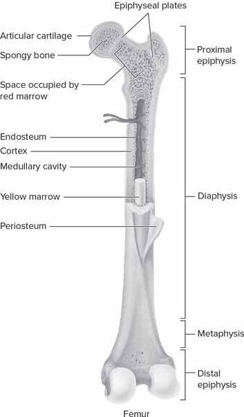
16. Label the parts of a long bone.

17. Joint type, movement, and plane of motion chart
Complete the joint type, movement, and plane of motion chart by filling in the type of diarthrodial joint and then listing the movements of the joint under the plane of motion in which they occur
Scapulothoracic Joint
Sternoclavicular Arthrodial X
Acromioclavicular Arthrodial X
Glenohumeral Joint
Enarthrodial X
Elbow Ginglymus X
Radioulnar joint Trochoid X Wrist
Condyloid X
1st carpometacarpal joint Sellar X
1st metacarpophalangeal joints
Thumb interphalangeal joint
2nd, 3rd, 4th & 5th metacarpophalangeal joints
2nd, 3rd, 4th & 5th proximal interphalangeal joints
2nd, 3rd, 4th & 5th distal interphalangeal joints
Atlantooccipital joint
Condyloid X X
Ginglymus X
Condyloid X X
Ginglymus X
Ginglymus X
Condyloid X X
Cervical spine C1-C2 Trochoid X
Cervical spine C-2-C7
Lumbar spine
Arthrodial X
Arthrodial X
Hip
Knee (tibiofemoral joint)
Knee (patellofemoral joint)
Ankle
Transverse tarsal & subtalar joints
Metatarsophalangeal joints
Great toe interphalangeal
2nd, 3rd, 4th & 5th proximal interphalangeal joints
2nd, 3rd, 4th & 5th distal interphalangeal joints
18. Joint position chart
Enarthrodial X X X
Ginglymus X X
Arthrodial X
Ginglymus X
Arthrodial X
Condyloid X X
Ginglymus X
Ginglymus X
Ginglymus X
Using proper terminology, complete the joint position chart by listing the name of each joint involved and its position upon completion of the multiple joint movement. In all cases, begin in a sitting position.
Multiple joint movement
Reach straight over the superior aspect of your head to touch the contralateral ear
Place the toe of one foot against the posterior aspect of the contralateral calf
Reach behind the back and use your thumb to touch a spinous process
Actively pull the knee as far as possible to the shoulder
Place the plantar aspect of both feet against each other
Joints and respective position of each
Scapula upward rotation, glenohumeral abduction, elbow flexion, wrist flexion, 2nd - 5th MCP, PIP, DIP flexion
Knee flexion & internal rotation, hip flexion, transverse tarsal/subtalar inversion, great toe metatarsophalangeal extension
Scapula abduction & downward rotation, glenohumeral extension & internal rotation, elbow flexion, wrist extension, thumb CMC, MP, & IP extension
Hip flexion & adduction, knee flexion, lumbar spine flexion, scapula abduction
Hip external rotation & abduction, knee internal rotation, ankle plantar flexion, transverse tarsal & subtalar inversion
19. Plane of motion and axis of rotation chart
For each joint motion listed in the plane of motion and axis of rotation chart, list the plane of motion in which the motion occurs and its axis of rotation.
Motion Plane of motion Axis of rotation
Cervical rotation Transverse Vertical
Shoulder girdle elevation Frontal Sagittal
Glenohumeral horizontal adduction Transverse Vertical
Elbow flexion Sagittal Frontal
Radioulnar pronation Transverse Vertical
Wrist radial deviation Frontal Sagittal
Metacarpophalangeal abduction Frontal Sagittal
Lumbar lateral flexion Frontal Sagittal
Hip internal rotation Transverse Vertical
Knee extension Sagittal Frontal
Ankle inversion Frontal Sagittal
Great toe extension Sagittal Frontal
20. List two sport skills that involve movements which are more clearly seen from the side. List the primary movements that occur in the ankle, knee, hip, spine, glenohumeral joint, elbow, and wrist. In which plane are these movements occurring in primarily? What axis of rotation is involved primarily?
Answers will vary depending upon the student’s choice.
Basketball free throws – ankle plantar flexion & dorsiflexion, knee flexion & extension, hip flexion & extension, lumbar & cervical spine flexion & extension, elbow flexion & extension, wrist flexion & extension. Primarily sagittal plane with a frontal axis.
Volleyball serve - ankle plantar flexion & dorsiflexion, knee flexion & extension, hip flexion & extension, lumbar & cervical spine flexion & extension, elbow flexion & extension, wrist flexion & extension. Primarily sagittal plane with a frontal axis.
21. List two sport skills that involve movements which are more clearly seen from the front or rear. List the primary movements that occur in the transverse tarsal/subtalar joint, hip, spine, glenohumeral joint, and wrist. Which plane are these movements occurring in primarily? What axis of rotation is involved primarily?
Baseball batting - Transverse tarsal / subtalar eversion frontal plane & anteroposterior axis, hip flexion sagittal plane frontal axis, hip rotation transverse plane vertical axis, lumbar rotation transverse plane vertical axis, shoulder horizontal abduction/adduction frontal plane sagittal axis, radioulnar pronation supination transverse plane vertical axis.
Soccer pass – Hip abduction/adduction frontal plane sagittal axis, trunk rotation transverse plane vertical axis, shoulder flexion/extension
sagittal plane frontal axis, shoulder abduction/adduction frontal plane sagittal axis.
22. List the similarities between the ankle/foot/toes and the wrist/hand/fingers regarding the bones, joint structures, and movements. What are the differences? There are many more similarities than differences. The major difference is the size and shape of some of the bones. The ankle is a hinge joint moving in the sagittal plane and the transverse tarsal/subtalar joint is an arthrodial joint moving in the frontal plane. Together, these are similar to the wrist which is condyloid and moves in the sagittal and frontal plane. There are eight wrist bones (carpals) and seven tarsals. In both cases these are small, short bones that work together as arthrodial joints. There are five metacarpals which make up the length of the hand and five metatarsals which make up the length f the foot. The thumb corresponds to the great toe in that it each have one interphalangeal joint. The thumb is different in that it originates with the 1st carpometacarpal joint. The four fingers correspond to the four lesser toes. Each is joined to the hand or foot at the metacarpals and metatarsals, respectively at the metacarpophalangeal and metatarsophalangeal joints which are all condyloid. Each of the four lesser toes and fingers have three phalanxes and two interphalangeal joints which are ginglymus.
23. Compare and contrast the glenohumeral and acetabulofemoral joints. Which one is more susceptible to dislocations and why? Both are enarthrodial and have a significantly large range of motion in all planes. The acetabulofemoral joint is a much deeper and more stable joint due to the significant osseous, ligamentous and muscular support. The glenohumeral joint is much shallower and has relatively little osseous and ligament support, making it much more dependent on muscular support. Glenohumeral subluxations and dislocations are very common whereas the hip rarely subluxes or dislocates.
24. Compare and contrast the elbow and knee joints. Considering the bony and joint structures, and their functions, what are the similarities and differences?
Both are ginglymus joints allowing flexion and extension in the sagittal plane, but the knee joint also allows internal and external rotation in the transverse plane. Both have similar ranges of motion from zero to around 150 degrees. In both cases, there is only one proximal bone of the joint which has two articular convex surface to articulate with the distal concave bony surfaces. In the elbow, both distal bones articulate with the proximal bone whereas in the knee the femur only articulates with the tibia. The knee has a
sesamoid patella to increase its mechanical advantage in active extension whereas the elbow does not, but depends on the prominent olecranon for the same purpose. The knee has menisci and the elbow does not.
LABORATORY EXERCISES
1. Choose several different locations on your body at random on your body and label as 1., 2., 3., etc. Then use correct anatomical terminology to describe the path you take as you move your index finger from one point to another without leaving the skin surface.
Answers will vary by student.
2. Determine which joints have movements possible in each of the following planes:
a. Sagittal
Interphalangeal, metatarsophalangeal ankle, knee, hip, spine, glenohumeral, elbow, wrist, metacarpophalangeal
b. Frontal
Metatarsophalangeal, transverse tarsal/subtalar, hip, spine, glenohumeral, wrist, metacarpophalangeal
c. Transverse Knee, hip, spine, glenohumeral, radioulnar, 1st carpometacarpal
3. List all the diarthrodial joints of the body that are capable of the following paired movements:
a. Flexion/extension
Interphalangeal of foot, metatarsophalangeal, talocrural, knee, hip, lumbar spine, cervical spine, glenohumeral, elbow, wrist, metacarpophalangeal, interphalangeal of hand
b. Abduction/adduction
Metatarsophalangeal, transverse tarsal, subtalar, hip, lumbar spine, cervical spine, glenohumeral, wrist, metacarpophalangeal
c. Rotation (left and right)
Lumbar spine, cervical spine
d. Rotation (internal and external)
Hip, glenohumeral, radiolulna, 1st
carpometacarpal
4. Determine the planes in which the following activities occur. Also, use a pencil to visualize the axis for each of the following activities.
a. Walking up stairs
Sagittal
b. Turning a knob to open a door
Transverse
c. Nodding the head to agree
Sagittal
d. Shaking the head to disagree
Transverse
e. Shuffling the body from side to side (sidestepping)
Frontal
f. Looking over your shoulder to see behind you Transverse
5. Individually practice the various joint movements, on yourself or with another subject.
Answers require demonstration by the student.
6. Locate the various types of joints on a human skeleton and palpate their movements on a living subject. Answers require demonstration by the student.
7. Stand in the anatomical position facing a closed door. Reach out and grasp the knob with your right hand. Turn it and open the door widely toward you. Determine all of the joints involved in this activity and list the movements for each joint.
Assumption: Using the right hand to open the door and the door swings from a hinge on the right. To reach for the knob, protract the right shoulder girdle, flex and slightly abduct the right humerus, flex the right elbow, pronate the right radioulnar joint, extend the right wrist, extend the right metacarpophalangeal joints slightly. Grasp the door knob by flexing the right metacarpophalangeal and interphalangeal joints of the fingers and thumb. Using all joints of the thumb oppose the door knob and fingers to grasp the door knob more firmly. Ulnar deviate the right wrist while simultaneously supinating the right radioulnar joint. Adduct the right shoulder slightly to assist in turning the knob. Then slightly abduct the shoulder while flexing the right elbow. Then extend the right shoulder while retracting the right scapula to fully open the door.
8. Utilize a goniometer to measure the joint ranges of motion for several students in your class for each of the following movements. Compare your results with the average ranges provided in Appendix 1.
a. External and internal rotation of the shoulder with the shoulder in 90 degrees of abduction while supine
b. Elbow flexion in the supine position
c. Wrist extension with the forearm in neutral and the elbow in 90 degrees of flexion
d. Hip external and internal rotation in the sitting position with the hip and knee each in 90 degrees of flexion
e. Knee flexion in the prone position
f. Ankle dorsiflexion with the knee in 90 degrees of flexion versus knee in full extension
Answers require demonstration by the student and will vary slightly depending upon the individual students’ range of motion.
9. Discuss the following joints among your classmates and place them in order from the least total range of motion in the sagittal plane to the most. Be prepared to defend your answer.
a. Ankle d. Hip
b. Elbow e. Knee
c. Glenohumeral f. Wrist
Answers may vary slightly depending upon the individual differences found in individuals but generally should be as follows:
Ankle
Elbow
Knee
Wrist
Hip
Glenohumeral
10. Is there more inversion or eversion possible in the transverse tarsal and subtalar joints? Explain this occurrence based on anatomy.
There is more inversion possible due to the lateral malleolus extending further distally to mechanically block excessive eversion
11. Is there more abduction or adduction possible in the wrist joint? Explain this occurrence based on anatomy. There is more adduction possible due to the distal ulna not extending as far distally as the radius.
