Instructor Solutions Manual, Study Guide, and Problems Book
Biochemistry,
7th Edition
Reginald H. Garrett
University of Virginia
Charles M. Grisham
University of Virginia

© 2024 Cengage Learning, Inc. ALL RIGHTS RESERVED.
WCN: 01-100-101
No part of this work covered by the copyright herein may be reproduced or distributed in any form or by any means, except as permitted by U.S. copyright law, without the prior written permission of the copyright owner.
For product information and technology assistance, contact us at Cengage Customer & Sales Support, 1-800-354-9706 or support.cengage.com.
For permission to use material from this text or product, submit all requests online at www.copyright.com.
ISBN: 978-0-357-72848-2
Cengage
200 Pier 4 Boulevard
Boston, MA 02210
USA
Cengage is a leading provider of customized learning solutions with employees residing in nearly 40 different countries and sales in more than 125 countries around the world. Find your local representative at: www.cengage.com.
To learn more about Cengage platforms and services, register or access your online learning solution, or purchase materials for your course, visit www.cengage.com.
Printed in the United States of America
Print Number: 02 Print Year: 2024
Garrett & Grisham FM-2
Table of Contents
Preface
Chapter 1 - The Facts of Life: Chemistry Is the Logic of Biological Phenomena
Chapter 2 - Water: The Medium of Life
Chapter 3 - Thermodynamics of Biological Systems
Chapter 4 - Amino Acids and the Peptide Bond
Chapter 5 - Proteins: Their Primary Structure and Biological Functions
Chapter 6 - Proteins: Secondary, Tertiary, and Quaternary Structure
Chapter 7 - Carbohydrates and the Glycoconjugates of Cell Surfaces
Chapter 8 - Lipids
Chapter 9 - Membranes and Membrane Transport
Chapter 10 - Nucleotides and Nucleic Acids
Chapter 11 - Structure of Nucleic Acids
Chapter 12 - Recombinant DNA: Cloning, Chimeric Genes, and Synthetic Biology
Chapter 13 - Enzymes Kinetics and Specificity
Chapter 14 - Mechanisms of Enzyme Action
Chapter 15 - Enzyme Regulation
Chapter 16 - Molecular Motors
Chapter 17 - Metabolism: An Overview
Chapter 18 - Glycolysis
Chapter 19 - The Tricarboxylic Acid Cycle
Chapter 20 - Electron Transport and Oxidative Phosphorylation
Chapter 21 - Photosynthesis
Chapter 22 - Gluconeogenesis, Glycogen Metabolism, and the Pentose
Phosphate Pathway
Chapter 23 - Fatty Acid Catabolism
Chapter 24 - Lipid Biosynthesis
Chapter 25 - Nitrogen Acquisition and Amino Acid Metabolism
Chapter 26 - Synthesis and Degradation of Nucleotides
Chapter 27 - Metabolic Integration and Organ Specialization
Chapter 28 - DNA Metabolism: Replication, Recombination, and Repair
Chapter 29 - Transcription and the Regulation of Gene Expression
Chapter 30 - Protein Synthesis
Chapter 31 - Completing the Protein Life Cycle: Folding, Processing, and Degradation
Chapter 32 - The Reception and Transmission of Extracellular Information
Glossary
Preface
In one scene in the movie Stripes (Columbia Picture Corporation 1981), privates John Winger and Russell Zissky (played by Bill Murray and Harold Ramis) attempt to persuade their platoon to an all night training session to prepare for the next day’s final parade. The troops are skeptical of the plan; however, Zissky wins them over by his testimony of the importance of cramming. He proudly reports that he had, in fact, once learned two semesters of geology in a single three-hour all-nighter.
It would seem unlikely that this approach would work well with biochemistry (or even geology). Rather a steady diet of reading, problem solving, and reviewing might be a better plan of attack. This study guide was written to accompany “Biochemistry” by Garrett and Grisham. It includes chapter outlines, guides to key points covered in the chapters, in-depth solutions to the problems presented in the textbook, additional problems, and detailed summaries of each chapter. In addition, there is a glossary of biochemical terms and key text figures.
Several years ago I spent part of a sabbatical in Italy and in preparation took a yearlong course in elementary Italian. I had not been on the student-end of an academic interaction for several years and taking a language course was an excellent opportunity to be reminded of the difficulties of learning something for the first time. Memorization is part and parcel to the study of any language and so I found myself committing to memory nouns, verbs, adverbs, adjectives, and complex, irregular verb conjugations. The study of biochemistry has parallels to language studies in that memorization is necessary. What makes the study of biochemistry somewhat easier, however, are the common themes, the interconnections between various facets of biochemistry, and the biological and chemical principles at work. The authors have done a marvelous job in presenting these aspects of biochemistry and I have attempted to highlight them here. Biochemistry is a demanding discipline but one well worth the effort for any student of the sciences. Buona fortuna.
Why Study Biochemistry?
This excerpt from Poetry and Science by the Scottish poet Hugh MacDiarmid (1892–1978), which first appeared in Lucky Poet (1943), might help with an answer.
Poetry and Science
Wherefore I seek a poetry of facts. Even as The profound kinship of all living substance Is made clear by the chemical route.
Without some chemistry one is bound to remain Forever a dumbfounded savage In the face of vital reactions. The beautiful relations Shown only by biochemistry
Replace a stupefied sense of wonder With something more wonderful Because natural and understandable. Nature is more wonderful When it is at least partly understood. Such an understanding dawns
On the lay reader when he becomes
Acquainted with the biochemistry of the glands
In their relation to diseases such as goitre
And their effects on growth, sex, and reproduction. He will begin to comprehend a little
The subtlety and beauty of the action
Of enzymes, viruses, and bacteriophages, These substances which are on the borderland Between the living and the non-living.
He will understand why the biochemist
Can speculate on the possibility
Of the synthesis of life without feeling
That thereby he is shallow or blasphemous. He will understand that, on the contrary, He finds all the more
Because he seeks for the endless
’Even our deepest emotions
May be conditioned by traces
Of a derivative of phenanthrene!’
Science is the Differential Calculus of the mind, Art is the Integral Calculus; they may be Beautiful apart, but are great only when combined.
Sir Ronald Ross
In this poem, MacDiarmid argues strongly for the importance of studying biochemistry to understand and appreciate Nature itself. The poem was published in 1943, well before the molecular revolution in biochemistry, well before the first protein structure was solved or the first gene cloned yet MacDiarmid seems to have appreciated the importance of enzyme kinetics and enzyme catalysis and to anticipate the value of recombinant DNA technology: “The subtlety and beauty of the action of enzymes, viruses, and bacteriophages. ” He even suggests that a fundamental understanding of life itself might be possible through biochemistry.
It is interesting to see how biochemists are portrayed in movies and films in this electronic age. In the 1996 film The Rock staring Sean Connery and Nicholas Cage, Cage plays a biochemist enlisted by the FBI to deal with a threat involving VX gas warheads. (VX is a potent acetylcholinesterase inhibitor.) Cage’s character, Stanley Goodspeed, delivers this memorable line, which informs the audience of his expertise: “Look, I’m just a biochemist. Most of the time, I work in a little glass jar and lead a very uneventful life. I drive a Volvo, a beige one. But what I’m dealing with here is one of the most deadly substances the earth has ever known, so what say you cut me some friggin’ slack!” Perhaps Stanley is overstating the danger inherent in his work but he is surely understating the importance of his occupation.
Chapter
1 The
Facts of Life: Chemistry Is the Logic of Biological Phenomena
Chapter Outline
• Properties of living systems
• Highly organized - Cells > organelles > macromolecular complexes > macromolecules (proteins, nucleic acids, polysaccharides)
• Structure/function correlation: Biological structures serve functional purposes
• Energy transduction: ATP and NADPH –energized molecules
• Steady state maintained by energy flow: Steady state not equilibrium
• Self-replication with high, yet not perfect, fidelity
• Biomolecules
• Elements: Hydrogen, oxygen, carbon, nitrogen (lightest elements of the periodic table capable of forming a variety of strong covalent bonds)
• Carbon -4 bonds, nitrogen -3 bonds, oxygen –2 bonds, hydrogen -1 bond
• Compounds: Carbon-based compounds –versatile
• Phosphorus- and sulfur-containing compounds play important roles
• Biomolecular hierarchy
• Simple compounds: H2O, CO2, NH4+, NO3–, N2
• Metabolites: Used to synthesize building block molecules
• Building blocks: Amino acids, nucleotides, monosaccharides, fatty acids, glycerol
• Macromolecules: Proteins, nucleic acids, polysaccharides, lipids
• Supramolecular complexes: Ribosomes, chromosomes, cytoskeleton
• Membranes: Lipid bilayers with membrane proteins
• Define boundaries of cells and organelles
• Hydrophobic interactions maintain structures
• Organelles: Mitochondria, chloroplasts, nuclei, endoplasmic reticulum Golgi, etc.
• Cells: Fundamental units of life
• Living state: Growth, metabolism, stimulus response and replication
• Properties of biomolecules
• Directionality or structural polarity
• Proteins: N-terminus and C-terminus
• Nucleic acids: 5’- and 3’- ends
• Polysaccharides: Reducing and nonreducing ends
• Information content: Sequence of monomer building blocks and 3-dimensional architecture
• 3-Dimensional architecture and intermolecular interactions (via complementary surfaces) of macromolecules are based on weak forces
• Van der Waals interactions (London dispersion forces)
• Induced electric interactions that occur when atoms are close together
• Significant when many contacts form complementary surfaces
• Hydrogen bonding
• Donor and acceptor pair: Direction dependence
• Donor is hydrogen covalently bonded to electronegative O or N
• Acceptor is lone pair on O or N
• Ionic interactions
• Stronger than H bonds
• Not directional
• Strength influenced by solvent properties
• Hydrophobic interactions: Occur when nonpolar groups added to water
• Water molecules hydrogen bond
• Nonpolar groups interfere with water H-bonding and to minimize this nonpolar groups aggregate
• Structural complementarity
• Biomolecular recognition depends on structural complementarity
• Weak chemical forces responsible for biomolecular recognition
• Life restricted to narrow range of conditions (temperature, pH, salt concentration, etc.) because of dependence on weak forces. Denaturation: Loss of structural order in a macromolecule
• Enzymes: Biological catalysts capable of being regulated
• Cell types
• Prokaryotes: Bacteria and archaea: Plasma membrane but no internal membranedefined compartments
• Archaea include thermoacidophiles, halophiles and methanogens
• Eukaryotes: Internal membrane-defined compartments: Nuclei, endoplasmic recticulum, Golgi, mitochondria, chloroplasts, vacuoles, peroxisomes
• Viruses and bacteriophages: Incomplete genetic systems
Chapter Objectives
Understand the basic chemistry of H, O, N and C.
H forms a single covalent bond. When bound to an electronegative element, like O or N, the electron pair forming the covalent bond is not equally shared, giving rise to a partial positive charge on the hydrogen (this is the basis of H bonds which will be covered in the next chapter). In extreme cases the H can be lost as a free proton.
O forms two covalent bonds and has two lone pairs of electrons. It is an electronegative element and when bound to hydrogen it will cause H to be partially positively charged. O is highly reactive due to its high electronegativity.
N forms up to three covalent bonds and has a single lone pair of electrons. It is an electronegative element and will create a partial positive charge on a hydrogen bonded to it.
C forms four covalent bonds. With four single bonds, tetrahedral geometry is predominant. With one double bond, carbon shows trigonal planar geometry, with an additional pair of electrons participating in a pi bond.
Macromolecules and subunits
Proteins are formed from amino acids composed of C, H, O, N, and in some instances S.
Nucleic acids are formed from nucleotides that are composed of phosphate, sugar and nitrogenous base components. (Nucleosides lack phosphate). Polysaccharides are made of carbohydrates or sugar molecules. Lipids are a class of mostly nonpolar, mostly hydrocarbon molecules.
Macromolecular structures
Macromolecular structures are composed of complexes of macromolecules (i.e., proteins, nucleic acids, polysaccharides and lipids). The ribosome, made up of protein and ribonucleic acid, is a prime example.
Organelles
Organelles are subcellular compartments defined by lipid bilayer membranes.
Cell types
There are two fundamental cell types: eukaryotic, having organelles and a defined nuclear region, and prokaryotic, lacking organelles and a membrane-enclosed region of genetic material. The archaea and bacteria comprise the prokaryotes.
Garrett & Grisham 1-3
Problems and Solutions
1. The Principle of Molecular Recognition Through Structural Complementarity
Biomolecules interact with one another through molecular surfaces that are structurally complementary.
a.What properties must proteins possess to interact with molecules as different as simple ions, hydrophobic lipids, polar but uncharged carbohydrates, and nucleic acids?
b.Consult Table 1.1 to compare the size of alanine (an amino acid) and immunoglobulin (a protein). Assume this protein can interact with alanine. What conclusions can you draw about the relative surface area of the protein and the parts of it that interact with alanine? (Section 1.4)
Answer:
a. The amino acid side chains of proteins can provide a range of shapes, polarity, and chemical features that allow a protein to be tailored to fit almost any possible molecular surface in a complementary way.
b.Immunoglobulin is hundreds of times bigger than alanine. It would take only a very small fraction of the surface area of IgG to accommodate alanine.
2. The Properties of Informational Macromolecules
What structural features allow biological polymers to be informational macromolecules? Is it possible for polysaccharides to be informational macromolecules? (Section 1.4)
Answer: Biopolymers, like proteins and nucleic acids, are informational molecules because they are vectorial molecules, composed of a variety of building blocks. For example, proteins are linear chains of some 20 amino acids joined head-to-tail to produce a polymer with distinct ends. The information content is the sequence of amino acids along the polymer. Nucleic acids (DNA and RNA) are also informational molecules for the same reason. Here, the biopolymer is made up of 4 kinds of nucleotides.
Monosaccharides can be linked to form polymers. When a polymer is formed from only one kind of monosaccharide, as for example in glycogen, starch, and cellulose, even though the molecule is vectorial (i.e., it has distinct ends) there is little information content. There are, however, a variety of monosaccharides and monosaccharide derivatives that are used to form polysaccharides. Furthermore, monosaccharides can be joined in a variety of ways to form branch structures. Branched polysaccharides composed of a number of different monosaccharides are rich in information.
3. Proteins and nucleic acids are informational macromolecules. What are the two minimal criteria for a linear informational polymer?
Answer: Informational macromolecules must be directional (vectorial) and they must be composed of unique building blocks. Both nucleic acids and proteins are directional polymers. The directionality of a single nucleic acid is 5’ to 3’ whereas that of a protein is N-terminus to C-terminus. The repeat units in nucleic acid polymers are four different nucleoside monophosphates. The repeat units in proteins are 20 amino acids. The information content of a nucleic acid, especially dsDNA, is its linear sequence. The same is true for proteins; however, proteins typically fold into unique three-dimensional structures, which show biological activity.
4. Structure and function.
Independent of their sequence, DNA molecules, polymers of nucleotide units, always adopt the famous double helix shape (see Figure 1.4). Proteins, polymers of amino acids, adopt varying, often amorphous, shapes (see Figures 1.10 and 1.16 and many more throughout the handbook). Read about the central dogma of molecular biology (see section 1.4b) and give a possible explanation for the reason why DNA molecules adopt only one type of shape and proteins multiple different types of shapes?
Answer: DNA molecules essentially have only one function: “store information on how to make a protein”. Proteins are the “workhorses” of biochemistry; the multitude of biochemical events happening in cells are mediated by a multitude of different proteins; requiring them to adopt a multitude of different shapes. Thus, a single type of shape for DNA molecules may be sufficient to fulfill its function. The multitude of shapes found in proteins is a reflection of the multitude of biochemical tasks proteins execute.
5. The Importance of Weak Forces in Biomolecular Recognition
Why is it important that weak forces, not strong forces, mediate biomolecular recognition? (Section 1.4)
Answer: Life is a dynamic process characterized by continually changing interactions. Complementary interactions based on covalent bonding would of necessity produce static structures that would be difficult to change and slow to respond to outside stimuli.
6.Biological Molecules Often Interact via Weak Forces
a.How can you change the kinetic energy in a system of interacting molecules?
b.Predict the effect on such interactions of 1)an increase in kinetic energy. 2)a decrease in kinetic energy.
Answer:
a.You can change the kinetic energy in a system of interacting molecules by raising the temperature.
b. An increase in the kinetic energy may overwhelm the energy of the weak forces and disrupt interactions between molecules. Meanwhile, a decrease in the kinetic energy
would result in the opposite, thus resulting in a stabilization of the energy of weak forces and stronger interactions between molecules.
7. Building Macromolecules
Biomacromolecules are often built by linking monomeric units through the removal of a water molecule between the monomers (see Figure 1.9). If one ton (1,000kg) of a monomer with a molecular mass of 200 g/mol is linked into a macromolecular structure, what volume (in L) of water would have been released (1mL water = 1g)?
Answer: 1,000 kg of monomer would correspond to 5,000 mol of monomer (1,000,000 g / 200 g/mol).
5.000 mol of monomer would form (5,000-1) mol of linkages. This would have generated the release of (5,000-1) mol of water. 4,999 mol water corresponds to 89,982 g water (4,999 mol x 18 g/mol). 89,820 g water would correspond to 89.982L water (1L water = 1,000g water).
8. Monomers and Macromolecules
Macromolecules are often formed by linking simple monomeric units through repeated chemical bonds between such monomers (see Figure 1.9). Consider the structure shown below and draw the structure of a macromolecule that could be created from this monomer based upon the chemistry shown in Figure 1.9.
H2N-CH2-O-CH2-COOH
Answer: As this compound has both an amine and a carboxylic acid functional group, it can make repeated amide bonds (as shown in Figure 1.9) with other molecules of its kind.
9. Solubility in Water
Calcium stearate, the calcium salt of stearic acid is poorly soluble in water despite being an ionic compound. Use your understanding of the “weak forces” (see section 1.4) to explain why this compound would prefer to separate itself from water.
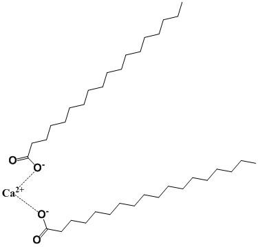
Structure of calcium stearate
Answer: The long-chain alkanes that make up the structure of calcium stearate do not contain any functional groups that could hydrogen bond with water. This inability to interact with water will lead to a decreased solubility and increased tendency to separate from water (the hydrophobic effect).
10. Interatomic Distances in Weak Forces versus Chemical Bonds
What is the distance between the centers of two carbon atoms (their limit of approach) that are interacting through van der Waals forces? What is the distance between the centers of two carbon atoms joined in a covalent bond? (See Table 1.4; See Section 1.4)
Answer: The limit of approach of two atoms is determined by the sum of their van der Waals radii, which are given in Table 1.4. For two carbon atoms the limit of approach is (0.17 nm + 0.17 nm) 0.34 nm. The distance between the centers of two carbon atoms joined in a covalent bond is the sum of the covalent radii of the two carbons or (0.077 nm + 0.077 nm) 0.154 nm. Clearly, two carbons sharing electrons in a covalent bond are closer together than are two carbons interacting through van der Waals forces.
11. The Strength of Weak Forces Determines the Environmental Sensitivity of Living Cells
Why does the central role of weak forces in biomolecular interactions restrict living systems to a narrow range of environmental conditions?
Answer: The weak forces such as hydrogen bonds, ionic bonds, hydrophobic interactions, and van der Waals interactions can be easily overcome by low amounts of energy. Slightly elevated temperatures are sufficient to break hydrogen bonds. Changes in ionic strength, pH, concentration of particular ions, etc., all potentially have profound effects on macromolecular structures dependent on the weak forces.
12. Cells As Steady-State Systems
Describe what is meant by the phrase "cells are steady-state systems”.
Garrett & Grisham 1-7
Answer: Life is characterized as a system through which both energy and matter flow. The consequence of energy flow in this case is order, the order of monomeric units in biopolymers, which in turn produce macromolecular structures that function together as a living cell.
13. The Biosynthetic Capacity of Cells
The nutritional requirements of Escherichia coli cells are far simpler than those of humans, yet the macromolecules found in bacteria are about as complex as those of animals. Because bacteria can make all their essential biomolecules while subsisting on a simpler diet, do you think bacteria may have more biosynthetic capacity and hence more metabolic complexity than animals? Organize your thoughts on this question, pro and con, into a rational argument.
Answer: Although it is true that Escherichia coli are capable of producing all of their essential biomolecules (e.g. there is no minimum daily requirement for vitamins in the world of wild-type E. coli) they are rather simple, single-cell organisms capable of a limited set of responses. They are self-sufficient, yet they are incapable of interactions leading to levels of organization such as multicellular tissues. Multicellular organisms have the metabolic complexity to produce a number of specialized cell types and to coordinate interactions among them.
14. Cell Structure
Without consulting figures in this chapter, sketch the characteristic prokaryotic and eukaryotic cell types and label their pertinent organelle and membrane systems.
Answer: Prokaryotic cells lack the compartmentation characteristic of eukaryotic cells and are devoid of membrane bound organelles such as mitochondria, chloroplasts, endoplasmic reticulum, Golgi apparatus, nuclei, peroxisomes and vacuoles. Both cell types are delimited by membranes and contain ribosomes.
15. The Dimensions of Prokaryotic Cells and Their Constituents
Escherichia coli cells are about 2 µm (microns) long and 0.8 µm in diameter. (Section 1.5)
a. How many E. coli cells laid end to end would fit across the diameter of a pinhead? (Assume a pinhead diameter of 0.5 mm.)
Answer:
Grisham
b. What is the volume of an E. coli cell? (Assume it is a cylinder, with the volume of a cylinder given by V = π r2h, where π = 3.14.)
Answer:
But,1 m100 cm10cm10 ml10L V110m110L1 fL (femtoliter)
c. What is the surface area of an E. coli cell? What is the surface-to-volume ratio of an E. coli cell?
Answer:
Surface
Surface Area6.0310m
Surface Area610m
Volume110mfrom b
Surface Area per volume610m
d. Glucose, a major energy-yielding nutrient, is present in bacterial cells at a concentration of about 1 mM. What is the concentration of glucose, expressed as mg/ml? How many glucose molecules are contained in a typical E. coli cell? (Recall that Avogadro’s number = 6.023 × 10²³.)
Answer:
Glucose110180molg Lmol
Glucose0.180.18gmg Lml moles of glucoseconcentrationvolume
of glucose110110L from b
e.A number of regulatory proteins are present in E. coli at only one or two molecules per cell. If we assume that an E. coli contains just one molecule of a particular protein, what is the molar concentration of this protein in the cell? If the molecular weight of this protein is 40 kD, what is its concentration., expressed as mg/ml? Answer
f. An E. coli cell contains about 15,000 ribosomes, which carry out protein synthesis. Assuming ribosomes are spherical and have a diameter of 20 nm (nanometers), what fraction of the E. coli cell volume is occupied by ribosomes?
Volume of 1 ribosome =3.14
Volume of 1 ribosome4.210m
Volume of 15,000 ribosomes15,0004.2106.310
Fractional volume
g. The E. coli chromosome is a single DNA molecule whose mass is about 3.0 × 10⁹ daltons. This macromolecule is actually a circular array of nucleotide pairs. The average molecular weight of a nucleotide pair is 660 and each pair imparts 0.34 nm to the length of the DNA molecule. What is the total length of the E. coli chromosome? How does this length compare with the overall dimensions of an E. coli cell? How many nucleotide pairs does this DNA contain? The average E. coli protein is a linear chain of 360 amino acids. If three nucleotide pairs in a gene encode one amino acid in a protein, how many different proteins can the E. coli chromosome encode? (The answer to this question is a reasonable approximation of the maximum number of different kinds of proteins that can be expected in bacteria.)
Answer: The number of moles of base pairs in 3.0 × 10⁹ Da dsDNA is given by
Length4.55100.3410m
Length1.5510m1.55 mm1,550 m
Length 2 m
Length DNA1,550 m 775
Length 2 m
To calculate the number of different proteins that would be encoded by the E. coli chromosome:
The exact number can be found at NCBI (http://www.ncbi.nlm.nih.gov). The genomes of a number of strains of E. coli have been sequenced but the first one was K-12 strain MG1655. At NCBI, search for MG1655 and view hits in the genome database. There should be 16 of them and NC_00913 should be one of them. Activate this link (or search for NC_00913 directly and then activate it). The returned page should indicate that this strain of E. coli has 4,145 protein coding genes.
16. The Dimensions of Mitochondria and Their Constituents
Assume that mitochondria are cylinders 1.5 mm in length and 0.6 mm in diameter. (Section 1.5)
a.What is the volume of a single mitochondrion?
b. Oxaloacetate is an intermediate in the citric acid cycle, an important metabolic pathway localized in the mitochondria of eukaryotic cells. The concentration of oxaloacetate in mitochondria is about 0.03 µM. How many molecules of oxaloacetate are in a single mitochondrion?
Answer:
# molecules = Molar concentrationvolume6.02310 mol mol molecules # molecules0.03104.2410L from a6.02310 L mol # molecules7.66 molecules (less than 8 molecules)
17. The Dimensions of Eukaryotic Cells and Their Constituents
Assume that liver cells are cuboidal in shape, 20 µm on a side. (Section 1.5)
a.How many liver cells laid end to end would fit across the diameter of a pinhead? (Assume a pinhead diameter of 0.5 mm.)
Answer:
b. What is the volume of a liver cell? (Assume it is a cube.)
Answer:
Volume of cubic liver cell810m
cm Volume of cubic liver cell810L8 pL
c. What is the surface-to-volume ratio of a liver cell?
Answer:
Surface
Volume810mfrom b
Surface Area
d. How does this compare to the surface-to-volume ratio of an E. coli cell? (Compare this answer to that of problem 3c.) What problems must cells with low surface-to-volume ratios confront that do not occur in cells with high surface-tovolume ratios?
Answer:
The surface-to-volume ratio of liver to that of E. coli is given by:
The volume of a cell sets or determines the cell's maximum metabolic activity while the surface area defines the surface across which nutrients and metabolic waste products must pass to meet the metabolic needs of the cell. Cells with a low surface-to-volume ratio have a high metabolic capacity relative to the surface area for exchange.
e.A human liver cell contains two sets of 23 chromosomes, each set being roughly equivalent in information content. The total mass of DNA contained in these 46 enormous DNA molecules is 4 × 10¹² daltons. If each nucleotide pair has a mass of 660 Da, what is the total number of nucleotide pairs in the 46 DNA molecules?
Answer:
f. If each nucleotide pair contributes 0.34 nm to the length of DNA, what would be the total length of the 46 liver-cell DNA molecules if laid end-to-end?
relative to liver cell1.0310 or about 100,000 times greater!
g.The maximal information in each set of liver cell chromosomes should be related to the number of nucleotide pairs in the chromosome set’s DNA. This number can be obtained by dividing the total number of nucleotide pairs calculated above by 2. What is this value?
Answer: The maximal information is 3.0 × 10⁹ bp.
If this information is expressed in proteins that average 400 amino acids in length and three nucleotide pairs encode one amino acid in a protein., how many different kinds of proteins might a liver cell be able to produce? (In reality livers cells express at most about 30,000 different proteins. Thus, a large discrepancy exists between the theoretical information content of DNA in liver cells and the amount of information actually expressed.)
Answer:
18. A Simple Genome and Its Protein-Encoding Capacity
The genome of the Mycoplasma genitalium consists of 523 genes, encoding 484 proteins, in just 580,074 base pairs (Table 1.5).
What fraction of the M. genitalium genes encodes proteins?
What do you think the other genes encode?
If the fraction of base pairs devoted to protein-coding genes is the same as the fraction of the total genes that they represent, what is the average number of base pairs per proteincoding gene?
If it takes 3 base pairs to specify an amino acid in a protein, how many amino acids are found in the average M. genitalium protein?
If each amino acid contributes on average 120 daltons to the mass of a protein, what is the mass of an average M. genitalium protein? (Section 1.5)
Answer:
What do you think the other genes encode?
Answer: The other genes likely code for ribosomal RNAs and transfer RNAs. To make a functional ribosome it takes at least three ribosomal RNAs, a small subunit rRNA, a large
subunit rRNA and a 5S rRNA. To decode 61 triplet codons requires a minimum of 32* tRNAs. So, a minimum set of tRNAs and rRNAs is 35 (32 + 3). Of the 523 genes, 484 are proteins leaving 39 genes to code for RNAs.
*Essentially 2 tRNA’s for each XXN triplet set except for TAN, which only requires one. This is because TAA and TAG are stop codons that require proteins for recognition. This would give 31 tRNAs but an extra one should be included for initiation of protein synthesis. In bacteria a methionine codon starts a protein-coding region and it is decoded by a special initiator tRNA, which is different from the one used at internal methionine codons.
Of the few RNAs that we are missing by this accounting one is the RNA portion of RNase P a ribonuclease involved in tRNA processing. Another is the so-called 10Sa RNA, a tRNA like RNA that is involved in decoding faulty mRNAs. The 4.5S RNA of the signal recognition particle, a complex involved in synthesis of membrane and secreted proteins is also coded in the genome. This leaves perhaps one or two RNAs unaccounted for whose functions are still unknown.
A complete listing of genes for M. genitalium may be found by doing a search at the NCBI web site (http://www.ncbi.nlm.nih.gov/) for this organism. You can either restrict your search to “Genome” using the pull down search menu or do a search on all databases and then inspect hits for the genome database. Information for M. genitalium G37 is in NC_000908. In August of 2011 the number of genes listed in this organism was 524, encoding 475 proteins. That these numbers are slightly different than those listed above emphasizes the dynamic nature of the interpretation of the genomic information.
If the fraction of base pairs devoted to protein-coding genes is the same as the fraction of the total genes that they represent, what is the average number of base pairs per protein-coding gene?
Answer: Assuming no overlap of genes, and using the numbers in the original problem: 484
Amount of genome devoted to proteins580,074536,818 bp 523 =×=
The average number of base pairs per protein-coding gene is found by dividing this number by the number of protein genes. Thus,
536,818bp
Average size of gene coding for protein 1,109 484protein ==
Note: This number is simply the genome size divided by the total number of genes.
If it takes 3 base pairs to specify an amino acid in a protein, how many amino acids are found in the average M. genitalium protein?
Answer:
1,109amino acids
Average number of amino acids3703protein ==
To calculate the actual average number of amino acids in M. genitalium proteins, visit NC_000908 at NCBI. You will find a table summarizing this organism’s genome. Activating the “Protein coding: 475” link will direct you to a table of all the proteins for M.genitalium. At the bottom of the page use the “Send to” pull down menu to select “Text”. This will return a tab delimited text file of the information in the table. Simply copy all of it except the very first line and paste this information into an Excel spread sheet. The information presented in the “length” column is the length in codons or amino acids for all the proteins. The average of this column is 369, which is in very good agreement with the average calculated above.
If each amino acid contributes on average 120 daltons to the mass of a protein, what is the mass of an average M. genitalium protein?
Answer:
Average protein size = 370 × 120 = 44,400 daltons
19. An Estimation of Minimal Genome Size for a Living Cell
One prominent study of existing cells to determine the minimum number of genes needed by a living cell has suggested that 206 genes are sufficient.
If the ratio of protein-coding genes to non-protein-coding genes is the same in this minimal organism as the genes of Mycoplasma genitalium, how many proteins are represented in these 206 genes?
How many base pairs would be required to form the genome of this minimal organism if the genes are the same size as M. genitalium genes? (Section 1.5)
Answer: For M. genitalium we determined in question 12 that 92.5% of the genes of this organism are protein-coding genes. Assuming the same percentage applies to a minimum set of genes then 191 of the 206 genes are protein-coding genes.
Protein-coding genes = 0.925 × 206 = 190.6 = 191
How many base pairs would be required to form the genome of this minimal organism if the genes are the same size as M. genitalium genes?
Answer: In question 12 we were told that 580,074 base pairs code for 523 genes. The genome size required to code for 206 genes is calculated as follows: 580,074x 523206 580,074 x206 228,480 523
Note: This calculation assumes that genes essentially do not overlap. A smaller genome size could be possible by allowing overlapping, but this would constrain the protein sequences.
Garrett & Grisham 1-17
20.An Estimation of the Number of Genes in a Virus
Virus genomes range in size from approximately 3,500 nucleotides to 280,000 base pairs. The genome of SARS-CoV-2, the virus responsible for the COVID-19 pandemic, consists of 29,811 nucleotides.
If viral genes are about the same size as M. genitalium genes, what is the minimum and maximum number of genes in viruses? (Section 1.6)
Answer: In Question 19 we determined that the average gene size in M. genitalium is 1109 (the genome size -580,074- divided by the number of genes -523). Applying this average gene size to viral genomes we find:
3,500
Minimum number of viral genes3.153 genes 1,109
280,000
Maximum number of viral genes 252 genes 1,109
21. Intracellular Transport of Proteins
The endoplasmic reticulum (ER) is a site of protein synthesis. Proteins made by ribosomes associated with the ER may pass into the ER membrane or enter the lumen of the ER. Devise a pathway by which: a.a plasma membrane protein may reach the plasma membrane. b. a secreted protein may be deposited outside the cell. (Section 1.5)
Answer:
Protein synthesis starts out on ribosomes located in the cytoplasm of cells. Proteins destined to be excreted or to become membrane proteins are synthesized with a signal sequence located near the N-terminus of the protein. (Protein synthesis begins at the Nterminus.) This signal sequence directs the ribosome to the endoplasmic reticulum where the ribosome docks with the reticular membrane. Endoplasmic reticulum studded with ribosomes is called rough endoplasmic reticulum. The signal-sequence-containing protein is synthesized by rough endoplasmic reticulum-bound ribosomes that synthesize the protein and simultaneously export it into the lumen of the endoplasmic reticulum. For a protein to be transported to the plasma membrane it must be packaged into membrane vesicles in the endoplasmic reticulum since the reticular membrane is separate from the plasma membrane. Vesicles from the endoplasmic reticulum containing membrane proteins do not, however, move directly to the plasma membrane. Rather they are routed to the Golgi apparatus where a variety of post-translational modifications occur. Once proteins move through the Golgi they are repackaged into vesicles that are directed to the plasma membrane. Secreted proteins follow the same pathway. Both membrane proteins and excreted proteins contain signal sequences that get them into the endoplasmic reticulum. Membrane proteins contain an additional domain or domains that are hydrophobic in nature and anchor the proteins into the reticular membrane.
Garrett & Grisham 1-1
Think-Pair-Share Question
Create Your Own Monomer
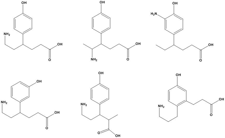
Many biopolymers are created through the repeated linking of monomeric units into a larger structure (see Figure 1.8). Such monomers have at least two structural elements (functional groups) that allows them to be linked to two other monomers.
Create your own monomer with the following requirements:
1) The general formula should be C12H17NO3.
2)The structure should contain one amine and one carboxylic acid functional group (see structures below).
3)Every carbon should have four bonds, every oxygen two bonds, every nitrogen three bonds and every hydrogen one bond.
4)Double bonds and cyclic (including aromatic) features can be present, but no triple bonds.
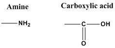
Answer: Various chemical structures are possible. The structure shown in the top left corner is a typical example. The other examples are so-called structural isomers. They have the same structural features but in different locations of the molecule. The structures shown all have a general formula of C12H17NO3. All structures shown contain an amine (NH2), an alcohol (-OH) and a carboxylic acid (-COOH). Such features can serve as “chemical handles” to form linkages as carboxylic acids and alcohols can make esters or carboxylic acids and amines can make amides.
Additional Problems
1.Silicon is located below carbon in the periodic chart. It is capable of forming a wide range of bonds similar to carbon yet life is based on carbon chemistry. Why are biomolecules made of silicon unlikely?
2.Identify the following characters of the Greek alphabet: α, β, γ, δ,
, λ, µ, ν, π, ρ,
,
,
,
,
, χ,
, ψ, and ω.
,
,
3. Give a common example of each of the weak forces at work.
4.On a hot dry day, leafy plants may begin to wilt. Why?
Abbreviated Answers
1. Covalent silicon bonds are not quite as strong as carbon covalent bonds because the bonding electrons of silicon are shielded from the nucleus by an additional layer of electrons. In addition, silicon is over twice the weight of carbon. Also, silicon oxides (rocks, glass) are extremely stable and not as reactive as carbon.
2. These Greek letters are commonly used in biochemistry but this set is not the complete Greek alphabet. alpha (α), beta (β), gamma (γ), delta (δ), capital delta (∆), epsilon (ε), zeta (ζ), theta (θ), kappa (κ), lambda (λ), mu (µ), nu (ν), pi (π), rho (ρ), sigma (σ), capital sigma (Σ), tau (τ), chi (χ), phi (φ), psi (ψ), and omega (ω), the last letter of the Greek alphabet.
3.Ice is an example of a structure held together by hydrogen bonds. Sodium and chloride ions are joined by ionic bonds in table salt crystals. A stick of butter is a solid at room temperature because of van der Waals forces. The energetically unfavorable interactions between water and oil molecules cause the oil to coalesce.
4.The tonoplast loses water and begins to shrink causing the plant cell membrane to exert less pressure on the cell wall.
Chapter Summary
The chapter begins with an outline of the fundamental properties of living systems: complexity and organization, biological structure and function, energy transduction, and self-replication. What are the underlying chemical principles responsible for these properties? The elemental composition of biomolecules is dominated by hydrogen, carbon, nitrogen and oxygen. These are the lightest elements capable of forming strong covalent bonds In particular, carbon plays a key role serving as the backbone element of all biomolecules. It can participate in as many as four covalent bonds arranged in tetrahedral geometry and can produce a variety of structures including linear, branched,
Garrett & Grisham 1-20
and cyclic compounds.
The four elements are incorporated into biomolecules from precursor compounds: CO2, NH4+, NO3 and N2. These precursors are used to construct more complex compounds such as amino acids, sugars, and nucleotides, which serve as building blocks for the biopolymers; proteins, polysaccharides, and nucleic acids, as well as fatty acids and glycerol which are the building blocks of lipids. These complex macromolecules are organized into supramolecular complexes such as membranes and ribosomes that are components of cells, the fundamental units of life.
Proteins, nucleic acids and polysaccharides are biopolymers with structural polarity due to head-to-tail arrangements of asymmetric building block molecules. In these biopolymers, the building blocks are held together by covalent bonds, but they assume an elaborate architecture due to weak, noncovalent forces such as van der Waals interactions, hydrogen bonds, ionic bonds and hydrophobic interactions. The three dimensional shape is important for biological function, especially for proteins. At extreme conditions such as high temperature, high pressure, high salt concentrations, extremes of pH, and so on, the weak forces may be disrupted, resulting in loss of both shape and function in a process known as denaturation. Thus, life is confined to a narrow range of conditions.
Life demands a flow of energy during which energy transductions occur in the organized, orderly, small, manageable steps of metabolism, each step catalyzed by enzymes.
The fundamental unit of life is the cell. There are two types: eukaryotic cells with a nucleus and prokaryotic cells without a nucleus. Prokaryotes are divided into two groups, eubacteria and archaea. All cells contain ribosomes, which are responsible for protein synthesis; however, prokaryotic cells contain little else in the way of subcellular structures. Eukaryotic cells, found in plants, animals, and fungi, contain an array of membrane-bound compartments or organelles, including a nucleus, mitochondria, chloroplasts, endoplasmic reticulum, Golgi apparatus, vacuoles, lysosomes, and perixosomes. Organelles are internal compartments in which particular metabolic processes are carried out.
Chapter 2
Water: The Medium of Life
Chapter Outline
• Properties of water: High boiling point, high melting point, high heat of vaporization, high surface tension, high dielectric constant, maximum density as a liquid: All due to ability of water to hydrogen bond
• Water structure
• Electronegative oxygen, two hydrogens: Nonlinear arrangement: Dipole
• Two lone pairs on oxygen: H-bond acceptors
• Partially positively charged hydrogens: H-bond donors
• Ice
• Lattice with each water interacting with 4 neighboring waters
• H-bonds: Directional, straight and stable
• Liquid: H-bonds present but less than 4 and transient
• Solvent properties of water
• High dielectric constant decreases strength of ionic interactions between other molecules
• Force of ionic interaction, F = e1e2/Dr2, inversely dependent on D
• Salts dissolve in water
• Interaction with polar solutes through H-bonds
• Hydrophobic interactions: Entropy-driven process minimizes solvation cage
• Amphiphilic molecules: Polar and nonpolar groups
• Colligative properties: Freezing point depression, boiling point elevation, lowering of vapor pressure, osmotic pressure effects: Depend on solute particles per volume
• Ionization of water
• Ions: hydrogen ion H+ (protons), hydroxyl ion OH–, hydronium ion H3O+(protonated water)
• Ion product: Kw = [H2O] × Keq = 55.5 × Keq = 10–14 = [H+][OH–]
• pH = –log10 [H+], pOH = –log10 [OH–], pH + pOH = 14
• Strong electrolytes: Completely dissociate: Salts, strong acids, strong bases
• Weak electrolytes: Do not fully dissociate: Hydrogen ion buffers
• Buffers
• Henderson-Hasselbalch equation: pH = pKa + log10 ([A–]/[HA])
• Biological buffers: Phosphoric acid (pK1 = 2.15, pK2 = 7.2, pK3 = 12.4); histidine (pKa = 6.04); bicarbonate (pKoverall = 6.1)
• “Good” buffers: pKa’s in physiological pH range and not influenced by divalent cations
Chapter Objectives
Water
Its properties arise because of the ability of water molecules to form H bonds and to dissociate to H+ and OH–. Thus, water is a good solvent, has a high heat capacity and a high dielectric constant.
Acid-Base Problems
For acid-base problems the key points to remember are: Henderson-Hasselbalch: pH = pKa + log([A–]/[HA])
Conservation of acid and conjugate base: [A–] + [HA] = Total concentration of weak electrolyte added.
Conservation of charge: Ʃ [cations] = Ʃ [anions] i.e., the sum of the cations must equal the sum of the anions. In many cases, simplifications can be made to this equation. For example, [OH–] or [H+] may be small relative to other terms and ignored in the equation. For strong acids, it can be assumed that the concentration of the conjugate base is equal to the total concentration of the acid. For example, an x M solution of HCl is x M in Cl–. Likewise for x M NaOH, the [Na+] is x M. In polyprotic buffers (e.g., phosphate, citrate, etc.), the group with the pKa closest to the pH under study will have to be analyzed using the Henderson-Hasselbalch equation. For groups with pKas 2 or more pH units away from the pH, they are either completely protonated or unprotonated.
The solution to a quadratic equation of the form, y = ax2 + bx + c is:
Problems and Solutions
1. Calculating pH from [H+]
Calculate the pH of the following.
a. 5 x 10⁻⁴ M HCl
Answer: HCL is a strong acid and fully dissociates into [H+] and [CL–]. Thus, [H+] = [Cl–] = [HCl]total added
b. 7 x 10⁻⁵ M NaOH
Answer: For strong bases like NaOH and KOH,
c. 2 µM HCl
Answer:
d. 3 x 10⁻² M KOH
Answer:
e. 0.04 mM HCl
f. 6 x 10⁻⁹ M HC1 = 0.06 x 10⁻⁷ M HCl
Answer: Beware! Naively one might fall into the trap of simply treating this like another strong acid problem and solving it like so:
However, something is odd. This answer suggests that addition of a small amount of a strong acid to water will give rise to a basic pH! What we have ignored is the fact that water itself will contribute H+ into solution so we must consider the ionization of water as well. There are two approaches we can take in solving this problem. As a close approximation we can assume that: 7 [H]10[HCl]or, +−=+ 79777 [H]10610100.06101.0610 +−−−−−
The exact solution uses the ion product of water. 14 w [H][OH]K10 +−− == (the ion product of water) (1)
In solution HCl fully dissociates into H+ + Cl . For any solution the sum of negative and positive charges must be equal. Since we are dealing with monovalent ion we can write: [H][Cl][OH](2) +−− =+
Now because HCl is fully dissociated: 9 total [Cl][HCl]610(3) ==×
Substituting this into equation (2), solving equation (2) for [OH–] and substituting in equation (1) we have a quadratic equation in H+: 2 9 14 [H]610[H]100 +−+− −×−= whose general solution is given by
Before firing up the calculator a little reflection suggests that the argument under the square root is dominated by 4 × 10-14 whose root is 2 × 10⁻⁷. Furthermore, of the two solutions (i.e., ±) it must be the + solution (- given rises to a negative [H+]!)
Therefore,
2. Calculating [H+] from pH
Calculate the pH or pOH of the following.
a. [H+] in vinegar
Answer:
b. [H+] in saliva
Answer:
From Table 2.3 we find that pH = 2.9 since pH = log10[H+]
pH2.9 3 [H]10101.2610M1.26mM +−−− ===×=
From Table 2.3 we find that in saliva pH = 6.6 since pH = log10[H+]
pH6.6 7 [H]10102.510M0.25M +−−− ===×=µ
c. [H+] in household ammonia
Answer:
The pH of ammonia is 11.4 thus pH11.4 12 [H]1010410M4pM +−−− ===×=
d. [OH–] in milk of magnesia
Answer:
e. [OH–] in beer
Answer:
The pH of milk of magnesia is 10.3.
From pH + pOH = 14, pOH = 14 10.3 = 3.7 pOH3.7 4 [OH]1010210M0.2mM ===×=
The pH of beer is 4.5.
From pH + pOH=14, pOH = 14 4.5 = 9.5
pOH9.5 10 [OH]10103.1610M0.316nM ===×=
f. [H+] inside a liver cell
Answer:
The pH of a liver cell is 6.9 thus pH6.9 7 [H]10101.2610M0.126M +−−−
3. Henderson-Hasselbalch Equation and Ionization
Consider the following equilibrium.
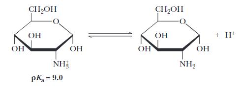
A scientist claims that at pH = 7.0, a solution of this molecule would contain about 90% of the species in their charged form (–NH3+) and about 10% of the species in their uncharged form (–NH2). Is this scientist correct and if not, provide the correct percentages for these two species in the solution?
Answer: The solution requires the Henderson-Hasselbach equation but shown according to the specifics of the equilibrium shown.
For the equilibrium shown, [–NH3+] corresponds to the molecule on the left side and [–NH2] corresponds to the molecule on the right side. Given the values of pH and pKa and using the Henderson-Hasselbalch equation one can determine that the ratio of uncharged species to charged species, [–NH2] / [–NH3+], is 10(7 – 9) = 10-2 = 0.01 or 1/100. Given that [–NH2] + [–NH3+] = 100, one can calculate that [–NH3+] = 100 / 1.01 = 0.99 and [–NH2] = 0.01. Thus about 1% would be in the uncharged form and about 99% would be in the charged form.
4. Effect of pH on Secondary Interactions
Three organic compounds are shown. Based upon the list of acidic compounds shown in Table 2.4, identify the acidic molecule.
Garrett & Grisham 2-5
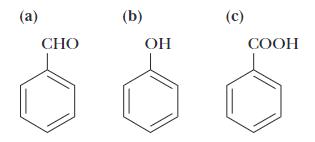
Answer: All three structures have the aromatic ring in common. Structure A contains an aldehyde functional group, Structure B contains an alcohol functional group and Structure C contains a carboxylic acid functional group. The carboxylic acid functional group is an acidic functional group as it can readily release a proton: R-COOH → RCOO- + H+
5. Effect of Solvent on Acidity
The pKa values that are typically discussed are only applicable in water. If an acidic compound is dissolved in an organic solvent (like liquid hexane C6H14), would this decrease or increase the acid’s pKa value? Explain.
Answer: When an acid dissociates in water it is a molecule of water that binds the released proton, H2O → H3O+, as water can act as a base. An organic molecule like liquid hexane does not have basic properties and could not pick up any released protons. Thus, in this type of environment the pKa values of acids would be much higher compared to an aqueous environment as the acids would not release protons that readily. This phenomenon is important when discussing the acidic or basic properties of functional groups within pockets of non-aqueous microenvironments inside larger biomolecules like proteins.
6. Calculating [H+] and pKa from the pH of a Solution of Weak Acid
The pH of a 0.02 M solution of an acid was measured at 4.6
a. What is the [H+] in this solution?
Answer:
b. Calculate the acid dissociation constant Ka and pKa for this acid.
Answer:
Assume that a small amount, x, of HA dissociates into equal molar amounts of H+ and A–. We then have:
HAHAor, →++− 0.02xxx −↔+
From (a) we know that [H]25MxA µ +− === And, 6 [HA]0.0225100.02 =−×≈ Thus,
7. Calculating the pH of a Solution of a Weak Acid; Calculating the pH of the Solution after the Addition of Strong Base
The Ka for formic acid is 1.78 × 10⁻⁴ M.
a. What is the pH of a 0.1 M solution of formic acid?
Answer: a10 a [A] [HA] pHpKlogor[H]K(1) [HA] [A ] + =
For formic acid, [H][A](2) +− ≈ and [HA][A]0.1Mor += [HA]0.1[A](3) =− and, using equation (2) we can write [HA]0.1[H] + =−
Substituting this equation and (2) into (1) we find: a 0.1[H] [H]K or [H] + + +
aa [H]K[H]0.1K0, +++−= a quadratic whose solutions are
The argument under the square root sign is greater than Ka. Therefore, the correct solution is the positive root. Further Ka2 is small relative to 0.4Ka and can be ignored.
[H]0.00413M
pHlog[H]2.38
b. 150 ml of 0.1 M NaOH is added to 200 ml of 0.1 M formic acid, and water is added to give a final volume of 1 L. What is the pH of the final solution?
Answer: The total concentration of formic acid is:
0.2L0.1M [HA] 0.02 M 1L × =
When 150 ml of 0.1 M NaOH is added its total concentration is: 0.15L0.1M [NaOH] 0.015M 1L × = =
Since NaOH is a strong base it will fully dissociate into equal amounts of Na+ and OH–. The OH– will react with an equivalent number of free protons and to compensate for loss of protons the protonated form of formic acid, i.e., HA, will dissociate. The final concentration of HA is found as follows: total [HA][HA][OH]0.02M0.015M0.0005Mand, [A]0.015M =−=−=
From the Henderson–Hasselbalch equation we have: a10 [A] pHpKlog [HA] =+
pH3.75log 0.005 =+
pH4.23 =
8. Prepare a Buffer by Combining a Solution of Weak Acid with a Solution of the Salt of the Weak Acid
Given 0.1 M solutions of acetic acid and sodium acetate, describe the preparation of 1 L of 0.1 M acetate buffer at a pH of 5.4.
Answer: From the Henderson–Hasselbalch equation, i.e., a10 [A]
pHpKlog [HA] =+
(pHpK)(5.44.76)0.64 [A] 1010104.37(1) [HA] ====
Further, we want [HA][A]0.1M(2) +=
Solving equation (1) for [A–] and substituting into (2) we find: [HA]4.37[HA]5.37[HA]0.1M [HA]0.0186M
Substituting this value of [HA] into (2), we find that [A–] = 0.0814 Therefore, combine 186 ml 0.1 M acetic acid with 814 ml 0.1 M sodium acetate.
9. Calculate the HPO42–/H2PO4– in a Muscle Cell from the pH If the internal pH of a muscle cell is 6.8, what is the [HPO42–]/[H2PO4–] ratio in this cell?
Answer: The dissociation of phosphoric acid proceeds as follows: 23 34
Each dissociation has the following pKa values: 2.15, 7.20 and 12.40. Now, at pH = 6.8, we expect the first equilibrium to be completely to the right and the last equilibrium to be to the left (i.e., we expect phosphoric acid to be in the doubly or singly protonated forms).
From the Henderson–Hasselbalch equations i.e.,
10. Preparing a Phosphate Buffer Solution of pH 7.5 from Solutions of Na3PO4 and H3PO4
Given 0.1 M solutions of Na3PO4 and H3PO4, describe the preparation of 1 L of a phosphate buffer at a pH of 7.5. What are the molar concentrations of the ions in the final buffer solution, including Na+ and H+?
Answer:
Na+ comes from the Na3PO4 solution, where it is 0.3 M. (Note: The solution is 0.1 M in Na3PO4, which dissociates into 3 Na+ and 1 PO43–.) To make the solution we need to add enough Na3PO4 to result in 0.1667 M Na+. This is calculated as follows:
Where x is the volume in ml of tribasic sodium phosphate. This will contribute 0.05557 M of phosphate to the solution. The final phosphate concentration must be 0.1 M and so the remainder must come from phosphoric acid. Therefore, 444.3 mL of phosphoric acid must be added.
The final solution will be:
11. Polyprotic Acids: Phosphate Species Abundance at Different pHs
What are the approximate fractional concentrations of the following phosphate species at pH values of 0, 2, 4, 6, 8, 10, and 12?
Answer: For phosphoric acid the following equilibria apply:
The Henderson–Hasselbalch equation may be used to calculate the ratio of any two species that differ by a proton. Thus,
By applying this equation at a pH value and at the 3 pKa’s we can calculate the following ratios:
The fraction of any one species, at a particular pH, is its concentration divided by the sum of the concentrations of all of the species. For example,
This fraction can be written as a function of x, y and z as follows:
Using this equation we can evaluate the fraction of the fully protonated species at each of the pH values given. For the other species, we can take a similar approach to find expressions for their fractional value as a function of x, y and z.
The values of x, y and z may be calculated by hand. A more efficient approach would be to use a spreadsheet to evaluate x, y and z at the pH values given and then to calculate the corresponding fractions at each of the pH values. The following two tables give this information (to three decimal places).
Note: This is the exact solution. A good approximation is to evaluate the Henderson–Hasselbalch for species whose pKa are nearest the pH under consideration. For example, for pH 0, 2 and 4, evaluation of the Henderson–Hasselbalch for pKa = 2.15 would give answers close to those presented above.
12. Polyprotic acids: Citric acid
species at various pHs
Citric acid, a tricarboxylic acid important in intermediary metabolism, can be symbolized as H3A. Its dissociation reactions are
If the total concentration of the acid and its anion forms is 0.02 M, what are the individual concentrations of H3A, H2A–, HA2–, and A3– at pH 5.2?
Answer: For citric acid
At pH = 5.2 the predominant equilibrium will involve
and HA2–.
And, considering the other two equilibria we have
These terms are related as follows:
Using equations (1), (2) and (3) we can relate the concentration of any one species to any other.
2 [HA]2.754[HA]from(1),and =× 32 [A]0.063[HA] =× from (2), or substituting from above
[A]0.0632.754[HA]0.174[HA]
Substituting these expressions into (4), we find:
And, substituting this value back into equations (1), (2), and (3) we find:
From(1)[HA]2.754[HA]2,7545.08mM14.00mM
From(2)[A]0.174[HA]0,1740.0051M0.88mM
From(3)[HA]0.009[HA]0,008510.0051M4.3410M0.04mM =×=×=×=
We could have anticipated these results because the pH is far from two of the pKas. Only the equilibrium between H2A– and HA2– with a pKa = 4.76 will be significant at pH = 5.2.
13. Calculate the pH Change in a Phosphate Buffer When Acid or Base Is Added
a. If 50 mL of 0.01 M HCl is added to 100 mL of 0.05 M phosphate buffer at pH 7.2, what is the resultant pH? What are the concentrations of H2PO4– and HPO4 2– in the final solution?
Answer: The relevant pKa for phosphoric acid is 7.2 governing the following equilibrium,
From the Henderson–Hasselbalch equation we find that
(pHpK)
( 7.207.20) 424
24 [HPO][HPO]10[HPO]10[HPO] =×=×= And, since 2 424 [HPO][HPO]0.05M +=
424 [HPO][HPO]0.025M ==
In 100 ml we have 100 ml
Now, addition of 50 ml of 0.1 M HCl accomplishes two things: (1) It dilutes the solution; and, (2) It introduces protons that will convert HPO42– to H2PO4–The moles of protons is given by:
(0.00250.0005)mol1000ml
Note: This amount of acid added to 100 ml of water gives pH = 2.5.
b. If 50 mL of 0.01 M NaOH is added to 100 mL of 0.05 M phosphate buffer at pH 7.2, what is the resultant pH? What are the concentrations of H2PO4– and HPO -42 in this final solution?
Answer: For NaOH, the same equations apply with one important difference: HPO42– is increased and H2PO4– is decreased. Thus,
(0.00250.0005)mol1000ml [HPO]
Note: If added to water instead, this amount of NaOH would result in a solution with pH = 11.5.
14. CO2 Buffer System
A consequence of the condition known as COPD (= chronic obstructive pulmonary disease) is the weakened capacity to exhale CO2 that accumulates as a consequence of the ongoing metabolism. This leads to an increased reabsorption of bicarbonate (HCO3-) into the circulation by the kidneys. Explain why this would happen?
Answer: CO2 is to be considered an acid as it can react with water releasing carbonate and a proton: CO2 + H2O → HCO3 - + H+. The chronic obstruction of the airways leads to a poor exhalation of CO2. This increase in CO2 levels in the circulation creates an acidosis. The kidneys try to counteract this situation by reabsorbing bicarbonate as a base to keep the pH balanced.
15. Explore the Bicarbonate/Carbonic Acid Buffering System of Blood Plasma
At 37°C, if the plasma pH is 7.4 and the plasma concentration of HCO3– is 15 mM, what is the plasma concentration of H2CO3? What is the plasma concentration of CO2(dissolved)? If metabolic activity changes the concentration of CO2 (dissolved) to 3 mM, and [HCO3–] remains at 15 mM, what is the pH of the plasma?
Answer: Given the pH and the concentration of bicarbonate, we can use the following equation to calculate the amount of H2CO3:
Garrett & Grisham 2-15
For the bicarbonate buffer system: The concentration of CO2(dissolved) is calculated by first solving for this term:
overallah whereKKK = and Ka = acid dissociation constant for 23 HCO, Kh = equilibrium constant for hydration of CO2 overall pK6.1(Seepage43.) =
16. How to Prepare a Buffer Solution: An Anserine Buffer
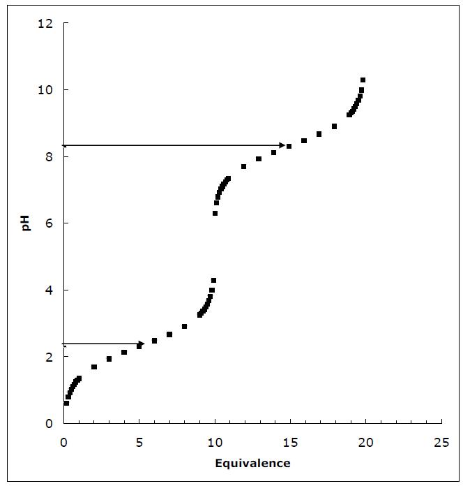
Draw the titration curve for anserine (Figure 2.16). The isoelectric point of anserine is the pH where the net charge on the molecule is zero; what is the isoelectric point for anserine? Given a 0.1 M solution of anserine at its isoelectric point and ready access to 0.1 M HCl, 0.1 M NaOH, and distilled water, describe the preparation of 1 L of 0.04 M anserine buffer solution, pH 7.2.
Answer:

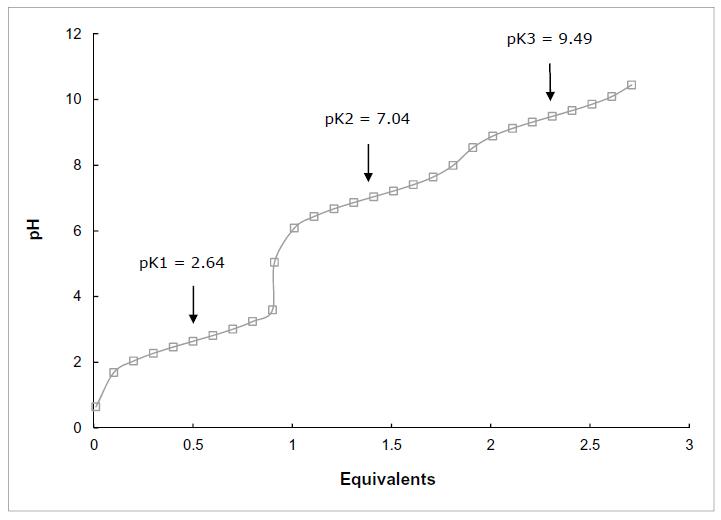
The structure of anserine is shown above. It has three ionizable groups: a carboxyl group pKa1 = 2.64, imidazole nitrogen pKa2 = 7.04 and amino group pKa3 = 9.49. Starting at acidic pH, all three groups will be protonated and thus the molecule will have a +2 charge. As base is added, the carboxyl group will be the first to deprotonate with a midpoint at 2.64. When fully deprotonated at about 4.64, anserine will have a +1 charge. As the pH approaches 7.0, the imidazole group will deprotonate, leaving anserine uncharged. Finally, as the pH passes 9.49, the amino group will deprotonate and by about pH 11.5, anserine will have a –1 charge.
The titration curve is shown below with the pKa’s labeled. The isoelectric point, pI, is the pH at which the molecule is uncharged. This will happen at a pH at which the carboxyl group’s –1 charge is balanced by positive charges from both the imidazole group and the amino group. The isoelectric point must be between the pKa’s of the imidazole and amino groups. Thus, the sum of the protonated imidazole group and the protonated amino group must equal to one equivalent of charge.
Let I and IH+ be the unprotonated and protonated imidazole groups, respectively. Let A and AH+ be the unprotonated and protonated amino groups.
[IH][AH]oneequivalent +++=
That is, the sum of the positively charged species for the imidazole and the amino groups must sum to the one equivalent of negative charge from the carboxylate. (Remember, there is one of each group in the molecule.) The concentrations of the imidazole species, protonated and unprotonated, must sum to one equivalent. The same is true for the amino species. Thus,
[I][IH][A][AH]andso +=+++ [I][IH] + + [IH] + = [AH](1) + + [A][AH] + + [IH][AH] =+++ (2)
From equations (1) and (2) we can see that: [AH][I]or[IH][A](3) ++ = =
The Henderson–Hasselbalch equations for each are:
Substituting (3) into these we have:
Garrett & Grisham 2-17
Solving these two equations for the log terms, which are inversely related, and setting them equal we have:
2 [AH]
pH-pKlog [IH] + + = 3 pH+pKloglog[IH][AH] [AH][IH] ++ ++ −=−=
Thus, 23 pH-pKpHpK =−+
pKpK23 7.049.49 pH pI 22
pI8.27 + + === =
In order to prepare 1 L of 0.04 M anserine, we need to use 400 ml (0.4 L) of 0.1 M anserine stock (1 L × 0.04 M = 0.1 M × 0.4 L). Since the pH = 8.27 we will have to titrate to pH = 7.2 using 0.1 M HCl. The ratio of the neutral to protonated anserine (unprotonated and protonated imidazole, respectively) at pH = 7.2 is determined using the Henderson–Hasselbalch equation..
Thus, we will need to add 164 mL of 0.1 M HCl to adjust the imidazole group, and the solution can then be diluted to 1 L.
Garrett & Grisham 2-18
17. On the Basis of Figure 2.12, what will be the pH of the acetate-acetic acid solution when the ratio [acetate]/[acetic acid] is 10?
a. 3.76
b. 4.76
c. 5.76
d. 11.24
Answer: The pKa of acetic acid is 4.76. To realize a ratio of acetate to acetic acid of 10 nearly an equivalent of base (NaOH) must be added and from Figure 2.12 we see that this corresponds to a pH of around 6. So, the correct answer must be “c”, 5.76. One can easily calculate this value using the Henderson–Hasselbalch equation, pH = pKa + log[A–]/[HA]. The term [A–]/[HA] is the ratio of acetate to acetic acid, which is given as 10. The log 10 = 1.0, thus the pH = 4.76 +1 = 5.76.
18. Determination of the Molecular Weight of a Solution by Freezing Point
Depression. A 100-g amount of a solute was dissolved in 1000 g of water. The freezing point of this solution was measured accurately and determined to be –1.12°C. What is the molecular weight of the solute?
Answer: In the section dealing with colligative properties we learn that 1 mol of an ideal solute dissolved in 1000 g of water (a 1.0 molal solution) depresses the freezing point by 1.86°C. We can set up a proportionality to calculate how many mol was added in this problem. 1.0molalx 1.86C1.12C =
1.12 x1.0molal 1.86 x0.60molal =× =
The 0.6 molal solution was made by adding 100 g solute, which represents 0.6 mol. Therefore, the solute’s molecular weight is: 100g 167Da 0.6mol =
19. How to Prepare a Triethanolamine Buffer
Shown here is the structure of triethanolamine in its fully protonated form:
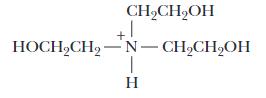
Its pKa is 7.8. You have available at your lab bench 0.1 M solutions of HCl, NaOH, and the uncharged (free base) form of triethanolamine, as well as ample distilled water. Describe the preparation of a 1 L solution of 0.05 M triethanolamine buffer, pH 7.6.
Answer: The free base form of triethanolamine is the molecule shown above but with its tertiary nitrogen unprotonated. The solution we are asked to make must be 0.05 M triethanolamine. So, the first step is to calculate the amount of triethanolamine that is needed to make 1 L of 0.05 M solution using a 0.1 M solution.
0.05 M × 1 L = 0.1 M × x x = 0.5 L = 500 ml
The pH of the free base solution is basic because when added to water triethanolamine protonates and depletes the solution of free protons. To adjust the pH to 7.6 we will have to add HCl. The amount of HCl needed is determined by application of the Henderson–Hasselbalch equation using 7.8 for pKa and 7.6 for pH. a [Triethanolamine] pHpKlog [TriethanolamineH] + =+ a (pHpK)
[Triethanolamine] 10 [TriethanolamineH] + =
(7.67.8)(0.2)
[Triethanolamine] 10100.6310 [TriethanolamineH] + ===
At pH 7.6 the ratio of free base to protonated triethanolamine is 0.631 and we know that the sum of the concentrations of these species is 0.05. Or,
[Triethanolamine] 0.6310 [TriethanolamineH] + = And, [Triethanolamine] + [Triethanolamine ⋅H+] = 0.05 M
There are two equations with two unknowns. Solving for one unknown and substituting gives:
[Triethanolamine] = 0.6310 × [Triethanolamine ⋅H+]
And, [Triethanolamine] + [Triethanolamine ⋅H+] = 0.05 M
Or, 1.6310 × [Triethanolamine] + [Triethanolamine ⋅ H+] = 0.05 M
And, 1.6310 × [Triethanolamine ⋅H+] = 0.05 M 0.05M [ ] 0.0307M Triethanolamin .163 e 10 H + = ⋅=
Garrett & Grisham 2-20
To adjust the pH to 7.6, which we should recognize is below the pKa, we will have to add 0.0307 moles of HCl. (We are making up 1L.) Using 0.1 M HCl, we will need
0.0307mole 0.307L307ml 0.1M ==
So, the solution is made by mixing 500 ml of 0.1 M triethanolamine (free base) with 0.1 M HCl using 307 ml to drop the pH to 7.6. Then adjust the final volume to 1 L.
20. How to Prepare a Tris Buffer Solution
Tris–hydroxymethyl aminomethane (TRIS) is widely used for the preparation of buffers in biochemical research. Shown here is the structure TRIS in its protonated form:
Its acid dissociation constant, Ka, is 8.32 × 10 9. You have available at your lab bench a 0.1 M solution of TRIS in its protonated form, 0.1 M solutions of HCl and NaOH, and ample distilled water. Describe the preparation of a 1 L solution of 0.02 M TRIS buffer, pH 7.8.
Answer: Using the 0.1 M TRIS solution the amount needed to make 1 L of 0.02 M is determined as follows:
Where x is the volume of 0.1 M to be used. x0.1M 0.02M 1L × =
Where x is the volume of 0.1 M to be used.
Solving for x we find:
Next let’s use the Henderson-Hasselbalch equation to calculate the ratio of TRIS base to protonated TRIS at pH = 7.6. Note: We are given the value for Ka, 8.32 x 10⁻⁹. The pKa is calculated as follows:
pKlog8.32108.0799 =−×=
Use the Henderson-Hasselbalch equation as follows: a [Trisbase] pHpKlog [TrisH] + =+ [Trisbase] 7.68.0799log [TrisH] + =+ ⋅ (7.68.0799)0.4799 [Trisbase] 10100.331 [TrisH] + === ⋅
But, [Trisbase][TrisH]0.02M + +⋅=
Use the last two equations to solve for the concentration of each species. [Trisbase]0.331[TrisH] + =×⋅
But,
And,
0.331[TrisH][TrisH]0.02M 1.331[TrisH]0.02M 0.02M [TrisH]0.0150 1.331 ++ +
[Tris base] = 0.02 M – [Tris • H+] = 0.02 M – 0.0150 = 0.005 M
The protonated Tris solution was likely made using Tris ⋅ HCl, which is the chloride salt of Tris base. Thus, it contains equal amounts of chloride and Tris distributed between its protonated and unprotonated forms but mainly as the protonated form. The 200 ml represents 0.02 mole of protonated Tris. To adjust the pH to 7.6 we will need to lower the protonated Tris from 0.02 moles to 0.015 mole and so we will need to add NaOH in the amount of 0.005 mole. This corresponds to the following volume of 0.1 M NaOH:
x × 0.1 M = 0.005 mol
x = 0.05 L = 50 ml
The final recipe is to use 200 ml of 0.1 M protonated Tris solution and add 50 ml of 0.1 M NaOH.
The final solution will actually be 0.02 M Tris buffer at pH = 7.6 but it will contain 5 mM NaCl produced when Tris HCl was adjusted with NaOH. A better way of preparing this solution is to use Tris base and then titrate with HCl to pH = 7.6.
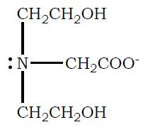
21. Plot the Titration Curve for Bicine and Calculate How to Prepare a pH 7.5 Bicine Buffer Solution Bicine (N, N–bis (2-hydroxyethyl) glycine) is another commonly
Garrett & Grisham 2-22
used buffer in biochemistry labs. The structure of bicine in its fully protonated form is shown below:
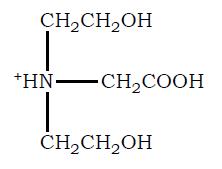
a. Draw the titration curve for Bicine, assuming the pKa for its free COOH group is 2.3 and the pKa for its tertiary amino group is 8.3.
b. Draw the structure of the fully deprotonated form (completely dissociated form) of bicine.
Garrett & Grisham 2-23
c. You have available a 0.1 M solution of Bicine at its isoelectric point (pHI), 0.1 M solutions of HCl and NaOH, and ample distilled H2O. Describe the preparation of 1 L of 0.04 M Bicine buffer, pH 7.5.
Answer: The volume of 0.1 M bicine needed is: x0.1M 0.04M 1L × =
Solving for x :
The isoelectric point of Bicine is simply the average of the two pKas.
pKpKala2 pI 2 + = 2.38.3 pI 5.3 2 + ==
At this pH the carboxyl group is mainly unprotonated whereas the tertiary nitrogen is nearly fully protonated and thus the average charge is zero. Using the HendersonHasselbalch equation we can calculate the ratio of protonated to unprotonated species for each group. For the carboxyl group:
[COO] pHpKlog [COOH] =+
(pHpK) (5.32.3) [COO] 1010 [COOH] = =
(3) [COO] 101000 [COOH] ==
This calculation shows that at pH 7.5 only approximately 0.1% is protonated. The calculation for the tertiary nitrogen of bicine is as follows: N [N] pHpKlog [NH] + =+ N (pHpK) (5.38.3) [N] 1010 [NH] + ==
(3) [N] 100.001 [NH] + ==
We should have anticipated this result, namely the ratios are inverse, because we are starting at a pH that is equidistant from each pKa
Garrett & Grisham 2-24
To adjust the pH from 5.3 to 7.5 will require addition of NaOH. The exact amount is determined by application of the Henderson-Hasselbalch equation to both groups to determine the ratio of protonated to unprotonated forms of both species. For the carboxyl group:
For the amino group:
Using these equations and remembering that the total sum of COO– and COOH is equal to 0.04 mol (0.04 M times 1 L) and that the same is true for the protonated and unprotonated nitrogen we can calculate the moles of each species at the starting pH (i.e., the pI) and at pH = 7.5. The results are shown to five places. The column labeled “delta” is the change in the species from pI to pH = 7.5.
Using 0.1 M NaOH we need 54.6 ml. The final solution is then adjusted to a final volume of 1L with 545.4 ml of water.
d. What is the concentration of fully protonated form of Bicine in your final buffer solution?
Answer: At any pH there will be four possible forms of bicine shown in the chart below. The fraction of each species is simply the fraction of the carboxyl species times the fraction of the nitrogen species.
For example, COO–/NH refers to Bicine with unprotonated carboxyl group and protonated nitrogen. The value 0.863 under the fraction column was calculated using data
Garrett & Grisham 2-25
in the chart in part c. The fraction of carboxyl group that is unprotonated is calculated by dividing the value for COO– in the above chart by the sum of COOH and COO–. The same is done for the nitrogen. The molar amount is the value under fraction times 0.04. The sum shows that all species are accounted for. There are only two species at significant levels, both have the carboxyl group unprotonated. Fully protonated Bicine is only at 0.2 µM.
Species Fraction
COO-/N
COO-/NH
COOH/N
COOH/NH
0.1364413
0.8635524
0.0000009
0.0000055
22. Calculate the Concentration of Cl– in Gastric Juice
0.0054577
0.0345421
0.0000000
0.0000002
Sum = 0.04
Hydrochloric acid is a significant component of gastric juice. If chloride is the only anion in gastric juice, what is its concentration if pH = 1.2?
Answer: Strong acids like HCl fully dissociate in solution and so the pH, which is –log[H+], is closely approximated by –log[HCl]
→++−
Thus, pH = –log[HCl] = 1.2 giving [HCl] = 10–1.2 = 0.0631 or 63.1 mM
So the chloride concentration is 63.1 mM.
23. Calculate the Concentration of Lactate in Blood Plasma at pH 7.4 if [Lactic Acid] = 1.5 µM
From the pKa for lactic acid given in Table 2.4, calculate the concentration of lactate in blood plasma (pH = 7.4) if the concentration of lactic acid is 1.5 pM.
Answer: From Table 2.4 the pKa of lactic acid is 3.86. To determine the concentration of lactate at pH = 7.4 we need to use the Henderson–Hasselbalch equation.
Garrett & Grisham 2-26
Note: The statement that the concentration of lactic acid is 1.5 pM was taken literally. It is possible that the concentration refers to the sum of lactate and its protonated, lactic acid form. In this case the solution is as follows:
pHpKlog lacticacid =+
[] [] a lactate
pH = 7.4, pKa = 3.86, [lactic acid] = 1.5 µM = 1.5 × 10 6 M = [lactate] + [lactic acid]
[lactate] = [lactic acid] × 10(7,4 3,86) = [lactic acid] × 103.54 = 3,467 × [lactic acid]
3,467 × [lactic acid] + [lactic acid] = 1.5 × 10 6 M
[lactic acid] = 6 10 1.510M 4.3210M,and 3,468 × =×
[lactate] = 3,467 × [lactic acid] ≈ 1.5 × 10 6 M
That is, the pH is so far from the pKa that essentially all the lactic acid is in the unprotonated lactate form.
24. Draw the Titration Curve for a Weak Acid and Determine Its pKa from the Titration Curve
When a 0.1 M solution of a weak acid was titrated with base, the following results were obtained:
Plot the results of this titration and determine the pKa of the weak acid from your graph.
Answer: In the chart shown below the values for pHcalculated were determined using the Henderson– Hasselbalch equation with a guess for pKa shown. Values of [A–] were assumed to be equal to the amount of base added while the values of [HA] were 1– [A–].
In the graph shown above pH is plotted against equivalence for the observed data (closed circles) and for the pHcalculated (x). There is very close agreement between the two data sets indicating that the guess for 4.7 is close to the true value. We know we are dealing with an acid (something with pKa lower that 7.0) because the initial pH is 3.42.
Garrett & Grisham 2-28
25. Calculate the Rate of an Enzymatic Reaction Where H+ is a Product
The enzyme alcohol dehydrogenase catalyzes the oxidation of ethyl alcohol by NAD+ to give acetaldehyde plus NADH and a proton:
CH3CH2OH + NAD+ → CH3CHO + NADH + H+
The rate of this reaction can be measured by following the change in pH. The reaction is run in 1 ml 10 mM TRIS buffer at pH 8.6. If the pH of the reaction solution falls to 8.4 after ten minutes, what is the rate of alcohol oxidation, expressed as nanomoles of ethanol oxidized per sec per ml of reaction mixture?
Answer: The number of protons produced after 10 minutes can be determined using the Henderson-Hasselbalch equation. The reaction is being conducted using Tris buffer at an initial pH of 8.6. In problem 16 we were given the following information about Tris:
We will use the Henderson-Hasselbalch equation to determine the ratio of protonated and unprotonated Tris at pH 8.6 and at pH 8.4.
For pH = 8.6
[Trisbase]
8.68.0799log [TrisH] [Trisbase] pHpKlog [TrisH]
(8.68.0799)0.5201 [Trisbase] 10103.312 [TrisH] +
For pH =8.4
[Trisbase] 8.48.0799log [TrisH] +
(8.48.0799)0.3201 [Trisbase] 10102.090 [TrisH] + ===
The total concentration of Tris is 10 mM and we are dealing with a volume of 1.0 ml. The total number of moles of Tris is calculated as follows:
39 5 1010mol1L 110nmol 10mM1ml 1 ml1.010mol 10,000nmol L 1000 ml mol
At pH 8.6, the ratio of unprotonated Tris to protonated Tris is 3.312. Using this information and the fact that both forms must sum to 10,000 nmol we can calculate the moles of each form at both pH values.
Garrett & Grisham 2-29
Tris base + Tris ⋅H+ = 10,000 nmol
Trisbase 3.312
TrisH + = ⋅
Tris base = 3.312 × Tris ⋅H+
Substituting into the top equation
3.312 × Tris ⋅H+ + Tris ⋅ H+ = 10,000 nmol
Or,
4.312 × Tris H+ = 10,000 nmol 10,000 TrisHnmole2319nmol 4,312 + ⋅==
And, substituting this back into the top equation
Tris base + 2319 nmol = 10,000 nmol
Tris base = 7681 nmol
The moles of each species at the two pH values are shown below pH 8.6 8.4 Delta Tris
The column headed “Delta” is the amount of acid in nmoles that must have been produced to change the ratio of Tris species. To be correct we should calculate the amount of acid needed to lower the pH from 8.6 to 8.4 without regard to buffering. This value, however, is very small. It is calculated as follows: pH = – log[H+ ]
[H+] = 10–pH At pH 8.6
[H+ ] = 10–86 = 2.51 x 10⁻⁹M At pH 8.4
[H+ ] = 10–8 4 = 3.98 x 10⁻⁹M
The change in protons is the difference inconcentrations times the volume ( ) 9 993 110nmol molesproduced3.9810M2.5110M110L 0.0015 nmol mol × =×−××××=
So, we have determined that 917 nmol of proton were produced in 10 minutes. The rate is calculated as follows:
917nmole1minnmole rate 1.53 10min60secsec =×=
Garrett & Grisham 2-30
26. The Bicarbonate Buffer System of Blood Plasma
The important buffer system of blood plasma is the bicarbonate/carbonic acid couple:
The relevant pKa, pK1 for carbonic acid is 3.77 at 25°C.
a. If [HCO3-] = 24mM, what is [H2CO3]?
b. What would be the pH of the blood plasma following a sudden influx of 4 x 10⁻⁶ M OH-?
However, remember that the concentration of H2CO3 is itself buffered by the available pools of CO2, which is in equilibrium with H2CO3.
Under the conditions prevailing in mammalian body fluids, the equilibrium constant for the reaction, Kb is 0.003 at 37°C. That is, the equilibrium lies far to the left, such that more than 300 CO2 molecules are present in solutions for every molecule of H2CO3
Kb = [H2CO3]/[CO2]
Thus,
[H2CO3] = Kb[CO2]
Putting this value for [H2CO3] into the expression for the first dissociation of H2CO3 gives
Kl = ([H+][HCO3-])/Kb[CO2]
Therefore, the overall equilibrium constant for the ionization of H2CO3 in equilibrium with CO2 is given by
K1Kb = ([H+]{HCO3-])/[CO2]
And KlKh, the product of two constants, can be defined as a new equilibrium constant, Koverall.
a. What is the value of Koverall?
b. What is the value of pKoverall?
c. If [CO2] = 1.2 mM, what is [HCO3-]?
Answer: a. 3.55 µM
b. Theoretically, 4 µM OH- would neutralize all of the H2CO3, swamping the buffer system, and leading to a dangerous rise in pH.
c. 1.2 mM
d. Koverall = 8.07 x 10⁻⁷
e. pKoverall = 6.1
27. Blood pH and Respiration
Hyperventilation, defined as a breathing rate more rapid than necessary for normal CO2 elimination from the body, can result in an inappropriately low (CO2) in the blood. Central nervous system disorders such as meningitis, encephalitis, or cerebral hemorrhage as well as a number of drug- or hormone-induced physiological changes, can lead to hyperventilation.
a. Which component of the bicarbonate/carbonic acid buffering system will be affected first?
b. What is the [H+] at pH 7.4? What is the [H+] at pH 7.74?
Answer:
a. As [CO2] drops due to excessive exhalation, [H2CO3] in the blood plasma falls, followed by a decline in [H+] and [HCO3-] in the blood plasma. Blood pH rises within 20 seconds of the onset of hyperventilation, becoming maximal within 15 minutes pH can change from its normal value of 7.4 to pH equals 7.74. This rise in plasma pH (increase in alkalinity) is termed respiratory alkalosis.
b. [H+] drops from its normal value of 40 nM (pH = 7.4) to 18 nM (pH = 7.74).
28. Hypoventilation and Blood pH
Hypoventilation is the opposite of hyperventilation and is characterized by an inability to excrete CO2 rapidly enough to meet physiological needs. Hypoventilation can be caused by narcotics, sedatives, anesthetics, and depressant drugs; diseases of the lungs also lead to hypoventilation. Which component of the bicarbonate/carbonic acid buffering system will be affected first during hypoventilation?
Answer: Hypoventilation results in respiratory acidosis, as CO2 accumulates, giving rise to H2CO3, which dissociates to form H+ and HCO3-.
Think-Pair-Share Question
Titration of an Unknown Acid
A 20mL solution containing an unknown, diprotic acid (AH2) was titrated with 0.2M NaOH. The figure shows the pH value of the mixture as a function of the mL 0.2M NaOH added.
a) Calculate the concentration of the unknown acid.
b) estimate the values of pKa,1 and pKa,2 of this unknown acid.
Answers:
a) Two inflection points can be observed in the titration curve: after the addition of 10 and 20mL 0.2M NaOH. This implies that the unknown diprotic acid H2A requires 10 mL of 0.2 mol/L NaOH to neutralize one H+ from this acid. Thus, the amount of H2A present is 0.01 L x 0.2 mol/L = 0.002 mol. This was present in 20 mL of the H2A solution, implying that [H2A] = 0.002 mol / 0.02 L = 0.1 M.
b) Given that every10 mL of 0.2M NaOH added corresponds to an inflection point, the addition of 5mL and 15mL 0.2M NaOH added corresponds to the half-way points. Thus, one can estimate the pKa values to be about 2.0 and 7.0.
Additional Problems
1. Tris (Tris[hydroxymethyl]aminomethane) is a commonly used buffer with a pKa = 8.06. In making up a Tris solution, an appropriate amount of the free base is dissolved in water and the pH of the solution is adjusted, often with HCl. The Mr of Tris is 121.1.
(a) . Describe the preparation of 1 L of 25 mM solution, pH = 8.4, using solid Tris base and 1 M HCl.
(b) Draw the structure of Tris in its protonated and unprotonated forms.
2. Many proteins are sensitive to divalent cations and often chelating agents are included in protein isolation solutions to bind divalent cations, lowering their concentration,
Garrett & Grisham 2-33
(a) . Draw the structures of EDTA and EGTA
(b) . These agents function by binding divalent cations. EDTA binds both Mg2+ and Ca2+ tightly while EGTA has a greater specificity for Ca2+ (Both will bind a number of other divalent cations as well.) However, cation binding is pH–dependent. In making a solution of EDTA, it is often suggested that the pH of the solution be adjusted to around 8. The pKas of the four acetic acid groups on EDTA are 2.0, 2.67, 6.16 and 10.26. At pH = 8.0, what is the predominant ionic species of EDTA?
(c) . Divalent cations bind avidly to the fully ionized forms of both EDTA and EGTA. What fault do you find with using a solution of 10 mM Tris (pH = 7.0) and 25 mM EDTA to both buffer a protein solution and chelate divalent cations?
3. To make up 1 L of a 0.5 M solution of EDTA starting with the free acid, approximately how much 10 M NaOH will have to be added to adjust the pH to 7.0? Do you expect this solution to have a pH-buffering capacity? Explain.
4. The "Good" buffers were developed to provide buffers that would not interact strongly with divalent cations such as Mg2+ and Ca2+. One such buffer is PIPES, (Piperazine–N,N'–bis[2–ethanesulfonic acid]), pKa = 6.8. You are asked to make 1L of 25 mM solution of PIPES, pH = 7.0. In preparing the solution starting from the free acid, you notice that PIPES is not very soluble in water. You find that the solution is acidic (around 2), and so you begin to add NaOH. After addition of approximately 3.75 mL of 10 M NaOH, the solution clears. The pH is around 7. Explain.
5. (a). On the same graph, sketch the titration curves of; acetic acid, pKa = 4.76; ethylamine, pKa = 10.63; and, glycine, pKa = 2.34 and 9.6.
(b). The titrations of acetic acid, ethylamine, and glycine involve a single carboxyl group, a single amino group, and a carboxyl and amino group respectively. Can you suggest why the carboxyl group and the amino groups on glycine have lower pKas than the same groups on acetic acid and ethylamine?
Garrett & Grisham 2-34 and removing them from proteins. Two common agents used for this purpose are EDTA (Ethylenediaminetetraacetic acid) and EGTA (Ethyleneglycol-bis-(βaminoethyl ether) N,N,N',N'-tetraacetic acid).
Abbreviated Answers
1. (a) Add 3.028 g Tris to approximately 900 mL of water. Carefully titrate the pH to 8.4 using approximately 7.8 ml of 1 M HCl. Finally, adjust the volume of the solution to 1 L.
(b)
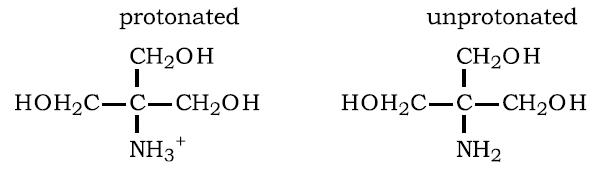
2 (a)
(b) EDTA3– (i.e., the singly-protonated species).
(c) Divalent cation binding to EDTA will result in release of a mole of proton per mole of cation bound to EDTA. The Tris functions as a proton buffer but it is one pH unit below its pKa. Thus, it will not buffer the solution very effectively.
3. To adjust the solution to pH = 7.0, sufficient NaOH must be added to fully titrate the two carboxyl groups with pKas = 2.0 and 2.67. This will require 100 mL of 10 M NaOH. Further, an additional 43.7 mL will be required to titrate the carboxyl group whose pKa = 6.16. This is calculated using the Henderson–Hasselbalch equation. The total amount of NaOH is 143.7 mL.
The solution will not be a good buffer at pH = 7.0 because the pH is 0.8 of a pH unit away from the nearest pKa.
Garrett & Grisham 2-35
4. To bring the pH of the solution to its pKa normally requires approximately one-half of an equivalent of (in this case) base. The 3.75 mL of NaOH represents (3.75 mL × 10 M = ) 0.0375 equivalents of OH–. One liter of 25 mM Pipes contains 0.025 moles of Pipes and 0.0375 equivalents of OH– corresponds to 1.5 equivalents of Pipes. Therefore, there must be two titratable groups on PIPES.
5. The pKas of the carboxyl group and the amino group of glycine are shifted to lower pH values relative to the same groups on acetic acid and ethylamine. Clearly, the groups are influencing each other’s pKas. The pKa of the carboxyl group is lowered because the positively charged amino group influences it. The unprotonated carboxyl group is negatively charged. This state is favored by having a positively charged amino group nearby. Thus, the carboxyl group becomes a slightly stronger acid and will give up its proton at lower pH values. Similarly, the amino group becomes a slightly stronger acid because in its protonated state it is positively charged.
Chapter Summary
The most abundant molecule in living systems is H20. What physical properties make water such an important component of life? The two hydrogens in a water molecule make covalent bonds with a single O. However, there is something special about these covalent bonds. Oxygen is an electronegative element with a high affinity for electrons, surpassed only by F. (This high attraction for electrons is a consequence of a positive ly–charged nucleus rather unshielded by electron clouds.) In covalent bonds with H, the electrons will be attracted to O, giving rise to charge separation or a dipole moment. The hydrogen becomes partially positively–charged whereas the oxygen becomes partially negatively–charged. The four electron pairs in H20 form a distorted tetrahedron, with two lone pairs separated by an angle greater than 109° and the two shared electron pairs (shared between O and H) at 104.5°. The partially positively–charged hydrogen can interact with lone–pair electrons to form hydrogen bonds. We will see that hydrogen bonds, although weak compared to covalent bonds, are important interactions in life.
Water molecules can interact with each other to form hydrogen–bonded structures. Ice is a hydrogen– bonded structure, with each water molecule bonded to four other water molecules. In two of these bonds, the hydrogens interact with lone–pair electrons on two other water molecules. In these cases, water is serving as a hydrogen–bond donor. The two electron pairs make hydrogen bonds, participating as hydrogen– bond acceptors, with positively–charged hydrogens on two other water molecules. The thermal properties of water are a consequence of hydrogen bonds. For example, the density of water decreases with decreasing temperature down to 4°C; as water molecules lose kinetic energy, they can approach each other more closely. As the temperature moves to the freezing point of water, more and more hydrogen bonds form, causing water molecules to move apart and density to decrease. Thus, ice floats. The environmental consequence is that ponds and lakes freeze from the surface, allowing life to continue in the liquid interior. The
Garrett & Grisham 2-36
transition from solid to liquid to gas is accompanied by disruption of hydrogen bonds. A great many hydrogen bonds remain in liquid water, as evidenced by the difference in the heat of melting versus the heat of sublimation. Water has a high heat capacity (the ability to absorb heat without a large increase in temperature), because energy in the form of heat is used to break hydrogen bonds rather than increase temperature (increase kinetic energy). Finally, the rather large amount of energy required to vaporize water means that water can absorb energy with little increase in temperature.
Water is a good solvent for ionic and polar substances, because it forms hydrogen bonds with these substances. Salts dissolve and dissociate because the ionic components become hydrated with water molecules. The high dielectric constant of water is responsible for decreasing the attractive force between two ionic components of a salt. (The force between two charges, e1 and e2, is given by F = e1e2/Dr2; it is inversely dependent on the dielectric constant.)
The attraction between water molecules is strong and the tendency of water molecules to make these interactions is great. For example, at an air/water interface, interaction of water molecules on the surface is responsible for surface tension. But surface tension comes at a price. Water molecules at an interface are forced to make irregular hydrogen bonds, often with less than perfect geometry. In fact, this occurs at any interface, and when nonpolar molecules are put into water, each molecule in effect represents an interface. To minimize the interface area, there is a tendency, driven by hydrogen bonding of H2O, for nonpolar substances to coalesce, to minimize their contact with water molecules. This tendency is termed hydrophobic interactions. We will see that biological membranes and proteins rely on hydrophobic interactions to maintain an ordered structure.
There are two other properties of water that are of extreme importance to biological systems: osmotic pressure and ionization of water. Osmotic pressure arises when a semipermeable membrane through which water can pass separates two aqueous compartments. If a solute is dissolved in one of the aqueous compartments (and the solute is not freely permeable to the membrane), water will move from the solute– free compartment into the compartment containing solute. The amount of pressure necessary to prevent this movement of water is the osmotic pressure. Osmotic pressure is a colligative property and as such its magnitude is directly proportional to concentration.
Water is capable of ionizing in solution to form hydrogen ions and hydroxyl ions. The pH is a measure of the hydrogen ion concentration: pH = –log[H+]. The pH scale in aqueous solution is set by the magnitude of the ion product. In aqueous solutions, pH + pOH = 14. Using this formula, the pH of solutions of strong acids or bases can be calculated. For weak acids and bases, the Henderson–Hasselbalch equation must be used: pH = pKa + log([A–]/[HA]). Several of the problems illustrate the use of this equation.
Chapter 3
Thermodynamics of Biological Systems
Chapter Outline
• Thermodynamic concepts
• Systems
• Isolated systems cannot exchange matter or energy with surroundings
• Closed systems exchange energy but not matter
• Open systems exchange both energy and matter
• First Law: ∆E = q + w
• ∆E = change in internal energy (state function), q = heat absorbed, and w = work done on the system
• Work = generalized force × generalized distance
• H = E + PV: H = enthalpy (energy transferred at P = constant): ∆H = q when work limited to P∆V
• ∆H° = Rd(lnKeq)/d(1/T): van’t Hoff plot RlnKeq vs 1/T (R = gas constant = 8 314J/mol K
• Second Law: Disorder or randomness
• S = klnW where k = Botzmann’s constant = 1.38 × 10 23 J/K
• W = number of ways of arranging the components of a system at fixed internal energy
• dS = dq/T for reversible process
• ∆S > 0 for irreversible process, ∆S = 0 for reversible process
• Third law: Entropy of a perfectly ordered, crystalline array at 0 K is exactly zero
• T pp 0 SCdlnT,Cheat = = ∫ capacity = dH/dT for constant P process
• Third law allows for calculation of S at any temperature provided Cp is known
• Gibbs free energy: G = H – TS
• ∆G = ∆H – T ∆S for constant T processes
• ∆G = ∆G° + RTln([P]/[R]) and AG° = –RTln([P]eq/[R]eq)
• When protons involved in process: ∆G°, = ∆G° ± RTln[H+]
• (“+” if reaction produces protons; “-” if protons consumed)
• Coupled processes: Enzymatic coupling of a thermodynamically unfavorable reaction with a thermodynamically favorable reaction to drive the unfavorable reaction. Thermodynamically favorable reaction is often hydrolysis of high-energy molecule.
• Energy transduction: high-energy phosphate in ATP and reduced cofactor NADPH
• Phototrophs use light energy to produce ATP and NADPH
• Chemotrophs use chemical energy to produce ATP and NADPH
• High-energy molecules
• Phosphoric anhydrides (ATP, ADP, GTP, UTP, etc.)
• Enol phosphates (phosphoenolpyruvate a.k.a. PEP)
• Phosphoric-carboxylic anhydrides (1,3-bisphosphoglycerate)
• Guanidino phosphates (creatine phosphate)
• Group transfer reactions
• Transfer of proton: pKa = ∆G/2 303RT
• Transfer of electron: ∆Eo = –∆G/nF
• Why is hydrolysis of high-energy bonds favorable
• Destabilization of reactant due to electrostatic repulsion
• Product isomerization and resonance stabilization
• Entropy factors
• Thermodynamics of ATP hydrolysis influenced by: pH, cation concentration, reactant and product concentrations
Chapter Objectives
Laws of Thermodynamics
The first law of thermodynamics is simply a conservation of energy statement. The internal energy changes if work and/or heat are exchanged. In biological systems, we are usually dealing with constant- pressure processes and in this case the term enthalpy, H, is used. Enthalpy is the heat exchanged at constant pressure. Since enthalpy is heat, it is readily measured using a calorimeter or from a plot of R(lnKeq) versus 1/T, a van't Hoff plot. (To get ahead of the story, the point is that if ∆G and ∆H are known, AS can be calculated.)
The second law of thermodynamics introduces the term entropy, S, which is a measure of disorder or randomness in a system. A spontaneous reaction is accompanied by an increase in disorder. For a reversible reaction, dSreversible = dq/T. Also, S = k ln W where k = Boltzmann's constant, and W = the number of ways to arrange the components of a system without changing the internal energy.
Gibbs Free Energy
The change is Gibbs free energy for a reaction is the amount of energy available to do work at constant pressure and constant volume. This is an important concept and should be understood. For a general reaction of the type
When a reaction is at equilibrium, clearly it can do no work (nor have work done on it) without change so it must be true that ∆G = O at equilibrium. Thus, ∆G° = RT ln Keq, and by measuring the equilibrium concentrations of reactants and products, AG° can be
Garrett & Grisham 3-2
evaluated. Knowing the initial concentrations of reactants and products and AG° allows a calculation of ∆G. Why is this important? The sign on ∆G tell us in which direction the reaction will proceed. A negative ∆G indicates that the reaction has energy to do work and will be spontaneous in the direction written. A positive ∆G indicates work must be done on the reaction for it to proceed as written, otherwise it will run in reverse. The magnitude of ∆G is the amount of energy available to do work when the reaction goes to equilibrium. Finally, the relationship ∆G = ∆H – T∆S can be used to evaluate ∆S, the change in disorder, if ∆G and ∆H are known.
High Energy Compounds
ATP is the energy currency of cells and its hydrolysis is used to drive a large number of reactions. For ATP and other high-energy biomolecules, you should understand the properties that make them energy- rich compounds. These include: destabilization due to electrostatic repulsion, stabilization of hydrolysis products by ionization and resonance, and entropy factors. Examples of high-energy compounds (from Table 3.3 in Garrett and Grisham) include: phosphoric acid anhydrides (e.g., ATP, ADP, GTP, UTP, CTP, and PPi), phosphoric-carboxylic anhydrides (acetyl phosphate and 1,3-bisphosphoglycerate), enol phosphates (PEP), and guanidinium phosphates (creatine and arginine phosphate). ATP is the cardinal high-energy compound. Hydrolysis of GTP is important in signal transduction and protein synthesis. UTP and CTP are used in polysaccharide and phospholipid synthesis, respectively. We will encounter 1,3- bisphosphoglycerate and PEP in glycolysis. These high-energy compounds, along with creatine and arginine phosphates, are used to replenish ATP from ADP. Other important high-energy compounds include coenzyme A derivatives such as acetyl-CoA, and succinyl-CoA important in the citric acid cycle. Aminoacylated-tRNAs, the substrates used by the ribosome for protein synthesis, are high-energy compounds.
Problems and Solutions
1. Using Thermodynamic Equations
A reaction was studied in the laboratory and the following thermodynamic data were obtained at 20ºC: ∆H = 600 kJ/mol and ∆S = 2,000 J/mol. This resulted in an estimate for ∆G = 14 kJ/mol; making the reaction “non-spontaneous”. However, in cells of the human body this reaction may actually occur spontaneously; even without the help of any proteins, enzymes or other biomolecules. Explain.
Answer: In the human body, the temperature is 37ºC (310K). Thus, applying ∆G = ∆HT ∆S one calculates ∆G = -20 kJ/mol; making the reaction spontaneous.
2. Calculating Keq and ∆G from Concentrations
An enzymatic hydrolysis of fructose-1-P:
++2i Fructose-1-PHOfructoseP
was allowed to proceed to equilibrium at 25°C. The original concentration of fructose-1P was 0.2 M, but when the system had reached equilibrium the concentration of fructose 1-P was only 6.52 × 10 5 M. Calculate the equilibrium constant for this reaction and the standard free energy of hydrolysis of fructose 1-P.
Answer: For Fructose-1-P + H2O ⇌ fructose + Pi the equilibrium constant, Keq is given by
At 25°C or 298 K (273 + 25), [fructose-1-P]eq = 6.52 × 10 5 M. Initially [fructose-1-P] = 0.2 M. The amount of the fructose produced is given by: 0.2 M – 6.52 × 10 5 M.
And, since an equal amount of [Pi] is produced, Keq may be written as follows:
GRTlnK
G(8.314J/molK)298Kln613
G15.9kJ/mol
3. Calculating ∆G° and ∆S° from ∆H°
The equilibrium constant for some process A ⇌ B is 0.5 at 20°C and 10 at 30°C. Assuming that ∆H° is independent of temperature, calculate ∆H° for this reaction. Determine ∆G° and ∆S° at 20° and at 30°C. Why is it important in this problem to assume that ∆H° is independent of temperature?
Answer:
At20C
GRTlnK
G(8.314J/molK)(27320)Kln0.5
1.69kJ/mol
At30C
GRTlnK
G(8.314J/molK)(27330)Kln10 5.80kJ/mol
From the equation ∆G° = ∆H° – TAS°, we see that ∆G° is linearly related to T when ∆H° is independent of temperature. If this is the case, then d∆H°/dT = 0 (i.e., the heat capacity
is zero). A plot of ∆G° versus T will be linear with a slope = –∆S° and a y intercept = ∆H°. 30C20C
∆H° can be calculated using ∆H° = AG° + TAS°.
For 20°C, ∆H° = 1.69 kJ/mol + 293 K × 0.75 kJ/mol K = 221.5 kJ/mol
For 30°C, ∆H° = –5.80 kJ/mol + 303 K × 0.75 kJ/mol K = 221.5 kJ/mol
Therefore, ∆H° = 221.5 kJ/mol and AS° = 0.75 kJ/mol K at both temperatures, and, ∆G° 20° C and ∆G° = –5.80 kJ/mol at 30° C.
4. Calculating ∆G from ∆G0
The standard state free energy of hydrolysis for GDP to GMP is ∆G0 = -32.8 kJ/mol.
GDP + H2O → GMP + Pi
Calculate the free energy change at 25ºC for the hydrolysis of GDP when [GDP] = 0.025 mM, [GMP] = 0.013 mM and [Pi] = 3.75 mM
Answer: Keq for the give conditions is calculated as: [ ] [ ] i eq [P] GMP 3.75 0.013 K GDP0.025 x = = = 1.95
∆G is calculated as ∆G = ∆G0 + RT ln(Keq) = -31.1 kJ/mol (with ∆G0 = -32.8 kJ/mol, R = 8.3144 J/(mol K) and T = 273 + 25 = 298K). Although the temperature is under standard conditions, the concentrations of the various chemicals at equilibrium are not standard conditions. Hence, ∆G is not equal to ∆G0.
5. Understanding State Functions
Define a state function. Name three thermodynamic quantities that are state functions and three that are not.
Answer: A state quantity is a variable that describes the condition of a system but is independent of the past history of the system. Variables like temperature, pressure and volume are state quantities. When a state quantity, a variable, can be mathematically related to another variable, the relationship is a state function. Functions are mathematical expressions that relate a variable to another variable such that each value of one variable is determined by or related to a single value of the other variable. A state function then is a variable quantity of a system that is related mathematically to another variable but is independent of the history of the system. It is a characteristic of the condition of a system. Equations that relate state quantities are known as equations of state. We know, for example, that the ideal gas law relates temperature, pressure, volume and number of moles of a gas. Each of these is a state quantity and the ideal gas law is a state function and an equation of state.
Garrett & Grisham 3-5
Temperature is a state function that depends on the energy of molecules of an object. An object’s temperature is independent of how energy was transferred to the object. Heating the object or cooling it or doing work on it could have all resulted in energy transfer to the object. The internal energy of an object is a state function. From the first law of thermodynamics we know that a change in internal energy occurs by heat or by work or by a combination of both. But, the internal energy does not depend on the events that brought it to its present value. The object could have been cooled, heated, worked on, allowed to do work or any combination to bring it to some internal energy value. Another example of a state function is gravitational potential energy of a closed systeman object that cannot exchange matter. The gravitational potential energy depends on the location an object is in a gravitational field and the object’s mass. For the case of an object on earth its potential energy is a function of the relative height of the object above some reference height (i.e., E = mgh). Gravitational potential energy is thus a state function. Changing the object’s height or altitude can change its gravitational potential energy. To accomplish this energy must be expended either by the object or on the object and this energy is not a state function. Suppose we decide to increase the gravitational potential energy of the object by raising its height. The energy expended in doing this will depend on path. If, for example, we drive it to the top of a mountain, energy will be expended as heat (from the car’s engine) and friction and only a portion of the energy released by combustion of say gasoline will be conserved as gravitational potential energy. The amount of energy expended will clearly depend on the route we take but the gravitational potential energy of the object will depend only on the final height of the object. The change in altitude experienced by the object is also a state function whereas the distance traveled by the object to reach its new altitude is not.
In the example above if we change an object’s gravitational potential energy by an adiabatic process then the object’s internal energy is increased by an amount equal to its gravitational potential energy increase. The first law of thermodynamics is a state function but heat and work are not.
To summarize, a list of state functions includes internal energy, temperature, volume, pressure, entropy and Gibbs free energy. Variables that are not state functions include work and heat, distance and time.
6. Thermodynamics and Spontaneous/Non-Spontaneous Processes
Passive diffusion, the movement of molecules from an area of high concentration to an area of low concentration, is an important transport mechanism in biochemistry. In almost all cases passive diffusion is a spontaneous process. Using the thermodynamic terms discussed in this chapter, explain why this process would be spontaneous.
Answer: The dispersion of a set of molecules confined to a small volume (concentrated solution) into a much larger volume (diluted solution) significantly increases the entropy (S) of the system. If all other conditions remain the same, the process of passive diffusion is a thermodynamically spontaneous process.
7. Calculating the Effect of pH on ∆G°
ATP hydrolysis at pH 7.0 is accompanied by release of a hydrogen ion to the medium
If the ∆G°' for this reaction is –30.5 kJ/mol, what is ∆G° (that is, the free energy change for the same reaction with all components, including H+, at a standard state of 1 M)?
Answer: The reaction produces H+ and we can use the following equation to calculate ∆G°:
ΔG° = ΔG°' RT ln [H+] where ΔG°' = 30.5 kJ/mol, T = 298 K, R = 8.314 J/mol K and [H+] = 10 7. Thus,
ΔG° = 30.5 kJ/mol-8.314 ×10 3kJ/mol K × (273 + 25) K × ln10 7.0
ΔG° = 30.5 + 39.9 = 9.4 kJ/mol
8. Calculating Keq and AG° for Coupled Reactions
For the process A ⇌ B, Keq(AB) is 0.02 at 37°C. For the process B ⇌ C, Keq(BC) is 1000 at 37°C.
a. Determine Keq(AC), the equilibrium constant for the overall process A ⇌ C from Keq(AB) and Keq(BC).
b. Determine standard state free energy changes for all three processes, and use
∆G°(AC) to determine Keq(AC). Make sure that this value agrees with that determined in part a, of this problem.
Answer: For AB and BC ,
[B] [C] K(AB),andK(BC) [A] [B]
By solving for [B]eq in the above two equations we find:
This equation can be rearranged to give:
[A]
The standard free energy change is calculated as follows:
G(AB)RTlnK(AB)8.314J/molK310Kln0.02
G(AB)10.1kJ/mol
G(BC)RTlnK(BC)8.314J/molK310Kln1000
G(AC)RTlnK(AC)8.314J/molK310Kln20
G(AC)7.72kJ/mol
G(AC)G(AB)G(BC)
G(AC)10.1kJ/mol17.8kJ/mol7.70kJ/mol
9 ∆G of Combination of Reactions
An important step of the glycolysis pathway is the conversion of 1,3-bisphosphoglycerate (BPG) in 3-phosphoglycerate (3-PG) with release of ATP: BPG + ADP 3-PG +ATP. This reaction typically functions close to equilibrium; ∆G ≈ 0 kJ/mol. If a second enzyme was present that hydrolyzes ATP into AMP and pyrophosphate (PPi): ATP → AMP + PPi.
a) Would the presence of the second enzyme increase or decrease the value of ∆G of the conversion of BPG into 3-PG?
b) What would be the sign of ∆G, negative or positive, for the combined reaction: BPG + ADP → 3-PG + AMP + PPi? Explain.
Answer: a) and b) The ∆G of the first reaction would not change. As outlined in Section 3.1a, G is a state function and its value is independent of the path the reaction(s) took to reach a particular state. However, the addition of the second enzyme catalyzing the hydrolysis of ATP (typically with ∆G < 0) would make this an example of “coupled reactions”. The ∆G of the combined reactions would be < 0 making the combined reactions spontaneous.
10. Understanding Resonance Structures
Draw all possible resonance structures for creatine phosphate and discuss their possible effects on resonance stabilization of the molecule.
Answer:
11. Calculating Keq from ∆G°'
Write the equilibrium constant, Keq, for the hydrolysis of creatine phosphate and calculate a value for Keq at 25°C from the value of AG°' in Table 3.2.
Answer: For the reaction:
creatine phosphate + HOcreatine + P, G43.3kJ/mol
From GRTlnK,wecanwrite:
12. Imagining Creatine Phosphate and Glycerol-3-phosphate as Energy Carriers
Imagine that creatine phosphate, rather than ATP, is the universal energy carrier molecule in the human body. Repeat the calculation presented in section 3.8, calculating the weight of creatine phosphate that would need to be consumed each day by a typical adult human if creatine phosphate could not be recycled. If recycling of creatine phosphate were possible, and if the typical adult human body contained 20 grams of creatine phosphate, how many times would each creatine phosphate molecule need to be turned over or recycled each day? Repeat the calculation assuming that glycerol-3-phosphate is the universal energy carrier, and that the body contains 20 grams of glycerol-3-phosphate.
Answer: The calculation presented in section 3.8 determined the number of moles of ATP that must be hydrolyzed under cellular conditions to provide 5,860 kJ of energy. Under standard conditions, ATP hydrolysis yields 35.7 kJ/mol (See Table 3.3) whereas under cellular conditions the value is approximately 50 kJ/mol. In order to repeat the calculation using creatine phosphate, we must estimate the free energy of hydrolysis
under cellular conditions. Alternatively, we can assume that the same number of moles of creatine phosphate must be hydrolyzed as ATP. The energy of hydrolysis of creatine phosphate is larger (i.e., more negative) than that of ATP. So, hydrolysis of an equivalent molar amount of creatine phosphate will release considerably more energy. Let us try both solutions.
First, let us assume that an equivalent number of moles of creatine phosphate is hydrolyzed. (The reason for considering this is that metabolic energy is in effect quantized as high-energy bonds.) From section 3.8 we see that 117 moles of ATP are required. An equal number of moles of creatine phosphate weigh:
117mol180g/mol21,060g ×=
And, the turnover of creatine phosphate is 21,060g 1.052times 20g =
To calculate the free energy of hydrolysis of creatine phosphate under cellular conditions, let us assume that in resting muscle, creatine phosphate is approximately 20 mM, the concentration of Pi is approximately 5 mM (See Problem 10), and approximately 10% of creatine phosphate or 2 mM is as creatine. Using these values and the standard free energy of hydrolysis of creatine phosphate (from Table 3.3) we calculate the energy of hydrolysis under cellular conditions as follows:
i [creatine][P] GGRTln [creatinephosphate] ∆=∆+ 33 3 3 (210)(510)
G43.3kJ/mol8.31410kJ/molK(27337)Kln 2010 ×× ∆=−+××+× ×
G62.9kJ/mol∆=−
The number of moles of creatine phosphate is given by 5860kJ 93.2mol 62.9kJ/mol =
The molecular weight of creatine phosphate is 180. Therefore 93.2 mol × 180 g/mol = 16,780 g is required.
The turnover of creating phosphate is 16,780g 839times 20g =
If one uses –43.3 kJ/mol for the AG of hydrolysis of creatine phosphate, the number of moles required is 5,860 kJ = 43.3 kJ/mol = 135.3 mol. The Mr of creatine is 131.1 and so it would require 131.1 g/mol × 135.3 mol = 17,738 g.
The solution to the problem using glycerol-3-phosphate is slightly more complicated. From Table 3.3 we see that the standard free energy of hydrolysis of the compound is only –9.2 kJ/mol, considerably lower than the –35.7 kJ/mol listed for ATP hydrolysis. So, we cannot simply assume that an equivalent number of moles of glycerol-3-phosphate
Garrett & Grisham 3-10
will substitute for ATP hydrolysis as we did for creatine phosphate above because hydrolysis of an equivalent number of moles of glycerol-3-phosphate will not supply sufficient energy. To solve the problem, we must estimate the energy of hydrolysis of glycerol-3-phosphate under cellular conditions as we did for creatine phosphate hydrolysis above. Let us assume that [Pi] = 5 mM, and that the ratio of [glycerol]:[glycerol-3-phosphate] is 1:10. Under these conditions,
5860kJ 637 9.2kJ/mol = mol or 637 mol × 172.08 g/mol = 109,615 g
The turnover of creating phosphate is 109,615g 5,480times. 20g = 3 3 510
G92.kJ/mol8.31410kJ/molK(27337)Kln 10 × ∆=−+××+×
G28.8kJ/mol∆=−
The number of moles of glycerol-3-phosphate is given by 5860kJ 203mol
28.8kJ/mol =
The molecular weight of glycerol-3-phosphate is 172.08. Therefore 203 mol × 172.08 g/mol = 35,013 g is required.
The turnover of glycerol-3-phosphate is 35,013g 1,751times. 20g =
13. Calculating ΔG in a Rat Liver Cell
Calculate the free energy of hydrolysis of ATP in a rat liver cell in which the ATP, ADP, and Pi concentrations are 3.4, 1.3, and 4.8 mM, respectively.
Answer: For [ATP] = 3.4 mM, [ADP] = 1.3 mM, and [Pi] = 4.8 mM, calculate the ΔG of hydrolysis of ATP. , i [ADP][P]
[ATP] ∆=∆+
∆G = 30.5 kJ/mol (From Table 3.3) + 8.314 × 10 3 × (293 + 25) K × ln
3 (1.310)(4.810) 3.410 ×× × ∆G = 46.1 kJ/mol At 37°C, ∆G = 46.7 kJ/mol
Garrett & Grisham 3-11
14. Calculating ΔG°' and Keq from Cellular Concentrations
Hexokinase catalyzes the phosphorylation of glucose from ATP, yielding glucose-6-P and ADP. The standard-state free energy change for hydrolysis of glucose-6-P is 13.9 kJ/mol. Calculate the standard-state free energy change and equilibrium constant for the hexokinase reaction.
Answer: Hexokinase catalyzes the following reaction: glucoseATPglucose-6-PADP ++
This reaction may be broken down into the following two reactions:
From Table 3.3, we find that ΔG°'= –13.9 kJ/mol for glucose-6-P hydrolysis. Thus, the reverse reaction, namely reaction (1), must have ΔG°' = +13.9 kJ/mol.
From Table 3.3, we also find that ATP hydrolysis has ΔG°' = –30.5 kJ/mol. The overall ΔG°' for phosphoryl transfer from ATP to glucose is:
15. Oxidation/Reduction Potential and Thermodynamics
According to the data presented in Table 3.3, oxygen has a very high reduction potential, meaning that it is a potent oxidizing agent. However, oxygen does not spontaneously oxidize nutrients. Read section 3.4 carefully and explain what may be needed to promote the oxidation reactions and explain the impact enzymes may have.
Answer: The oxidation reaction may require the input of activation energy to initiate the oxidation reactions. Enzymes typically lower the required activation energy and as such promote reactivity.
16. Evaluating the Reactivity of
High-Energy Molecules
Would you expect the free energy of hydrolysis of acetoacetyl-coenzyme A (see diagram) to be greater than, equal to, or less than that of acetyl-coenzyme A? Provide a chemical rationale for your answer.
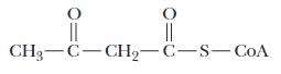
Answer: Hydrolysis of acetyl-coenzyme A produces free coenzyme A and acetate whereas hydrolysis of acetoacetyl-coenzyme A releases acetoacetate and coenzyme A.
Garrett & Grisham 3-12
Acetate is relatively stable; however, acetoacetate is unstable and will break down to acetone and CO2. Thus, the instability of one of the products of hydrolysis of acetoacetylcoenzyme A, namely acetoacetate, will make the reverse reaction (i.e., production of acetoacetyl-coenzyme A) unlikely. Furthermore, the terminal acetyl group of acetoacetylcoenzyme A is electron-withdrawing in nature and will destabilize the thiol ester bond of acetoacetyl-CoA. Thus, the free energy of hydrolysis of acetoacetyl-coenzyme A is expected to be greater than that of acetyl- coenzyme A and in fact, the free energy of hydrolysis is 43.9 kJ/mol for acetoacetyl-CoA and 31.5 kJ/mol for acetyl-CoA.
17. Assessing the Reactivity of Carbamoyl Phosphate
Consider carbamoyl phosphate, a precursor in the biosynthesis of pyrimidines:
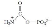
Based on the discussion of high-energy phosphates in this chapter, would you expect carbamoyl phosphate to possess a high free energy of hydrolysis? Provide a chemical rationale for your answer.
Answer: Is carbamoyl phosphate destabilized due to electrostatic repulsion? The carbonyl oxygen will develop a partial-negative charge causing charge-repulsion with the negatively charged phosphate. So, charge destabilization exists. Are the products stabilized by resonance or by ionization? Without regard to resonance states of the phosphate group, there are two possible resonance structures for carbamoyl phosphate:
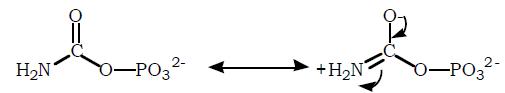
However, we expect that the carbonyl-carbon must pass through a positively charged intermediate and in doing so, affect phosphate resonance. Thus, the products are resonance stabilized. Finally, are there entropy factors? The products of hydrolysis are phosphate and carbamic acid. Carbamic acid is unstable and decomposes to CO2 and NH3 unless stabilized as a salt by interacting with a cation. With these considerations in mind, we expect carbamoyl phosphate to be unstable and therefore a high energy compound. The free energy of hydrolysis of carbamoyl phosphate is –51.5 kJ/mol, whereas for acetyl phosphate it is only –43.3 kJ/mol.
& Grisham 3-13
18. Assessing the Effect of Temperature on Equilibrium
You are studying the various components of the venom of a poisonous lizard. One of the venom components is a protein that appears to be temperature sensitive. When heated, it denatures and is no longer toxic. The process can be described by the following simple equation.:
T (toxic) → N (nontoxic)
There is only enough protein from this venom to carry out two equilibrium measurements. At 298 K, you find that 98% of the protein is in its toxic form. However, when you raise the temperature to 320 K, you find that only 10% of the protein is in its toxic form.
a. Calculate the equilibrium constants for the T to N conversion at these two temperatures.
Answer: For the reaction T → N
[N] K [T] =
At 298 K, [T] = 98% of the total protein concentration and [N] = 2%. Thus,
[N] 2 K 0.0204 [T]98 ==
At 320 K, [T] = 10% and [N] = 90%. Thus,
[N] 90 K9 [T]10 ==
b. Use the data to determine the ΔH°, ΔS°, and ΔG° for this process.
Answer: Using Figure 3.2 as a guide, the following chart calculates 1/T and RlnKeq for the data. (R = 8.314J/mol-K)
The slope of this van’t Hoff plot is –ΔH° in kJ/mol and so ΔH°= 291.5 kJ/mol. In general,
Values of ΔG° at both temperatures were calculated above.
G°= –RTlnKeq
(°K) Keq
19. Assessing the Effect of pH on Metabolic Reactions
The difference between ΔG and ΔG°' was discussed in Section 3.3. Consider the hydrolysis of acetyl phosphate (Figure 3.7) and determine the value of ΔG° for this reaction at pH 2, 7, and 12. The value for ΔG°' for the enolase reaction (Figure 3.8) is 1.8 kJ/mol. What is the value of ΔG° for enolase at pH 2, 7, and 12? Why is this case different from that of acetyl phosphate?
Answer: When protons are involved in process: ΔG°' = ΔG° ± RTln[H+] where the plus sign is used when protons are produced and the minus sign when protons are consumed. Hydrolysis of acetyl phosphate produces acetate, phosphate, and a proton and hence the plus sign is used. The ΔG°' for acetyl phosphate is given as –43.3 kJ/mol.
∆G°' = ∆G° + RT ln [H+] (1)
Solving for ∆G°
∆G°= 43,300 J/mol (8.314 J/mol ⋅ K) × (273 + 25)K × ln[10 7]
∆G° = 33,700 J/mol = 33.7 kJ/mol
We were given the ΔG°' value, which is for a reaction at pH 7. The value of AG° is the same for pH 2, 7, and 12 because it is the value when [H+] = 1 M. We could, however, use equation (1) to calculate ΔG°' at pH 2.0 and pH 12. Remembering that [H+]= 10 pH we find ΔG°' values of 14.78 kJ/mol and –71.82 kJ/mol for pH 2.0 and 12.0 respectively (and of course –43.3 kJ/mol for pH 7.0).
The enolase reaction does not involve a proton and so ΔG° = ΔG°'.
20. Analyzing the Energetics of Coupled Reactions
What is the significance of the magnitude of ΔG°' for ATP in the calculations in the Deeper Look box “ATP Changes Keq by a Factor of 108”? Repeat these calculations for
Garrett & Grisham 3-15
the case of coupling of a reaction to 1,3-bisphosphoglycerate hydrolysis to see what effect this reaction would have on the equilibrium ratio for components A and B under the conditions stated on this page.
Answer: The value of ΔG°' for hydrolysis of ATP, a highly favorable reaction, is –30.5 kJ/mol. Conversion of A to B under standard conditions has ΔG°' of + 13.8 kJ/mol, which is unfavorable. By coupling the two reactions the overall ΔG°' is 16.7 kJ/mol, which indicates that under standard conditions the coupled reaction will proceed toward hydrolysis of ATP and conversion of A to B. But, net conversion of A to B is exaggerated in the example given in the textbook because the concentrations of ATP, ADP and Pi are likely not equilibrium values. If they were and ATP hydrolysis was not coupled to anything the values would predict a ΔG°' of approximately +17 kJ/mol. It is likely the case that the concentrations are being maintained by coupling of favorable catabolic processes to maintain ATP concentrations high. Given this, the ratio of B to A would be greatly skewed in favor of production of B.
To make the same calculation but using 1,3-bisphophoglycerate hydrolysis one would have to know the concentrations of 1,3-BPG and 3-phosphoglycerate for E. coli. Table 18.2 gives values of 0.001 mM and0.12 mM in erythrocytes for 1,3-BPG and 3-PG and 1 mM for Pi. The overall ΔG°' for coupling of A to B with 1,3-BPG to 3-PG would be +13.8 kJ/mol –49.6 kJ/mol = –35.8 kJ/mol. The corresponding Keq is:
The reaction is 2i A1,3BPGHOB3-PGP ++→++
[B][3PG][P] K [A][1,3BPG]
i [B]K[1,3BPG] 1.89100.00001 [A][3PG][P]0.00012.001
9 eq [B] 1.5710 [A] =×
21. The acyl-CoA Synthetase Reaction is:
a. ΔG°’ = -0.8 kJ/mol. Use data from Table 3.1 to determine the standard-state free energy of hydrolysis of fatty-acyl CoA.
b. The concentrations of various metabolites in the human red blood cell are: [CoASH] = 0.12 mM, [fatty acid] = 0.001 mM, [AMP] = 0.14 mM, [ATP] = 1.85 mM, PPi = [0.5 mM]
c. What is the equilibrium constant for the fatty-acyl CoA synthetase reaction?
d. What is the cellular free energy change for the fatty-acyl CoA synthetase reaction at 37°C
Answer:
a. ΔG°’ = -31.5 kJ/mol
b. Keq = 1.38
c. The concentration of fatty-acyl CoA is not specified in this problem, since it varies in a typical cell. Assuming [fatty-acyl CoA] = 1 mM, ΔG = 58.8 kJ/mol.
22. The Standard-State Free Energy Change (ΔG°’) for Hydrolysis of Carbamoyl Phosphate is -40.47 kJ/mol.
a. What is the free energy change (ΔG) for carbamoyl phosphate hydrolysis in a solution in which the concentrations are [carbamoyl phosphate] = 2.5 mM, [carbamate] = 1.5 mM, [Pi] = 1 mM?
b. What is the equilibrium constant for the carbamoyl phosphate hydrolysis reaction?
c. In the carbamoyl phosphate synthetase reaction, ADP is phosphorylated by carbamoyl phosphate to form ATP:
ADP + carbamoyl phosphate ATP + carbamate →
What is the standard-state free energy change for this reaction?
d. What is the equilibrium constant for the carbamoyl phosphate synthetase?
Answer:
a. The temperature is not specified. Assuming T = 298 K, ΔG = -58.8 kJ/mol
b. Keq = 1.24 x 10⁷
c. ΔG°’ = -9.97 kJ/mol
d. Keq = 55.7
23. The Hydrolysis of 1,3-Bisphosphoglycerate is Favorable, Due in Part to the Increased Resonance Stabilization of the Products of the Reaction. Draw resonance structures for the reaction and the products of this reaction to establish that this statement is true.
Answer: This exercise is left to the student. Use Figure 3.5 as a guide.
24. The acyl-CoA synthetase reaction activates fatty acids for oxidation in cells:
The reaction is driven forward in part by hydrolysis of ATP to AMP and pyrophosphate However, pyrophosphate undergoes further cleave to yield two phosphate anions. Discuss
Garrett & Grisham 3-17
the energetics of this reaction both in the presence and absence of pyrophosphate cleavage.
Answer: Without pyrophosphate cleavage, the acyl-CoA synthetase reaction is only slightly favorable, with ΔG°’ of 0.8 kJ/mol. With pyrophosphate cleavage included, the net ΔG°’ for the reaction is -33.6 kJ/mol, a far more favorable value.
25. You have been hired to work in an independent drug- and chemical-testing lab and your first assignment involves creatine, a molecule found in skeletal muscle that reacts with ATP to produce creative phosphate and ADP (the so-called creatine kinase reaction, which has a ΔG in skeletal muscle of 2100 J/mol.)
During the first few seconds of intense muscular effort, this reaction runs in reverse, producing extra ATP to support muscle contractions. For your first assignment, your supervisor asks you to determine the concentration of creatine in resting human muscle. She tells you that the muscle tissue contains 1.85 mM ATP, 0.14 mM ADP, and 25 mM creatine phosphate. Use the information provided here as well as other information contained in this exam to determine the creatine concentration in muscle.
Answer: Use the data in Table 3.1 to determine Δ G°’ for creatine kinase reaction:
Cr + ATPCrP + ADP = →
ΔG°’ +12.8 kJ/mol
Use ΔG in muscle of 8.04 kJ/mol with Equation 3.8 to calculate [Cr] = 0.012M = 12mM
Think-Pair-Share Question
Create an Electron Transport Pathway
Consider the figure shown. Electrons need to move from NADH to O2 through a number of intermediates. The following intermediates are available (in no particular order): UQ, Cytochrome c (Fe3+), FMN, Cytochrome bL (Fe3+). Based upon the information given in Table 3.3:
a) Assign appropriate intermediates to beakers B, C, D and E.
b) For each pair of beakers write a balanced equation and calculate the overall reduction potential ∆ε0 ’ of the redox reactions.
c) For the entire system write a balanced equation and calculate the overall reduction potential ∆ε0 ’ .
Garrett & Grisham 3-18
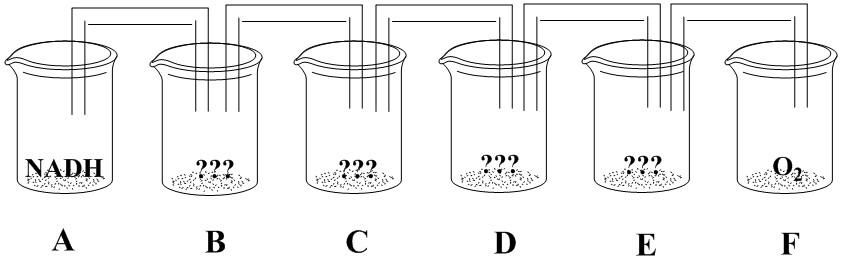
a) The various reagents should be organized with increasing values of ε0’. Beaker B should contain FMN, beaker C should contain cytochrome bL (Fe3+), beaker D should contain UQ and beaker E should contain cytochrome c (Fe3+).
b) Beakers A and B:
Beakers B and C:
NADH + H+ + FMN NAD+ + FMNH2
∆ε0 ’ = -0.219V – (-0.32V) = 0.101V
FMNH2 + 2 cyto bL (Fe3+) FMN + 2 cyto bL (Fe2+) + 2H+
Beakers C and D:
∆ε0 ’ = -0.100V – (-0.219V) = 0.119V
2 cyto bL (Fe2+) + 2H+ + UQ 2 cyto bL (Fe3+) + UQH2
∆ε0 ’ = 0.060V – (-0.100V) = 0.16V
Beakers D and E: UQH2 + 2 cyto c (Fe3+) UQ + 2 cyto c (Fe2+) + 2H+
∆ε0 ’ = 0.254V – (0.060V) = 0.194V
Beakers E and F: 2 cyto c (Fe2+) +2H+ + ½ O2 2 cyto c (Fe3+) + H2O
∆ε0 ’ = 0.816V – (0.254V) = 0.562V
c) Overall reaction
NADH + H+ + ½ O2 NAD+ + H2O
∆ε0 ’ = 0.816V – (-0.32V) = 1.136V
Answers:
a) The various reagents should be organized with increasing values of ε0’. Beaker B should contain FMN, beaker C should contain cytochrome bL (Fe3+), beaker D should contain UQ and beaker E should contain cytochrome c (Fe3+).
b) Beakers A and B:
NADH + H+ + FMN NAD+ + FMNH2
∆ε0 ’ = -0.219V – (-0.32V) = 0.101V
Beakers B and C: FMNH2 + 2 cyto bL (Fe3+) FMN + 2 cyto bL (Fe2+) + 2H+
Beakers C and D:
∆ε0 ’ = -0.100V – (-0.219V) = 0.119V
Garrett & Grisham 3-19
2 cyto bL (Fe2+) + 2H+ + UQ 2 cyto bL (Fe3+) + UQH2
∆ε0 ’ = 0.060V – (-0.100V) = 0.16V
Beakers D and E: UQH2 + 2 cyto c (Fe3+) UQ + 2 cyto c (Fe2+) + 2H+
∆ε0 ’ = 0.254V – (0.060V) = 0.194V
Beakers E and F: 2 cyto c (Fe2+) +2H+ + ½ O2 2 cyto c (Fe3+) + H2O
∆ε0 ’ = 0.816V – (0.254V) = 0.562V
c) Overall reaction NADH + H+ + ½ O2 NAD+ + H2O ∆ε0 ’ = 0.816V – (-0.32V) = 1.136V
Questions for Self-Study
1. True or False.
a. The internal energy of an isolated system is conserved.
b. A closed system can exchange matter but not energy with the surroundings.
c. An open system includes a system and its surroundings.
d. An open system can exchange matter with another open system.
e. The internal energy of an open system is always constant.
2. The first law of thermodynamics states that there are only two ways to change the internal energy of any system. What are they?
3. Enthalpy, H, is defined as H = E + PV and ΔE = q + w. Under what conditions is ΔH = q?
4. Define the terms in the following expression: S = k ln W.
5. Match the items in the two columns.
a. ΔG = ΔH – TΔS
b. ΔG = ΔG° + RT ln ([P]/[R])
c. ΔG° = 0
d. ΔG = 0
e. ΔG > 0
f. ΔG < 0
g. ΔG° = –RT ln Keq
1. Reaction spontaneous as written.
2. Used to determine standard Gibbs free energy change.
3. Reaction unfavorable.
4. Used to calculate amount of free energy released when reaction proceeds to equilibrium.
5. Definition of change in Gibbs free energy.
6. System at equilibrium.
6. The compounds shown below include ATP, pyrophosphate, phosphoenolpyruvate, creatine phosphate, and 1,3-bisphosphoglycerate. They are all examples of high-energy
Garrett & Grisham 3-20
compounds. Identify each, locate the high-energy bond and list the products of hydrolysis of this bond.
7. What are the three chemical reasons for the large negative energy of hydrolysis of phosphoric acid anhydride linkage for compounds such at ATP, ADP, and pyrophosphate?
8. Creatine phosphate is an example of a guanidinium phosphate, a high-energy phosphate compound. This compound is abundant in muscle tissue. What is its function?
9. During protein synthesis amino acids are joined in amide linkage on the ribosome. The substrates for this reaction are not free amino acids but rather amino acids attached via their carboxyl groups to the 3' hydroxyl group of tRNAs. What kind of bond is formed between the amino acid and the tRNA? Is this a high-energy bond? Given the fact that aminoacyl-tRNA formation is accompanied by hydrolysis of ATP to AMP and PPi and that PPi is subsequently hydrolyzed to 2 Pi, how many high-energy phosphates are consumed to produce an aminoacyl-tRNA?
10. Although the standard free energy of hydrolysis of ATP is around 30 kJ/mol the cellular free energy change is even more negative. What factors contribute to this?
Answers
1. a.T; b.F; c.F; d.T; e.F.
2. Energy flow in the form of heat or work.
Garrett & Grisham 3-21
3. In general, ΔH = ΔE + PΔV + VΔP = q + w + PΔV + VΔP. When the pressure of the system remains constant (i.e., ΔP = 0) and work is limited to only mechanical work (i.e., w = –PΔV ) then ΔH = q.
4. S is the entropy, k is Boltzmann's constant, ln W is the natural logarithm of the number of ways, W, of arranging the components of a system.
5. a.5; b.4; c.7; d.6; e.3; f.1; g.2.
6. a./pyrophosphate, hydrolysis products 2 Pi. b./1,3-bisphosphoglycerate, hydrolysis products 3- phosphoglycerate and Pi. c./ATP, hydrolysis products either ADP and Pi or AMP and pyrophosphate. d./phosphoenolpyruvate, hydrolysis products pyruvate and Pi. e./creatine phosphate, hydrolysis products creatine and Pi. The location of high-energy bonds is shown below.
7. Bond strain due to electrostatic repulsion, stabilization of products by ionization and resonance, and entropy factors.
8. Creatine phosphate is used to replenish supplies of ATP by transferring phosphate to ADP in a reaction catalyzed by creatine kinase.
9. Amino acid ester bonds in aminoacyl-tRNAs are high-energy bonds produced at the expense of two high- energy phosphate bonds.
10. The presence of divalent and monovalent cations and the maintenance of low levels of ADP and Pi and high levels of ATP are responsible for the large negative free energy change of ATP under cellular conditions.
Additional Problems
1. Show that ΔG° = –RT ln Keq
2. The term ΔG° may be evaluated by measuring ΔG for a reaction in which reactants and products start out at 1 M concentration. The ΔG measured in this case is the standardstate free energy and is equal to ΔG°. Prove this.
3. If a particular reaction is allowed to reach equilibrium, is the equilibrium concentration of product ever dependent on the initial concentrations of reactant and product?
4. Confusion reigns supreme when work and energy expenditure are discussed. Define the term work. Are work and energy expenditure synonymous?
5. There are two statements about spontaneity of reactions: ΔS > 0, and ΔG < 0. Justify that these statements are in fact true descriptions of spontaneity.
6. DNA ligase catalyzes formation of a phosphodiester bond between a 5'-phosphate and a 3'-hydroxyl group on the ends of two DNAs to be joined. Many ligases use hydrolysis of ATP to drive phosphodiester bond synthesis; however, the E. coli ligase uses NAD+ as a high-energy compound. Explain why NAD+ is considered a high-energy compound. What type of reaction is required to release energy from NAD+ and what are the products?
7. You find yourself in the laboratory, late at night, working on an important assay and you discover that the last of the ATP stock is used up. After frantically searching everywhere, you find a 10 mg bottle of 2'- deoxyadenosine 5'-triphosphate. Is this a highenergy compound? Will it substitute for ATP in your assay?
8. Stock solutions of ATP are usually adjusted to around neutral pH to stabilize them. Why?
9. Intense muscle activity depletes ATP and creatine phosphate stores and produces ADP, creatine, AMP, and Pi. ADP is produced by ATP hydrolysis catalyzed by myosin, a component of the contractile apparatus of muscle. Creatine is a product of creatine kinase activity, and AMP is produced by adenylate kinase from two ADPs. Given this information, explain how high-energy phosphate compounds in muscle are interconnected.
10. The standard free energy of hydrolysis of glucose-1-phosphate is –21 kJ/mol whereas it is only –13.9 kJ/mol for glucose-6-phosphate. Provide an explanation for this difference.
Abbreviated Answers
1. At equilibrium ΔG = 0 and the concentration of reactants and products are at their equilibrium values. Using , [C][D]
∆=∆+
GGRTlnweseethat [A][B]
G0GRTln [A][B]
or, solving for ∆G° we find that:
[C][D]
GRTln RTlnK [A][B] ∆=− =−
2. Using , [C][D]
∆=∆+
GGRTlnweseethat [A][B]
GGRTlnGRTln1
But, ln1 = 0, and ∆G = ∆G°
3. The equilibrium constant is independent of the initial concentrations of reactants and products but the equilibrium constant is the ratio of the product of the concentration of products to the product of the concentration of reactants. This ratio is independent of initial concentrations. Clearly, the absolute amount of product depends on the initial concentration of reactant
4. Work is application of a force through some distance the consequence of which is a change in the energy of the object upon which the force is acting. Application of a force on an object at rest will cause the object to accelerate and in the absence of friction this will impart kinetic energy to the object. In the presence of friction, energy will be dissipated as heat raising the temperature of the object. However, one can have energy expenditure without doing work. As an example, holding an object at some position in a gravitational field requires a force equal and opposite to the force of gravity and creation of this force requires expenditure of energy yet no work is done if the object does not move.
5. Entropy is a measure of disorder and for a reaction to be spontaneous AS > 0 or since Sf Si > 0, Sf > Si. A spontaneous reaction results in an increase in disorder. Gibbs free energy is the amount of energy available to do work at constant pressure and temperature. Using G as a criterion for spontaneity requires that AG < 0 or since Gf G i < 0, Gf < Gi.
Garrett & Grisham 3-24
The amount of energy available to do work decreases for a spontaneous reaction.
6. NAD+ contains a single high-energy phosphoric anhydride linkage between AMP and nicotinamide monophosphate nucleotide. Hydrolysis will release AMP and nicotinamide nucleoside monophosphate.
7. 2'-Deoxyadenosine 5'-triphosphate or dATP contains two high-energy phosphoric anhydride bonds. However, if the assays being performed are enzymatic assays (as opposed to some chemical assay), then it is out-of-the-question to even think about substituting dATP for ATP. ATP is a ribonucleotide; dATP is a deoxyribonucleotide. dATP is used for synthesis of DNA and little else.
8. At first thought it might seem reasonable to make the ATP solution slightly acidic. This will neutralize the negatively-charged phosphates and should lead to stabilization of the phosphoric anhydride linkages. The problem with this idea is that the N-glycosidic linkage is acid labile. This bond is more stable in alkaline solution but the anhydride bonds are readily cleaved by hydroxide attack.
9. The key here is to recognize that kinases are enzymes that transfer the y-phosphate of ATP to a target molecule. Thus, creatine kinase phosphorylates creatine and adenylate kinase phosphorylates AMP. The following scheme outlines how ATP, ADP, AMP, creatine phosphate, creatine, and Pi are related.
ATPcreatineADPcreatinephosphate(creatine kinase)
10. Phosphate is an electron-withdrawing group that is attached to quite different carbons in glucose-6- phosphate versus glucose-1-phosphate. In the latter, carbon-1 is already bonded to an electronegative oxygen in the pyranose form.
Chapter Summary
Thermodynamics - a collection of laws and principles describing the flows and interchanges of heat, energy and matter - can provide important insights into metabolism and bioenergetics. Thermodynamics distinguishes between closed systems, which cannot exchange matter or energy with the surroundings; isolated systems, which may exchange heat, but not matter, with the surroundings; and open systems, which may exchange matter, energy or both with the surroundings.
The first law of thermodynamics states that the total energy of an isolated system is conserved. E, the internal energy function, which is equal to the sum of heat absorbed and
Garrett & Grisham 3-25
work done on the system, is a useful state function, which keeps track of energy transfers in such systems. In constant pressure processes, it is often more convenient to use the enthalpy function (H = E + PV) for analysis of heat and energy exchange. For biochemical processes, in which pressure is usually constant and volume changes are small, enthalpy (H) and internal energy (E) are often essentially equal. Enthalpy changes for biochemical processes can often be determined from a plot of R(ln Keq) versus 1/T (a van’t Hoff plot).
Entropy is a measure of disorder or randomness in the system. The second law of thermodynamics states that systems tend to proceed from ordered (low entropy) states to disordered (high entropy) states. The entropy change for any reversible process is simply the heat transferred divided by the temperature at which the transfer occurs: dSrev = dq/T.
The third law of thermodynamics states that the entropy of any crystalline, perfectly ordered substance must approach zero as the temperature approaches 0 K, and at 0 K, entropy is exactly zero. The concept of entropy thus invokes the notion of heat capacitythe amount of heat any substance can store as the temperature of that substance is raised by one degree K. Absolute entropies may be calculated for any substance if the heat capacity can be evaluated at temperatures from 0 K to the temperature of interest, but entropy changes are usually much more important in biochemical systems.
The free energy function is defined as G = H – TS and, for any process at constant pressure and temperature (i.e., most biochemical processes), ΔG = ΔH – TΔS. If ΔG is negative, the process is exergonic and will proceed spontaneously in the forward direction. If ΔG is positive, the reaction or process is endergonic and will proceed spontaneously in the reverse direction. The sign and value of ΔG, however, do not allow one to determine how fast a process will proceed. It is convenient to define a standard state for processes of interest, so that the thermodynamic parameters of different processes may be compared. The standard state for reactions in solution is 1 M concentration for all reactants and products. The free energy change for a process at concentrations other than standard state is given by:
( ) GGRTln[C][D]/[A][B]
and the standard state free energy change is related to the equilibrium constant for the reaction by ΔG° = –RT ln Keq. This states that the equilibrium established for a reaction in solution is a function of the standard state free energy change for the process. In essence, ΔG° is another way of writing an equilibrium constant. Moreover, the entropy change for a process may be determined if the enthalpy change and free energy change calculated from knowledge of the equilibrium constant and its dependence on temperature are known.
The standard state convention of 1 M concentrations becomes awkward for reactions in which hydrogen ions are produced or consumed. Biochemists circumvent this problem by
Garrett & Grisham 3-26
defining a modified standard state of 1 M concentration for all species except H+, for which the standard state is 1 × 10 7 M or pH 7.
Thermodynamic parameters may provide insights about a process. As an example, consider heat capacity changes: Positive values indicate increase freedom of movement whereas negative values indicate less freedom of motion.
Many processes in living things must run against their thermodynamic potential, i.e., in the direction of positive ΔG. These processes are driven in the thermodynamically unfavorable direction via coupling with highly favorable processes. Coupled processes are vitally important in intermediary metabolism, oxidative phosphorylation, membrane transport and many other processes essential to life.
There is a hierarchy of energetics among organisms: certain organisms capture solar energy directly, whereas others derive their energy from this group in subsequent chemical processes. Once captured in chemical form, energy can be released in controlled exergonic reactions to drive life processes. High energy phosphate anhydrides and reduced coenzymes mediate the flow of energy from exergonic reactions to the energyrequiring processes of life. These molecules are transient forms of stored energy, rapidly carrying energy from point to point in the organism. One of the most important high energy phosphates is ATP, which acts as an intermediate energy shuttle molecule. The free energy of hydrolysis of ATP (for hydrolysis to ADP and phosphate) is less than that of PEP, cyclic-AMP, 1,3-BPG, creatine phosphate, acetyl phosphate and pyrophosphate, but is greater than that of the lower energy phosphate esters, such as glycerol-3phosphate and the sugar phosphates. Group transfer potential - the free energy change that occurs upon hydrolysis (i.e., transfer of a chemical group to water) - is a convenient parameter for quantitating the energetics of such processes. The phosphoryl group transfer potentials for high energy phosphates range from –31.9 kJ/mole for UDP-glucose to –35.7 kJ/mole for ATP to –62.2 kJ/mole for PEP.
The large negative ΔG°' values for the hydrolysis of phosphoric acid anhydrides (such as ATP, GTP, ADP, GDP, sugar nucleotides and pyrophosphate) may be ascribed to 1) destabilization of the reactant due to bond strain caused by electrostatic repulsion, 2) stabilization of the products by ionization and resonance, and 3) increases in entropy upon product formation. Mixed anhydrides of phosphoric and carboxylic acids - known as acyl phosphates - are also energy-rich. Other classes of high energy species include enol phosphates, such as PEP, guanidinium phosphates, such as creatine phosphate, cyclic nucleotides, such as 3’,5’-cyclic AMP, amino acid esters, such as aminoacyl-tRNA and thiol esters, such as coenzyme A. Other biochemically important high energy molecules include the pyridine nucleotides (NADH, NADPH), sugar nucleotides (UDPglucose) and S-adenosyl methionine, which is involved in the transfer of methyl groups in many metabolic processes.
Though the hydrolysis of ATP and other high energy phosphates are often portrayed as simple processes, these reactions are far more complex in real biological systems. ATP, ADP and similar molecules can existin several different ionization states, and each of
Garrett & Grisham 3-27
these individual species can bind divalent and monovalent metal ions, so that metal ion complexes must also be considered. For example, ATP has five dissociable protons. The adenine ring amino group exhibits a pKa of 4.06, whereas the last proton to dissociate from the triphosphate chain possesses a pKa of 6.95. Thus, at pH 7, ATP is a mixture of ATP4 and HATP3 . If equilibrium constants for the hydrolysis reactions are re-defined in terms of total concentrations of the ionized species, then such equilibria can be considered quantitatively, and fractions of each ionic species in solution can be determined. The effects of metal ion binding equilibria on the hydrolysis equilibria can be quantitated in a similar fashion.
The free energy changes for hydrolysis of high energy phosphates such as ATP are also functions of concentration. In the environment of the typical cell, where ATP, ADP and phosphate are usually 5 mM or less, the free energy change for ATP hydrolysis is substantially larger in magnitude than the standard state value of 30.5 kJ/mole.
High energy molecules such as ATP are rapidly recycled in most biological environments. The typical 70 kg human body contains only about 50 grams of ATP/ADP. Each of these ATP molecules must be recycled nearly 2,000 times each day to meet the energy needs of the body.
Chapter 4
Chapter Outline
Amino Acids and the Peptide Bond
• Amino acids: Tetrahedral alpha (α) carbon with H, amino group, carboxyl group, and side chain
• Amino acids in proteins joined by peptide bonds
• Amino group reacted with carboxyl group and removal of water
• Peptide bonds join amino acid residues in polypeptides
• Classification
• Nonpolar: Ala, val, leu, ile, pro, met, phe, trp
• Polar uncharged: Gly, ser, thr, tyr, asn, gln, cys
• Acidic: Asp, glu
• Basic: His, lys, arg
• Amino acids come in a variety of forms: There are far more than 20 amino acids
• The 20 amino acids presented above are coded for in the genetic code
• Selenocysteine and pyrrolysine are the 21st and 22nd amino acids to be coded for
• Modified amino acids: Hydroxylysine, hydroxproline, thyroxin, methyl histidine, methyl arginine, methyl lysine, γ-carboxyglutamic acid, pyroglutamic acid, aminoadipic acid
• Small molecules derived from amino acids: GABA, histamine, serotonin, β alanine, epinephrine, penicillamine, ornithine, citrulline, betaine, homocysteine, homoserine
• Ionic properties of amino acids
• α-Carboxyl group: pKa’s range from 2.0 to 2.4
• α-Amino group: pKa’s range from 9.0 to 9.8
• Side chains
• Acidic pKa’s for asp, glu
• Basic pKa’s for his, arg, lys
• Amino acid chemistry: Edman reagent
• Used to modify N-terminus and to sequence peptides
• Chirality: Asymmetric carbon has four different groups attached
• Optically active compounds
• Dextrorotatory (+), clockwise rotation
• Levorotatory (-), counterclockwise rotation
• Asymmetric carbon compounds
• One asymmetric carbon: Mirror image isomers or enantiomers
• More than one asymmetric carbon: enantiomers and diastereomers
• D,L System: Typically naturally occurring amino acids: L-amino acids; related to L-glyceraldehyde
• (R,S) System: Assign priorities to groups about the chiral carbon
• SH > OH > NH2 > COOH > CHO > CHOH > CH3
• First four above: Priority order is Mr of heaviest element
• Last four: Priority is oxidation state of carbon
• Ultraviolet spectral properties of amino acids: Only phe, try, trp absorb UV light above 250 nm
• NMR signal from amino acids sensitive to magnetic environment
• Separation of amino acids
• Ion exchange
• Anion exchangers: Matrix is positively charged so anions bind
• Cation exchangers: Matrix is negatively charged so cations bind
• Gas chromatography and high-performance liquid chromatography (HPLC)
• Peptide bonds: Carboxyl group + amino group – water = peptide (amide) bond
• Trans arrangement (usually) of carbonyl oxygen and amide hydrogen
• Double-bond character
• Pi electron delocalization prevents free rotation about peptide bond
• Amide plane includes carbonyl, amide and alpha carbons
• Peptide classification
• Up to 12 amino acid residues, dipeptide, tripeptide, tetrapeptide, etc.
• Numerical prefix refers to number of amino acid residues not peptide bonds
• Oligopeptides: 12 to 20 residues
• Polypeptides: Several dozen residues
• Proteins: Polypeptides
• Monomeric proteins have one chain
• Multimeric proteins have more than one chain
• Homomultimeric: Identical chains
• Hetromultimeric: Different chains
Chapter Objectives
It is imperative to learn the structures and one-letter codes for the amino acids. Amino acids are key molecules, both as components of proteins and as intermediates to important biomolecules. The structures are shown in Figure 4.3 of Biochemistry. Figure 4.3 is presented on the following two pages.
Classification of Amino Acids
Here is an alternative classification scheme.
Acidic amino acids and their amides: aspartic acid, asparagine, glutamic acid, glutamine.
Basic amino acids: histidine, lysine, arginine.
Aromatic amino acids:
phenylalanine, tyrosine, tryptophan
Sulfur containing amino acids: cysteine, methionine
Imino acid: proline.
Hydrophobic side chains: glycine, alanine, valine, leucine, isoleucine.
Hydroxylic amino acids: serine, threonine, (tyrosine).
To memorize the structures of the 20 amino acids, here are some aids: The side chain of glycine is H and that of alanine is a methyl group, CH3.
The side chain of valine is just the methyl side chain of alanine with two methyl groups substituting for two Hs. (In effect it is just dimethylalanine.)
The side chain of leucine is just valine with a –CH2– group between the valine side chain and the α-carbon (or, leucine is alanine with the valine side chain attached).
Isoleucine is an isomer of leucine with one of the methyl groups exchanged with a H on the carbon attached to the α-carbon. Several of the amino acids have alanine as a base.
Phenylalanine has a phenyl group (i.e., benzene ring) attached to alanine.
Tyrosine is hydroxylated phenylalanine.
Serine has a hydroxyl group substituting for a H on the methyl group of alanine.
Cysteine can be thought of as the sulfur variety of serine i.e., with a sulfhydryl group in place of the hydroxyl group of serine.
Threonine, a hydroxylic amino acid, can be thought of as methylated serine.
The acidic amino acids and their amides are quite easy to remember. Aspartic acid and glutamic acid have carboxylic acid groups attached to –CH2– and (–CH2–)2, respectively. The amides of these amino acids, namely asparagine and glutamine, have nitrogen attached to the side chain carboxyl groups in amide linkage.
The basic amino acids all have six carbons, counting the carboxyl carbon and the αcarbon. Lysine's side chain is a linear chain of four –CH2– groups with a terminal amino group. Arginine has a linear side chain of three –CH2– groups to which a guanidinium group is attached. (The sixth carbon is the central C of the guanidinium group.)
Guanidinium is like urea but with a nitrogen group replacing the carbonyl oxygen.
Histidine has an imidazole ring attached to alanine. The ring contains two nitrogens and three carbons, a five-membered ring.
An easy way to remember the structure of proline is to know that it derives from glutamic acid. The carboxyl side chain forms a C-N bond to the amino group to form this imino acid. (In the process, the carbon is reduced so the side chain carboxyl oxygens do not show up in proline.)
Methionine is a sulfur-containing amino acid. Biologically, it is also used as a methyl group donor in the molecule S-adenosylmethionine (SAM). These two facts indicate that the side chain ends in a S-CH3, which is attached to (–CH2–)2. The remaining amino acid is tryptophan. Its side chain is an indole ring attached to a –CH2– group. In effect it can be thought of as alanine with a indole ring. Substituted indole rings are widely used in biochemistry. The basic structure is a six-membered benzene ring fused to a fivemembered ring containing a single N.
Nomenclature
The one-letter code for the amino acids must be memorized. In journals, the one letter codes are used to show amino acid sequences. See the answer to Problem 2 for a discussion of how the one letter codes are organized.
Chirality
The α-carbons of amino acids, excepting Gly, have four different groups attached to them, and hence they are chiral carbons. Two different arrangements of the four groups about the chiral carbon can be made, giving rise to two structural isomers. Structural isomers that are mirror images of each other are called enantiomers. When two or more chiral centers exist on a single molecule, 2n structural isomers are possible, where n = the number of chiral centers. The various structural isomers include enantiomers (mirrorimage-related isomers) and non mirror-image-related isomers known as diastereomers.
The structural isomers can be distinguished from each other using the R and S prefixes assigned to each chiral center of a molecule. The R and S prefixes are assigned according to a set of sequence rules. The four groups about a chiral center are assigned priority numbers with the highest priority given to the atom with the highest atomic number, thus S > O > N > C > H (for elements found in amino acids). When two or more groups have
Garrett & Grisham 4-4
the same primary element, then elements attached to the primary element are considered. So for example, SH > OR > OH > NH2 > COOH > CHO > CH2OH > CH3. Once priority assignments are made, the molecule is viewed with the group having the lowest priority away from the reader. Moving from highest to lowest priority, for the three remaining groups, clockwise movement indicates R and counterclockwise indicates S configuration.
UV-absorbing Properties
Only three amino acids, phenylalanine, tyrosine, and tryptophan, absorb light in the near UV range (i.e., 230 nm to 310 nm). These amino acids will dominate the UV absorption spectra of proteins. The wavelength maxima for tyrosine and tryptophan are around 280 nm. In addition, these two amino acids have extinction coefficients quite a bit larger than the extinction coefficient of phenylalanine, whose λ max = 260 nm. Thus very many proteins have maximum absorbance around 280 nm. In contrast, nucleic acids absorb light with a maximum around 260 nm due to the nucleotides. The extinction coefficients of the nucleotides are larger than those for the aromatic amino acids. In general, nucleic acid contamination of a protein sample will contribute to UV absorption at 260 nm. Often the 260/280 ratio is used as a criterion for purity.
Figure 4.3 The amino acids that are the building blocks of most proteins can be classified as (a) nonpolar (hydrophobic), (b) polar, neutral, (c) acidic, or (d) basic. Also shown are the one-letter and three-letter codes used to denote amino acids. For each amino acid, the ball-and-stick (left) and space-filling (right) models show only the side chain.