
Making waves
SONOGRAPHY
Contributors
Karen Barton

Karen is a highly experienced sonographer and tutor sonographer with 28 years of clinical practice spanning both public and private sectors. Throughout her career, she has developed a strong professional focus on sonographer education, supervision, and mentorship. Karen holds a Postgraduate Certificate in Health Professional Education and is a committed advocate for the development of teaching and learning frameworks within the profession. She is a member of the ASA Clinical Supervisors Special Interest Group (SIG), where she contributes to the advancement of supervisory practice and support for clinical educators. Her work is underpinned by a passion for fostering professional growth and maintaining high standards of clinical education in sonography.
Helen Beets

Helen is a sonographer coordinator and tutor based in Perth, Western Australia. She has been an active member of the Australasian Sonographers Association (ASA) Special Interest Groups (SIGs) since 2019. Helen is a member of the Clinical Supervisors SIG and currently serves as Chair of the Research SIG. After completing her master’s degree by research in ultrasound at Monash University, Helen has continued to contribute to research projects. She has a strong passion for mentoring and supporting students and early career sonographers.
Madonna Burnett

Madonna Burnett obtained her DMU in Ultrasound in 1997 and became a member of the Australasian Sonographers Association (ASA) shortly after, around 1998. She joined the ASA Queensland Branch Committee in 1997–98 and served in various roles over the years, including Chairperson, Secretary, and Treasurer, before stepping down from the committee in 2022. Madonna has also been a long-standing member of the Paediatric Special Interest Group (SIG) Committee, where she continues to contribute. Her career spans several decades, having worked as a radiographer/ sonographer at Mater Health Services from 1983 to approximately 2018. Since 2014, she has worked at the Queensland Children’s Hospital, where she is now employed full-time in the same role.
Madonna has been married to her husband, Rob, for 37 years. They have 4 children and one granddaughter.
A/Prof Michelle Fenech, FASA

A/Prof Michelle Fenech is a sonographer and radiographer and associate professor at Central Queensland University (CQU), where she is Head of Course for
Postgraduate Medical Sonography, teacher and active researcher. Michelle has an interest in researching imaging of musculoskeletal and lymphatic structures, point-of-care ultrasound, and investigating learning, teaching and curriculum and pedagogy development. Michelle maintains clinical relevancy through working with Queensland Health. She is a member of the ASA Research and Musculoskeletal (MSK) Special Interest Groups (SIGs), and is deputy editor of the BMUS journal, Ultrasound
Andrew Grant

Andrew is a regular presenter on MSK, vascular, paediatrics and general anatomy both here in Australia and internationally. Andrew is a passionate educator and holds numerous casual lecturing positions throughout Australia. Andrew has also worked for one of the top medical centres in the western USA, UCLA Medical Centre, where he assisted in setting up the musculoskeletal ultrasound program.
Andrew was the 2023 ASA Victorian Sonographer of the Year and is the current Secretary of the Victorian Branch of the ASA. He practises at I-Med East Melbourne Radiology, where he is the ultrasound supervisor.
Fiona Hilditch

Fiona is a dedicated healthcare professional with 16 years of experience in radiology, spanning radiation therapy, mammography, breast ultrasound, and cancer support. Over the past 9 years, she has worked in 2 of Brisbane’s leading tertiary breast imaging clinics, developing strong expertise in breast imaging and oncologyrelated radiology. Passionate about women’s imaging, Fiona is committed to patient-centred care, quality assurance, training, and best practice. She thrives in multidisciplinary teams and actively contributes to improving departmental systems and processes. Her goal is to help build advanced radiology departments recognised for delivering high quality imaging and fostering compassionate, skilled teams.
Contributors
Mitchell Hubble

Mitchell is a cardiac sonographer with a Postgraduate Diploma in Cardiac Ultrasound from QUT and a recent graduate from a Master of Cardiac Sonography at WSU. He is now pursuing an MBA through Melbourne Business School.
Since entering the industry in 2017, he has specialised in heart failure prevention and is passionate about education and training. Mitchell collaborates with prominent institutions like Guidepoint Advisors, Melbourne HeartCare, Advara HeartCare, and Heart of Australia. He is an active member of professional organisations, including ASA, ASAR, ASE, ESC, and PiSCA, consistently striving for optimal patient care outcomes.
Emma Jardine, AFASA

Emma is a senior sonographer working in Victoria. She has a passion for professional growth and believes that investing in ongoing education and training will benefit individual sonographers, contribute to better patient outcomes and overall healthcare experience.
Joanne King

As a dedicated and passionate healthcare professional, Joanne brings a wealth of experience and expertise to the field of medical imaging as a dual-qualified radiographer and sonographer. Jo qualified as a radiographer in 2007 from Curtin University and qualified as a sonographer from the University of South Australia in 2014. For the past 13 years, Jo has been working in the rural and remote region of Kimberley, WA. She is the manager of the Broome Radiology department and enjoys clinical and admin
roles. Jo is an active member of the ASA SIG ET, volunteers on her children’s school P&C and is the secretary of the Broome Floorball Association.
With a track record of excellence, a commitment to continuous improvement and a genuine passion for healthcare, Jo stands as a trusted and respected radiographer and sonographer, dedicated to advancing the field and providing exceptional care to patients.
Ling Lee, AFASA

Ling Lee (AFASA) is a clinical sonographer specialised in the field of maternal fetal medicine and women’s health. Her role as a clinical educator allows her to participate in the training and mentoring of student sonographers and fellow sonographers, which is a rewarding experience to be involved in the growth of sonography. Her ongoing research focuses on providing better support for the education of future sonographers through a social psychological lens. She believes that research is a vital part of our clinical role and pivotal to the advancement of our profession.
James Maunder

James studied Zoology, Anthropology and Biochemistry at the Australian National University (ANU), graduating in 2000, before beginning his vascular ultrasound training (DMU Vasc) in 2004. James is currently the Chief Vascular Sonographer for Dr Peter Bray (MBBS [UWA] FRACS [Vascular]), in Subiaco, WA. He has worked as a dedicated vascular sonographer for over 20 years, and has presented at local, state, national and international conferences. He was awarded the ASA Western Australian Sonographer of the Year in 2019.
Donna Napier, FASA

Donna Napier is a radiographer/ sonographer and the Acting Sonography Section Manager at the Tweed Valley Hospital in northern NSW, having over 24 years’ industry experience. She holds a master’s degree in medical sonography and is a Fellow of the Australasian Sonographers Association (FASA). Donna has a passion for education and is an advocate for expanding the sonographer’s scope of practice. She is a frequent presenter at conferences and the author of multiple ultrasound publications. Donna has a keen interest in emergency ultrasound applications, and more recently has developed a passion for the use of ultrasound to improve men’s health outcomes.
Sophie O’Brien

Sophie O’Brien is a sonographer supervisor based in the Hunter Valley. Sophie initially trained in nuclear medicine before moving into ultrasound in 2011. Sophie has interests in MSK and Women’s Health ultrasound and has been involved in both SIGs since 2021. Sophie is currently the Chair of the Women’s Health SIG and has been involved in education with the ASA for many years.
Claire O’Reilly

Claire is a general sonographer at Monash Health and is the Ultrasound Site Supervisor at the Victorian Heart Hospital. She has presented at local, national and international conferences.
Contributors
Lino Piotto, FASA

Lino Piotto (FASA, FASMIRT, MMedRad, DMU, AMS) is the tutor sonographer at the Women’s and Children’s Hospital, Adelaide. He has been specialising in paediatric ultrasound for 35 years. He completed his fellowship in ultrasound for the Australian Society of Medical Imaging and Radiation Therapy and the Australasian Sonographers Association. Lino enjoys learning and sharing his experience.
Ian Schroen, AFASA

Ian has built a diverse and accomplished career in diagnostic imaging. Starting as a radiographer, he specialised in vascular ultrasound before progressing to all aspects of diagnostic ultrasound. His career evolved further with a successful role in corporate healthcare, where he gained extensive experience in sales and marketing.
Holding a Master of Medical Ultrasound, Ian is passionate about all facets of clinical ultrasound, including imaging technology, teaching, and research. He previously served as President of the Australasian Sonographers Association and currently as an associate lecturer at Monash University, where he teaches the vascular ultrasound subject in the postgraduate ultrasound program.
Christopher Thomas

Christopher Thomas graduated from The University of Queensland in 1997 with a Bachelor of Applied Science in Human Movement Studies. A Master of Scientific Studies was completed, specialising in cardiac physiology and rehabilitation.
Chris made an 18-year contribution to The Prince Charles Hospital’s Echocardiography Department and has trained many specialist echocardiologists working in Australia and abroad.
Chris manages and is the Senior Cardiac Sonographer at Queensland Echocardiography, a stand-alone and fully accredited echocardiography imaging service, the first of its kind in Queensland.
In addition to presenting his original research at the World Congress of Cardiology in 2002, Chris has been involved in countless research initiatives as new technologies arrived on the market, such as ASD/VSD closure devices, ventricular assist devices and transcatheter heart valve replacements/interventions.
Jane Wardle

Jane Wardle is a dedicated educator and the Head of Course for Medical Sonography at CQUniversity, overseeing the course across multiple campuses in Australia. With a clinical background in radiography and sonography, she has extensive experience in healthcare and academic leadership. Jane is passionate about advancing sonographer education, emphasising patient-centred care and fostering critical thinking in her students. A lifelong learner, Jane is currently exploring educational neuroscience, combining her interests in teaching and the science of learning to enhance student outcomes.
Alison White, AFASA

Alison White (BSc MSc DMU (cardiac) AMS AFASA FASE SFHEA), began her career in the health industry working in 1999 as a cardiac physiologist and went on to train to become an accredited medical sonographer, progressing over 20 years to Senior Managing Scientist and National Clinical Examiner for the DMU. Her clinical expertise has been recognised at both national (Australian Sonographer of the Year, ASUM, 2012; Associate Fellow of the Australasian Sonographers Association, 2023) and international levels (Fellow of the American Society of Echocardiography, 2017). She joined Griffith University in 2012 as a senior lecturer and program director. Alison is passionate about creating positive environments in the clinical workplace for both sonographers and their patients and has published on reflective practice and communication skills for sonographers.
CARDIAC Left atrial function and incident heart failure in adults 6
CARDIAC
Guidelines for the evaluation of prosthetic valve function with cardiovascular imaging: A report from the American Society of Echocardiography developed in collaboration with the Society for Cardiovascular Magnetic Resonance and the Society of Cardiovascular Computed Tomography 7
GENERAL Utility of contrast enhanced ultrasound (CEUS) in penile trauma 9
GENERAL
Sonographic localisation of neck lymph nodes using surgical neck level classifications 10
VASCULAR
Contrast enhanced ultrasound after endovascular aortic repair: Supplement and potential substitute for CT in early- and long-term follow-up 11
VASCULAR Diagnostic test accuracy of pedal acceleration time to identify peripheral artery disease 12

WOMEN’S HEALTH
Placental biomarker and fetoplacental Doppler abnormalities are strongly associated with placental pathology in pregnancies with small-for-gestationalage fetus: prospective study 13

WOMEN’S HEALTH
Sonographic localisation of lymph nodes suspicious of metastatic breast cancer to surgical axillary levels 15
MUSCULOSKELETAL
Ultrasound-guided hydro-dissection of the superficial fascia for cervical myofascial pain: A case series 17
MUSCULOSKELETAL
Posterior tibial nerve ultrasound assessment of peripheral neuropathy in adults with type 2 diabetes mellitus 18
EMERGING TECHNOLOGIES
Automated classification of liver fibrosis stages using ultrasound imaging 19
EMERGING TECHNOLOGIES
Progress in the application of artificial intelligence in ultrasoundassisted medical diagnosis 20
PAEDIATRIC
Optic nerve sheath diameter measurement for the paediatric patient with an acute deterioration in consciousness 21
PAEDIATRIC
Ultrasound in paediatric inflammatory bowel disease – A review of the state of the art and future perspectives 22
RESEARCH
Recommendations for the extraction, analysis and presentation of results in scoping reviews 23
RESEARCH
A comparison of ChatGPT-generated articles with human-written articles 24
HEALTH & WELLBEING
Exploring the impact of obstetric sonographers dealing with and supporting patients who receive negative outcomes 25
HEALTH & WELLBEING
Building emotional resilience to foster well-being by utilising reflective practice in the sonography workplace 26
CLINICAL SUPERVISORS
Sonography education in the clinical setting: The educator and trainee perspective 27
CLINICAL SUPERVISORS
Push and pull factors impacting the pedagogical approaches used by sonographers to teach scanning skills 28
Left atrial function and incident heart failure in older adults
Why the study was performed
The risk of developing heart failure (HF) in the general population by the age of 45 has been noted to vary between 20–46%. The risk of developing HF is higher in adults of older age and particularly older adults with hypertension. Patients without obvious symptoms of HF, but at risk of developing HF (New York Heart Association Class I), may already have left ventricular (LV) myocardial dysfunction. Echocardiography has been used to evaluate both left ventricular and left atrial (LA) dysfunction in patients with HF, including the assessment of the presence of increased LA volumes and decreased LA strain values. Studies have revealed that these changes in LA structure and function may be predictive factors in the progression to HF. However, the participants in these studies had a known prior history of cardiac disease, including atrial fibrillation (AF) and ischaemic heart disease (IHD), both of which have clear correlations with LA abnormalities. The question remains about the risk level of older adults in the general population who have no prior history of cardiac disease developing HF, and whether LA derangements in this cohort could be utilised to risk profile the development of HF. This study aimed to determine the association between LA dysfunction and the development of HF in older patients without prior cardiovascular events.
How the study was performed
A series of studies, including the Northern Manhattan Study (1993–2001) and the CABL study (2005–2010), were used to identify appropriate participants. A total of 795 patients from a tri-ethnic community-based cohort were included, and the participants were all above the age of 55. The participants had an LV EF of > 50% and had no history of HF, atrial AF, stroke or myocardial infarction (MI). All LA structural and functional measurements were available for the included participants. LA strain (performed using speckle tracking offline from the 4 and 2 chamber views), LA volume (by 3-D), LA stiffness, and LA coupling index (LACI – which is the ratio of the maximum LA volume and tissue Doppler imaging a’) were measured and longitudinal follow-up was conducted over 11 years. Over the study period, 345 participants (43.4%) developed new-onset HF.
What the study found
• All measures of LA structure and function were associated with the occurrence of new-onset (incident) HF; however, LACI was found to be the strongest predictor of the development of HF in patients with no previous cardiovascular history.
• In addition, the study showed that the association between traditional measures of LA dysfunction, such as LA volumes and LA strain when used in isolation, loses significance in predicting HF predisposition when corrected for LV global longitudinal strain (GLS).
• It should be noted that the presence of LA myopathy may be one of the factors that contributes to the progression to HF instead of merely being a marker of the severity of HF.
Relevance to clinical practice
• Many practices routinely perform mitral annual DTI evaluation to measure E and e’ values. Given that the a’ is captured during this technique, the a’ should be measured to allow the calculation of the LACI.
• Many practices routinely measure biplane LA volumes using 2D imaging. The addition of 3D biplane LA volumes will enable the calculation of LACI.
• LACI is an expression of the coupling of LA and LV function and is beneficial in differentiating between normal and abnormal LV function.
• Higher than normal values of LACI can indicate a decrease in LV function. This is because LA function can influence LV function, so when LA function deteriorates, this can cause a subsequent deterioration in LV function.
• In summary, the LACI can be beneficial for supporting the classification of the risk of developing HF in older patients. Considering the ageing population of Australia, the LACI may provide an additional tool in the management of older patients to predict deterioration to HF.
• The limitations of this study:
a. As the study population was primarily older adults, the results may not be applied to a younger population of patients.
b. The mechanism of HF (HFrEF vs HRpEF) could not be elucidated.
Reviewer: Mitchell Hubble and Alison White, AFASA
Authors & Journal: Mannina C, Ito K, Jin Z, Yoshida Y, et al. 2024; Journal of the American Society of Echocardiography (JASE), 38(2):103–110
Open Access: Yes
Read the full article here
Left atrial dysfunction can be helpful in understanding the early changes in pressure and volume in LV chamber.
Guidelines for the evaluation of prosthetic valve function with cardiovascular imaging:
A report from the American Society of Echocardiography developed in collaboration with the Society for Cardiovascular Magnetic Resonance and the Society of Cardiovascular Computed Tomography
Why the study was performed
The latest 2024 guideline replaces the previous guidelines for prosthetic valve function. The previous guidelines are the 2009 American Society of Echocardiography guideline on prosthetic heart valves (PHV) and the 2019 guideline on the evaluation of valvular regurgitation after percutaneous valve repair or replacement.
Although many principles and recommendations detailed in the 2009 ASE guideline are still current and valid, it lacks several important developments: function of percutaneous valves, the use of 3-dimensional (3D) echocardiography, and the role of computed tomography (CT) and cardiac magnetic resonance (CMR) in the evaluation of PHVs, all of which are addressed in the updated 2024 guideline.
How the study was performed
Reflecting the complexity of current valve disease interventions, the 2024 guideline involved a broader collaboration beyond the American Society of Echocardiography (ASE) and included the Society for Cardiovascular Magnetic Resonance (SCMR) and the Society of Cardiovascular Computed Tomography (SCCT). These guidelines were written based on a comprehensive literature review and expert consensus.
What the study found and its relevance to clinical practice
• Transthoracic echocardiography (TTE) remains the first-line modality to assess prosthetic valve function.
• The prevalence of mechanical valve implantation has declined over the last 10 years. Transcatheter valve repair and replacement have changed the demographics and clinical characteristics of patients undergoing surgical valve replacements.
• Bileaflet tilting disk variants are the most used in current practice, with limited use of single leaflet and a cessation in the implantation of ball-in-cage valves; however, these may still be encountered in day-to-day practice.
• Transcatheter aortic valve implants continue to evolve. Variants include a) balloon expandable intra-annular devices, b) selfexpanding supra-annular valves, and c) intra-annular valves. Mitral and tricuspid transcatheter valves are currently under clinical investigation.
• Assessment of prosthetic valve function, regardless of position (that is, aortic, mitral, etc.), must start with knowledge of the type and size of the valve.
• A key point to note from this article is that ‘Standardised measurements of cardiac chambers, systolic and diastolic function, aortic root and ascending aorta per ASE guidelines are recommended in patients with PHVs (page 7). This highlights that cardiac function as a whole should be assessed, not just the function of the prosthetic valve in isolation. Assessing chamber size and function can help identify potential reversibility of chamber remodelling that occurred before valve replacement or detect lingering or progressing deteriorations in chamber structure and function.
• Both colour and spectral Doppler evaluation of prosthetic valve function are crucial additions to 2D evaluation, providing critical information about haemodynamic function and possible complications.
• 3D echo has been emphasised for structural assessment and regurgitation detection, in which 3D echo should be performed firstly without colour Doppler and secondly with colour Doppler.
• In the 2009 guidelines, specific parameters were used for individual valves and a large table of parameters was used. This has been simplified in the 2024 guideline to an algorithm for normal function, possible stenosis and stenosis based on Doppler jet contour and peak valve velocities. The algorithms provide a quick reference guideline, which is a more practical tool in busy clinical practice, rather than sifting through pages of tables.
• In patients with suspected prosthetic valve dysfunction, complementary imaging utilising CT and CMR is recommended.
Reviewers: Christopher Thomas and Alison White, AFASA
Authors & Journal: Zoghbi WA, Jone PN, Chamsi-Pasha MA, et al. 2024; Journal of the American Society of Echocardiography (JASE), 37(1)2–63.
Open Access: Yes
Read the full article here
Zoom imaging with multiple views should be used to evaluate all components of the prosthetic valve.
Guidelines for the evaluation of prosthetic valve function with cardiovascular imaging (continued)
Changes to previous guidelines specific for the assessment of aortic valve interventions:
• Doppler Velocity Index (DVI) is now the preferred term and uses the ratio of VTI (previous guidelines used the terminology of Dimensionless Performance Index (DPI) or Dimensionless Severity Index (DSI)).
• LVOT diameter and PW Doppler measurement vary between surgical aortic valve replacement (SAVR) and transcatheter aortic valve insertion (TAVI). For SAVR, the LVOT diameter should be measured just below the valve plane, and the PW Doppler sample volume positioned in the same location. In comparison, for a TAVI, the LVOT diameter is measured from the outer edge to the outer edge of the stented valve, and the PW Doppler sample volume should be placed beyond the stent frame towards the left ventricular apex (immediately before the point of flow acceleration at the inlet to the stent). In the situation that the TAVI has been placed not in the outflow tract but more towards the apex, the inner-edge to inner-edge method can be used to measure the LVOT diameter, BUT the PW Doppler sample volume then needs to be adjusted to be placed inside the stent proximal to the site of flow acceleration.
• It is no longer recommended that the size of the prosthetic valve be used as a substitute for measuring the LVOT diameter.
Conclusion
This analysis is a brief snapshot of the updates to the complex analysis of prosthetic valve function. All registered cardiac sonographers are encouraged to read the original document in full to support their clinical competency.
CARDIAC
A comprehensive assessment of prosthetic valve function includes echocardiographic imaging (2D and 3D), Doppler evaluation, and pertinent clinical information.
Utility of contrast-enhanced ultrasound (CEUS) in penile trauma
GENERAL
Reviewer: Donna Napier, FASA
Authors & Journal: Gómez-Bermejo MA, Huang DY, Bertolotto M, et al. 2023; Insights Imaging, 14:158.
Open Access: Yes
Read the full article here
Why the literature review was written
Penile trauma can have significant longterm sequela and alter a patient’s quality of life if incorrectly managed. Ultrasound plays an important role in differentiating penile traumatic disease due to its accessibility, high spatial resolution and ability to add additional information to the clinical examination. Although having many advantages in penile evaluation, conventional ultrasound can still be limited in cases that are atypical or where a definitive diagnosis cannot be convincingly determined from differentials. Furthermore, baseline sonographic assessment of typical traumatic presentations can be compromised due to technical limitations inherent to ultrasound or through haematoma masking the extent of injury. This educational literature review aims to demonstrate the value of contrast enhanced ultrasound (CEUS) as an adjunct to conventional ultrasound assessment, with its use providing higher confidence for accurate diagnosis and contributing to favourable patient outcomes when appropriate management is subsequently undertaken.
What the literature review found
Penile trauma has a vast classification spectrum, ranging from cutaneous haematoma to total penectomy, incorporating various grades of soft tissue and vascular injury between the extremes. Traumatic penile injuries are considered a urological emergency, with misdiagnosis or conservative management of higher grade lesions resulting in devastating consequences such as infarction, ischaemia, atrophy, curvature, infertility and chronic pain. As such, a test with a high diagnostic confidence is imperative. In addition to its higher diagnostic confidence over clinical examination and conventional ultrasound, CEUS requires no patient preparation or pre-procedure testing, is not nephrotoxic and lacks the use of radiation. This literature review provides a concise explanation of the intrinsic principles of penile anatomy, pathophysiology of disease, physics of CEUS and relative sonographic anatomy.
The hallmark feature of this article focuses on sonographic descriptors of penile injuries with CEUS, and the added value that this technique provides over standard B-mode and colour Doppler imaging.
Penile fracture – CEUS provides a greater delineation of tunical defects, demonstrated as a non-enhancing interruption of the tunica albuginea layer. Its greatest advantage is to differentiate between an associated haematoma and cavernosal herniation at the level of the fracture site, with cavernosal tissue represented as an enhancing mass communicating with the cavernosal body. Dissimilarly, haematoma does not enhance.
Intracavernosal haematoma and high flow priapism – the extent of haematoma formation secondary to injury of the cavernosal arteries can be accurately delineated by CEUS as a non-enhancing region. Active bleeding from the damaged vessel and subsequent formation of a pseudoaneurysm are also clearly defined by contrast.
Thrombosis of penile veins and corpora cavernosum – when the presence of venous thrombosis is inconclusive, CEUS can confirm vessel patency with the demonstration of microbubbles within the vessel in question. A lack of cavernosal enhancement in all or part of a corporal body represents a lack of vascularity and confirms thrombosis.
Relevance to clinical practice
With a higher diagnostic confidence, the addition of CEUS to the clinical examination and standard ultrasound assessment allows for either a conservative or surgical management pathway to be clearly determined. CEUS can also refine the surgical approach to a localised incision rather than an extensive surgical exploration.
The addition of CEUS to the clinical examination and standard ultrasound assessment allows for either a conservative or surgical management pathway to be clearly determined.
Sonographic localisation of neck lymph nodes using surgical neck level classifications
GENERAL
Reviewer: Emma Jardine, AFASA
Authors & Journal: Fenech M, Connolly J, Clarke C. 2024; Sonography, 11(4):348–363.
Open Access: Yes
Read the full article here
Why the article was written
This article was written to enhance sonographers’ knowledge and understanding of how to accurately describe the position of cervical lymph nodes using surgical neck level classifications. Given the predictable lymphatic spread of head and neck cancers, ultrasound can be used to identify and document the location of suspicious lymph nodes, which allows for staging and treatment planning. The authors aim to equip sonographers with knowledge of anatomy and ultrasound imaging techniques to aid in the correlation of ultrasound findings with surgical and oncology classification systems.
How the study was performed
This article serves as an educational and anatomical guide by combining existing literature, anatomical knowledge, clinical imaging standards, and surgical classifications to provide a structured approach to ultrasound localisation of neck lymph nodes. By aligning ultrasound practice with surgical neck levels, the article promotes consistency in reporting and reduces variability in how lymph node locations are described.
What the study found
The article outlines key sonographic features that differentiate benign from malignant neck lymph nodes, including shape, echogenicity, vascularity, and cortical thickness –emphasising that size alone is not a reliable indicator of malignancy.
It provides detailed sonographic techniques and anatomical landmarks for identifying each of the 6 neck levels and their sublevels:
• Level I (IA & IB): Submental and submandibular nodes
• Level II (IIA & IIB): Upper jugular nodes (anterior and posterior)
• Level III: Middle jugular nodes
• Level IV: Lower jugular nodes
• Level V (VA & VB): Posterior triangle nodes (superior and inferior)
• Level VI: Central anterior nodes
Each level is illustrated with ultrasound images, anatomical diagrams, transducer
positioning, and scanning techniques. The article also discusses how lymph node location can suggest the likely primary tumour site and stresses the importance of being able to correlate ultrasound findings with CT, MRI, or PET imaging for accurate biopsy targeting and surgical planning.
Relevance to clinical practice
This article is highly useful for sonographers performing neck ultrasounds, particularly in oncology settings. It supports standardised, reproducible documentation of neck lymph node locations, which is important for staging, treatment planning, and follow-up. The practical guidance and visual aids make it useful for both novice and experienced sonographers.
Accurate sonographic localisation of the cervical lymph nodes using surgical neck level classifications is important for accurate diagnosis and effective multidisciplinary care. Knowledge of anatomical landmarks and systemic ultrasound scanning allows sonographers to identify suspicious lymph nodes and report their location.
Contrast-enhanced ultrasound after endovascular aortic repair: Supplement and potential substitute for CT in earlyand long-term follow-up
VASCULAR
Reviewer: James Maunder
Authors & Journal: Morell-Hofert D, Gruber L, Gruber H, Glodny B, Gruber I, Loizides A. 2024; Annals of Vascular Surgery, 102,9–16.
Open Access: No
Read the full article here
Why the study was performed
Endoleaks are the most common complication after endovascular aneurysm repair (EVAR) and are defined by persistent flow within the excluded aneurysm sac. Computer tomography angiography (CTA) is presently the gold standard for post-EVAR surveillance; however, a significant portion of patients undergoing AAA repair or post-EVAR may have chronic kidney disease (34–63%) and may suffer from contrast-induced acute kidney injury.
This study was to further validate the inclusion of CEUS for post-EVAR surveillance, especially in cases where the residual AAA sac was increasing in anterior-posterior diameter, but no endoleak could be detected with routine duplex ultrasound.
How the study was performed
This was a retrospective study, looking at 101 patients who had received CEUS, along with post-intervention CTA or MRA.
A non-nephrotoxic contrast agent was used, along with the Philips Epiq Ultrasound System. Endoleak detection and classification were compared between modalities.
What the study found
Initial CEUS showed an endoleak sensitivity of 91.2%, a specificity of 100%, a positive predictive value of 100%, and a negative predictive value of 84.6%.
Also, in this study, CEUS showed 8 type II endoleaks that had not been detected by early CTA. It was speculated that the small-sized and delayed appearance of these endoleaks using contrast further supports CEUS as a superior method regarding classification of endoleak type, mostly because of its dynamic capability, making it easier not only to detect the presence of endoleak at a precise site but also to evaluate its exact origin.
Relevance to clinical practice
As endoleaks are a known, frequent complication post-EVAR, and in respect to the ALARA principle (‘as low as reasonably achievable’ application of possibly adverse radiation, medication, etc.) that if a routine duplex ultrasound has identified an endoleak of
unknown type, or has detected an unexplained increase in residual AAA sac size, then CEUS could be considered a non-nephrotoxic, reduced radiation exposure alternative to CTA. By utilising CEUS, a confirmed type II endoleak may continue to be monitored conservatively, whereas a confirmed type Ia, Ib or type III endoleak may be operated on to reduce the risk of aneurysm rupture.
This single site study correlated well with several meta-study findings, including the 2019 Journal of Vascular Surgery ‘Metaanalysis of the accuracy of contrast enhanced ultrasound for the detection of endoleak after endovascular aneurysm repair’, that CEUS is NOT inferior to CTA for the detection AND classification of endoleaks, and for AAA sac diameter measurements post-EVAR.
Contrast-enhanced ultrasound (CEUS) has similar effectiveness as CTA in detecting and characterising endoleaks after EVAR.
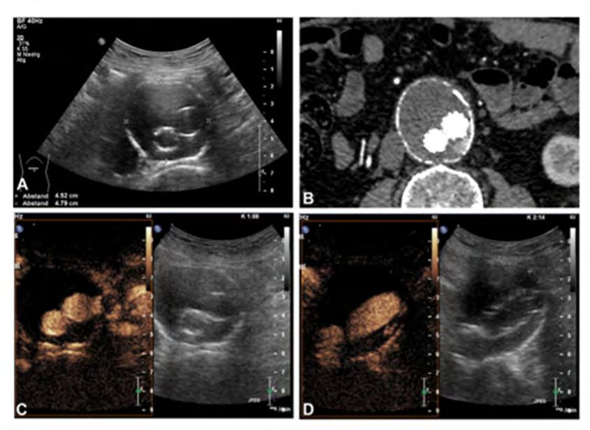
Figure 1: Initial native sonographic B-mode assessment with measurement of the maximal AAA diameter (A). CEUS evaluation showing an endoleak type II in transversal (C) and sagittal (D) planes. Postinterventional CTA showing the same results (B).
Diagnostic test accuracy of pedal acceleration time to identify peripheral artery disease
VASCULAR
Reviewer: Claire O’Reilly
Authors & Journal: Somerset J, Teso D, Mills JL Sr, Sebastian M, Kayssi A, Leask S, Rounsley R, Tehan PE. 2025; Eur J Vasc Endovasc Surg, 69;876e886.
Open Access: Yes
Read the full article here
Why the study was performed
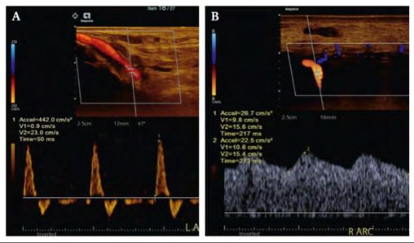
Traditional noninvasive tests for PAD include ABI (ankle brachial index) and TBI (toe brachial index). Both have limitations and can be unreliable in patients with noncompressible vessels (calcified vessels), wounds or amputations. This study aimed to assess how accurate pedal acceleration times are at diagnosing peripheral arterial disease (PAD) in patients already suspected of having the disease. PAT measures the time it takes blood to accelerate in the foot’s arteries using Doppler ultrasound and is usually measured in 4–5 arteries within the foot (Fig. 1). PAT values increase proportionally to the severity of PAD and return a zero value in occluded or dormant vessels where no flow is seen or recorded.
How the study was performed
This was a multicentre, cross-sectional study. One hundred and eighty-eight patients were recruited, with a mean age of 71 years, with males being more dominant (70%), for a total of 227 limbs. Colour Duplex ultrasound was used as the reference standard, as it is considered the gold standard for non-invasive imaging. The sonographer performing the scan was blinded to the study. PAD was graded using standard criteria. ABI and TBI were measured. A second sonographer, unaware of other results, measured the PAT of the 5 pedal arteries (lateral plantar, medial plantar, dorsal metatarsal, arcuate and deep plantar).
What the study found
In the statistical analysis, the AUC (area under the curve) ranged from 0.71 to 0.77 across the different arteries. The worst case PAT achieved the best overall accuracy with an AUC of 0.79. Worst case PAT AUC (0.79) was similar to TBI (0.78) and superior to ABI (0.7). In terms of predictive values, the threshold that optimised performance was PAT > 85 ms. It showed acceptable interoperator reliability.
Relevance to clinical practice
Pedal acceleration time is a promising, noninvasive ultrasound method to detect PAD and is particularly useful when standard measures fail. With its comparable accuracy to TBI and ease of use, PAT could help bridge the gaps
in vascular care. It doesn’t require any new equipment. The operator can target the artery of concern in case of wound care. It needs broader validation across diverse populations, but the study proved that it could help enhance early detection of PAD and is useful as an adjunct for vascular labs in complex patients. It may improve decision-making for revascularisation as well as monitoring woundhealing potential.
Takeaway message: PAT is a promising, noninvasive ultrasound method to detect PAD and is particularly useful when standard measures fail. It could help bridge the gaps in vascular care.
Using the worst case (highest or zero) pedal acceleration time (PAT) value yielded the highest diagnostic test accuracy of all PAT measures, with an optimal cut-off of > 85 ms determined for estimating peripheral arterial disease (PAD).
Figure 1: Pedal acceleration times in a (A) normal (50 ms) and (B) abnormal (213 ms) artery.
Placental biomarker and fetoplacental
Doppler
abnormalities are strongly associated with placental pathology in pregnancies with small-for-gestationalage fetus: prospective
study
Why the study was performed
Placental dysfunction leads to small-forgestational-age (SGA) and fetal growth restriction (FGR), resulting in poor perinatal outcomes and impacting intrauterine growth of appropriate-for-gestational-age (AGA) fetuses. Clinicians use common fetoplacental Doppler indices (for example, umbilical artery (UA); pulsatility index (PI); fetal middle cerebral artery (MCA); PI, uterine artery (UtA); PI, cerebroplacental ratio (CPR); and placental biomarkers of maternal biochemistry (for example, placental growth factor (PlGF), soluble fms-like tyrosine kinase-1 (sFlt-1)/PlGF ratio) to assess placental function and predict risks of adverse pregnancy outcomes. However, the evidence connects these prenatal abnormalities to specific postnatal placental histopathologies (for example, maternal vascular malperfusion (MVM), fetal vascular malperfusion (FVM), villitis of unknown aetiology (VUE), chronic histiocytic intervillositis (CHI) and delayed villous maturation (DVM)) is limited.
This large prospective study was conducted to bridge this gap, aiming to associate CPR, other fetoplacental Doppler indices, PlGF, and sFlt-1/PlGF ratio with detailed placental abnormalities in SGA/FGR pregnancies, confirming the relationship between prenatal placental dysfunction markers and specific placental pathologies postnatally.
How the study was performed
This prospective cohort study, conducted at a maternal-fetal medicine centre in Queensland, Australia (May 2022 – March 2024), investigated singleton pregnancies diagnosed with SGA or FGR. Pregnancies with genetic/ structural abnormalities or fetal infection were excluded. SGA was defined as abdominal circumference (AC) or estimated fetal weight (EFW) < 10th centile, while FGR used Delphi consensus criteria and is classified as early onset (< K32) or late onset (≥ K32).
Experienced obstetrics sonographers performed fetal biometry and Doppler assessments (UtA-PI, UA-PI, MCA-PI, CPR), with defined abnormal Doppler thresholds. Maternal-fetal medicine specialists reported ultrasound findings and were blinded to placental biomarker levels. Maternal blood
samples were collected on the first visit and repeated every 4 weeks for PlGF and sFlt-1 levels, with abnormal levels defined by specific thresholds.

Reviewer: Ling Lee, AFASA
Authors & Journal: Hong J, Crawford K, Cavanagh E, Clifton V, et al. 2025; Ultrasound Obstet Gynecol, 65:749–760.
Open Access: Yes
Read the full article here
Abnormal fetoplacental Doppler thresholds
CPR < 5th centile
UA-PI > 95th centile
UA Doppler AREDF
Mean UtA-PI > 95th centile
Post-birth, placentae underwent blinded histopathological examination based on Amsterdam Placental Workshop Group Consensus criteria by a perinatal pathologist. Primary abnormalities assessed were MVM, FVM, VUE, CHI, and DVM, categorised by predominant placental lesion. Multivariable logistic regression models, adjusted for preeclampsia, analysed association between abnormal Doppler/biomarkers and specific placental abnormalities.
What the study found
Abnormal placental biomarkers thresholds
PIGF < 100 ng/L
Elevated sFlt-1/PIGF ratio based on gestation
(> 5.78 for < K28)
(> 38 for > K28)
In a cohort of 367 women with singleton SGA pregnancies, maternal vascular malperfusion (MVM) was the most prevalent placental abnormality, identified in 43.3% of cases. Less frequent abnormalities included FVM (5.4%), VUE (13.4%), DVM (5.2%), and CHI (1.6%). The study revealed a strong association between abnormal prenatal Doppler findings and placental biomarkers (as shown in Table 1 above) and the presence of any placental abnormality upon histopathological examination, even after adjusting for preeclampsia.
Women with these abnormal markers had significantly lower median placental weight and poorer pregnancy outcomes, such as lower median gestational age at birth and birth weight, increased rates of preterm birth (PTB) (including medically indicated), higher caesarean rates, emergency operative
Table 1: Abnormal thresholds for fetoplacental Doppler and placental biomarkers.
Placental biomarker and fetoplacental Doppler abnormalities are strongly associated with placental pathology in pregnancies with small-for-gestational-age fetus: prospective study (continued)

deliveries for non-reassuring fetal status and severe neonatal non-neurological morbidity. Mean UtA-PI > 95th centile showed the strongest association with placental abnormalities, specifically MVM.
Relevance to clinical practice
This study demonstrated that routine prenatal fetoplacental Doppler findings and placental biomarkers in SGA/FGR pregnancies effectively identify underlying placental pathologies. Elevated mean UtA-PI (> 95th centile) and low CPR (< 5th centile) are key indicators in predicting placental abnormality that enable timely medical interventions, enhancing pregnancy management and preventing adverse outcomes.
A low CPR is associated with perinatal mortality, intrapartum fetal compromise and adverse neonatal outcome in both SGA and AGA fetuses.
Sonographic localisation of lymph nodes suspicious of metastatic breast cancer to surgical axillary levels

Reviewer: Fiona Hilditch
Authors & Journal: Fenech M, Burke T, Arnett G, Tanner A & Werder N. 2025; Journal of Medical Radiation Sciences, 72(1):119–138.
Open Access: Yes
Read the full article here
Why the review was undertaken
The review was undertaken to address the current gap in knowledge and standardised protocols for assessing axillary lymph nodes (LNs) in breast cancer patients. Although sonographic assessment is widely used in clinical practice to evaluate axillary involvement, which is crucial for staging, treatment planning, and surgical decisionmaking, there is significant variability in how these assessments are conducted, and documentation is often inconsistent. By outlining the anatomy, surgical levels and sonographic techniques needed to differentiate between benign and suspicious nodes and accurately localise them, this review aimed to:
• provide a systematic approach to sonographic imaging of axillary LNs
• improve the diagnostic accuracy and consistency of identifying and characterising suspicious LNs
• facilitate reliable localisation of LNs in correlation with other imaging modalities (for example, CT, PET-CT, MRI)
• support better clinical decision-making and surgical planning
• enhance overall patient management.
How the review was performed
The article offers a comprehensive review of the anatomy of the breast’s axillary lymph nodes, with detailed insight into surgical levels, lymphatic drainage, sonographic classification (benign vs suspicious), scanning techniques and other imaging modalities such as MRI, which is a valuable addition to the diagnostic process for breast cancer. MRI can further evaluate LN metastasis, especially in the internal mammary and supraclavicular regions. It presents in-depth guidance on sonographic assessment of axillary LNs to support informed patient management and surgical planning. Anatomical illustrations are paired with corresponding sonographic images and scanning techniques, highlighting axillary anatomy, surgical levels, and distinguishing features between benign and suspicious LNs.
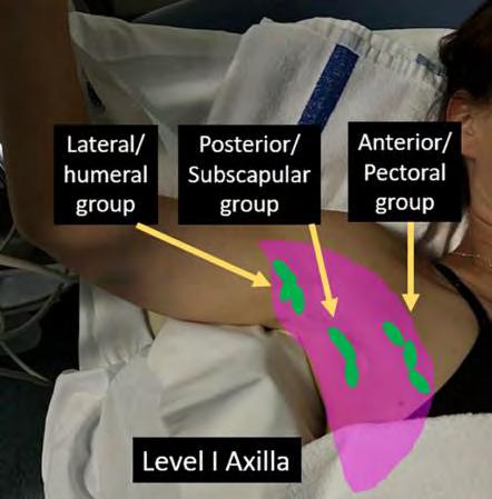
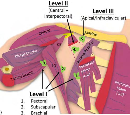
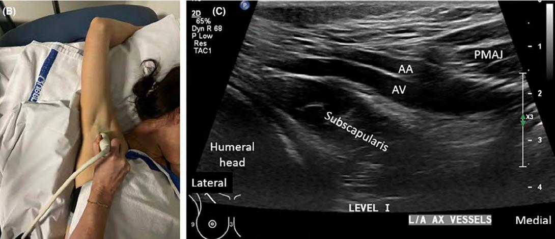
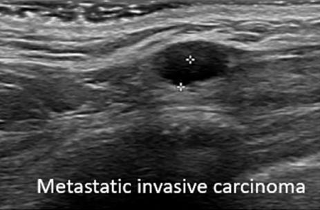
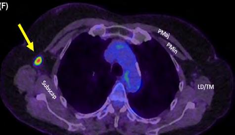
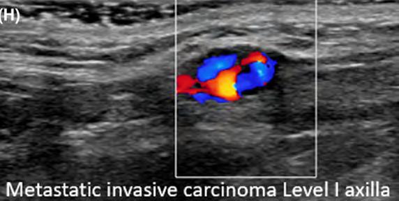
Metastatic axillary level I LN demonstrated on sonographic and axial PET CT in patient with HER2 positive breast cancer
Example images, surgical levels of the axillary LNs
Example images, ultrasound and PET CT images of metastatic invasive carcinoma in LNs
Sonographic localisation of lymph nodes suspicious of metastatic breast cancer to surgical axillary levels (continued)

What the review found
The review found that the accurate assessment of axillary LN involvement is critical in breast cancer management, as it significantly influences treatment strategies and prognosis. It emphasises the need for detailed anatomical knowledge, standardised imaging techniques, and consistent application of diagnostic criteria to distinguish between benign and malignant LNs for optimal breast cancer treatment planning. The findings support the importance of:
• Sonographic imaging is essential for identifying, characterising and localising suspicious axillary and regional lymph nodes in breast cancer patients.
• Detailed anatomical knowledge of the axilla, including surgical levels and muscular landmarks, is necessary for effective sonographic evaluation.
• Consistent application of specific sonographic criteria (such as cortical thickness, shape, and vascularity) is vital to differentiate benign from malignant nodes, which differ slightly from the criteria used for other lymph node regions.
• Accurate imaging and reporting of lymph node status support informed surgical planning and overall patient care decisions.
Relevance to clinical practice
The relevance of this article for clinical practice lies in its potential to improve the accuracy and consistency of preoperative axillary staging in breast cancer patients.
The article provides a standardised sonographic approach to assessing axillary LNs, improving the diagnostic accuracy of identifying metastatic involvement, which is crucial for staging and treatment planning.
The article serves as a comprehensive reference for sonographers, radiologists and trainees, reinforcing best practice in axillary ultrasound technique and interpretation.
Accurate sonographic evaluation of axillary lymph nodes – integrating anatomical knowledge, surgical level localisation, and malignancyspecific criteria – is critical for accurate staging, consistent reporting.
Ultrasound-guided hydro-dissection of the superficial fascia for cervical myofascial pain: A case series
Reviewer: Andrew Grant
Authors & Journal: Razaq S, Ricci V, Ricci C and Özçakar L. 2025; J Clin Ultrasound, 53:940–945.
Open Access: Yes
Read the full article here
Why the review was performed
The fascial system is gaining attention for its potential role in a variety of pain disorders. Owing to the increased understanding of the rich innervation of the fascia, it is emerging as a potential pain generator and a target for treatment in many cases of cervical myofascial pain.
The fascial tissue is divided into 2 types: the deep and the superficial fasciae. The deep consists of epimysia and aponeurotic fascia, while the superficial fascia (SF) comprises a fibrotic layer that is partly areolar and partly membranous. Previous histological studies have shown an abundant sensory and autonomic innervation of the SF, making it one of the richest sensory tissues, in some cases, only secondary to the skin.
Myofascial pain in the cervical region is a common and challenging condition. Previous studies have focused on the role of the deep fascia in chronic myofascial pain and the utility of ultrasound (US) in its diagnosis. To date, the role of SF as a pain generator in myofascial pain, especially in the cervical and periscapular region, has not been reported.
This report of 3 case studies aims to discuss the possible role of US-guided hydro-dissection of the superficial fascia to manage cervical and periscapular myofascial pain and stiffness.
What the review described
On ultrasound, the SF appears as a very thin hyperechoic line within the subcutaneous tissue, between the superficial and deep subcutaneous fat. The deep fascia appears as a thick and multilayered hyperechoic structure located between the muscle and the subcutaneous tissues (Figures 1 & 2).
The very thin nerves and vessels within the fascia are occasionally visible. Using US guidance, injections in and around the SF can be performed in a number of ways, namely: perifascial injection – injection to the histological interfaces between the fascia and the surrounding soft tissues; fascial hydro-dissection – high-volume injection within the thickness of the fascia; and fascial fenestrations – multiple back and forward movements of the needle crossing the fascia.
The case studies highlighted patients with focal superficial tenderness overlying the trapezius muscle, often with focal tightening of the skin. Thickening of the SF was observed in 2 cases, while in one case, the SF appeared normal; however, during needle insertion, resistance was felt while penetrating the SF.
Hydro-dissection of the SF was performed (Figure 3), and in all cases, immediate relief of symptoms was observed.
Relevance to clinical practice
Enhanced understanding of the fascial tissue innervation has led to a better understanding of the SF as a pain generator. To this end, when evaluating a patient with myofascial pain, isolated SF involvement in the periscapular/ cervical region should be considered. High resolution US is invaluable to localise and assess the SF as well as to guide the needle during interventional procedures.
Further research is needed to confirm the potential role of SF in chronic cervical myofascial pain, cervicogenic dizziness, and paresthesias.
Ultrasound has emerged as an excellent imaging technique to visualise and assess the fascial structures in detail and to help guide pain-relieving interventions.
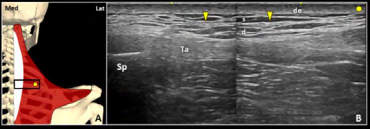
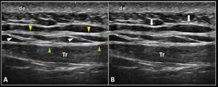
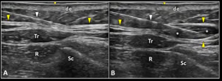
Figure 1: Positioning the transducer (black rectangle) in a transverse plane (A) over the right trapezius muscle (red), the focal thickening of the superficial fascia (yellow arrowheads) can be accurately observed in between the superficial (s) and deep (d) layers of the subcutaneous adipose tissue (B). de, Dermis; Lat, Lateral; Med, Medial; Sp, Spinous process; white (A)/Ta (B), Trapezius aponeurosis.
Figure 2: The superficial (yellow arrowheads) and deep (green arrowheads) fasciae are widely interconnected to each other through thin fibrous stripes (white arrowheads), forming a tridimensional fascial structure (A). The subcutaneous fibrous scaffold (white arrows) that stabilises the fat lobules should not be misinterpreted as the cervical superficial fascia (B).
Figure 3: The needle (white arrowhead) is advanced inside the superficial fascia (yellow arrowheads) to perform a high volume hydro-dissection, releasing the tiny nerves running inside the fascial structure in a patient with cervical and periscapular pain (A, B). de, Dermis; R, Rhomboid muscle; Sc, Scapula; Tr, Trapezius muscle; white asterisks, mixture.
Posterior tibial nerve ultrasound assessment of peripheral neuropathy in adults with type 2 diabetes mellitus
Reviewer: Sophie O’Brien
Authors & Journal: Latifat Tunrayo OduolaOwoo, Adekunle Ayokunle Adeyomoye, Olubukola Abeni
Omidiji, Bukunmi Michael Idowu, et al. 2024; Journal of Medical Ultrasound, 32(1):62–69.
Open Access: Yes
Read the full article here
Why the study was performed
We are currently experiencing an obesity epidemic, with the diagnosis of type II diabetes mellitus on the rise. Diabetic peripheral neuropathy (DPN) is a prevalent complication of type 2 diabetes associated with significant morbidity. Traditional nerve conduction studies are considered the diagnostic gold standard but are resource-intensive, invasive, lack specificity and provide limited anatomical context. This study aimed to evaluate whether high resolution ultrasound could serve as a non-invasive, low-cost screening tool for DPN by measuring the cross-sectional area of the posterior tibial nerve (PTN).
How the study was performed
The study was a case-control study performed prospectively. The study recruited 80 adults with type 2 diabetes and 80 healthy control adults. DPN was assessed using the Toronto Clinical Neuropathy Score (TCNS). The cross-sectional area of the left posterior tibial nerve was measured at 1 cm, 3 cm, and 5 cm proximal to the medial malleolus using ultrasound. Measurements were averaged, and correlations between modalities were analysed using receiver operating characteristic curves and correlation statistics versus TCNS severity and glycaemic markers.
What the study found
Over the 160 participants in the study, 58 participants (72.5%) were diagnosed with diabetic peripheral neuropathy based on the Toronto Clinical Neuropathy Score. Ultrasound measurements showed that the cross-sectional area (CSA) of the posterior tibial nerve (PTN) was significantly larger in diabetic patients with DPN compared to both diabetic patients without neuropathy and healthy controls. This indicated that CSA enlargement strongly correlated with TCNS severity. Notably, at 5 cm proximal to the medial malleolus, a CSA cutoff of 14 mm² showed 73.8% overall accuracy, with 77.6% sensitivity and 63.6% specificity. The optimum point of CSA of the PTN was 5 cm above the medial malleolus, providing the most accurate, reproducible measurement. Additionally, CSA values showed a weak but statistically significant positive correlation with
both fasting plasma glucose and HbA1c levels, suggesting a relationship between nerve enlargement and glycaemic control.
Relevance to clinical practice
The obesity epidemic is a global issue fuelling the increase in cases of diabetes mellitus type II. While the gold standard for assessing DPN is nerve conduction study, this is resource-intensive, invasive and costly. Ultrasound measurement of the posterior tibial nerve’s CSA presents a promising, accessible, cost-effective, non-invasive modality for early detection and staging of DPN. It may complement or partially substitute traditional nerve conduction testing in settings with limited access to electrophysiology services, addressing the issue of accessibility. Ultrasound is easily reproducible, costeffective, and correlates with both clinical and biochemical indicators of neuropathy. With established protocols and measurements, PTN CSA is easily reproduced by multiple sonographers and sonologists. This improves the ability of referrers to follow up, monitor progression and initiate timely intervention to potentially prevent the progression of DPN in those with type II diabetes before significant symptoms develop. Further research could validate the results and establish diagnostic thresholds for the CSA of the PTN.
The crosssectional area of the posterior tibial nerve increases significantly with the severity of diabetic peripheral neuropathy and may serve as a useful screening tool.
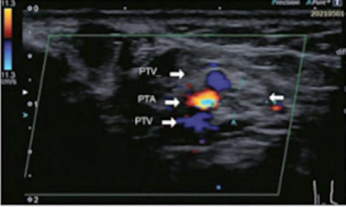
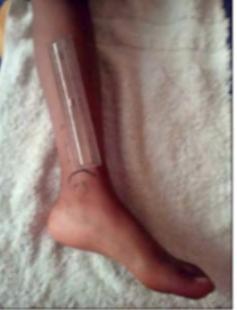
Figure 1: The transducer positions at 1, 3, and 5 cm proximal to the MM. MM Medial malleolus.
Figure 2: Transverse colour Doppler ultrasonogram of the PTN showing its minor axis (A-A) and major axis (B-B), with the accompanying PTA and paired PTVs. PTN: Posterior tibial nerve, PTA: Posterior tibial artery, PTV: Posterior tibial vein.
Automated classification of liver fibrosis stages using ultrasound imaging
EMERGING TECHNOLOGIES
Reviewer: Ian Schroen, AFASA
Authors & Journal: Park HC, Joo Y, Lee OJ, et al. 2024; BMC Med Imaging, 24;36.
Open Access: Yes
Read the full article here
Why this study was performed
Liver cirrhosis is a progressive condition that can lead to fibrosis and liver failure. Accurate staging of fibrosis is essential for timely intervention and management. Although liver biopsy is the gold standard, it is invasive and carries risks. Ultrasound is widely used as a non-invasive imaging method, but its diagnostic accuracy can be limited by operator dependence and subjective interpretation. This study by Park et al. (2024) was conducted to explore whether artificial intelligence (AI), specifically deep learning, could be used to automatically classify liver fibrosis stages from standard B-mode ultrasound images. The goal was to develop a more objective, reproducible, and accessible tool for liver fibrosis assessment that could enhance the reliability of ultrasound in clinical practice.
How this study was performed
The researchers obtained 7,920 B-mode ultrasound images from 933 patients who had undergone liver biopsy or surgical resection at 2 major university hospitals. The images were gathered from a range of ultrasound systems manufactured by GE Healthcare, Philips Medical Systems and Siemens Medical Systems. Each patient’s liver fibrosis stage was confirmed histologically, via biopsy, using the METAVIR scoring system, and classified from F0 (no fibrosis) to F4 (cirrhosis). The ultrasound images were then used to train and validate 5 types of deep convolutional neural network (DCNN) models: VGGNet, ResNet, DenseNet, EfficientNet, and Vision Transformer (ViT).
The models were trained to predict the fibrosis stage directly from the ultrasound images without the need for additional imaging modalities. Performance was evaluated using metrics such as the area under the receiver operating characteristic curve (AUC), sensitivity, specificity, and accuracy. All models performed strongly with clinically relevant measures.
Relevance to clinical practice
For sonographers, this study illustrates the growing potential of AI to augment ultrasound interpretation. The automated models demonstrated high diagnostic accuracy using only conventional B-mode images, making them feasible for use in routine clinical settings. By reducing variability between operators and standardising fibrosis assessment, AI tools could support more consistent and objective liver evaluations. This is particularly valuable in resource-limited environments or clinics without access to elastography or advanced imaging technologies.
In the future, integrating AI-based fibrosis classification into ultrasound systems may help sonographers provide more reliable preliminary assessments, facilitate early detection, and improve communication with referring physicians. This study represents a promising step towards AI-assisted ultrasound becoming a practical, everyday tool in hepatology and general abdominal imaging.
Deep learning
AI
models demonstrated high diagnostic performance in classifying liver fibrosis stages using B-mode ultrasound images only, suggesting the potential for AIassisted ultrasound to provide accurate and operatorindependent liver fibrosis assessment.
Progress in the application of artificial intelligence in ultrasound-assisted medical diagnosis
Why the study was performed
The study was conducted to review and evaluate the current progress, challenges and potential of artificial intelligence (AI) technologies in ultrasound-assisted medical diagnosis. Ultrasound is widely used due to its non-invasive, real-time imaging capabilities, but its effectiveness heavily depends on the operator’s expertise. The study sought to explore how AI could mitigate this variability. The authors also aimed to investigate how AI tools could improve diagnostic precision, reduce human error and assist clinicians in interpreting complex ultrasound data more effectively. Additionally, by reviewing the current state of research, the study aimed to highlight the limitations, ethical concerns and data challenges in AI-driven ultrasound, therefore suggesting future directions.
How the study was performed
The research was structured as a narrative review. The authors employed the following approaches:
1. Compilation of literature – by gathering and analysing a broad range of studies.
2. Technological assessment – reviewing ultrasound AI technologies such as machine learning, deep learning and convolutional neural networks and evaluating their roles in enhancing image acquisition, quality assessment and disease diagnosis.
3. Application analysis – examining current AI ultrasound applications, including automated image analysis, diagnostic assistance and medical education and highlighting how these applications contribute to improved lesion detection and reduced physician workload.
4. Future perspectives – exploring emerging trends, such as the potential for AI to promote standardisation, personalised treatment and intelligent healthcare, especially in underserved areas and remote areas.
What the study found
AI enhances ultrasound diagnostic accuracy and workflow efficiency, particularly through machine learning and deep learning. AI enables real-time image acquisition and interpretation, enhancing diagnostic precision and alleviating the workload of radiologists. AI promotes consistent imaging interpretations and enables personalised treatment by analysing patientspecific data, therefore enhancing the precision of medical interventions. Portable AI ultrasound can improve diagnostic access in remote or low-resource areas. The study also concluded that challenges remain – algorithm validation, data privacy, and ethical concerns must still be addressed, and it is crucial for the responsible and effective integration of AI in the medical field.
Relevance to clinical practice
The study provides a holistic understanding of how AI is transforming ultrasound-assisted medical diagnosis to guide future research and clinical applications in this evolving field. The advancements in AI are important as they have the potential to improve patient outcomes and therefore enhance the efficiency of healthcare delivery.
EMERGING TECHNOLOGIES
Reviewer: Joanne King
Authors & Journal: Li Yan, Qing Li, Kang Fu, Xiaodong Zhou, Kai Zhang. 2025; Bioengineering, Natural Library of Medicine – PubMed Central
Open Access: Yes
Read the full article here AI enhances diagnostic accuracy and consistency in ultrasound by reducing operatordependent variability and improving workflow efficiency.
PAEDIATRIC
Optic nerve sheath diameter measurement for the paediatric patient with an acute deterioration in consciousness
Why the study was performed
The study aimed to explore the use of optic nerve sheath diameter (ONSD) measurement as a non-invasive diagnostic tool for assessing intracranial pressure (ICP) in paediatric patients who are experiencing an acute deterioration in consciousness. Elevated ICP is a critical concern in these cases and can be challenging to assess in children, especially without invasive procedures.
The motivation was to find a practical, quick, and less invasive method to assist healthcare professionals in monitoring these patients’ neurological status.
How the study was performed
Design: This was an observational study involving paediatric patients who presented with acute changes in consciousness.
Methods: The researchers measured the optic nerve sheath diameter using ultrasonography, which can detect changes in ICP based on the enlargement of the optic nerve sheath. The measurements of the ONSD were then compared with clinical markers of elevated ICP to assess their accuracy.
Participants: The study focused on children who showed signs of deteriorating consciousness, which could be indicative of increased ICP.
What the study found
The study found that ONSD measurement via ultrasound was a reliable tool for detecting signs of increased ICP in paediatric patients with acute deterioration in consciousness.
Correlation with increased ICP: The results showed that larger ONSD measurements were strongly associated with elevated ICP in these patients.
Accuracy: The technique was shown to have good sensitivity and specificity, making it a potentially effective method for rapid, pointof-care evaluation in paediatric emergency settings.
Relevance to clinical practice
Non-invasive diagnostic tool: The study’s findings suggest that ultrasonographic ONSD measurement can be used as a rapid, non-invasive alternative to other methods (for example, invasive ICP monitoring) for assessing increased ICP in children.
Clinical decision-making: It can help guide clinical decisions, especially in emergency settings where quick evaluation of ICP is critical.
Wider application: This technique could be useful in various paediatric clinical environments, including emergency departments, intensive care units, and even in settings with limited access to advanced imaging equipment.
Overall, the study highlights the potential of ONSD measurement as a non-invasive and easily accessible method to assess ICP in paediatric patients with deteriorating consciousness, offering significant clinical value in the management of these high risk patients.
Reviewer: Lino Piotto, FASA
Authors & Journal: Ali A, McCreary DJ. 2023; Ultrasound J, 15(1):45.
Open Access: Yes
Read the full article here
Optic nerve sheath diameter (ONSD) has been demonstrated to correlate closely with intracranial pressure (ICP) and an elevated measurement can detect raised ICP readily.
PAEDIATRIC
Ultrasound in pediatric inflammatory bowel disease – A review of the state of the art and future perspectives
Reviewer: Madonna Burnett
Authors & Journal: Hoerning A, Jüngert J, Siebenlist G, Knieling F, Regensburger AP. 2024; Children, 11(2):156.
Open Access: Yes
Read the full article here
Why the review was performed
Inflammatory bowel disease (IBD) can affect adults, children and adolescents. To detect relapses of inflammation, patients need to have multiple examinations, which may include laboratory and stool sample assessment, endoscopic and classical cross-sectional imaging. Some of these examinations may be invasive and quite often require sedation for the younger cohort of patients. Intestinal ultrasound (IUS) is becoming more important as a non-invasive assessment of the intestine and its inflammatory involvement. This review sheds some light on the current state of the art and provides an outlook on developments in this field that may spare these younger patients more invasive follow-up procedures.
What the review looked at
Crohn’s disease (CD) and ulcerative colitis (UC) are chronic, relapsing inflammatory conditions of the gastrointestinal tract (GI). IBD in children and adolescents often has unusual manifestations, is exhibited more frequently, and is usually more severe and difficult to treat. The need for immunosuppressive therapy and the need for surgery are much higher in paediatric IBD than in adult patients.
CD can be localised throughout the GI tract and is characterised by segmental, discontinuous involvement and inflammatory changes affecting all layers of the intestinal wall. The paper describes symptoms of CD. In paediatric patients, an appendicitis-like clinical picture may present if the terminal ileum is affected.
UC affects the distal rectum and continuously spreads orally. Clinical symptoms are discussed.
Along with ultrasound, diagnostic imaging of the GI tract also includes magnetic resonance imaging (MRI). Oral mannitol administration is commonly used – termed magnetic resonance enterography (MRE), to better visualise the small intestine and pelvic MRI to assess anal fistulas or perianal abscesses. It is difficult to evaluate the upper GI tract with US. MRE/ MRI performs better than US in detecting IBD in paediatric patients and is better at distinguishing between CD and UC.
The European Society of Paediatric Radiology abdominal imaging taskforce recommends the first-line use of US. MRE is used for further workups in the case of unclear US findings.
The paper goes on to discuss US in IBD, describing transducer selections and positioning. US anatomy of the intestinal wall (correlating to US images) is discussed at length with a good table to refer to. Intestinal wall thickness is discussed in normal and IBD conditions, along with ultrasound images to refer to.
Current ultrasound characteristics used in IBD diagnostics, US Doppler signals, including the ultrasound (Doppler) scoring of inflammatory activity according to Limberg, mesenterial or ‘Creeping Fat’, fibrostenosis and intestinal strictures are all discussed.
A US scoring system would be desirable. A table of the ‘Overview of paediatric sonographic US indices’ for CD, UC and IBD is in this review.
What the review found
While intestinal US techniques progress and standardisation of examination methods have been introduced in the field of adult medicine, some of these developments are still pending in paediatrics.
The advantage of US compared to other diagnostics in paediatric IBD is the non-invasiveness, no radiation or need for anaesthesia, the broad availability, the low infrastructure costs and the easy-to-learn imaging modality with simple comprehensibility.
Relevance to clinical practice
From the perspective of a physician, the implementation of basic US categorisation into the clinical routine follow-up procedure for IBD should rely on the quantitative assessment of the intestinal wall thickness. A wall thickness that exceeds 2–3 mm in inflamed segments with increased blood flow should alert doctors to possible IBD lesions or a flare in already diagnosed patients.
In the future, technological improvements and new technologies could provide a large number of other imaging biomarkers, making US-based imaging the firstline diagnostic method in pediatric IBD imaging.
RESEARCH
Recommendations for the extraction, analysis, and presentation of results in scoping reviews
Reviewer: A/Prof Michelle Fenech, FASA
Authors & Journal: Pollock D, Peters MDJ, Khalil H, McInerney, P, et al. 2023; JBI Evidence Synthesis, 21(3):520–532.
Open access: Yes
Read the full article here
Why the study was performed
Scoping reviews are a type of journal paper used to scope the body of literature, clarify concepts, identify knowledge gaps or investigate research conduct. They are different to systematic reviews and can be precursors to systematic reviews and further research. Scoping reviews are useful for examining emerging evidence, when it is still unclear what other more specific questions need to be posed and addressed by a systematic review.1,2 Like systematic reviews, they require a rigorous and transparent method in their conduct to ensure the results are trustworthy. However, scoping reviews can be challenging for authors to undertake and write.2 This paper outlined the process to extract, analyse, and present results in a scoping review.
How the study was performed
This paper outlined an approach to extracting, analysing and presenting the results of a scoping review.
What the study found
Scoping reviews need to identify and map available evidence and clarify key concepts and definitions related to a concept or idea, and identify and analyse knowledge gaps.1 Due to the large amount of literature that needs to be searched during a scoping review, this paper highlighted that a team approach is good to use to extract, analyse and present data. The type of data collected needs to be agreed upon by the team. A template to compare data is required to be developed to determine whether papers (data) collected are relevant, appropriate, meaningful, and effective in addressing the scoping review question. The methodology to qualitatively assess and analyse the literature collected needs to be defined and followed. The literature collected needs to be organised and analysed using either an inductive (coding) or deductive approach. The results should be communicated in a cohesive, coherent form. Using tables helps to summarise a large amount of information related to the literature used to develop the scoping review. A PRISMA scoping review
(PRISMA-ScR) checklist is helpful to guide the communication of the results.
Relevance to clinical practice
The difference between scoping reviews and systematic reviews, including how to interpret them, should be appreciated by sonographers, as both these reviews can be used to inform clinical practice and highlight gaps in knowledge and signpost further research required.
Scoping reviews have been defined as a ‘type of evidence synthesis that aims to systematically identify and map the breadth of evidence available on a particular topic, field, concept, or issue, often irrespective of source (that is, primary research, reviews, nonempirical evidence) within or across particular contexts’.
References:
1. Munn Z, Peters MDJ, Stern C, Tufanaru C, McArthur A, Aromataris E. Systematic review or scoping review? Guidance for authors when choosing between a systematic or scoping review approach. BMC Med Res Methodol. 2018;18(1):143.
2. Peterson J, Pearce PF, Ferguson LA, Langford CA. Understanding scoping reviews: Definition, purpose, and process. J Am Assoc Nurse Pract. 2017;29(1):12-6.
A comparison of ChatGPT-generated articles with human-written articles
RESEARCH
Reviewer: Helen Beets
Authors & Journal: Ariyaratne S, Iyengar KP, Nischal N, et al. 2023; Skeletal Radiol, 52:1755–1758.
Open Access: No
Read the full article here
Why the study was performed
The artificial intelligence (AI) of ChatGPT can generate scientific papers similar to those written by humans, utilising natural language and some limited comprehension. While interest in the use of ChatGPT in research is an expanding topic, there are some concerns about the accuracy of AI-generated research papers. Additionally, while the format and structure of these papers can be convincing, there are rising questions about the authenticity and accuracy of the data produced. This paper aimed to compare research articles compiled by ChatGPT with those previously written/ published by humans to assess for accuracy.
How the study was performed
Five random research papers written before 2021 were selected, and ChatGPT version 3.0 was asked to write these articles, including the references.
Two independent radiologists were asked to review each paper, and the references were cross-checked. Each article was graded 1–5, with 1 being bad and inaccurate and 5 being excellent and accurate. Comments were made if articles were totally incorrect or different. Validity and accuracy of the data were checked.
What the study found
The ChatGPT articles that were generated were brief, at around 400 words or less, and compiled within around 15 seconds. They were written in a format expected of journal articles, and overall, the introduction, main body and conclusions were mostly good. All papers, however, were factually incorrect, and some were unrelated to the actual topic at all. One of the articles went on to suggest surgical intervention for cases where surgery is not recommended, and another described an intervention technique with incorrect steps. In some of the articles, the references were wrong and in one of the articles, the references did not exist and were fictitious. All 5 articles were graded as a 1.
Relevance to clinical practice
ChatGPT has been widely used around the globe since its introduction in 2022, and AI is increasing in prevalence across all industries. One of the key failures of ChatGPT is the inability to discern which web-based resources are suitable for the extraction of information to produce an accurate literature review.
This study raises awareness of the risks of relying on AI to produce written reports. An untrained or unknowing individual may fall victim to scientific misinformation, and early career researchers or untrained individuals may use this misinformation to teach clinical practice and train with dangerous outcomes. For example, one of the papers reproduced by ChatGPT in this study was about a widely used cartilage imaging protocol currently used in practice, and the formulated report described the steps and methods incorrectly. Some potential benefits for using ChatGPT include the drafting process or spelling and grammar checking; however, caution is required with an understanding of the current limitations and pitfalls of AI-produced papers. While ChatGPT versions continue to improve and learn, without proper checking, inaccurate papers may cascade and lead to further misinformation, which can be dangerous for evidence-based clinical practice.
ChatGPT can generate authenticlooking scientific articles; however, in this study, the articles generated were largely inaccurate and unreliable.
Exploring the impact of obstetric sonographers dealing with and supporting patients who receive negative outcomes
Why the article was written
The authors aimed to shed light on the experiences of obstetric sonographers who deliver unexpected and often distressing news to patients. Recognising a significant gap in the literature, they aimed to explore how this challenging aspect of their role impacts sonographers both professionally and personally. The article examines the support systems available to sonographers across different clinical settings and highlights the need for better resources and training to support sonographers.
How the study was performed
The authors reviewed existing literature to gather data on the experiences of obstetric sonographers. They focused on various obstetric work environments, including maternal-fetal medicine (MFM) clinics, general obstetric/gynaecology practices, and emergency departments, examining the roles and responsibilities of sonographers in delivering unexpected news in these settings, the support they receive from colleagues, and the impact of these factors on their mental health and professional wellbeing.
What the study found
The authors noted that obstetric sonographers face significant stress and emotional challenges when delivering unexpected news to patients. Key findings:
• Obstetric sonographers experience high levels of burnout, driven by feelings of guilt, uncertainty, and stress. The emotional toll of delivering unexpected news, coupled with the demanding nature of their role, contributes to burnout.
• Sonographers working in MFM clinics benefit from better support systems compared to those in obstetric/gynaecology practices and emergency departments. In MFM clinics, teamwork with physicians and other healthcare professionals is more established, providing a collaborative environment that helps manage stress.
• Effective communication and understanding of roles and responsibilities within the healthcare team are important for managing difficult situations and reducing stress. The authors highlighted that proper support could make communication with patients less stressful.
• There is a need for targeted training materials and resources to help sonographers manage the emotional toll of delivering unexpected news.
Relevance to clinical practice
This report highlights the importance of supporting obstetric sonographers in their roles when delivering difficult news to patients. Recommendations for clinical practice include:
• Implementing training programs focused on delivering unexpected news, medical ethics, and patient support to better equip sonographers for this challenging aspect of their role.
• There is a high level of burnout among obstetric sonographers, driven by high workloads, lack of support and the emotional toll of delivering unexpected news. Workplaces should develop support systems and provide access to mental health services to ensure sonographers can manage the stress associated with their role.
• Create a positive work environment that fosters mutual trust and respect among colleagues, which can help reduce burnout and improve patient care. Effective collaboration between healthcare providers can help manage difficult situations and reduce stress.
HEALTH & WELLBEING
Reviewer: Emma Jardine, AFASA
Authors & Journal: Butwin AN, Smith KA, Volz KE. 2024; Journal of Diagnostic Medical Sonography, 40(5):520–523.
Open Access: Yes/No (preferred, but not required)
Read the full article here
To prevent obstetric sonographer burnout, it is important to consider ways to create a positive work environment and provide resources to protect mental health.
Building emotional resilience to foster well-being by utilising reflective practise in the sonography workplace
HEALTH & WELLBEING
Reviewer: Jane Wardle
Authors & Journal: White A, Humphreys L, Oomens D. 2024; Sonography, 11(3):243–250.
Open Access: Yes
Read the full article here
Why the article was written
This article was written to explore the value of reflection, particularly affective reflection and reflective writing, as resilience-building tools for sonographers. Sonography professionals often face emotionally challenging and traumatic scenarios due to the nature of their work, including delivering difficult news or witnessing distressing situations. Such continual exposure can lead to compassion fatigue and burnout, impacting mental health and the quality of patient care. While reflection is widely practised in other healthcare fields for self-awareness and resilience, little research addresses its application within the sonography profession. The authors aim to demonstrate how reflective practices can support sonographers’ emotional resilience, helping to safeguard their wellbeing and sustain high standards in clinical practice.
How the review was performed
This article primarily reviews existing literature on reflective practice, affective reflection, and resilience within healthcare. It examines how sonographers could benefit from adopting reflective strategies, drawing from established methods and models in the literature, including the works of Donald Schön, Gibbs, and others on experiential learning. By incorporating theoretical frameworks and real-world examples, the authors present practical strategies for reflective writing tailored to sonography. The article outlines a structured approach to reflection, emphasising cyclical processes such as critical incident analysis, which help identify and address gaps in knowledge, skills, and emotional responses. Practical guides, including guided prompts for reflective writing, are provided to support the implementation of these strategies in sonography.
What the article found
The article suggests that affective reflection and reflective writing can effectively support emotional resilience, self-awareness, and selfregulation among sonographers. Reflective practice allows professionals to process emotions, reframe responses, and develop coping mechanisms to manage ongoing
emotional stress. The authors observed that sonographers who engage in reflective writing can better adapt to stressful situations, potentially preventing burnout and promoting empathy in patient interactions. Additionally, the article suggests that reflective practice can enhance clinical skills and decision-making by helping sonographers bridge gaps between theory and practice, creating opportunities for growth in cognitive, psychomotor, and emotional domains.
Relevance to clinical practice
This article highlights the importance of integrating reflective practices into the sonography field, emphasising their potential to foster emotional resilience and enhance patient care. By making reflection a routine part of clinical practice, sonographers may experience improved emotional stability and reduced stress reactivity, which can positively impact patient interactions and clinical outcomes. Introducing structured reflective writing and mindfulness practices can help sonographers better manage the emotional challenges of their work, ultimately contributing to their professional development and personal wellbeing.
Given the clear link between the practice of reflection and the development of resilience in other healthcare professions, this concept can be adopted by sonographers to reduce their degree of emotional fatigue and enhance their overall wellbeing.
Sonography education in the clinical setting: The educator and trainee perspective
CLINICAL SUPERVISORS
Reviewer: Helen Beets
Authors & Journal: Burnley, K and Kumar, K. 2019; Australasian Journal of Ultrasound in Medicine, 22:279–285.
Open Access: Yes
Read the full article here
Why the study was performed
Sonography is a complex clinical skill with little research on how these skills are taught. The most common clinical training methods include a ‘see one, do one’ approach or a step-bystep structured model. The research study was performed to identify the perspectives of sonographers involved in teaching and learning, and sonographer trainees/newly qualified graduates, regarding the methods used in clinical skills training. A secondary aim was to determine who and where training was delivered within Australia.
How the study was performed
This was a prospective study involving 2 groups. The first group were sonographers registered with the Australian Society for Ultrasound in Medicine (ASUM), and the second group were trainees or newly qualified sonographers from one clinical workplace.
The first group participated in an online survey while the second group undertook semi-structured interviews with the first author. A thematic analysis of the online survey and interviews was performed to identify a thematic framework.
What the study found
In the first group, 49% of participants identified as tutor sonographers, while 52% were primary clinical supervisors. Overall, 58% of the total group has no formal clinical training qualifications. In terms of the clinical teaching method utilised, 22% adopted the ‘see one, do one’ method, with 9% reporting an undisclosed method. The remaining 52% of participants used a structured, step-by-step model approach such as the George and Doto five-step model and the Walker and Peyton four-step model.
From the thematic analysis of 6 interviews and 49 survey respondents, 4 main themes relating to sonography teaching skills emerged:
1. The importance of repeated observation and practice – identified by both educators and students. Students also desired more supervised practice.
2. Identification of teaching model – 5 of the 6 interview participants recognised that their clinical training followed the five-step
model. The sixth participant preferred a model that included a comprehension step, as this helped to reinforce their learning.
3. Opportunity for feedback – feedback was universally valued by the participants; however, time constraints and a lack of training in feedback delivery were barriers to consistent implementation.
4. Flexibility to adapt skill teaching and method – the educator participants indicated that the teaching models had limitations relating to the level of student competency, examination complexity and time constraints. However, they were able to adapt their teaching to the student and the examination.
Relevance to clinical practice
This study recognises the important role of structured, adaptable teaching methods in sonography education. The findings indicate that trainees valued clear instructional models with repeated observation and would prefer more feedback and greater time dedicated to the feedback process. This requires organisations to recognise that the trainee and the clinical supervisor may need more time for the education process. Furthermore, the lack of formal teaching qualifications highlights a need for professional development in clinical education to invest in the sonographer educator workforce and to optimise the experience of clinical training for trainees.
Quality sonography education is underpinned by regular feedback, frequent observation and practice and the adaptation of sonography clinical skills training and teaching practices according to context.
Push and
pull
factors impacting the pedagogical approaches used by sonographers to teach scanning skills
CLINICAL SUPERVISORS
Reviewer: Karen Barton
Authors & Journal: Nicholls D, Sweet L, Hyett J, Müller A. 2020; Australasian Journal of Ultrasound in Medicine, 23:220–226.
Open Access: Yes
Read the full article here
Why the study was performed
The aim of the study was to report and analyse the instructional methods currently used by Australian sonographers to teach sonography scanning skills, and the contextual features that help or hinder effective teaching. Understanding pedagogical approaches is essential, as evidence-based teaching practices not only enhance student learning outcomes but also contribute to improved patient care and wellbeing.
How the study was performed
A purpose-built survey tool called the SonoSTePs instrument was developed to investigate how sonographers teach scanning skills in clinical practice. The study analysed data from a national cross-sectional electronic survey distributed to all ASAR-registered sonographers. Key themes and pedagogical approaches were identified from the survey responses, which were coded as relating to a barrier or enhancement to best practice.
What the study found
The paper identified 5 complex and interrelated themes influencing current sonography clinical education. These were:
1. limited protected teaching time
2. perceptions of skill complexity
3. perceptions of the learner’s skill level
4. avoiding overwhelming the learner
5. patient wellbeing and willingness to be scanned.
The identified themes incorporate evidencebased educational pedagogy, especially scaffolding and cognitive load theory, which support psychomotor skill learning. However, variable, opportunistic and unplanned teaching practice was also revealed, consequently highlighting a gap between current clinical teaching practice and optimal pedagogical approaches outlined in motor-learning literature. Limited protected teaching time due to educators’ workloads and departmental time pressure was identified as a significant barrier to best teaching practice. Furthermore, the ad hoc subjective assessment of learner skill level based on qualification or perceived skill level
was also identified as a potential barrier to an optimal pedagogical approach.
A novel finding of the study identified 4 previously unreported, sonographer-specific skill education time points that reflect a nuanced and responsive approach to teaching, tailored to both learner needs and clinical context.
• Pre-task: clarifying objectives and guidance
• In-task: real-time verbal information and scanning support
• Post-task: support during interpretation and documentation
• End-task: reflective feedback and planning for future learning.
Relevance to clinical practice
This research provides a comprehensive, evidence-based insight into how sonographers teach psychomotor skills, providing valuable guidance for educators and education providers in shaping clinical supervision practices. It highlights the benefits of pedagogical approaches, such as scaffolding skills, where complex skills are broken into manageable components to reduce cognitive load and enhance learning. To improve current practice, the study recommends increasing protected teaching time, implementing more planned and frequent learning opportunities, expanding formal sonographer education in pedagogy and enhancing recognition of the educator’s role.
Whilst opportunistic teaching is valuable, it is also important that educators prioritise their teaching role, and plan and enact dedicated teaching sessions that are frequent and short in duration and reduce the sporadic and long teaching sessions.
