3 minute read
Posterior tibial nerve ultrasound assessment of peripheral neuropathy in adults with type 2 diabetes mellitus


Posterior tibial nerve ultrasound assessment of peripheral neuropathy in adults with type 2 diabetes mellitus
Reviewer: Sophie O’Brien
Authors & Journal: Latifat Tunrayo OduolaOwoo, Adekunle Ayokunle Adeyomoye, Olubukola Aben
Why the study was performed
We are currently experiencing an obesity epidemic, with the diagnosis of type II diabetes mellitus on the rise. Diabetic peripheral neuropathy (DPN) is a prevalent complication of type 2 diabetes associated with significant morbidity. Traditional nerve conduction studies are considered the diagnostic gold standard but are resource-intensive, invasive, lack specificity and provide limited anatomical context. This study aimed to evaluate whether high resolution ultrasound could serve as a non-invasive, low-cost screening tool for DPN by measuring the cross-sectional area of the posterior tibial nerve (PTN).
How the study was performed
The study was a case-control study performed prospectively. The study recruited 80 adults with type 2 diabetes and 80 healthy control adults. DPN was assessed using the Toronto Clinical Neuropathy Score (TCNS). The cross-sectional area of the left posterior tibial nerve was measured at 1 cm, 3 cm, and 5 cm proximal to the medial malleolus using ultrasound. Measurements were averaged, and correlations between modalities were analysed using receiver operating characteristic curves and correlation statistics versus TCNS severity and glycaemic markers.
The crosssectional area of the posterior tibial nerve increases significantly with the severity of diabetic peripheral neuropathy and may serve as a useful screening tool.
What the study found
Over the 160 participants in the study, 58 participants (72.5%) were diagnosed with diabetic peripheral neuropathy based on the Toronto Clinical Neuropathy Score. Ultrasound measurements showed that the cross-sectional area (CSA) of the posterior tibial nerve (PTN) was significantly larger in diabetic patients with DPN compared to both diabetic patients without neuropathy and healthy controls. This indicated that CSA enlargement strongly correlated with TCNS severity. Notably, at 5 cm proximal to the medial malleolus, a CSA cutoff of 14 mm² showed 73.8% overall accuracy, with 77.6% sensitivity and 63.6% specificity. The optimum point of CSA of the PTN was 5 cm above the medial malleolus, providing the most accurate, reproducible measurement. Additionally, CSA values showed a weak but statistically significant positive correlation with
both fasting plasma glucose and HbA1c levels, suggesting a relationship between nerve enlargement and glycaemic control.
Relevance to clinical practice
The obesity epidemic is a global issue fuelling the increase in cases of diabetes mellitus type II. While the gold standard for assessing DPN is nerve conduction study, this is resource-intensive, invasive and costly. Ultrasound measurement of the posterior tibial nerve’s CSA presents a promising, accessible, cost-effective, non-invasive modality for early detection and staging of DPN. It may complement or partially substitute traditional nerve conduction testing in settings with limited access to electrophysiology services, addressing the issue of accessibility. Ultrasound is easily reproducible, costeffective, and correlates with both clinical and biochemical indicators of neuropathy. With established protocols and measurements, PTN CSA is easily reproduced by multiple sonographers and sonologists. This improves the ability of referrers to follow up, monitor progression and initiate timely intervention to potentially prevent the progression of DPN in those with type II diabetes before significant symptoms develop. Further research could validate the results and establish diagnostic thresholds for the CSA of the PTN.

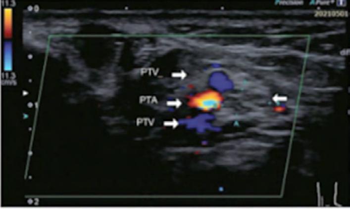
Figure 1: The transducer positions at 1, 3, and 5 cm proximal to the MM. MM Medial malleolus.
Figure 2: Transverse colour Doppler ultrasonogram of the PTN showing its minor axis (A-A) and major axis (B-B), with the accompanying PTA and paired PTVs. PTN: Posterior tibial nerve, PTA: Posterior tibial artery, PTV: Posterior tibial vein.