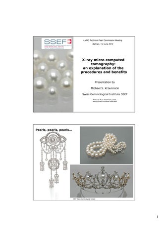
pearls, pearls...


LMHC Technical Pearl Commission Meeting Bahrain, 1-2 June 2010
Presentation by Michael S. Krzemnicki
Swiss Gemmological Institute SSEF

Photos © M.S. Krzemnicki, SSEF, except where indicated otherwise
1
X-ray micro computed tomography: an explanation of the procedures and benefits
© SSEF Swiss Gemmological Institute Pearls,
Natural or cultured pearls ?


Large quantities of „new“ pearls arrive on the pearl market.

Cultured pearls: the options
from H.A. Hänni, 2008
- Saltwater or freshwater Gonad grown or mantle grown Beaded or beadless

2
© Swiss Gemmological Institute SSEF
© Swiss Gemmological Institute SSEF
Saltwater beadless cultured pearls


The bead is rejected just after implantation. The remaining inserted mantel tissue (epithelium) starts pearl formation in the gonads.

Usually round to button and drop shapes.
After the first harvest, a second bead was implanted in the existing pearl sac, but shortly after was rejected. The pearl sac collapses, but will continue to produce a pearl. Usually baroque shapes.


Cross sections

3
Gonad grown “Keshi” first generation
Gonad grown “Keshi” second generation
© Swiss Gemmological Institute SSEF
Photos © H.A. Hänni, SSEF
© Swiss Gemmological Institute SSEF
Photos © H.A. Hänni, SSEF
Beaded cultured pearl
FW
beadless cultured pearl
Natural
pearl
SW
beadless cultured pearls
X-ray radiography
Problems when interpreting radiographies
X-rays
- a three-dimensional object is projected to on a planar film fracture or cavity structures sometimes small structures

the visibility of internal structures (e.g. irregular cavity in a beadless cultured pearl) may vary strongly depending to the direction of the X-ray projection.


4
© Swiss Gemmological Institute SSEF
Pearl mix, cultured
Natural pearls beadless cultured freshwater
© Swiss Gemmological Institute
SSEF
from H.A. Hänni, 2008
X-ray micro tomography


a three-dimensional object is slowly rotated and repeatedly exposed to projected to X-rays on a planar detector

the projected scans are then reconstructed into a three dimensional model
With specific software, we then can virtually scroll through the object
X-ray micro tomography (X-ray !-CT)

SkyScan1172 high-resolution micro-CT
fully distortion corrected 11Mp X-ray camera up to 8000x8000 pixels in every slice down to 1 !m isotropic resolution
dynamically variable acquisition geometry for shortest scan at any magnification computer cluster for 3D reconstruction - software for 2D / 3D image analysis
Operating conditions (example):
Source voltage (kV) = 88
Source current (!A) = 100
Image pixel size (!m) = 3
Exposure (ms) = 2356
Rotation step (deg) = 0.30°
Rotation (deg) = 360°
Frame averaging = 2
Scan duration = approx. 2 hrs
Reconstruction duration = approx. 2hrs
5
© Swiss Gemmological Institute SSEF
©
Swiss
Gemmological Institute SSEF
X-ray micro tomography


6
© Swiss Gemmological Institute SSEF
Pearl mounted for X-ray micro CT analysis
Projected X-ray scan of the pearl
in one
position
X-ray micro tomography © Swiss Gemmological Institute SSEF Scrolling in 3 directions through the pearl
Reconstructed slice of a beadless cultured pearl
Three dimensional scrolling 5 microns resolution
Cultured pearl (beadless)
Cultured pearl (beadless)


8
© SSEF Swiss Gemmological Institute
scrolling
Beadless cultured pearl (P. maxima) Sample mxt 61-14
© SSEF Swiss Gemmological Institute
Irregular cavity scrolling
Beadless freshwater cultured pearl (China) Sample mxt-1 (5 microns resolution)
Micro X-ray tomography


9
© Swiss Gemmological Institute SSEF
© Skyscan, Belgium
Foam: Visualization of as an X-ray absorptivity model
Modelling the irregular cavity and rotating the model
Beadless cultured pearl
© Swiss Gemmological Institute SSEF
Beadless freshwater cultured pearl
(China)
Sample mxt-1 (5 microns resolution)
X-ray micro tomography

Beadless freshwater cultured pearl (China) mxt 1


Please note: The flat cylindrical shape is only reflecting the modelled part of the CT-reconstruction and has nothing to do with the pearl shape!


Please note: Drilling may be a problem!
Two different pearls from Pinctada maxima
Irregular cavity
scrolling

10
© Swiss Gemmological Institute SSEF
© SSEF Swiss Gemmological Institute
Beadless cultured pearl
!
Cultured pearl (beaded)
Beaded cultured pearl (Pinctada maxima) Sample mxt-14b (5 microns resolution)
Cultured pearl (beaded)

Beaded cultured pearl (Pinctada maxima) Sample mxt-14b (5 microns resolution)


11
scrolling
© Swiss Gemmological Institute SSEF
bead
scrolling
© Swiss Gemmological Institute SSEF
Special cases of cultured
Special cases of Cultured


12
pearls:
Small
additional beadless cultured pearls forming during pearl cultivation
Photo © H.A. Hänni, SSEF 2010
Beaded
cultured pearl (Pinctada maxima) with attached small additional cultured pearl Sample mxt 21_1 (4 microns resolution) bead scrolling
pearls © Swiss Gemmological Institute SSEF Additional cultured pearl
Special cases of Cultured pearls
Beadless cultured pearl (Pinctada maxima) with included small additional cultured pearl Sample mxt 21_2 (4 microns resolution)


Special cases of Cultured pearls


Beadless cultured pearl (Pinctada maxima) grown during pearl cultivation Sample mxt 37_20 (2.3 microns resolution)


13
Large cavity due to collapsed pearl sack after bead rejection
scrolling
© Swiss Gemmological Institute SSEF
Additional cultured pearl
scrolling
© Swiss Gemmological Institute SSEF
cultured pearl
Pearl Structures


14
© Swiss Gemmological Institute SSEF
“New” beaded Cultured Pearls: the next challenge...
© Swiss Gemmological Institute SSEF
Beaded cultured pearl from P. maxima with a natural pearl used as „bead“
Conclusions, part 1
“New” pearls enter the market in large quantities


Their quality is often outstanding compared to natural pearls
Their internal structures may be difficult and misleading
We assume that part of these pearls are purposely selected (and/or produced) to enter the market as natural pearls!

Conclusions, part 2
X-ray micro tomography is a non-destructive method for pearl testing.

No sample preparation is required.
We get a three-dimensional reconstruction of the pearl The analytical time per pearl (incl. reconstruction) is approx. 4 hours
Large data accumulation for reconstruction
Only for single pearls where traditional X-ray radiography has not enough sensitivity Metal mounting produces artefacts
See also: Krzemnicki M., Friess D., Chalus P., Hänni H. A., Karampelas, S. (2010). Micro X-ray Computed Tomography: Distinguishing natural from cultured. G&G, Vol. 37, No. 2 (submitted).
Karampelas S., , J. Michel, M. Zheng-Cui, J.-O. Schwarz, F. Enzmann, E. Fritsch, L.Leu Krzemnicki, M.S. (2010). X-ray Computed Micro-Tomography Applied to Pearls: Advantages and Limitations. G&G, Vol. 46, No. 2 (submitted).
Krzemnicki M., Friess D., Chalus P., Hajdas I., Hänni H. A. (2009). New developments in pearl analysis: X-ray micro-tomography and 14C radiocarbon age dating. Proceedings of the 31st International Gemmological Conference, Arusha, Tanzania, 11-14 October, pp. 29-31;
Krzemnicki, M.S. (2010a) Keshi” cultured pearls are entering the natural pearl trade. SSEF Press release (12 May 2010) http://www.ssef.ch/en/news/news_pdf/newsletter_pearl_2010May.pdf
Krzemnicki, M.S. (2010b)...And what happens with the beaded cultured pearls? SSEF Press release (20 May 2010) http://www.ssef.ch/en/news/news_pdf/newsletter_pearl_2010May_add.pdf
15
© Swiss Gemmological Institute SSEF
© Swiss Gemmological Institute SSEF

16
Thank you for your attention
© Swiss Gemmological Institute SSEF