
Turfgrass Diseases
FOURTH EDITION
Part I. Infectious Diseases Diseases of Foliage and Crowns
Diseases affecting the foliage and crowns of turfgrass plants often induce distinct symptoms that can be recognized visually, and signs of the causal agent such as fungal hyphae or reproductive structures are often visible with the naked eye, a hand lens, or a dissecting microscope. Foliar diseases such as red thread, frost scorch, and brown patch were among the first turfgrass diseases described in the late 1800s to early 1900s. During the formative years of turfgrass pathology, most research focused on these and other foliar diseases such as dollar spot and leaf spots. Many foliar- and crown-infecting turfgrass pathogens have since been documented worldwide.
Foliar and crown diseases such as anthracnose, dollar spot, brown patch, and leaf spots remain some of the most widespread and damaging turfgrass diseases. Much has been learned about these diseases through many years of research, and integrated management strategies have been developed to lessen their impact on turfgrass health. Other foliar diseases, such as those caused by bacteria, are less well understood and have been the subject of more-recent research efforts.
The taxonomy of fungal pathogens has changed dramatically in recent years, brought about by techniques such as DNA sequencing and whole-genome analysis. Some of the most wellknown turfgrass pathogens have been reclassified or are now referred to by the names of their sexual stages. For example, the dollar spot pathogen, originally described as Sclerotinia homoeocarpa, is now recognized as a complex of at least six different species in the new genus Clarireedia. Microdochium nivale is now referred to by its sexual stage, Monographella nivalis, and Rhizoctonia zeae is now referred to by its sexual stage, Waitea zeae
Epidemics of new or previously unrecognized diseases are a regular occurrence in turfgrass systems. Whether these outbreaks are the result of new introductions or are brought about by changing management practices or climatic conditions is often unknown. Recently described diseases such as rapid blight, brown ring patch, bacterial decline, and lethal viral necrosis are foliar diseases that are new additions to this fourth edition of Compendium of Turfgrass Diseases
(Prepared by R. W. Smiley, P. H. Dernoeden, and B. B. Clarke; revised by L. P. Tredway)
Anthracnose Diseases of Cool- Season Turfgrasses
“Anthracnose” is the name given to foliar diseases caused by fungi that produce asexual spores in acervuli. These diseases occur in cool- and warm-season turfgrass species throughout the world. In cool-season turf, the causal fungus, Colletotri-
chum cereale (formerly known as C. graminicola), often colonizes naturally senescing leaves and tillers, but it can also attack younger plant tissues. In cool, humid climates, anthracnose is particularly damaging in Poa annua. Agrostis stolonifera is most susceptible when grown on putting greens in the transition zone or subtropical environments. Additional cool-season turfgrass hosts include Festuca, Lolium, and other Poa spp. Anthracnose also occurs on warm-season turfgrasses, including Axonopus, Cynodon, Eremochloa, Paspalum, and Zoysia spp., but it is typically less destructive than on cool-season turfgrasses and is caused by other species of Colletotrichum (see Anthracnose Diseases of Warm-Season Turfgrasses)
Symptoms and Signs
Anthracnose symptoms may include foliar blight or a rotting of basal tissues (crowns, stolons, and roots). The two phases of the disease, referred to as “anthracnose foliar blight” and “anthracnose basal rot,” are more common at different times of the year depending on turfgrass species. In P. annua, basal rot typically begins to develop in winter to spring and may progress into summer, whereas anthracnose foliar blight occurs more frequently during high-temperature stress of midsummer. In A. stolonifera, anthracnose basal rot develops during hot, humid conditions in the summer, whereas foliar blight can occur throughout the season, either during periods of heat stress in midsummer or under cloudy, wet conditions in the spring through autumn.
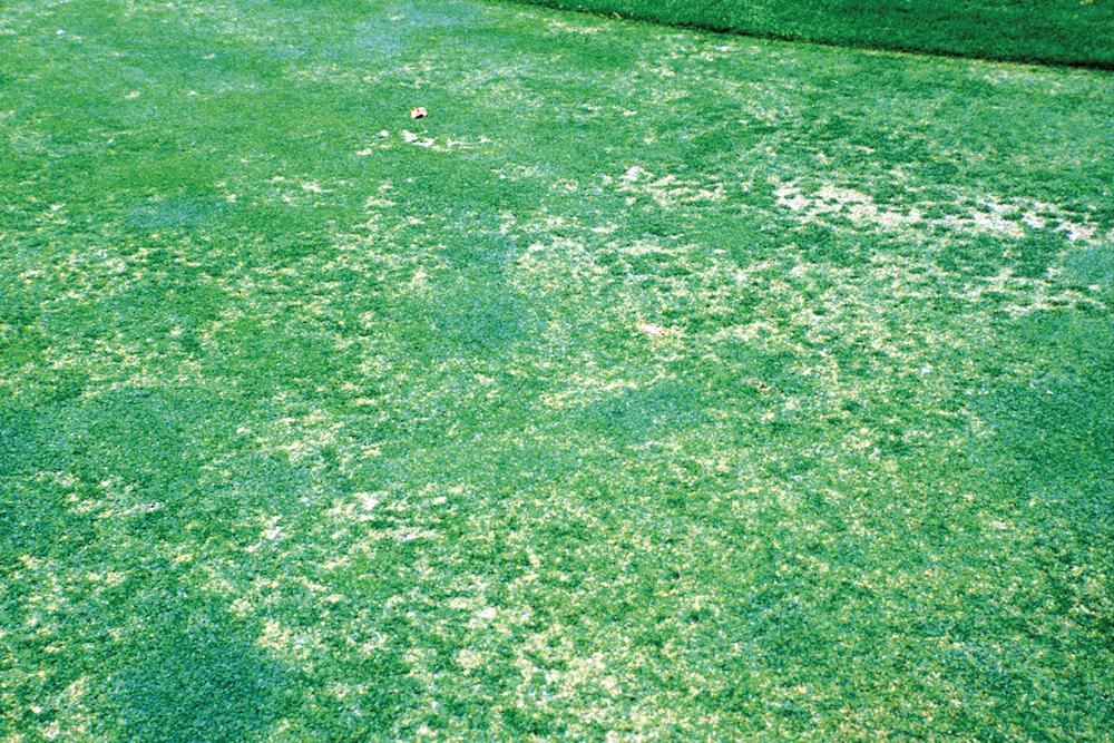
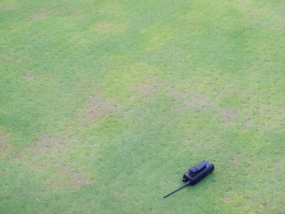
Foliar blight. Symptoms of anthracnose foliar blight include yellow or reddish brown discoloration of leaves (Fig. 3) and eventually a loss of shoot density (Fig. 4). It occasionally
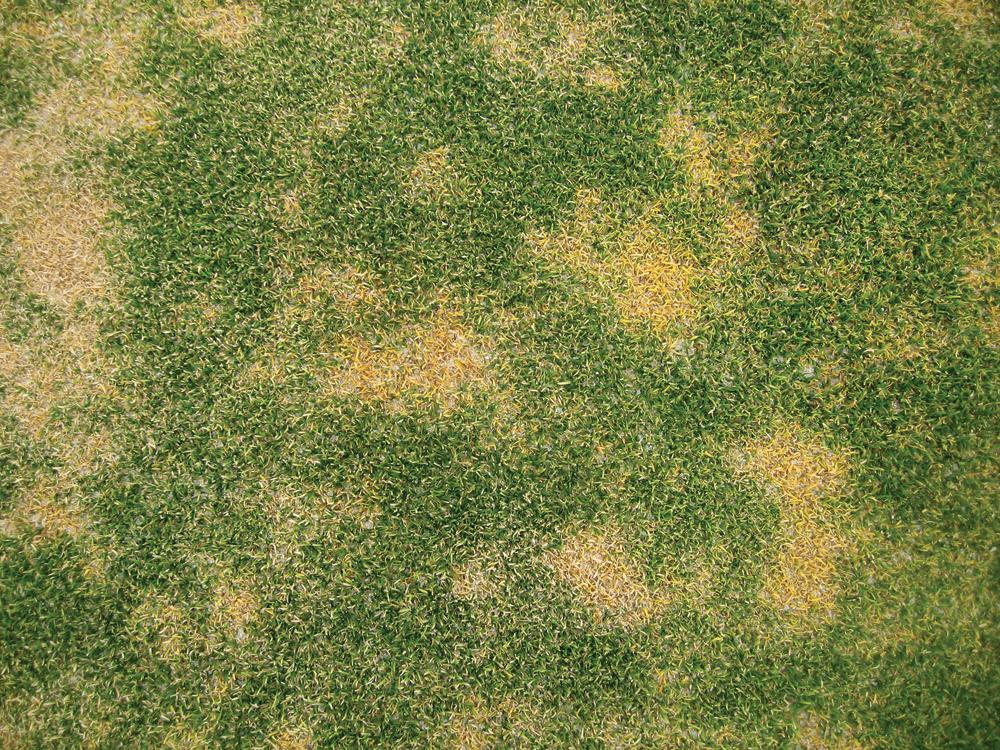
Fig. 3. Yellowing of Poa annua on a golf green caused by anthracnose. (Cour tesy J. Hempfling— © APS)
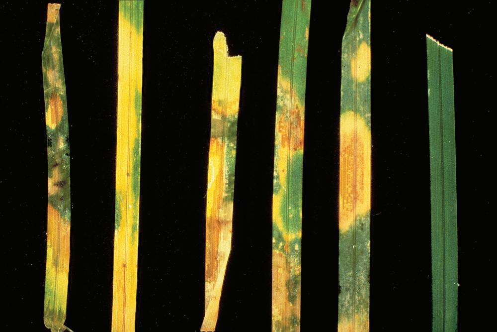
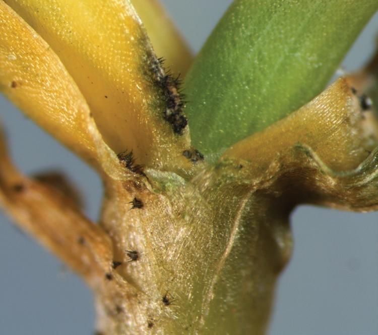
causes oblong, reddish brown leaf lesions (Fig. 5). The distinctive acervuli (fruiting bodies) of the pathogen, with their protruding black setae (hairlike structures), can be observed on green, yellow, or tan tissues of leaves, sheaths, or stems (Fig. 6). Large numbers of acervuli are often observed on dead plant tissue and are not necessarily diagnostic of an anthracnose outbreak. Therefore, it is important to carefully look for appressoria (Fig. 7) and acervuli on green or discolored tissue of living plants. Bipolaris sorokiniana and Curvularia lunata may be found in association with C. cereale, causing a decline in P. annua in the summer in some regions.
Basal rot. Anthracnose basal rot initially appears as orange or yellow spots about 6–12 mm in diameter. As crown tissues become infected, small patches of plants or scattered individual plants turn yellow and die (Figs. 8 and 9). Individual plants may have both green, healthy-appearing tillers and yellowish orange, infected tillers. The central, or youngest, leaf is often the last to change color. Removal of the sheath tissue to expose the infected stem base often reveals a water-soaked or black rot of crown tissues where roots and new buds are produced (Fig. 10). As the disease progresses, it develops into patches or
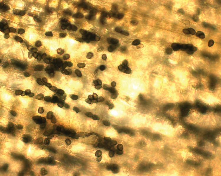
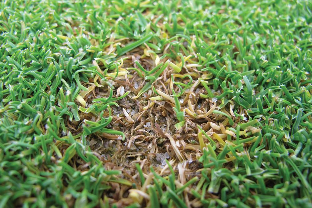
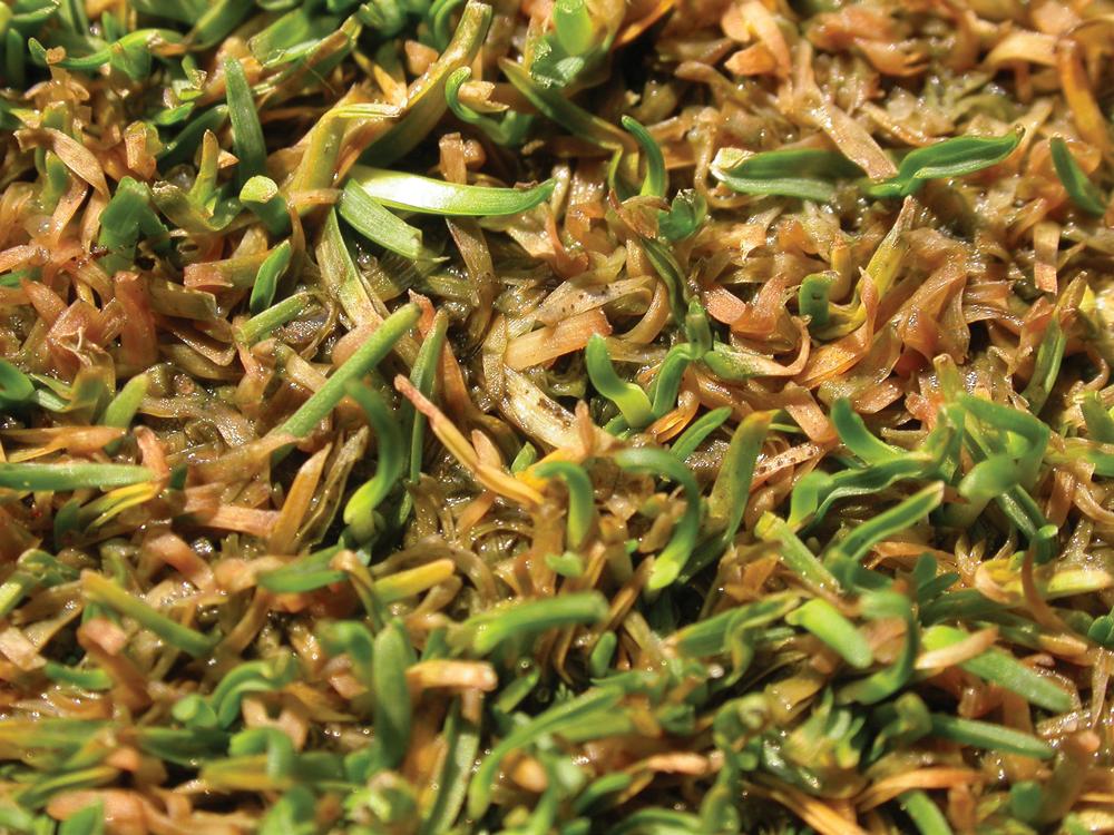
Fig. 4. Anthracnose of Poa annua on a golf fairway. (Cour tesy R. W. Smiley— © APS)
Fig. 5. Anthracnose foliar blight lesions on Poa pratensis. (Courtesy R. W. Smiley— © APS)
Fig. 6. Acervuli of Colletotrichum cereale on leaves and a stem of Poa annua. (Cour tesy B. B. Clarke— © APS)
Fig. 7. Appressoria of Colletotrichum cereale on a leaf of Poa annua. (Cour tesy L. Beirn)
Fig. 8. Small, yellow spot caused by anthracnose basal rot of Poa annua on a golf green in the spring. (Cour tesy B. B. Clarke— © APS)
Fig. 9. Foliar bronzing caused by anthracnose basal rot of Poa annua on a golf green in the spring. (Cour tesy J. E. Kaminski— © APS)
irregularly shaped patterns (Fig. 11) that are initially orange to reddish brown before turning yellow and dying. When large areas thin or die out completely (Fig. 12), the disease may be confused with melting- out or red leaf spot (see Bipolaris and Exserohilum Diseases and Pyrenophora (Drechslera) Diseases). Individual clones or genotypes of A. stolonifera may be attacked more severely than others, forming more-distinct patches in some cases.
In warm weather, anthracnose basal rot of P. annua and A. stolonifera appears as irregularly shaped patterns 1 m or more in diameter. Brown, circular patches similar to those caused by brown patch may also form. Plants often turn bright yellow or orangish yellow before dying; this symptom can be confused with symptoms of summer patch
When discoloration or thinning is first observed, managers and diagnosticians are advised to carefully examine exposed stem bases or stolons to look for appressoria or hyphopodia, grayish black infection mats, or rotting tissues (Fig. 7). Appressoria and hyphopodia are small (64–316 µm in diameter), dark brown or black, and dome shaped. Infection mats first appear
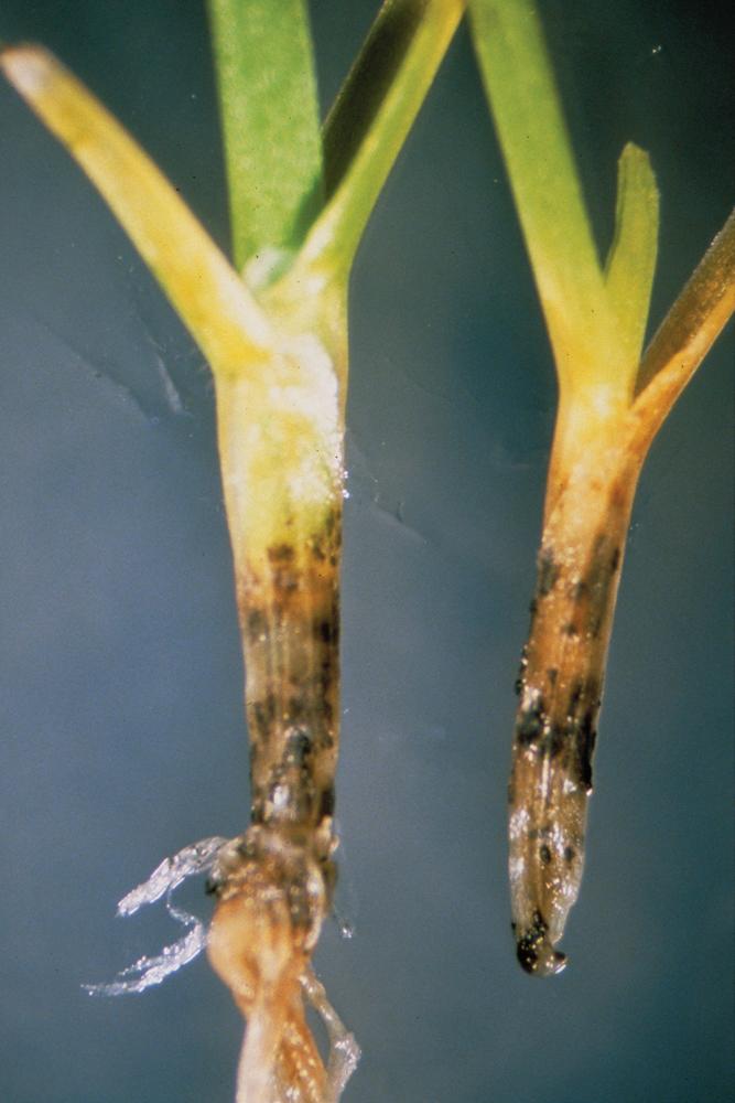
as small, pinhead-sized black specks (Fig. 13). The mats enlarge and turn black and may produce acervuli and setae (darkcolored bristle- or hairlike structures). Normally, there are no diagnostic lesions or signs of the pathogen on green leaf or sheath tissue during the early stages of basal rot. Turf managers often notice the discoloration only after stem tissues have begun to rot. In advanced stages, black aggregates of fungal mycelium, hyphopodia, infection mats, and acervuli are often present on infected stolons or stem bases. Acervuli and black mycelial aggregates on stem bases and stolons can be seen with a hand lens. In advanced stages of basal rot, acervuli develop on sheath or leaf tissue, at which point the plants generally die. Conclusive diagnosis of anthracnose basal rot depends on microscopic inspection of the conidia produced in acervuli. For unknown reasons, the pathogen seldom attacks both P. annua and A. stolonifera on the same green or even on the same golf course.
Causal Agent
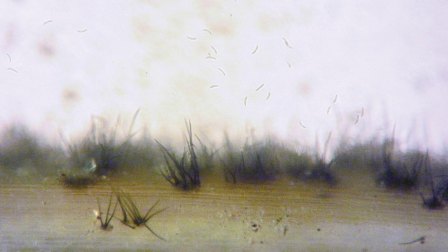
C. cereale causes anthracnose of cool-season turfgrasses. This fungus is very common and forms small, dark, elongated acervuli within infected tissues. The presence of black setae arising from the acervuli is often used as a rapid diagnostic criterion for anthracnose. The setae are long, dark brown or black, septate spines (Fig. 6) measuring 32–120 × 6–8 µm; they are wider at the bases and taper toward the tips. Conidia are one celled, crescent shaped, guttulate, and 4–34 µm long (most are 19–30 µm) (Fig. 14). The sexual stage (Glomerella graminicola) rarely occurs in nature.
Research with isolates of C. cereale from A. stolonifera and P. annua indicates a degree of host specificity. However, the
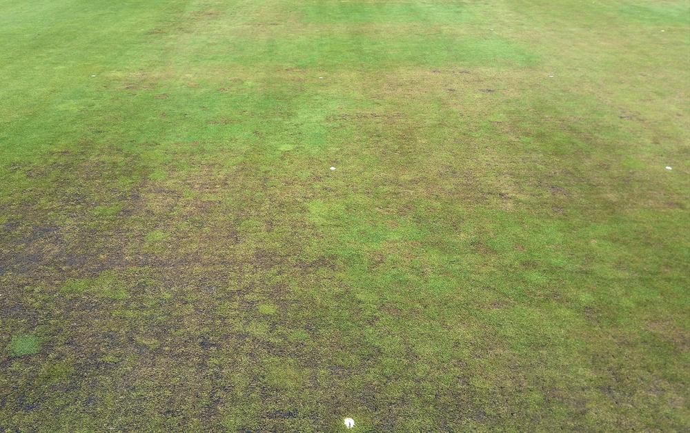

Fig. 10. Water- soaked, rotted, blackened lower stems of Poa annua caused by anthracnose basal rot. (Cour tesy P. J. Landschoot— © APS)
Fig. 11. Anthracnose basal rot on an Agrostis stolonifera putting green. (Cour tesy M. Tomaso- Peterson— © APS)
Fig. 12. Anthracnose of Poa annua on a golf green in August. Note that the dark gray to black patches have coalesced into a large area colonized by algae. (Cour tesy B. B. Clarke— © APS)
Fig. 13. Infection mat of Colletotrichum cereale. (Cour tesy P. H. Dernoeden— © APS)
occurrence of races of the pathogen in turfgrasses is inconclusive. The apparent genetic similarities among isolates from the same host may account for the attack of P. annua in mixed stands of P. annua and A. stolonifera at one location and the attack of only A. stolonifera in similar mixtures elsewhere. Similarly, the preferential attack on Festuca spp. in turfs in which fescue is mixed with P. pratensis could also be explained by the presence of physiologic races.
Two other fungi have also been associated with turfs diagnosed with symptoms of anthracnose: Microdochium bolleyi and Lectera colletotrichoides. Their importance as potential pathogens of turfgrass is unknown.
M. bolleyi is common in some regions and appears similar to C. cereale upon initial inspection. The fruiting structure of M. bolleyi is a sporodochium, which may not be easily distinguished from an acervulus, except that M. bolleyi does not form setae. Conidia of M. bolleyi are one celled, crescent shaped, and 5–9 µm long and resemble conidia of C. cereale
L. colletotrichoides has been isolated from Festuca rubra exhibiting symptoms and signs of anthracnose and was demonstrated to be pathogenic to Festuca spp. in greenhouse studies. It produces small, hemispherical sporodochia. Dark setae, about 100 µm long, surround the sporodochia and, like those of C. cereale, are clearly visible. Conidia are one celled, bean shaped, and about 5 µm long. Distinguishing between Lectera and Colletotrichum spp. requires diagnosis in a laboratory.
Disease Cycle
C. cereale is thought to survive unfavorable periods as mycelium in decaying plant debris or living host tissue. The fungus can penetrate foliage, stem, or root tissues of turfgrass plants growing under stressful conditions and can infect the plants when atmospheric humidity is high and leaves are wet. Acervuli form on blighted or necrotic tissue, and conidia are disseminated to uninfected plants by wind or water or mechanically by foot traffic or equipment. Conidia germinate within 2–6 hours and infect host tissue.
Epidemiology
Anthracnose is favored by overcast weather and other conditions that promote high humidity or allow excess water to remain in the leaf canopy. Conditions that weaken plants, including low mowing height, low fertility, drought stress, and extreme temperatures, can also stimulate anthracnose development. Symptoms may initially appear on mounds or other areas prone to moisture stress. Plants growing in soils deficient in phosphorus, potassium, or nitrogen are particularly susceptible to infection.
In P. annua, anthracnose can occur at almost any time of year. Anthracnose foliar blight generally occurs during periods of high-temperature stress in summer. Spring outbreaks of anthracnose basal rot in P. annua putting greens can be very
destructive. P. annua weakened by environmental stress or low mowing during the previous summer or by winter injury is particularly susceptible. On putting greens, deep vertical cutting practices that extensively wound plant crowns may enable C. cereale to be more invasive; however, practices such as topdressing, other cultivation practices, and aerification do not increase the severity of this disease.
Anthracnose is a common disease of A. stolonifera putting greens in the transition zone and subtropical climates and is likely encouraged by the physiological stresses imposed by these environments. Anthracnose basal rot is most common on A. stolonifera during hot, humid conditions and may be encouraged by excessive thatch accumulation and poor soil drainage. Basal rot often causes severe damage to A. stolonifera on putting surfaces and recovery is slow because the stolons and crowns are attacked. Anthracnose foliar blight is observed on A. stolonifera during prolonged periods of high heat or during periods of cloudy, wet weather.
Management
The effects of cultural practices on anthracnose in P. annua have been thoroughly investigated. Providing proper fertility, increasing mowing height, topdressing with sand, and avoiding drought stress are important measures for reducing disease severity. Turf should be fertilized to avoid deficiencies of nitrogen, phosphorus, and potassium. Nitrogen should not be applied at high rates during periods of drought or high-temperature stress. When applied frequently at low rates and in moderation, however, nitrogen and potassium help to reduce disease symptoms on putting greens and fairways in summer. A foliar nitrogen concentration of 3.4% (dry weight) is considered a minimum critical level for disease suppression on P. annua putting greens. Symptoms are more severe on P. annua grown at soil potassium levels below 50 ppm and less than 2% potassium in clipping tissue (dry weight). Disease symptoms are also more severe on P. annua that frequently experiences wilt stress. Water should be applied at levels sufficient to avoid both excessively wet and dry conditions. Light, frequent applications of topdressing sand throughout the growing season reduce disease symptoms and promote recovery of injured turf. On putting greens, midseason cultivation practices (e.g., solid tine aeration, vertical mowing, and grooming) do not enhance anthracnose severity, except for deep vertical cutting when the disease is active. The use of growth regulators that stress or weaken P. annua or delay recovery of injured turf should be avoided.
Observations of A. stolonifera support many of the same cultural recommendations for anthracnose management. Routine aerification and topdressing are important to minimize thatch accumulation and maintain soil drainage. During periods of summer stress, mowing height should be increased and mowing frequency reduced, and sufficient irrigation and fertility to support turf growth should be provided. Removal of trees and/ or installation of fans can help to improve overall turf health and reduce anthracnose development.
In temperate climates, reducing P. annua populations in favor of A. stolonifera can reduce anthracnose incidence. Modern creeping bentgrass cultivars bred for improved heat tolerance are typically less susceptible to anthracnose in transition zone environments.
Preventive applications of fungicides are recommended where the disease is a chronic problem, but the use of bestmanagement practices can substantially reduce fungicide inputs needed to control anthracnose. Strains of C. cereale that are resistant to one or more fungicides (e.g., benzimidazoles and quinone outside inhibitors) have been detected in the United States. Tank mixes can be used, or fungicides with different modes of action can be alternated. Sequential applications of active ingredients considered to have a high potential for fungicide resistance should be avoided.
Fig. 14. Agrostis stolonifera leaf with numerous acervuli and onecelled, guttulate conidia of Colletotrichum cereale. (Cour tesy M. Tomaso- Peterson— © APS)
Selected References
Avila-Adame, C., Olaya, G., and Köller, W. 2003. Characterization of Colletotrichum graminicola isolates resistant to strobilurin-related QoI fungicides. Plant Dis. 87:1426-1432.
Backman, P. A., Landschoot, P. J., and Huff, D. R. 1999. Variation in pathogenicity, morphology and RAPD marker profiles in Colletotrichum graminicola from turfgrasses. Crop Sci. 39:1129-1135.
Bailey, J. A., and Jeger, M. J., eds. 1992. Colletotrichum: Biology, Pathology and Control. CAB International, Wallingford, U.K.
Browning, M., Rowley, L. V., Zeng, P., Chandlee, J. M., and Jackson, N. 1999. Morphological, pathogenic, and genetic comparisons of Colletotrichum graminicola isolates from Poaceae. Plant Dis. 83:286-292.
Crouch, J. A., Clarke, B. B., and Hillman, B. I. 2006. Unraveling evolutionary relationships among the divergent lineages of Colletotrichum causing anthracnose disease in turfgrass and corn. Phytopathology 96:46- 60.
Danneberger, T. K., Vargas, J. M., Jr., and Jones, A. L. 1984. A model for weather-based forecasting of anthracnose on annual bluegrass. Phytopathology 74:448- 451.
Dernoeden, P. H. 2017. Creeping Bentgrass Management. 2nd ed. CRC Press, Boca Raton, FL. Hempfling, J. W., Schmid, C. J., Wang, R., Clarke, B. B., and Murphy J. A. 2017. Best management practices effects on anthracnose disease of annual bluegrass. Crop Sci. 57:602- 610.
Inguagiato, J. C., and Guillard K. 2016. Foliar N concentration and reflectance meters to guide N fertilization for anthracnose management of annual bluegrass putting green turf. Crop Sci. 56:3328-3337.
Inguagiato, J. C., Murphy, J. A., and Clarke, B. B. 2008. Anthracnose severity on annual bluegrass influenced by nitrogen fertilization, growth regulators, and verticutting. Crop Sci. 48:1595-1607.
Inguagiato, J. C., Murphy, J. A., and Clarke, B. B. 2009. Anthracnose disease and annual bluegrass putting green performance affected by mowing and lightweight rolling. Crop Sci. 49:1454-1462.
Inguagiato, J. C., Murphy, J. A., and Clarke, B. B. 2010. Anthracnose development on annual bluegrass affected by seedhead and vegetative growth regulators. Appl. Turfgrass Sci. 7. doi:10.1094/ ATS-2010- 0923- 01-RS
Inguagiato, J. C., Murphy, J. A., and Clarke, B. B. 2012. Sand topdressing rate and interval effects on anthracnose severity of an annual bluegrass putting green. Crop Sci. 52:1406-1415.
Khan, A., and Hsiang, T. 2003. The infection process of Colletotrichum graminicola and relative aggressiveness on four turfgrass species. Can. J. Microbiol. 49:433- 442.
Roberts, J. A., Inguagiato, J. C., Clarke, B. B., and Murphy, J. A. 2011. Irrigation quantity effects on anthracnose disease of annual bluegrass. Crop Sci. 51:1244-1252.
Roberts, J. A., and Murphy, J. A. 2014. Anthracnose disease on annual bluegrass as affected by foot traffic and sand topdressing. Plant Dis. 98:1321-1325.
Schmid, C. J., Clarke, B. B., and Murphy, J. A. 2017. Anthracnose severity and annual bluegrass quality as influenced by nitrogen source. Crop Sci. 57:S-285-S-292.
Schmid, C., Clarke, B. B., and Murphy, J. A. 2018. Potassium nutrition affects anthracnose on annual bluegrass. Agron. J. 110:2171-2179.
Smith, J. D. 1954. A disease of Poa annua. J. Sports Turf Res. Inst. 8:344-353.
Sutton, B. C. 1980. The Coelomycetes: Fungi Imperfecti with Pycnidia, Acervuli and Stromata. Commonwealth Mycological Institute, Kew, England.
(Prepared by R. W. Smiley, P. H. Dernoeden, and B. B. Clarke; revised by J. A. Murphy and B. B. Clarke)
Anthracnose Diseases of Warm- Season Turfgrasses
Tanso-byô (anthracnose) was described on Eremochloa ophiuroides in 2006 in Japan. Anthracnose of E. ophiuroides and Axonopus compressus was subsequently described in the
United States and China, respectively. Three additional Colletotrichum species have been isolated from Axonopus, Paspalum, and Zoysia spp., but pathogenicity to these grasses has not been documented.
Symptoms
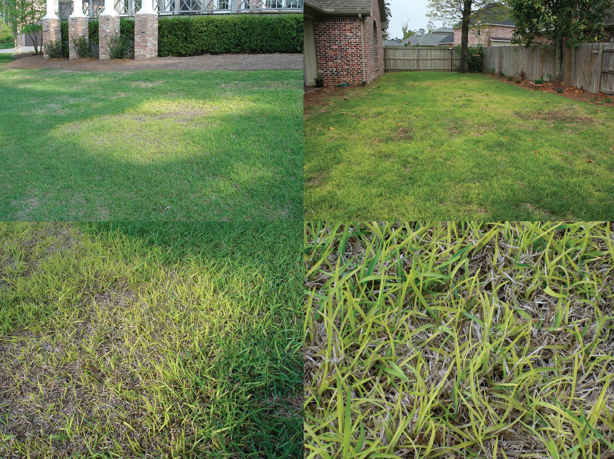
Anthracnose of E. ophiuroides affects the crown and leaf sheaths. The oldest leaf sheaths appear necrotic, while young leaf sheaths and leaves are chlorotic. Leaf blight progresses from the leaf tips to the bases of older leaves. Foliar lesions, as well as acervuli and setae, are absent on infected leaf sheaths. The stolons and roots are not affected. Irregular, diffuse patches of various sizes in the turf appear bright yellow or chlorotic. A loss of turf density may occur. Necrotic patches and subsequent bare areas develop and persist into the summer as plants die (Fig. 15).
Tanso-byô of E. ophiuroides is a foliar disease resulting in fusiform to ellipsoid necrotic lesions that develop a grayish white appearance as they mature. Acervuli and setae develop within lesions of infected tissues.
On A. compressus, conspicuous foliar lesions have pale brown centers surrounded by reddish brown borders. Lesions can coalesce to form large, irregular areas. Excessive moisture promotes infection of crowns and leaf sheaths, resulting in reddish brown areas within a turf stand (Fig. 16). Stolons and roots are not affected. Acervuli and setae develop in lesions and on associated infected leaves.
Causal Agents
Colletotrichum eremochloae infects E. ophiuroides. Light gray, septate mycelium may be observed colonizing the epidermal tissue of the inner leaf sheaths. Abundant, grayish brown to dark brown appressoria (12.5 × 12.5 µm) are produced in association with mycelium and serve as a diagnostic feature. In culture on potato dextrose agar (PDA), a 7- day- old colony produces mycelium that is gray on the inner half and white on the outer half. The perimeter of the colony has a distinct fluorescent yellow pigment when cultured in 12–16 hours of light per day. Conidia are hyaline, aseptate, falcate to fusiform, and average 23.4 × 5.6 µm.
C. caudatum, the causal agent of tanso-byô, produces acervuli and setae within necrotic lesions. The conidia are aseptate, hyaline, falcate to fusiform, and 20.5–38.5 × 2.8–4.2 µm. On PDA, the colony is grayish white to gray. The appressoria are grayish brown, ellipsoid to round, and 10.0–16.8 × 7.6–11.5 µm.
C. hainanense infects A. compressus. Falcate, aseptate, hyaline conidia (19.5–27.5 × 4–7 µm) are produced in acervuli. Appressoria are pale to brown, globose or lobate, and 10–16 × 8.5–11.5 µm.
C. axonopodi was isolated from A. affinis in Australia. In pure culture, mycelium is white, overlaid on brown mycelium with a pale tan orange pigment. Falcate to fusiform conidia average 24.2 × 4.2 µm. Appressoria are globose, ovoid or clavate, smooth or lobate to multilobate, and average 10.1 × 7.5 µm.
C. paspali was isolated from Paspalum notatum in Japan. The colony is whitish cream, and setae are absent. Conidia are falcate or fusiform and average 21.2 × 4.1 µm. Appressoria are variable in shape, averaging 12.3 × 9.9 µm.
C. zoysiae was isolated from leaves of Zoysia tenuifolia in Japan. On PDA, mycelium is gray to light gray and pale olivaceous gray at the margin. Caudate conidia are aseptate, hyaline, falcate, and 25–47.5 (including appendage) × 4.0–6.5 µm. Appressoria are elongate to round, lobate to multilobate, black, and 17.0–22.5 × 10.0–14.5 µm.
Colletotrichum spp. can be accurately identified by their morphological characteristics and by analyses of the nuclear rDNA internal transcribed spacer region, the 5' end of the DNA lyase gene (Apn2), the 3' end of Apn2, the 5' end of the matingtype gene Mat1-2 (Mat1/Apn2), and the manganese superoxide dismutase (Sod2) gene.

Disease Cycle, Epidemiology, and Management
Anthracnose diseases of warm-season grasses are polycyclic and primarily occur when plants are transitioning out of winter dormancy, but they can persist into the summer months if disease pressure is high. Cool, springlike temperatures and abundant moisture favor disease outbreaks. Conidia and infected plant parts are disseminated by mowing and water movement.
The turf recovers during the summer months when conditions are favorable for growth.
To date, little research has been conducted on control measures; however, fungicides that are effective on C. cereale may prove efficacious for controlling anthracnose of warm-season turfgrasses.
Selected References
Crouch, J. A. 2014. Colletotrichum caudatum s.l. is a species complex. IMA Fungus 5:17-30.
Crouch, J. A., Clarke, B. B., White, J. F., Jr., and Hillman, B. I. 2009. Systematic analysis of the falcate-spored graminicolous Colletotrichum and a description of six new species from warm-season grasses. Mycologia 101:717-732.
Crouch, J. A., and Tomaso-Peterson, M. 2012. Anthracnose disease of centipedegrass turf caused by Colletotrichum eremochloae, a new fungal species closely related to Colletotrichum sublineola. Mycologia 104:1085-1096.
Fuke, K., Hozumi, N., Enami, Y., Matsuura, K., and Tajimi, A. 2006. Anthracnose of centipede grass caused by Colletotrichum caudatum. J. Gen. Plant Pathol. 72:74-75.
Tomaso-Peterson, M., Crouch, J. A., and Balbalian, C. 2012. Anthracnose of centipedegrass turf. Appl. Turfgrass Sci. 9. doi:10.1094/ ATS-2012-1126- 01-MG
Zhang, W., Damm, U., Crous, P. W., Groenewald, J. Z., Niu, X., Lin, J., and Li, Y. 2020. Anthracnose disease of carpetgrass (Axonopus compressus) caused by Colletotrichum hainanense sp. nov. Plant Dis. 104:1744-1750.
(Prepared by M. Tomaso-Peterson)
Fig. 15. Eremochloa ophiuroides lawns exhibiting irregular, diffuse areas of yellow, thinning turf caused by anthracnose. (Cour tesy M. Tomaso- Peterson— © APS)
Fig. 16. Axonopus compressus with anthracnose symptoms caused by Colletotrichum hainanense. (Cour tesy W. Zhang— © APS)
Ascochyta
Leaf Blight
Leaf spots and blights caused by Ascochyta and Neoascochyta spp. are common on many grasses. On turfgrasses, the principal symptom is a leaf blight that seldom causes widespread damage. These fungi are common colonizers of senescent leaf tissue and inflorescences.
Symptoms and Signs
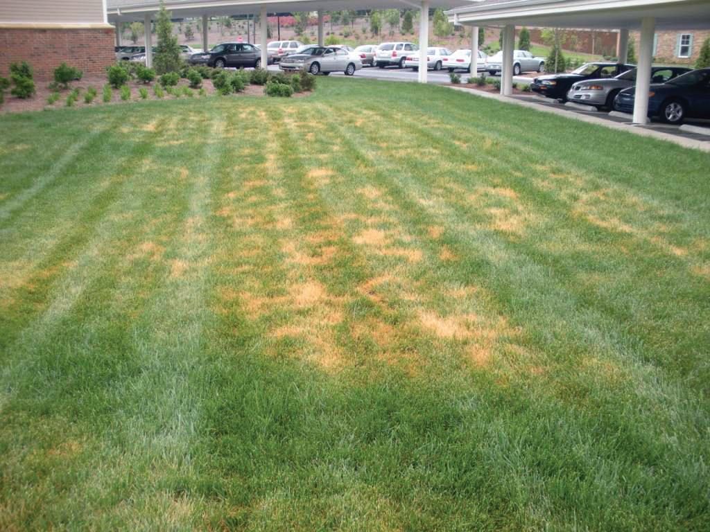
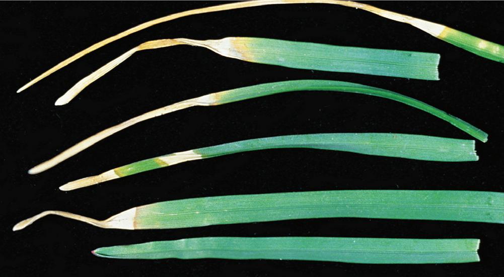
Large areas of turf may assume a uniformly blighted appearance, or localized areas of heavy infection may cause a patchy appearance (Fig. 17). Individual leaves generally start dying back from the tips (Fig. 18), and uniform lesions affecting the entire leaf blade may develop. When leaf spots are present, they are dark brown and circular to elliptical and appear similar to those caused by Pyrenophora and Bipolaris spp. Less commonly, there may be minute chlorotic, purple, or brown areas where the pathogen has directly penetrated the leaf mid-blade. These spots enlarge into bleached lesions that girdle the leaf and cause it to become chlorotic and die. Round, brown pycnidia (fruiting bodies) of the pathogen generally form in the lesions or blighted leaf tissue (Fig. 19), where they can be seen with a hand lens.
Causal
Agents
More than 15 species of Ascochyta and Neoascochyta are associated with turfgrasses as pathogens or saprophytes; refer to the appendix, Diseases of Turfgrasses, for a comprehensive list. On the basis of recent phylogenetic studies, certain species have been moved to the newly erected genus Neoascochyta. Several species associated with turfgrasses have not been studied and remain in Ascochyta. Morphological characteristics
of pycnidia and conidia are highly variable and overlap across species; thus, they are no longer considered useful for identification to species.
Pycnidia formed by most Ascochyta and Neoascochyta species are 70–200 µm in diameter and yellow brown, rust brown, or brick red. Conidia (Fig. 20) are mostly hyaline to pale or straw yellow, fusoid, one to three septate, and 2.5–5.5 × 7–21 µm. Conidia of A. rhodesii are 8–10 × 24–32 µm, and this species forms pycnidia that are dark brown to black and up to 240 µm in diameter.
Disease Cycle
The pathogen survives as mycelium and pycnidia in host debris. The fungus generally enters plants soon after the grass is mowed and begins to grow from the freshly cut ends toward the leaf bases. Pycnidia form in leaf spot lesions or blighted tissues and produce conidia, which are released during wet periods and splashed or mechanically dispersed to uninfected leaves.
Epidemiology
Ascochyta leaf blight occurs throughout the year. Like Septoria and Stagonospora leaf spots, it can occur in late winter

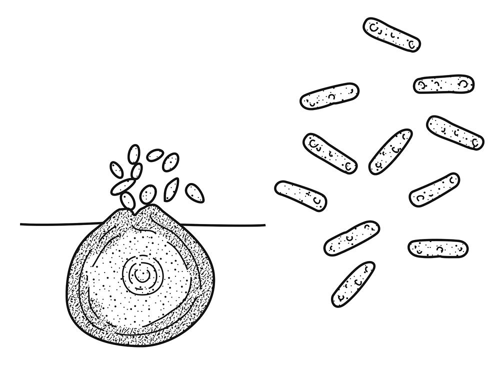
Fig. 17. Ascochyta blight in a Festuca arundinacea landscape. (Cour tesy J. P. Kerns— © APS)
Fig. 18. Leaf necrosis caused by an Ascochyta sp. on Poa pratensis. (Cour tesy R. W. Smiley— © APS)
Fig. 19. Pycnidia of an Ascochyta sp. in a leaf spot on Poa pratensis. (Cour tesy R. W. Smiley–© APS)
Fig. 20. Conidia emanating from a pycnidium (left) and conidia (right) of an Ascochyta sp. (Cour tesy L. P. Tredway— © APS. Drawing by S. McKinley.)
