


Bacterial wilt was first reported by Thomas Burrill in 1890, and the causal pathogen was first described as Bacillus solanacearum in 1896. Since then, bacterial wilt has become one of the most destructive plant diseases worldwide in semitropical and tropical climatic regions. This disease is also referred to as potato brown rot; bacterial wilt of tomato, tobacco, and eggplant; and Moko disease on banana. It affects at least 50 different families representing more than 200 different plant species. This disease has been reported on roses from only three countries: the Netherlands, Switzerland, and Korea.
Symptoms
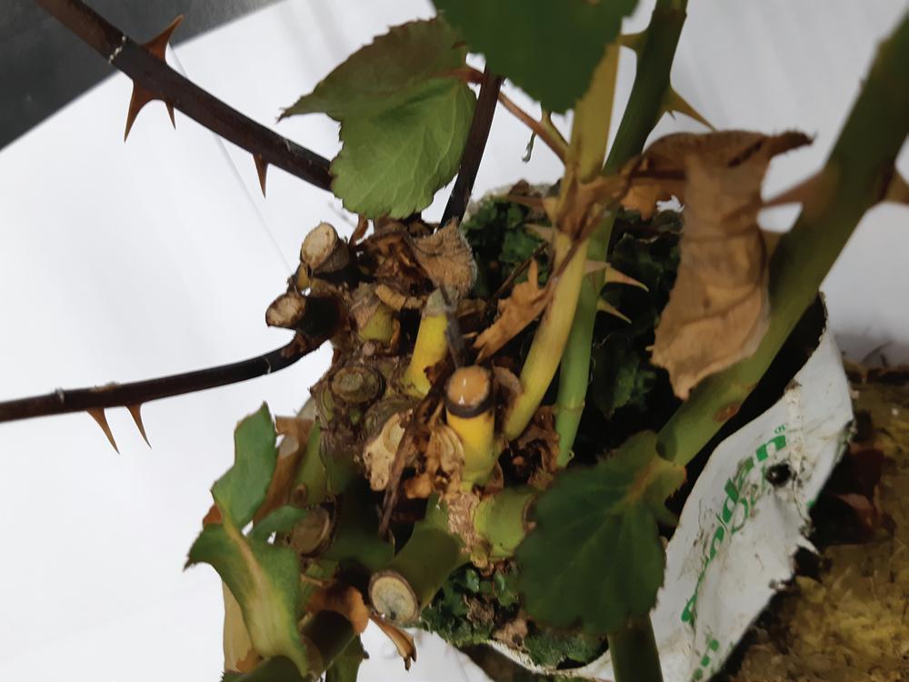
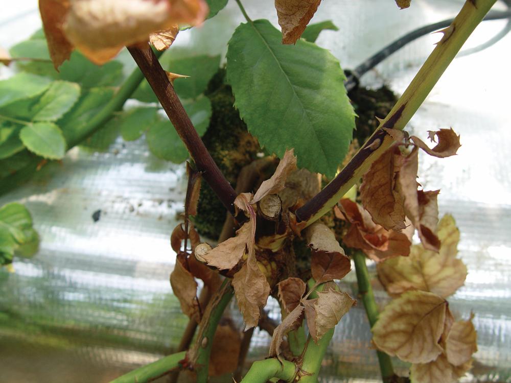
The most common symptoms are wilting or blighting of lower leaves on an infected plant (Fig. 8), production of bacterial ooze from a cut stem (Fig. 9), and discoloration with black necrosis on the stem just above the soil or rockwool surface (Fig. 10). Lower leaves that exhibit wilting or blighting at late stages of the disease are typically yellow at the early stages. Milky bacterial ooze can be released from a cut site, and streams of it are observed when cut stems are placed in pure water. Depending on the rose type, for example, on the cultivar Red Eagle, wilting in the form of a crook sometimes occurs on flower stalks (Fig. 11). All these symptoms are typical of a soilborne bacterium that infects plant roots through wounds, root tips, and cracks at sites of lateral root emergence. Subsequently, the invading bacterium colonizes the root cortex, enters the xylem vessels,
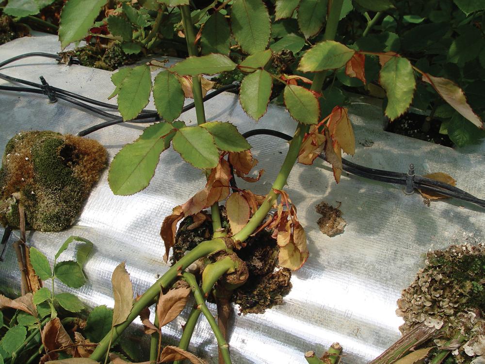
reaches the stem, and finally goes up into the aerial parts of the plants through the vascular bundle.
Causal Organism
Erwin F. Smith first described the causal organism of bacterial wilt as Bacillus solanacearum in 1896. The genus was changed to Burkholderia in 1992 and then to Ralstonia in

1995 on the basis of DNA analysis. Thus, the causal pathogen of bacterial wilt is Ralstonia solanacearum. It is a member of the class Betaproteobacteria in the family Ralstoniaceae and consists of gram-negative, aerobic, rod-shaped cells that are motile with one to four polar flagella. Its colonies on complex agar medium are irregularly round, fluidal, mucoid, and cream colored. Because of its variation in virulence on diverse hosts, R. solanacearum is posited to be a heterogeneous species now referred to as the “R. solanacearum species complex” (RSSC).
Traditionally, RSSC strains are divided into five races based on host range and six biovars based on utilization of three disaccharides and three hexose alcohols. In addition, the four phylotypes resulting from analysis of DNA sequences are divided into phylotype I originating in Asia, phylotype II from America, phylotype III from Africa, and phylotype IV from Indonesia, Australia, and Japan. RSSC strains are divided into 57 sequevars based on multilocus phylogeny with three individual genes: the endoglucanase gene egl, the DNA repair gene mutS, and the flagellin gene fliC. Classification based on these traditional and phylogenetic criteria for RSSC strains shows that R. solanacearum causing bacterial wilt on roses belongs to race 1, biovar 3, phylotype I, and sequevar 1-33.
Etiology and Epidemiology
R. solanacearum is favored by conditions of warmth and high humidity and is thus widespread in semitropical and tropical regions. It can persist in loamy and sandy, rather than clay, soils for extended periods. When populations on host tissues and in soils are large, it can persist for as long as 4 years in soil.
R. solanacearum strains that affect solanaceous crops can penetrate via wounds and natural openings beyond the root surface and enter the cortex. The characteristics of pathogenic strains on roses are similar to those of strains affecting solanaceous species, but less is known regarding infection routes and behaviors. However, because roses are propagated with plant material including roots and aerial parts such as shoots, outbreaks can occur even when propagation materials harbor only low populations of the pathogen. Because propagation plant materials are frequently traded between different continents, such as European countries and Korea, bacterial wilt on roses can be introduced into new areas where wilting symptoms on roses were never previously observed.
Management
In rose cultivation areas that have been free of R. solanacearum, the possible routes of natural dispersal of the pathogen include latently contaminated propagation plant materials. Thus, border quarantine regulations must be enforced when
plant materials are traded. R. solanacearum is known to infect more than 40 rose cultivars, but there are significant variations in the symptoms produced on leaves and stems depending on the cultivar. Therefore, although bacterial wilt on roses was reported relatively recently, resistant cultivars must be developed.
Bergsma-Vlami, M., van de Bilt, J. L. J., Tjou-Tam-Sin, N. N. A., Westenberg, M., Meekes, E. T. M., Teunissen, H. A. S., and Van Vaerenbergh, J. 2018. Phylogenetic assignment of Ralstonia pseudosolanacearum (Ralstonia solanacearum phylotype I) isolated from Rosa spp. Plant Dis. 102:2258-2267.
Fegan, M., and Prior, P. 2005. How complex is the “Ralstonia solanacearum species complex”? Pages 449- 461 in: Bacterial Wilt Disease and the Ralstonia solanacearum Species Complex. C. Allen, P. Prior, and A. C. Hayward, eds. American Phytopathological Society, St. Paul, MN.
Kado, C. I. 2010. Vascular wilt diseases and their pathogens. Pages 143-152 in: Plant Bacteriology. American Phytopathological Society, St. Paul, MN.
Kim, Y. S., Lim, S. R., Kim, J.-W., Lee, H.-J., and Park, D. H. 2019. First report of Ralstonia solanacearum phylotype I causing bacterial wilt on Rosa spp. in Korea. Plant Dis. 103:1407.
Mansfield, J., Genin, S., Magori, S., Citovsky, V., Sriariyanum, M., Ronald, P., Dow, M., Verdier, V., Beer, S. V., Machado, M. A., Toth, I., Salmond, G., and Foster, G. D. 2012. Top 10 plant pathogenic bacteria in molecular plant pathology. Mol. Plant Pathol. 13:614- 629.
Tjou-Tam-Sin, N. N. A., van de Bilt, J. L. J., Westenberg, M., BergsmaVlami, M., Korpershoek, H. J., Vermunt, A. M. W., Meekes, E. T. M., Teunissen, H. A. S., and Van Vaerenberg, J. 2017. First report of bacterial wilt caused by Ralstonia solanacearum in ornamental Rosa sp. Plant Dis. 101:378.
Yabuuchi, E., Kosako, Y., Yano, I., Hotta, H., and Nishiuchi, Y. 1995. Transfer of two Burkholderia and Alcaligenes species to Ralstonia gen nov.: Proposal of Ralstonia pickettii (Ralston, Palleroni and Doudoroff 1973) comb. nov., Ralstonia solanacearum (Smith 1896) comb. nov. and Ralstonia eutropha (Davis 1969) comb. nov. Microbiol. Immunol. 39:897-904.
(Prepared by D. H. Park and I. Lee)
Crown gall has been reported in all parts of the temperate world on a wide range of plants. Besides species of Rosaceae, crown gall occurs on members of more than 60 dicotyledonous plant families. Inoculation experiments with a single isolate resulted in infection of plants in additional families, but many of these rarely, if ever, develop disease except when inoculated. Some common hosts are almond, apple, apricot, blackberry, cherry, chrysanthemum, euonymus, nectarine, peach, pear, plum, poplar, raspberry, rose, Shasta daisy, walnut, and willow; crown gall is rarely found on conifers. Individual isolates possess host specificity, but this is not dependent on the host from which the isolate was obtained. Other species of the bacteria occur on Ficus spp., grape, and occasionally on raspberry. The disease occurs worldwide on roses and is common in the United States.
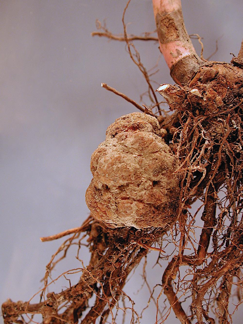
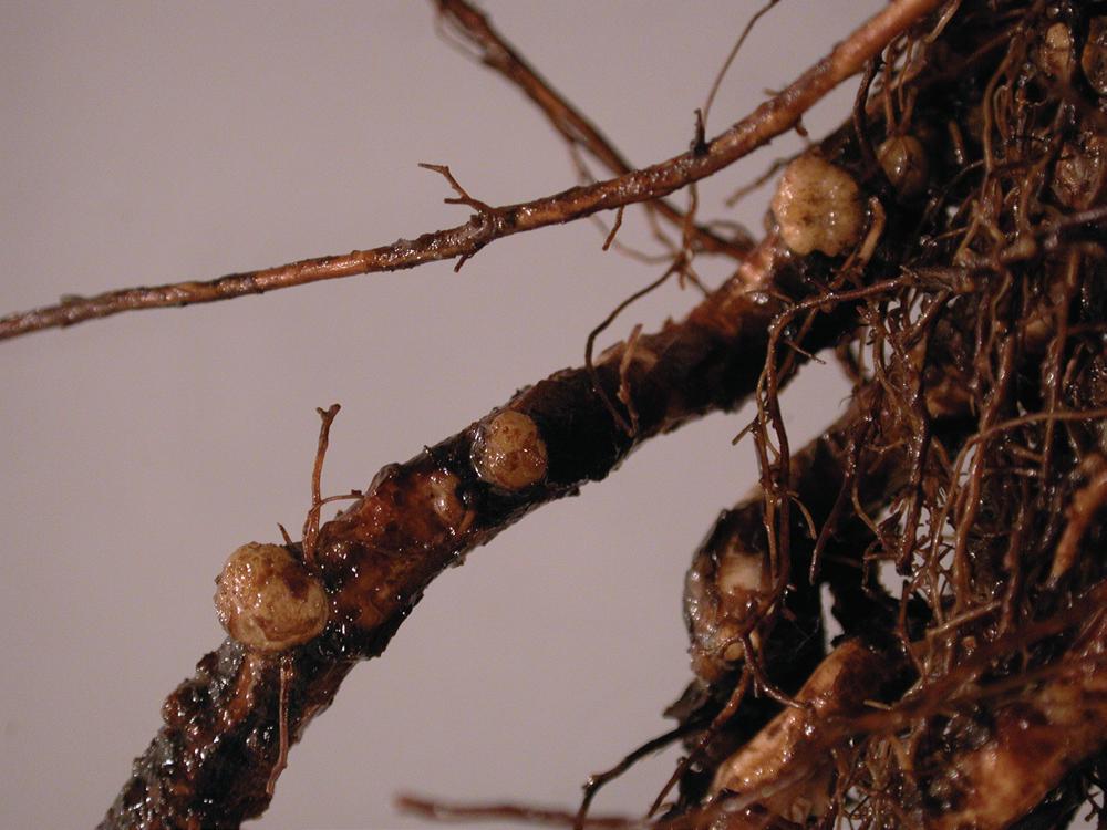
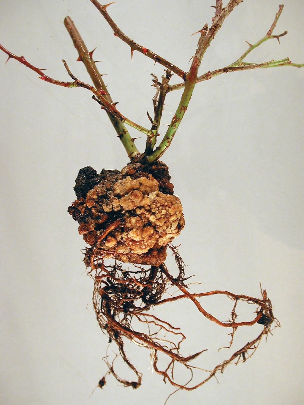
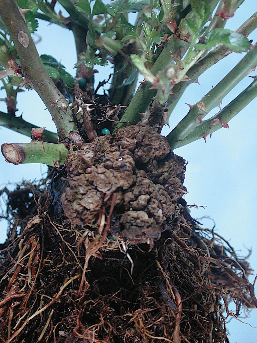
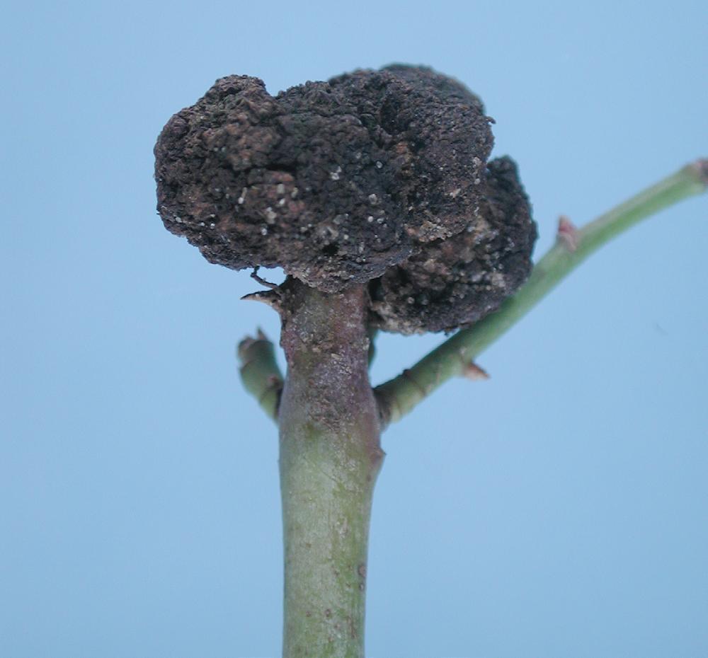
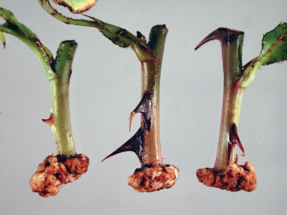
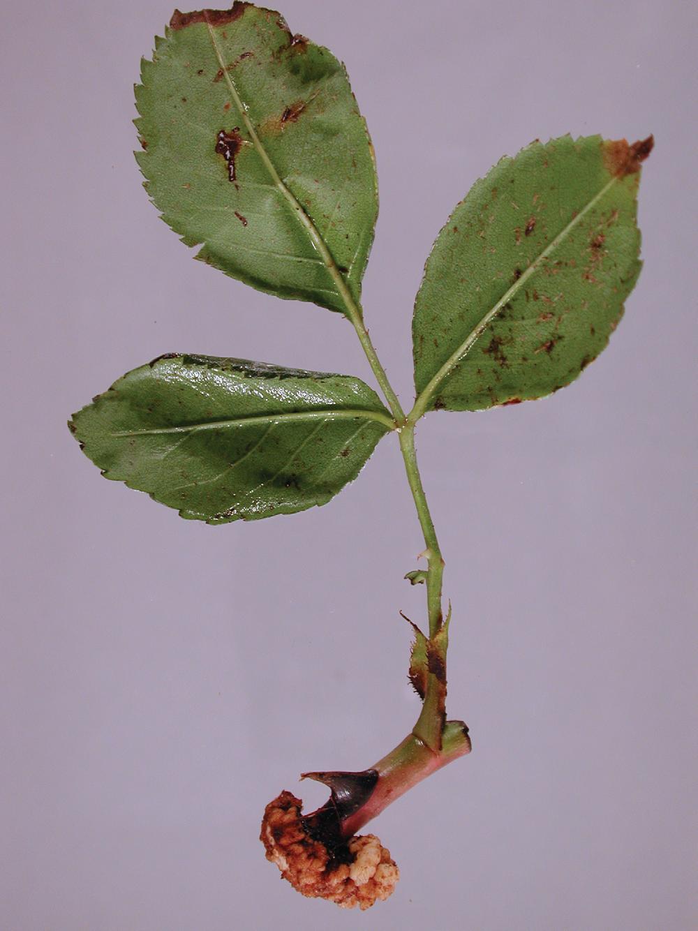
Crown gall is a disease primarily of the parenchyma tissue, starting with a rapid proliferation of cells in meristematic tissues and the formation of more or less convoluted, soft or hard overgrowths called galls or tumors. The galls may be formed on the roots (Figs. 12 and 13) or more commonly at or just below the soil surface in the basal stem or crown region (Figs. 14 and 15) and where tissues were wounded (Fig. 16). The galls are usually round with rough, irregular surfaces. They first appear as small protuberances on the root surface (Fig. 13). Galls may vary from 0.5 to 8 cm in diameter, depending on
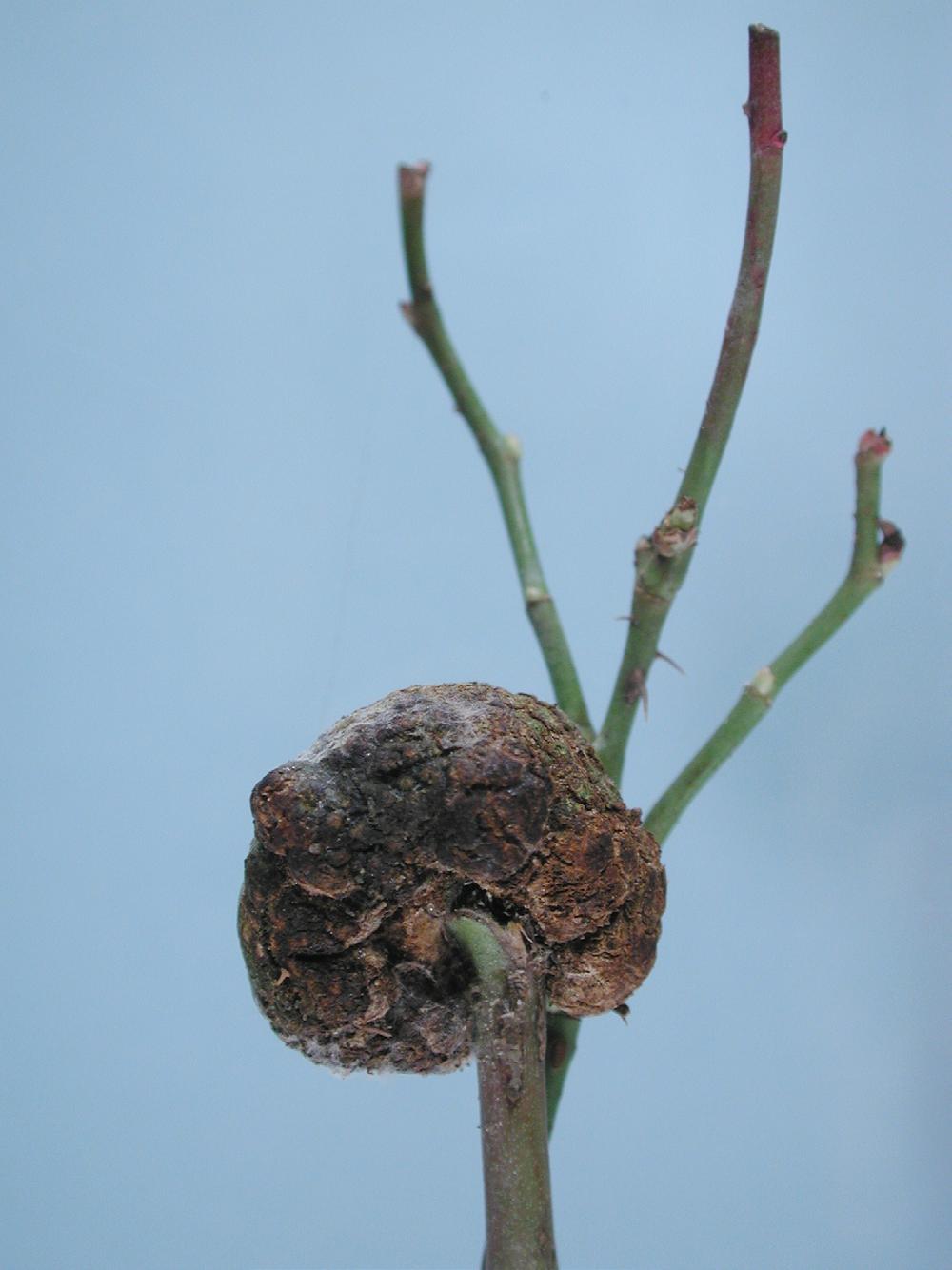
the plant’s age and vigor and the time since infection. Young, actively developing galls are light green or nearly white (Fig. 12), and the tissue is soft. As they age, galls become darkened, woody (Fig. 17), and friable. Outer portions of galls can slough off with age because of normal rotting, weathering, or the action of other microorganisms. The appearance of galls at the bases of cuttings or at graft unions makes it difficult to differentiate disease (Fig. 18) from callus tissue (Fig. 19). Symptoms of crown gall differ from those of hairy root in that hairy root galls do not become woody and small roots develop from hairy root galls and later form large masses of fibrous roots (see Hairy Root).
The economic importance of crown gall is primarily in rose propagation and production; minor amounts of disease can
agrobacteria.
losses of 30% from crown gall have been reported. Losses in ornamentals nurseries are less well documented, but it is reasonable to assume a similar impact.
often be tolerated in landscapes and home gardens. Plants with crown gall should not be used for propagation in commercial settings, and affected cuttings should be discarded. Presence of the disease in production settings can necessitate stringent sanitation efforts, leading to lost revenue from discarded plants and time and resources spent on cleaning propagation facilities. In East Africa, losses in rose production facilities caused by crown gall have been as great as 60%. In fruit tree nurseries,
Sometimes, plants severely infected by the crown gall bacteria show few adverse effects, while in other instances, plants with a few galls may decline and die. It is likely that the location of the galls determines the effect of the disease on plants; a single gall at the base of a plant may be more detrimental than several galls on the canes and roots.
Crown gall is caused by Agrobacterium tumefaciens, which traditionally was subdivided into phenotypic groups called bi-
ovars. Biovar 1 included the crown gall-inducing isolates; biovar 2 isolates induced hairy root disease and were termed A. rhizogenes; and biovar 3 was associated primarily with grapes and was subsequently renamed A. vitis. A. tumefaciens biovar 1 is now recognized as a polyphyletic group containing multiple subspecific groupings termed “genomospecies.” The nomenclature of agrobacteria has been changed repeatedly since 1907, when it was recognized that bacteria caused galls. The bacteria were placed in the genus Agrobacterium in 1940. In 2001, a proposal was put forward to combine gall-forming bacteria in the genus Agrobacterium with the closely related genus Rhizobium. Under this proposal, both pathogenic (A. tumefaciens) and nonpathogenic (A. radiobacter) bacteria should be called Rhizobium radiobacter. This was contested, and various arguments have been put forth for upholding various nomenclatural schemes based on multilocus sequence analysis, DNA:DNA hybridization, extrachromosomal topologies, and other means. Gall-forming bacteria currently are placed in Agrobacterium, Rhizobium, Pararhizobium, and Allorhizobium. More recently, whole-genome analyses of nearly 1,500 bacteria in the family Rhizobiaceae, which includes the above genera, suggests that differentiation of seven genera within that family is not supported and that all are congeneric. Clearly, the nomenclature remains in flux. Here, the vernacular term “tumorigenic agrobacteria” is used for gall-forming isolates. In general, the term “nonpathogenic agrobacteria” is used for those isolates that do not cause disease and the term “biovar system,” which groups isolates based on biochemical phenotypic characters, is used, recognizing that there are isolates that do not fit conveniently within that system. The majority of crown gall- causing isolates obtained from rose are biovar 2. Additional novel isolates have been recovered from rose, including a single pathogenic isolate, originally isolated in 1960 in South Africa from a tumor found on a hybrid tea rose (Rosa × hybrida); the name Agrobacterium rosae was proposed for the isolate. Isolates recovered in Iran from rose galls were deemed sufficiently diverse from known agrobacteria to be considered new genomospecies, although these results are based on only four-gene phylogenies.
Agrobacteria are gram-negative, non-spore-forming, rodshaped cells, 0.5–1.0 × 1.2–3.0 µm, and are motile, each with one to six flagella arranged around the circumference of the cell, often at one end. Ability to cause disease is determined by the presence of a transferable tumor-inducing or root-inducing (sometimes both) plasmid. Different genotypes may be recovered from a single gall, and tumor-inducing or root-inducing plasmids are not restricted to a particular species of gall- or root-forming agrobacterium or biovar.
Etiology and Epidemiology
Bacterial activity is greatest during the summer months, and tumor growth occurs fastest at 24–27°C. The pathogen enters plants through wounds, either natural or caused by pruning, grafts, mechanical injury from cultivation, heaving of frozen soils, chewing insects, or the emergence of lateral roots. Galls become visible 45 days to 36 months after infection.
Plant cells, in response to wounding, form compounds that attract tumorigenic agrobacteria, which, after entering a plant, transfer a section of plasmid-borne DNA into the nuclear genome of the plant cell. This transforms normal cells into tumor cells, which express, in addition to other compounds, the plant hormones auxin and cytokinin. Once the cells are transformed, the abnormal proliferation of tumor cells becomes an autocatalytic process that continues independently of the bacterium. Cells immediately surrounding the transformed cells usually divide rapidly. New growth centers may also form and contribute to the distorted structure of the gall. The orientation and size of proliferating cells vary, and vascular elements may differentiate as a continuation of existing xylem elements or as discontinuous groups. The result is a gall composed of a more or less disorganized mass of hyperplastic and hypertrophic tissues.
In rose, agrobacteria are systemic, and tumorigenic agrobacteria have been recovered from roots, stems, and canes, although the bacteria are unevenly distributed within a plant. Agrobacteria attach to the walls of the xylem, where they remain until stimulated by a wound.
Anatomy of the host vascular tissue, host vigor, stage of growth, environmental conditions, and bacterial isolate all influence the rate of gall development. Infections may remain latent, such that infected plants remaining in the field or in cold storage may not show symptoms until late the following spring. The bacteria are generally more abundant between cells located at or near the surfaces of developing galls. Thus, pruning tools that cut through galls can become contaminated with the bacteria, spread them to cut surfaces of subsequently pruned plants, and thereby induce new infections. As few as 100 tumorigenic bacterial cells applied to a wound can induce a gall.
Bacteria shed from galls can be transported by moving soil or water. In the absence of host plant roots, populations of pathogenic bacteria in soil gradually decrease; however, the pathogen may survive in soil for as long as 3 years. Propagation from and movement of infected plants is a major means of disease spread.
Research indicates that rootstocks may differ in susceptibility to tumorigenic agrobacteria, but the studies were superficial in that they were done with only a few isolates and without specifying the genomospecies used. No rootstock is known to be immune.
Several recommended practices reduce the occurrence of crown gall. First, only disease-free plants should be used. Since plants with latent infections appear to be disease free even in the presence of the pathogen, propagation should be from tested elite material to ensure freedom from infection. Second, injury to the roots and crown during planting and cultivating should be avoided. Third, roses should be planted in soil that has been steam-sterilized or has no history of crown gall. Common soil fumigants do not control the pathogen. Fourth, infected plants in nurseries should be removed as soon as galls are observed. If possible, soil from around the area of infected plant roots should be removed to ensure that all galls and infected roots are discarded. And fifth, cutting and pruning tools should be washed thoroughly with soap and water and disinfested between plants. Quaternary ammonium compounds are effective on hard surfaces when given sufficient exposure time.
Soil solarization has been effective in killing agrobacteria in the upper 20 cm of soil and can reduce populations, but it is ineffective against those present deeper in the soil profile. Bacterial populations can regrow once a susceptible plant is replanted to the site.
Biological control of crown gall by the antagonistic agrobacterium strain K84 is effective against some isolates of tumorigenic agrobacteria. Sensitive strains of tumorigenic agrobacteria do not produce crown galls when wounds are treated with K84. Strain K84 contains a plasmid that carries genes for both production of the antibiotic agrocin 84 and protection from agrocin 84, which is lethal to sensitive agrobacteria. An improved biocontrol product contains the strain K1026, which carries a mutation that prevents conjugal transfer of the plasmid to other bacteria. Otherwise, a pathogenic isolate could obtain the plasmid and become able to produce the toxin and protect itself from its effect.
These two biocontrol products are effective only when sensitive strains of agrobacteria are present and when used in a preventive manner. They have no ability to eradicate the pathogen from an infected plant and are ineffective when the bacteria are already present in the vascular system.
Strain K84 has been used successfully with roses in Australia, New Zealand, and Spain but is not widely effective in the United States. It is possible that tumorigenic agrobacteria
isolates immune to K84 already exist in some fields. Strain K1026 has been used successfully in Australia and is once again available in the United States. An oil and water emulsion containing 2,4-xylenol and meta- cresol has been effective when painted directly on galls. However, tumor growth often recurs, and in rose, where tumorigenic agrobacteria infection is systemic, this type of treatment is ineffective.
Selected References
Cooksey, D. A., and Moore, L. W. 1982. High frequency spontaneous mutations to agrocin 84 resistance in Agrobacterium tumefaciens and A. rhizogenes. Physiol. Plant Pathol. 20:129-135.
de Lajudie, P., and Young, J. P. W. 2017. International Committee on Systematics of Prokaryotes Subcommittee for the Taxonomy of Rhizobium and Agrobacterium: Minutes of the meeting, Budapest, 25 August 2016. Int. J. Syst. Evol. Microbiol. 67:2485-2494.
Jones, D. A., and Kerr, A. 1989. Agrobacterium radiobacter strain K1026, a genetically engineered derivative of strain K84, for biological control of crown gall. Plant Dis. 73:15-18.
Kerr, A. 1980. Biological control of crown gall through production of agrocin 84. Plant Dis. 64:24-30.
Kuzmanović, N., Puławska, J., Smalla, K., and Nesme, X. 2018. Agrobacterium rosae sp. nov., isolated from galls on different agricultural crops. Syst. Appl. Microbiol. 41:191-197.
López-López, M. J., Vicedo, B., Orellana, N., Piquer, J., and López, M. M. 1999. Behavior of a virulent strain derived from Agrobacterium radiobacter strain K84 after spontaneous Ti plasmid acquisition. Phytopathology 89:286-292.
Mafakheri, H., Taghavi, S. M., Puławska, J., de Lajudie, P., Lassalle, F., and Osdaghi, E. 2019. Two novel genomospecies in the Agrobacterium tumefaciens species complex associated with rose crown gall. Phytopathology 109:1859-1868.
Massey, L. M. 1950. Crown gall. Am. Rose Annu. 35:145-153.
Penyalver, R., Vicedo, B., and Lopez, M. M. 2000. Use of the genetically engineered Agrobacterium strain K1026 for biological control of crown gall. Eur. J. Plant Pathol. 106:801-810.
Ryder, M. H., and Jones, D. A. 1991. Biological control of crown gall using Agrobacterium strains K84 and K1026. Aust. J. Plant Physiol. 18:571-579.
Schroth, M. N., and Hildebrand, D. C. 1968. A chemotherapeutic treatment for selectively eradicating crown gall and olive knot neoplasms. Phytopathology 58:848-854.
(Prepared by R. K. Horst; revised by M. L. Putnam)
The hairy root (rhizogenic) agrobacteria were reported by Hildebrand to infect roses in the northeastern United States in 1934 and again in 1937. The disease was of minor importance until 1952–1956, when serious losses were experienced in commercial rose production in southern California. Although rhizogenic agrobacteria cause significant problems in hydroponic production, the incidence of infection in rose, which is not produced hydroponically, has diminished and remains insignificant.
Symptoms
Hairy root is characterized by swellings on stems or roots below the soil surface and usually at wounds, such as disbud scars or the ends of cuttings. The swellings are firm but not woody, as in crown gall, and discrete roots protrude from the swellings (Fig. 20). Large masses of fibrous roots 2–25 cm long are later formed. In storage under cool, moist conditions, many white roots are sometimes formed from the “hairy root” areas. Usually, no characteristic symptoms appear on aerial plant parts. However, in the second year after planting, infected plants have been observed to grow slowly and may die. The death rate increases during the third and fourth years after planting.
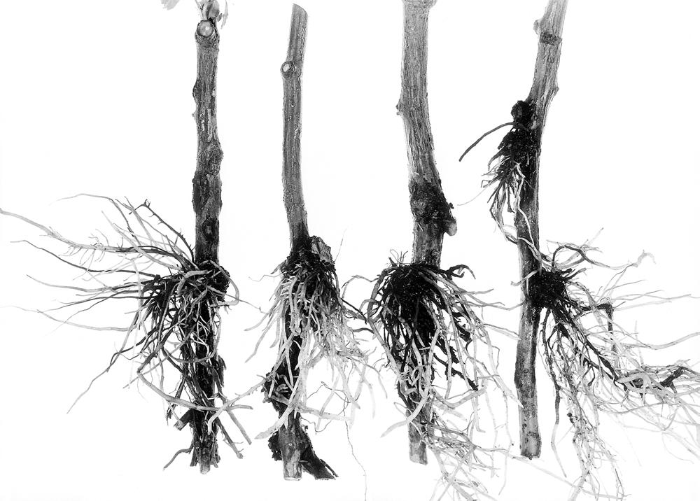
Fig. 20. Hairy root, caused by rhizogenic agrobacteria. Note the discrete, fibrous roots protruding from the swellings or galls. (Cour tesy R. K. Horst. Reproduced by permission—© Cornell University)
In 2010, the International Committee on Systematics of Prokaryotes recognized that the hairy root-inducing biovar 2 isolates of Agrobacterium, known as A. rhizogenes, were sufficiently distinct to be their own species and should be called Rhizobium rhizogenes. The term “rhizogenic agrobacteria” is used here to refer to isolates that cause hairy root. Rhizogenic isolates carry a transmissible root-inducing plasmid on which the genes for pathogenicity are located. Root-inducing plasmids are not restricted to biovar 2 isolates, however, and may be found in bacteria with a biovar 1 chromosomal background. Plants in 49 families have been found to be susceptible upon inoculation with rhizogenic agrobacteria, but the number of families that are affected without inoculation is closer to 10.
The maximum development of hairy root occurs when host growth is optimal. The disease is more severe at a soil temperature of 20°C than at 26°C. Infection is initiated when a plant is wounded in the presence of the pathogen, which is soilborne. Genes from the root-inducing plasmid are integrated into the genome of an infected host cell, resulting in the production of auxin and cytokinin and the formation of an abnormal proliferation of roots. The bacteria remain in infected roots and can persist in plant debris and soil. Infection occurs when plants are replanted into soil in which diseased plants had recently been living. Movement of water through infested soil likely also transmits the bacteria; hairy root disease can become widely distributed in hydroponic production systems.
There is no cure for infected plants; hence, all management hinges on prevention. If hairy root disease is found in a production setting, affected plants should be rogued and discarded, followed by strict sanitation to disinfect all potentially infested materials. Steam sterilization is an effective preplanting soil treatment, but soil fumigation does not effectively control this disease. The biocontrol strains K1026 and K84, marketed as Agrobacterium radiobacter, are not labeled for control of hairy root.
Selected References
Hildebrand, E. M. 1937. Infectious hairy root on rose. Plant Dis. Rep. 21:86-87.
Kado, C. I. 2010. Plant Bacteriology. American Phytopathological Society, St. Paul, MN.
Lindstrom, K., and Young, J. P. W. 2011. International Committee on Systematics of Prokaryotes, Subcommittee for the Taxonomy
of Rhizobium and Agrobacterium: Minutes of the meeting, 7 September 2010, Geneva, Switzerland. Int. J. Syst. Evol. Microbiol. 61:3089-3093.
Munnecke, D. E., Chandler, P. A., and Starr, M. P. 1963. Hairy root (Agrobacterium rhizogenes) of field roses. Phytopathology 53:788-799.
Porter, J. R. 1992. Host range and implications of plant infection by Agrobacterium rhizogenes. Crit. Rev. Plant Sci. 10:387- 421.
White, F. F., and Nester, E. W. 1980. Hairy root: Plasmid encodes virulence traits in Agrobacterium rhizogenes. J. Bacteriol. 141:1134-1141.
White, F. F., and Nester, E. W. 1980. Relationship of plasmids responsible for hairy root and crown gall tumorigenicity. J. Bacteriol. 144:710-720.
(Prepared by R. K. Horst; revised by M. L. Putnam)
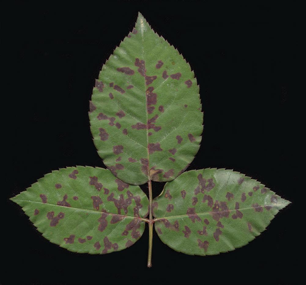
Xanthomonas leaf spot was first observed on shrub roses collected from production nurseries or retailers or received by plant diagnostic clinics in Florida and Texas between 2004 and 2010.
Symptoms
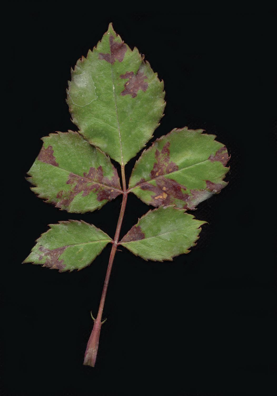
Symptoms of the disease include small, angular to circular, water-soaked lesions that form between leaf veins, along the leaf midrib, or in the leaf margins surrounding hydathodes (Fig. 21). Lesions, which may be surrounded by slightly chlorotic halos, eventually become necrotic. The lesions may occur in different parts of the plant, including foliage, fruit, and stems. As disease progresses, lesions coalesce (Fig. 22), and blighting and premature defoliation can occur.
Causal Organism
The disease- causing agent was originally described as Xanthomonas sp. pv. rosa, later reclassified as Xanthomonas euvesicatoria based on whole-genome sequence analysis. Isolates from rose form a distinct clade within Xanthomonas group 9.2. In cross-inoculation experiments, Rosa spp. and Rhaphiolepis indica (Indian hawthorn) have been reported to be susceptible to rose isolates, indicating that the host range is possibly lim-
ited to the family Rosaceae. Xanthomonads are gram-negative, rod-shaped bacteria, each with a single polar flagellum. Colonies are mucoid and convex and display a yellow pigment when grown on yeast extract–dextrose–calcium carbonate or nutrient agar media.
Like many other bacterial pathogens, spread and infection of xanthomonads are favored by splashing water and high temperatures; thus, disease development is more common during the summer months when temperatures are high and overhead irrigation is used to provide water to plants. The pathogen enters plants through wounds or natural openings because it is not able to penetrate tissues directly. X. euvesicatoria is capable of both long-term survival (overwintering) in dead plant material and short-term survival in water and soil.
Some preventive measures, including proper watering and sanitation practices, may help to control Xanthomonas leaf spot. Since water facilitates the diffusion of the bacterium and wounds provide entry points for the pathogen, it is recommended to avoid overwatering, to irrigate at times when foliage can dry rapidly, and to minimize mechanical injury to plants. Infected plants should be destroyed and plant debris (which represents an inoculum source) should be removed promptly when symptomatic plants are discovered within the garden or nursery. The application of copper-based products, which have bactericidal activity, can be effective when used as a preventive measure. Biological control products that contain Bacillus sp. can also be used to protect roses from infection because the beneficial bacterium acts as a competitor on the plant surface.