
IN THIS ISSUE
MESSAGE FROM THE CHAIR
2 Navigating Growth, Excelling in our Missions
FEATURES
Green Radiology
6 Leading the Way to Decarbonize Radiology
8 Sustainable Aviation Fuels (SAF) reduce carbon footprint for MRI scanner delivery
Patient Care
9 The Promise of Mid-Field MRI
11 Ultrasound-Guided Injection Relieves Pain, Enables Mom to Care for Newborn
14 Imaging Technologists Redefine Patient Care
17 Talking Tech & the Human Touch
17 A Magic Moment at ZSFG
A Sci-Fi Novel and a Children's Book
18 Just City by Olga Tymofiyeva, PhD
20 Radiology for Kids by John Mongan, MD, PhD
PEOPLE
Academic Affairs
21 Promoting Academic Success in Radiology and Biomedical Imaging
23 Faculty Promotions 2023
24 New Faculty
28 Faculty Retirements
30 Roster, Clinical and Research Faculty
33 Faculty Awards
Education
36 Training the Future Radiology Workforce: Collaboration, Independence, Mentorship
37 Welcome: Class of 2027
40 Residency Program Graduates, Class of 2023
41 Clinical Fellows & Clinical Instructors 2023-24
43 Master of Science in Biomedical Imaging
44 Continuing Medical Education
Wellbeing and Professional Climate
45 Culture and Connection
46 RIDR Summer Intern Symposium
48 Diversity in Radiology: Resident Xiao Wu, MD, Discusses Research on the Topic
49 Radiology Investigators Awarded NIH Diversity Supplements in 2022-23
Honors and Awards
50 Highlights from Across the Department
Staff Leadership
54 Amy Pradhan, MPH, MS Appointed Chief Administrative Officer
Alumni
55 Gratitude for Our Alumni
56 Margulis Society Career Evening Connects Radiology Alumni and Trainees
57 In Memoriam
56 Alumni News
THE YEAR IN PICTURES
60 The Many Faces of Radiology
HOW WE WORK
65 Clinical Operations & Strategy
67 Informatics: Focus on AI Evaluation & Deployment
68 Capital Investments in Our Clinical & Research Imaging Fleet
69 MRI Safety Practices: “Every Minute, Every Day”
CLINICAL DIVISIONS
Clinical Stories
71 Pediatric Imaging Without Anesthesia at UCSF is Highly Successful and Cost-Saving
73 A Labor of Love: Emergency Radiology at ZSFG
74 Breast Cancer Screening Guidelines Prompt Comments from Bonnie Joe, MD, PhD, and Kimberly Ray, MD
75 For Some Prostate Cancer Patients, PSMA PET May Be Better for Initial Staging of Disease
76 Mount Zion is Once Again an Inpatient Hospital Where Imaging Plays an Important Role
78 Neuroendovascular Surgery: Reflections on the First Year
79 Brain Focused Ultrasound: A New Era of Image-guided Neuro-intervention
80 Research Lab Group Photos
RESEARCH
Research Achievements and Highlights
81 Vice Chair's Overview
Specialized Resource Groups
83 Chemistry, Probes, and Molecular Therapy
85 Advanced Imaging Technologies: HMTRC
90 Intelligent Imaging: AI partnership with Christian-Albrechts-Universität
91 Center for Intelligent Imaging hosts Scientific Sessions
Research Groups
92 Body Imaging
93 Advanced Imaging of Peripheral Pain Generators
94 Imaging Cortical Bone Vasculature
95 Neuroimaging Research Group
98 Vascular and Cardiac Research Group
Research Stories
99 Parkinson’s Disease and Deep Brain Stimulation
100 Faculty Awarded $3.93M Team Science Grant
101 Celebrating 20 Years of Innovation at the Center for Molecular and Functional Imaging
Front Cover: 4D Flow MRI, image courtesy of Jesse Courtier, MD
Back Cover: Shan McBurney-Lin, MD, MBA, Residency Class of 2026
Executive Editor: Christopher Hess, MD, PhD
Managing Editor: Rita Gaber
Writers: Arleen Bandarrae, Francis Horan, Rebecca Wolfson
Photography: Elisabeth Fall, James Ramirez, Andrea Rowe, Marco Sanchez
Design: Victoria Odson
© 2024 The Regents of the University of California
MESSAGE FROM THE CHAIR
Shared values create the elements of our thriving community.” “
Dear Friends,
The contents of this annual magazine serve as a written record of achievements over the past year that reflect the contributions of many exceptional individuals and teams in the UCSF Department of Radiology & Biomedical Imaging. From my perspective, they also represent the tangible realization of our department’s deep and longstanding commitment to excellence and leadership in how, when, why, and where imaging is applied to improve health. They provide context for what has fueled our success in the past and for what is to come in 2024 and beyond.
I am frequently asked how our department persistently remains at the vanguard of radiology and imaging science year after year. Without exception, my first answer always emphasizes our people, and how our shared values create the elements of our thriving community. To this end, the term ‘force multiplier,’ although it originated in military science, is especially relevant to the 1,100+ faculty, trainees, healthcare staff, and administrators who work in our department. I would posit that there are more force multipliers amongst our ranks than any other academic organization and as a result, the impact and influence of our work is amplified within the field of radiology.
The second ingredient that underpins our success is an immutable commitment to the academic mission. Without an academic mission, our organization would take the path of regression to a mediocre mean. When asked to opine on ‘standard of care’ issues in clinical affairs, my response is identical in every instance: we don’t practice the standard of care at UCSF, we define the standard of care. Our unfaltering focus on our academic mission –advancing imaging science and training future thought leaders – serves as the fundamental strategy that sets us apart from other organizations and enables us to continually elevate the bar for what is standard of care.
Christopher Hess, MD, PhD

As clinical radiologists, an expanding demand for our expertise and an increasingly central role for imaging in healthcare has driven unprecedented growth across our clinical specialties. Growth helped to mitigate new financial challenges faced by healthcare in 2023, but only through the hard work of our people and stretched resources. We have hired more than 50 faculty in less than three short years, with at least six new members joining us in 2024. Our continued success will require not only further recruitment, but also career pipeline development, major capital renewal, and increased investment in our academic programs. Clinical expansion can only be fueled by proportionate growth in academic mission. We will continue to support the cultivation of our faculty as national and international leaders in their specialties, so that they can magnify reciprocal relationships between training, clinical practice, and research.
Later this year UCSF will acquire, from Dignity Health, St. Mary’s Medical Center (SMMC) and Saint Francis Memorial Hospital (SFMH). These are positioned as community hospitals with leadership teams that report to the President of the UCSF Health Care Network. Our department, at least in the short term, will not assume the operation or management of current imaging operations at SMMC and SFMC. However, UCSF Health as an entity must grow and innovate to overcome expected financial stresses and space constraints that it faces in an increasingly competitive healthcare environment. Boosting our growth through the Dignity purchase is an economical way to quickly add space and nearly 600 beds while construction for our new Parnassus hospital continues through 2032.
As educators, we have redoubled our focus on meeting the growing need for training academic leaders in Radiology and imaging science. For 10 years running,

Doximity has ranked our diagnostic residency #1 in the US, thanks to our stellar residents and the reputation for excellence on the part of our alumni. We have been fortunate to hire 13 of our outstanding fellowship graduates as faculty since 2021. Our T32 program, now in its 18th year and with three fellows in its current cohort, enjoys a nearly 70% faculty position placement rate in the best academic institutions across the United States. After a pandemic hiatus, CME is back with new destinations and refreshed course offerings that are specifically designed to share and disseminate excellent clinical care across the broader community of radiologists. And our graduate and many research programs continue to cultivate some of the most innovative minds in imaging science around the world.
As researchers, our investigators and their teams continue along a robust trajectory of success by all metrics, including securing nearly $60M in NIH funding in fiscal year 2023, publishing more than 575 peer-reviewed manuscripts, and expanding programs in image analysis, image acquisition, and device development. Our annual Imaging Research Symposium returned as an in-person event, and was highly attended and full of exciting talks and posters that showcased the interdisciplinary and groundbreaking work by our faculty and researchers. Similarly, our annual Research Conference held at the Asilomar conference grounds brought our research community together outside of UCSF to spotlight the department’s most successful research programs across body systems and technology domains.
A source of both excitement and consternation across the world, research this year also ushered in rapid advancement in artificial intelligence (AI). Generative AI has taken center stage as a computational force multiplier and potential disruptive innovation poised to dramatically impact multiple industries. Chancellor Sam Hawgood’s annual state of the university address spotlighted the central role that UCSF is poised to play in adopting AI to effect the modern transformation of healthcare. I have no doubt that radiologists and scientists in our department will play a critical role in the development and translation of AI to improve the quality and practice of healthcare when it comes to imaging. (By the way, I should note that no large language models were used in writing this text.)
I hope you enjoy this issue of Images 2023. You’ll see stories that show how we do ‘force multiplication’ in a myriad of ways, amplifying our effectiveness as individuals through partnerships and collaborations, and celebrating our successes along the way. As always, our tripartite mission to teach, heal, and discover grounds us in the rewards and satisfactions of doing right by our patients, our people, and the local, national, and international stakeholders who count on us to lead imaging innovation.

Christopher Hess, MD, PhD
Alexander R. Margulis Distinguished Professor, and Chair Department of Radiology and Biomedical Imaging

GREEN RADIOLOGY
Leading the Way to Decarbonize Radiology
By Rebecca Wolfson
In September 2020 the skies of San Francisco turned orange and ashes fell on the sidewalks. The city was surrounded by wildfires. The smoky air obstructed the sun, stinging peoples’ eyes and throats, and posed a particular hazard for sensitive groups such as children and people with asthma. For Sean Woolen, MD, assistant professor of radiology, this moment served as a wake-up call.
“It was an almost dystopian experience, tending to COVID-19 patients in the hospital after being exposed to hazardous air quality due to nearby wildfires,” Woolen said. He was aware of the healthcare sector’s contribution to greenhouse gas emissions and its adverse effects on public health due to climate change. As a result, he felt compelled to take action. Woolen is now leading efforts to ensure that UCSF’s imaging fleet is the first carbon-neutral imaging fleet in the world.
While the exact size of radiology’s carbon footprint remains unknown, the energy-intensive nature of its equipment makes it a significant contributor to the healthcare sector’s 10 percent share of carbon emissions. A single MRI, for example, expends the energy equivalent to powering 12 US homes and one CT unit equates to three US homes.
Woolen collaborated with like-minded individuals at UCSF and Siemens to form a partnership in 2021 aimed at improving sustainability in radiology. Together, they assessed energy consumption, carbon footprint, and actively sought ways to reduce environmental impact.
A Simple Solution with Profound Savings
In April 2023, a retrospective evaluation of MRI power and energy consumption published in the journal Radiology, revealed a groundbreaking solution. Co-authored by Woolen and Christopher Hess, MD, PhD, UCSF chair of radiology, the study demonstrated that switching MRIs from idle to off during the night reduced power consumption by 25-to-33 percent. Implementing an “Eco Power mode” further decreased the power draw by 28%. By powering off and implementing “Eco Power mode,” the US healthcare system could save up to $11 million and reduce CO2 emissions equivalent to 50,000 tons.
“The idea of powering down is so simple,” Woolen said. He hopes that hospitals and healthcare systems will begin to make the change and has been speaking at conferences across the country on the subject. An encouraging aspect of reducing energy consumption is that it enables simultaneous cost reduction while paving the way for sustainable healthcare and decarbonization. “I hope the potential savings also serves as a catalyst for the healthcare sector to explore more eco-friendly approaches to practicing medicine,” Woolen said.
The department’s sustainable strategy revolves around the 3Ms: Measure, Message, and Manage. The “Measure” phase seeks to gain a better understanding of the environmental impact of imaging and encompasses the entire lifecycle of scanners, from production and delivery to operations. “We have to measure each of those in order

to communicate the impact we can have when designing steps to manage our carbon footprint.” [See related article: Sustainable Aviation Fuels (SAF) reduce carbon footprint for MRI scanner delivery for more on how we’re addressing the delivery and supply chain stage.]
A Collaborative Approach
“A single sector can’t do this alone,” Hess said. “To reduce the energy use of healthcare and radiology, it requires a coordinated strategy among academics, government, industry and patients.”
To develop “Eco power mode” a multidisciplinary team came together under a shared vision of working to make a more sustainable healthcare system. Experts in radiology, energy monitoring and technology, high-voltage electricity, facility engineering, and informatics convened to launch the software update that can be installed on all brands of MRI to reduce energy usage.
The team harnessed existing energy technologies to create a novel energy monitoring program, enabling them to better understand energy consumption and identify methods that decrease usage to ensure a more sustainable path forward.
“I think the most important part of the initiative for me is identifying achievable and simple changes that make a significant impact,” Woolen said.
Alastair Martin, PhD, professor of radiology and the department’s associate chair for capital projects, noted that there are a lot of other pieces of sustainability beyond energy use that are critical. “We’re focused on energy not because we think it’s the only important thing, it’s just an area where we see we can have a great impact.”
“I see our department serving as a key leader in this sustainability space,” Hess said. “We have the people in the department to tackle complex issues like this. It allows us to be pioneers and set an example for other medical disciplines.”
Learn more at https://tiny.ucsf.edu/CarbonFootprint

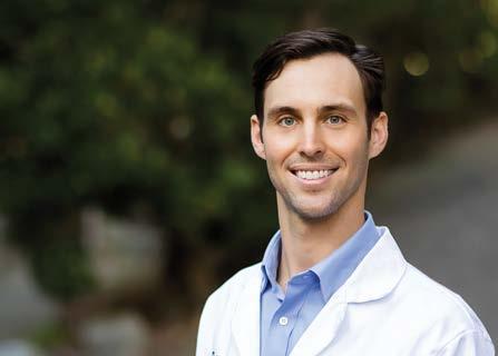
Sean Woolen, MD, assistant professor of radiology
Sustainable Aviation Fuels (SAF) Reduce Carbon Footprint For MRI Scanner Delivery
By Rita Gaber
In another first for Green Radiology, the UCSF Department of Radiology & Biomedical Imaging and Siemens Healthineers have shown how using sustainable strategies for device delivery can substantially reduce greenhouse gas production in the medical imaging community. Supply chain carbon emissions, together with device manufacturing and operation, represent the largest contributors to carbon emissions in medical imaging.
In mid-July, the department received a Siemens MAGNETOM Sola 1.5T MRI system, delivered from Germany to our China Basin Landing site in San Francisco using a combination of sustainable aviation fuels (SAF) and short road transportation for an optimal mix of speed and sustainability. The scanner has been in use for clinical patients since early September.
Alastair Martin, PhD, who leads Capital Projects for UCSF Radiology & Biomedical Imaging, partnered with Vibhas Despande, PhD, from Siemens on using SAF for this scanner delivery. “As part of our commitment to Green Radiology we are focused on understanding and mitigating imaging’s energy demands,” Martin said. “At the same time, we are making strides to meet our goal of becoming the world’s first carbon neutral imaging fleet by choosing the most energy efficient modes of transport when we invest in new scanners.”
Bio SAF are typically derived from corn, plant waste, or other agricultural byproducts, while synthetic SAF may be produced using renewable energy, water and carbon dioxide (CO2). Because various greenhouse gases have different warming potentials, CO2 equivalent (CO2e) standardizes the climate effects of a mixture of greenhouse gases. Siemens uses the industry-standard Global Logistics Emissions Council (GLEC) framework to calculate the average CO2e emissions for both aviation and road transport. The GLEC framework estimated 52,500 kg CO2e (well-to-wheel) for transportation in this specific case. This is compensated by approximately 22,000 liters of SAF.
Using SAF enables a 75-90% reduction in CO2e emissions compared to conventional jet fuel kerosene. To bring delivery emissions for this particular scanner to almost zero, UCSF and Siemens Healthineers co-funded the cost of offsetting road transport emissions by procuring 110% to 125% of the SAF needed. Logistics consultants Kuehne+Nagel issued the third-party certificates for this low-carbon delivery using the “book and claim” accounting and reporting method. Book and claim is a versatile chainof-custody model that tracks, documents, and verifies the attributes – including sustainability benefits – of products as they move through the supply chain.
Department chair, Christopher Hess, MD, PhD, noted that “As climate change continues along an increasingly precipitous course and to achieve the Paris Agreement’s goal of limiting global temperatures to 1.5°C above pre-industrial levels, it is vital for the medical-industrial complex to develop strategies to reduce its carbon footprint. To reduce CO2e emissions for future Siemens scanner deliveries, we will strive to use sea freight rather than air freight for the longest portion of transport from Germany to California. We are committed to reducing our carbon footprint through efficiency and innovation, and will continue to adapt our emissions-reduction efforts as market-based mechanisms evolve.”


We are proud to be reducing our carbon emissions by using sustainable strategies for delivery of this Siemens MAGNETOM Sola 1.5T MRI to our China Basin Landing site in San Francisco. Photo credits: Alastair Martin and Craig DeVincent.

PATIENT CARE
The Promise of Mid-Field MRI
By Rebecca Wolfson
Magnetic Resonance Imaging (MRI), plays a pivotal role in healthcare, determining treatment paths and eligibility for procedures like hip or knee replacements. Yet, approximately 90% of the global population lacks access to this vital technology, creating barriers to essential care, particularly in rural or remote areas, and even within parts of the United States.
The prospect of a more compact, affordable, and easily installable MRI machine raises an intriguing question: How many lives could be saved, and how much suffering could be alleviated with improved access to this transformative technology?
In 2021, UCSF became the first of a handful of healthcare organizations to acquire a mid-field MRI, the Siemens 0.55T Free Max. Since then, UCSF has been conducting research to better understand the promise of these machines and to improve the artificial intelligence associated with them.
“Mid-field MRIs use less space, less power, and can be more accessible,” said Yang Yang, PhD, associate professor of radiology and director of mid-field MRI. Historically, MRI development focused on increasing field strength, reaching up to 7T and beyond. However, the midfield strength, often overlooked, holds substantial potential for innovation, according to Christopher Hess, MD, PhD, chair of the radiology department. “We have to provide the evidence that it’s an effective screening tool beyond what is currently available,” Hess said.


Mid-field MRI has significantly lower field strengths than conventional MRI, yet there are many reasons to use it. For one, it has the potential to bring the life-saving technology of MRI to more people. Conventional MRIs are expensive, heavy and require special piping to be installed in a room in a hospital. Mid-field MRIs require none of that. In fact, they can be utilized on the trailer of the truck with the potential to bring them out into the community.
“In the future, we might bring the scanner to the patient, enhancing accessibility and potentially revolutionizing routine follow-ups or screenings,” Yang said.
Hess agrees: “When you have a 0.55 T scanner there’s more promise to implement that at scale,” he said. “You’re able to deploy more scanners in the community because it’s less expensive and more accessible.”
“Lower cost and easier installation make mid-field scanners a practical solution for various locations,” Yang said.
Leveraging artificial intelligence, Yang aims to enhance quality and reduce scan times. Preliminary results show promising outcomes, with some scans reduced by 5073%. “The ideal future is a full stack of AI tools, from acquisition to image reconstruction, enhancement, postprocessing, contour drawing, and reporting,” Yang said.
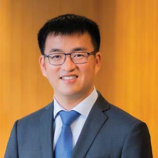
Jae Ho Sohn, MD, MS
Yang Yang, PhD
The Clinical Benefits of Mid-Field MRI
Because of the different contrast mechanisms, mid-field MRIs have the potential to screen for some conditions more effectively than current technologies.
Jae Ho Sohn, MD, MS, a cardiothoracic radiologist and assistant professor who is conducting research with Yang, said that patients with lung conditions can benefit from mid-field MRI. “Traditionally, we’ve pushed MRIs to be high field, but in situations like the lungs, lower field is more helpful.”
Mid-field MRI excels in anatomic lung evaluation, offering advantages over traditional CT without radiation exposure. In the lungs, air can cause image degradation, and lower field MRI – in combination with artificial intelligence – can compensate for motion challenges.
“Mid-field MRI is useful for anatomic evaluation of lung parenchyma, offering advantages over traditional CT, involving no radiation and allowing tissue characterization,” Sohn said. "Functional MRI allows us to examine ventilation and perfusion patterns, providing valuable information for characterizing diseases and assessing their severity." Sohn is conducting studies to explore the full clinical potential and limitations of the 0.55T Free Max machine, including correlations with CT for diverse pathologies like cancer, infection, and lung diseases.
Traditionally, since cardiac and lung MRIs are offered separately, a combined cardiac and pulmonary screening protocol that Sohn’s team is developing using mid-field MRI can save a significant amount of time, improving the patient experience. Machine learning will then be used to improve image quality and expedite the scanning process. The team is also developing predictive models to address potential image degradation proactively. This modality offers crosssectional imaging of the chest without radiation, making it a valuable option for radiation-sensitive populations like pregnant patients.
The wider bore of mid-field MRI, exemplified by the 0.55 Free Max, brings additional benefits. It accommodates patients with claustrophobia or obesity, ensuring accessibility for diverse populations. For individuals with implanted devices like pacemakers, artificial joints, or hips, mid-field MRI becomes a viable option.
Because of the many benefits of mid-field MRI, Yang and Sohn are working to amplify its potential and provide evidence of its success as a viable alternative to conventional MRI through scientific publications, presentations and other channels. “We’re striving to make this transformative technology as widely known as possible,” Yang said.

Example of sample image from Free.Max
• 6-fold faster scan image without any further processing, image is noisy but faster to get
• Traditional denoising using Block-matching and 3D filtering, SNR is improved by details are blurred
• Deep learning denoising using spatial temporal attentions, details are well preserved while SNR is also improved
• Reference clinical scan image quality, the scan time is 6 times longer than the previous 3 images


Ultrasound-Guided Injection Relieves Pain, Enables Mom to Care for Newborn
By Arleen Bandarrae
Maryana was over-the-moon to be a mom for the first time when she learned she was pregnant in May 2022. Energetic and lively, Maryana was taken by surprise when she began experiencing difficulties halfway into her pregnancy.
At 16 weeks, the 32-year-old tech sales executive from Marin was diagnosed with severe symphysis pubis dysfunction (SPD), which refers to pain caused by pubic symphysis separation – the loosening of the joint between the left and right pelvic bones that occurs during pregnancy to allow the pelvic bones to widen during birth. Due to the pressure of her growing baby weighing on her pelvis, Maryana was in pain, the kind of pain that prevented her from walking up and down stairs. At 30 weeks, she was basically confined to her bed.
Maryana counted down the days until her due date. She believed everything would be better, once her baby was safely delivered. Doctors had advised – and her own Google searches confirmed – that in most cases this condition would go away shortly after birth. Determined to get through her pregnancy, Maryana pushed aside her feelings of fear and isolation.
She had no idea her toughest health battle would only just begin after she gave birth to Emily, a healthy baby girl, on December 27, 2022.
Maryana returned home from the hospital with her newborn, strong pain medication, and unbeknownst to her, pelvic pain (caused by pubic symphysis separation) that would be so debilitating once the medication wore off that she’d be confined to a wheelchair for nearly a month.
The Worst Day
“It was the worst and most shocking day of my life,” Maryana said about the day she stopped taking opioid pain medication and began breast feeding. The mind-blowing pain meant she could not walk, or even stand. “My legs were literally not holding me. My daughter is five feet away from me, crying in her crib, and I can’t get up. It is forever ingrained in my body how horrible that was.”
Maryana knew something was very wrong. She received an X-ray at the hospital, but it did not show the structural bone damage that had occurred to her pelvis because she was lying down for the procedure. Later, Maryana learned she
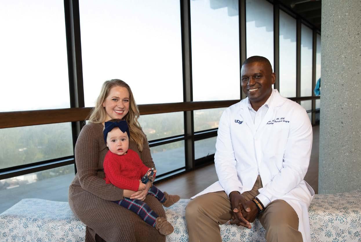
UCSF Patient Maryana Kessel, with her daughter Emily, and Kevin McGill, MD, MPH, Director of Musculoskeletal Interventions and assistant professor of musculoskeletal imaging in the Department of Radiology and Biomedical Imaging.
suffered from traumatic separation of the pubic symphysis, the joint that holds the pelvic bones together, and bone marrow edema, a bruising of the pelvic bones.
Maryana’s condition was far worse than anyone expected, and to her shock, it seemed no one knew what to do. She felt like a hot potato being passed from clinicians in obstetrics, to pain management, to orthopedics. Unfamiliar with the severity of her injury, doctors waited weeks for insurance authorizations for each step of the evaluation process, as well as scheduling delays. Maryana was left with a deep sense of distrust. How could she have access to what felt like all the resources in the world, and yet no one was able to help alleviate her condition so she could fully care for her newborn baby? She was taking Advil and Tylenol daily to manage the pain, but she was still only able to walk from her bed to her bathroom most days.
Searching for answers, Maryana was referred to University of California, San Francisco (UCSF).
UCSF’s Breadth and Depth of Specialty Care
Here, with the breadth and depth of expert care, two specialists teamed up to get Maryana the help she needed. Melanie M. Henry, MD, MPH, FASA, an anesthesiologist, pain management specialist, professor, and Director of Telehealth and Outreach at UCSF Health, collaborated with Kevin McGill, MD, MPH, an assistant professor of musculoskeletal imaging and the Director of Musculoskeletal Interventions in the Department of Radiology and Biomedical Imaging.
“I was hardly able to get to the chair for my video call,” said Maryana, who met Henry for a telehealth visit about one month after she gave birth. Once they talked, she felt seen
for the first time. With emotion choking her words, Maryana described the feeling of “being taken into warm arms” as she heard Henry’s first words: “I am a mother too, and I am so sorry you are going through this.”
“For Dr. Henry to see me as a human and a mother – it brings tears to my eyes to know what she did for me in that moment.” After connecting with Henry, everything started to change. After a series of evaluations, Henry contacted her colleague McGill, who specializes in image-guided joint interventions.
“Dr. McGill is an angel. He put me back on my feet, quite literally,” said Maryana, who to this day shares photos of Emily with him. “He forever has a piece of my story and my heart.”
The Role of the Radiologist
“When I initially met Maryana, she was in a lot of pain and desperate for a solution. I recommended she come in for a diagnostic ultrasound with our expert musculoskeletal sonographers. After the images were obtained and a subsequent neurogram was performed, I had a conversation with her about her condition and the procedure,” said McGill, who performed an ultrasoundguided steroid injection into Maryana’s pubic symphysis to relieve her pain. Though she had an initial bad reaction called a steroid flare, it did not impact her recovery.
“She handled the procedure remarkably well. I called her the next day to see how she was doing. The pain had not completely resolved at that time, but she seemed optimistic,” said McGill, who continued correspondence with Maryana over email and as her symptoms improved.
For the first time during her ordeal, Maryana felt like doctors genuinely cared about her care and how she was doing. “When the level of pain you are in is seen and believed, and a doctor reaches out to you unprompted, it changes everything about that experience,” she said.
“ We all worked together, and Maryana allowed both of us to be the best physicians possible.
~ Melanie Henry, MD, MPH, FASA
Previously, Maryana imagined a radiologist as someone who sat in a dark room and looked at X-rays all day. Today, she continues to be amazed by the personal care and attention she received from McGill and the fact that a radiologist was the answer to her grave condition.
“Dr. McGill has made a huge impact on my life. I am so grateful.”
“I am honored to have been given an opportunity to provide care for Maryana. She is the type of patient that reminds me why I chose a career in medicine,” said McGill, reflecting on this experience.
After receiving the injection, Maryana slowly got better, feeling more and more relief from the pain as the weeks progressed. After being nearly immobile for 6 months, she was able to move around more and more and continue with physical therapy to rebuild her muscles.
“ I am honored to have been given an opportunity to provide care for Maryana. She is the type of patient that reminds me why I chose a career in medicine.”
~
Kevin McGill, MD, MPH

UCSF Patient Maryana Kessel, with her daughter Emily, and Kevin McGill, MD, MPH, Director of Musculoskeletal Interventions and assistant professor of musculoskeletal imaging in the Department of Radiology and Biomedical Imaging.
In early fall, about 10 months after giving birth, Maryana felt no more pain. She was able to take her daughter to the grocery store by herself for the first time. Although she realized her status is what most other moms experience at two weeks postpartum, she was elated to be able to fully participate in her daughter’s life, without mobility being an issue.
“Maryana was an advocate for herself and pursed all options to find the appropriate treatment. We all worked together, and Maryana allowed both of us to be the best physicians possible,” said Henry.
A Future Not Yet Written
“I don’t take for granted that I was able to go on a 15-minute walk yesterday,” said Maryana with renewed self-assurance and a sense of inner strength. She’s looks forward to chasing after her 1-year-old soon enough.
Her advice to others who may be experiencing a similar challenging condition is to keep searching until you find the right experts.
Once a self-described non-support group person, Maryana now turns to her community on Facebook of other people around the world who also experienced severe pubic symphysis separation. “Some have gone on to have other children and are ok, some need mobility aids permanently, and some have their pelvis bolted together afterwards,” said Maryana. “I have the providers, I know what to expect, but no one can tell me it will be ok.”
“Despite all this, becoming a mom is the best thing that ever happened to me. I’d go through it a million times over to have my daughter,” said Maryana. “She is my entire universe. The joy I get from seeing her grow is incredible.”
Symphysis pubis dysfunction (SPD)
Symphysis pubis dysfunction (SPD) is a common condition that can lead to debilitating pain during pregnancy and in the post-partum period. The goals of treatment focus on decreasing pain while improving muscle function and joint stability using a combination of physical therapy, activity modification, a pelvic support belt, and/or oral medications. Alleviating pain in a timely manner is important to enable patients to adequately care for their newborns and/or return to work.
Diagnostic ultrasound is an excellent tool to assess pubic symphysis separation, which is often associated with SPD, in the peripartum period. If conservative measures fail, ultrasound is also the ideal modality for an image guided injection of anesthetic +/- steroids into the pubic symphysis for pain relief.
The rapid growth of the musculoskeletal ultrasound program at UCSF over the past few years has created wonderful opportunities for meaningful interactions with both patients and referring providers.

Kevin McGill, MD, MPH, Director of Musculoskeletal Interventions
and assistant professor of musculoskeletal imaging in the Department of Radiology and Biomedical Imaging.
Imaging Technologists Redefine Patient Care
By Arleen Bandarrae

Behind every diagnosis and image-guided treatment is a highly-trained and dedicated radiologic technologist. Their expertise is crucial to the patient experience at UCSF Health and we are proud of their accomplishments as educators and collaborators in cutting-edge clinical care. Over the past year, technologists have also dedicated their time and insights to value-improvement projects that improve patient access to imaging and make available new services and therapies. Read on to learn more about these efforts, and please join us in acknowledging our technologists’ essential and outstanding contributions.
Diagnostic radiology technologists performed more than 223,000 imaging exams – 46 percent of all imaging scans – in fiscal year 2022-23. The team is comprised of two managers, eight supervisors, and more than 100 radiologic technologists. “Technologists are highly trained in a variety of exams including x-ray, portable imaging, fluoroscopy exams, intra-operative fluoro and CT, and bone densitometry. Our technologists are very creative and flexible and can obtain excellent imaging views regardless of the patient’s mobility or range of motion,” said Jeff Geiger, Manager of Mission Bay. Some diagnostic radiology technologists advanced their imaging skills to include bone density scans (DXA or DEXA) and expanded our services to our Montgomery Street clinic in San Francisco and the Berkeley Outpatient Center (our latest site of expansion.) Bone densitometry, a standard method for diagnosing osteoporosis, marks the successful completion of a value improvement project.
“I’d like to give a shout out to my team of supervisors for managing it all, keeping a positive attitude, and holding it together during a challenging year,” said Chief Manager of Diagnostic Radiology Tosca Bridges, MS, RT.
The Interventional Radiology (IR) technologist team collaborates with ultrasonographers and crossspecialty physician teams to deliver minimally invasive,
comprehensive cancer care at UCSF. Yttrium90 (Y90) radiation for liver tumors and radiofrequency ablation (RFA) for thyroid cancer are two therapies now offered to patients that limit damage to healthy tissue and improve quality of life for cancer patients. In anticipation of expanding pediatric services, Interventional Radiology technologists at Mission Bay are training in advanced pediatric procedures. The IR team is managed by Alpana Patel Camilli, BS, CRA, RT (CT), along with two supervisors, Julio Gonzalez, ARRT, Vincent Ramirez, BSRT (R) (VI) ARRT CRT and includes advanced-trained interventional technologists.
Committed to expanding accessibility for patients, the MRI team implemented strategic growth and expanded services to include new locations including the Berkeley Outpatient Center and other Bay Area sites. In addition, the team continued its partnership with Gurnick Academy, a distinguished nursing and healthcare school, to offer students clinical training and experience that will help shape and prepare future MRI technologists. “Rising to the occasion every day, the MRI team has done incredible work this past year to provide excellent patient care, especially given the record-setting volumes this year,” said Craig DeVincent, MRI manager.
The Molecular Imaging & Therapeutics team developed the infrastructure for a robust radioligand therapy practice
at UCSF, as part of a value improvement project. “This enabled us to provide increased patient access to PSMA617 (Pluvicto), a targeted radioligand therapy for prostate cancer which received FDA approval in March of 2022,” said Molecular Imaging & Therapeutics Manager Michelle Swenson. “We continue to expand capacity for this important service to better serve our patients.”
Nuclear medicine technologists Kimberly Rosales, Levi Fujimoto, and Harminder Grewal received their computed tomography (CT) license, to perform diagnostic CT in combination with PET imaging on the hybrid PET/CT scanners. To earn CT credentials, they fulfilled additional requirements in structured education, clinical experience, and examination, after meeting the supporting discipline prerequisite (CNMT).
The Computed Tomography (CT) team led by Jessica Pfannenstiel, MS, RT (CT) (R), increased access for patients at UCSF Bakar Cancer Hospital in Mission Bay by implementing a value improvement project that enhanced the automatic scheduling process for CT machines and has resulted in significant additional revenue each quarter. In addition, the CT team began cross-training one of two diagnostic X-ray technologists, a collaborative effort between seasoned CT technologists and leadership to promote growth within the department. The CT team has successfully cross-trained diagnostic X-ray technologists in the past, many of whom are now passing along their knowledge on to new staff.
Contrast Agent Bar
Coding Saves
More Than $5 Million. In a value-improvement project led by Pharmacy and facilitated by Sherilyn Hutchinson, Senior Analyst with UCSF Health, the Radiology team collaborated on implementing a bar code medication administration program that reduced overall drug costs by maximizing purchasing at lower pricing (GPO and 340B levels), identifying and preventing billing errors, and streamlining stock lists. This project, which was fully implemented in October 2023, has saved more than $5 million in radiology contrast agent purchasing costs since the program began in 2021.
Key milestones in the project include a strategic reduction in the number of package sizes and strengths being purchased and adding bar code scanning to accurately capture products administered to patients. With bar code scanning, UCSF can trace every contrast agent administered, even if it was purchased outside the scope of the plan, which proved to be invaluable during the global contrast shortage that began affecting operations in spring 2022. Now all radiology technicians scan the contrast agent bar code as part of daily operations. UCSF Radiology members of the contrast agent workgroup included David Sostarich, Devin Dixon, and modality leaders Jessica Pfannenstiel, Jeffrey Geiger, Alpana Patel Camilli, Craig DeVincent, Lisa Burke, and Tosca Bridges.

In early January, UCSF welcomed visitors from Korle Bu Teaching Hospital in Accra, Ghana. Dr. Alfred Otoe Ankrah gave a presentation about medical care in Ghana. Molecular Imaging & Therapeutics Manager Michelle Swenson, supervisor Harmi Grewal, and technologist Erika Padilla Morales helped host the guests who included Emmanuel Nii Boye Hammond, MD, and technologists Miriam Naa Yarley Yartey, Clement Korsah, and Asimeng Adu Sarkodie. SNMMI helped sponsor the visit.
“
The nurse and doctor that treated me patiently explained the procedure that was going to be performed and listened and answered all my concerns and questions. They made me feel so confident they were going to be successful with my procedure. Shortly after returning home, I was alerted that my procedure notes were already posted to My Chart.”
Interdepartmental collaborators included Senior Willow Analyst, Danielle Alves, Rev Cycle Informaticist, Charlene Joe, and support from Pharmacy Leadership.
The UCSF Breast Imaging team now offers contrastenhanced mammography, an imaging technique that involves the injection of a contrast agent into the bloodstream to highlight blood vessels and lesions in the breast. This enhances the visibility of abnormalities, particularly in dense breast tissue, and provides a more detailed and accurate assessment of breast health. It is ideal for patients not amenable to MRI and it is faster and less expensive than MRI.
“This innovative approach has proven invaluable in cases where traditional mammography may yield inconclusive results,” said Breast Imaging Manager Amy Vincent, BSRS, CRT. “Contrast-enhanced mammography aids in the early detection of abnormalities, including tumors and lesions, ultimately improving diagnostic accuracy and patient outcomes.”
Committed to providing accessible and high-quality breast imaging services, the mammography team expanded its reach to a new location in San Mateo, in addition to the Berkeley Outpatient Center, which has been offering mammography since October 2020.
As part of the mammography team’s focus on innovation, they have implemented new software systems and completed the process of upgrading to Digital Breast Tomosynthesis (3D mammography) to enhance diagnostic capabilities. Artificial intelligence software Volpara AI provides real-time positioning feedback to technologists and performance improvement analytics to ensure the highest standards of patient care. QC-Trak software facilitating paperless quality control tracking, inspection reporting, ACR credentials, QC event tracking, and document management.
“Always struck by the deep kindness of every single person I interact with - thank you! The CT technicians were especially warm and helpful.”


Chief of Body Interventional Radiology K. Pallav Kolli, MD, and IR Fellow Charlene Ofosu, MD, at Parnassus Heights.
Mary Beier Keane, RN, and technologists Christie Schavitz, CRT, and Julio Gonzalez.
Jessica Nelson, CNMT, Thomas Hope, MD, and Carlos Todd, from Gurnick Academy collaborate in the PET CT exam room at Parnassus Heights. ~ Mammography patient
Tech & the Human Touch
By Arleen Bandarrae
Nuclear Medicine Technologist Erika Padilla
Morales Presents at International Conferences

In the early morning hours, just after her bike commute to the UCSF Imaging Center at China Basin, we caught up with Erika Padilla Morales, BS, CNMT, a nuclear medicine technologist in the Molecular Imaging & Therapeutics group, to hear about presentations she’s given on nuclear medicine innovations for the Society of Nuclear Medicine and Molecular Imaging (SNMMI) and South African Society of Nuclear Medicine conferences last year.
Erika presented on the Copper Cu 64 Dotatate isotope used with PET CT neuroendocrine tumor imaging at the SNMMI conference in Vancouver in 2022, then co-presented on “Radioligand Therapy for Prostate Cancer Patients” at SNMMI’s western regional meeting in Lake Tahoe, California in October. She was then invited to the South African Society of Nuclear Medicine conference in August of 2023 where she was asked to present on molecular breast imaging, a technique used in addition to traditional mammography that can account for a patient’s unique genetics, risk factors, and breast constitution/density.
“It is an honor to be invited to speak, and it is really exciting as a technologist to connect and collaborate with my colleagues and get first-hand information about what’s happening in the field,” said Erika, thinking back to the many virtual meetings held during the pandemic. “There’s so much expertise in the room, and I have the opportunity to share and to get feedback too.”
While she gets excited to “talk tech” at conferences, Erika never loses sight of her compassion. “It’s powerful when you center the human in technology,” she said. "The technology is amazing, but if the patient doesn’t feel comfortable and in partnership with their caregivers, then the medical care is not truly in alignment with the patient’s needs."
For her talk on Lutetium-177 PSMA therapy, she often begins by sharing an anecdote. As Erika prepped a patient for his prostate cancer treatment, he showed her a photo and said, “I just want more time with my grandson.” Reflecting on this, Erika notes that imaging technology supports the human touch: technologists position patients or offer warm blankets to ease a patient through the scan. It’s one of the things she loves about nuclear medicine – the opportunity to develop relationships with patients over the course of their care. It’s similar to the joy she felt as a teacher in her early career, working with her students and watching them progress over time. Now, she’s motivated by a desire to further the mission of personalized medicine – getting the right treatment to the right patient. As she recalled her patient’s desire for more time with his grandson, Erika said, “In the end, that’s what we’re doing and why we are doing it.”
A Magic Moment
By Francis Horan
In mid-September, vice chair and interventional radiologist Mark Wilson, MD, was caring for a patient in Zuckerberg San Francisco General Hospital when he happened to look up and notice that everyone on the interventional radiology team was Black.
Thinking back, Dr. Wilson realized that they had all worked together before, but on that morning he was struck by the significance of the moment: “Representation is an area where radiology, not just IR, can suffer. Imagine being a patient at ZSFG from an underrepresented community, and seeing a team of professionals who look like you. As a patient, you know that this team will be better able to understand your experience.”
Research consistently shows that Black patients have disparate access to diagnostic imaging which leads to worse outcomes. Black physicians are underrepresented in radiology, and among the radiology subspecialties, neuroradiology and interventional radiology fellowships have the least Black physician representation. Physicians from underrepresented backgrounds are more likely to choose to practice in areas with low access to medical care, and those patients are more likely to receive the highest quality of attention and care when paired with medical personnel who share their backgrounds.
Reflecting on this recent experience, Dr. Wilson said, “I’m proud of our Department of Radiology and grateful to be practicing medicine here. We’ve worked to recruit and retain underrepresented physicians in radiology, and we have been intentional in fostering our community, including technologists and nurses, to help moments like these happen. We know there is always more work to do, even as we pause for a moment to celebrate how far we’ve come.”

(L to R) Christopher Brunson, MD, Elaine Martin, RN, Viana Larkin, RN, Antoine Pierce, RN, Alana Walker, RT, Mark Wilson, MD
A SCI-FI NOVEL & A CHILDREN'S BOOK
Just City Explores Compassion in Virtual Reality
By Francis Horan
About ten years ago, soon after she had moved to the United States, Olga Tymofiyeva, PhD, associate professor in the UCSF Department of Radiology and Biomedical Imaging, watched a lecture by Michael Sandel, professor of government at Harvard, speaking on the nature of justice. Not only did Sandel’s passionate and interactive lecture inform Dr. Tymofiyeva’s own teaching style to this day, but it also inspired her science fiction novel, Just City. Tymofiyeva recalls that “Sandel spoke of the ‘veil of ignorance’ that innately clouds our understanding, formed by our preferences and preconceptions. This is true even in scientists who strive to be without bias. As humans we do not see all the factors that predetermine our path in life, and often are instinctively resentful when they are pointed out.”
This aversion to the invisible rails of nature and nurture that determine so many outcomes in our life was the seed that grew into the novel Just City. At first, Tymofiyeva imagined creating a virtual reality game that would allow people to walk in someone else’s shoes. Then, she took a conceptual step back to write a story about the effect that virtual reality program might have. Somewhere between real-life virtual reality therapeutic treatments and The SIMS, the titular program in Just City allows its users to inhabit different bodies and lives to experience who they would be if their circumstances were fundamentally different.
In a near-future San Francisco, a young man named Nathan dreams of establishing his own start-up to solve homelessness with an app that should make people choose to work. As part of Nathan’s effort to raise money for his dream company, he signs up for a scientific experiment held in a virtual reality game that places players into the lives and experiences of people from very different backgrounds and circumstances. With “success” and “failure” at everyday life displayed as a game with unbalanced odds, Nathan’s once firm belief in meritocracy and our contemporary paradigms of justice begins to crack, placing him in conflict with his friends and his own self-perception.
What does justice mean when our actions are dominoes set in motion by our biology and environment? Tymofiyeva is an optimistic skeptic who sees this complex interplay as an opportunity for compassion. “If the causes influencing a person’s course in life are invisible, observers are much more likely to assign blame instead of sympathy. What we can see first-hand is given more explanatory weight against equally real but visually diffuse causes. And there is so much we don’t see.”

Olga Tymofiyeva, PhD
At UCSF, Tymofiyeva uses imaging technology to reveal what isn’t readily visible to the naked eye. Her research delves deep into MRI as a tool to detect and understand the hidden causes and effects of adolescent mental illness, with a goal of preventing and ameliorating this suffering. “The experience of writing Just City deepened my interest in the variables of prosociality, compassion, self-compassion, and value/meaning-finding as they play out in adolescents’ lives. These factors can have a significant impact on wellbeing and potentially serve as protective factors in the face of depression.”
The four-and-a-half-year journey of writing Just City was an adventure whose success Olga credits as much to all those who helped her as she does to her own drive. She expressly thanks all the people who gave aid as Beta readers, editors, friends who test read, sensitivity readers, her book coach, writing buddies. When she experienced blockages, Tymofiyeva noted that “Sometimes simply having someone on the other end of a silent zoom call can give the accountability necessary to buckle down and put words on the page.”
Just as the writing process was made all the better by the support of those around her, radiology colleagues have been enthusiastic readers and reviewers. Department chair Christopher Hess, MD, PhD, writes that, “Olga's novel draws from her research to tie together the impact of electronic devices on adolescents with everyday issues in San Francisco such as bridging the gap between compassionate care and dispassion in managing the homeless.” David Saloner, PhD, noted that that Tymofiyeva’s novel “cleverly weaves together a number of current concepts in a coming-of-age tale embedded deep in the fabric of San Francisco. The book is a fun read with broad appeal – even for those as young as high schoolers.” In expressing her gratitude to colleagues, Tymofiyeva said, “The reception of the book by everyone at UCSF has been amazing. Hearing people’s thoughts about the book has created real connections, real conversations. It’s precious.”
While a fully immersive virtual empathy engine is unfortunately not for sale at the moment, Tymofiyeva suggests that the lesson at the heart of Just City is caring enough to hear the life stories of others, which is possible to accomplish right now in our physical reality. She adds that, “We can also educate ourselves and others on the strong interplay between physical injury or illness and what makes up our personality. The most important avenue, and indeed the simplest approach to compassion and understanding, is to simply get out into the world of other people and pay attention to the stories they are living.”


Radiology for Kids Children’s Book
By Rita Gaber

John Mongan, MD, PhD, associate professor in our Abdominal Imaging division, recently published Radiology for Kids with co-authors Brandon Pham, MD, and Betty Nguyen. The book is the tenth title in Medical School for Kids, a charming, beautifully illustrated series by Pham and Nguyen that introduces children to medical specialties.
We caught up with Dr. Mongan for a Q&A about this fun project.
What inspired you to write a children's book? How did your collaborators influence the creation of this book?
My co-authors are my cousins. I saw one of their earlier books on ophthalmology (Dr. Pham's specialty) and, half in jest, asked them where the radiology version was. They were extremely enthusiastic about the idea, and the more I thought about it the more it seemed like a fun project and a great way to help the next generation have a better idea of what "behind the scenes" doctors like us do.
What insights did you gain about your teaching, clinical or scientific work in the process of writing this book?
It's really challenging to describe technical things in a way that's engaging, doesn't use too many words that are unfamiliar to children, is technically accurate and fits in the three-to-four lines available on each page of a picture book! The process really made me think about the fundamentals of what we do as radiologists.
What did you edit out of the book?
My first version of the text had about a paragraph per page; my co-authors showed me that it really needed to be distilled into just a few sentences per page. There was also some more detailed material about different subspecialties of radiology and applications of different modalities that ended up being a little much and was condensed into single summary pages.
If you have a next book project in mind, could you tell us a bit about that... the idea or topic, audience?
No big ideas at the moment, but I know my coauthors are working towards a complete series of children's books that cover every medical specialty.
Your closing thoughts about this project?
Stretching my mind in a new direction to do something different from anything I've done before was a great experience.


PEOPLE

ACADEMIC AFFAIRS
By Christine Glastonbury, MBBS, Vice Chair for Academic Affairs
Promoting Academic Success in Radiology & Biomedical Imaging
This last year has seen many challenges for faculty in the department as the imaging volume rises. Everyone seems incredibly busy, research funding is more difficult to come by, and researchers and clinical faculty are re-evaluating being in the office versus working from home. Our team's mission is to support our faculty, seek opportunities to enhance their success, and find ways to make the necessary complexities of an academic career easier to navigate. Our academic affairs team of manager Lorna Kwok, RN; coordinator Jocelyn Pulido, and project coordinators Apple Palad and Connie Jang, have been working hard to try to meet faculty needs.
It has been a whirlwind year, and it’s only when we were asked to provide data to the Dean’s office that we realized that we have hired 51 faculty since the beginning of 2020 (including 5 starting in the coming months)! This is an achievement to be celebrated, although there is little time to rest as we continue the search and hiring process for additional faculty in the Musculoskeletal, Neuroradiology, Cardiothoracic, Abdominal, Interventional, Pediatrics, Molecular Imaging and Breast divisions, as well as a Nuclear Medical Physicist and a Radiopharmaceutical Chemist!
The academic affairs team is so appreciative of the many faculty who have chaired our recent search committees –Jane Wang, Pallav Kolli, Vinil Shah, Mark Wilson, Thomas Link, and Ray Sze. Thank you also to the many faculty for attending candidate presentations, one-on-one and group interviews, and for welcoming candidates to our campuses. Successfully expanding our teams is a time-consuming process but has been highly rewarding this past year.


The Academic Affairs team, left to right, Apple Palad, Susan Wall, Connie Jang, Christine Glastonbury, Lorna Kwok, Jocelyn Pulido
As of June 2023, our Department of Radiology has 146 faculty, with 33 Imaging Scientists and 101 Radiologists, including 12 emeritus faculty working on recall. We have welcomed 8 new Radiology faculty since July, 2023, and at the time of writing, have hired 5 more to start work after the new year 2024. We are delighted to welcome our new faculty who you will ‘meet’ on the following pages.

During Mentoring Month in January, we gifted water bottles to faculty and displayed posters at our various clinical and research locations.
tiny.ucsf.edu/radmentor

Faculty Mentoring
Our Faculty Mentoring Program, designed to create mentoring networks for each of our assistant professors, continues to guide and support the faculty through career direction decisions and research collaborations, as well as providing sponsorship opportunities. We currently have 34 MD assistant professors and 6 PhD assistant professors in the mentoring program. Sri Nagarajan, PhD, is mentoring facilitator for the imaging scientist PhD faculty.
It is only through the dedication of each our faculty that the mentoring program is so successful and it is always
impressive how many faculty are enthusiastic to become mentors to our Assistant Professors. In January this year we again honored and celebrated all faculty mentors during National Mentoring Month, with our mentoring flasks. This June we were thrilled to acknowledge the extraordinary mentoring work of Jane Wang, MD, who was the recipient of the 2023 Radiology Award for Outstanding Faculty Mentoring. Jane has been an extraordinary mentor for many faculty in our department, in addition to being the career co-mentor for the junior faculty in the abdominal division she now leads.
Faculty Development
Susan Wall, MD, advisor to the Chair, leads these efforts and continues to source opportunities for faculty development, available grants, courses and faculty awards. Together with Elissa Price, MD, she continues to lead the highly sought-after faculty speaker training course. This small group intensive, conducted in three afternoon workshops, focuses on teaching lecturing skills through practice, feedback and re-presentation.
Academic Advancement
Our M&P committee includes 12 full professor faculty and myself, with representatives from each of our affiliate sites at the VA Medical Center, Zuckerberg San Francisco General (ZSFG) and Benioff Children’s Hospital - Oakland. Each of our committee members contributes many hours of packet review and preparation and contributes to the often-complex discussions over our six meetings. Their service to our department and their support for and mentoring of our faculty is greatly appreciated.
For the most recent cycle with advancements effective July 2023, our M&P committee reviewed and discussed 51 packets including 11 promotions and 10 accelerations approved. We are enormously proud of our faculty’s hard work. We especially wish to congratulate those faculty promoted to full professor this July: Pallav Kolli, professor of clinical radiology, and Drs. Duygu Tosun-Turgut, Janine Lupo Palladino, Galateia Kazakia, and Dimitrios Mitsouras as professors in residence.
The advancements, the recruitment, faculty development, the hiring and visa organization for non-faculty academics, faculty credentialing, visiting clinical professor programs, the mentoring program and the faculty celebrations would not be possible without an extraordinary team of dedicated academic affairs coordinators who are creative and hardworking and committed to supporting our faculty. An enormous thank you to Connie, Apple, Jocelyn, and the team manager, Lorna. They are an amazing team and it is an honor and pleasure to work with you!
2023 Mentoring Month

Congratulations Faculty Promotions
We are honored to announce that eleven faculty members – six new associate professors and five new full professors – received academic promotions effective July 1, 2023.
Our department and UCSF recognizes each promoted faculty member for their significant contributions to our mission: Advancing Health Worldwide. To do this well, each faculty member balances teaching, clinical practice, scientific inquiry and clinical translation, and service activities to create an outstanding portfolio of accomplishments. We are incredibly proud of each faculty member and their unique qualities as teachers and mentors; department and university leaders committed to Diversity, Equity and Inclusion; and physicians and imaging scientists known for their national or international stature in the field of radiology.

Associate Professor Clinical Radiology Cardiac & Pulmonary Imaging


Associate Professor Clinical Radiology Cardiac & Pulmonary Imaging


Yan Li Associate Professor In Residence Research
Kim Kallianos
Brian Haas
Rina Patel Associate Professor Clinical Radiology Musculoskeletal
Tatiana Kelil Associate Professor Clinical Radiology Breast Imaging
Yi Li Associate Professor Clinical Radiology Neuroradiology
Pallav Kolli Professor Clinical Radiology Interventional Radiology
Galateia Kazakia Professor In Residence Research
Janine Lupo Professor In Residence Research
Dimitrios Mitsouras Professor In Residence Research
Duygu TosunTurgut Professor In Residence Research
Welcome New Faculty
We are delighted to introduce our ten new faculty members for 2023. One imaging scientist has joined our research groups and 9 clinical faculty will deliver patient care in five of our nine subspecialities. All of our faculty contribute to trainee education as well as new and ongoing initiatives that ensure our department’s reputation for innovation.
As each new faculty member joins our UCSF community, we extend a warm welcome and our best wishes for successful, fulfilling careers.


Maggie Chung, MD
Assistant Professor In Residence Breast Imaging
Dr. Chung earned her medical degree at the Warren Alpert Medical School, Brown University, followed by an internship at Scripps Mercy Hospital in San Diego. Dr. Chung completed her diagnostic radiology residency at UCSF, where she was recognized in 2022 with the Elmer Ng Outstanding Resident Award, the Margulis Society Resident Research Award, and the RSNA’s Roentgen Resident/Fellow Research Award. Dr. Chung has served as a clinical fellow in our breast imaging section since 2022.
Dr. Chung is a member of the Science and Technology Resource Group pillar in the Center for Intelligent Imaging (ci2). She has served on the UCSF Residency Admission Committee, the Residency Quality Assurance Committee, and was the UCSF Representative to the California Radiological Society.
With a strong record of publication, Dr. Chung’s current research focuses on the use of neural networks in imaging. Her projects cover simulated contrast-enhanced breast MRI using convolutional neural network, convolutional neural networks for radiographic detection of surgical sponges, and PET/CT metabolic tumor segmentation using a multi-modality multi-t ask neural network. Her previous research at UCSF focused on the use of ultrasound for the evaluation of palpable masses in lactating women.
Daehyun Yoon, PhD
Assistant Adjunct Professor Research
Dr. Yoon holds an MS and PhD in electrical engineering from the University of Michigan and a BS in computer science & engineering from Seoul National University. Upon completing his postdoctoral fellowship at Stanford, Dr. Yoon continued on as senior research associate working
on musculoskeletal MRI and PET/MRI. He has been involved with development of research imaging techniques and translation of those into clinical protocols for patient care.
Dr. Yoon’s research interest is in the development of PET/MRI and MRI methods for identifying peripheral pain generators in the musculoskeletal system. His recent projects include PET/ MRI of various chronic pain conditions (low back pain, complex regional pain syndrome, CSF leak, etc.), highresolution/quantitative MRI of peripheral nerves, and artifact-free MRI near metallic implants. Dr. Yoon is embarking on a new NIH/ NIAMS-funded project as a co-investigator, which aims to enable [18F]FDG PET/MRI near metallic hip implants for the improved identification of local sources causing persistent pain following total hip arthroplasties.

Jonathan Friedman, MD
Assistant Professor
Clinical Radiology
Musculoskeletal Imaging
Dr. Friedman earned his medical degree at The George Washington University School of Medicine in Washington, DC. Following his internship at University of Chicago Medical Center (NorthShore) in Evanston, IL, he completed his diagnostic radiology residency at Boston University Medical Center and a fellowship in musculoskeletal imaging at UCSF.
Dr. Friedman has published research on imaging of common hip pathologies in runners. He has conducted resident lectures on hip injuries in runners, benign osseous lesions that can be diagnosed radiographically, and imaging findings of non-accidental trauma or child abuse. He has presented research on “Comparison of DLP-Based Effective Dose to Monte Carlo-Based Effective Dose in Low Dose Chest CTs.”

Weiya Mu, MD
Assistant Professor
Clinical Radiology
Neuroradiology, ZSFG
Dr. Mu earned her medical degree at the Albert Einstein College of Medicine in Bronx, NY, with a distinction in research. Following an internship at California Pacific Medical Center in San Francisco, she completed her diagnostic radiology residency at the Einstein Medical Center in Philadelphia, where she was chief resident. She completed her neuroradiology fellowship at the Stanford Medical Center in Palo Alto.
Dr. Mu has experience in emergency radiology and has published research on diagnosing fat-containing lesions in thoracic imaging, traumatic brain injury in combat veterans, neuroplasticity, and neck strength in young adults. Her presentation on “The Vomer Bone and Vomerovaginal Canals: A Pictorial and Educational Review of Neuroimaging Anatomy and Pathology” won the ASNR Educational Exhibit Certificate of Merit in 2022. She has received an award in the Einstein Medical Center annual case report competition, as well as several research fellowships.

Christopher Brunson, MD
Assistant Professor Clinical Radiology
Interventional Radiology, ZSFG
Dr. Brunson received his medical degree at UCSF. After an internship with the UCSF-East Bay Surgery Program at Highland Hospital and Kaiser Oakland, he completed his integrated interventional radiology residency at the University of Arizona in Tucson. Dr. Brunson was chief interventional radiology resident and served Graduate Medical Education as an executive member to the advisory and diversity committees, and as a search committee member for associate dean. Dr. Brunson has received the RSNA Roentgen Resident/ Fellow Research Award and the University of Arizona GME Resident Academic Career Scholarship. While he was a UCSF medical student,
Brunson was an organizer with #whitecoatsforBlacklives, a movement to draw attention to the effects of racial disparities on health care.
Dr. Brunson’s clinical practice and research program focuses on oncologic outcomes and healthcare disparities, particularly in patients with NASH/NAFLD, as well as advanced dosimetry, particularly tumor perfusion augmentation. His publications have focused on interventional radiology methods for managing liver tumors, the use of percutaneous radiofrequency ablation, and comparisons of 2-D perfusion angiography and Tc-99m MAA SPECT/CT.

Allen Ye, MD, PhD Assistant Professor Clinical Radiology Neuroradiology, ZSFG
Dr. Ye obtained his MD and his PhD in bioengineering in 2017 at the University of Illinois Chicago in the Medical Scientist Training Program. After his internship at St. Francis Hospital in Evanston, Illinois, he completed his diagnostic radiology residency at the University of California San Francisco in 2022. During that time, he was selected as an NIH T32 research fellow and remained at UCSF to complete a neuroradiology fellowship in 2023. He has received multiple honors for his work including the Dr. Edward P. Cohen Medical Scientist Training Award, the UCSF Resident Research Fund Award, and the UCSF Radiology
Outstanding Fellow/Instructor Teaching Award. Dr. Ye has served as director of the Resident Informatics Team and as a representative on the AIIMS (PACS) Steering Committee.
Dr. Ye’s research focuses on utilization and application of advanced magnetic resonance imaging techniques such as anomalous diffusion, elastography, and connectomics in the clinical setting. He has translated these techniques for better characterization of vascular, neurodegenerative, and psychiatric diseases. Dr. Ye also has a strong interest in trauma research and has performed retrospective studies that categorize cervical spine trauma with respect to established trauma classifications and to evaluate the utility of MRI in the setting of isolated prevertebral edema.
Kevin Sweetwood, MD
Assistant Professor
Clinical Radiology
Musculoskeletal Imaging
Dr. Sweetwood earned his medical degree at Baylor College of Medicine in Houston, TX, where he graduated with highest honors and a certificate of medical ethics. After an internship at St. Mary’s Medical Center in San Francisco, he completed a diagnostic radiology residency at UCSF, where he served as chief resident, and a musculoskeletal radiology fellowship. He received the Stanley W. Olson Award for Academic Excellence and Service and is inducted into the Alpha Omega Alpha Honor Medical Society.
At UCSF, Dr. Sweetwood has served on the Residency Recruitment Committee and as a chief resident representative to the Executive Committee, as well as on the Radiology COVID Response (RICS) Team. He has conducted lectures and teaching sessions for medical students and residents on multiple topics including ultrasound, chest imaging, and musculoskeletal imaging, as well as preparatory presentations for trainee call and board exams. Dr. Sweetwood has published on digital techniques to improve consensus among radiologists, MRI-guided prostate biopsy, and spindle cell lesions and carcinoma of the breast. His current research focuses on the clinical application of molecular imaging using 99mTc-MDP SPECT/ CT and NaF PET/CT and PET/MRI in the evaluation of low back pain and guidance for therapeutic intervention. His clinical interests include sports imaging and image-guided procedures for pain management.

Joelle Harwin, MD Assistant Professor Clinical Radiology Abdominal Imaging
Dr. Harwin earned her medical degree at Rosalind Franklin University of Medicine and Science. She completed an internship in internal medicine at the Virginia Mason Medical Center in Seattle, followed by diagnostic radiology residency and an abdominal imaging fellowship at UCSF.
Dr. Harwin served as chief resident during her final year of residency. In the radiology department, Dr. Harwin served on the Quality & Safety committee where she authored guidelines on imaging modality size/weight limitations and reading room safety during the COVID-19 pandemic. Both documents were implemented by the department and published on the UCSF website. Dr. Harwin served on the Patient Experience Leadership team where she helped with hospital and departmental transition in response to the 2020 Cures Act. She also served on the resident recruitment committee and was part of the hospital-wide Clinical Performance Improvement committee. On a state level, Dr. Harwin served as both president and vice president of the California Radiological Society Residents and Fellows Section where she organized the third annual UCLA Career Symposium and helped plan the Radiology Leadership Institute Westcoast Kickstart Your Career Sampler Workshop. Dr. Harwin is the Young and Early Career Professional Society 2024 grant recipient from the American College of Radiology.
Dr. Harwin has published on multiple topics including multidisciplinary approach to cancer in pregnancy and liver imaging in hereditary hemorrhagic telangiectasia. Her research abstract “Going Beyond FDG-PET: The Next Generation of Targeted Nuclear Medicine Imaging for Breast Cancer” won the American Roentgen Ray Society 2021 Certificate of Merit. Dr. Harwin was invited to a lecture at the Association of University Radiologists 2022 on creating a sustainable workforce by optimizing relationships between program directors, trainees and program coordinators.

Omar Hassan, MD
Assistant Professor
Clinical Radiology
Abdominal Imaging
Dr. Hassan earned his medical degree at Chicago Medical School in, North Chicago, IL, receiving a distinction in basic sciences. He completed a general surgery internship, diagnostic radiology residency, and fellowship in abdominal imaging and diagnostic sonography all at UCSF.
During residency, Dr. Hassan co-led the radiology section’s quality improvement initiative at the Zuckerberg San Francisco General Hospital, examining delays in morning turnaround times of overnight, resident-read radiology examinations and implemented a system to improve the time from wet read to final attending reads of reports.
Dr. Hassan conducts research as part of the Cirrhosis & Hepatocellular Carcinoma Working Group, establishing a database of over 200 patients with treated HCC for the current largest radiologic-pathologic correlation of treatment modalities in a multidisciplinary collaboration of diagnostic and interventional radiologists and transplant hepatologists. Dr. Hassan has published papers imaging in gender affirmation surgery, organic cation transporter-mediated clearance of cardiovascular drugs, and currently has a paper in progress on the imaging reliability of liver tumor grading (LI-RADS)

Kang Wang, MD, PhD
Assistant Professor In Residence
Abdominal Imaging
Dr. Wang earned his PhD in biomedical engineering at Cornell University and his MD at the University of California, San Diego. Following his internship at Kaweah Delta Health District in Visalia, CA, Dr. Wang completed his diagnostic radiology residency at UC San Diego followed by a body MRI diagnostic radiology fellowship at Stanford.
Dr. Wang has received the Resoundant Innovation Research Award from the Society of Computed Body Tomography and Magnetic Resonance, as well as the RSNA Resident Research Award, and the Society of Abdominal Radiology Wylie J. Dodds Research Award. He was the founding member and manager of the ultrasound clinic at UC San Diego Free Clinic, and a founding member of RAD-AID chapter at UC San Diego Health. He has published research on various aspects of quantitative imaging biomarkers for non-alcoholic fatty liver disease, including automated CT and MRI liver segmentation and biometry, and hepatic MR elastography, with a longterm interest in developing automated, accurate, and reproducible quantitative imaging biomarkers to diagnose, stage, and monitor chronic liver disease.

Faculty Retirements

Vickie Feldstein, MD
Professor Emeritus Abdominal Imaging
Dr. Feldstein is an expert in many applications of diagnostic ultrasound and the use of Doppler techniques to analyze blood flow. Dr. Feldstein received her medical degree from Dartmouth Medical School / Brown University Program in Medicine, Rhode Island in 1986, and completed her residency in Radiology at UCSF, serving as chief resident in 1991.
In 2012, Dr. Feldstein helped initiate a new program at the UCSF Medical Center dedicated to the care of twin pregnancies. She is a part of the multidisciplinary team of experts who comprise the UCSF Fetal Treatment Center,
a ground-breaking collaboration in the diagnosis and treatment of birth defects before delivery.
Dr. Feldstein’s research focuses on obstetric, gynecologic and abdominal applications of sonography in evaluation of a variety of pathologic conditions, in particular the etiology and pathophysiology of complicated monochorionic (single placenta) twin pregnancies. Dr. Feldstein has written, lectured, and investigated widely on ways to use ultrasound to safely and effectively guide needle procedures. She has researched and written about ultrasound findings noted in the prenatal evaluation of fetuses with anomalies. Dr. Feldstein has written over 62 published articles and 10 book chapters.
She has received many honors and recognition for her work, including the department’s Elmer Ng Award for Outstanding Resident (1991), Outstanding Alumni Award (2020), and the Clinical Faculty Excellence Award (2023).
Career Retrospective: Michael Weiner, MD
By Francis Horan

Fueled by his curiosity, persistence in the face of obstacles, and a personal philosophy to “do good, help people, and have a good time,” Michael Weiner, MD, has reinvented his scientific focus more than once during his now half-century-long career. In 1980, Weiner was an Assistant Professor of Medicine (Nephrology) at Stanford, researching kidney metabolism at the Palo Alto Veterans Administration (VA) Medical Center. He thought that his grants
and publications, in addition to his Young Investigator Award from the American College of Cardiology, meant his career growth at Stanford was a sure thing.
Being passed over for tenure at Stanford was a shock that in retrospect turned out to be, as Weiner recalls, “the best gift I ever received.” Soon after this disappointment, Floyd Rector, MD, who was Chair of Nephrology at UCSF, offered Weiner the position of Associate Professor of medicine and Chief of Hemodialysis at the VA Medical Center. During his last few months at Stanford, Weiner got the idea to study the metabolism of the rat kidney using Nuclear Magnetic Resonance (NMR) spectroscopy on the rat kidney with an implanted radiofrequency coil. This proved to be one of the first experiments to perform NMR on an intact animal. He brought this technology to UCSF, where, he says, “I realized that in the near future, human patients would be
I’m a scientist. I’m interested in everything.” “
scanned with NMR (soon to be called MRI), as soon as we developed magnets big enough to fit someone larger than a rat. I decided I wanted to be a part of that.”
Weiner credits UCSF and the VA for providing a welcoming scientific home for his career: “Not getting tenure at Stanford was heart-breaking. But a scientist has to adapt and take advantage of the unexpected. All my success has happened at UCSF, and the VA has been incredibly supportive. I owe my entire career to these institutions.”
As he developed his MR imaging program at the VA, Weiner’s interests quickly expanded from nephrology to brain health. In the late 1980s, knowing that to secure consistent research funding he needed to “pick a disease,” Weiner’s interest in aging and brain scans led him to focus on Alzheimer’s disease. Alzheimer’s disease had both a spotlight in public health and reliable grant money, as well as a tantalizingly unsolved puzzle: it could not be diagnosed in living patients. Weiner was at the forefront of this research and wrote UCSF’s first NIH grant for imaging Alzheimer’s disease.
Never content with only academic research, Weiner has been involved in several public-private partnerships beginning with Synarc in the late 1990s. Synarc was founded by UCSF faculty to develop bone-density imaging products,
resolving the conflict between the realities of university staffing and the trial-by-trial nature of grant funding. Weiner collaborated with Harry Genant, MD, to form a neurology division at Synarc to focus on Alzheimer’s disease, observing that, “The point of research is to make advances that are translated to help people’s lives. When any discovery reaches the patient, it will be coming from a private company.”
Weiner’s next great advance, the Alzheimer’s Disease Neuroimaging Initiative (ADNI), is also a public-private partnership co-funded by the NIH and pharmaceutical and diagnostic companies. Since 2004, ADNI has been the largest multi-site longitudinal study on Alzheimer’s disease, mild cognitive impairment and dementia. This unique sample pool, which follows healthy and symptomatic patients for years and often decades, offers invaluable data from people as they first develop symptoms of mild cognitive impairment. The deidentified data are available to the entire scientific community on the ADNI website with no embargo leading to over 5500 publications to date. ADNI was followed by the Brain Health Registry (BHR), an internet-based registry involving more than 100,000 participants, designed to accelerate development of effective treatments for brain diseases, which was a landmark in its use of all-online data collection for medical research.
Looking back at these initiatives, Weiner described the incredible demand for data from the many companies pivoting into immunotherapy and imaging, “It was like getting on a rocket ship. The growth and success of ADNI was amazing.” In 2011, the Alzheimer’s Association presented the Ronald and Nancy Reagan Research Award to ADNI and in 2021, Dr. Weiner was awarded the Henry Wisniewski Lifetime Achievement Award in Alzheimer’s Disease Research by the Alzheimer’s Association.
“How does this help patients?” is the underlying question that animates Weiner’s work. In 2020, he established the ADNI Diversity Taskforce to improve recruitment of underrepresented research participants. Similarly, the BHR has pioneered initiatives to recruit underrepresented communities such as the CEDAR study, which aims to increase engagement by Black participants, and the California Latino Brain Health Registry. As Weiner says, “even ADNI is not representative, and this problem is present across the entire medical research field. We need to engage and enroll people from underrepresented communities into research to better serve patients.”
Perhaps unusually for a principal investigator, Weiner is also an ADNI study participant. For the past 18 years, he has come in for clinical tests, samples, and scans, observing that, “It is not for my benefit. Participating myself can show that the process is not scary and illustrates how it contributes to the common good.”
With that attention to the common good in research, Weiner is especially proud of his role as a mentor to hundreds of graduate students, postdoctoral fellows (120+) including Alan Koretsky, PhD, Greg Karczmar, PhD, Hoby Hetherington, PhD, Anthony Baker, MD, and a great number of faculty, including Linda Chao, PhD, Susanne Mueller, MD, Duygu Tosun, PhD, Rachel Nosheny, PhD, and Scott Mackin, PhD. Weiner describes this true passion: “Mentoring students, postdoctoral fellows, and junior faculty is my single most important function at UCSF. People can grow and adapt quickly, so we mentors must adapt with them. Most of all, I try to mentor by example rather than by instruction.”
Weiner’s avocations are further expressions of his interests in health and originality. A member of the Dolphin Club in the Marina District, Weiner routinely swims in the open water of the San Francisco Bay, even in January. To those skeptical about taking the plunge, he insists, “Swimming in the morning makes you feel better all day long. It really wakes you up.” An accomplished jazz pianist, Weiner has provided entertainment at functions for UCSF, the VA, and various charities. “In a way,” he says, “jazz is like science in that it is problem solving. You explore, improvise, discover new things, test hypotheses. The goal is always to create something new.” As a teenager, Weiner discovered jazz when he hopped a bus down to New York City’s Greenwich Village, where a friend’s father owned a nightclub. There, Weiner heard Count Basie and Thelonious Monk and was seized with the desire to learn to play as they did. His bands in high school and college were largely “just noise,” but after his own two children left for college, Weiner decided to give jazz another serious effort. Thirty years later, he now plays San Francisco venues such as Pier 23. He has been happily married to his wife Barbara for more than 61 years.
Now, as an emeritus professor at UCSF though still active with ADNI, Weiner is grateful to the Department of Radiology and Biomedical Imaging and to the leaders who supported him and his research over the years: “We are extremely fortunate to have Christopher Hess as our Chair (I interviewed him back when he applied for residency at UCSF!). It was quite sad to retire from UCSF in June 2023. I had tears, and I am very thankful. Sometimes you appreciate things more once you leave them.”
■ Dr. Weiner was named a “Rockstar of Science” in 2011
■ 928 peer-reviewed papers
■ 152 conference abstracts
■ 246 invited lectures since 2010
■ Served on 4 editorial boards
■ Served on committee of 15 professional organizations, 7 as chair or co-chair
Faculty Roster, Clinical and Research
July 1, 2022–December 31, 2023
Abdominal Imaging
Zhen Jane Wang, MD Professor In Residence
Spencer Behr, MD Associate Professor, Clinical Radiology
Hailey Choi, MD Assistant Professor, Clinical Radiology
Vickie Feldstein, MD Emeritus
Ruth Goldstein, MD Emeritus
Joelle Harwin, MD Assistant Professor, Clinical Radiology
Omar Hassan, MD Assistant Professor, Clinical Radiology
Cheng William Hong, MD Assistant Professor, Clinical Radiology
Sina Houshmand, MD Assistant Professor, Clinical Radiology
Marc Kohli, MD Professor, Clinical Radiology
Joseph Leach, MD, PhD Assistant Professor In Residence
John Mongan, MD, PhD Associate Professor, Clinical Radiology
Liina Poder, MD Professor, Clinical Radiology
Dorothy Shum, MD HS Clinical Associate Professor
Mark Sugi, MD
Assistant Professor, Clinical Radiology
Derek Sun, MD
HS Clinical Associate Professor
Kang Wang, MD, PhD Assistant Professor In Residence
Emma Webb, MD Professor, Clinical Radiology
Sean Woolen, MD Assistant Professor, Clinical Radiology
Benjamin Yeh, MD Professor In Residence
Breast Imaging
Bonnie Joe, MD, PhD Professor In Residence
Elissa Amans - Price, MD Professor, Clinical Radiology
Maggie Chung, MD Assistant Professor In Residence
Rita Freimanis, MD Professor, Clinical Radiology
Heather Greenwood, MD Associate Professor, Clinical Radiology
Jessica Hayward, MD Associate Professor, Clinical Radiology
Tatiana Kelil, MD Associate Professor, Clinical Radiology
Amie Le, MD Associate Professor, Clinical Radiology
Kimberly Ray, MD Associate Professor, Clinical Radiology
Edward Sickles, MD Emeritus
Cardiac & Pulmonary Imaging
Brett Elicker, MD Professor, Clinical Radiology
Kimberly Kallianos, MD Associate Professor, Clinical Radiology
Yoo Jin Lee, MD Assistant Professor, Clinical Radiology
Jonathan Liu, MD Assistant Professor, Clinical Radiology
Jamie Schroeder, MD, DPhil Assistant Professor, Clinical Radiology
Jae Ho Sohn, MD Assistant Professor In Residence
Shravan Sridhar, MD Assistant Professor, Clinical Radiology
Maya Vella, MD Assistant Professor, Clinical Radiology
Interventional Radiology
Pallav Kolli, MD Professor, Clinical Radiology
Jemianne Bautista, MD Assistant Professor, Clinical Radiology
Nicholas Fidelman, MD Professor, Clinical Radiology
Ryan Kohlbrenner, MD Associate Professor, Clinical Radiology
Alexander Lam, MD Assistant Professor, Clinical Radiology
Gina Landinez, MD HS Clinical Assistant Professor
Evan Lehrman, MD
Associate Professor, Clinical Radiology
R. Pete Lokken, MD, MPH Associate Professor, Clinical Radiology
Jaehoon Shin, MD, PhD Assistant Professor, In Residence
Andrew Taylor, MD, PhD
Associate Professor, Clinical Radiology
Molecular Imaging & Therapeutics
Rob Flavell, MD, PhD Associate Professor In Residence
Miguel Hernandez Pampaloni, MD, PhD Professor, Clinical Radiology
Thomas Hope, MD Professor In Residence
Roxanna Juarez, MD Assistant Professor, Clinical Radiology
Courtney Lawhn Heath, MD Assistant Professor, Clincal Radiology
Yingbing Wang, MD
Associate Professor, Clinical Radiology
Musculoskeletal Radiology
Thomas Link, MD, PhD Professor In Residence
Matt Bucknor, MD Associate Professor In Residence
Jonathan Friedman, MD
Assistant Professor, Clinical Radiology
Kevin McGill, MD, MPH
Assistant Professor, Clinical Radiology
Daria Motamedi, MD Associate Professor, Clinical Radiology
Rina Patel, MD Associate Professor, Clinical Radiology
Lynne Steinbach, MD Emeritus
Kevin Sweetwood, MD Assistant Professor, Clinical Radiology
Neuroendovascular Surgery
Steven W. Hetts, MD Professor In Residence
Matthew Amans, MD Associate Professor, Clinical Radiology
Daniel Cooke, MD Associate Professor In Residence
Christopher F. Dowd, MD Emeritus
Van Halbach, MD Emeritus
Randall T. Higashida, MD Professor, Clinical Radiology
Kazim Narsinh, MD Assistant Professor In Residence
Neuroradiology
Vinil Shah, MD Associate Professor, Clinical Radiology
Matthew Barkovich, MD Assistant Professor In Residence
Soonmee Cha, MD Professor In Residence
Cynthia Chin, MD Emeritus
William Dillon, MD Professor
Elizabeth George, MBBS Assistant Professor, Clinical Radiology
Christine Glastonbury, MBBS Professor, Clinical Radiology
Orit Glenn, MD Professor, Clinical Radiology
Christopher Hess, MD, PhD Professor
Yi Li, MD
Associate Professor, Clinical Radiology
Andreas Rauschecker, MD, PhD
Assistant Professor In Residence
Leo Sugrue, MD, PhD Associate Professor In Residence
Javier Villanueva-Meyer, MD Associate Professor, Clinical Radiology
David Wilson, MD, PhD Professor In Residence
Xin Cynthia Wu, MD Associate Professor, Clinical Radiology
Pediatric Radiology
Raymond Sze, MD, MAMS Professor, Clinical Radiology
Jesse Courtier, MD Professor, Clinical Radiology
Taylor Chung, MD
HS Clinical Professor
Pierre-Alain Cohen, MD
HS Clinical Professor
Kayla Cort, DO
HS Clinical Assistant Professor
Rachelle Durand, DO Assistant Professor, Clinical Radiology
Bamidele Kammen, MD
HS Clinical Professor
Alexander Wai, MD
HS Clinical Professor
Matthew Zapala, MD, PhD Associate Professor, Clinical Radiology
Research
Sharmila Majumdar, PhD Professor
Robert Bok, MD, PhD Adjunct Professor
Linda Chao, PhD Adjunct Professor
Michael Evans, PhD Associate Professor In Residence
Myriam Chaumeil, PhD Associate Professor in Residence
Jeremy Gordon, PhD Assistant Adjunct Professor
Grant Gullberg, PhD Emeritus
Michael Hoff, PhD Associate Professor, Clinical Radiology
Nola Hylton, PhD Professor
Galateia Kazakia, PhD Professor In Residence
John Kurhanewicz, PhD Emeritus
Thomas Lang, PhD Emeritus
Peder Larson, PhD Professor In Residence
Yan Li, PhD Associate Professor In Residence
Jing Liu, PhD Associate Adjunct Professor
Janine Lupo, PhD Professor In Residence
Alastair J. Martin, PhD Adjunct Professor
Dimitrios Mitsouras, PhD Professor In Residence
Melanie Morrison, PhD Assistant Professor In Residence
Susanne Mueller, PhD Adjunct Professor
Srikantan S. Nagarajan, PhD Professor
Susan Noworolski, PhD Adjunct Professor
Eugene Ozhinsky, PhD
Assistant Adjunct Professor
Donna Peehl, PhD Adjunct Professor
Ashish Raj, PhD Professor In Residence
Sabrina Ronen, PhD Emeritus
David Saloner, PhD Professor In Residence
Youngho Seo, PhD Professor In Residence
Renuka Sriram, PhD
Assistant Adjunct Professor
Duygu Tosun-Turgut, PhD Professor In Residence
Olga Tymofiyeva, PhD Associate Adjunct Professor
Henry VanBrocklin, PhD Professor In Residence
Dan Vigneron, PhD Professor
Pavithra Viswanath, PhD
Associate Adjunct Professor
An (Joseph) Vu, PhD
Assistant Adjunct Professor
Michael Weiner, MD Emeritus
Duan Xu, PhD Professor In Residence
Yang Yang, PhD
Associate Professor In Residence
Daehyun Yoon, PhD
Assistant Adjunct Professor
SF Veterans Affairs
Michael Parsa, MD Clinical Professor
Yungtong (Lorin) Ma, MD
Assistant Professor, Clinical Radiology
Pratik Mukherjee, MD, PhD Professor In Residence
Rajiv Sawhney, MD
HS Clinical Professor
Zuckerberg SFGH
Mark Wilson, MD Professor In Residence
Christopher Brunson, MD
Assistant Professor, Clinical Radiology
Ellen Chang, MD
HS Clinical Assistant Professor
Miles Conrad, MD, MPH
HS Clinical Professor
Shital Gandhi, MD
HS Clinical Associate Professor
Brian Haas, MD Associate Professor, Clinical Radiology
Vishal Kumar, MD Associate Professor, Clinical Radiology
Terry Lynch, MD
HS Clinical Professor
Weiya Mu, MD Assistant Professor, Clinical Radiology
Sujal Nanavati, MD
HS Clinical Professor
Jared Narvid, MD Associate Professor, Clinical Radiology
Michael Ohliger, MD, PhD Associate Professor In Residence
Preethi Raghu, MD
Assistant Professor, Clinical Radiology
Amrutha Ramachandran, MD
Assistant Professor, Clinical Radiology
Alexander Rybkin, MD
HS Clinical Professor
Lori Strachowski, MD
HS Clinical Professor
Jason Talbott, MD, PhD
Associate Professor, Clinical Radiology
Thienkhai Vu, MD, PhD
HS Clinical Professor
Allen Ye, MD, PhD
Assistant Professor, Clinical Radiology
Esther Yuh, MD, PhD Professor In Residence


Outstanding Achievements
We congratulate four department faculty members who received awards at the June 2023 Commencement Ceremony. These awards honor faculty who demonstrate clinical excellence, outstanding mentorship, heartfelt teaching, and exemplary volunteer service.
In addition, the department honors an Outstanding Alumnus of our residency program who demonstrates sustained accomplishment, leadership, collaboration, and professionalism in the wider radiology community.
Jane Wang, MD | Outstanding Faculty Mentor
Presented annually since 2015, the Outstanding Faculty Mentor Award recognizes extraordinary service by a faculty mentor. Dr. Susan Wall presented this year's award to Dr. Jane Wang.
Dr. Wang is distinguished by her selflessness, generosity with time and knowledge, and ability to foster the careers of junior colleagues. Dr. Wang possesses all the qualities of a truly outstanding faculty mentor, generously reviewing junior faculty CVs with them, encouraging and supporting their applications for developmental opportunities, awards and grants, and helping them with their applications. Her nominators noted that she is sensitive to the stresses of faculty, demonstrated by her ability to listen, be available, respond, and understand.
One nominator remarked that “Jane has done a phenomenal job not only for her assigned junior faculty mentees, but also for her section members. It is impressive to see how section members are flourishing in the abdominal section under her leadership. I also would like to highlight Jane’s tenure as Associate Director of the T32 program, where she has done wonderful work mentoring and supporting our T32 trainees.”

Another nominator noted that “Jane is currently mentoring three junior faculty, each of whom has commended Jane’s contributions to their careers, her patience and her thoughtful professional advice on deal with the complexities of raising young families while in academic medicine.”
Jane Wang receiving Outstanding Faculty Mentor Award from Susan Wall.

Vickie Feldstein, MD | Clinical
Faculty Excellence Award
The Clinical Faculty Excellence Award was established in 2021 to honor and celebrate our faculty who are distinguished by their dedication to clinical radiology, their superb skills in diagnostic and/or interventional radiology, and the exceptional care they provide to our patients. This honor is awarded annually. This year, the award was presented by Dr. Chris Hess.
Dr. Feldstein received a number of nominations from within and outside the department for this award, described by colleagues and trainees as a phenomenal, steadfast, exemplary diagnostician and mentor. One nominator noted that “Vickie’s efforts go beyond her brilliant clinical skills. Her persistence and fine-tuned detective work in seeking the potentially lost yet important facts in a patient’s medical history inspires us to always think and act one step further. Vickie is precisely an example of why humans will never be replaced completely by AI or
other technology. What she practices is truly an art of radiology and medicine. She is willing and available, at any time of the day, to help her colleagues seek out the right diagnoses or the appropriate treatment pathway.”
Another nominator noted that “Dr. Feldstein is a true master clinician when it comes to OBGYN ultrasound. She has been instrumental in the education of radiology and ob-gyn residents. She is always willing to teach while simultaneously providing excellent direct patient care, especially in complex cases, often going beyond the discipline of radiology to interact with a broad range of specialties. Her skills and diagnostic acumen have helped numerous patients that seek care in the Fetal Treatment Center and the Multidisciplinary Approach to the Placenta Service.”

Mark Sugi, MD | Hideyo Minagi Outstanding Teacher
Named in honor of faculty member Hideyo Minagi, MD, this award recognizes an outstanding faculty teacher that most exemplifies the commitment, enthusiasm and dedication to teaching demonstrated by Dr. Minagi over his long UCSF career.
Dr. Mark Sugi, this year’s recipient of the Hideyo Minagi Outstanding Teacher Award, is recognized by trainees for his skill and knowledge as a radiologist, his availability as a teacher, and his stature as an outstanding role model. Graduating residents Amanda Liu, Emmanuel Carrodeguas, and Will Fletcher presented the award to Dr. Sugi:
“Back when we were starting residency, we had the pleasure of working with Mark when he was a fellow in abdominal imaging and ultrasound. And even back then it was very clear just how passionate he was about teaching. Residents and fellows alike look forward to
working with Mark because no matter how busy the clinical service is, he always makes time for teaching. He goes out of his way to show us challenging cases and share interesting papers. Outside of the reading room, his resident conferences are always very thoughtfully put together and interactive.
Mark treats everyone with the utmost kindness and respect, whether they are patients, trainees, technologists, or even clinicians who call the reading rooms at all hours. He’s incredibly patient while teaching us ultrasound scanning or procedures, and conveys calm to both trainees and patients.
Thank you, Mark. We want to congratulate you and thank you for all you have taught us over the last four years.”
Ying Fung, MD | Outstanding Volunteer Clinical Faculty
Presented by Dr. Mark Wilson, for outstanding service to the UCSF Department of Radiology & Biomedical Imaging as a volunteer member of the Clinical Faculty.
Dr. Fung has provided invaluable support at ZSFG since 2011, where she has played a crucial role in reducing wait times for mammography appointments and addressing the need for qualified breast imaging radiologists at the Avon Breast Center. She continues to volunteer regularly, offering vital breast cancer screening and diagnostic services.
Dr. Fung is highly respected by the staff at Zuckerberg San Francisco General Hospital, where she serves as a beloved teacher to residents and fellows. Her private practice experience provides valuable perspectives, and she generously offers ad hoc career mentoring to fellows considering local private practice opportunities.
A nominator noted that “Dr. Fung consistently demonstrates a commitment to professionalism, a grounding in compassionate patient care, and an understanding of the importance of diversity. The impact that Dr. Fung has had on our patients and learners cannot be overstated. She is the model volunteer clinical provider and is very deserving of this long overdue recognition.”
Elaine Caoili, MD | Outstanding Alumnus
Presented to a graduate of the UCSF Department of Radiology & Biomedical Imaging residency program who has achieved a high level of professional accomplishment, who has provided outstanding service to the field of radiology through their leadership, and who has promoted the culture of our department through collaboration, professionalism and excellence. This year, the award was presented by Dr. Jane Wang.
Dr. Caoili graduated from UCSF residency in 1998, followed by fellowship training at Duke. She has been on the faculty at the University of Michigan since then and is currently the Saroja Adusumilli Collegiate Professor of Radiology at Michigan. Dr. Caoili is an internationally renowned radiologist, and has made exceptional contributions to the field of genitourinary imaging. In particular, she is a pioneer in adrenal imaging and CT urography. Her work has led the clinical adoption of these imaging techniques and has improved diagnostic accuracy for radiologists worldwide. Dr. Caoili has delivered over 50 invited lectures, and authored over 130 manuscripts. Her expertise in abdominal imaging is also reflected by her numerous leadership roles of committees at major radiology societies, including RSNA, American Roentgen Ray Society, Society of Abdominal Radiology, American College of Radiology, and American Board of Radiology. Dr. Caoili’s distinguished career exemplifies the spirit of the UCSF Radiology Outstanding Alumni Award.
Her nominator made these comments: “As a mentor during my


Elaine Caoili receiving Outstanding Alumnus Award from Jane Wang. residency, Elaine demonstrated a remarkable ability to inspire, uplift, and educate. She consistently brought a humble, approachable, and supportive attitude to the reading room, and her dedication to developing the next generation of radiologists was unparalleled. Elaine's mentorship has had a lasting impact on my career and personal growth, and her legacy lives on through my efforts to emulate her values and mentorship style with current residents at UCSF.”

are immensely grateful for the dedication and contribution of our
EDUCATION
Training the Future Radiology Workforce: Collaboration, Independence, Mentorship
By Soonmee Cha, MD, Vice Chair for Education
Our program continues to provide diverse and challenging clinical cases to ensure a well-rounded education and training experience at all sites to our residents. All residents are thriving during their independent call training reflecting the effectiveness of the educational curriculum and the program’s commitment to preparing residents for gradual independence as a radiologist.
Many of our residents are involved in research and scholarly activities including publishing peer-reviewed research papers, obtaining grants, presenting at conferences, and working closely with their research supervisors and mentors. The outstanding quality and expertise of our faculty educators are crucial to the success of our residency program; their experience, dedication, and knowledge contribute to effective teaching, mentorship, and guidance for residents.
Collaboration with other medical specialties and healthcare professionals is essential for providing comprehensive patient care. We take great pride in fostering an environment that encourages interdisciplinary communication and interactions, and use that mindset to continue to refine and
improve our curriculum. This past year we have instituted new curricular elements in IT informatics, medical physics, and a healthcare economics curriculum with much engagement and participation from residents and faculty. With an ever-increasing volume of imaging studies, our residents are exposed to a diverse range of clinical cases, patient populations, and medical conditions to develop the skills they will need to handle a wide variety of scenarios in their future careers. Regular constructive feedback and ongoing evaluations from faculty contribute to our residents’ professional development to identify areas for improvement and skill building.
Our program recognizes the importance of resident well-being and promotes a healthy work-life balance by addressing burnout, stress, and mental health issues and developing programmatic strategies to help our residents who are in need. Our graduated residents continue to thrive in their fellowship training and all have secured their top choice jobs in academic or private practice. We are so proud that our residency is Doximity’s top-ranked program in the country for 10 years in a row, with thanks to our education staff members and alumni for helping us achieve this distinction.
We
education team members. From left to right: Anna Zheng, Sandria Wong, Melinda Parangan-Chu (Director), Samira Zebarjadian, Sora Kang, and Mike Leon.
The Class of 2027
Incoming Residents, July 2023

Barrett Anderies, MD
Barrett Anderies earned his medical degree in 2022 from the Mayo Clinic Alix School of Medicine, in Rochester, MN. During his training there he was a student researcher in the Radiology Informatics Laboratory with a focus on machine learning techniques for liver lesion identification and segmentation. He also has interests in eye-tracking applications in research and is co-first author of a paper reporting the effects of reconstructive surgery on facial attention. He co-holds a patent for a neurosurgical device titled “3d tracking-assisted functional brain region mapping,” issued in February 2023. In July 2023, he became a Diagnostic Radiology Resident at UCSF after completing a one-year transitional internship at Scrips Mercy Hospital in San Diego.

Priyanka Boddu, MD
Priyanka Boddu earned her medical degree from the Icahn School of Medicine at Mount Sinai in New York, NY in 2022, where she received a commendation in teaching. There she was a member of the Clinical Data Science Team conducting anesthesiology research, and later a member of the Thoracic Imaging Radiology Research Group.
She comes from a software engineering background and served as Team Lead of Engineers without BordersCornell. While at Mount Sinai, she presented on the creation of an integrated mapping system for drug libraries. After a one-year transitional internship at Santa Clara Valley Medical Center, she joined the UCSF department of diagnostic radiology as a first-year resident in July 2023

Kaelan Chan, MD
Kaelan Chan earned his medical degree from the University of Cincinnati in 2022, where he was a member of the Alpha Omega Alpha Medical Honor Society. He is passionate about mentorship and medical education and has participated in pre-medical student mentorship throughout his time in medical school and residency. He has contributed an Introduction to Neuroradiology module to his medical school’s third year radiology rotation curriculum and published a review of chest tube management practical points for trainees. Currently, he is researching the economics of interventional radiology business models. In July 2023, he joined the diagnostic radiology class at UCSF.

Nikita Deshpande, MD
Nikita Deshpande earned her medical degree in 2022 from the Georgetown University School of Medicine in Washington, DC, where she was the keynote speaker of the doctoring course and a Health Justice Scholar.
She created two courses and their curriculum on Clinical Reasoning. She has received the Gold Medal from the American Society of Head and Neck Radiology. She is a co-investigator with the departments of Radiology, Otolaryngology, and Medicine at the MedStar Georgetown University Hospital. She has published two cases with the Human Diagnosis Project, and was first author on the Management of Invasive Ductal Carcinoma in Hypermobile Ehlers-Danlos Syndrome, as well as her study on Association Between Twitter Mentions and Citations in Otolaryngology Literature. She became a diagnostic radiology resident at UCSF in July 2023.

Kevin Ding, MD
Kevin Ding earned his medical degree from the David Geffen School of Medicine at University of California, Los Angeles, in 2022, where he was the Leo G. Rigler Outstanding Medical Student in Radiology. He researched skull-base tumor surgery and radiotherapy outcomes with the UCLA department of neurosurgery. He also was a member of the program for the Advancement of Surgical Equity at UCLA and performed work in Cameroon, establishing a national trauma quality improvement committee and a phone follow-up tool for hospital triage and disability assessment.
He completed a preliminary general surgery internship as part of the general surgery residency program at the Swedish Medical Center in Seattle, WA, before joining the UCSF diagnostic radiology residency in July 2023.

Anne Hu, MD
Anne Hu earned her medical degree from the UCSF School of Medicine in 2022, where she received the Dean’s Commendation for Exceptional Volunteerism and Community Service for her contributions to the Covid-19 response and anti-racism. She has researched imaging placental disorders, the impact of vaccinations on breast imaging, and ultrasound mammography. She worked with the UCSF Department of Internal Medicine to present her findings on a cohort of chronic Hepatitis C patients, for which she received the UCSF Inquiry Conference Presentation Award. In June 2023, she completed a preliminary internal medicine internship at California Pacific Medical Center in San Francisco, before she became a diagnostic radiology resident at UCSF in July 2023.

Jose Mendez, MD
Born and raised in San Juan, Puerto Rico, Jose David Mendez earned his medical degree in 2022 from University of Puerto Rico School of Medicine. At UPR, his research focused on automating radiation treatment planning for rib metastasis, building language models that detected and assessed dental artifacts in CT scans, and building data repositories of animal species social networks. He has authored and coauthored multiple studies which have been published in journals, including Nature – Scientific Data and Computerized Medical Imaging and Graphics.
During medical school, Jose was vice-president of the Radiology Interest Group, served as the Student Council class representative for two years, and was very involved in medical student teaching and mentoring. He was awarded the Gold Humanism Honor Society and Alpha Omega Alpha awards during his medical school journey. Jose has also been the recipient of multiple societal awards, including the Grants for Education of Medical Students (GEMS) Scholarship and the SIR Resident-inTraining Award. Before joining UCSF, Jose completed a Fulbright Scholarship to research dengue vaccine implementation strategies in Ho Chi Minh City, Vietnam, where he got to perform research and present in multiple international conferences around the Asian-Pacific region. He completed his internship year in Auxilio Mutuo Hospital in San Juan, Puerto Rico, before joining the UCSF diagnostic radiology residency in July 2023.

John Morkos, MD, MSE
John Morkos earned his medical degree and MSE in biomedical engineering in 2022 from Johns Hopkins University in Baltimore, MD. There he researched MRI evidence of manganese deposition and AI techniques in measuring stroke infarcts and detecting nystagmus in videos of eye movements.
He was first author and presenter on the outcomes from a 10-year study on cryoablation for renal cell carcinoma, for which he won Best Abstract ‘Other’ at Society of Interventional Oncology 2020. He has also first-authored presentations on ventriculostomy and pituitary interruption syndrome. He became a diagnostic radiology resident at UCSF in July 2023.

Simona Morochnik, MD, PhD
Simona Morochnik earned her medical degree in 2022 from the University of Illinois College of Medicine in Chicago, IL, and her PhD in Biological Sciences and Biomedical Engineering from Northwestern University in Chicago. In 2019, she was a visiting researcher to the Department of Radiology & Biomedical Imaging at UCSF, where she researched a deep learning model to predict outcomes
in prostate cancer and MRI mapping in the treatment of desmoid tumors, on which she published as first author. At Northwestern, she researched bone-promoting thermoresponsive macromolecules, for which she now holds a patent. Prior to joining the UCSF diagnostic radiology residency program, she completed an internship at Weiss Memorial Hospital in Chicago.

Alexander Moushey, MD
Alexander Moushey earned his medical degree in 2022 at Yale University School of Medicine in New Haven, CT. He is an avid videographer with a passion for medical education, having created over 80 educational videos to date. His notable film projects include seven peer-reviewed educational procedure videos for interventional radiology and otolaryngology residents, as well filming and editing a 50-video series to companion the popular radiology textbook Search Pattern: A Systematic Approach to Diagnostic Imaging by Long H. Tu, MD. He has published research in interventional radiology and biomechanics, with projects related to fibroadipose vascular anomalies and cryoablation of venous malformations. After completing a transitional year residency at Riverside Regional Medical Center in Newport News, VA, he entered UCSF’s diagnostic radiology resident program in July 2023.

Simon Pan, MD, PhD
Simon Pan earned his medical degree and PhD in Neuroscience in 2022 at the University of California, San Francisco School of Medicine. He has published extensively within the field of myelin plasticity, and was the first author on a Nature Neuroscience paper describing a novel role of new myelin formation in fear memory. He has also worked on projects in clinical radiology including examining the spectrum of neuroradiological findings in COVID-19 patients. He joined UCSF’s diagnostic radiology program in June 2023, after completing an internal medicine internship at California Pacific Medical Center.
Shan Sivanushanthan, MD, MS

Shan Sivanushanthan earned both his medical degree and his MS in Physiology and Biophysics in 2022 at Georgetown University in Washington, DC, where he also was an anatomy teaching assistant and served on the admissions committee. He was co-first author on a paper and presentation where he worked alongside RAD-AID’s mobile airship program to use geographic information systems to identify populations in rural Canada with decreased access to radiological services. He completed a preliminary internal medicine residency at Medstar Washington Hospital Center in Washington, DC, before joining the UCSF diagnostic radiology residency in July 2023.

Maximilian Stahl, MD
Maximilian Stahl earned his medical degree in 2022 from the Albert Einstein College of Medicine, in the Bronx, NY, where he received distinction in research. There he researched the impact of the COVID-19 pandemic on the university’s lung cancer screening program, and the imaging features of pituitary macroadenoma with the Department of Radiology. In addition, he performed research with the Department of Otolaryngology, publishing a systematic review on enlarged vestibular aqueducts. He entered the diagnostic radiology residency at UCSF in July 2023.

Justin Yoon, MD
Justin Yoon earned his medical degree in 2022 from Case Western Reserve University in Cleveland, OH. He researched pancreatic cystic neoplasms, myeloid sarcoma, metastatic prostate cancer, and CAR T-cell therapy, for which his firstauthored work was selected for the AJR Editor’s Choice list. He completed a transitional internship at the University Hospitals Cleveland Medical Center Community Consortium in June 2023, before entering the UCSF diagnostic radiology residency program in July 2023. In his free time, Justin enjoys trying out new recipes and exploring the amazing hiking and cycling trails that the Bay Area has to offer.

Theresa Yu, MD
Theresa Yu earned her medical degree in 2022 from the University of Maryland, Baltimore, School of Medicine, where she graduated magna cum laude and received the Joseph E. Whitely Memorial Award for Academic Excellence in Radiology. She received the RSNA Trainee Research Prize for her paper on pelvic fracture severity grading. She completed a transitional year internship at Santa Clara Valley Medical Center in June 2023, before becoming a diagnostic radiology resident at UCSF in July 2023.

Residency Program Graduates, Class of 2023
Emmanuel Carrodeguas, MD Fellowship, Abdominal Imaging-Ultrasound, UCSF
Andrew Fenster, MD Fellowship, Neuroradiology, UCSF
Will Fletcher, MD, MPhil Fellowship, Abdominal Imaging-Ultrasound, UCSF
Isaac Ghansah, MD Fellowship, Musculoskeletal Radiology, UCSF
Ashley Hastings-Robinson, MD Fellowship, Breast Imaging, UCSF
Masis Isikbay, MD Fellowship, Neuroradiology, UCSF
Michael Khanjyan, MD Fellowship, Independent Interventional Radiology
Sarasa Kim, MD Fellowship, Breast Imaging, UCSF
Amanda Liu, MD Fellowship, Pediatric Radiology, Children’s Hospital of Philadelphia, PA
Rana Rabei, MD PGY6, Interventional Radiology, UCSF
Ethan Speir, MD Fellowship, Independent Interventional Radiology, University of Washington, Seattle
Kevin Terashima, MD Fellowship, Neuroradiology, UCSF
Parmede Vakil, MD, PhD Fellowship, Independent Interventional Radiology, UCSF
Adam Yen, MD Fellowship, Abdominal Imaging-Ultrasound, UCSF

Congratulations to our outstanding 2023 graduates!
(L to R) Back row: Soonmee Cha, MD, Vice Chair for Education; Emmanuel Carrodeguas, MD; Will Fletcher, MD, MPhil; Isaac Ghansah, MD; Amanda Liu, MD; Andrew Fenster, MD; Parmede Vakil, MD, PhD; Ethan Speir, MD; Kevin Terashima, MD; Christopher Hess, MD, PhD, Department Chair. Front row: Ashley Hastings-Robinson, MD; Adam Yen, MD; Sarasa Kim, MD; Rana Rabei, MD; Masis Isikbay, MD; Michael Khanjyan, MD.
Clinical Fellows & Clinical Instructors of 2023-24
ABDOMINAL IMAGING/ULTRASOUND
■ Eric Foo, MD
■ Jonathan Levine, MD
■ Min Kong, MD, MS
■ Gabriel Duhancioglu, MD, MS
■ Ivy Nguyen, DO
■ Brandon Wang, MD
■ Michio Taya, MD, MS
■ Hyunjoong Kim, MD
■ Jonathan Tran, DO
■ John Sowinski, MD
■ Kevin Eng, MD
■ Will Fletcher, MD
■ Reza Sirous, MD, MPH
■ Emmanuel Carrodeguas, MD
■ Adam Yen, MD
■ Rachael Latshaw, DO
■ Alyssa Kirsch, MD
BREAST IMAGING
■ Phoebe Chang, MD
■ Yun An Chen, MD
■ Ashley Etchison, MD
■ Ashley Hastings-Robinson, MD
■ Sarasa Kim, MD
■ Ran Pang, MD
CARDIOTHORACIC IMAGING
■ Sayedomid Ebrahimzadeh (Omid), MD
■ Hannah Ahn, MD
INTERVENTIONAL RADIOLOGY
■ Jason Kim, MD
■ Jeffrey Worthley, MD
■ Michael Khanjyan, MD
■ Parmede Vakil, MD
■ Charlene Ofosu, MD
MUSCULOSKELETAL RADIOLOGY
■ Isaac Ghansah, MD
■ Justin Junus, DO
■ Kevin Junus, DO
■ Eric Pang, MD
NEURO INTERVENTIONAL
■ Raghav Mattay, MD
■ Woody Han, MD
■ Edgar Perez, MD
NEURORADIOLOGY
■ Michael Baggett, MD
■ Nour Dababo, MD
■ Andrew Fenster, MD
■ Aubrey Frazzitta, MD
■ Elianna Goldstein, MD, MS
■ Masis Isikbay, MD
■ Christopher Lee, MD
■ Joel McLouth, MD
■ Kevin Terashima, MD
■ Benjamin Voss, MD
CLINICAL INSTRUCTORS
■ Jeffrey Huang, MD
■ Brian Tsui, MD
■ Ngoc-Anh Tran, MD
■ Ryan Fisicaro, MD
NUCLEAR MEDICINE
■ April Garcia, DO
■ Stefano Johnson, MD
PEDIATRIC IMAGING
■ Kalpana Manral, MBBS


Clinical
Fellows & Clinical Instructors, 2023-24


Musculoskeletal
Neuroradiology
Master of Science In Biomedical Imaging
By Susan Noworolski, PhD
Graduate Studies in Medical Imaging
The Master of Science in Biomedical Imaging (MSBI) program annually enrolls 10-20 students from around the world, who come to UCSF to spend one year learning the fundamentals of medical imaging. The MSBI program is designed to bring students with diverse backgrounds rapidly up to speed on the scientific underpinnings of medical imaging technologies. Students entering the program typically have an undergraduate degree in engineering or the physical or biological sciences.
The MSBI program is led by Director of Graduate Studies Susan Noworolski, PhD, and Program Director Alastair Martin, PhD. Mike Leon is the Program Coordinator, rounding out the administrative team. The MSBI faculty includes 13 PhD faculty from the Department of Radiology and Biomedical Imaging. A wide range of clinical faculty contribute to the MSBI program by giving guest lectures and hosting MSBI students for shadowing or demonstration experiences.
Students learn both the fundamental physics and applications of in-vivo imaging modalities, including MRI, CT, PET, SPECT, and ultrasound and gain training in image processing. The vast majority of the students pursue research during their tenure, with many of the current class of 2024 having started by early in the fall quarter. The small student body, close interaction with faculty, lots of hands-on instruction and the plethora of research opportunities are regularly hailed as strengths of the program by the students.


T32 Fellows, 2023-24
We are delighted to have Madhavi Duvvuri, MD, MPhil, Joe Baal, MD, and Charlie Wang, MD, PhD in this year’s T32 cohort. Funded by a grant from the National Institute of Biomedical Imaging and Bioengineering, the program is designed to give early career radiologists and nuclear medicine physicians the skills required to become independent investigators and leaders in academic biomedical imaging. The T32 curriculum includes focused mentoring and formal training in clinical research, biostatistical methods, and grant writing.


T32 leaders are program director Thomas Link, MD, PhD (Musculoskeletal Imaging), and associate program directors Z. Jane Wang, MD (Abdominal Imaging), and David Wilson, MD, PhD (Neuroimaging).
The MSBI graduating class of 2023 with two students missing.
The MSBI graduating class of 2024 with two students missing.
Charlie Wang, MD, PhD Madhavi Duvvuri, MD, MPhil Joe Baal, MD
Continuing Medical Education
Tradiology.ucsf.edu/cme
he UCSF Radiology Postgraduate Education Committee has been working on a full list of courses and destinations for the 2024 CME calendar. “Beginning in 2024, we will begin rebuilding our CME portfolio of offerings back to pre-pandemic levels. This will include adding a few exciting new destinations to our travel locations, and also introducing some of our new faculty along with our seasoned speakers,” remarks Dr. Brett Elicker, Chair of the Radiology PGE Committee and Chief of Cardiothoracic Imaging.
We are pleased that we will be reinstating the UCSF Radiology Annual Review: Comprehensive Imaging course, April 29 – May 3. This will be a weeklong, full-day, live virtual course featuring over 40 UCSF Radiology Faculty. Each half-day session will cover one of the ten radiology subspecialties (GI, GU, ObGyn, Breast, Chest, MSK, Neuro, Peds, IR, Nuclear Medicine) utilizing CT, MR, US, PET and Breast imaging. Course content will include didactic lectures, rapid-fire cases, and multiple choice-questions. The course will also be recorded and available to live-course registrants for 30 days post course.
In addition to returning to the big island of Hawaii, Palm Springs, and Kauai, we are pleased to be holding courses at the following NEW locations:

April 8 - 12
Napa Valley, St. Helena, California
The Alila Hotel (World of Hyatt) is centrally located in Napa Valley and is exclusively designed for adults-only. Attendees can easily venture out to the region’s premier wineries and restaurants. The course will focus on abdominal and chest imaging and include skeletal, neuro, and nuclear medicine.

July 22 - 26
Vancouver, British Columbia, Canada
Canada’s cool west-coast city is known for its picturesque waterfront, cosmopolitan offerings, food and restaurant highlights, and many tourist activities showcasing British, French, and indigenous cultures. Vancouver is a gateway to Canada’s great outdoors, be it along the Pacific Coast or up in the Pacific Ranges. Within an hour’s drive from Vancouver’s high-rise downtown hub, you’ll find gorgeous beaches and forested mountains.

September 15 - 20 Lisbon, Portugal
One of the current top travel locations, Portugal’s capital city is centered around a beautiful bay on the west coast with several beaches. Lisbon is a wonderful mix of old-world and new-world charm, and its historic neighborhoods can be explored via foot, tram, or hop-on/off bus to explore the city’s many monuments, museums, and parks. The course will be offered at the Hyatt Regency Lisbon.

December 8 - 13
Los Cabos, Baja Sur, Mexico
Dreams Los Cabos Suites Golf Resort & Spa (World of Hyatt) is located on a stunning beachfront at the southern tip of the Baja peninsula on the west coast. Dreams Resorts & Spas are committed to redefining and elevating the all-inclusive experience. With Unlimited-Luxury, everything is included with your stay, wristband free.
If you are unable to attend one of our live courses, you have the option for self-study available thru Oakstone publishing. You may choose between the CMEinfo “Insider” program and pick & choose individual lectures, or a themed series from one of our recent conferences: https://radiology.ucsf.edu/cme/self_study.
2024 UCSF Radiology CME Calendar
January 14 – 19: Body Imaging: Abdominal and Thoracic
February 4 – 9: Neuro and Musculoskeletal Imaging
February 18 – 20: Thoracic Imaging Update
February 21 – 23: Abdominal and Pelvic Imaging
Fairmont Orchid, Kona, Hawaii
Fairmont Orchid, Kona, Hawaii
Hyatt Indian Wells, Palm Springs, CA
Hyatt Indian Wells, Palm Springs, CA
February 25 – March 1: Breast Imaging Update ................................................................................................. Fairmont Orchid, Kona, Hawaii
April 8 – 12: Imaging in Napa Wine Country Alila Hotel & Resort, St Helena, CA
April 29 – May 3: Radiology Review: Comprehensive Imaging Live Virtual Program
July 22 – 26: Imaging Update in Vancouver Fairmont Vancouver, Vancouver, BC, Canada
September 15 – 20: Imaging Update in Lisbon Hyatt Regency Lisbon, Lisbon, Portugal
October 20 – 25: Imaging Update on Kauai Grand Hyatt Kauai, Koloa, Hawaii
December 8 – 13: Imaging Update in Los Cabos Dreams Los Cabos, Baja Sur, Mexico
WELLBEING & PROFESSIONAL CLIMATE
Culture and Connection
Matthew Bucknor, MD, MFA, Associate Chair, Wellbeing
and Professional Climate


Looking back at 2023, I’m so impressed with the many ways faculty and staff are actively fostering a department culture based on connection and continuous improvement. These values are always explicit in our formal programming. It is especially gratifying to see how they permeate the many informal interactions I’ve been fortunate to observe and in the many creative projects that department members have initiated.
Our annual Net Promoter Survey (NPS) results are one indicator of faculty well-being and an appraisal of our professional climate. In recent months, we have analyzed surveys related to wellbeing – the UCSF Health NPS and our internal Department Climate Survey – to better understand areas where we have done well and areas that need continued focus to meet our goals. Our current strategy centers the US Surgeon General’s framework on the five essentials of workplace wellbeing: work-life harmony, mattering at work, connection and community, opportunity for growth, and protection from harm. Faculty have voiced the need for more staffing support, including the need for additional faculty hires, reading room coordinators, and ancillary support staff. We have been encouraged by the record-breaking pace of recruitments and have strategized new funding approaches to bring the chronically lagging practice coordinator staffing levels for the reading rooms up to an appropriate number. Related efforts include the work of our Incentive Task Force and Space Task Force with recommendations from those groups set to roll out in 2024.
We hosted two special faculty professional development opportunities this year. A workshop on Your Personal Digital Brand offered insights on translating real-world network, reputation, experience, and thought leadership into the digital world. Christine Chung, MD, of UCSD, delivered a Grand Rounds on Developing Your Research Skills as a Clinician.
Each year we host a variety of speakers across the department, with several focused explicitly on wellbeing and professional development or climate. The RadWomen speaker series, ongoing since 2020 and under the
leadership of Kimberly Kallianos, MD and Christine Glastonbury, MBBS, is focused on advocacy for women at all career stages. This year, RadWomen hosted Lea Azour, MD, of NYU Langone Health for a lecture. Dr. Azour’s topic was Growing Wellbeing: Embracing Equity in our Work (Eco)Systems. The Residents’ Visiting Professor Lecture has, since 1996, brought a distinguished professor to give an extended teaching session especially for third-year residents. Karen Ordovas, MD, MAS, an alumna of our department, and now at the University of Washington, presented The Heart Avatar: Advanced Imaging to Create Virtual Simulations for Management of Heart Disease.
Our faculty DEI committee, co-chaired by Tatiana Kelil, MD, and Preethi Raghu, MD, has continued to lead in this space, organizing outreach events which enable students from diverse backgrounds to get a closer look at our incredible field in addition to running several excellent pathway programs, including the Research Initiative to promote Diversity in Radiology (RIDR), which expanded to 18 students in summer 2023.
The DEI Staff Committee, co-chaired by Martin RawlingsFein and Cindy Cheng, hosted three events this year. Screaming Queens: The Riot at Compton’s Cafeteria was a film screening and panel discussion with filmmaker Susan Stryker, PhD, Community Activist Cecilia Chung, and San Francisco Transgender District Representative Gabby Burgos-Colón. For Mental Health Month, Diane Ngo, MPA, from the Department of Surgery and Kim Lapean, from the Office of the Senior Vice Chancellor presented Mental Health Resilience. Early in the year, Tyler Gregory from the Jewish Community Relations Council Bay Area presented Modern Antisemitism in America.
We remain focused on fostering relationships across our department and encouraging faculty, trainees, and staff to develop forums for authentic connection. Some examples of this include a faculty night out at Presidio Bowl, our book and podcast clubs, Town Halls, faculty dinners hosted by the department chair Chris Hess and me, monthly lunches with the Chair at our different locations, the annual staff retreat, staff appreciation day, the annual research conference, the holiday party, our annual gratitude campaign on social media, a Spotlight series that profiles members of our department, and more.
I am very grateful and humbled to work with such an outstanding group of faculty, staff, and trainees. As we look ahead to 2024, I am confident that we will continue to enhance our professional environment and offer a range of opportunities for our people to thrive.
RIDR Summer Intern Symposium 2023
Eighteen student interns from the 2023 RIDR cohort – including 5 high school students, 2 undergraduate college students, and 11 medical school students – presented their final research projects at the RIDR Symposium on Friday, August 18.
Developed by UCSF Radiology and Biomedical Imaging’s Diversity, Equity and Inclusion (DEI) Committee, the Research Initiative to promote Diversity in Radiology (RIDR) offers an 8-week summer internship for students from diverse backgrounds. Students get an early opportunity to experience what a career in radiology would be like with hands-on research in the labs and oneon-one mentorship from faculty members.
"The RIDR program had another successful year, introducing high school, college, and medical school students to radiology through mentorship and research


projects at UCSF. This year, we were truly impressed by the caliber of participants and the wide range of research questions they were able to explore over the course of the summer, in collaboration with their mentors,” said Tatiana Kelil, MD, who co-chairs the DEI Committee with Preethi Raghu, MD.
“We would like to express immense gratitude for all the mentors that volunteered their precious time to work with RIDR students, as the program would not be possible without their participation. We are incredibly proud that RIDR program alumni have amassed many accolades, and some have been accepted into radiology residency, reaffirming the importance of early exposure to radiology," added Kelil.
The virtual symposium was organized by RIDR program directors Raghu and Kelil and program coordinator Samira Zebarjadian and featured live and recorded presentations.


Research Initiative to promote Diversity in Radiology
Preethi Raghu, MD
Tatiana Kelil, MD
■ Intern: Azin Anees
“Head Computed Tomography and Brain Injury Guidelines (BIG) to Select Low-Risk Traumatic Brain Injury Patients for Discharge From the Emergency Department”
Mentor: Esther Yuh, MD, PhD
■ Intern: Kelly Du
“Reproducibility of pancreatic MRI fat fraction measures based upon manually drawn ROIs”
Mentor: Susan Noworolski, PhD
■ Intern: Luis A. Garcia Cruz
“The potential use of 134Ce-PSMA-617 as an imaging surrogate agent for 225Ac-PSMA-617 prostate cancer therapy”
Mentors: Robert Flavell, MD, PhD, Kondapa Naidu
Bobba, PhD
■ Intern: Ibukunoluwa Ibrahim
“Brain Mapping: Using fMRI to Study DBS Effects in OCD”
Mentor: Melanie Morrison, PhD
■ Intern: Alexandra Jellinger
“Dedicated Breast PET Imaging to Monitor Estrogen Receptor Positive Breast Cancer During Neoadjuvant Therapy”
Mentors: Natsuko Onishi, MD, PhD, Nola Hylton, PhD
■ Intern: Vanya Krishna
“Age Prediction in Healthy and Alzheimer’s Diseased Subjects”
Mentor: Ashish Raj, PhD
■ Intern: Jyothikaa Ramann
“ADC and SNR Comparative Analysis, Repeatability Studies, and Gradient Non-Linearity Correction techniques to Improve Reliability and Accuracy of Quantitative Metrics regarding DWI Imaging in Breast Phantom”
Mentor: Wen Li, PhD
■ Intern: Sophia Ramirez
“Validating Automated MR Visible Thermometer on Breast DWI Phantom”
Mentor: Bonnie Joe, MD, PhD
■ Intern: Pamela Rodriguez de la Torre
“Cerebral oxygenation assessment in CHD using NIRS and MRI”
Mentor: Elizabeth George, MBBS
■ Intern: Alennie Roldan
“Multi-omics analysis of Adenocarcinoma vs Small Cell Neuroendocrine Castration-Resistant Prostate Cancers”
Mentor: Renuka Sriram, PhD
■ Intern: Kaitlyn Sabb
“MRI Segmentation for the Assessment of Body Composition in People Living with HIV”
Mentors: Galateia Kazakia, PhD, Dr. Zehra Akkaya
■ Intern: Brandon Schonour
“Predictors of 0.55T Respiratory Navigated Lung MRI
Image Degradation”
Mentor: Jae Ho Sohn, MD, PhD
■ Intern: Amritpal Singh
“Developing Patient Cohort with Sufficient Follow-up to Identify Novel CT-based Biomarkers of Abdominal Aortic Aneurysm Progression”
Mentor: Dimitrios Mitsouras, PhD
■ Intern: Theodore Su
“Evaluating optimal positions for deep brain stimulation electrodes in Parkinson’s patients”
Mentor: Melanie Morrison, PhD
■ Intern: Jenny Vo-Phamhi
“Investigating the Effects of HIV on Bone Health using High-Resolution Peripheral Quantitative Computed Tomography”
Mentor: Galateia Kazakia, PhD
■ Intern: Kasen Wong
“A Phantom Study of Secretin-Enhanced MRCP with Pancreatic Fluid Injection into Duodenum at Slow Rate Over Time: Comparing Image Quality Across Eccentric Contrast Agents”
Mentor: Benjamin Yeh, MD
■ Intern: Hossam Zaki
“Predicting IR CPT codes using NLP”
Mentor: Jae Ho Sohn, MD, MS
■ Intern: Luann Zerefa
“Evaluation of 225-Ac radioimmunotherapy in preclinical metastatic prostate cancer models using sub-organ alpha particle dosimetry methods”
Mentor: Robert Flavell, MD, PhD
Diversity in Radiology: Resident Xiao Wu, MD, Discusses Research on the Topic

The paper, "Diversity in Radiology: Current Status and Trends Over the Past Decade," was firstauthored by Dr. Wu and is published in Radiology. The senior author, Dr. Ajay Malhotra, professor of radiology at the Yale School of Medicine, joined Dr. Wu and podcast host Dr. Linda Chu, associate professor of radiology at Johns Hopkins Medicine, to discuss diversity on Radiology Podcasts from the Radiological Society of North America (RSNA).
In the article, Wu et al. discuss the proportion of diverse faculty at various levels of professorship and department leadership, and the changes in those proportions from 2010 to 2019.
On the podcast, Drs. Wu and Malhotra discuss strategies to improve recruitment of diverse residents and faculty, including increased exposure to radiology during medical school.
"Studies in the past have shown that patients feel more comfortable with a more diverse group of physicians," said Dr. Wu on the podcast. "Gender, as well as racial and ethnic representation, can help reduce healthcare disparities."
The researchers discussed the current status and trends of diversity in radiology, including the following findings:
■ One of the greatest reductions along the pipeline was from female medical students to female radiology residency applicants, from 52% to 29%, nearly halved.
■ The low representation of African American and Hispanic radiology faculty is seen at all levels and has not changed over time.
■ Among underrepresented minority groups, female faculty members had increased representation relative to their male counterparts from 2010 to 2019.
Dr. Wu received the RSNA Trainee Research Prize for her presentation on the topic, "Gender and Racial Diversity in Radiology: Current Status and Trends in the Last Decade," in 2022.
Studies in the past have shown that patients feel more comfortable with a more diverse group of physicians"


Read full article https://pubs.rsna.org/doi/10.1148/radiol.220643
Listen to Podcast https://rsnaradiology.libsyn.com/diversity-in-radiology
Radiology Investigators Awarded NIH Diversity Supplement


MD, and Peder Larson, PhD


Researchers in our department have received six NIH diversity supplements over the past year, critical support for achieving our vision of a diverse and inclusive pipeline of innovative, high-performing imaging scientists. We are delighted to share overviews of their projects and congratulate the PIs and trainees for their outstanding work.
2023 NIH Diversity Supplement PIs & Trainees
Richard Souza PhD, PT, and Sharmila Majumdar, PhD, received two diversity supplements in 2023.
Hector Mendez, a PhD student in the UCSF/ UC Berkeley Joint Program in Bioengineering, is contributing to Simultaneous Imaging of Tissue Biochemistry and Metabolism associated with Biomechanics in Patella Femoral Joint Osteoarthritis. Mendez is working on a longitudinal cohort study of the cross-sectional relationships between bone and cartilage imaging biomarkers, investigating how the patterns of interactions are mediated by gait biomechanics and bone morphology. Mendez has expertise in technology innovations in movement science using recent advancements in mobile technology and machine learning; he is based in Souza’s lab in the Department of Physical Therapy and Rehabilitation Science.
Joshua Johnson, BA, is working with Souza and Majumdar on their project Structural, Biochemical and Functional Connectivity in Osteoarthritis using Quantitative Magnetic Resonance Imaging and Skeletal Biomechanics. Johnson assists patients with varying gait analysis tasks, conducts functional assessments and strength assessments, and assists with data processing to identify abnormalities or differences in an individual's movement pattern. This work aims to better understand the progression of osteoarthritis and help determine what movement patterns or lifestyle choices impact disease progression. As a member of Souza’s lab, Johnson’s longterm research interest is mitigating age-related diseases by improving metabolic flexibility.
Jane Wang, MD, and Peder Larson, PhD, received a supplement for Ernesto Diaz, BS for Translating Hyperpolarized 13C Metabolic MRI to Predict Renal Tumor Aggressiveness. Diaz, the first in his family to graduate college, earned a BS in computer science from San Francisco State University and is now developing computational tools at UCSF for more accurate characterization of cancer metabolism using hyperpolarized carbon-13 MRI. Applying this imaging method to renal tumors addresses an unmet need for noninvasive predictors of tumor aggressiveness. This will enable better cancer risk stratification and aid future management of renal tumor patients by reducing overdiagnosis and treatment of indolent tumors while enabling early detection of aggressive renal cancers that require timely surgery.
Peder Larson, PhD, and Renuka Sriram, PhD, received funding for Avantika Sinha, MS, to contribute to Co-Clinical Quantitative Imaging of Small Cell Neuroendocrine Prostate Cancer Using Hyperpolarized 13C MRI. Sinha will be working on the establishment of an online resource of quantitative HP 13C MRI protocols, data analyses tools, and correlative biology data allowing for a consensus on how quantitative HP 13C MRI can be used in co-clinical imaging trials to improve the assessment of therapeutic response and resistance. While this project focuses on advanced prostate cancer, these new quantitative metabolic imaging techniques could ultimately benefit the clinical management of other cancers and diseases.
From March 2022 through February 2023, Dan Vigneron, PhD, and Jeremy Gordon, PhD, funded work by Daniel Gebrezgiabhier, PhD candidate, on their project Development and Translation of Hyperpolarized C-13 Prostate Cancer MRI Methods. From September 2023 through August 2024, Gebrezgiabhier has continued supplement funding to work with mentor Vigneron on New Hardware and Software Developments for Improving Prostate Metabolic MR Imaging.
Sharmila Majumdar, PhD, Renuka Sriram, PhD, Dan Vigneron, PhD, Jeremy Gordon, PhD, Richard Souza PhD, PT, Jane Wang,

HONORS AND AWARDS


Abdominal Imaging
■ Vickie Feldstein, MD, received the Clinical Faculty Excellence Award.
■ Cheng William Hong, MD, received the RSNA Resident Research Award.
■ Sina Houshmand, MD, received the Society for Abdominal Radiology Howard S. Stern Research Grant.
■ Marc Kohli, MD, was inducted into the Society for Imaging Informatics in Medicine (SIIM) College of Fellows.
■ John Mongan, MD, PhD, received the RadioGraphics Editor’s Recognition Award.
■ Liina Poder, MD, received the RadioGraphics Editor’s Recognition Award, and was appointed chair of the Radiological Society of North America’s Obstetrics/Gynecology Subcommittee. She also served as a member of the OB/Gynecology Imaging Annual Meeting Program Planning Committee for RSNA 2023.
■ Shravan Sridhar, MD, MS, was selected to attend the Clinical Educator Development Program (CEDP) hosted by ARRS.
■ Mark Sugi, MD, received the Hideyo Minagi Outstanding Teacher Award & the RadioGraphics Editor’s Recognition Award.
■ Z. Jane Wang, MD, received the Outstanding Faculty Mentoring Award and was appointed a board member of the Society for Abdominal Radiology.
■ Sean Woolen, MD, received the 2023 Rahul Desikan Award at the Imaging Research Symposium.
■ Benjamin Yeh, MD, received the RadioGraphics Editor’s Recognition Award.
Breast Imaging
■ Bonnie N. Joe, MD, PhD, received the Honored Educator Award at RSNA 2023.
Cardiothoracic Imaging
■ Yoo Jin Lee, MD, received the Bracco Diagnostics Inc/RSNA Research Scholar Grant.
Molecular Imaging & Therapeutics
■ Robert Flavell, MD, PhD, received a Pilot for Established Investigators award from the Cancer Center Support Grant for Treatment of Prostate Cancer using Thorium-227 AlphaRadioimmunotherapy Targeting CD46
■ Roxanna Juarez, MD, was selected to attend the Clinical Educator Development Program (CEDP) hosted by ARRS.
Musculoskeletal Radiology
■ Matthew D. Bucknor, MD, was a member of the Noninterpretive/Practice Management Annual Meeting Program Planning Committee for RSNA 2023.
■ Kevin McGill, MD, MPH, is guest editor for Skeletal Radiology.
Neuroradiology
■ Soonmee Cha, MD, received the Exceptional Service Award to Neuropathology from the UCSF Neuropathology Department for her outstanding contributions to science and exemplary professionalism.
■ Elizabeth George, MD, received the RadioGraphics Editor’s Recognition Award and the RSNA Research Scholar Grant.
Liina Poder, MD
Bonnie N. Joe, MD, PhD
Yoo Jin Lee, MD
RIDR alum Hossam Zaki and Matt Bucknor, MD at RSNA
Soonmee Cha, MD


■ Christine Glastonbury, MBBS, was elected Secretary-Treasurer of ARRS and received the RadioGraphics Editor’s Recognition Award with Special Distinction.
■ Christopher P. Hess, MD, PhD, was named senior fellow of ISMRM.
■ Yi Li, MD, received the Medical Student Teaching Award and is the Society for Pediatric Radiology’s Representative to The Academy for Radiology & Biomedical Imaging Research.
■ Vinil Shah, MD, was named Secretary of the International Pain & Spine Intervention Society (IPSIS), and appointed to the Executive Committee of the Board of Directors.
■ Xin Wu, MD, was a member of the Head & Neck Imaging Annual Meeting Program Planning Committee for RSNA 2023.
Neuroendovascular Surgery
■ Matthew Amans, MD, MSc, was elected Radiology Member at Large of the 2023 Board of Directors for the Society of NeuroInterventional Surgery (SNIS).
■ Christopher Dowd, MD, emeritus professor, and Diane Newton, MD, alumna, were awarded the WNRS Gold Medal for their contributions to neuroradiology at the Western Neuroradiological Society (WNRS) annual meeting.
■ Kazim Narsinh, MD, received a Pilot for Early Career Investigators RAP award for Contrastfree MRI methods for assessment of blood-brain barrier opening upon MR-guided lowintensity focused ultrasound treatment.
Pediatric Imaging
■ Matthew Zapala, MD, PhD, won the UCSF Health PRIDE Experience Award, physician category.
ZSFG
■ Mark Wilson, MD, served as the chair of the 2023 John A. Watson Faculty Scholar Selection Committee.
■ Allen Ye, MD, PhD, received the Outstanding Fellow/Clinical Instructor Teaching Award.
■ Michael Ohliger, MD, PhD, received the RadioGraphics Editor’s Recognition Award.
Residents, Fellows, and Medical Students
■ Amanda Baker, MD, was awarded an ASPNR Grant.
■ William C. Chen, MD, received the 2023 Roentgen Resident/Fellow Research Award from the RSNA.
■ Brandon K.K. Fields, MD, was named Vice-Chair of the RSNA Resident and Fellow Committee; he was also named to the American Board of Radiology Initial Certification Advisory Committee (ICAC) for Diagnostic Radiology.
■ Masis Isikbay, MD, was named the 2023 William W. Olmsted Trainee Editorial Fellow by RSNA. At RSNA, he received the Derek Harwood-Nash International Education Scholar Grant and the 2023 Roentgen Resident/Fellow Research Award. At commencement, he received the Margulis Society Resident Research Award and the Elmer Ng Outstanding Resident Award.
■ Kevin Terashima, MD, received the ASPNR V. Michelle Silvera and Robert Heist Educational Travel Award.
■ Lynne Leng, BS, received an RSNA Research Medical Student Grant.
■ Amanda Liu, MD, received the Elmer Ng Outstanding Resident Award.
■ Felicia Tang received the Phillips/RSNA Research Medical Student Grant.
Elizabeth George, MD
Diane Newton, MD & Christopher Dowd, MD
(Left) Matthew Zapala, MD, PhD
Masis Isikbay, MD
Amanda Baker, MD


■ Lucia West received an RSNA Research Medical Student Grant.
■ Samuel Vydro received the RSNA Medical Student Grant.
■ Yannan Yu, MD, received the Krevans Award for excellence in patient care.
■ Yolanda Yu, medical student, received the ASPNR 2024 Medical Student Travel Award.
Research Faculty
■ Robert Bok, MD, PhD, received a Team Science Grant RAP award for Development of New Hyperpolarized C-13 MRI Tools for Measuring Neuroendocrine Cancer Aggressiveness and Therapeutic Response.
■ Michael Evans, PhD, received the Roger Tsien Award from the World Molecular Imaging Society.
■ Yan Li, PhD, received an REAC Shared Technology Award for Dual-tuned 1H/2H Head Coil for 7T MRI.
■ Janine Lupo, PhD, was named to the Academy of Radiology Research’s Council of Distinguished Investigators.
■ Eugene Ozhinsky, PhD, received a Seed Grant.
■ Renuka Sriram, PhD, received an Institutional Matching Instrumentation Award (IMIA-RRP) for Replacement of an Obsolete Pre-clinical DNP Polarizer to Enable Higher Throughput and Improved HP 13C MR Studies.
■ Henry VanBrocklin, PhD, received the 2023 the MSBI Student Award for Outstanding Teaching.
■ An Vu, PhD, received an Under-represented Faculty in Clinical & Translational Research RAP award for Integration of markerless motion correction and high-temporal resolution technologies for fast, robust, sub-millimeter gSLIDER fMRI at 3T
Research Postdocs and Trainees
■ Garima Arvikar, PhD, was recognized by Society of Nuclear Medicine and Molecular Imaging as a 2023 “Ones to Watch.”
■ Rupsa Bhattacharjee, PhD, received a summa cum laude poster award at ISMRM.
■ Anil Bidkar, PhD, received the DoD Prostate Cancer Research Program (PCRP) Early Investigator Research Award.
■ Shalini Chopra, PhD, received the Center for Molecular Imaging Innovation & Translation Young Investigator Award at the SNMMI annual meeting, where she was also named a 2023 “Ones to Watch.”
■ Di Cui, PhD, received a Seed Grant.
■ Jacob Ellison’s abstract was selected for the 2023 Neuroimaging Award and featured in the Annual Meeting of the Society for Neuro-Oncology.
■ Kisoo Kim, PhD, received the 2023 Bruce Hasegawa Award at the Imaging Research Symposium.
■ Yaewon Kim, PhD, received a summa cum laude Merit Award at ISMRM.
■ Xiaoxi Liu, PhD, received a magna cum laude Merit Award at ISMRM, and a Seed Grant.
■ Ivina Mali won Best Poster at the UCSF Imaging Research Symposium, November 25, 2023
■ Changhua Mu, PhD, received a Clinical & Translational Science Institute Pilot award for Development of 18F-Labeled Zinc Biosensors and PET Imaging Techniques.
■ Tiffany Ngan won Pest Podium at the UCSF Imaging Research Symposium, November 25, 2023
■ Apurva Pandey, PhD, received the Prostate Cancer Foundation’s Young Investigator Award, and was named a 2023 “Ones to Watch” at the SNMMI annual meeting.
■ Devin Schoen, a PhD candidate in the Melanie Morrison Lab, was selected as the bio-engineering recipient of the 2023 Achievement Rewards for College Scientists (ARXS) Scholarship.
Shalini Chopra, PhD
Kisoo Kim, PhD
An Vu, PhD
Michael Evans, PhD
Janine Lupo, PhD

Alumni
■ Ying Fung, MD, received the Outstanding Volunteer Clinical Faculty Award.
■ Elaine Caoili, MD, received the Outstanding Alumni Award.
UCSF Health
■ The Radiology-Ultrasound team received the 2023 Friends of the Float Pool Award.
■ Maggie Kwan was awarded the 2024 Richard Sollitto, MD Award for outstanding Medical Center employee.
■ Ebony Holbert-Mammo was recognized with the 2024 Lanna Lee Outstanding Technologist award.
Campus Staff
■ Emma Bahroos received the 2022 Cathy Garzio Award at the spring staff retreat.
■ Stephanie Murphy was honored with the 2023 Cathy Garzio Award at the holiday party.
■ Lorna Kwok, academic affairs manager, Tijana Popovic, MS, clinical imaging analyst, and Rita Gaber, director of communications, all graduated from the UCSF School of Medicine’s Leadership Development Program (LDP).
Henry VanBrocklin Receives the Prestigious Aebersold Award at SNMMI

Congratulations to Henry VanBrocklin, PhD, FSNMMI, FSRS, recipient of the 2023 Paul C. Aebersold Award! Dr. VanBrocklin is a professor in the Department of Radiology and Biomedical Imaging and director of the Radiopharmaceutical Research Program in the Center for Molecular and Functional Imaging at the University of California, San Francisco (UCSF). He is also one of the core faculty for the Master's in Biomedical Imaging (MSBI) program. Dr. VanBrocklin received the award on June 25 at the Society of Nuclear Medicine and Molecular Imaging (SNMMI) Annual Meeting in Chicago.
The Aebersold Award recognizes outstanding achievement in basic science applied to nuclear medicine. It is named for Paul C. Aebersold, a pioneer in the biologic and medical application of radioactive materials and the first director of the Atomic Energy Commission's Division of Isotope Development.
"It's an honor to be recognized by my colleagues and receive the prestigious 2023 SNMMI Aebersold Award," said Dr. VanBrocklin. "I have had the opportunity to collaborate with so many talented nuclear medicine and molecular imaging professionals throughout my career, and I look forward continuing our effort to advance nuclear medicine agents for the benefit of our patients."
For decades, Dr. VanBrocklin has led research on the preparation and application of novel radiopharmaceuticals, including receptor-based imaging agents for hormone receptors, growth factor receptors, neuroreceptors and prostate-specific membrane antigen. His expertise includes short-lived radioisotope production and the creation of fluorine-18 and carbon-11 labeling chemistry strategies for new radiotracer preparations and applications. Chris Hess, MD, PhD, UCSF Radiology department chair, noted, "Henry is a most deserving recipient of the Aebersold Award. He is a distinguished member of the international nuclear medicine and molecular imaging community and has immeasurably improved patient care through his innovative collaborations with basic scientists and clinicians."
"It is a distinct pleasure to present the Aebersold Award to Henry," noted Alan B. Packard, PhD, FSNMMI, chair of the SNMMI Committee on Awards. "His work in the field spans the full spectrum of radiopharmaceutical science disciplines and has resulted in tracers for blood flow measurement, receptor-targeted tracers for prostate, pancreatic and breast cancer, neurological imaging agents for neuroinflammation and excitatory amino acid transport and more."
(Left) Emma Bahroos
New Chief Administrative Officer STAFF LEADERSHIP APPOINTMENT

Amy Pradhan, MS, MPH, was appointed Chief Administrative Officer (CAO) for the Department of Radiology & Biomedical Imaging, effective April 1, 2023.
Many of you know Ms. Pradhan through her work as our Director of Academic Affairs and Site Operations for the Parnassus Campus, and since August 2022 as our interim CAO. Amy is an experienced and highly skilled department administrator with over a decade of experience in our department and 15 years total at UCSF. Amy earned a Master of Public Health at San Jose State University and a Master of Science in Healthcare Administration at California State University, East Bay. She also holds credentials as a UCSF DEI Champion and as a member of the 2016 cohort of the School of Medicine’s Leadership Development Program.
During her time in the department and as interim CAO, Amy has distinguished herself as an energetic, collaborative, and highly effective leader in her work with faculty and staff. She has been successful in organizing and developing robust processes to improve administrative functions across department operations and has led cross-functional teams in effective change management. As Director of Academic Affairs, Amy partnered with the Vice-Chair of Academic Affairs to build a world-class academic affairs unit that has been extremely successful in supporting faculty advancement and development, recruiting new faculty, and enhancing our professional environment. Amy has also worked with the Chair and our teams to enhance and make transparent our compensation plan, strengthen our research operations, and build administrative support teams.
As CAO, Amy will partner with the Chair and our faculty and staff leadership to guide continuous improvement of our department’s strategy, operations, and culture. She will work with leaders to ensure our department’s sustainable growth in faculty recruitment; work with Health system leadership to make thoughtful and data-driven investments in operations and resources; and work with faculty and staff to foster a professional culture that encourages opportunities for career growth and inclusion at every level. In each of these areas, Amy’s leadership will help to ensure timely decision-making and smooth administrative functions across faculty affairs, finance, clinical services, education, human resources, and communications.

ALUMNI
Gratitude for Our Alumni
This year Margulis Society donors funded 15 residents to attend the 6-week ACR Institute for Radiologic Pathology (AIRP) course in Washington, DC.


Camilo Campo, MD, Class of 2025, shared his appreciation for the valuable opportunity to attend AIRP in-person —
I was able to have a month dedicated to didactic learning that not only helped fill in knowledge gaps, but it also taught me new fundamental concepts in radiologic pathology that will help me become a better radiologist. Although the learning that takes place in the reading room and while on call is indispensable, being free of clinical duties and going back into “student mode” for a month is a great complement to the exceptional clinical training we receive at UCSF. I believe residents will continue to benefit immensely from attending AIRP both as future practicing radiologists and as preparation for the CORE exam.
Our donors also support residents and fellows with enhanced education opportunities, designated research funding, and powerful connections with esteemed alumni. A few additional highlights from 2023 —
■ Masis Isikbay, MD, now a neuroradiology fellow, received the Margulis Society Resident Research Award
■ Amanda Liu, MD, now a pediatric radiology fellow at CHOP, received the Elmer Ng Outstanding Resident Award
■ Our fall Career Conference was held in-person, after several virtual years during the pandemic. Alumni and trainees enjoyed frank and lively discussions about the nuances of private practice, academic medicine, and industry.
■ For the tenth year in a row, Doximity ranked our diagnostic residency #1 in the US, thanks to the stellar reviews of our alumni network.
In 2024, we will host a renewed Margulis Society Gala in San Francisco and focus on engaging our most recent alumni. Please be in touch if you’d like to join our inspiring alumni community.


Amanda Liu, MD Elmer Ng Award Winner
Camilo Campo, MD Class of 2025
Christopher Mutch (left) presents the Margulis Society Resident Research Award to Masis Isikbay at commencement.
Margulis Society Career Evening Connects Radiology Alumni
and Trainees

Current residents and fellows connected with alumni at the Margulis Society Career Evening on September 28, 2023, for an informative discussion on radiology careers moderated by departmental alumnus Erik Gaensler, MD, of the Bay Area Imaging Radiology Group. In-person for the first time since the start of the pandemic, this event organized by the Margulis Society allowed Department of Radiology and Biomedical Imaging trainees to meet with radiologists from academic and private practice to discuss radiology career paths, develop relationships, and learn the nuances of private practice, academic medicine, and industry.
The panelists included UCSF Radiology faculty Spencer Behr, MD, and Soonmee Cha, MD, as well as alumni panelists Miriam Bredella, MD, Eric Hu, MD, John Jordan, MD, Todd Lempert, MD, Chris Sonne, MD, and Camilla Lindan, MD. The discussion was followed by a questionand-answer session in which attendees could tap the panelists for their expertise and experience.
The Margulis Society hosts the annual Career Evening to connect UCSF Radiology and Biomedical Imaging trainees with alumni to foster relationships within the radiology community, with support from Margulis Society members who foster a culture of service and philanthropy among alumni with an annual giving campaign that underwrites trainee research & professional development opportunities.

MARGULIS SOCIETY BOARD OF DIRECTORS
PRESIDENT Christopher Mutch, MD, PhD
IMMEDIATE PAST-PRESIDENT
Nick Costouros, MD
BOARD MEMBERS
Alexander Adduci, MD, PhD
Avanti Ambekar, MD
Soonmee Cha, MD
Erik Gaensler, MD
Christine Glastonbury, MBBS
Christopher Hess, MD, PhD
Donna Hoghooghi, MD
Kimberly Kallianos, MD
Naveen Kumar, MD
Jafi Lipson, MD
Peter Moskowitz, MD
Derk Purcell, MD
Gautham Reddy, MD, MPH
Diego E. Ruiz, MD
Christopher J. Schultz, MD
Volney F. Van Dalsem, III, MD
Javier Villanueva-Meyer, MD
RESIDENT REPRESENTATIVES
PGY-5: Adam Yen, MD
PGY-4: Alex Ward, MD
PGY-3: Christopher Hyde, MD
PGY-2: Shan McBurney-Lin, MD, MBA
L-R and top to bottom: Spencer Behr, MD; Christopher Sonne, MD; Miriam Bredella, MD; Todd Lempert, MD; John Jordan, MD; Eric Hu, MD; Erik H.L. Gaensler, MD; Soonmee Cha, MD; Camilla Lindan, MD
In Memorium
By Michael W. Weiner, MD

Jerry Matson, PhD August 25, 1938 – February 19, 2023
Those of us who were fortunate to know Jerry Matson were very saddened to learn that he passed away in February 2023 following hospitalization for COVID.
Jerry did his PhD training in nuclear magnetic resonance (NMR) with Edward Stejskal in 1967, at University of Wisconsin-Madison, and a postdoc with Tom Gerig at UC Santa Barbara. Later, when he was the NMR facility manager at UC Davis, Jerry became involved in what was, at the time, called “in vivo NMR.” Tom Schleich was very interested in NMR spectroscopy of the eye and developed a collaboration with Jerry to use Rf field profiling to obtain spectra from localized regions. Mike Garwood was a graduate student doing this work for his PhD thesis. This was the beginning of Jerry’s (and Mike’s) careerlong interest in developing and optimizing rf pulses for experimental and clinical use. It is hard to think of another scientist who truly loved MRI as much as Jerry.
In 1986 Jerry moved to UCSF joining our group at the Veterans Affairs Medical Center to create and build the Magnetic Resonance Unit. Jerry was the NMR expert and while at UC Davis/UCSF, he mentored or interacted with many scientists on hardware/pulse sequence issues who went on to their own independent careers. To name a few: Alan Koretsky, Greg Karczmar, Dieter Meyerhoff, Michael Boska, Hoby Hetherington, Joyce Suhy. Andrew Maudsley, Don Tweig, and Norbert Schuff, along with dozens of postdoctoral fellows and research assistants, were close collaborators. Jerry authored and co-authored countless abstracts, published papers and presented at meetings. Jerry is particularly known for the pulse simulation software called “Matpulse” which is still in use today. Years ago, Jerry was recognized at a special surprise party in his honor at our home, held when ISMRM was in San Francisco.
Jerry was devoted to his wife Diana whom he met while in Madison. Diana developed medical issues, requiring more and more of Jerry’s care. Then an additional tragedy

struck when their home was completely destroyed by the Tubbs Fire in Santa Rosa, CA, in 2017, and they managed to narrowly escape with the clothes on their back, two suitcases, and their pets. I will never forget Jerry’s description of how they woke in the middle of the night to see their neighbor’s roof aflame, quickly pulled on some clothes, ran out of the house and drove through a smokefilled burning landscape of horror.
Jerry and I worked very closely together for many hours each day at the San Francisco VA Medical Center affiliated with UCSF, especially in the early years of “whole body MRI/MRS” from 1986 onward extending into recent years. I understood some of the medical and biological questions that NMR could answer, and Jerry understood the feasibility of the technology. We especially bonded when making many trips to visit the Philips team in Eindhoven. We learned a lot from each other about science and about life. During his later years, Jerry’s twin sister shared with us, “from our many discussions I know how much he enjoyed his work with MRI and the people at the VA. He glowed when he talked about those years and his work.”
For those of us who were fortunate to know and work with Jerry, he was a beautiful man. He didn’t have a mean or cruel bone in his body. He was a really good person who cared about others, which made him a very devoted teacher, mentor, and collaborator. He also had an extremely strong fundamental knowledge about NMR including the theory, construction of the hardware including magnets, gradients, and coils, and the pulses and software programming behind imaging sequences. Jerry was known as an exceptional resource to our lab, with his vast knowledge of NMR and MRI. His life as a scientist and as a person serves as an inspiration to all of us.
Alumni News


1992
1967
Drs. Charles (1967) and Gretchen (1975) Gooding enjoyed a family reunion at Villa Montebella in Glen Ellen, CA in summer 2023.
1971
Stan Handel, MD, shares that he has been “Married for 60 years and we have 4 children and 5 grandchildren. I spend my time with family and writing my autobiography. Just published my second book, Harbordale Food Market now out on Amazon, which recounts my high school years in Houston in our family’s grocery/ beer joint/ice house. Other projects include working on medical education curriculum and a few children’s stories. Hope to have them published in 2024 and 2025 to include 3 1/2 years at UCSF. Many tales of residents, faculty and San Francisco.”
1982
Hans Ringertz MD, PhD, (adjunct professor, 1982-2009), is a proud grandfather and now great-grandfather of Hans Ebbe, pictured here. Ringertz asks, "If grandchildren are the dessert of life, what do you call great-grandchildren?"
Ziv J. Haskal, MD, (Residency 1991, IR Fellowship 1992) writes with the news that he received “the Gold Medal of the Society of Interventional Radiology (SIR) this year. I am honored to be one of a handful to have been awarded the SIR Leaders in Innovation, the Gold Medal, and given its Dotter Lecture, and the only one to have also served as the (two term) Editor in Chief of the Journal of Vascular and Interventional Radiology.” In addition to designing and leading ongoing several multi-center research trials, Ziv has taken up painting and mountaineering.


1) Dr. Haskal’s remarks during the Gold Medal awards ceremony. 2) Drs. Michael Dake (1986) and Ziv Haskal preparing to moderate their popular 5-hour Extreme IR session at the 2023 SIR Annual Meeting, during which faculty presentations were given to a crowd of over 1,000. 3) Dr. Haskal recently published the 1st edition of Extreme IR: Extraordinary Cases in Interventional Radiology and Endovascular Therapies.


1993
Judy Yee, MD, FACR, (Fellowship, Abdominal Imaging; UCSF faculty 1993-2017),
received the Society of Abdominal Radiology Gold Medal at the annual meeting in Austin, Texas in March 2023. Dr. Desiree Morgan from University of Alabama presents Dr. Yee’s Gold Medal.

1999
Allen Nalbandian, MD, noted that “Wendy and I met in 1991 in San Francisco while I was a first-year medical student and this year we are celebrating our 25th anniversary. We have many fond memories of San Francisco and UCSF. We wish all UCSF alumni and particularly UCSF Radiology alumni all the best.”

2004
Miriam Bredella, MD, writes that “After almost 20 years at the Massachusetts General Hospital and Harvard Medical School, I was recruited to NYU Langone and the Grossman School of Medicine as Associate Dean and Director of the Clinical and Translational Science Institute. I also have a leadership role in the Department of Radiology as Vice Chair for Strategy.”

2006
Andrew M. Ciccarelli, MD, who was a Cross Sectional Imaging Fellow SF VA, recently transitioned from on-site staff radiologist at Cooley Dickinson Hospital, Northampton, MA to remote teleradiologist in Amherst, MA.
2010
Shilpa Kumbhani, MD, (Fellowship 2011) recently met with radiology alums for dinner in San Francisco. “We are all either residents from Class of 2010, 2009, and/or fellows around that same time.”


2) Left to right: Nayela Keen, Shilpa Kumbhani, Peter Jun, Lauren Raher, Natasha Brasic, Peter Chuang 2
1) From front left around the back to the right: Tom Urbania, Tommy Nguyen, Lauren Raher, Shilpa Kumbhani, Natasha Brasic, Nayela Keen, Katherine To’o, Yee-Li Sun, Peter Jun.


2011
Rainer Poley, MD, (Residency 2010, IR Fellowship 2011) and Catherine Zaruba were married in Paris, France, on June 3, 2023.
2023
Jeffrey Dieden, MD (1986) and Camilla Lindan, MD (volunteer clinical faculty) attended the festivities for department communication director Katie Murphy’s retirement. reception in June.
THE YEAR IN THE YEAR IN PICTURES PICTURES

Clinical fellow Noor Dababo, MD, and neuroradiologist Andreas Rauschecker, MD, PhD, review images of a patient’s brain.

Masis


Clinical fellow
Isikbay, MD, reviews scans in the Neuroradiology reading room at the UCSF Helen Diller Medical Center at Parnassus Heights.
Xin Cynthia Wu, MD, and clinical fellow Aubrey Frazzitta, MD.
Marc Kohli, MD with Anthony Blackburn, MD, and Brandon Wang, MD, in the abdominal reading room at 400 Parnassus Ave.
Radiology resident Alice Zhou, MD.
Chair Christopher Hess, MD, PhD, reviews scans in the Neuroradiology reading room.
Thoracic Imaging Chief Brett Elicker, MD.
Radiology resident Alexander Moushey, MD. Neuroendovascular surgery team at Parnassus

Matthew Ribeiro, MD (front), Noor Dababo, MD (back), Andreas Rauschecker, MD, PhD, and Radiologic Tech Jing Zhu.


Musculoskeletal Imaging’s Carol Tran, MD (front), Thomas M. Link, MD, PhD, Daria Motamedi, MD, Emily Huang, MD, Kevin Junus, DO, and Isaac Ghansah, MD, MS.


Chief of Neuroendovascular Surgery Dr. Steven Hetts & Nurse Practitioner Shaddi Settcase.
Administrative staff gathered for retreat in May 2023 at Oyster Point.
Resident Theresa Yu, MD
Thomas Link, MD, PhD, Isaac Ghansah, MD, MS, and Kevin Junus, DO.
Chief of Body Interventional Radiology Kanti Pallav Kolli, MD.


Shaddi Settcase, NP, Neuroendovascular Surgery
Surbeck Lab at Mission Bay
Radiology technologist at the Precision Cancer Medical Building
Yi Li, MD, (center) with neuroradiology fellowship alumni Benita Tamrazi, MD (left) and Priya Rajagopalan, MBBS, MPH (right) at ASPNR 2023
Research Conference at Asilomar
Abdominal Imaging faculty, Liina Poder, MD (left) and Jane Wang, MD


Neuroendovascular Surgery team at Parnassus
L – R: Terea White, Stephanie Calangian, Lena Borodina
Interventional radiology cross training, technologists Matthew McCann and Mira Jee Ventura
RSNA 2023, L – R: Connie Jang, Janay Hansen, Rita Gaber, Mike Hoff, Mitch Mitchell, Robin Ippisch, Amy Pradhan, Liz Odell, Apple Palad, Melinda Parangan-Chu
Holiday Party 2023 (L-R) Marjorie Galicha, Hanh Ryan, Melinda Parangan-Chu, Francis Horan, Janay Hansen, Victoria Odson, Arleen Bandarrae
L – R: Christine Sarmiento, Jackie Jay-Farmer, Kathryn Wold-Murphy, Stephanie Murphy, Sean Sotelo, Susan Lin, Celestine Fry
Molecular Imaging & Therapeutics faculty and technologists

Katie Murphy


retirement party. (L-R) Lena Borodina, Cindy Cheng, Jammie Aguilar, Tym Peters, Katie Murphy, Lorel Hiramoto, Malena Ryan
(L-R) Stephanie Murphy, winner of the Cathy Garzio Award for Outstanding Staff Employee, Maggie Kwan, winner of the Richard A. Sollitto, MD, Award, and Ebony Holbert-Mammo, winner of the Lanna Lee Outstanding Technologist Award.
L - R: Jackie Jay-Farmer, Kim Semien, Barbara Yip, Sharmila Majumdar, Susan Lin, Liana Tran, Stephanie Murphy, Sean Sotelo
L-R: Pierre Nedelec, MS, Andreas Rauschecker, MD, PhD, and Salvatore (Sam) Torrisi, PhD
(L-R) Alastair Martin, PhD, Soonmee Cha, MD, K. Pallav Kolli, MD, and Steven Hetts, MD.
(L-R) Apple Palad, Kristine Chooey, Janay Hansen, Connie Jang, Christopher Hess, MD, PhD, Amy Pradhan (included via poster print), Lorna Kwok, Jocelyn Pulido, & Jordan Zhu.
HOW WE WORK
CLINICAL OPERATIONS AND STRATEGY
Directing Growth to Maintain Academic Mission

MThomas Hope, MD
Vice Chair, Clinical Operations and Strategy
Associate Chair, Health Partnerships
aintaining our academic mission and excellence in a setting of briskly increasing volumes is straining our infrastructure, both in terms of people as well as equipment. UCSF, like other health systems in the US, is operating in an environment of inflation, tight margins, rising costs and lower reimbursement. To address some of these challenges, the Operations Committee has transformed during the past 12 months to focus on developing our people and efficient utilization of our existing resources. Our goal is to create better alignment between Campus and Health and ensure that our staffing levels of technologists and nurses are adequate for increased volumes.
To help us achieve this, we were fortunate to welcome Elizabeth Odell to the team as the new Director of Clinical Affairs. Liz has already proven to be an invaluable resource, working with me, Javier Villanueva-Meyer, MD, Vice Chair for Quality and Technology, CAO Amy Pradhan, and Michael Haynes, Interim Director of Radiology to design and implement processes that affect overall radiology clinical operations including quality and safety, patient relations, and risk management.
While increasing volumes have put a burden on our faculty, one of the approaches to direct and manage this is increasing the number of assistants to help improve workflow in our reading rooms and ensure that faculty and fellows are better able to practice at the top of their license.
Developing New Analytic Tools:
Mitch Mitchell has been working over the past year to develop tools that will support decision making within departmental operations. Healthcare produces an abundance of data, yet much of it is underutilized. A strategic initiative for UCSF Radiology is to harness the power of our healthcare data. From schedule utilization workflows to machine resource
management, radiology analytics delves into how datadriven insights can help steer our operational decisions towards increased efficiency and success.
We are doing this by developing key performance indicators (KPIs) that align with our operational goals. The KPIs which will be central to our success in focusing on patient access, strategic growth, and productivity. These KPIs tell us how we are doing from a top level, while more granular KPIs are also being developed. An example is the White Space Dashboard, which reports the percentage of the schedule that is filled. The dashboard aligns with our patient access and strategic growth KPIs. We continue to refine our measures and look forward to working with the wider Radiology teams to develop helpful measures.
Geographic Expansion / Bringing Imaging to Patients: In addition to improvements in efficiency, UCSF is continuing to develop additional outpatient facilities, which frequently involve imaging resources, including the Peninsula Outpatient Center (POPC) and Bayfront Medical Building, both of which are slated to open in the next year. Additionally, projects are being planned to significantly expand outpatient imaging at Mt. Zion as well as potentially China Basin.
Dignity Health Acquisition: In July 2023, UCSF Health announced the intended purchase of Dignity Health’s clinical presence in San Francisco, including St. Mary’s Medical Center (SMMC), Saint Francis Memorial Hospital (SFMH) and associated outpatient and urgent care clinics. Approved by the UC Regents, the transaction is in negotiation and review. Together, SMMC and SFMH have approximately 500 beds. If the acquisition is executed as planned in 2024, we expect it to have a significant effect on our operations, staffing, and imaging volumes in the future.

Elizabeth Odell, BSW Director of Clinical Affairs
Liz has over 20 years’ experience in operational leadership roles, with a strong background in healthcare quality improvement, compliance, and clinical team development. She comes to UCSF from LifeStance Health, where she served as the Operations Director for multiple California offices providing support and leadership for their mental and behavioral health services team. Prior to that, Liz spent six years as the Executive Director of Medical Operations for La Clínica de la Raza in the East Bay, a large Federally Qualified Health Center (FQHC). Liz has lived in the Bay Area for seven years and enjoys cooking, weightlifting, skiing, hosting parties, nurturing her house plants, and outdoor adventures with friends.

Mitch Mitchell joined the department in March 2022 as a data scientist and developer. He has over ten years of healthcare analytics experience and is experienced in coding and technical solutions. Mitch earned a master’s degree in Medical Informatics at Northwestern University, Chicago. Before joining Radiology, Mitch was a clinical reporting supervisor at UCSF Benioff Children’s Hospital. Prior to his UCSF roles, he was a manager of value-based care at Northwestern Medicine in Chicago, IL.

Christopher (Mitch) Mitchell, MS Director of Radiology Analytics
Operations Committee Members, Front: Craig DeVincent, Tom Hope, Javier Villanueva-Meyer, Marc Kohli
Middle: Amy Pradhan, Jessica Pfannenstiel, Tosca Bridges, Michelle Swenson, Liz Odell, Katelyn Chooey
Back: Jeffrey Geiger, Devin Dixon, Tom McElderry, Rudy Baltodano, Chelsy Coco, Alpana Patel
Infomatics: Focus on AI Evaluation & Deployment
By Marc Kohli, MD and John Mongan, MD, PhD
The UCSF Radiology Informatics continues to be a dynamic area of growth, change and new technology. Artificial intelligence is on everyone’s mind lately, and is one of the largest areas of focus for informatics. Mindful of “First, do no harm” we have developed a framework for evaluating whether an AI algorithm has benefits that outweigh its risks and costs, and if so how it can be most safely, effectively and efficiently deployed. We have applied this to several internally developed or validated AI algorithms as well as several commercial AI products, leading to successful deployments. The clinical stakeholders who participate in applying the framework have found it useful. AI evaluation and governance have become priority topics across the University; the framework we developed in radiology was foundational for the processes implemented by UCSF-wide AI Oversight Committee, where we actively participate.
In addition to standalone AI products, we’re seeing an increase in AI-based features within our conventional clinical applications. We’re part of several beta testing programs, enabling us to pilot, test, and provide feedback on AI-enabled features that aim to enhance our digital work environment’s capability and efficiency.
Catalyzing the ability of UCSF investigators to expand the frontiers of radiology, particularly through AI, is a key priority in our research. Our existing infrastructure includes radiology and pathology report search for cohort assembly, the AIR self-service image data extraction and deidentification platform, web-based collaborative image and text annotation platforms, and shared GPU compute for training AI. To this, we have added Center for Intelligent Imaging (ci2) Datasets for Medical Imaging (DMI) a public repository for sharing imaging datasets (https:// imagingdatasets.ucsf.edu/). DMI leverages cloud provider open data programs to enable sharing datasets at no cost to investigators, and provides a convenient way to meet new NIH Data Management and Sharing requirements. Additionally, we have implemented a research instance of our PACS to facilitate evaluation and testing of AI and other informatics tools prior to clinical deployment, as well as to provide an environment for review of research imaging studies when those research workflows don’t fit into the increasingly tightly controlled clinical workflows.
As a tertiary referral center, a substantial percentage of our patients have imaging studies performed outside of UCSF. We import a large volume (up to 30% of our annual data ingestion) of external studies into PACS, accounting for ~30% of our annual data creation. With such large volumes, our historical process had increasingly become
a bottleneck which we alleviated through a new image exchange application that is able to automate import for the vast majority of external studies, reducing both the delay and effort required for each study imported. We also decentralized the ability to import outside studies allowing clinics to self-serve rather than relying on the imaging library touching each study.
This expanding portfolio of informatics activities requires expanded leadership to ensure continued success. Recognizing that, we have opened an internal search for a Medical Director of Apex Imaging Informatics, who will focus on optimizing our RIS, study catalog and ordering process. We look forward to welcoming this new informatics leader to our team.


John Mongan, MD, PhD, Associate Chair, Translational Informatics
Marc Kohli, MD, Associate Chair, Clinical Informatics
Capital Investments in our Clinical & Research Imaging Fleet
By Alastair Martin, PhD, Associate Chair, Capital Projects
Capital investment is a critical requirement for maintaining our clinical radiology resources and for supporting our biomedical imaging academic mission. Our department contains over 100 fixed install systems that include MRI, CT, PET/CT, SPECT/CT, angiography, mammography and a range of x-ray systems. We further have numerous portable systems including ultrasound, C-arms and mobile x-ray. All of these feed into an Imaging IT infrastructure that must store, process and distribute our imaging data. Each of these elements requires regular capital investment to assure we’re acquiring and disseminating the highest quality imaging studies. Fixed equipment poses a particular challenge as it requires site preparation work and San Francisco is the most expensive place in the world for the required

design and construction activities. UCSF Health also finds itself in a challenging financial situation as it plans for the new Parnassus hospital, which is slated to open in the early 2030s, and deals with lower than projected revenue.
New imaging resources. Despite the challenging climate for capital investment, the department has brought online some critical new resources in the past year. The loss of the Langley Porter MRI in spring 2023 created a deficit of inpatient MRI scanning at Parnassus that was finally rectified this fall with the installation of a Philips Ambition 1.5T MRI. This MRI addresses important inpatient needs as it is a wide-bore system and is well equipped for the special needs of patients with implants that may be contraindicated for 3T scanning. The system further supports minimally invasive neurosurgical procedures and is especially capable for cardiac and vascular studies. This same project also brought back PET/CT services to Parnassus, which had been missing for several years. A Siemens Vision PET/ CT was installed that mirrors the system that has been in service at China Basin for the past two years. It will support inpatient needs and will permit the expansion of molecular imaging services to include cardiac PET imaging.
Investments in outpatient care. We further continue to invest in our outpatient services at China Basin. We introduced GE’s StarGuide, which is a unique new style of SPECT/ CT camera. It achieves higher sensitivity and resolution by bringing the detectors into close proximity with the tissue under investigation.

The system is capable of shorter acquisition times and will help support our growing Theranostic needs. We also refreshed our 1.5T resource at China Basin, with the installation of a Siemens Sola 1.5T MRI. This completes a set of current generation Siemens MRI with field strengths of 0.55T, 1.5T and 3T. The Sola is also wide bore, will accommodate patients with implants, and will be especially strong for cardiac imaging. Over the next 1-2 years, expansion of our outpatient imaging resources will continue at China Basin and at new facilities in Mission Bay and Burlingame.
Ongoing inpatient and imaging research investments. There are a number of ongoing projects that will address key needs for the department. At Parnassus, we’re working to refresh CT resources in the emergency department and on the third floor, and the 3North MRI systems are slated to be upgraded to wide bore GE Premier systems. The coming year will also see a refresh and expansion of our research MRI fleet. At Byers Hall on the Mission Bay campus the 3T MRI will be upgraded to a GE Premier system and a novel compact 7T will be introduced under the supervision of Duan Xu, PhD. Two new Siemens Cima.X 3T systems will be installed at the UCSF Nancy Friend Pritzker Psychiatry building as part of a collaboration with the Departments of Psychiatry and Behavioral Sciences and Neurology.
These activities highlight the department’s commitment to feature state-of-the-art imaging resources for both clinical application and research explorations. We continue to work with UCSF Health to assure the necessary investments in this critical infrastructure are being made.

The new PET/CT scanner at Parnassus, with Harminder Grewal, Senior Nuclear Medicine/PET-CT Technologist
The new 1.5 MRI at Parnassus: in the control room with MRI Manager Craig DeVincent
Alastair Martin, PhD
MRI Safety Practices: "Every Minute, Every Day"
We strive to ensure patient safety in all our imaging modalities, and this includes providing patients with the highest level of MR safety. MR Safety is overseen by the MR Safety Committee, led by Michael Ohliger, MD, PhD, associate professor and MR Modality Director. Additional leaders contributing to our MR Safety focus are Michael Hoff, PhD, associate professor and MRI Safety Expert (MRSE) and Christina Calvin, MRI Safety Officer (MRSO) and Supervisor.
The charge of the MR Safety committee is to establish, maintain and routinely review UCSF MRI Safety policies and procedures for both clinical and research imaging; to maintain national and international MR standards; and to provide a formal review process for significant changes to the hardware or software of our MRI systems.
"The MR Safety Committee has a presence at all levels," says Dr. Ohliger. "We administer all MR Safety protocols at UCSF. In addition to Radiology's scanners, the Committee establishes safety protocols for the MR scanners in Radiation Oncology, Neurology and Neuroscience. The Committee also serves as a resource, providing MR Safety information through trainings, the department's MR Safety webpages, publications and outreach to the broader radiological community."
Dr. Ohliger says that MR safety requires a system of constant vigilance. "We screen patients for metallic implants and objects at every point of contact on their journey as a patient," says Dr. Ohliger. "We have a rigorous vetting process organized by Christina Calvin and Dr. Michael Hoff that includes documenting hardware and implants to decrease the likelihood of something slipping through into the MR environment." He notes that this can be a challenge because of the quantity and variety of implanted medical devices in use today. According to Dr. Ohliger, attention to detail, such as carefully following protocols, is key to maintaining a safe MR environment: "What you are preventing through attention to detail is the possibility of larger safety incidents." In situations where there is a concern or an incident, the MR Safety Committee provides both follow up and process improvement to ensure that it is not repeated.
There is a proactive and a reactive element to safety, according to Dr. Hoff. "We carefully set up imaging protocols while considering national and international standards. We also analyze imaging outcomes as a group and adjust our procedures and protocols as needed to ensure that we maximize future imaging efficiency and safety. When you subjecting patients to powerful and complex magnetic fields, you need comprehensive oversight over all patients and personnel in that environment, and that oversight is provided by the MR Safety Committee."
Education is extremely important to MR Safety, according to Calvin, who does an MRI safety class with all new hires, including nurses (LVNs), patient navigators and MRI technologists: "They spend time with me during their first two weeks to review the procedures and expectations for MRI safety." In addition, formal quarterly meetings are held for all staff that work in the MR arena. The twohour meetings feature guest speakers, discussion and information about new procedures. Drs. Ohliger and Hoff are included on the schedule, and Calvin does a regular safety presentation. Calvin recently instigated a new education initiative in partnership with UCSF Occupational Health that facilitates MR Safety training offerings through their learning portal.
UCSF Health Hospitality staff also receive important MR Safety training geared to their job duties. "We are implementing an MR-safe hospitality cart and special cleaning procedures for Zone 4, the magnet room, which has high magnetic fields," says Calvin. "We just completed a training with 60 hospital staff to ensure that they understand safe practices in the MRI zone." Calvin has also created an MR Safety Alert email to communicate with frontline staff, including those at ZSFG. The alert advises on national and international MR safety incidents, UCSF MR updates and best practices.
According to Calvin, a challenge is cross-departmental education. "We spend 80% of our time in the MR environment, and MR safety is part of our department's clinical training whereas many RNs and some physicians spend most of their time outside the MR environment.

Michael Hoff, Christina Calvin, Javier Villanueva-Meyer
It is not always built into their practice that MR Safety is at the forefront." Calvin works extensively to provide education and share knowledge with multi-departmental teams, including the surgical operating room, where the Committee recently developed new workflow for interoperative MRI. Calvin also attends Radiation Oncology's monthly meetings to provide MR Safety guidance for staff treating patients with cancer.
Calvin and Craig DeVincent, RT, a Radiology MRI Manager, gave a presentation on MR Safety at RSNA in December, bringing our department's MR knowledge and best practices to a large audience. "This type of outreach is an extension of the MR Safety committee," says Dr. Ohliger. "It is important that we share our expertise with the broader community".
"When you think of new technologies that are coming to MRI, remote scanning is at the forefront," says Dr. Hoff. "American College of Radiology (ACR) guidance has stipulated that remote scanning should only be used in

(L-R) Christina Calvin and Craig DeVincent present their RSNA Educational Session: “Wait!! WHAT?! An MRI System is Going Where????”
situations where specialized personnel are unable to be present for scanning, as opposed to as a costsavings measure to reduce staff. Our MRI Safety Committee will reinforce that theme by utilizing the latest technologies and techniques to provide optimal patient care, while still maintaining the highest level of patient safety."
"I want to highlight that technologists are the bedrock of patient MRI safety," says Dr. Ohliger. "Eventually MRI scanners may operate more automatically but what will never be replaced is the ability to image safely and make sure the patient is getting the correct experience. Trained technologists are highly aware, with extraordinary skill and knowledge. We are lucky that we have excellent technologists who can bring patients safely through the system."
"MRI safety really starts with the person who is looking in the mirror every morning," says Calvin. "The following is a mantra that I keep close to my heart: This is our workplace, our work environment, our patients, our co-workers and our MRI Safety responsibility


Michael Ohliger, MD, PhD, associate professor and MR Modality Director, leads the MR Safety Committee
Christina Calvin, MRI Safety Officer and Supervisor
Michael Hoff, PhD, associate professor and Director, Diagnostic Medical Physics
CLINICAL DIVISIONS


CLINICAL STORIES
Pediatric Imaging Without Anesthesia at UCSF is Highly Successful and Cost-Saving
In pediatric imaging, anesthesia is commonly thought to be necessary for obtaining high-quality diagnostic imaging, but an innovative program at the University of California, San Francisco (UCSF) is challenging that idea. The Scan Without Anesthesia Program (SWAP) is a collaborative effort by clinicians, radiographic technologists, nurses and Child Life Specialists to prepare children for successful imaging without anesthesia.
Life can also provide kid-friendly preparation books, show the family around the scan suites ahead of time and give guidance on practicing at home.
“Some kids get really into practicing, they’ll practice staying still and hearing the sounds,” says Meyer. “For some parents, too, watching their kid practice helps them feel confident, which is also huge predictor for success. Anesthesia use can come with risk, particularly in the pediatric population. Additionally, the use of anesthesia lengthens the imaging appointment, can increase costs and requires additional cross-department coordination. SWAP aims to address these challenges for UCSF pediatric patients.

“It’s been a very successful program, and we get a lot of good feedback from families about it,” says Lauren Meyer, a Child Life Specialist at UCSF. “A lot of families have concerns about anesthesia or want an alternative, so it’s huge for providers to be able to offer SWAP to families.”
Preparation for the scan starts about a week ahead of time. A Child Life Specialist calls the family and walks the parents through the process — what to expect, what supports will be available, what aspects may be particularly challenging for the child and how to cope. Child
Typically, calm, confident parents are going to have a calm, confident kids.”
On the day of the scan, Child Life Specialists meet the patient in the waiting room and offer support throughout the appointment, from sitting with the patient during IV placement to ensuring the patient has equipment like movie goggles or music.
Collaboration is critical for SWAP’s success. Technologists use child-friendly language to communicate with and comfort young patients. Nurses use innovative techniques, including a small buzzing bee toy, to help distract from uncomfortable moments like placement of the IV.
“The process is very customized for each patient and their family,” says Jordan Kaitz, RN, an Interim Nurse Supervisor at UCSF Health Radiology Mission Bay. “Some families have very specific requests for what they want. We ask all those questions ahead of time so that we don’t do something that they wouldn’t want.”
Scanner for children, one with a cable car theme.

The SWAP Team #UCSFProud
Patients are recommended for SWAP at the time of the imaging request. The patients are usually 5 years old or older, but infants can be good candidates for “feed-and-swaddle” to help them sleep through the scan without anesthesia.
“I’m excited to watch SWAP continue to grow,” says Kaitz. “It’s a hard thing to wrap your head around, that we can actually do scans successfully without anesthesia. We’re planting the seed and getting the word out to more providers that it’s a great thing we can try.”
In 2023, a team of researchers from UCSF and the University of California, Berkeley, published findings on the factors for success and the labor cost savings associated with SWAP. The investigators, led by first author Joshua Vic Chen, BA, and senior author Yi Li, MD, published “Factors and Labor Cost Savings Associated with Successful Pediatric Imaging without Anesthesia: a SingleInstitution Study” in Abdominal Radiology.
The researchers conducted a retrospective chart review of patients who participated in SWAP between 2019 and 2022, and the findings show a remarkable 95% success rate. Of the 731 patients that participated in SWAP, 698 had successful scans. Estimated labor cost savings of SWAP equals $139,367.80 per year.
“We hope that this analysis will help to promote the creation of similar programs in hospitals across the country,” write the authors. Future areas of research include analyzing indirect cost savings through increased imaging efficiency.
Mr. Chen is a student at the UCSF School of Medicine. Dr. Li is an assistant professor at UCSF Radiology and the director of the Neuroradiology Fellowship Program. Additional authors from UCSF Radiology include Matthew Zapala, MD, PhD, Alice Zhou, MD, Lauren Meyer, MA, Mikaela Demartini Smith, MA, Chloe Kelleher, MA, Orit Glenn, MD, and Jesse Courtier, MD. Nola Vu, BA, is a student at the UC Berkeley School of Public Health.

A Labor of Love: Emergency Radiology at ZSFG


In operation for 18 months, the Emergency Radiology section at Zuckerberg San Francisco General Hospital (ZSFG) is humming along and meeting a real need for patients and providers. Vice Chair of Radiology at ZSFG
Mark Wilson, MD, and Jason Talbott, MD, PhD, lead this effort in partnership with Malini Singh, MD, MPH, MBA, Vice Chief of the Department of Emergency Medicine at ZSFG. With nearly 4,000 trauma activations annually and a service area of 1.5 million people, ZSFG is the only Level 1 Trauma Center serving the City and County of San Francisco and northern San Mateo County.
Dr. Singh observed that “Before our Emergency Radiology (ER) section was established, on-call radiology residents did a preliminary interpretation of after-hours studies and the attending radiologist gave a final report the following day. While our call-backs were relatively low, we needed to do better for ZSFG’s patient population, many of whom struggle to keep stable housing which makes contacting them after discharge from the ED a challenge.”
“As a teaching hospital, preserving on-call resident autonomy and enhancing the resident learning experience are important aspects of our mission,” Dr. Wilson said. “On-call residents are responsible for the protocol, interpretation and initial wet reads for more than 95% of imaging exams at ZSFG. Our residents value this opportunity for independence. The Emergency Radiology section decreases the turnaround time for final interpretation of imaging studies and ensures safe and excellent patient care.”
In the 2022-23 academic year, approximately 11,000 CT and MRI exams were ordered and performed from the ED and Urgent Care during the Emergency Radiology shift which runs from 5 pm to midnight. During this period, ER faculty updated ~450 studies read by residents during ER shift hours; ~240 of those overreads had the potential to require a call back if the patient had been discharged. In the first six months of the ER section’s operation (July –Dec 2022), the average turnaround time from wet read to final read dropped from 30 hours (July – Dec 2021) to just over two hours. The current average time from wet read to final read is ~45 minutes. Dr. Talbott said, “Our faculty and trainees are proud of these improvements, and they are excellent partners in delivering high-quality clinical
care and providing a trainee educational experience that is autonomous and distinctive.”
“Having access to Emergency Radiologists avoids missed findings for our most vulnerable patients,” Dr. Singh said, underscoring the value of Emergency Radiology for patient care. “Afterhours, from midnight to 8 am, we have a lower threshold now for calling an attending if needed. The ER section has also been a morale boost for our providers, allowing us to deliver the best possible care and improve our patient outcomes. We waited a long time for this. It’s been a labor of love and Mark and Jason are wonderful collaborators and partners in bringing this vision to fruition.”
What’s ahead? Wilson, Talbott, and Singh agree: “Our goal is to continue expanding after-hours coverage by attending emergency radiologists while preserving our focus on patient care and preserving the educational value of call for our trainees.”
Along with Wilson (interventional radiology), Talbott (neuroradiology), and Singh, the ER section is staffed by Shital Gandhi, MBBS, a specialist in ultrasound and neuroradiology, and Amrutha Ramachandran, MBBS, who has expertise in neuroradiology, trauma imaging, and pediatric neuroradiology. Eight per-diem faculty round out Emergency Radiology staffing.
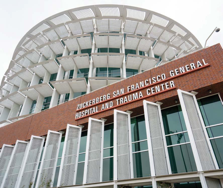
Jason Talbott, MD, PhD Malini Singh, MD, MPH, MBA
Mark Wilson, MD

USPSTF’s Proposed Breast Cancer Screening Guidelines
On May 9, the U.S. Preventive Services Task Force (USPSTF) proposed a significant change to current breast cancer screening guidelines. The USPSTF’s draft proposal recommends lowering the recommended breast cancer screening age for average risk women to 40. Current USPSTF guidelines, in place since 2016, recommend starting biennial screening by age 50.
The new recommendations are a positive step, but don’t go far enough, according to Bonnie Joe, MD, PhD, professor and chief of Breast Imaging at UCSF. “The goal of breast cancer screening is to reduce breast cancer deaths and morbidity - the adverse impact of the disease on the person. We should stick to that goal, and annual screening - not biennial – is the best way to meet this goal for those at average risk.”
A concern, says Dr. Joe, is the fact that USPSTF recommendations are tied to insurance coverage under the Affordable Care Act. Under the proposed guidelines, many people who want to be screened annually may lose coverage for yearly exams. “Those with more resources will still be able to afford to be screened every year while those with fewer resources may be screened less frequently, increasing their chance of their cancers not being diagnosed until they are at a more advanced stage.”
The draft also noted that beginning mammography screening at a younger age and screening more frequently may increase the risk for overdiagnosis (OD) and subsequent overtreatment. “Interval has no effect on OD,” counters Dr. Joe. “Tumors don’t go away, so a truly over-diagnosed cancer will still be there at the next mammogram – no matter whether it is one year or two years between exams.”
Kimberly Ray, MD, associate professor of clinical radiology at UCSF added that the USPSTF recommendations for biennial rather than annual screening will disproportionately affect minorities noting that one-third of all breast cancers in Black, Asian, and Hispanic women are diagnosed under age 50. She added that:

■ Among all women with breast cancer, minority women are 72% more likely to be diagnosed with invasive breast cancer under age 50 years and 58% more likely to be diagnosed with advancedstage disease under age of 50 years than non-Hispanic white women.

■ Black women have 40% higher breast cancer mortality than non-Hispanic white women. Contributing factors include higher incidence of BRCA1 and BRCA2 mutations and twice the incidence of aggressive, triple-negative breast cancers in Black women. These cancers with aggressive biology tend to present at a younger age, more often before age 50.
Dr. Ray says that the above disparities can be reduced if more minorities are encouraged to undergo routine annual screening mammography. “Annual screening results in cancer detection at an earlier stage and fewer interval cancers (symptomatic cancers that present in between screening exams) than longer screening intervals. Diagnosing cancers at an earlier stage means that people will have better survival outcomes and may be spared the morbidity of more aggressive treatments."
Also potentially impacted by the proposed guidelines, Ray notes, are LGBTQ patients, who are less likely to present for cancer screening than non–LGBTQ patients. The American College of Radiology (ACR) recommends annual screening mammography beginning at age 40 for transfeminine (maleto-female) patients who have used hormones for ≥5 years, as well as for transmasculine (female-to-male) patients who have not had a mastectomy.
Finally, in addition to routine annual screening at age 40, the ACR now recommends that all women undergo breast cancer risk assessment at age 25. This will enable those women who are at elevated risk to undergo high risk screening beginning at age 25-30 with annual breast MRI in addition to mammography as well as to consider other risk reduction strategies.
Bonnie Joe, MD, PhD
Kimberly Ray, MD
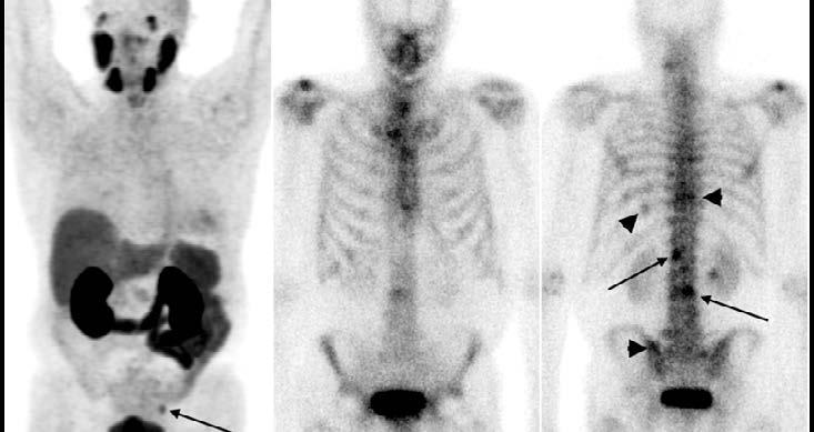

For Some Prostate Cancer Patients, PSMA PET May Be Better for Initial Staging of Disease
Arecent article in the Journal of Nuclear Medicine reports findings of a multi-center retrospective study that compares bone scan with PSMA PET for initial staging of prostate cancer. Authors include UCSF Radiology and Biomedical Imaging faculty members Thomas Hope, MD, Roxanna Juarez, MD, Miguel Hernandez Pampaloni, MD, PhD, and clinical research coordinator Daniel Thompson.
Titled “Do Bone Scans Overstage Disease Compared with PSMA PET at Initial Staging? An International Multicenter Retrospective Study with Masked Independent Readers,” the purpose of the study was to improve our understanding of how to apply clinical trial data based on conventional imaging to patients staged using PSMA PET. To do this, the researchers evaluated the ability of bone scans to detect osseous metastases using PSMA PET as a reference standard. Regarding potential improvements to patient care, overstaging for metastasis – when local disease is a more accurate diagnosis – may subject patients to treatments that increase therapy-related morbidity.
The study included 167 patients with prostate cancer, who were imaged with bone scans and PSMA PET performed within 100 days of one another to compare interpretations of the bone scans at various stages of disease, using the results of the PSMA PET as a reference standard.
In a recent post on Twitter/X, Dr. Hope noted, “If you thought PSMA PET always led to stage migration, you might want to think again. At initial staging over half of patients with bone mets on bone scan were negative on PSMA PET. Sometimes increased accuracy leads to fewer mets!” His tweet has more than 79,000 views to date.
The authors summarize the study’s conclusions: “In this multicenter retrospective diagnostic study using PSMA
PET as the reference standard, the positive predictive value (PPV) of bone scans at initial staging was low (0.43). This results in incorrect staging (as having osseous metastasis) of more than half of patients in this group. This overstaging by bone scans is important when applying clinical data from trials such as the STAMPEDE M1 RT trial to patients being staged with PSMA PET. Before patients receive prostate bed RT in low-volume metastatic disease on PSMA PET, further work should be performed to understand if the results of the STAMPEDE M1 RT trial are generalizable.”
Study co-authors include Matthias Benz, Martin AllenAuerbach, Pawan Gupta, and senior author Jeremie Calais of UCLA; Fei Jiang of UCSF Epidemiology and Biostatistics; and Francesco Barbato and Wolfgang P. Fendler of the University of Duisburg–Essen.

If you thought PSMA PET always led to stage migration, you might want to think again. At initial staging over half of patients with bone mets on bone scan were negative on PSMA PET. Sometimes increased accuracy leads to fewer mets! Check it out from JNM: bit.ly/3qLnMWO Do Bone Scans Over Stage Disease Compared to PSMA PET at Initial Staging?

Thomas Hope, MD

Mount
Zion

is Once Again an Inpatient Hospital Where Imaging Plays an Important Role
By Francis Horan
The resumption of in-patient care at Mount Zion Hospital in 2020 after many years as an outpatient site presented an opportunity for imaging expansion. The pandemic-provoked transformation was not the first time this hospital has reinvented itself in its long history. On November 3, 1887, 43 members of San Francisco’s Jewish community met for the first time to plan a hospital in the San Francisco area, "for the purpose of aiding the indigent sick without regard to race or creed, to be supported by the Jewish community." The hospital, dubbed Mount Zion, was given the mission to practice healing, grounded in learning and supported by acts of personal kindness. In 1990, UCSF acquired Mount Zion, more than a century after its founding as San Francisco’s first Jewish hospital.
When COVID-19 began to spread in California in March 2020, Mount Zion sprang into action, opening an intensive care unit and two acute care units to provide capacity in case of a surge. However, San Francisco’s success at “flattening the curve” and slowing the initial speed of the pandemic meant that much of that capacity was not needed.
“Mount Zion reopening as an inpatient hospital has provided UCSF Health with a significant increase in capacity. Mount Zion plays a critical role in fulfilling UCSF Health’s 2025 vision as a leading complex care provider and a destination for tertiary and quaternary care in the Bay Area.”
When Mount Zion Hospital went 24/7, all the imaging modalities based there had to become quite creative, working together to develop plans for how to provide full services even with temporarily reduced staff levels. Leading imaging’s organizational response during this transition were David Sostarich, Radiology Operations Director, Jessica Pfannenstiel, BSRT (R)(CT), Manager of CT Operations, Tosca Bridges, M.S., R.T.(R)(CT)(QM), Chief Manager of Diagnostic Radiology, Craig DeVincent, R.T.(MR), Manager of MRI, Amy Vincent, BS, R.T.(M), Chelsy Coco, BS, RDMS, Manager of Ultrasound, Alpana Patel Camilli, BS, CRA, RT (CT), Manager of Interventional Radiology and Neuroendovascular Surgery.
Alpana described the Mount Zion radiology teams in familial terms, “We’ve always known that we are a part of something greater here: a family, a community. Mount Zion has a smaller footprint than other UCSF operations. The departments and modalities are closely located so the various modality staff know each other well, creating a unity that is hard to match.” Cooperation between the modality leaders and their respective teams was the key foundation for making the transition successful. Alpana described it so, “The Diagnostic, CT, MRI, Ultrasound and Mammography leadership were all part of the remarkable joint effort that enabled Mount Zion’s transformation. One of the wonderful aspects of Mount Zion is the modalities are not so separated. This physical proximity helps bring on a collegial atmosphere.”
Supervisors and managers took the lead to provide coverage, as the teams used an all-on-call process to staff the gaps. Radiology technologists with experience in multiple modalities were the key, as their knowledge and adaptability provided the flexibility to meet this new level of demand. These skills are still being cultivated; two diagnostic techs are currently cross training in interventional radiology, several diagnostic techs successfully trained in MRI and CT. When orthopedic services first expanded at Mount Zion, the technologists, X-Ray & CT techs in particular, needed to shuffle and stretch operations to quickly match the demand. Yet despite these extraordinary demands, the team fell into place and executed the transition with efficiency and unfailing professionalism.
Mount Zion radiology fosters a learning environment with a feeling of unity and a loyal community among its physicians, staff, and technologists. Chester Lim, Diagnostic Supervisor, has been a part of the team since the onset of Mount Zion operation, over 20 years ago. Those who work with Chester have called him, “the heart and soul of the department.” Chester has helped train dozens of techs through his own valuable experience, providing classes on positioning and patient safety. This contribution is volunteered without having ‘educator’
anywhere in his official title. Alpana noted that Chester “is extremely passionate about education and this is his contribution, his legacy at UCSF. He obtains great satisfaction from seeing the students graduate, pass their board exams, and then apply for a role with us at UCSF. Chester is the person to contact if something unexpected happens or something goes wrong.”
Yelena Borodina, known as Lena to her coworkers and friends, is the Mount Zion Site Manager, responsible for imaging administration and facilities coordination. She is also the best person to educate anyone on the organizational history of the hospital from its earliest days to the present, even though Lena’s official offices are located in the Breast Imaging section at Mission Bay. Camilli observed that, “Lena has a vision and a passion for supporting people that allows her to work so well together with campus and medical.”
Camilli remarked that team members like Chester and Lena are crucial to Mount Zion’s thriving: “Our teams embrace change and view workflows and practices through the lens of continuous improvement. We directly involve our respective teams in the improvement process. For example: have your team follow a workflow idea suggested by the technologist or nurse. Have the team provided feedback at the end of the day; can any step be adjusted or fine-tuned? We create an environment where it is allowable to stumble, and then to try again.”
MRI Lab at Mount Zion Mount Zion may not be the largest medical center at UCSF, but it is busy. At Mount Zion, imaging includes X-ray, MRI, CT, bone density, ultrasound, mammography, and interventional radiology provided at the Main Hospital Building on Divisadero Street, the Women’s Health Center on Sutter Street, and the Medical Offices Building on Post Street. Mount Zion imaging recently added two MRI resources (a 3T and 1.5T) to accommodate advanced studies, with additional imaging services in the planning stages. Change is a constant at Mount Zion, with Camilli observing that, “We understand that flexibility and adaptation are strengths for us to foster as we move into the future. It is the individuals of our department that contribute to those hundreds of small steps that will move us all together in the right direction.”


Mount Zion reopening as an inpatient hospital has provided UCSF Health with a significant increase in capacity. Mount Zion plays a critical role in fulfilling UCSF Health’s 2025 vision as a leading complex care provider and a destination for tertiary and quaternary care in the Bay Area.” “
~ Christopher Holland, Executive Director and Site Administrator
Left to Right: Wes Maupin, RN, Cecilia Villar, BSN, Michelle Devaux, RN, and Mike Wollslair, RN
Chester Lim, RT supervisor
Alpana Patel Camilli, manager BIR/NES, and Yelena Borodina, Mount Zion Site Manager
Neuroendovascular Surgery: Reflections on the First Year
Neuroendovascular Surgery (NES) was established as a single division on November 1, 2022, when the Neurointerventional Radiology (NIR) team joined the Neurosurgery Cerebrovascular Surgery team, in a partnership between the Department of Radiology & Biomedical Imaging and the Department of Neurological Surgery, with support from the UCSF School of Medicine and UCSF Health.
“This combined structure has allowed our faculty to focus on delivering the highest quality patient care and developing innovative clinical and research programs that leverage the diverse skills of our seven full-time, three emeritus faculty, and seven advanced practice professionals across five hospitals,” said Dr. Steven Hetts, Co-Chief of Neuroendovascular Surgery at UCSF Mission Bay with Luis Savastano, MD, PhD.
“What’s really exciting is the combination of endovascular and surgical expertise, building on UCSF’s pioneering work in stroke treatment over the past four decades,” noted Dr. Hetts. Now entering its second year, NES division leadership is focused on merging administrative processes between neurosurgery and radiology, expanding clinical trial participation, and building community outreach. “The vision goes beyond unifying the neuroendovascular surgery group,” said Dr. Savastano. “For too long patient care has been managed by a single department, rather than thinking from the perspective of a particular disease. And this idea of combining two specialties that deal with vascular problems or neurovascular problems is a step towards that vision of having a more disease-centered practice.”
One of the largest neuroendovascular practices on the West Coast, the NES team is using its deep expertise in translational research to continue building successful subspecialty practices – pulsatile tinnitus, hereditary hemorrhagic telangiectasia (HHT), birthmarks, and vascular anomalies – with newly focused efforts in interventional neuro-oncology, moyamoya disease and obliterative arteriopathy, and brain arteriovenous malformations (AVM) to improve patient outcomes. Sub-specialization allows the team to push the understanding of diagnosis and treatment for a particular disease forward and to share that expertise with colleagues.
Interventional Neuro-oncology. NES is branching out beyond the blood vessels to focus on neuro-interventional oncology to treat tumors in the central nervous system, head, neck, and spine. An area of significant success is in treating retinoblastoma patients with endovascular chemotherapy, saving 80% of eyes in babies that may have otherwise been removed. With body interventional radiology, NES clinicians are exploring primary tumor

(L-R) Neuroendovascular Surgery Division Co-Chiefs Steaven Hetts, MD, Professor, Department of Radiology, and Luis Savastano, MD, PhD, Assistant Professor, Department of Neurological Surgery

treatment in the head and neck using MR-guided highintensity ultrasound for tumor ablation and to temporarily make the brain more permeable for drug delivery, which is expected to be transformational in the next decade.
SPOTLIGHT ON NES CLINICAL AND RESEARCH INNOVATION
Pediatric Patients Undergo a Single Anesthetic for Surgery and Imaging. Pediatric AVM treatment at UCSF Medical Center at Mission Bay leverages a fusion between MR imaging and x-ray angio imaging for embolization and surgical planning, and MB OR 23 allows state-of-the-art craniotomies for surgical AVM resection combined with the best quality intraoperative angiography, thus allowing children to undergo a single anesthetic for both surgery and imaging.
Neuro Hub at New UCSF Medical Center at Parnassus Heights. Planning for a comprehensive “neuro hub” is underway for the new UCSF Medical Center at Parnassus Heights to improve care for patients with stroke, brain tumors, and cerebrovascular disease. Modeled after Zuckerberg San Francisco General Hospital’s new facility that brings imaging services to the patient, three neuro angio suites and one operating room will connect directly to an intraoperative MRI suite, combining x-ray angio and MRI imaging capabilities in one location.
Research collaborations include colleagues at the VA Advanced Imaging Research Center. Recent highlights include Department of Defense grants for HHT MRI and DAVF MRI; an NIH grant for ChemoFilter device development; industry contracts for clinical trials Core Lab Services and venous sinus flow and stent analysis; genetic analysis of CNS vascular malformations; and endovascular and percutaneous device development. In the past two years, NES faculty have authored more than 100 publications and are co-founders of four active startup companies.
Brain Focused Ultrasound: A New Era of Image-guided Neuro-intervention
AUCSF team that includes members of the Department of Radiology and Biomedical Imaging, is addressing new clinical and research neuro applications of High Intensity Focused Ultrasound (HIFU) and Low Intensity Focused Ultrasound (LIFU).
"From the outset we wanted the UCSF Focused Ultrasound in Neuroscience Program to be multidisciplinary with close ties between the clinical and research arms," says Leo Sugrue, MD, PhD, associate professor who co-directs UCSF’s clinical program in focused ultrasound with Doris Wang, MD PhD, assistant professor in the Department of Neurological Surgery. Dr. Sugrue, Kazim Narsinh, MD, assistant professor and neurointerventional radiologist, Eugene Ozhinsky, PhD, assistant professor, Kisoo Kim, PhD, postdoctoral scholar, and Tommaso Di Ianni, PhD, recently hired assistant professor in the Department of Psychiatry, will present their work on brain applications of HIFU and LIFU in a webinar on March 28 at noon PDT.
“We traditionally think of ultrasound in terms of its uses in imaging parts of the body,” says Dr. Sugrue. “‘Focused’ ultrasound concentrates ultrasound waves at a particular location in order to deliver energy in a targeted manner to a particular part of the body to produce a physiological effect.”
His presentation will introduce the Focused Ultrasound in Neuroscience Program and describe how he is using HIFU clinically to treat Essential Tremor, the most common movement disorder, by ablating a specific part of the thalamus – an important structure deep in the brain. Dr. Sugrue will explain the key role that imaging plays in guiding these treatments and ensuring excellent tremor reduction while minimizing potential side effects.
Dr. Narsinh’s talk will explain applications of LIFU in Neuro Oncology. LIFU is an innovative technique that uses low

Leo Sugrue, MD, PhD, associate professor, co-directs UCSF’s clinical program in focused ultrasound

Kazim Narsingh, MD, assistant professor and neurointerventional radiologist
intensity and lower frequency US to produce non-thermal effects on the target tissue. One specific use of LIFU is to temporarily change the permeability of the blood brain barrier, a specialized barrier that protects the brain, so that anti-cancer drugs can be delivered in high concentrations to brain tumors. Another application is to allow liquid biopsy – the genotyping of tumors based on tumor DNA circulating in the blood – which has the potential to replace invasive tissue biopsy.
Eugene Ozhinsky, PhD and Kisoo Kim, PhD will discuss their current research which focuses on an important research area: MR techniques to monitor focused ultrasound imaging effects on the brain in real time. Specifically, they are developing advanced thermometry (in the case of thermal ablation) and perfusion and diffusion-based MR imaging techniques (in the case of the blood brain barrier opening). Dr. Kim says that while MRI technologies have improved, there is still a need for further development to advance MRguided focused ultrasound in the brain.
Dr. Tommaso Di Ianni’s talk will provide an overview of promising research using LIFU for neuromodulation –modifying the activity of brain circuits in neuropsychiatric applications. Although LIFU is not yet used clinically to treat neuropsychiatric disorders, ongoing research is exploring its use in clinical applications that this modality could potentially address. Some of the disorders that are currently being investigated for ultrasound neuromodulation treatment are Major Depression, Obsessive Compulsive Disorder, sleep disorders and substance abuse.
According to Dr. Di Ianni, LIFU neuromodulation has the potential to provide a new tool for noninvasive mapping of brain circuits in individual patients – allowing for personalized targeting of circuit-based treatments with either focused ultrasound or more traditional approaches like Deep Brain Stimulation.
Eugene Ozhinsky, PhD, assistant professor
Kisoo Kim, PhD, postdoctoral scholar

Tommaso Di Ianni, PhD, assistant professor in the Department of Psychiatry
Some of Our Research Lab Groups


Rob
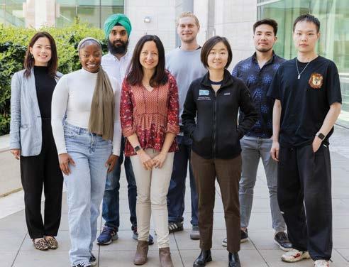


Raj Lab: (L to R) Huaqing Jin, PhD, Ashish Raj, PhD, Parul Verma, PhD, and Benjamin Sipes, MS.
Chemistry, Probes, and Molecular Therapy PIs: (L to R)
Flavell, Mike Evans, Henry VanBrocklin, David Wilson
Breast Research Group: Bottom (L to R) Judith Zimmerman, Nola Hylton, Bonnie Joe, Lisa Wilmes, Margarita Watkins, Teffany Bareng. Top (L to R) Julia Carmona-Bozo, Natsuko Yamashita, Jessica Gibbs, Wen Li
Cardiothoracic radiologist Yoo Jin Lee, MD (left) with cardiologists Tilo Thottakara, MD, & Roselle Abraham, MD
Lupo Lab: (L to R) Jenny Li, Oluwaseun Adegbite, Paramjot Singh, Janine Lupo, Jacob Ellison, Hui Lin, Nate Tran, Bo Liu
Kazakia Lab: (L to R) Front: Gabrielle Joseph, Galateia Kazakia, Isra Saeed, Izzie Yu (UC Berkeley undergrad) Back: Pranav Kolluri (UC Berkeley undergrad), Minhao Zhou, Po-Hung Wu, Gaik Mkrtchian (UC Berkeley undergrad)
RESEARCH

RESEARCH ACHIEVEMENTS AND HIGHLIGHTS

WVice Chair's Overview
Sharmila Majumdar, PhD Vice Chair for Research
e are proud to present here our collective efforts and commitment to research excellence. Over the past year, our grant funding has continued to grow, and we are #2 in NIH funding nationally, which is a testament to our journey of growth, innovation, the dedication of our researchers, the resilience of our teams, and the unwavering support of our collaborators. In an ever-evolving landscape of pandemic changes, rapid technological advancements, and dynamic global changes in human resources, we have steadfastly pursued meaningful basic, translational, and clinical research. In this edition of Images, you will find an in-depth exploration of our key accomplishments and strategic partnerships that are propelling our vision of innovative imaging in patient care.
From renewing our NIH funding for the Hyperpolarized MRI Technology Resource Center (HMTRC) to the advanced studies conducted with FDA-approved gallium PSMA, our efforts in molecular imaging, theranostics and probe development flourish and grow with publications and funded research.
This year we celebrated the third anniversary of the Center for Intelligent Imaging, hosting international collaborators from two German universities: Frederich-Alexander Universität in Erlangen and Christian Albrecht Universität of Kiel. These events featured lectures by world-renowned researchers, publications in leading journals, and research grants for interstitial lung disease, lower back-pain, neuroscience, oncology and more.
In Imaging and Neuroscience, innovation continues in basic and translational research, with significant strides into the clinic. Two highlights in this arena are high-intensity focused ultrasound to treat essential tremors and applying imaging insights to deep brain stimulation for Parkinson’s disease. These are just a few examples, but each of our research achievements reflects our commitment and dedication to creating meaningful advances in patient care.
We anticipate 2024 with optimism, enthusiasm, and ambitious goals. We have new collaborations planned with our colleagues in Psychiatry and other departments at UCSF.
We are dedicated to the success and well-being of our staff, faculty and researchers. On behalf of the entire department, I can only end with extending our deepest gratitude to all who have been part of our success.
Research Administration Leadership
As our research enterprise has grown exponentially in the past several years, the Research Administration Leadership team is designed to support faculty programs and new initiatives and ensure alignment with our strategic plan. Reporting to Amy Pradhan, CAO, and in partnership with the Chair and Vice Chair of Research, this team proactively evaluates existing processes and systems, designs new approaches where needed, and serves as a resource for faculty in their areas of expertise and responsibility.

Robin Ippisch, PhD, Director of Research Development, oversees the scientific aspects of research administration. This includes coordinating clinical research activities and monitoring human and physical resources to ensure growth and development of the research enterprise. Robin manages the Radiopharmaceutical Facility (RPF) and the Clinical Research Coordinator (CRC) Core, interacts with all scientific steering committees to prioritize strategic research directions, reviews scientific content on new proposals for Innovation Ventures (licensing and agreements) and Industry Contracts for feasibility, and enforces and creates new standards for research conduct in partnership with the Vice Chair of Research.

Kathryn Wold-Murphy, Director of Research Finance, oversees the financial aspects of all sponsored research in the department including supervision of the Post Award team, oversight of research financial compliance and award verification and reconciliation, and proposal review to ensure eligibility. Kathryn is also the liaison with the pre-award teams (Research Management Services and Industry Contracts), other departments, and the controller’s office.

Mabel Zelaya, Director of Site Operations, manages the administrative operations and physical facilities at our China Basin site and Mission Bay campus. In addition to oversight of space allocation, construction, and upgrade projects for research labs, Mabel manages day-to-day staff administrative services and supervision of several research and clinical admin units. She is the liaison with HR for the full range of research staff and post-doc recruitment and hiring. She is also responsible for short- and long-range needs assessments to appropriately support the department’s overall goals and missions.

Kathryn Wold-Murphy
Mabel Zelaya
Robin Ippisch, PhD
Specialized Resource Groups
Chemistry, Probes, and Molecular Therapy
By David Wilson, MD, PhD
The Chemistry, Probes, and Molecular Therapy (CPMT) Specialized Resource Group represents a varied collection of clinicians, physician-scientists, and basic scientists in the Department of Radiology and Biomedical Imaging at UCSF. As a group, we want to image the biologic processes underlying cancer, infection, dementia, and other diseases- using innovations in chemistry. This central goal of molecular imaging is increasingly accompanied by radiotherapy, whereby alphaand beta-particle emitting radioisotopes are delivered specifically to diseased tissues. The breadth of CPMT research is reflected by recent accomplishments by the Evans, Flavell, VanBrocklin, and Wilson labs that develop new radiochemistry to better diagnose and treat patients. These accomplishments include numerous NIH and other extramural awards, first-in-human radiotracer studies, and the high-impact publications highlighted here.
The Evans group recently published two articles in ACS Central Science. The first manuscript (Zhao et al., 2021) was written in collaboration with the Craik laboratory in the Department of Chemistry and Chemical Biology and described a new method to image human granzymes- an enigmatic class of enzymes found in natural killer and cytotoxic T-cells. The strategy used an innovative strategy whereby granzyme B activates a positron emission tomography reporter (64Cu-labeled) based on proteolytic cleavage (Figure 1). With R01 funding confirmed for granzyme B-targeted human studies, the Evans lab turned to targeted radionuclide therapies that use covalent
(irreversible) binding to their targets, resulting in the second publication (Klauser et. al., 2023) in collaboration with Dr. Flavell’s lab and Lei Wang’s lab in the School of Pharmacy. This manuscript describes the attachment of an alphaemitting 225Ac-labeled nanobody to tumors for more efficient radiotherapy.
The Flavell group has recently explored radiotherapy using both alpha and beta-emitters and imaging using the novel radionuclide 134Ce, resulting in several exciting and impactful publications. Dr. Flavell’s group published a manuscript in Clinical Cancer Research (Bidkar et al., 2023) describing a new CD46-targeted antibody tagged with the alpha emitter 225Ac, to target prostate cancer (Figure 2). Interestingly, the antibody used binds to a tumor-selective conformational epitope of CD46 allowing for selective cancer treatment. This new radiotherapy suppressed tumor size and prolonged survival in animal models- including those bearing PSMA negative tumors. In the future, patients who cannot be successfully treated with 177Lu or 225Ac PSMA-targeted therapies will have other options for radiopharmaceutical therapy.
The VanBrocklin group published an article in Nature Communications that might revolutionize the way latent HIV infection is understood and treated (Beckford-Vera et al., 2022). One issue with long-term antiretroviral (ART) therapy is the presence of chronically infected cells in a viral reservoir, that may contribute to long-term symptoms. In collaboration with the Henrich lab, the VanBrocklin group labeled an HIV-neutralizing antibody with the positron

Figure 1. Strategy used in the Evans group whereby enzymatic cleavage by granzyme B produces PET signals in tumorassociated cytotoxic T cells.
emission tomography isotope 89Zr (Figure 3). The uptake of this probe depended on detectable viremia, and signals depended on HIV protein expression in lymph nodes. This technology may allow longitudinal characterization of latent HIV infection and has motivated immunoPET imaging of other viruses including SARS-CoV2, which the team is currently studying to better understand long COVID.
The Wilson group has developed a way to rapidly transform the common radiotracer [18F]FDG into other complex radiolabeled sugars using enzymes, several of which are bacteria-specific. As described by a manuscript published in Journal of the American Chemical Society (Sorlin et al., 2023) enzymes could control the way [18F]FDG and other sugars are attached to each other, resulting in rare oligosaccharides including [18F]sakebiose (Figure 4). Sakebiose is a nonfermentable disaccharide found in sake (as the name suggests), beer, and honey. Since the metabolism of sakebiose is poorly understood, the team is currently investigating how bacteria use it, with studies in infected UCSF patients planned. In the future, rapid chemoenzymatic transformation of [18F]FDG will allow radiotracers to be produced “on demand” for efficient patient studies.
Figure 2. Many prostate tumors do not produce PSMA, leading the Flavell lab to develop radioimmunotherapy targeting a new cancer antigen.

Figure 3. The VanBrocklin lab developed a way to image latent HIV with higher signals in lymph nodes in virem ic patients.

Figure 4. The Wilson lab used enzymes to rapidly convert the common radiotracer [18F]FDG to bacteria- and fungus-specific disaccharides.


Permissions
Figure 1: In Vivo Measurement of Granzyme Proteolysis from Activated Immune Cells with PET. ACS Cent. Sci. 2021, 7, 10, 1638–1649. Publication Date: September 2, 2021. https://pubs.acs.org/doi/full/10.1021/acscentsci.1c00529. Used under Creative Commons Attribution CC-BY-NC-ND 4.0.
Figure 2: Treatment of Prostate Cancer with CD46-targeted 225Ac Alpha Particle Radioimmunotherapy. Clinical Cancer Research. Volume 29, Issue 10. 15 May 2023. https://doi.org/10.1158/1078-0432.CCR-22-3291.
Figure 3: First-in-human immunoPET imaging of HIV-1 infection using 89Zr-labeled VRC01 broadly neutralizing antibody. Nat Commun 13, 1219 (2022). https://doi.org/10.1038/s41467-022-28727-5.
Figure 4: Chemoenzymatic Syntheses of Fluorine-18-Labeled Disaccharides from [18F] FDG Yield Potent Sensors of Living Bacteria In Vivo. J. Am. Chem. Soc. 2023, 145, 32, 17632–17642. Publication Date: August 3, 2023. https://doi.org/10.1021/jacs.3c03338. Used under Creative Commons Attribution CC-BY 4.0.
ADVANCED IMAGING TECHNOLOGIES
Leadership in Team Science: HMTRC
By Z. Jane Wang, MD and Dan Vigneron, PhD
2023 Accomplishments
Clinically, several advances in technology development have opened avenues for improved disease diagnosis and treatment monitoring. A milestone achievement in HP C-13 MR technology was

detailed in NMR in Biomedicine, “Probing Human Heart TCA Cycle Metabolism and Response to Glucose Load using Hyperpolarized [2-13C] pyruvate MR Spectroscopy.” This work, by Hsin-Yu Chen et al., demonstrated for the first time HP [213C] pyruvate MRI measurements of metabolic intermediates of the TCA cycle and acetyl carnitine in the fasting and fed states in human heart. The pharmaceutical and MR methods for HP [2-13C] pyruvate developed in this study will enable future studies of metabolic flexibility in the human heart and its alteration in heart disease.
A second major achievement was outlined by Philip M. Lee et al., in the J Magn Reson Imaging article “Whole-Abdomen Metabolic Imaging of Healthy Volunteers Using Hyperpolarized [1-13C]pyruvate
MRI”. The authors developed novel approaches for acquiring HP 13C metabolic images of multiple organs in the human abdomen. Multi-echo gradient echo 1H acquisition was used to inform 13C center frequencies and to overcome challenges of broad spatial coverage as well as B0 inhomogeneities that were typical of previous abdominal imaging. This allowed for characterization of the metabolism of multiple organs including the liver, kidneys, pancreas, and spleen. Metabolite energetics in the abdomen compiled for these healthy volunteers are being used to design larger clinical trials in patients with metabolic diseases. This normative data will be highly valuable for the clinical translation of HP C-13 MRI investigations of cancer and metabolic diseases in the abdomen.
Several clinical equipment upgrades made these studies possible. Through an NIH S10 high-end instrumentation grant, the Center added a second SPINlab polarizer which has increased reliability and optimized workflow for human studies. In December 2023 the Center upgraded the 3T MR750 scanner to the GE Premier platform which will enable improvements in gradient and RF performance to significantly advance HP 13C molecular imaging technology and accompanying 1H MRI.
About the Center
The Hyperpolarized MRI Technology Resource Center (HMTRC) is an NIH National Center for Biomedical Imaging and Bioengineering (NCBIB) funded by the National Institute of Biomedical Imaging and Bioengineering (NIBIB).
Founded in 2011, the HMTRC focuses on development and dissemination of new dissolution DNP (dynamic nuclear polarization) techniques and instrumentation, specialized data acquisition methodology, and analysis software for biomedical research. Hyperpolarized (HP) MR is an emerging molecular imaging method to monitor enzymatic conversions through key, previously inaccessible biochemical pathways. In numerous studies to date, HP carbon-13 (C-13) MR has shown outstanding research and potential clinical value but requires major technological development to realize its full potential. The HMTRC aims to collaboratively develop new technology with training and broad dissemination to advance this field to better detect, characterize and monitor human diseases and ultimately improve clinical care.
HMTRC at a Glance
265+ publications both pre-clinical & clinical HP studies
15 sites doing clinical HP studies
90+ human research publications in HP C-13 MRI
29 grants totaling $97M at UCSF
19 other funded grants supported at other institutions
Philip Lee, PhD Postdoctoral Researcher
Hsin-Yu Chen, PhD Senior BioEngineer
Preclinical Studies
Preclinical studies play a critical role developing and testing first-in-human HP imaging probes as exemplified by a recent article in ACS Sensors, “Clinically Translatable Hyperpolarized 13C Bicarbonate pH Imaging Method for

Use in Prostate Cancer” by Changhua Mu et al. This landmark publication demonstrated the production of highly polarized and safe HP H13CO3, a pHsensitive contrast agent suitable for human injection. Preclinical imaging studies validated the reliability and accuracy of measuring acidification in healthy kidney and prostate cancer. These methods and findings were used to support an Investigational New Drug application to the Food and Drug Administration and the recent use of this probe in a clinical trial assessing the role of tumor pH in predicting prostate cancer aggressiveness.

Co-clinical studies that mimic the human situation allow researchers to test and optimize probes and approaches to imaging and data analyses. A recent publication in Tomography, “Animal Models and Their Role in Imaging-Assisted CoClinical Trials” by Donna Peehl et al., describes well-characterized and phenotyped genetically engineered mouse models (GEMM) and patient-derived xenografts (PDX) with promise for improved therapeutic response. PDX and GEM models are being used in a large number of HMTRC-funded studies of prostate, renal, and brain cancer as well as studies of traumatic brain injury and multiple sclerosis.
These studies were supported by several equipment upgrades within the Preclinical Magnetic Resonance Imaging and Spectroscopy Core Laboratory. These investments allow the acquisition of higher signal-to-noise (S/N) preclinical HP 13C MR studies thereby allowing new less sensitive HP 13C probes and additional metabolic research in preclinical models.

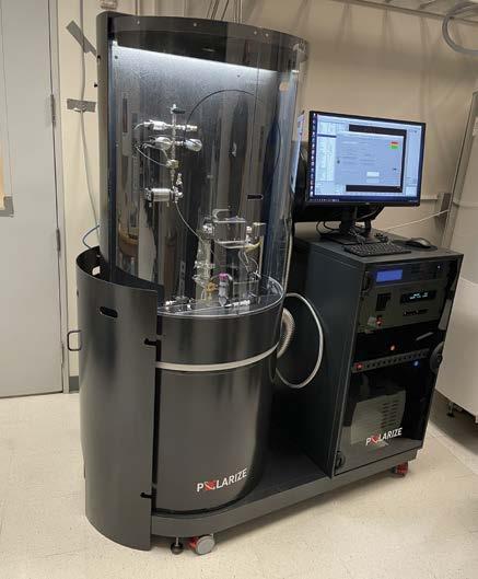


Changhua Mu, PhD Postdoctoral Researcher
New NIH S10-funded preclinical Bruker 9.4T MR scanner with 1H/13C MRI cryoprobe capabilities.
Next generation higher field (6.7T), lower temperature preclinical SpinAligner DNP Polarizer.
Improved RF coils.
Donna Peehl, PhD Professor
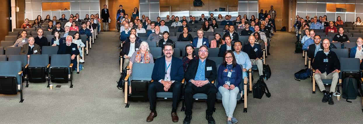
Training & Dissemination
The HMTRC aims to develop new HP MR instrumentation, techniques, acquisition and analysis software through push-pull interactions with external sites. To accomplish this goal we use the Center’s website as a main resource for training and dissemination and for encouraging multisite studies.
At a Glance
■ 55,500+ total views
■ 59.9% of website traffic originates outside California
Research Tools & Dissemination pages
■ 164+ video talks and tutorials
■ 55+ videos added in 2023
■ 17,606 views
■ 31.7% of website traffic
Education & Training pages
■ 20,979 views
■ 37.8% of website traffic
Annual Workshop page
■ 67 presentations
■ 31 presentations added in 2023
■ 1,347 views
ANNUAL WORKSHOPS

April 13-14, 2023
On April 13-14, 2023, the HMTRC celebrated its 11th year as a Center and its annual Workshop continues to have a national and global impact on the advancement of HP C-13 MR molecular imaging technology. The 2023 Workshop boasted the highest ever attendance with 230 participants from academia, industry, and government organizations. International representatives came from China, Denmark, England, Germany, Israel, Japan, Singapore, South Korea, Spain, and Taiwan.
April 3-5, 2024
Our 2024 workshop is scheduled for April 3-5.
Spring 2023 HMTRC Annual Workshop attendees.
At the 2023 HMTRC Workshop, Dr. Z. Jane Wang, MD presented on HP C-13 Clinical Updates at UCSF.
Personnel Highlights
The successes and accomplishments of the HMTRC are a quintessential example of team science. Each member brings unique and valuable talents, experience, and expertise in advancing HP C-13 technology. Here we highlight two Center members.

Daniel Gebrezgiabhier, PhD candidate NIH F31 NRSA Predoctoral Diversity Fellowship Awardee
Danny is a talented imaging scientist with expertise in advanced technology development for MRI. Danny was born in Eritrea surrounded by a turbulent and politically oppressive environment. He survived life-threatening challenges in immigrating to the United States, where he graduated from the University of Michigan with an engineering degree. In the UC BerkeleyUCSF Bioengineering PhD program he focused on developing novel imaging technology to improve clinical care for prostate cancer and other diseases. In 2023, Danny was awarded as PI an NIH F31 NRSA Diversity Fellowship for “New Hardware and Software Developments for Improving Prostate Metabolic MR Imaging.” The goal of this project is to develop new MR molecular imaging hardware and techniques to better detect aggressive cancers in the prostate and local spread to the prostatic bed and lymph nodes. This is relevant for treating prostate cancer patients presenting with advanced disease which occurs two-fold more often in African American men.

Duy Dang, PharmD: HMTRC HP Research Pharmacist
Dr. Dang joined the HMTRC team as an HP Research Pharmacist in June 2022. Prior, he worked at the Regional Medical Center San Jose as a Clinical Pharmacist, and is a board-certified pharmacotherapy specialist. He previously worked at SOFIE, a radiopharmaceutical drug company that manufactures PET (Positron Emission Tomography) drugs. At the Center, Dr. Dang focuses on the clinical translation of novel HP 13C MR probes and their preparations.
With Dr. Jim Slater, Senior Research Pharmacist for Hyperpolarized Isotope Imaging, and two licensed pharmacy technicians, Dr. Dang oversees the HP pharmacy team in developing new methods, probes, and hardware for faster, more robust production of sterile HP stable-isotope C-13 for clinical research MRI studies. Since joining the HMTRC, Dr. Dang has assisted in the development and testing of three probes for FDA-IND submissions of which all agents have been IND- and IRB-approved for clinical studies. In addition, Dr. Dang collaborates with industry on implementing and optimizing new human studies workflows. Moreover, he and the UCSF Hyperpolarize Manufacturing Facility are working on new HP agents to probe additional metabolic pathways and physiological parameters.
International Efforts: Collaborative Development of HP C-13 MRI technology

In 2023 the Center convened researchers from academic institutions, hospitals and industry for collaborative discussions. We host the bi-weekly International SPINlab Users webconference for sites and new users of this advanced equipment. Fifteen sites now perform human studies, for which the Center has shared technical experience. The Center continues to support the SpinAligner webconference of 13 sites with 50 members.
Future Directions
The Center has supported development of five first-in-human probes: [1-13C]pyruvate, [2-13C]pyruvate, urea+pyruvate, alphaketoglutarate, 13C-glycerolcarbonate and HP [13C,15N2]urea. With the FDA-IND and IRB approval, volunteer and patient studies are performed to investigate their clinical value in monitoring and evaluating diseases and predicting treatment response. An IND application for HP 13C-lactate is in the planning stages.
As clinical HP 13C MRI research advances, there is a growing need to build consensus for best practices, which are critical for comparing data across sites, performing multi-site trials, deploying methods to new sites, partnering with vendors, and obtaining regulatory approvals. With this in mind, the HP C-13 MRI Consensus Group, established in 2022, is designing an HP C-13 Consensus Survey to gather recommendations for repeatable and comparable human studies at multiple sites using [1-13C]pyruvate.
A new NIH R01 grant led by Jeremy Gordon, PhD, and Zhen Jane Wang, MD, “Translating Hyperpolarized 13C MRI as a Novel Tool to Predict Treatment Response in Pancreatic Cancer.” aims to develop a new approach to monitor and predict treatment response in patients with pancreatic ductal adenocarcinoma. Data from this first study will inform future investigations of metabolic imaging biomarkers for defining clinically relevant thresholds, enable timely therapy switch in non-responders, and guide high-stake decisions about the benefit of a highly morbid surgery, with the overarching goal of optimizing treatment decisions to improve survival for patients with this deadly disease.
HP C-13 MRI Consensus Group
In 2023, this group, with 28 sites and 60+ members, published a position paper on ArXiv titled “Current Methods for Hyperpolarized [1-13C]pyruvate MRI Human Studies” which aims to capture the current successful practices for HP MRI human studies with [1-13C]pyruvate.
UCSF members of the Consensus Group include Peder Larson, Duan Xu, Dan Vigneron, Jeremy Gordon, Jim Slater, Bob Bok, John Kurhanewicz, Renuka Sriram, Michael Ohliger, Myriam Chaumeil, Yaewon Kim, Jenna Bernard, Jenny Che
Consensus Group Members
ASIA
■ Chang Gung Memorial Hospital, Taiwan
■ National Institutes for Quantum Science & Technology, Japan
■ University of Toronto & Sunnybrook
EUROPE
■ Aarhus University, Denmark
■ ETH Zurich, Switzerland
■ IBEC, Barcelona
■ Kiel University, Germany
■ Laboratory for Functional and Metabolic Imaging (LIFMET), Switzerland
■ MIAAI (Medical Image Analysis & AI), Austria
■ NVision Imaging Technologies, Germany
■ Technical University of Denmark (DTU), Copenhagen
■ Technical University of Munich, Germany
■ University of Freiburg, Germany
UNITED KINGDOM
■ Cambridge University
■ Kings College London
■ University College London
■ University of Nottingham
■ Oxford University
UNITED STATES
■ GE Healthcare
■ MD Anderson Cancer Center
■ MSK Cancer Center
■ NIH/NCI
■ Oxford Instruments
■ University of Florida
■ University of Maryland
■ University of Texas Southwestern
■ Washington University CANADA
INTELLIGENT IMAGING SRG
Delegation from Germany visits UCSF to launch AI partnership
with Christian-Albrechts-Universität
By Francis Horan
On Wednesday, June 7, the UCSF Department of Radiology and Biomedical Imaging hosted a delegation of German political leaders and academics from the state of Schleswig-Holstein for the official launch of AI FLEX, a federated learning exchange project with great promise for advancing biomedical imaging and improving patient care. AI FLEX will begin with a large repository of orthopedic radiographs archived at UCSF, training a model to find biomarkers that predict which patients are at risk of suffering hip fractures based on indicators in previous scans.
Sharmila Majumdar, PhD, and Dr. Claus-C. Glüer, research collaborators since the 1990s, are the principal investigators for AI FLEX. Dr. Majumdar is a UCSF faculty member. Dr. Glüer is a faculty member at Christian-Albrechts-Universität zu Kiel (CAU) and the University Medical Center SchleswigHolstein (UKSH), the second largest university hospital in Germany. Frailty and aging are significant population health issues in the US and Germany, hence the PI’s focus on AI research with the potential to reduce morbidity and improve quality of life in aging populations.
Dr. Majumdar said, “This partnership will allow us to use AI imaging to address meet unmet patient needs. AI can see far more than a human reading these images. It can assess an image in its entirety, with all the biology that shows up.” Dr. Glüer observed that, “Establishing a framework for effective cooperation while maintaining data privacy would allow UCSF, CAU, UKSH, and other institutions to scale these approaches to other scientific questions. The advent of advanced AI systems to process vast arrays of data could present answers to urgent public health concerns, further illuminate the implications of climate change, and provide insight to predict and model other world-changing matters.”
Daniel Günther, Minister-President of Schleswig-Holstein, led the German delegation and expressed his enthusiasm for this scientific collaboration showcasing the strengths of UCSF and UKSH and the warm Sister City relationship
AI AI

between San Francisco and Kiel. “Today we are building a new bridge between our regions, one that will benefit our universities, healthcare industry, and patients. The exchange of knowledge always makes us better.”
UCSF participants in the event included Catherine Lucey, MD, UCSF Executive Vice Chancellor and Provost, who delivered opening remarks. Atul Butte, MD, PhD, Chief Data Scientist at UC Health and director, Bakar Computational Health Sciences Institute, spoke to the unique scale of the University of California’s data repositories. Paul Voigt and Alexander Roth, intellectual property attorneys based in the Berlin and San Francisco offices of Taylor Wessing, LLP, addressed data privacy issues in international law. Mark Chandler, director of International Trade and Commerce in the San Francisco Mayor’s Office, was on hand to foster the sister city relationship of San Francisco and Kiel, Germany.
Dr. Simone Fulda, president of CAU, joined in the celebration by video from Germany, along with an enthusiastic audience of late-night watchers. She observed that, “The combination of AI and health is so very important, and this kind of international cooperation and scientific exchange is key to addressing the great challenges that we all face.”
Several speakers at the AI Flex launch, from left: Atul Butte, MD, PhD; Dr. Claus-C. Glüer; Sharmila Majumdar, PhD; Catherine Lucey, MD; and Minister-President Daniel Günther.
Center for Intelligent Imaging hosts Scientific Sessions
By Francis Horan
On Wednesday, March 29, the UCSF Department of Radiology and Biomedical Imaging and the Center for Intelligent Imaging (ci2) welcomed a global crowd of researchers to our Mission Bay Campus. The scientific sessions inaugurated a new collaboration with Friedrich-Alexander University (FAU) of Erlangen-Nürnberg, Germany, and helped mark the ci2’s third anniversary celebrations.
The experts on imaging and artificial intelligence convened to discuss AI applications in neurological, oncological and musculoskeletal imaging. Christopher Hess, MD, PhD, Chair of Radiology and Biomedical Imaging, and Sharmila Majumdar, PhD, Co-Executive Director of ci2 and Vice Chair for Research in Radiology gave introductory remarks. Joachim Hornegger, PhD, (Dr.-Ing), President of FAU, shared FAU’s past and present history of innovation with attendees.
This meeting of research universities was intended to foster an environment of collaboration between FAU and UCSF/ci2. FAU, founded in 1743, has been a European leader in technological development for nearly three centuries. FAU’s partnerships with German and EU industrial and technological innovators match well with UCSF’s linkages to Bay Area biotech innovators.
UCSF and ci2 were represented by presenters Yang Yang, PhD, Aniket Tolpadi, Valentina Pedoia, PhD, Christoph Krist, PhD, Jae Ho Sohn, MD, MS, Beck Olson, Andreas Rauschecker, MD, PhD, Abhejit Rajagopal, PhD, Peder Larson, PhD and Andrew Burghardt.
Dorin Comaniciu, PhD, Senior Vice President for Artificial Intelligence and Digital Innovation at Siemens Healthineers,


concluded the scientific sessions with a keynote entitled “Artificial Intelligence for Healthcare, the Road Ahead.” After highlighting the chronically severe shortage of staffing in healthcare, he discussed the AI evolution as a major component of the increased automation and productivity in healthcare that is required to meet increased demand in patient care. He also advanced the argument that "artificial intelligence is not here to replace doctors, it’s here to augment their intelligence and relieve them from repetitive tasks so they can focus on what they do best – patient care."
The UnConference breakout sessions on Thursday, March 30, were an opportunity for attendees to engage more fully with the topics presented in Wednesday’s scientific talks. Questions of possible collaboration, other research avenues, and discussion over results from the first day sprouted an intense series of break-out groups, in a flexible conference environment designed to adapt to these emergent topics. While the international arrivals had been disappointed by their arrival in sunny California being greeted by the quenching rain, old Sol emerged on Thursday to welcome the thriving creativity and connections. After a light breakfast and a bit of socializing, groups comprising of students and faculty from both UCSF and FAU formed organically to continue the discussions that had begun the previous day. After a few hours of discussions, the groups identified 10 potential projects in the areas of advanced imaging and artificial intelligence that also would involve the exchange of students. Over the course of the year, teams comprised of both UCSF and FAU researchers will submit grant proposals that support these efforts.


RESEARCH GROUPS

Peder Larson, PhD Director, Body Imatging Research Group


Body Imaging
In this issue of Images, we are featuring work by Daehyun Yoon, PhD, who joined the Body Imaging Research Group in January 2023. Yoon is focusing his image acquisition and analysis expertise on diagnosing a broad range of peripheral pain conditions. His research uses PET and MRI to improve the visualization and characterization of peripheral pain generators for enabling directly targeted treatments. UCSF is unusual – perhaps unique in the US – for having MRI scanners with 0.55, 1.5, and 3.0 Tesla main magnetic field strengths from the same vendor (Siemens). This distinction presents a
rare opportunity to rigorously assess the benefits and trade-offs of all three field strengths in postoperative imaging of the rapidly increasing patient population with metallic implants.
Our second story highlights bone quality research. Galateia Kazakia, PhD, and her lab are building on a well-established program in bone quality research to develop novel image analysis approaches to improve our understanding of how bone structure changes in Type 2 diabetes and the implications for clinical status of patients.
People are living longer, and as a result, we’re seeing more patients experiencing chronic pain conditions associated with implant/joint replacement pain. At UCSF, we have the opportunity to develop an optimal MRI strategy to diagnose and treat pain sources for these patients.”
~ Daehyun Yoon, PhD, Assistant Professor

Advanced Imaging of Peripheral Pain Generators
By Daehyun Yoon, PhD

Imaging at 0.55, 1.5, and 3.0 Tesla. The main magnetic field strength of clinical MRI scanners has shifted from 1.5 Tesla to 3 Tesla over the past decades. This is because higher field strength offers a higher signal-to-noise ratio, which, in turn, enables higher spatial-resolution imaging. Contrary to this trend, a new 0.55 Tesla whole-body MRI scanner was recently released. The primary motivation for this mid-field MRI scanner is to improve the accessibility to MRI with significantly reduced initial and maintenance costs.
Aside from the cost-effectiveness, the 0.55 Tesla scanner offers a unique advantage over the 1.5 Tesla and 3 Tesla scanners: substantially reduced magnetic field inhomogeneity. This is particularly useful for MRI of patients with metallic implants. Metal in the human body perturbs the applied magnetic field proportionally to the main field strength of the MRI scanner. The resulting main field inhomogeneity causes image distortion artifacts, which are much more severe in 3 or 1.5 Tesla MRI than in 0.55 Tesla MRI.

Conventional fast spin-echo (FSE) and 3D multispectral imaging (MSI) MRI images of a water phantom with three stainless steel pedicle screws and one plastic replica screw (third screw from the top) acquired at 0.55 Tesla, 1.5 Tesla, and 3 Tesla are shown on the right. At each field strength, 3D MSI images present reduced signal pile-up (abnormally bright) and signal loss (abnormally dark) artifacts compared to conventional FSE images. In both FSE and 3D MSI images, 0.55T images show the least metal-induced artifacts among different field strengths.
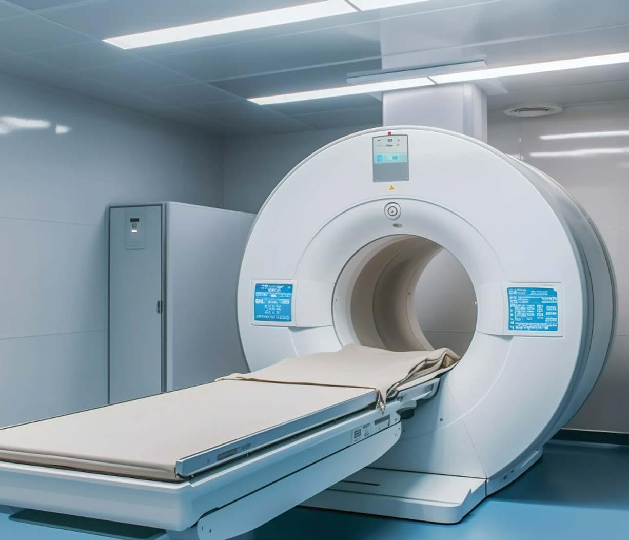
Approximately one million joint arthroplasties and 400,000 spinal fusions are performed in the United States annually. The number of cases is estimated to double by 2030 with the steady increase of the aging population and younger patients. Unfortunately, 10 percent to 30 percent of patients suffer from symptomatic postoperative complications. MRI can enable the early detection of periprosthetic tissue damage if the prohibitive imaging artifacts near implants can be corrected. The substantial metal artifact reduction at 0.55 Tesla MRI provides a new opportunity to develop the next-generation MRI examination of postoperative complications.
116 Million
Adults in the US suffer from common chronic pain conditions, more than the number affected by heart disease, diabetes, and cancer combined.
The cost of pain management for people who suffer from these chronic conditions
$635 Billion

30%
Treatments are only effective 30 percent of the time, partly due to our inability to pinpoint pain generators.
Daehyun Yoon, PhD, Assistant Professor

Imaging Cortical Bone Vasculature
By Galateia Kazakia, PhD
Adecade ago, Thomas Link, MD, PhD, was senior author on a paper in J Bone Miner Res1 that suggested that severe deficits in cortical bone quality are responsible for fragility fractures in postmenopausal diabetic women. Building on that work, our Bone Quality Research Lab has developed a technique to visualize intra-cortical vessels and assess the structural changes that can degrade bone strength.
To understand how the normal vascular structure is altered, rendering the compact cortical shell very porous in some patients, our lab developed a technique that uses both dynamic contrast-enhanced MRI and highresolution peripheral quantitative computed tomography (HR-pQCT)2. Using this new capability, we have analyzed baseline data of the distribution and size of intracortical vessels in patients with diabetes compared to healthy controls.
One imaging challenge is deducing which cortical bone pores house blood vessels and which canals are filled with fat. Using this technique, contrast is injected into the patient and multiple MRI scans are taken to track how the contrast moves through the veins, allowing researchers to image their structure. The cortical bone microstructure

itself does not show up well on MRI, but HR-pQCT can visualize the very fine details of the internal bone structure, allowing researchers to see the pores and canals in the bones through which the blood vessels could travel, while the MRI identifies which actually have vessels threaded through them. Earlier work by the Bone Quality Research Lab has documented that pores that do not contain vessels are filled with fat3.
Results indicate that patients with diabetes have fewer and larger vessels through their bone, whereas in the controls the vessels are more homogeneously distributed and small. We expect that determining the content and spatial distribution of cortical pore space will reveal the biological systems influencing pore expansion.
The overall goal of this study is to understand the longitudinal evolution of human diabetic bone disease and to investigate and visualize the underlying biological processes that drive increased cortical porosity in the setting of T2D. Filling this knowledge gap will elucidate appropriate cellular targets for drug development to prevent or reverse pathological pore development and the associated skeletal fragility.
Dual modality bone vasculature imaging


1) Patsch JM, Burghardt AJ, Yap SP, Baum T, Schwartz AV, Joseph GB, Link TM. Increased cortical porosity in type 2 diabetic postmenopausal women with fragility fractures. J Bone Miner Res. 2013 Feb; 28(2):313-24. doi: 10.1002/jbmr.1763
2) Löffler MT, Wu PH, Kazakia GJ. MR-based techniques for intracortical vessel visualization and characterization: understanding the impact of microvascular disease on skeletal health. Curr Opin Endocrinol Diabetes Obes. 2023 Aug 1; 30(4):192-199. doi: 10.1097/ MED.0000000000000819. Epub 2023 Jun 19
Wu PH, Gibbons M, Foreman SC, Carballido-Gamio J, Han M, Krug R, Liu J, Link TM, Kazakia GJ. Cortical bone vessel identification and quantification on contrast-enhanced MR images. Quant Imaging Med Surg. 2019 Jun; 9(6):928-941. doi: 10.21037/qims.2019.05.23. PMID: 31367547; PMCID: PMC6629562
3) Garita B, Maligro J, Sadoughi S, Wu PH, Liebenberg E, Horvai A, Link TM, Kazakia GJ. Microstructural abnormalities are evident by histology but not HR-pQCT at the periosteal cortex of the human tibia under CVD and T2D conditions. Med Nov Technol Devices. 2021 Jun; 10:100062. doi: 10.1016/j.medntd.2021.100062. Epub 2021 Feb 3. PMID: 37383338; PMCID: PMC10306320
Galateia Kazakia, PhD

Neuroimaging Research Group
By Sri Nagarajan, PhD, Director, Neuroimaging Research Group

The Neuroimaging Research Group includes basic science and clinical faculty with diverse interests in brain imaging and its applications. The vision of this faculty group is to:
1. Understand the relationship between brain and behavior in health and disease;
2. Integrate information between molecules and mind;
3. Translate neuroimaging advances to the clinic.
The approach taken by this group to achieve these goals are: 1) Developing computational and informatics tools for multimodal structural and functional brain imaging; and 2) Understanding structural and functional correlates of behaviors in health and disease. The ultimate goal is to develop and disseminate powerful resources for multimodal structural and functional brain imaging including state-of-the-art computational tools and validated multimodal neuroimaging processes.
A major thrust of the research group is on technology developments in:
■ Imaging data acquisition (Anh Vu)
■ Machine Learning and Artificial Intelligence Algorithm Development (Pratik, Janine, Duygu, Ashish, Sri)
■ Biophysical and Computational Modeling (Ashish, Sri, Yan)
■ Preclinical model development and validation (Pavithra, Myriam)
■ Imaging to understand mechanisms of disease processes (All members)
■ Multimodal imaging data fusion (All members)
a. Structural MRI
b. Diffusion MRI
c. Functional MRI
d. PET Imaging
e. MEG Imaging
f. MR Spectroscopic Imaging
■ Neuromodulation (Leo, Leighton, Sri, Karuna, Melanie)
a. Transcranial Magnetic Stimulation
b. Deep brain stimulation
c. Electrical cortical stimulation
d. Focused ultrasound


■ Mass Spectrometry
■ Theranostics (Leo, Leighton, Sri, Karuna, Melanie)
■ Behavioral interventions (Olga, Tracy, Karuna, Carly, Pratik, Sri)
■ Behavioral Assessments and Human Psychophysics (Tracy, Melanie, Linda, Olga, Leo, Carly, Karuna, Sri, Kamalini, Leighton, Corby)
These technological developments have occurred in the context of:
■ Basic Neuroscience and Biology
■ Brain Tumors
■ Neurodegenerative disorders (Dementia, MS, Parkinson’s disease, Huntington’s disease)
■ Neurodevelopment (Longitudinal study of prematurity, birth asphyxia, and congenital heart defects)
■ Neurodevelopmental disorders (Autism Spectrum Disorders, Dyslexia, Callosal Agenesis, SPD)
■ Neurological disorders (Epilepsy, Stroke, Traumatic Brain Injury, Movement Disorders)
■ Psychiatric disorders (Schizophrenia, Depression, Gulf war illness)
■ Hearing, Speech, and Voice Disorders (Deafness, Tinnitus, Laryngeal Dystonia, Stuttering)
Research Publication Highlights
A fundamental neuroscience topic is the link between the brain’s molecular, cellular, and cytoarchitectonic properties and structural connectivity. Recent studies relate inter-regional connectivity to gene expression, but the relationship to regional cell-type distributions remains understudied.
In a recent paper published in the prestigious journal Cell Reports from Dr. Ashish Raj’s Lab, “Spatial cell-type enrichment predicts mouse brain connectivity,” examined the relationships between regional brain connectivity, gene expression data, and cell-type distributions. They utilized whole-brain mapping of neuronal and non-neuronal subtypes via the matrix inversion and subset selection algorithm, developed by them in an earlier publication, to model inter-regional connectivity as a function of
regional cell-type composition with machine learning. They deployed random forest algorithms for predicting connectivity from cell-type densities, demonstrating surprisingly strong prediction accuracy of cell types in general, and particular non-neuronal cells such as oligodendrocytes.
We found evidence of a strong distance dependency in the cell connectivity relationship, with layer-specific excitatory neurons contributing the most for long-range connectivity, while vascular and astroglia were salient for short-range connections. These results demonstrate a link between cell types and connectivity, providing a roadmap for examining this relationship in other species, including humans.
Dynamic hyperpolarized (HP)-13C MRI has enabled real-time, non-invasive assessment of Warburg-related metabolic dysregulation in glioma using a [1-13C] pyruvate tracer that undergoes conversion to [1-13C]lactate and [13C] bicarbonate. Using a multi-parametric 1H/HP-13C imaging approach, Dr. Yan Li’s lab investigated dynamic and steadystate metabolism, together with physiological parameters, in high-grade gliomas to characterize active tumor.
In a recent paper published in Neuroimage Clinical titled “Multi-parametric hyperpolarized 13C/1H imaging reveals Warburg-related metabolic dysfunction and associated regional heterogeneity in high-grade human gliomas”, they acquired multi-parametric 1H/HP-13C MRI data from fifteen patients with progressive/treatment-naïve glioblastoma [prog/TN GBM, IDH-wildtype (n = 11)], progressive astrocytoma, IDH-mutant, grade 4 (G4AIDH+, n = 2) and GBM manifesting treatment effects (n = 2). Regional analysis of Prog/TN GBM metabolism revealed statistically significant heterogeneity in 1H choline-to-N-acetylaspartate index (CNI)max, [1-13C]lactate, modified [1-13C]lactate-to[1-13C]pyruvate ratio (CELval > NELval > NAWMval); [1-13C] lactate-to-[13C]bicarbonate ratio (CELval > NELval/NAWMval); and 1H-lactate (CELval/NELval > NAWMundetected).
Imaging biomarkers remain mostly unavailable for nonAlzheimer’s disease neuropathological changes (non-ADNC) such as transactive response DNA-binding protein 43 (TDP-43) proteinopathy, Lewy body disease (LBD), and cerebral amyloid angiopathy (CAA). A recent publication from Dr. Duygu Tosun’s group in Alzheimer’s and Dementia titled “Identifying individuals with non-Alzheimer’s disease copathologies: A precision medicine approach to clinical trials in sporadic Alzheimer’s disease” addressed this problem.
A multilabel non-ADNC classifier using magnetic resonance imaging (MRI) signatures was developed for TDP-43, LBD, and CAA in an autopsy-confirmed cohort (N = 214). A model using demographic, genetic, clinical, MRI, and ADNC variables (amyloid positive [Aβ+] and tau+) in autopsy-confirmed participants showed accuracies of 84% for TDP-43, 81% for LBD, and 81% to 93% for CAA,
outperforming reference models without MRI and ADNC biomarkers. In an ADNI cohort (296 cognitively unimpaired, 401 mild cognitive impairment, 188 dementia), Aβ and tau explained 33% to 43% of variance in cognitive decline; imputed non-ADNC explained an additional 16% to 26%.
Accounting for non-ADNC decreased the required sample size to detect a 30% effect on cognitive decline by up to 28%. These results lead to a better understanding of the factors that influence cognitive decline and may lead to improvements in clinical trial design for dementia patients.
Sleep is a highly stereotyped phenomenon, requiring robust spatiotemporal coordination of neural activity. Understanding how the brain coordinates neural activity with sleep onset can provide insights into the physiological functions subserved by sleep and the pathologic phenomena associated with sleep onset. A collaboration between the laboratories of Drs. Nagarajan, Ranasinghe and Raj, published in the Journal of Neuroscience titled “Cortical synchrony and information flow during transition from wakefulness to light non-rapid eye movement sleep,” quantified whole-brain network changes in synchrony and information flow during the transition from wakefulness to light non-rapid eye movement (NREM) sleep. The investigators used magnetoencephalography imaging in a convenient sample of 14 healthy human participants (11 female; mean 63.4 y [SD 11.8]). They also performed computational modeling to infer excitatory and inhibitory properties of local neural activity.
The transition from wakefulness to light NREM was identified to be encoded in spatially and temporally specific patterns of long-range synchrony. Within the delta band, there was a global increase in connectivity from wakefulness to light NREM, which was highest in fronto-parietal regions. Within the theta band, there was an increase in connectivity in fronto-parieto-occipital regions and a decrease in temporal regions from wakefulness to N1.
Patterns of information flow revealed that mesial frontal regions receive hierarchically organized inputs from broad cortical regions upon sleep onset, including direct inflow from occipital regions and indirect inflow via parietotemporal regions within the delta frequency band. Finally, biophysical neural mass modeling demonstrated changes in the anterior-to-posterior distribution of cortical excitationto-inhibition with increased excitation-to-inhibition model parameters in anterior regions in light NREM as compared to wakefulness. Together, these findings uncover whole-brain corticocortical structure and the orchestration of local and long-range, frequency-specific cortical interactions in the sleep-wake transition.
Figure 1. Graphical Abstract of Spatial cell-type enrichment predicts mouse brain connectivity from Dr. Ashish Raj’s lab.

Figure 2. Spatial maps of local neural synchrony across sleep-wake states. A) Mean synchrony across regions in wake (purple), N1 (green) and N2 (orange) sleep states. B) Frequency specific spatial maps of regional local neural synchrony across the sleep-wake states.

Learn more at https://tiny.ucsf.edu/neuroimaging

Vascular and Cardiac Research Group
By David Saloner, PhD
The Vascular and Cardiac Imaging Research Group (VCIRG) has broadened and deepened the scope of investigations pursued by team members. An important development has been the formation of a new service line in Neuroendovascular Surgery (co-led by Drs. Steven Hetts and Luis Savastano) that pools the efforts of Neurointerventional Radiology with Neurovascular Surgery. While esteemed colleagues have retired (Randy Higashida, Chris Dowd, and Van Halbach), the added faculty (Savastano, Ethan Winkler, and Dan Raper) provide new dynamism and expertise. In addition to changes in personnel, advances have been enabled by a broadened array of hardware capabilities including the new 0.55T FreeMax MR scanner which has the potential to show that bigger is not always better!
Technical Imaging Advances. The VCIRG aims to pursue technical developments in quantitative compositional characterization of tissue in the heart and in the vessel wall. Additionally, an important focus has been in utilizing improved acquisition methods together with novel contrast agents for comprehensive evaluation of the vasculature. A promising approach has been to use ultrasmall paramagnetic iron oxides (USPIO) which, unlike other contrast agents, provide constant luminal enhancement over hours rather than tens of seconds.
David Saloner, PhD Director VAMC Research Programs


These are representative images acquired of the head (with 0.5mm isotropic resolution (2-minute acquisition) followed by a chest and abdomen study at 1.2mm isotropic resolution. These methods offer the clinician the ability to provide a comprehensive multiple organ study in one imaging session as opposed to the current practice where visits over multiple days are required.

Research Stories
Parkinson’s Disease and Deep Brain Stimulation
By Francis Horan

Melanie Morrison, PhD, is an assistant professor in residence in the Department of Radiology and Biomedical Imaging at UCSF, and a core member of the UCSF-UC Berkeley Joint PhD Program in Bioengineering. With her team in the new Neuromodulation Lab, she researches imaging techniques with the potential to treat the symptoms of Parkinson’s disease.
Dr. Morrison and her collaborators use MRI and other advanced imaging techniques to improve neuromodulation therapies for patients with neuropsychiatric conditions such as Parkinson’s disease and obsessive-compulsive disorder (OCD). This neuromodulation technique, called Deep Brain Stimulation (DBS), uses an implant that delivers electrical current to targeted brain areas to restore function. Dr. Morrison and her colleagues extract quantitative information from brain MRI images to study and predict patients’ neural and clinical responses to DBS.
DBS was first approved for Parkinson’s treatment in 2002, and there are currently around 200,000 patients worldwide with DBS implants. During the initial procedure, the surgeon places a neurostimulator, a small, battery-operated device, under the skin near the collarbone. This device is connected to electrodes in the brain via wires that run under the skin. The neurostimulator sends electrical impulses to the targeted areas of the brain, which can help to reduce the symptoms of various neurological disorders such as Parkinson’s and OCD.
DBS implants help patients achieve greater stability through the day than medication alone, as the medication levels in their bloodstream vary with natural biological cycles. As Dr. Morrison described it, “DBS is a second line therapy. Patients see a lot of fluctuations in their daily medication cycle, inconsistencies in the medication levels. It doesn’t mean the medication stops working, it’s just less effective as Parkinson’s progresses. DBS helps when they would otherwise feel their motor symptoms come back more frequently throughout the day.”
This therapy has seen great success, but there remains a need to address variability in patient outcomes and the bottleneck to wider implementation arising from limited number of multidisciplinary clinical teams of neuropsychologists, neurologists, neurosurgeons who conduct DBS procedures and provide patient follow up.
Fortunately, there are many potential ways to use MRI to improve patient outcomes and streamline the therapy. As Dr. Morrison describes it, “The automated tools we are currently aiming to develop will help identify the best candidates, and the optimal parameters and settings for the medical device to personalize it to the individual. We are looking for ways to streamline all parts of the implementation from pre-operative candidate selection and surgical planning all the way through post-op optimization of therapeutic device setting.”
Dr. Morrison observed that “The lab is at the beginning of what I hope is going to be a pretty exciting research program. We are developing new tools and imaging techniques for studying different populations that benefit from DBS. Our team includes neurologists and neurosurgeons, as well as neuroscientists and psychiatrists to support us, along with our network of collaborators, and we all work in a very close level of connectivity. This is very in-person, collaborative research, in an exciting growing field.”


Deep Brain Stimulation: Figure design Dr. Sandoval-Pistorius, Neurology.
Faculty Awarded $3.93M Team Science Grant
Metabolic Imaging for Evaluating Brain Cancer
By Rita Gaber

Researchers from UCSF’s Department of Radiology & Biomedical Imaging were recently awarded a $3.93 million Translational Team Science Award from the Department of Defense. This team project is led by Yan Li, PhD, associate professor, Janine Lupo, PhD, professor, and Eugene Ozhinsky, PhD, assistant professor of the VA Advanced Imaging Research Center (VAARC) in collaboration with doctors from the UCSF Department of Neurosurgery.
The purpose of the grant is to create new, artificial intelligence (AI)-based approaches that will enable direct translation of non-invasive metabolic MR imaging methods (MR spectroscopic imaging) into clinical practice. The award will support the research team to develop these tools for evaluating tumor metabolism in patients with glioma, a type of brain tumor, with three specific aims.
The first aim will develop strategies for rapidly scanning and generating high-resolution metabolic images of the whole brain using customized AI-based algorithms to define the optimal scan plane and orientation automatically by finding the general location of tumor within the brain while avoiding regions outside the brain that can cause artifacts.
The team will then develop a fully automated postprocessing workflow to enable spectral processing, quantification, and quality control at the push of a button. These AI-based pipelines will be installed on the scanner console to automatically generate accurate, high-resolution metabolic maps in only a few minutes.
The last aim will evaluate the final tools in patients with recurrent glioblastoma who are about to receive surgery in order to determine their impact on patient care and predicting progression-free survival when combined with other routinely acquired clinical MRI.
“ As metabolic changes often precede anatomic and microstructural changes, employing noninvasive metabolic imaging can potentially enable earlier intervention for treatment modification. We will develop AI-based methods to automate the process of acquiring and generating maps of brain metabolism, which can be incorporated into clinical workflow. We want it to be as simple as the clinician pressing a button.”
~ Yan Li, PhD, Associate Professor
Dr. Lupo noted that “UCSF has been one of the pioneers in performing metabolic imaging in patients with glioma, but it still requires highly specific knowledge and training to acquire good quality data. This has prohibited its translation from a research tool to clinical practice, despite its demonstrated benefits in identifying infiltrating tumor cells that are invisible on standard clinical MRI protocols. By automating the entire workflow, this project will allow the technique to be easily scanned by any technologist and ultimately accessible to all patients with glioblastoma as part of their routine clinical MRI exam.”
Dr. Ozhinsky added that “the result of this study will be a collection of whole-brain, super high resolution metabolic images that represent maps of tumor activity or aggressiveness at a resolution similar to that of standard structural MRIs. Acquiring the data in less than 10 minutes will allow it to easily be added to any existing clinical MRI protocol, which makes it both cost effective and non-taxing on the patient.”
Eugene Ozhinsky, PhD, Yan Li, PhD, and Janine Lupo, PhD
Celebrating 20 Years of Innovation: UCSF’s Center for Molecular and Functional Imaging
By Francis Horan
Twenty years ago, the Center for Molecular and Functional Imaging (CMFI) at China Basin Landing opened its doors with San Francisco’s first 3T MRI and two 1.5T clinical scanners, followed a few years later by a cyclotron which launched our in-house radiopharmaceutical program. Since then, the CMFI has been at the forefront of 3T research and major advances in theranostics and precision medicine. We’re pleased to share highlights and milestones achieved along the way to this 20th anniversary and look forward to celebrating the CMFI’s first two decades with a reception in January.
In the early 2000s, Ronald Arenson, MD, chair of UCSF’s Radiology Department from 1992 to 2017, knew the department needed to expand. “We were totally out of space for research and clinical work at Parnassus. An external review of on-campus construction predicted a very high cost-per-square-foot, but at the moment San Francisco was in the aftermath of the dot-com bust with plummeting commercial real-estate prices. One of our vendor partners mathematically evaluated the optimum location in the city for clinical space, and on that map there was a bull’s-eye over China Basin.”
The CMFI space was built on time and within budget on the third floor of the China Basin Landing and on December 12, 2003, opened with two research instruments: the pioneering 3T MRI and a 16-slice CT. Sharmila Majumdar, PhD, explained the impact of this new technology, “It was the first 3T magnet in San Francisco, and one of the first in any public institution. This made UCSF a leader in high-field imaging.” Dan Vigneron, PhD, also reflects on those early days of high field 3T MRI, “I received a grant to develop unprecedented 3T prostate cancer MRI at a time when 3T was thought to only be useful for brain imaging. But now due to the pioneering work at China Basin, 3T is now stateof-the-art for prostate imaging and other body applications. Also, new high-sensitivity multichannel brain coils were developed in the MRI electronics shop there, and these types of detectors are now widely used worldwide.”
The China Basin facility provided a home for radiology faculty representing physics, nuclear medicine, bioengineering, musculoskeletal research, and neuroradiology. UCSF Health also opened an outpatient imaging location in the same building, and the co-location of these powerful research scanners fostered immediate benefits and a seamless flow between clinical needs and CMFI’s research.
Youngho Seo, PhD, who, with Bruce Hasegawa, PhD, was one of the first researchers to move his lab into CMFI, described the culture that grew there, “There is power in a close network of researchers. The UCSF


approach is to bring in many individual faculty who can establish themselves and their work, rather than relying on one figurehead researcher.” Arenson agreed with this assessment and the results it produced, and noted that “After we opened China Basin, UCSF rose up to be number two or three in the nation in federal (NIH) funded research. The 3T MRI was pioneering in research and it is now UCSF’s clinical norm.” This reflects the rapid translation of research into clinical arenas for which the department is renowned.


The CMFI’s 3T research program generated UCSF’s first 3T prostate exams, first 3T knee images, cutting-edge brain tractography research, and the diffusion imaging program that now is a cornerstone in neurosurgery clinical evaluations. The Center’s first external research collaboration was a project between UCSF professor Ben Francs, MD, MS, MBA, and Clifford Berkman, PhD, of San Francisco State University, who developed early prostate-specific membrane antigens (PSMAs)-targeting PET and SPECT agents for prostate cancer imaging. Now, faculty members Michael Evans, PhD, and Robert Flavell, MD, PhD, lead research on novel theranostic compounds which use paired isotopes to combine imaging and therapy in one molecular structure, and which are in the process phased human trials toward potential FDA approval. Every year, the research group submits two to three Investigational New Drug (IND) applications to the FDA for new compounds submitted for human trials. Henry VanBrocklin, PhD, director of the Radiopharmaceutical Research Program within CMFI, described this pace of constant science, “We’ve translated a number of new radiopharmaceuticals here and have seen significant growth of our radiopharmaceutical research program.”


CMFI’s early days were marked by prominent investments in growth and infrastructure. On Labor Day 2005, the cyclotron was installed in the basement of CMFI’s building, and by December of 2006, UCSF produced its first FDAapproved clinical radiopharmaceutical. Seo described how the cyclotron has facilitated CMFI’s growth, “We are very good at first-in-human imaging studies; they are our forte. We know how to develop compounds from the bench and on into human trials.” Now, with theranostics and precision medicine, CMFI’s close linkage between research and clinical implementation keeps them at the forefront of this rapidly expanding field.
An FDA registered cyclotron both expanded UCSF’s capability for clinical applications and gave access to new radiopharmaceuticals for research. Gone were the days of driving compounds through traffic across the Bay Bridge from Berkeley, and now UCSF researchers are developing many new radiopharmaceutical with potential for translation for human imaging and therapy. VanBrocklin has developed over 10 compounds with FDA approval, and has provided leadership to help vital work like Seo’s research on radionuclide and x-ray imaging instrumentation and physics, Evans and Flavell’s theranostics breakthroughs, and Dave Wilson, MD, PhD’s development of new molecular probes for infection and brain imaging, as well as new agents for hyperpolarized 13C spectroscopy with positron emission tomography (PET) correlates for imaging.
Today, Seo, from his desk in the office that formerly seated the late medical physicist Bruce Hasegawa, PhD, is proud of what he and his CMFI colleagues have accomplished in 20 years, “Our people are very successful today and all help CMFI do great things.”


