DEPARTMENT OF RADIOLOGY AND BIOMEDICAL IMAGING 2021–2022
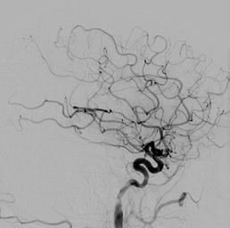
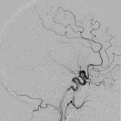
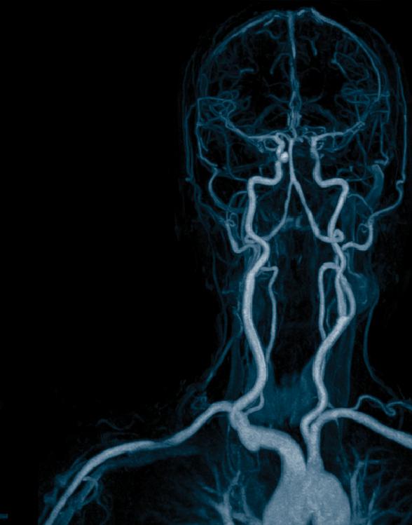
I mages from the embolectomy procedure performed on the patient whose CT perfusion image is on the cover. Both the “before” and “after” are lateral projections with the x-ray camera positioned over the patient’s head.
The “before” image has a paucity of blood vessels because there is a blood clot in the middle cerebral artery that is occluding flow into all of the branches from the middle cerebral artery. The “after” image was obtained after the embolectomy procedure was performed to remove the blood clot in the middle cerebral artery. This shows restoration of normal blood flow to the brain.
Comparing the “before” and “after” images shows the very large number of blood vessels, and subsequent area of the brain, that were at risk of causing permanent stroke for the patient. Thankfully, the blood vessels were able to be reopened in time and the patient made a full recovery.
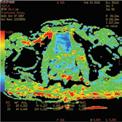
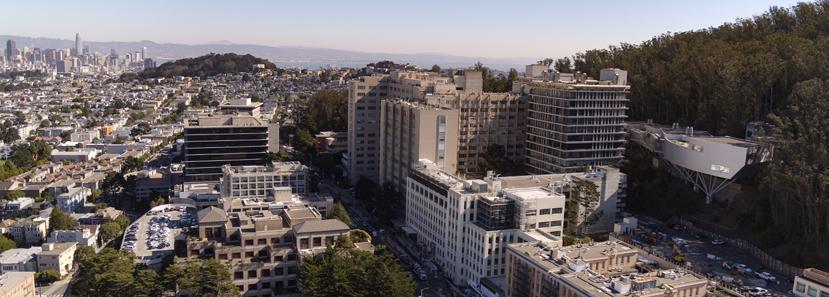
COVER: CT perfusion image showing slow blood flow to the patient’s left middle cerebral artery territory due to a blood clot in the proximal left middle cerebral artery. This study shows the volume of brain that is at risk of becoming permanently damaged by the blood clot starving the brain of normal blood flow.
Executive Editor: Christopher Hess, MD, PhD
Managing Editor: Katie Murphy
Editorial Consultants: Rita Gaber and Steaven Campbell
Copyediting: DEF Communications
Design: Victoria Odson
© 2022 The Regents of the University of California
BEFORE AFTER
TABLE OF CONTENTS
MESSAGE FROM THE CHAIR 02 Reflections on Changes and Accomplishments, 2021-22 OUR MISSION 04 Breast Cancer Survivor Advocates for Screening, Early Detection 06 Collaboration at UCSF Peripheral Nerve Center Helps Difficult-to-Diagnose Patients 08 Neurointerventional Radiology Saves Stroke Patient 10 Transforming Radiology Practice Using Artificial Intelligence 12 New Developments in PSMA PET Imaging for Prostate Cancer PEOPLE: Academic Affairs 14 Promoting Academic Success in Radiology and Biomedical Imaging 16 Faculty Promotions 2021-22 18 Faculty Leadership Appointments 22 New Faculty 28 Faculty Retirements 29 All Faculty List, Clinical and Research Education 32 Training Programs 2021-2022 34 The Class of 2025 38 The Year in Pictures 40 The Class of 2026 43 Residency Program Graduates, Classes of 2021 and 2022 46 Clinical Fellows & Clinical Instructors 2021-22 and 2022-23 48 Goldberg Center 49 Master of Science in Biomedical Imaging 50 Continuing Medical Education 51 Inaugural ER and Trauma Imaging Course Honors and Awards 52 Highlights from Across the Department People 57 Alumnae Reflect on Their Radiology Training and Careers 60 Alumni News 62 Margulis Society 2021-2022 63 In Memoriam: W. Richard “Rick” Webb, MD, 1945-2022 STRATEGIC PLAN PORTFOLIO 64 Five Strategic Aims 65 Expand Our Reach: Faculty Visibility & Service 67 Team Science: Hyperpolarized MRI Technology Resource Center (HMTRC) 70 Team Science: Automated AI Coronary Artery Calcium Scoring is Live 71 Professional Development: Narrative Medicine & Well-Being in New Ultrasound Course 72 Enhance Community: Faculty and Staff DEI Committee Leadership 74 Strengthen Operations: New Emergency Radiology Section at ZSFG 75 Wellbeing and Professional Climate: Connection, Communication, Our Future 76 Strengthen Operations: Successful Collaboration Solves a PACS Challenge IMAGES 2021–2022 1
IN THIS ISSUE
MESSAGE FROM THE CHAIR
Dear Friends:
It has been said that the only constant in life is change. Catalyzed by so many external challenges over the last few years – a pandemic, shifts in healthcare and healthcare economics, historical levels of political division, the list goes on – the forces of change are evident across almost every aspect of what we do in the department. We have new hybrid work environments, new imaging technologies and resources, new image-guided interventional treatments, new sites of practice, new organizational and operating structures, and new paradigms for how we recruit and train the next generation. And this list includes only a few of the changes!
A flag flown in the wind changes direction quickly. Like any healthy organization, we have a history of adapting and responding to new challenges, keeping our flag raised and flying high even amid significant turbulence. But unlike a flag in the wind, as a department we also have unique agency: the ability to harness the winds of change, even during the most extreme storms, and to use them to thoughtfully and intentionally transform the way we work. Across the field of radiology, we are respected as the change-makers, the questioners who constantly challenge the status quo and always strive to improve. We accomplish much more than adapting to change, we create change.
Some things change so precipitously that they are obvious to anyone who experiences them. It’s a badge of pride that we are capable of quickly developing and
bringing new tools and techniques to our patients in our reading rooms, our interventional suites, and our research laboratories. But other things change so imperceptibly that they are evident to most of us only in hindsight. Our culture and our academic community are prime examples. Because clinical excellence, intellectual curiosity, and cultivating future leaders are core values to our identity, challenges to these values take place over a much longer time scale. But even our culture and our community have changed. As an organization and as a department, our culture has become more collaborative, inclusive, diverse, and rooted in teamwork than ever before.
Whether fast or slow, the primary instruments of change at UCSF are the world class people in our department. Our people bring the will, the insight, and the vision to change in new and positive ways. This issue of Images focuses on new people, new programs, new resources and new ideas that have been inspired by forces of change and led to transformation in the UCSF Department of Radiology & Biomedical Imaging. These are communicated on the pages that follow through snapshots and stories of our department’s people and the patients we serve.
Clinical Care. Clinical care drives our enterprise, and since 2021 we have been in a phase of substantial growth across our practices at UCSF Health, Zuckerberg San Francisco General Hospital, Benioff Children’s Hospital Oakland, and the San Francisco
 Christopher Hess, MD, PhD
“ 2 IMAGES 2021–2022
Nothing gets better by chance, it gets better by change.”
Veterans Administration Health System. We have seen a transition from inpatient to ambulatory imaging and a substantial overall growth in volume. We have also experienced a consistent increase in exam complexity, with fewer plain films and an increasing volume of crosssectional imaging.
These trends have required that we bring more people to our group, add operating hours, improve the efficiency of our work, and increase capital investments to meet the ever-rising demand for our services. Our experience with geographical expansion – to Berkeley in the East Bay, and to San Mateo and Redwood Shores on the Peninsula – has proven that demand for imaging outpaces supply within two months of launching new services. In fact, demand for our imaging brand increasingly exceeds our capacity to provide imaging services for our patients, a growing challenge for our department and for the institution. Despite the pressure of keeping up with growth, we continue to bring innovative, new clinical programs to our patients, like MR-guided focused ultrasound for neurologic illness, powerful new molecular diagnostics and therapeutics for cancer, and robotic endovascular neurointerventions.
Research. Our mission to discover and innovate continues to excel. Though because of pandemic shutdowns our NIH funding was lower in 2020 and 2021, our faculty were awarded nearly $60M in 2022, surpassing our previous high of $56M in 2019. Thriving programs in molecular imaging, metabolic imaging, and artificial intelligence complement new programs in accessible midfield MRI 0.55T and environmentally sustainable imaging through “Green Radiology.” Last year our annual departmental symposium on campus, and our research conference at Asilomar, returned as in-person events. P41 funding for our world-class Hyperpolarized MRI Technology Resource Center was renewed. Together with the Departments of Psychiatry and Neurology we launched a plan to develop a stateof-the-art MRI and neuromodulation program in the new Nancy Friend Pritzker Psychiatry Building. And this year, the Center for Intelligent Imaging (ci2) celebrates its third anniversary with a series of events including a debate on the question, Is AI Ready for Primetime in Radiology?
Education. Our education programs are recognized as the premier imaging training at all levels, from medical students through CME. Doximity has ranked our diagnostic residency #1 in the country for the ninth year in a row, and applications to our residency continue to increase at breakneck pace. Our T32 program, now in its 17th year, and sponsoring three fellows, continues its remarkable track record of placing more than 75% of graduates in academic faculty positions around the US.
Conclusion. Our strategic plan 2020-25, implemented just as the pandemic changed everything, continues to guide our success. In the strategic plan portfolio section of this magazine, you’ll see a selection of projects that show how we’re expanding our reach, strengthening operations, delivering on team science and ‘moonshots’ and enhancing wellbeing and professional climate. These achievements are only possible through the inspired efforts of our people, and the snapshots printed here are just a fraction of the creativity and enterprise we are fortunate to harbor.
As we close our first century, we continue a path that has served us well, always charting the future and asking ourselves, “What can we do better?” In 2023 and beyond, we will focus on recognizing prevailing trends, identifying new and ongoing challenges, and responding collectively and individually.
 Christopher Hess, MD, PhD Alexander R. Margulis Distinguished Professor, and Chair of the Department of Radiology and Biomedical Imaging
MESSAGE FROM THE CHAIR
IMAGES 2021–2022 3
OUR MISSION
Breast Cancer Survivor Advocates for Screening, Early Detection
By Rebecca Wolfson
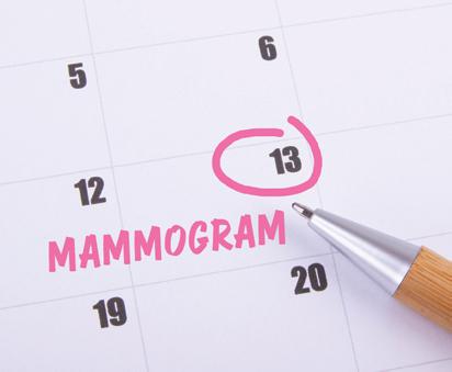
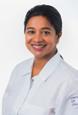
When 53-year-old photographer and single mom Pia Navales went to the Berkeley Outpatient Center for her annual mammogram in December 2021, she had no reason to suspect any problems. “I felt completely healthy. I’d lost weight. I had more energy than ever before,” Navales said. “Suddenly, my life changed on a dime.” The mammogram identified three masses on her left breast, which Navales would later refer to as her triplet Loch Ness monsters.
After a follow-up mammogram and ultrasound with UCSF Professor of Clinical Radiology Rita Freimanis, MD, Navales drove from Berkeley across the Bay Bridge to get a core biopsy and fine needle aspiration done at the UCSF Breast Imaging Clinic at Mission Bay. She arrived for the procedure feeling optimistic, but nervous. After filling out paperwork and changing into a gown, Medical Assistant Sandy Champa offered her a choice of lavender or ylang ylang essential oil for aromatherapy and a choice of spa music. The lights were dimmed, and there was a photograph of a nature scene on display. “I said, ‘Sandy, I feel like I’m about to get a massage at a spa rather than a biopsy at a hospital.’”
As a result of both the soothing environment and the excellent care team, Navales said she felt calm throughout the procedure. “I was given a play-by-play of everything happening,” Navales said. “They checked in with me throughout the entire process.” Champa held her hand whenever she got scared or nervous. “Sandy made me feel that my comfort was her number-one priority during the procedure.” She felt a strong human connection with everyone at the clinic. “I felt so seen, so heard, and I never felt invisible.”
Bonnie N. Joe, MD, PhD, the chief radiologist for UCSF Breast Imaging, said the Mission Bay and Berkeley Outpatient Center breast imaging clinics were designed to create a comfortable, calming experience for patients. “The idea is to alleviate anxiety and provide a nurturing, safe environment, and build patients’ confidence in the expert level of care,” Dr. Joe said. “I think this is the best way to provide breast imaging services.”
Despite the calming environment, the biopsy confirmed what Navales feared: She had breast cancer. While a devastating diagnosis, she was heartened by the confidence she had in her doctors and the knowledge that it was caught early and was treatable.
She received a call from a nurse navigator two days later confirming that she had three malignant growths that would need to be removed. She had invasive ductal carcinoma, non-aggressive Stage 1 breast cancer, that was HR-positive/HER2-negative, with a Ki-67 score of 2-3%. The nurse navigator walked Navales through her diagnosis, and a plan of action, with a next step of selecting a breast surgeon. Depending on the pathology and surgery results, she would either need chemotherapy or radiation. Navales selected Associate Clinical Professor of Surgery Karen Goodwin, DO, FACS, as her surgeon, and hasn’t regretted her choice. “Dr. Goodwin is warm, caring and provides exceptional patient care.”
At this point, Navales had to make another difficult decision: When would she tell her teenage kids? Her diagnosis arrived just before the holidays. Her son was coming home from college for winter break. “On the one hand, I felt an urgency to share the news and thought it would be a huge release for me, but on the other hand, I didn’t want them to look back on Christmas 2021 as something really sad and worrisome. I ultimately waited until the new year to share the news.” Both kids understood and thanked her for waiting — they appreciated being able to experience a more joyous and worry-free season, Navales said.
4 IMAGES 2021–2022
Meanwhile, as a precaution, Dr. Goodwin had ordered a bilateral breast MRI, which revealed that the cancer was larger than originally suspected.
In February 2022, Dr. Goodwin performed a lumpectomy on Navales’ left breast. She worked in tandem with Navales’ plastic surgeon, Dr. Christian Kirman, who performed a bilateral mastopexy (breast lift) and breast reduction on the right breast to make her symmetrical. She also removed three lymph nodes. The surgery went well, and her results came back with clean margins.
After six weeks of recovery, her next step was five weeks of daily radiation treatment overseen by Assistant Professor in Radiation Oncology Lisa Singer, MD, PhD. Navales chose to undergo treatment at the UCSF Breast Imaging Clinic at Mission Bay because she says the patient care protocols are the best of the best. “I felt at ease starting radiation treatment because of the calm demeanor and level of expertise of my UCSF doctors.”
If Navales had not gotten her mammogram in 2021, she suspects that the cancer might have begun to grow more aggressively, leading to a worse prognosis and more intense treatment. “These triplet Loch Ness masses would have continued to grow in stealth mode in my left breast,” Navales said. “By the time I would’ve found out, I would’ve been at a later stage.
“Breast cancer doesn’t mean a death sentence, especially if it’s detected early,” Navales said. “Schedule your mammogram, show up, and follow up if you need to—do not procrastinate!”
Dr. Joe recommends that women start annual mammograms at age 40. “Early screening can make the difference between a tiny tumor or metastatic disease,” Dr. Joe said. Peak breast cancer incidence occurs for women of color in their 40s and screening at this age gives all women the opportunity to detect breast cancer early.
“It can be so personal and so devastating to hear the words ‘you have cancer,’” Dr. Joe said. “Knowing that we help patients through this difficult time is really an honor.”
By sharing her story, Navales hopes to destigmatize breast cancer and empower other women to take charge of their health. “How many other women out there might also have breast cancer growing inside them unbeknownst to them because they have no symptoms and they’ve put off their mammograms?” As a domestic violence survivor and now as a breast cancer survivor, she has experienced the love and support of her community by sharing her story publicly.
“I have breast cancer and that’s ok because it can be treated,” Navales said. “I’m not going to let it bring me down. I’m not going to let it stop me. I’m going to keep fighting it.”
Breast cancer doesn’t mean a death sentence, especially if it’s detected early,” Navales said.
“ OUR MISSION
“Schedule your mammogram, show up, and follow up if you need to—do not procrastinate!”
IMAGES 2021–2022 5
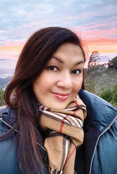
Breast Cancer Survivor Pia Navales. Photo credit: Pia Navales
Collaboration at UCSF Peripheral Nerve Center Helps Difficult-to-Diagnose Patients
 By Rebecca Wolfson
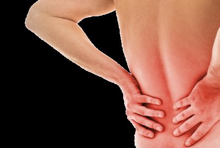
The Peripheral Nerve Center at UCSF is comprised of a multidisciplinary team devoted to precisely assessing and addressing often painful and difficult-todiagnose conditions of the peripheral nervous system. The Precision Spine Center run by the neuroradiology section of the Department of Radiology and Biomedical Imaging collaborates closely with the Peripheral Nerve Center and is one of the only centers in the world that routinely offers highresolution imaging techniques including MR neurography as well as CT, MR and ultrasound image guided injections.
One patient who was eventually diagnosed with thoracic outlet syndrome, was in constant pain and bed-ridden for 18 months unable to work or take care of himself. “It was so demoralizing going from a healthy athlete to disabled with no medical diagnosis or treatment plan,” the patient wrote in an email to Associate Professor and Chief of Neuroradiology
 Vinil Shah, MD.
This patient was eventually diagnosed, and treated, at the UCSF Precision Spine and the Peripheral Nerve Center, for neurogenic thoracic outlet syndrome, a condition in which there is entrapment of nerves of the brachial plexus in the neck. Using sonographic guidance Dr. Shah gave this patient Botox injections into the anterior and middle scalene muscles.
“The scalene Botox treatment has worked like a miracle and has given my life back. I am pain free and back to 90-95% function. I am working, surfing, and rock-climbing after eight months from my first Botox,” the patient said in an email.
It is patients like these — difficult to assess and therefore treat — that the Peripheral Nerve Center’s team of neuroradiologists, US and MSK radiologists, neurosurgeons, neurologists, orthopedic surgeons and pain physicians are working to help.
“The impact on patients is dramatic,” Dr. Shah said. “These patients would not have many places to go — even many other major academic medical centers are not able to offer the degree of expertise and image interpretation.”
The peripheral nerves, which reside throughout the entire body outside the brain and spinal cord, are often a few millimeters or less in size. Although there have been major advances in understanding the anatomy and impact of these tiny nerves, they still remain somewhat of an enigma to many in the medical community. For patients with peripheral nerve disorders or injuries, getting an accurate diagnosis and effective treatment can be challenging.
Professor of Clinical Radiology and Neurosurgery Cynthia Chin, MD, along with Professor of Neurology John Engstrom, MD, and Professor Emeritus Philip Weinstein, MD, Department of Neurological Surgery, developed the protocols for the nerve imaging techniques and sequences used at UCSF over a period of more than 20 years.
MR neurography allows clinicians to see the anatomy of the peripheral nerves in various parts of the body in exclusive detail, which provides essential information about whether
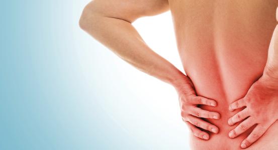 Photo Caption: (l-r) Cynthia Chin, MD, William Dillon, MD, and Vinil Shah, MD, Department of Radiology and Biomedical Imaging; John Engstrom, MD, Department of Neurology; Philip Weinstein, MD and Line Jacques, MD, Department of Neurological Surgery.
Cynthia Chin, MD William Dillon, MD Vinil Shah, MD John Engstrom, MD Philip Weinstein, MD Line Jacques, MD
Photo Caption: (l-r) Cynthia Chin, MD, William Dillon, MD, and Vinil Shah, MD, Department of Radiology and Biomedical Imaging; John Engstrom, MD, Department of Neurology; Philip Weinstein, MD and Line Jacques, MD, Department of Neurological Surgery.
Cynthia Chin, MD William Dillon, MD Vinil Shah, MD John Engstrom, MD Philip Weinstein, MD Line Jacques, MD
6 IMAGES 2021–2022
the nerve is injured so that the neurologists and surgeons can diagnose a problem and figure out a plan for treatment, said Dr. Chin. “It took many years to develop it to be a routine study that we now do every day,” Dr. Chin said. About 25 patients per week undergo these advanced imaging studies at UCSF.
During MR neurography the fat signal prevalent in muscle and bone is suppressed — so the nerve signal becomes more conspicuous. “If you’re trying to find a needle in a haystack, make the haystack all one color” and disappear into the background so you can see the needle, Dr. Chin said.
Diffusion tensor imaging is another MR technique that explores the integrity of the nerve in even greater detailtracking the movement of the water within nerve fibers, allowing physicians to look at the ultrastructure of the nerves and to characterize the organization of the axons within the peripheral nerve. Physicians then create a 3D reconstruction, or tractography of the nerve, which indicates if the nerve fibers are intact or disrupted.
“We can also indirectly measure the speed of the water movement going up and down along the nerve fibers,” Dr. Chin said. “If there’s a tumor or injured nerve, water motion will be abnormal, and may be relatively reduced or increased depending upon the degree of tumor cellularity, stage of injury and treatment. We can measure this water motion activity and give the physicians taking care of the patient some indication of what might be going on.”
“When it comes to imaging, MR neurograms aren’t difficult to obtain, but are exceptionally difficult to read well,” Dr. Engstrom said. “We have a concentration of expertise in one place, which can be leveraged onto diagnostic problems that folks have been stumped by.”
Through the training provided in the Neuroradiology fellowship program, the next generation of radiologists are also learning to interpret these highly specialized studies.
Professor of Neurological Surgery Line Jacques, MD, regularly refers patients for advanced peripheral nerve imaging. “These advanced images allow us to act faster, wait times for an intervention can be reduced by 2-3 weeks,” Dr. Jacques said. For example, if the MR neurogram indicates a laceration of a peripheral nerve, Dr. Jacques can schedule surgery immediately, rather than waiting several weeks.
Professor of Radiology William P. Dillon, MD, has been working as a neuroradiologist for over 35 years. He says the Precision Spine and the Peripheral Nerve Center represents a shift toward greater precision in radiology. “When I was training, we did not have the tools to interrogate the peripheral nerves,” Dr. Dillon said, “Now it’s routine.”
Dr. Dillon recently saw a patient who had a cystic lesion around an upper thoracic nerve root and had pain in the shoulder and back area. “We noticed she had a little mass near her chest along a nerve root that hadn’t been recognized before. We’re going to work that up to make sure that’s not the cause of her pain.”
“The ability to see in different parts of the body and then precisely place needles for diagnosis or therapeutic injections into very sensitive areas is a real advantage,” Dr. Dillon said.
Dr. Shah said he enjoys working with referring clinicians to improve the health of these often difficult-to-diagnose patients. “It’s very satisfying to be able to identify the diagnosis and try to help them.”
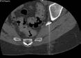
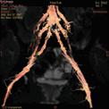
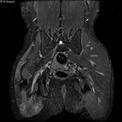
A) Coronal T2 IDEAL, B) axial diffusion tensor imaging (DTI), and C) coronal DTI fiber tracking sequences of the lumbosacral plexus and sciatic nerves demonstrates increased caliber and signal of the right sciatic nerve as it traverses the sciatic notch underneath the piriformis muscle.
D) Prone CT scan of the pelvis at the sciatic notch demonstrates a spinal needle placed at the sciatic nerve with injection of contrast and anesthetic: steroid mixture filling the perineural spaces of the sciatic nerve during a sciatic nerve block.
OUR MISSION
A C D
B IMAGES 2021–2022 7
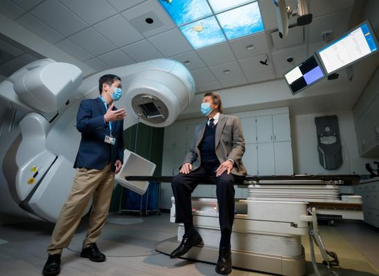
Neurointerventional Radiology Saves Stroke Patient
By Rebecca Wolfson
In September 2021, Kevin Tuckman, a 48-year-old sales manager, was doing a demo for a potential client in Foster City when he started having difficulty talking. “I would want to say something, and some words came out, but others I couldn’t say,” Tuckman said. After he dropped his pen five times within a one-minute period, Tuckman’s colleague, Melissa Jackson, asked him if he was OK. “I remember looking at Melissa, taking a deep breath and saying ‘Give me a minute.’” He continued to discuss the digital technology he was trying to sell, going over numbers and figures, but his math was completely wrong. Eventually, Jackson said: “You aren’t OK, we need to take you to the hospital.”
After they left the meeting, Jackson called Tuckman’s fiancée Karla Wargo who said: “Take him to UCSF.” For Wargo, who grew up in San Francisco, UCSF is always her first choice for loved ones in need of medical attention. “I know UCSF leads the way in so many areas of medical care.”
Stroke therapy and intervention are among the most successful advances in medical history, according to Matthew Amans, MD, MSc, an associate professor of clinical radiology, who ultimately treated Tuckman. “You can take someone who’s dying and not only save their life, but often allow them to be normal, functioning members of society within 30 minutes,” Dr. Amans said.“It’s an incredible revolution in medicine that’s only happened within the last 10 years.” His group in neurointerventional* radiology treats about 150 stroke patients per year.
When Tuckman arrived at the hospital, he and Wargo were brought to triage where tests were conducted. “We were waiting in the ER for a room, and I was sitting on a bed in the hallway with Karla,” Tuckman said. A nurse came over and asked him to put his hands up, but he didn’t understand what she said. “At that moment, I thought this was my new life – not being able to communicate anymore,” said Tuckman, who is a father to 13-year-old twin boys. “I thought my life was over as I knew it.” For one of the rare times in his life, Tuckman broke down in tears.
But he didn’t have time to dwell on it. “Once they thought it was a stroke, I remember hearing ‘code stroke,’ and within about 30 seconds I had 15 people on top of me.” Tuckman said he was immediately placed on a gurney, had an IV placed and rushed to the angio suite. For every minute that the blood vessels close in the brain, it’s estimated that 1.9 million neurons are damaged permanently.
An occlusion of a blood vessel in Tuckman’s brain caused the downstream brain to not function properly. “If left untreated the survivability from that is very low,” said Dr. Amans. Using an X-ray camera, Dr. Amans and his team guided their tools, starting in the groin and up through the blood vessels, to the brain. They saw that in the neck there was no flow in the carotid artery and there was a clot sitting in the blood vessel in the neck extending all the way up and into the brain. They deployed a stent fused to a wire within the clot, and pulled the clot down and out of the body reopening the blood vessels in the brain. All of this was done from inside the blood vessels (as it always is), making it a minimally invasive procedure. In other words, the skull does not need to be surgically opened to access the clot.
Tuckman was awake during this procedure because general anesthesia can worsen an injury to the brain. Once the clot was removed, Tuckman was able to speak again. “I remember at one point, I told them it hurt,” Tuckman said. The doctors told him the pain would subside quickly, and it did. “Before that, I had lost the ability to speak.”
After the clot was removed, Dr. Amans turned his attention to Tuckman’s problem in the neck. The clot had come from a dilated and injured segment of the carotid artery in the neck. Leaving that blood vessel as it was, would
*In fall 2022, the Neurointerventional Radiology faculty joined the Neurosurgery Cerebrovascular Surgery faculty to establish Neuroendovascular Surgery (NES), a new transdisciplinary service line.
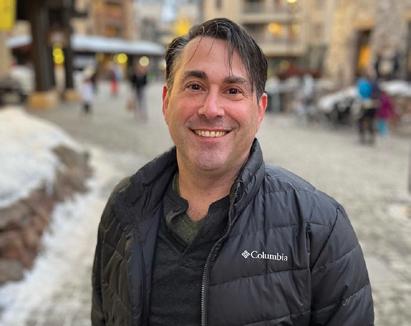 Stroke Survivor Kevin Tuckman
8 IMAGES 2021–2022
send another clot downstream and he’d be back in the same life-threatening situation. At this point, Dr. Amans had two options: He could use a series of stents, a big construct of tubes to try and open up the blood vessel — or — he could close that blood vessel off entirely. Both options carried the risk of a second stroke.
To make the decision Dr. Amans and his team (including the vascular neurology Fellow) did a physical exam on the operating table. They checked Tuckman’s neurological function to see if his brain was working after they had restored the blood flow within the head.
“He had started to recover immediately on the table after we reopened the blood vessel in the brain,” Dr. Amans said. “Since he was recovering, we figured he probably didn’t need the severely diseased blood vessel in the neck in order for his brain to survive. We made the decision to sacrifice the blood vessel in the neck, and we sealed it off with a few coils.”
For some surgeons, the more intuitive therapy in a case like this would be to install a series of hardware stents to restore the blood flow to the vessel, but in this situation it would have been more dangerous, Dr. Amans said. “While it’s not the most intuitive therapy to deconstruct the vessel, it’s sometimes the better therapy.” Placing a series of stents likely would have pushed more blood clot downstream into the brain circulation. The entire procedure was minimally invasive and completed in about 30 minutes. “The only way you can do something with that level of efficiency is to have a really good team of people who are highly trained to work in parallel and do our jobs at the same time,” Dr. Amans said. “That’s what allows for the very rapid restoration of blood flow to the brain, even in the most complex patients.”
About two years earlier Tuckman dove three-to-four feet off a pontoon boat into a lake. “I hit the water wrong,” Tuckman said. “I remember coming out and noticed my
hand was a little numb.” He even sought out medical care for nerve testing, but medical staff couldn’t find anything wrong. That dive likely damaged his carotid artery, which caused the blood clot.
Today, aside from the occasional miscommunication of words – for example saying “season” instead of “game” – Tuckman is essentially back to normal and feeling great. “I feel like I won the lottery,” Tuckman said. “I’m living my life again.” He spent six days in February skiing and hiking with his sons at Lake Tahoe. In November 2022, he married Wargo. Their first wedding was postponed because of COVID, their second wedding was postponed in 2021 because of his stroke, but Tuckman said their wedding in November 2022 would have happened no matter what. “Come hell or high water, we were getting married!”
OUR MISSION
For every minute that the blood vessels close in the brain, it’s estimated that 1.9 million neurons are damaged permanently.
Learn more at http://tiny.ucsf.edu/nes IMAGES 2021–2022 9
Transforming Radiology Practice Using Artificial Intelligence
 By Sharmila Majumdar, PhD and Christopher Hess, MD, PhD
As the saying goes, an image is worth a thousand words. Every medical image is replete with countless information about every patient’s health – new diagnoses, latent health issues, charting the course of disease, and predicting short- and long-term outcomes. Radiologists are experts at understanding a patient’s medical story. They integrate the manifold available sources of information to understand each patient. And with the assistance of new intelligent analytics, especially those that utilize artificial intelligence and machine learning, imaging contains an even greater wealth of information that can be used to optimize and precisely tailor high-quality care for a broader and more diverse group of patients. Artificial intelligence magnifies the impact of imaging.
The Center for Intelligent Imaging (ci2) was launched in 2018 in the UCSF Department of Radiology and Biomedical Imaging and focuses on using new techniques to enhance image acquisition, improve image quality, enable quantitative analysis of images and extract new information from images. The Center brings together imaging experts across an array of clinical and scientific backgrounds, from radiologists, surgeons, cardiologists and oncologists to engineers, informatics experts and students—to create an ecosystem for the study of how best develop and apply new artificial intelligence technology and intelligent analytic techniques for imaging. Imaging is a window into biology, physiology and disease. The construction of images from biological data is complex. Utilizing AI and machine learning, ci2 teams are working to overcome the limitations of traditional data
acquisition and analysis in imaging. New technology, especially in artificial intelligence, has enabled faster, more robust and higher-quality imaging. It has also launched the ability to extract quantitative data from images.
Ci2 is developing technology that optimizes patient health. Some of the recent successes of the teams within ci2 are highlighted below.
Clinical Deployment
The ci2 Clinical Deployment team has developed a framework for evaluating whether an AI algorithm should be deployed for routine clinical usage, and if so, how. The framework evaluates the balance between the benefits provided by the algorithm and the costs and risks incurred by deployment. On the benefit side, the framework considers the breadth and depth of the potential improvement, the effect on workload and efficiency, and compares this to the current workflow or standard of care. On the cost side, likelihood of erroneous outputs, detectability and correctability of those errors, and patient impact of uncorrected errors are evaluated. The financial costs of deployment, maintenance and training are considered, and potential unintended consequences are explored, including creation or exacerbation of health care disparities, delays in care and adverse effects on radiologist performance.
For algorithms where the balance of these factors is favorable for deployment, the form of integration with
10 IMAGES 2021–2022
the clinical environment and workflow is designed. This takes into consideration how the AI results should be presented, how they should be contextualized and how they should be stored as part of the medical record. A post-deployment monitoring plan is developed.
This framework has been successfully employed to guide the deployment of the BunkerHill Coronary Artery Calcification scoring algorithm and will soon be applied to several ci2-developed algorithms, including brain tumor volumetrics.
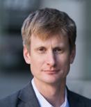
John Mongan, MD, PhD
Extramural Funding
In April 2021, researchers in the ci² received a five-year R01 grant from the National Institutes of Health for “UltraFast Knee MRI with Deep Learning” to improve the study of joint degeneration, injury and osteoarthritis. This study is paradigm-shifting in that it may provide a first step toward the integration of accelerated image acquisition, fully automatic image inspection and personalized imaging protocoling. The integration of ultrafast MRI image acquisition, online image reconstruction and post-processing opens new horizons of opportunities and different use-cases to facilitate the translation of fast MRI techniques.
The integrated pipeline the investigators are developing has the potential to characterize patients automatically based on quantitative features at the time of data acquisition and potentially to modify the MRI protocol dynamically by adding sequences to better tailor them to patient needs using real-time precision imaging. An ultrafast MRI-based preliminary patient triage system can open new possibilities on the use of accelerated MRI. If ultrafast MRI is used for online patient triage and personalization of image protocoling, sequences that are optimally acquired and optimized based on patient characteristics would be available at the time of study interpretation by the radiologist. This would help the radiologist provide a better and more precise assessment of abnormalities. Real-time automatic image processing and interpretation of highly accelerated MRI acquisition may change drastically the musculoskeletal radiology scenario.
Valentina Pedoia, PhD
Federated Learning, AI and Clinical Deployment at UCSF – Lessons Learned from COVID-19 Modeling
Early in the pandemic, predicting oxygen requirements for incoming patients was important, given the limited availability of respirators and hospital resources. At that time, no single hospital had sufficient data to train a predictive AI model capable of making such predictions. It was clear that the only short-term option would require pooling data collaboratively among multiple sites. However, directly sharing medical data is challenging.
This led a team at ci2, to turn its attention to federated learning, a privacy-preserving method capable of training neural networks on decentralized data that enables sensitive patient information to remain securely at each institution. The team included Jae Ho Sohn, MD, assistant professor, Pablo Damasceno, PhD, senior data scientist, Peter Storey, manager of scientific computing services, Jed Chan, systems administrator and Jeff Block, director of infrastructure, along with Wyatt Tellis, PhD, director of innovation and analytics, among others.
UCSF collaborated with 20 sites worldwide using federated learning to develop a model for predicting COVID-19 outcomes from chest X-rays, labs and vitals collected during an emergency room visit. Remarkably, the modeling was completed in 5 months and achieved an average area under the curve (AUC) >0.92 for predicting outcomes at 24 and 72 hours from time of presentation (Nature Medicine; 27, pages 1735–1743. 2021). The project demonstrated the power of AI methodologies combined with federated learning to collaboratively develop robust solutions to critical public health problems in a timely fashion. The work has since paved the way for a multi-center effort focused on predictive modeling in prostate cancer (PI: Peder Larson, PhD, UCSF/UCLA).
Translating such methods into the clinic safely and efficiently is a key goal. This requires the development of frameworks capable of deploying clinically integrated models, work being led by Carolina Ramirez and James Hawkins (data scientists, ci2 Computational Core) in collaboration with UCSF’s Apex Enabled Research (AER link) and under the oversight of ci2’s Clinical Deployment pillar team, led by Dr. John Mongan and Jason Crane, PhD
OUR MISSION
IMAGES 2021–2022 11
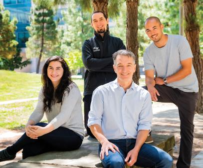
Members of the computational core team at Mission Bay: (l-r) Carolina Ramirez, Beck Olson, Dr. Jason Crane & James Hawkins.
New Developments in PSMA PET Imaging for Prostate Cancer
Dr. Hope answers some questions about the significance of these developments and gives insight into what’s next for PSMA PET.
Thomas Hope, MD
Professor In Residence
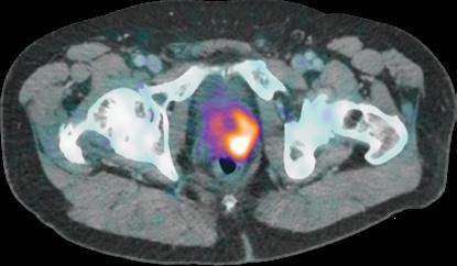
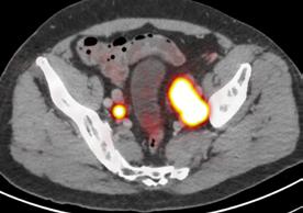
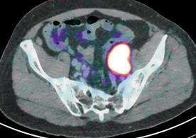
In the 2020-21 issue of Images, we shared the story of the U.S. Food and Drug Administration’s approval of Prostate-Specific Membrane Antigen (PSMA) PET imaging, based on research conducted at UC San Francisco and UCLA. The technique uses positron emission tomography in conjunction with a PET-sensitive drug that is highly effective in detecting prostate cancer throughout the body so that it can be better and more selectively treated. The PSMA PET scan also identifies cancer that is often missed by current standard-of-care imaging techniques.
At this time, UCSF and UCLA are the only two medical centers in the U.S. that can offer PSMA PET to the public through this FDA approval. A limited number of other U.S. medical centers are currently using PSMA as an investigational technique, generally as part of a clinical trial. However, more hospitals will have the opportunity to adopt the technology in 2022 after applying for expedited FDA approval, which is now possible as a result of the initial FDA approval gained by UCLA and UCSF.
In September 2021, a team led by Thomas Hope, MD, at UCSF and Jeremie Calais, MD, at UCLA, published a paper in JAMA Oncology (doi:10.1001/ jamaoncol.2021.3771) detailing the phase 3 diagnostic efficacy trial that led to FDA approval of PSMA PET.
In addition, the National Comprehensive Cancer Network (NCCN) and the Society of Nuclear Medicine and Molecular Imaging (SNMMI) included PSMA PET in published prostate guidelines and established appropriate use criteria (AUC) for this new imaging technique.
What is the main focus of the JAMA Oncology paper?
The paper focuses on the role of 68Ga-PSMA-11 PET at time of initial staging. The goal was to compare the imaging results to nodes found at time of surgery in order to determine the sensitivity and specificity of PSMA PET. This study showed that PSMA PET has a high specificity for the detection of nodal metastases, although the sensitivity for small pelvic nodes was lower than expected.
What is the significance of NCCN including PSMA PET in their published prostate guidelines?
This is a very important development. NCCN guidelines are used by many insurance companies to determine what tests to cover. The inclusion in these guidelines will help increase the likelihood of insurance coverage of PSMA PET at time of initial staging and biochemical recurrence. Additionally, the NCCN guidelines recently convinced the FDA to include 68Ga-PSMA-11 PET at time of initial staging for patients with prostate cancer.
What are some highlights from the SNMMI’s appropriate use criteria for PSMA PET imaging?
Similar to the NCCN guidelines, the SNMMI AUC document will have an impact on insurance coverage. Through the Protecting Access to Medicare Act (PAMA), high-cost imaging studies will be required to use clinical decision support mechanisms. The AUC document will provide the required documentation to support the use of PSMA PET in the appropriate indications.
What’s next for PSMA PET imaging?
Next up is to grow the use of PSMA PET for patient selection in PSMA radioligand therapy.
Currently there are no FDA approved agents for PSMA radioligand therapy, but we expect approval of 177Lu-PSMA-617 in the coming months, and so we will start using PSMA PET in this patient population. Additionally, now that PSMA PET will be widely available, we will need to complete clinical trials in order to understand how to manage patients better based on the results of PSMA PET imaging studies.
Learn more at https://www.ucsf.edu/news/2020/12/419196/ucsf-ucla-gain-fda-approval-prostate-cancer-imaging-technique
12 IMAGES 2021–2022
PSMA PET scan identifies cancer that is often missed by current standardof-care imaging techniques.
OUR MISSION
Radiation oncologist Felix Feng, MD (left), with patient Dennis Brod, in a radiation treatment room at the UCSF Precision Medicine Cancer Building. Photo by Maurice Ramirez
IMAGES 2021–2022 13
Promoting Academic Success in Radiology & Biomedical Imaging


Under the direction of the Chair, Dr. Christopher Hess, the Radiology Academic Affairs team manages the complexities of the often-opaque academic processes for all our faculty and our non-faculty academics [NFAs]. Our team have worked over the last 3 years to streamline and clarify many processes including faculty and NFA hiring, annual reviews, department committee composition, and the academic advancement processes, to promote transparency, equity, and diversity in our department. We are building on our faculty mentoring program and have revamped and created new faculty development workshops and courses. We are continually improving on systems so that we can better support and celebrate our faculty in their career pathway to success.
Recruitment
Our department has been extraordinarily busy with a much-increased clinical workload, along with strategic expansion of our radiology clinical sites – BCH Oakland, Berkeley Outpatient Center, and the development of a new Emergency Radiology section at ZSFG. This, coupled with the retirement of three beloved faculty has necessitated multiple faculty searches.
As of June 2022, our Department of Radiology has 140 faculty, with 37 Imaging Scientists and 103 Radiologists, including 8 emeritus faculty working on recall. Four of our new Radiology faculty commenced work since February 1, 2022, and 11 additional faculty joined our Department between July 1 and November 1 this calendar year. During the last 9 months we have also successfully completed five national searches for section chiefs with the appointments of Dr. Jane Wang for Abdominal Imaging, Dr. Pallav Kolli for Interventional Radiology, Dr. Vinil Shah for Neuroradiology, Dr. Jesse Courtier for
Mission Bay Pediatric Radiology, and Dr. Raymond Sze for Oakland Children’s Pediatric Radiology. It has been an extraordinary recruitment year for our department, and our academic affairs team is so appreciative of the many faculty who have been part of, or chaired, our search committees. Thank you to all faculty for attending presentations, interviews, and welcoming candidates to our campuses! While it can be such a time-consuming process, successfully expanding our teams has been highly rewarding this past year. Please see the separate introduction pages for each of our new faculty.
Faculty Mentoring Program
Our Faculty Mentoring Program, designed to create mentoring networks for each of our assistant professors, continues to guide and support the faculty through career direction decisions and research collaborations, as well as providing sponsorship opportunities. We currently

PEOPLE
Amy Pradhan, MS MPH Director of Academic Affairs
Apple Palad Academic Project Coordinator
Jocelyn Pulido Academic Affairs Coordinator
Lorna Kwok, RN Academic Affairs Coordinator
Christine Glastonbury, MBBS, Vice Chair for Academic Affairs
Susan Wall, MD Advisor to the Chair, Faculty Development
Connie Jang Project Coordinator, Academic Affairs
14 IMAGES 2021–2022
ACADEMIC AFFAIRS By Christine Glastonbury, MBBS
have 31 MD Assistant Professors and 7 PhD Assistant Professors in the mentoring program. Sri Nagarajan, PhD, was appointed as Director of Mentoring for the Imaging Scientist PhD faculty in 2021.
It is only through the dedication of each our faculty that the mentoring program is so successful. In January 2022 we again honored and celebrated all faculty mentors during National Mentoring Month, with our custom department tote bags. This June we were thrilled to acknowledge the extraordinary mentoring work of Dr. Dan Vigneron who was the recipient of the 2022 Radiology Award for Outstanding Faculty Mentoring. Dan was previously the Director of Mentoring for our imaging scientists and has been an extraordinary mentor for imaging scientists and radiologists in our department.
MENTORING MONTH
January 2023
http://tiny.ucsf.edu/radmentor

Faculty Development
Susan Wall, MD, advisor to the Chair, leads these efforts and continues to source opportunities for faculty development, available grants, courses and faculty awards. Together with Elissa Price, MD she continues to lead the highly sought-after faculty speaker training course. This small group intense three afternoon workshop focuses on teaching lecturing skills through practice, feedback and re-presentation.
Academic Advancement
Our Merits & Promotions (M&P) committee consists of 12 Full Professor faculty and myself, with representatives from each of our affiliate sites at VAMC, ZSFG and BCHOakland. Each of our committee members contributes many hours of packet review and preparation and contributes to the often-complex discussions over our six meetings. Their service to our department and their support for our faculty is greatly appreciated.
For the most recent cycle with advancements effective July 2022, our M&P committee reviewed and discussed 65 packets including 10 promotions and 11 accelerations approved. We are enormously proud of our faculty’s hard work, particularly through the challenges brought by the pandemic over the last two years.
Non-Faculty Academics (NFAs)

Our academic affairs team also manages and in 2020 formalized the hiring, visa applications, and on-boarding process for our NFA faculty who contribute enormously to our research efforts in the department. In 2021 we instituted a formalized annual review process for all NFAs was well as more formalized merits and promotions process to promote successful development of their career potential at UCSF.
Faculty Inclusion, Recognition and Celebration

We have many department committees with opportunities for our faculty to be involved in shared decision making, governance and management of our work practice. We have endeavored over the last 3 years to increase representation on these committees across academic series and ranks, and across different hospital and research working sites. Committee chairs have been charged with diversifying their membership along these lines and have enthusiastically addressed this issue.
Since July 2020 we have created an Annual Celebration of our faculty (online and hardcover) to recognize the new faculty, retiring faculty, academic promotions, and faculty Departmental awards.
Celebrations
July 2022
http://tiny.ucsf.edu/celebrations
With so much growth in our department and expansion of the activities of our academic affairs team, we have welcomed outstanding and dedicated new team-members Apple Palad and Connie Jang. We all remain committed to unraveling the complexities of the academic administrative process for our faculty and the department.
PEOPLE: ACADEMIC AFFAIRS
IMAGES 2021–2022 15
FACULTY PROMOTIONS 2021-22

Our department and UCSF recognizes each promoted faculty member for their significant contributions to our mission: Advancing Health Worldwide. To do this well, each faculty member balances teaching, clinical practice, scientific inquiry and clinical translation, and service activities to create an outstanding portfolio of accomplishments. We are incredibly proud of each faculty member and their unique qualities as teachers and mentors; department and university leaders committed to Diversity, Equity and Inclusion; and physicians and imaging scientists known for their national or international stature in the field of radiology.
2021
We are honored to announce that eleven faculty members – seven new associate professors and four new full professors – have received academic promotions effective July 1, 2021.
Robert Flavell, MD, PhD Associate Professor, In Residence Molecular Imaging & Therapeutics
Jared Narvid, MD Associate Professor, Clinical Radiology Neuroradiology
Esther Yuh, MD, PhD Professor, In Residence Neuroradiology
Matthew Zapala, MD, PhD Associate Professor, Clinical Radiology Pediatrics
Stefanie Weinstein, MD Professor, Clinical Radiology Abdominal Imaging / Ultrasound
Vishal Kumar, MD Associate Professor, Clinical Radiology Interventional Radiology
Bhavya Rehani, MD Associate Professor, Clinical Radiology Neuroradiology
Jesse Courtier, MD Professor, Clinical Radiology Pediatrics
Evan Lehrman, MD Associate Professor, Clinical Radiology Interventional Radiology
Leo Sugrue, MD, PhD Associate Professor, In Residence Neuroradiology
Marc Kohli, MD Professor, Clinical Radiology Abdominal Imaging / Ultrasound
16 IMAGES 2021–2022
We are honored to announce that ten faculty members – five new associate professors and five new full professors – have received academic promotions effective July 1, 2022.
 Ryan Kohlbrenner, MD Associate Professor of Clinical Radiology, Interventional Radiology
Valentina Pedoia, PhD Associate Professor In Residence, Body Imaging Research Group
R. Pete Lokken, MD, MPH Associate Professor of Clinical Radiology, Interventional Radiology
Olga Tymofiyeva, PhD Associate Adjunct Professor, Neuroimaging Research Group
Thienkhai Vu, MD, PhD HS Clinical Professor, Cardiac and Pulmonary Imaging, ZSFG
Thomas Hope, MD Professor In Residence, Molecular Imaging & Therapeutics
Javier Villanueva-Meyer, MD Associate Professor of Clinical Radiology, Neuroradiology
Elissa Amans Price, MD Professor of Clinical Radiology, Breast Imaging
Robert Bok, MD, PhD Adjunct Professor, Body Imaging Research Group
Peder Larson, PhD Professor In Residence, Director, Body Imaging Research Group
Ryan Kohlbrenner, MD Associate Professor of Clinical Radiology, Interventional Radiology
Valentina Pedoia, PhD Associate Professor In Residence, Body Imaging Research Group
R. Pete Lokken, MD, MPH Associate Professor of Clinical Radiology, Interventional Radiology
Olga Tymofiyeva, PhD Associate Adjunct Professor, Neuroimaging Research Group
Thienkhai Vu, MD, PhD HS Clinical Professor, Cardiac and Pulmonary Imaging, ZSFG
Thomas Hope, MD Professor In Residence, Molecular Imaging & Therapeutics
Javier Villanueva-Meyer, MD Associate Professor of Clinical Radiology, Neuroradiology
Elissa Amans Price, MD Professor of Clinical Radiology, Breast Imaging
Robert Bok, MD, PhD Adjunct Professor, Body Imaging Research Group
Peder Larson, PhD Professor In Residence, Director, Body Imaging Research Group
PEOPLE: ACADEMIC AFFAIRS IMAGES 2021–2022 17
2022
FACULTY LEADERSHIP APPOINTMENTS
Kimberly Kallianos, MD, was appointed the Modality Director for CT on March 1, 2021.
Dr. Kallianos is an assistant professor of Clinical Radiology in the Cardiac and Pulmonary Imaging section. She specializes in interpreting cardiothoracic CT and MRI exams, chest radiographs, and CT-guided lung biopsies. Dr. Kallianos is active in both clinical decision-making and teaching conferences including preoperative trans-catheter aortic valve replacement, pediatric cardiac surgery, thoracic tumor board, adult congenital cardiology, and multidisciplinary conferences for thoracic conditions (VA) and interstitial lung disease.
Dr. Kallianos conducts collaborative research focusing on the use of cardiac magnetic resonance for assessment of myocardial disease including strain imaging in pulmonary hypertension and T2 analyses for myocardial iron overload. She also conducts quality improvement research, including radiation dose reduction and resident/faculty agreement in interpretation of coronary computed tomography angiography.
We are grateful to Dr. Kallianos for her longstanding commitment to patient care.
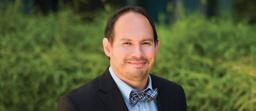
K. Pallav Kolli, MD, was appointed Chief of Interventional Radiology on October 18, 2021.
Dr. Kolli has served as the department’s associate chair and medical director for Quality & Safety since 2018. During the pandemic, he has also served on the Radiology Incident Command System (RICS) team and has prior experience as the department’s director of operations, Interventional Radiology from 2018-20 and as a member of the Strategic Planning Committee (2018-20). With our department Diversity and Operations Committees, Dr. Kolli contributes to evaluating health and healthcare disparities in the radiology clinical context and defining structures to address them. Within UCSF Health, Dr. Kolli chairs the Quality Improvement Executive Committee (QIEC) and is a member of the Clinical Performance Improvement Committee (CPIC) and the Executive Medical Board (EMB). Dr. Kolli was named a fellow of the Society of Interventional Radiology in 2020, and is a frequently invited speaker at national and international conferences.
After earning his bachelor’s degree in biomedical engineering and his medical degree from Northwestern University in Chicago, Dr. Kolli completed his transitional medicine internship at Evanston Northwestern Healthcare where he was named Transitional Intern of the Year. Dr. Kolli subsequently completed his residency in diagnostic radiology at UCSF, followed by a fellowship in vascular and interventional radiology at UCSF.
Dr. Kolli’s clinical expertise and research interests span management of acute and chronic venous diseases; minimally invasive therapies to treat patients with solid tumors of the liver, kidneys, and lungs; and complex hepatobiliary interventions in patients with portal hypertension or complex biliary tract disease. Dr. Kolli has led UCSF involvement in multicenter clinical trials in the areas of chronic venous disease and portal hypertension and sits on the steering committee of a prospective multicenter national trial evaluating outcomes in patients undergoing creation of transjugular intrahepatic portosystemic shunts (TIPS).
A committed educator and strong advocate for our trainees, Dr. Kolli has mentored several dozen residents and fellows in interventional and diagnostic radiology. Most recently, Dr. Kolli has focused his mentoring on projects conducted by early career faculty in our department engaged in quality and safety work.
Vinil Shah, MD, was appointed Chief of Neuroradiology on August 1, 2021.
Dr. Shah has been an associate professor and faculty member in the department since 2014. From 2017-22, Dr. Shah was Neuroradiology Fellowship Director, stewarding a two-year program of nine first-year ACGME fellows and six secondyear clinical instructors.
Dr. Shah earned his bachelor’s degree in economics from the Wharton School, University of Pennsylvania and his medical degree from the University of Pittsburgh School of Medicine where he received the Brinton Prize for the Most Outstanding Medical Student. He completed his internship at the Memorial Sloan Kettering Cancer Center in New York. During his residency at UCSF, Dr. Shah served as chief resident and received the Elmer Ng Outstanding Resident Award in 2011. After fellowships in neuroradiology and musculoskeletal radiology at Massachusetts General Hospital, where he also served as neuroradiology chief fellow, he returned to UCSF and joined the faculty.
18 IMAGES 2021–2022
Recognized locally, nationally, and internationally for his clinical expertise, Dr. Shah’s primary interests focus on imaging evaluation of spinal and peripheral nerve disorders and image-guided intervention. His research explores biomarkers of pain generation in the spine, efficacy of image-guided intervention for treatment of acute and chronic back pain, and emerging technologies and techniques for treatment of chronic pain, cerebrospinal fluid leaks, and spinal tumors.
Dr. Shah has served in numerous leadership and service activities for the section, our department, and in national professional organizations. Outside of UCSF he serves on the Board of Directors of the Spine Intervention Society (SIS) and on the Executive Committee of the American Society of Spine Radiology (ASSR) and is a frequently invited speaker at national and international neuroradiology conferences. Known as a passionate educator and strong advocate for our trainees, Dr. Shah has actively engaged in recruitment efforts to increase the diversity and number of underrepresented trainees, including recruiting more women into our fellowship program.
Jesse Courtier, MD, was appointed Chief, UCSF BCH-SF Pediatric Radiology, on January 1, 2022.
Dr. Courtier earned his medical degree from the University of Iowa College of Medicine. After completing his internship in preliminary surgery at University of Hawaii, his diagnostic radiology residency at the University of Kansas, and fellowships in both abdominal imaging and pediatric radiology at UCSF, he joined the UCSF faculty in 2010.
From 2014-22, Dr. Courtier served as Pediatric Radiology Fellowship Director and Residency Site Director of Training for Mission Bay, and participated on numerous departmental committees. He has also played an important operational role in our health system as Clinical Director for UCSF Mission Bay Hospitals. A strong advocate for diversity, equity and inclusion, he has participated as a RIDR mentor in the department.
An array of accolades including the UCSF Radiology Outstanding Clinical Fellow Teaching Award, the AAME Excellence in Teaching Award, the Hideyo Minagi Outstanding Teaching Award, and the UCSF Catalyst Award in Digital Health stand as testament to Dr. Courtier’s influence as an educator, mentor and innovator. A respected mentor in the department, Dr. Courtier has guided multiple mentees, three of whom have received UCSF Radiology Resident Research Awards.
Outside of UCSF, Dr. Courtier is a subject review committee member for the Pediatric Radiology section of the American Board of Radiology, reviewer for multiple journals including Radiology, AJR, and Pediatric Radiology, and a frequent invited speaker at national and international society meetings.
The overarching theme of Dr. Courtier’s research practice is to develop and translate innovative technologies to improve the practice of clinical pediatric imaging. As a clinician and researcher, Dr. Courtier is a recognized expert in pediatric abdomino-pelvic MRI applications, including urography and enterography. Key clinical innovations include advanced motion-resistant high-resolution MR sequences for use in pediatric body and musculoskeletal imaging, the first use of the contrast agent gadofosveset at UCSF, and creation and implementation of rapid MRI protocols for abdominal imaging which, in a partnership with Child Life Services, has allowed MRI exams to be performed on children as young as four years old without anesthesia or sedation. His most recent collaborative research projects and publications focus on augmented reality and its application to trainee education and surgical planning.
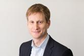
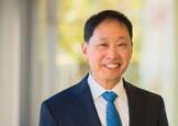
Thomas Hope, MD, professor & director of molecular therapy in the Molecular Imaging & Therapeutics Section, was appointed Vice Chair, Clinical Operations & Strategy, on June 1, 2022.
In this new role, Dr. Hope will provide vision and leadership for the department’s growing clinical footprint in imaging, working with UCSF Health to manage our clinical operations and expand our regional services. Dr. Hope will also advise on the deployment of tools to improve datadriven decision-making in clinical operations, to ensure that faculty, trainees, and clinical teams benefit from systems and support that foster innovation and continuous practice improvement.
Dr. Hope earned his medical degree from Stanford University School of Medicine. After completing his internship at Kaiser Permanente, San Francisco, his diagnostic radiology residency at UCSF, and a fellowship in body MRI and nuclear medicine at Stanford Medical Center, Dr. Hope joined the UCSF faculty in 2013.
Since 2016, Dr. Hope has served as co-director of the UCSF Neuroendocrine Tumor Destination Program and since 2018 has served as Chief of Nuclear Medicine at the San Francisco VA Medical Center. Dr. Hope also chairs the Cancer Center’s Molecular Imaging & Radionuclide Therapy Site Committee. Since 2020, Dr. Hope has served as the department’s Associate Chair for Business
PEOPLE: ACADEMIC AFFAIRS
IMAGES 2021–2022 19
Strategy, a role that will be subsumed in his new position. A respected teacher and mentor in the department, Dr. Hope has mentored multiple residents and fellows in abdominal imaging and nuclear medicine, several of whom have joined our faculty in recent years.
Dr. Hope is an international leader in prostate cancer imaging research and the translation of new molecular imaging agents to improve clinical care. After a collaborative effort with UCLA in which Dr. Hope played a leading role, FDA approved the clinical use of prostatespecific membrane antigen PET imaging (PSMA PET) in 2020. PSMA PET has the potential to revolutionize care for prostate cancer patients with metastatic disease and patients whose cancer may be missed by current standard-of-care imaging techniques.
A sampling of Dr. Hope’s awards over the past few years includes the Henkin Fellowship and Marc Tetalman Memorial Award from the Society of Nuclear Medicine and Molecular Imaging, the Young Investigator Award from the Prostate Cancer Foundation, the Wylie J. Dodds Research Award from the Society of Abdominal Radiology, two Outstanding Teacher Awards from the ISMRM, and the Hal O’Brien Rising Star Award from the SNMMI/ERF.
Zhen Jane Wang, MD, professor in the UCSF Department of Radiology & Biomedical Imaging, was appointed Chief, Abdominal Imaging and Ultrasound, on June 15, 2022.
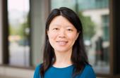
In her new role, Dr. Wang will build on the international stature of the section and provide vision and leadership for its continued growth through innovations in clinical practice, research and education. A respected teacher and mentor, Dr. Wang will also support the professional development of trainees and faculty.
Dr. Wang earned her undergraduate degree in electrical engineering from Brown University and her medical degree from Northwestern University Medical School. During her diagnostic radiology residency at UCSF, she also served as a chief resident. Dr. Wang completed an abdominal imaging fellowship at UCSF before joining the faculty in 2008.
With a practice that spans multiple areas of abdominal imaging, Dr. Wang is recognized for her expertise in oncological imaging of kidney, pancreas and liver. Dr. Wang serves as principal investigator on several NIH grants that focus on kidney and pancreas imaging with hyperpolarized carbon-13 MRI and PET/MRI. She has a significant list of peer-reviewed publications and has received numerous awards and distinctions over the
years. Some of her recent awards include the Roscoe E. Miller Best Paper Award from the Society of Abdominal Radiology, the Summa Cum Laude Award from the International Society of Magnetic Resonance in Medicine and the Distinguished Investigator Award from the Academy for Radiology & Biomedical Imaging Research.
Since 2020, Dr. Wang has served as the Associate Chair of Strategic Planning and partnered with many in our department to develop and implement initiatives aligned with our Strategic Plan 2020-2025. She is the Medical Director for the UCSF Hyperpolarized MRI Technology Resource Center and an Associate Program Director for the T32 Clinician Scientist Training Program. Dr. Wang also serves on multiple departmental committees including Merit and Promotion, and co-chairs the Clinical Research Coordinator Core steering committee. Dr. Wang is a member of the UCSF Resource Allocation Program Technology Review Committee, and recently completed service on the UCSF Institutional Review Board. Nationally, Dr. Wang has held leadership positions on multiple program committees and panels over the years, including currently serving as the Chair of the Radiological Society of North America Gastrointestinal Imaging committee and as the Portfolio Director for Lifelong Learning for the Society of Abdominal Radiology.
Javier Villanueva-Meyer, MD, was appointed Vice Chair, Quality and Technology on September 7, 2022.
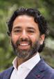
In this role, Dr. Villanueva-Meyer is responsible for maintaining the quality and safety of diagnostic and interventional clinical imaging services and provide medical director oversight of imaging technologies at UCSF Health and affiliate sites. He also serves as the primary liaison with UCSF Health for quality issues in imaging, works to develop new tools and benchmarks to continuously improve clinical outcomes in Radiology, ensures compliance with regulatory agencies, and maintains and disseminates departmental quality and safety guidelines.
Dr. Villanueva-Meyer has served as Medical Director of MRI since 2021 and has distinguished himself as a leader across our mission areas as a talented clinician, patient advocate, transdisciplinary partner, and top-notch educator and mentor.
The department thanks K. Pallav Kolli, MD, who has served as Associate Chair of Quality and Safety since February 2018. Dr. Kolli, who was appointed Chief, Interventional Radiology in October 2021, is leaving a lasting footprint on the quality and safety of imaging in our department through his nearly five years of service in this role.
20 IMAGES 2021–2022
Raymond Sze, MD, MAMS, joined UCSF on October 1, 2022. He is Radiologist in Chief, Pediatrics and Vice Chair, Radiology, Benioff Children’s Hospital - Oakland. In his leadership roles, Dr. Sze will continue to build the clinical, educational, and academic programs at UCSF, strengthen collaborations between our East and West Bay pediatric programs, and ensure that BCH Oakland continues to provide the highest possible level of imaging care for our pediatric patients.
Dr. Sze comes to UCSF with an exemplary record of program development, having spearheaded strategic growth at both Children’s National in Washington, DC, and Children’s Hospital of Philadelphia (CHOP). In his prior roles, Dr. Sze has overseen the development of robust Bioengineering, Quality & Safety, and Physician and Staff Wellness programs, fostered multiple strong inter-departmental collaborations, instituted recruitment projects that expanded faculty numbers, and design projects resulting in award-winning clinical environments. A sustained commitment to diversity, equity, and inclusion characterizes Dr. Sze’s work, and includes his personal advocacy and mentoring of trainees and faculty as well as developing and sponsoring outreach and recruitment programs for underrepresented minorities.
Dr. Sze earned his MD at UMDNJ-Robert Wood Johnson, Piscataway, NJ, followed by a Masters of Associated Medical Sciences (MAMS) in biomedical visualization at University of Illinois in Chicago. After an intern year at University of Hawaii in Honolulu, Dr. Sze completed a diagnostic radiology residency at New England Deaconess Medical Center in Boston and a second radiology residency at Stanford followed by a fellowship in pediatric radiology at Cincinnati Children's Hospital Medical Center.
With a track record of building radiology services, Dr. Sze has collaborated on developing programs in pediatric surgical innovation, nanotheranostics, MR guided robotics, and augmented reality. At CHOP, Dr. Sze was instrumental in building labs and research programs in artificial intelligence and 3D printing. The 3D printing program produces medical devices, surgical guides, and procedural simulators in addition to PPE and testing devices during the pandemic. Dr. Sze has facilitated AI research lab projects that focus on pediatric imaging challenges such as automation of time-consuming tasks (leg length discrepancy) and screening imaging studies for evidence of child abuse (rib fractures on chest radiographs).
Steven Hetts, MD, FACR
Effective November 1, 2022, the UCSF Radiology
Neurointerventional Radiology (NIR) faculty joined UCSF Neurosurgery
Cerebrovascular Surgery
faculty to establish a new transdisciplinary service line: Neuroendovascular Surgery (NES). This service line is a partnership between our department and the Department of Neurological Surgery and sponsored by the UCSF School of Medicine and UCSF Health.
Steven Hetts, MD, FACR from UCSF Radiology and Adib Abla, MD from UCSF Neurosurgery have been appointed as co-chiefs of the new NES service line. As co-chiefs, their charge is to build a unified, collaborative, multidisciplinary service for all neuroendovascular procedures at UCSF.
Dr. Hetts provides cutting-edge, minimally invasive endovascular therapy for children and adults. He founded the Interventional Neuroradiology services at San Francisco General Hospital and the San Francisco Veteran’s Administration Hospital, where he served as Chief until 2015. Honored in 2017 as an Exceptional Physician by UCSF Health, Dr. Hetts has an active clinical practice treating patients with stroke, brain aneurysms, arteriovenous malformations, dural arteriovenous fistulas, spinal vascular malformations, and tumors including meningioma and retinoblastoma. Dr. Hetts is the founding Co-Director of the UCSF Hereditary Hemorrhagic Telangiectasia Center of Excellence wherein he and his colleagues provide care for patients with vascular malformations of the brain, severe nosebleeds (epistaxis), and arteriovenous malformations of the lung and other organs.
“By reorganizing in this way, we expect that NES will better support education and research, building upon the illustrious traditions of each department. The support teams – administrative staff, advanced practice professionals, technologists, and nurses – from NIR and Vascular Neurosurgery will merge to create a seamless patient care experience,” said Christopher Hess, MD, PhD.
“The department thanks Dr. Higashida for his 25 years of leadership and stewardship in our world-class NIR section,” said Dr. Hess. “Dr. Higashida developed one of the first intracranial stents for aneurysm treatment and UCSF pioneered the use of stents in patients with aneurysms that otherwise could not be treated endovascularly. He is a leading intellect in the ischemic stroke sphere, helping to develop the TICI scale by which success in intracranial revascularization is measured and leading some of the first trials for intra-arterial lytic agents in stroke treatment.”
PEOPLE: ACADEMIC AFFAIRS IMAGES 2021–2022 21
NEW FACULTY
Gina Landinez, MD Assistant Professor of Clinical Radiology Interventional Radiology
Gina Landinez joined the department in March 2021.
Dr. Landinez’s clinical, research and teaching interests include lung ablation and multimodality interventional radiologic techniques for treating cancer-related pain and sarcoma. Dr. Landinez is a member of the Society of Interventional Radiology and has served on its Women in Interventional Radiology, Early Career Section and SIR Gala committees since 2019.
Dr. Landinez received her medical degree from St. George’s University School of Medicine, Grenada. She completed an internship in general surgery and a diagnostic radiology residency at the University of Texas Health Science Center in Houston, followed by an interventional radiology fellowship at the University of Texas MD Anderson Cancer Center.
Renuka Sriram, PhD Assistant Adjunct Professor
Renuka Sriram joined our department as faculty in May 2021.
After earning her PhD in biophysics from UC Davis in 2007, Dr. Sriram worked as a senior imaging scientist at Pfizer, Inc., joining UCSF as a postdoctoral scholar in 2010. Prior to her faculty appointment, Dr. Sriram served as technical director of Pre-Clinical MR Imaging and Spectroscopy Core and as a professional researcher in the department. Dr. Sriram earned her Bachelor of Science degree in instrumentation and control engineering at the University of Madras in Chennai, India.
Dr. Sriram’s research focus lies in biomarker discovery using MR technology for applications such as infection, inflammation and oncology. Her investigations are directed at validating metabolic biomarkers to discern tumor aggressiveness using hyperpolarized carbon-13 MR and high-resolution MR in urologic cancers. She strives to establish relevant and unique tumor models and bioengineering tools for her work.
Elizabeth George, MBBS Assistant Professor of Clinical Radiology Neuroradiology
Elizabeth George became faculty in July 2021.
Dr. George earned her MBBS in 2012 at the All-India Institute of Medical Sciences, New Delhi. Dr. George then held a two-year radiology research fellowship and completed her residency at Brigham and Women’s Hospital, Boston, MA. Dr. George served as a neuroradiology fellow and clinical instructor at UCSF.
Dr. George’s research focuses on integrating neuroimaging with genetics and clinical variables in pediatric disorders to create meaningful improvements in patient outcomes while expanding the frontiers of advanced imaging. Her specific areas of interest lie in the evaluation of neurodevelopmental, inflammatory and neoplastic disease.
Dr. George’s past collaborative research focused on identifying victims of intimate partner violence (IPV) in a large urban center in the US via objective imaging markers. This study has sparked further research and initiatives to increase awareness among radiologists and the general medical community of the prevalence and signs of IPV.
Jae Ho Sohn, MD, MS Assistant Professor In Residence
Cardiac and Pulmonary Imaging
Jae Ho Sohn joined in July 2021.
As a physician with an engineering background, Dr. Sohn works at the intersection of big data and radiology, specifically natural language processing and cardio-thoracic imaging. Dr. Sohn’s current research projects span imaging biomarker discovery in lung cancer imaging, deep survival analysis and integration of machine learning innovations with clinical radiology practice. Recent work includes longitudinal lung nodule tracking and characterization from chest CT, radiologyspecific word embedding, automated radiology protocoling and detection of urgent findings from radiology text reports, and prediction of health care cost from chest radiographs.
Dr. Sohn completed his residency in Radiology at UCSF in 2020 and was a 2019-2020 NIH T32 Scholar. In 2021, Dr. Sohn completed a one-year fellowship in Cardiothoracic Imaging at UCSF. He is the co-founder of the Big Data in Radiology (BDRAD) research team, now part of the UCSF Center for Intelligent Imaging (ci2).
22 IMAGES 2021–2022
Maya Vella, MD
Assistant Professor of Clinical Radiology Cardiac and Pulmonary Imaging
Maya Vella became faculty in July 2021.
Dr. Vella completed her MD, residency and fellowship at UCSF while also serving as an NIH T32 Scholar in 2020-21. As a clinician scientist, Dr. Vella has published on using PET/CT in lung cancer staging in the veteran population, MRI sequences in screening for cerebral arteriovenous malformations in hereditary hemorrhagic telangiectasia, and gender disparities between education leadership and other forms of leadership in academic radiology.
Her current projects include the creation and evaluation of imaging-based criteria for the diagnosis of hypersensitivity pneumonitis, analysis of medical student grading schema and evaluation of the natural history of pulmonary metastases in gastrointestinal tumors.
As a fellow, Dr. Vella has contributed to one-on-one teaching with residents, the Thoracic Multidisciplinary Committee/Tumor Board and the interstitial lung disease conferences at the San Francisco Veterans Affairs Hospital, the department Fellows’ Council and the GME Curriculum Committee.
Sina Houshmand, MD Assistant Professor of Clinical Radiology Abdominal Imaging / Ultrasound
Sina Houshmand joined as faculty in August 2022.
Dr. Houshmand earned an MD at the Shahid Beheshti University of Medical Sciences, Tehran, Iran, followed by a postdoctoral research fellowship in radiology at the University of Pennsylvania. After an internal medicine internship at Albert Einstein College of Medicine/Montefiore Medical Center Wakefield Campus in New York City, Dr. Houshmand completed a diagnostic radiology residency at University of Pittsburgh Medical Center where he was awarded the Radiology Resident Distinguished Achievement Award for exemplifying consistent commitment to scholarship, accomplishment, service, and innovation. Dr. Houshmand completed his abdominal imaging and ultrasound fellowship at UCSF.
Dr. Houshmand’s recent research has included novel applications of PET for pediatric disorders, the use of 18F-NaF and 18F-FDG as molecular probes to evaluate atherosclerosis, and the evolving role of molecular imaging with 18F-sodium fluoride PET as a biomarker for calcium metabolism.
Zoe Doyle, MD Assistant Professor of Clinical Radiology Musculoskeletal Imaging
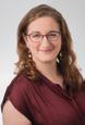
Zoe Doyle joined the department in September 2021.
Dr. Doyle earned her MD at the University of California, San Diego in 2015, followed by a diagnostic radiology residency and musculoskeletal imaging fellowship at Stanford University Hospital, Palo Alto, CA. During her time at Stanford, Dr. Doyle worked on quality improvement initiatives such as improving the efficiency of ultrasound exams, streamlining the informed consent process for pregnant patients and reducing time spent protocoling MSK MRI exams.
Dr. Doyle’s research interests include collaborations to study how novel MRI metal-suppressive techniques can be clinically implemented in patients with hip arthroplasties and the clinical use of new accelerated isotropic sequences which would allow for the elimination of multi-planar acquisitions and potentially improve evaluation of anatomy that is not as well seen on the traditional planes acquired.
Kayla Cort, DO Assistant Professor Pediatric Radiology
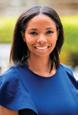
Kayla Cort, DO, joined our faculty as an assistant professor of Clinical Radiology in the Pediatric Radiology section in February 2022.
Dr. Cort earned her DO degree at the Philadelphia College of Osteopathic Medicine. She completed an internship in medicine at Morristown Medical Center in Morristown, NJ, a diagnostic radiology residency at SUNY Downstate Medical Center in Brooklyn, NY, and a pediatric radiology fellowship at Children’s Hospital of Philadelphia, PA. Following the fellowship, Dr. Cort became an assistant clinical professor and director of pediatric ultrasound at UC Davis Medical Center. At UCD, Dr. Cort served on the Inclusion, Diversity, Anti-Racism and Equity (IDARE) taskforce which promotes an environment that embraces diversity, equity and inclusion.
Dr. Cort works at the UCSF Benioff Children’s Hospitals in San Francisco and Oakland. Among Dr. Cort’s clinical focus areas are the implementation of high-quality pediatric protocols and the expansion of the applications of contrast-enhanced ultrasound.
PEOPLE: ACADEMIC AFFAIRS
IMAGES 2021–2022 23
Kazim Narsinh, MD Assistant Professor Neurointerventional
Kazim Narsinh, MD, joined our faculty as an assistant professor in residence in the Neurointerventional Radiology section in February 2022.
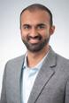
Previously, Dr. Narsinh obtained his medical degree at the UC San Diego School of Medicine where he was a Howard Hughes Medical Research Fellow. He then completed a surgical internship at Stanford, a radiology residency at UC San Diego, an interventional radiology fellowship at the University of Pennsylvania, and fellowships in neuroradiology and neurointerventional at UCSF.
As a clinician and scientist, Dr. Narsinh has authored more than 60 articles on topics including single cell transcriptomics, cellular and molecular imaging, carcinogenesis and vascular disease. He works with colleagues at UCSF to develop and strengthen clinical and research programs of excellence.
Jamie L. Twist Schroeder, MD, DPhil Assistant Professor Cardiac and Pulmonary Imaging
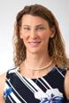
Jamie Schroeder, MD, DPhil, joined our faculty as an assistant professor of Clinical Radiology in the Cardiac & Pulmonary Imaging section in March 2022.
Dr. Schroeder completed a master’s degree in Bioengineering at Stanford University, Palo Alto, CA, and earned a DPhil at the University of Oxford in England. She earned her medical degree at Johns Hopkins School of Medicine, Baltimore, MD, and completed an internal medicine internship at Sinai Hospital of Baltimore. She completed a four-year diagnostic radiology residency at Johns Hopkins Hospital, followed by a one-year fellowship in cardiac and pulmonary imaging at UCSF. Prior to joining the UCSF Radiology faculty, Dr. Schroeder was a staff physician in the Cardiovascular/Thoracic Imaging section of the Stanfordaffiliated Palo Alto Veterans Affairs Medical Center.
Dr. Schroeder’s recent projects focus on improving clinical decision-making in cardiac imaging and include the development of an unsupervised machine learning pathway to analyze motion-corrected time-course data from myocardial perfusion MRI with the goal of systematizing the diagnostic process.
Michael Hoff, PhD Associate Professor
Advanced Imaging Technologies
Specialized Resource Group
Michael Hoff, PhD, joined our faculty as an associate professor in April 2022. As director of diagnostic medical physics, Dr. Hoff supports best practices with imaging technology, regulatory compliance and facilitating translation of imaging physics innovations to improvements in clinical care.
Dr. Hoff completed his master’s and doctoral degrees in medical physics at the University of British Columbia in Vancouver, Canada. He joined the Department of Radiology at the University of Washington in Seattle in 2013, where he was an associate professor in the Diagnostic Physics section. His primary research interest is to exploit complex MR data to glean highly accurate information on tissue physiology and function.
“As a scientist, Dr. Hoff comes to us with a history of prestigious awards and funding and a strong collaborative profile across departments, institutions and research areas,” said Christopher Hess, MD, PhD, UCSF Radiology chair.
Jaehoon Shin, MD, PhD Assistant
 Professor, In Residence
Interventional Radiology
Jaehoon Shin, MD, PhD, became faculty in July 2022. He earned his MD at Seoul National University, South Korea, followed by a PhD at Johns Hopkins University. He completed an internship at MedStar Harbor Hospital in Baltimore followed by a diagnostic radiology residency concurrently with early specialization in interventional radiology (ESIR) at UCSF. Dr. Shin completed an interventional radiology fellowship, also at UCSF.
Dr. Shin received the prestigious Samsung Scholarship for doctoral study, and was awarded Best Case in Neuroradiology at the 2019 American Institute for Radiologic Pathology. Dr. Shin is a collaborator on a Medical Research Award from the Robert J. & Helen C. Kleberg Foundation, and PI for a SIR Pilot Research Grant. Dr. Shin’s research addresses novel T cell-based imaging (synNotch-PET), which has the potential to detect early cancers and pre-cancerous lesions. Additional research projects include locoregional T cell therapy to enhance treatments for pancreatic cancer and glioblastoma and single-cell RNA sequencing to better understand venous insufficiency and druggable target biological processes.
24 IMAGES 2021–2022
Jonathan Liu, MD, MS
Assistant Professor of Clinical Radiology Cardiothoracic Imaging
Jonathan Liu became faculty in July 2022.

Dr. Liu earned his MD at the Keck School of Medicine, University of Southern California and an MS in electrical engineering at Stanford. After an internship in internal medicine at Santa Clara Medical Center, Dr. Liu completed residency at the Mallinckrodt Institute, Washington University followed by a thoracic radiology fellowship at Stanford.
Dr. Liu’s clinical and research interests include 3D printing applications in nuclear medicine and artificial intelligence applications in thoracic and cardiovascular radiology. During residency, Dr. Liu was instrumental in a collaborative project at Washington University to introduce radiology into the anatomy curriculum, providing more clinical experiences for medical students earlier in their training.
Shravan Sridhar, MD Assistant Professor of Clinical Radiology Cardiothoracic Imaging
Shravan Sridhar joined as faculty in July 2022.
Dr. Sridhar earned an MS in Biomedical Engineering summa cum laude at Arizona State University followed by an MD at University of Arizona College of Medicine where he was a Service Learning Scholar and Peer Tutor. Dr. Sridhar completed an integrated clinical internship and diagnostic radiology residency at the University of New Mexico in Albuquerque. Dr. Sridhar completed a cardiac and pulmonary imaging fellowship in 2022. at UCSF. Dr. Sridhar has been recognized with the Resident Educator Award (2021), RSNA Roentgen resident/fellow research award (2020) and RSNA certificate of merit (2019), among others.
Dr. Sridhar’s research includes examining the role of molecular biomarkers in brain metastasis of non-small cell lung cancer and multi-modality imaging features of sternoclavicular joint infection and impact on operative management. Dr. Sridhar has served as a consulting editor for American Journal of Radiology (AJR)
Xin Cynthia Wu, MD Associate Professor
of Clinical Radiology Neuroradiology
Cynthia Wu joined the department in August 2022.
Dr. Wu earned her MD at the University of Pennsylvania with a Certificate in Clinical Neuroscience. After an internship year at Allegheny General-Western Pennsylvania Hospitals Medical Education Consortium, Dr. Wu completed a diagnostic radiology residency at NYU School of Medicine followed by a neuroradiology fellowship at UCSF, where she served as chief fellow. Dr. Wu has been an assistant professor of radiology and imaging sciences at Emory University School of Medicine in Atlanta.
Dr. Wu’s clinical practice has included general neuroradiologic procedures and dedicated ENT imaging including complex head and neck studies and multidisciplinary oncology clinics. As a researcher, Dr. Wu focuses on application of advanced MR techniques to head and neck tumor imaging, improving imaging delineation of rare and unconventional head and neck cancer spread patterns, and characterizing head and neck postoperative findings that may mimic disease. Additional research interests include MRI imaging of brain tumors and tumor mimics as well as advanced MR imaging of mild traumatic brain injury.
Cheng William Hong, MD, MS
Assistant Professor of Clinical Radiology
Abdominal Imaging / Ultrasound
Cheng William Hong joined the department in September 2022.
Dr. Hong received an MD with special qualification in Biomedical Research from the Cleveland Clinic Lerner College of Medicine, followed by an MS in biomedical investigation from Case Western Reserve University in Cleveland, where he was in the Clinical Research Scholars Program. After an internal medicine internship year at St Mary’s Medical Center in San Francisco, Dr. Hong completed a diagnostic radiology residency on the T32 Clinician-Scientist Track at UC San Diego, where he received the RSNA Roentgen Resident Research Award and the UCSD Robert Mattrey, MD Research Award which recognize outstanding research among graduating residents. Dr. Hong completed a body MRI fellowship at Stanford.
As a clinician, Dr. Hong focuses on advanced interpretation, protocoling, and troubleshooting in body MRI and PET/MRI, as well as performing MRI-guided procedures. Dr. Hong’s research focuses on noninvasive hepatic fat and iron quantification using MRI and ultrasound, and the clinical outcomes of LI-RADS.
IMAGES 2021–2022 25 PEOPLE: ACADEMIC AFFAIRS
Ellen Chang, MD, MS
Assistant Professor, HS Clinical
Musculoskeletal Radiology, Zuckerberg San Francisco General Hospital
Ellen Chang joined our faculty in September 2022.
Dr. Chang earned an MD from Washington University in St Louis and an MS in biological chemistry from UCLA. During her general surgery internship at Harbor-UCLA Medical Center, Dr. Chang was named Intern of the Year. Dr. Chang completed a diagnostic radiology residency with a fourth year neuroradiology concentration at Dartmouth Hitchcock Medical Center followed by a musculoskeletal fellowship at Mallinckrodt Institute of Radiology. From 2020-22, Dr. Chang was an assistant professor in the Musculoskeletal Division of the USC Department of Radiology.
Dr. Chang’s clinical practice focuses on musculoskeletal imaging across MRI, CT, radiography and ultrasound as well as procedures that include joint injections, biopsies, and spine interventions. Recent research has focused on image-guided spine interventions, acute ACL injury, and trainee education projects.
Jemianne Bautista, MD Assistant Professor of Clinical Radiology Interventional Radiology
Jemianne Bautista became faculty in September 2022.
Dr. Bautista earned her MD from UC Irvine, followed by an internal medicine internship and diagnostic radiology residency at Kaiser Permanente Los Angeles Medical Center. During residency, Dr. Bautista elected early specialization in interventional radiology, additionally serving as chief resident and Wellness Chair. Dr. Bautista completed a fellowship in vascular and interventional radiology at University of California, San Diego.
As a clinician, Dr. Bautista brings expertise in women’s health and interventions as well as more complex arterial and venous work such as trauma management, peripheral arterial disease interventions, and aortic endografts. Dr. Bautista’s research focuses on interventional oncology and women’s interventions, including targeted therapies for colorectal carcinoma and uterine fibroid embolization. Future research projects are expected to explore targeted palliative nerve therapies.
Yang Yang, PhD Associate Professor, In Residence Director, Mid-field
MRI Research
Yang Yang became faculty in September 2022.
Dr. Yang holds an MSE in biomedical engineering from Xi’an Jiaotong University in Shaanxi, China, and a PhD in biomedical engineering from University of Virginia, Charlottesville. Dr. Yang’s dissertation: Whole-heart Coverage Quantitative First-pass Spiral Perfusion in Cardiac Magnetic Resonance Imaging. Dr. Yang was an assistant professor at the Icahn School of Medicie at Mount Sinai, in the Biomedical Engineering and Imaging Institute and Department of Diagnostic, Molecular and Interventional Radiology. Dr. Yang was an adjunct assistant professor in the Department of Medicine, Division of Cardiovascular Medicine at the University of Virginia.
Dr. Yang’s expertise is MR sequence development, nonCartesian trajectory design and application, robust motion correction, and artificial intelligence. The overall goal of Dr. Yang’s research is to develop and evaluate novel pulse sequences and techniques to improve the clinical utility of MRI. Current research projects are focused on fast magnetic resonance imaging technique development and its translational/clinical applications. During the COVID-19 pandemic, Dr. Yang’s research on COVID-19 imaging and artificial intelligence resulted a technique for rapid diagnosis of COVID and a highly cited Nature Medicine AI paper.
Yingbing Wang, MD
Associate Professor of Clinical Radiology
Molecular Imaging & Therapeutics
Yingbing Wang joined as faculty in September 2022.
Dr. Wang earned an MD from Stanford University followed by an internal medicine internship at Kaiser Permanente in Santa Clara, CA. Dr. Wang completed a radiology residency and a mini-fellowship in nuclear radiology at Massachusetts General Hospital. Dr. Wang was an assistant professor of radiology at Harvard Medical School, and held directorships of NMMI education and radionuclide therapies, and served as program director for the Nuclear Radiology Fellowship at Massachusetts General Hospital.
26 IMAGES 2021–2022
Dr. Wang’s clinical interests and expertise span conventional single photon nuclear medicine, positron emission tomography, nuclear cardiology, and nuclear therapies. Dr. Wang’s scholarship encompasses retrospective data mining, improving the diagnostic interpretation of clinically approved nuclear radiotracers, and exploring novel PET radiotracers for diagnosis and therapy monitoring in oncologic applications. The long-term goals of this research are to validate imaging techniques – novel radiopharmaceuticals and AI methods – that can improve diagnostic accuracy, predict therapy response, and provide meaningful prognostic information. Recent projects include developing quantitative methods to augment conventional qualitative interpretation of PET images.
Amrutha Ramachandran, MD Assistant Professor of Clinical Radiology Neuroradiology and Emergency Room, Zuckerberg San Francisco General Hospital
Amrutha Ramachandran became faculty in November 2022.
Dr. Ramachandran completed an MD at Kasturba Medical College, Manipal in Karnataka, India. After an internship at MetroWest Medical Center Framingham, MA, Dr. Ramachandran completed a radiology residency at Jackson Memorial Hospital and University of Miami where she served as chief resident, followed by a neuroradiology fellowship at UCSF. In 2021-22, Dr. Ramachandran was a neuroradiology clinical instructor at UCSF.
As a neuroradiologist, Dr. Ramachandran focuses on pediatric neuroimaging with an interest in fetal MRI, pediatric tumor imaging, and emergency department trauma imaging. Dr. Ramachandran contributes to radiology QA/QI, including optimizing imaging protocols and standardizing reporting templates. Dr. Ramachandran’s research interests are in musculoskeletal imaging and pediatric neuroimaging.
Melanie Morrison, PhD Assistant Professor, In Residence Research
Melanie Morrison became faculty in December 2022.
Dr. Morrison holds a PhD in medical biophysics from the University of Toronto, and has completed postdoctoral research at Imperial College London and UCSF, where she was a UC Chancellor’s Postdoctoral Fellow. While at University of Toronto, Dr. Morrison received the James Lepock Memorial Award and the Walker-Marshall Award, both for doctoral student academic excellence. Dr. Morrison has held honorary fellowships at Imperial College London and University of Edinburgh, and was the recipient of the Bruce Hasegawa, PhD Award for Early-Stage Scientist Research Excellence at UCSF.
Dr. Morrison’s research concentrates on advanced MRI methods to evaluate neurological movement and psychiatric disorders, including the surgical neuromodulation therapies used to treat severe conditions. Current collaborative projects include a multimodal 7T imaging study of Huntington’s disease; a multimodal imaging study of advanced Parkinson’s disease; a multicenter surgical gene-therapy trial for the treatment of Parkinson’s disease; and a functional imaging study of obsessive-compulsive disorder, specifically patients who are currently benefiting from deep brain stimulation.
IMAGES 2021–2022 27 PEOPLE: ACADEMIC AFFAIRS
FACULTY RETIREMENTS
Christopher Dowd, MD Professor Emeritus, Radiology, Neurological Surgery, Neurology, Anesthesia, and Preoperative Care Co-Director of Birthmarks and Vascular Anomalies Center
Dr. Dowd specializes in the diagnosis, management and treatment of cerebrovascular diseases and vascular birthmarks. Dr. Dowd earned his medical degree from the University of Virginia, Charlottesville, and completed his diagnostic radiology at UC San Diego, followed by a fellowship in neuroradiology and interventional neuroradiology at UCSF.
Dr. Dowd has served as the Primary Investigator and Co-Investigator of many IRB/CHR approved research projects on topics related to his clinical practice such as endovascular treatment of intracranial aneurysms, subarachnoid hemorrhage-induced vasospasm, cutaneous vascular birthmarks and similar conditions. He has published 178 articles, 300 scientific abstracts, and 58 book chapters.
A selection of notable awards over the years includes recognition as a US News and World Report Top Doctor in 2012, Clinical Teacher of the Year in 2001 in the UCSF Department of Dermatology, and notation as an Honorary Overseas Member and First Honored Guest Lecturer for the British Skull Base Society.
Ronald Cohen, MD Clinical Professor, Radiology
Medical Director, Pediatric Diagnostic Imaging UCSF Benioff Children’s Hospital-Oakland
Dr. Cohen has been a physician at Benioff Children’s Hospital-Oakland since 1984. He received his medical degree at UC Davis followed by a diagnostic radiology residency at Stanford University and a fellowship in pediatric radiology at Children’s Hospital Medical Center in Cincinnati, OH.
His special interests include imaging of the digestive, urinary and reproductive systems, advanced applications of CT imaging, and imaging related to child abuse detection. Dr. Cohen has mentored pediatric radiology fellows for 25 years, and has served for a number of years as a mentor to radiology residents from Travis Air Force Base. During his career, he has engaged in teaching opportunities in Russia, Vietnam, Guatemala, Panama, Israel, Zambia, Zimbabwe. Since 1990, Dr. Cohen has sponsored an annual visiting lectureship honoring Hooshang Taybi, MD.
Outside of the reading room, Dr. Cohen is an avid cyclist and adventure traveler.
Ronald Zagoria, MD Professor
Emeritus, Section Chief,
Abdominal Imaging and Ultrasound, Vice Chair, Clinical Operations
Dr. Zagoria earned his medical degree at the University of Maryland School of Medicine followed by a diagnostic radiology residency and a fellowship in abdominal imaging and interventional radiology at Wake Forest University and Massachusetts General Hospital. Prior to joining UCSF in 2013, Zagoria served as chief of genitourinary imaging and intervention as well as executive vice chair for the radiology department at Wake Forest School of Medicine where he was on the faculty for 26 years.
In a storied career, a selection of notable awards include a Gold Medal from the Society for Abdominal Radiology in 2022, a Lifetime Service Award from the American Board of Radiology in 2015, Felson Lecturer at the Cincinnati College of Medicine in 2000, and numerous awards for teaching excellence over the years. Castle Connolly and U.S. News & World Report, among other national organizations, have repeatedly recognized Dr. Zagoria as one of the best doctors in America and specifically as a top doctor for cancer and a top radiologist.
Dr. Zagoria’s clinical interests include urologic oncology, urolithiasis and infertility. He specializes in image-guided tumor ablation for kidney cancer. He has given more than 300 lectures and presentations, and has written more than 300 published pieces, including textbooks, book chapters, journal articles and medical abstracts.
Having enjoyed a varied and active career as a mentor and educator, clinician, and researcher, Dr. Zagoria notes that his greatest successes are having a wonderful partnership with his wife of 39 years, Kat Zagoria, and raising two wonderful children, David and Michael.
28 IMAGES 2021–2022
ALL FACULTY LIST, CLINICAL AND RESEARCH
July 1, 2021–December 31, 2022
Abdominal Imaging Ultrasound
Jane Z. Wang, MD Professor In Residence
Spencer Behr, MD Associate Professor, Clinical Radiology
Peter Callen, MD Emeritus Faculty
Hailey Choi, MD Assistant Professor, Clinical Radiology
Vickie Feldstein, MD Professor, Clinical Radiology
Ruth Goldstein, MD Emeritus Faculty
Cheng William Hong, MD Assistant Professor, Clinical Radiology
Sina Houshmand, MD Assistant Professor, Clinical Radiology
Priyanka Jha, MD Associate Professor, Clinical Radiology
Marc Kohli, MD Professor, Clinical Radiology
John Mongan, MD, PhD Professor, Clinical Radiology
Tara Morgan, MD Associate Professor, Clinical Radiology
Liina Poder, MD Professor, Clinical Radiology
Dorothy Shum, MD HS Clinical Professor
Mark Sugi, MD Assistant Professor, Clinical Radiology
Derek Sun, MD HS Clinical Associate Professor
Emma Webb, MD Professor, Clinical Radiology
Sean Woolen, MD Assistant Professor, Clinical Radiology
Benjamin Yeh, MD Professor In Residence
Ronald Jay Zagoria, MD Professor, Clinical Radiology
Breast Imaging
Bonnie Joe, MD, PhD Professor In Residence
Elissa Amans - Price, MD Professor, Clinical Radiology
Rita Freimanis, MD Professor, Clinical Radiology
Heather Greenwood, MD Associate Professor, Clinical Radiology
Jessica Hayward, MD Associate Professor, Clinical Radiology
Tatiana Kelil, MD Assistant Professor, Clinical Radiology
Amie Le, MD Professor, Clinical Radiology
Kimberly Ray, MD Associate Professor, Clinical Radiology
Edward Sickles, MD Emeritus Faculty
Cardiac & Pulmonary Imaging
Brett Elicker, MD Professor, Clinical Radiology
Kimberly Kallianos, MD Assistant Professor, Clinical Radiology
Yoo Jin Lee, MD Assistant Professor, Clinical Radiology
Jonathan Liu, MD Assistant Professor, Clinical Radiology
Jamie Schroeder, MD, DPhil
Assistant Professor, Clinical Radiology
Jae Ho Sohn, MD Assistant Professor In Residence
Shravan Sridhar, MD Assistant Professor, Clinical Radiology
Maya Vella, MD Assistant Professor, Clinical Radiology
Interventional Radiology
Pallav Kolli, MD Associate Professor, Clinical Radiology
Jemianne Bautista, MD Assistant Professor, Clinical Radiology
Nicholas Fidelman, MD Professor, Clinical Radiology
Michael Heller, MD Assistant Professor, Clinical Radiology
Ryan Kohlbrenner, MD Associate Professor, Clinical Radiology
Alexander Lam, MD Assistant Professor, Clinical Radiology
Gina Landinez, MD HS Clinical Assistant Professor
Evan Lehrman, MD Associate Professor, Clinical Radiology
Pete Lokken, MD, MPH Associate Professor, Clinical Radiology
Jaehoon Shin, MD, PhD Assistant Professor, In Residence
Andrew Taylor, MD, PhD Associate Professor, Clinical Radiology
Molecular Imaging & Therapeutic
Rob Flavell, MD, PhD Associate Professor In Residence
Miguel Hernandez Pampaloni, MD, PhD Professor, Clinical Radiology
Thomas Hope, MD Professor In Residence
Roxanna Juarez, MD Assistant Professor, Clinical Radiology
Courtney Lawhn Heath, MD Assistant Professor, Clincal Radiology
Yingbing Wang, MD Associate Professor, Clinical Radiology
PEOPLE: ACADEMIC AFFAIRS IMAGES 2021–2022 29
Musculoskeletal Radiology
Thomas Link, MD, PhD Professor In Residence
Matt Bucknor, MD Associate Professor In Residence
Kevin McGill, MD, MPH Assistant Professor, Clinical Radiology
Daria Motamedi, MD Associate Professor, Clinical Radiology
Rina Patel, MD Assistant Professor, Clinical Radiology
Lynne Steinbach, MD Emeritus Faculty Neuroendovascular Surgery
Steven W. Hetts, MD Professor In Residence
Randall T. Higashida, MD Professor, Clinical Radiology
Matthew Amans, MD Professor, Clinical Radiology
Daniel Cooke, MD Professor In Residence
Christopher F. Dowd, MD Emeritus Faculty
Kazim Narsinh, MD Assistant Professor In Residence
Neuroradiology
Vinil Shah, MD Associate Professor, Clinical Radiology
Matthew Barkovich, MD Assistant Professor In Residence
Soonmee Cha, MD Professor In Residence
Cynthia Chin, MD Emeritus Faculty
William Dillon, MD Professor
Elizabeth George, MBBS Assistant Professor, Clinical Radiology
Christine Glastonbury, MBBS Professor, Clinical Radiology
Orit Glenn, MD Professor, Clinical Radiology
Christopher P. Hess, MD, PhD Professor
Yi Li, MD Assistant Professor, Clinical Radiology
Andreas Rauschecker, MD, PhD Assistant Professor In Residence
Leo Sugrue, MD, PhD Professor In Residence
Javier Villanueva-Meyer, MD
Associate Professor, Clinical Radiology
David Wilson, MD, PhD Professor In Residence
Xin Cynthia Wu, MD Associate Professor, Clinical Radiology
Pediatric Radiology
Raymond Sze, MD, MAMS Professor, Clinical Radiology
Jesse Courtier, MD Professor, Clinical Radiology
Taylor Chung, MD HS Clinical Professor
Pierre-Alain Cohen, MD HS Clinical Professor
Ronald Cohen, MD HS Clinical Professor
Kayla Cort, DO HS Clinical Assistant Professor
Rachelle Durand, DO Assistant Professor, Clinical Radiology
Bamidele Kammen, MD HS Clinical Professor
Alexander Wai, MD HS Clinical Professor
Matthew Zapala, MD, PhD
Associate Professor, Clinical Radiology
Research
Sharmila Majumdar, PhD Professor
Robert Bok, MD, PhD Adjunct Professor
Linda Chao, PhD Adjunct Professor
Michael Evans, PhD Associate Professor In Residence
Myriam Chaumeil, PhD Associate Professor in Residence
Jeremy Gordon, PhD Assistant Adjunct Professor
Grant Gullberg, PhD Emeritus Faculty
Michael Hoff, PhD Associate Professor, Clinical Radiology
Nola Hylton, PhD Professor
Galateia Kazakia, PhD Professor In Residence
Roland Krug, PhD Associate Professor
John Kurhanewicz, PhD Emeritus Faculty
Peder Larson, PhD Professor In Residence
Yan Li, PhD Assistant Adjunct Professor
Jing Liu, PhD Associate Adjunct Professor
Janine Lupo, PhD Associate Professor In Residence
Alastair J. Martin, PhD Adjunct Professor
Dimitrios Mitsouras, PhD Associate Professor In Residence
Melanie Morrison, PhD Assistant Professor In Residence
Susanne Mueller, PhD Adjunct Professor Srikantan S. Nagarajan, PhD Professor
Susan Noworolski, PhD Adjunct Professor
Eugene Ozhinsky, PhD Assistant Adjunct Professor
Valentina Pedoia, PhD Associate Adjunct Professor
Donna Peehl, PhD Adjunct Professor
Ashish Raj, PhD Professor In Residence
Sabrina Ronen, PhD Emeritus Faculty
David Saloner, PhD Professor In Residence
Youngho Seo, PhD Professor In Residence
Renuka Sriram, PhD Assistant Adjunct Professor
Duygu Tosun-Turgut, PhD Associate Professor In Residence
Olga Tymofiyeva, PhD Associate Adjunct Professor
30 IMAGES 2021–2022
Henry VanBrocklin, PhD Professor In Residence
Dan Vigneron, PhD Professor
Pavithra Viswanath, PhD Associate Adjunct Professor
Joseph Vu, PhD Assistant Adjunct Professor
Michael Weiner, MD Professor In Residence
Duan Xu, PhD Professor In Residence
Yang Yang, PhD Associate Professor In Residence
SF Veterans Affairs
Michael Hope, MD Professor In Residence
Michael Parsa, MD Interim Chief, On Detail
Zoe Doyle, MD Assistant Professor, Clinical Radiology
Eugene Huo, MD HS Clinical Assistant Professor
Joseph Leach, MD, PhD Assistant Professor In Residence
Pratik Mukherjee, MD, PhD Professor In Residence
Rajiv Sawhney, MD HS Clinical Professor
Preeti Sukerkar, MD, PhD
Assistant Professor, Clinical Radiology
Stefanie Weinstein, MD Professor, Clinical Radiology
Zuckerberg SFGH
Mark Wilson, MD Professor In Residence
Ellen Chang, MD HS Clinical Assistant Professor
Miles Conrad, MD, MPH HS Clinical Professor
Shital Gandhi, MD HS Clinical Associate Professor
Brian Haas, MD Assistant Professor, Clinical Radiology
Vishal Kumar, MD Associate Professor, Clinical Radiology
Terry Lynch, MD HS Clinical Professor
Sujal Nanavati, MD HS Clinical Professor
Jared Narvid, MD Associate Professor, Clinical Radiology
Michael Ohliger, MD, PhD Associate Professor In Residence
Preethi Raghu, MD Assistant Professor, Clinical Radiology
Amrutha Ramachandran, MD Assistant Professor, Clinical Radiology
Bhavya Rehani, MD Associate Professor, Clinical Radiology
Alexander Rybkin, MD HS Clinical Professor
Lori Strachowski, MD HS Clinical Professor
Jason Talbott, MD, PhD Professor In Residence
Thienkhai Vu, MD, PhD HS Clinical Professor
Esther Yuh, MD, PhD Professor In Residence
PEOPLE: ACADEMIC AFFAIRS IMAGES 2021–2022 31
EDUCATION
CHALLENGES AND ACCOMPLISHMENTS
UCSF Training Programs 2021-2022
By Soonmee Cha, MD
Iam so proud of the resiliency and dedication of all our trainees. Despite the challenging environment of 20212022, our trainees had a tremendous year punctuated by learning and achievement. They provided excellent clinical care with a focus on professionalism, safety and creating a positive experience for our patients at UCSF hospitals.
Residency Program
In July 2021, we welcomed the Class of 2025, an outstanding group of 15 diagnostic radiology residents and one integrated interventional resident. This past July the class of 2026 joined us. We were pleased to welcome 14 diagnostic radiology residents and 1 integrated interventional resident. I am amazed to see how our newest trainees have grown, both as people and as physicians. They have embraced learning and have moved from being beginners in medical imaging to becoming skilled colleagues who have gained confidence in their diagnoses and work effectively with our fellows and attendings to support the highest standards of patient care. I appreciate the attention, dedication and skill that our newest recruits bring to their duties.
Graduation 2021 was again virtual. However, the many people who attended were moved by the meaningful ceremony acknowledging the accomplishments of our graduates. Friends and relatives logged in from across the country to celebrate our trainees with us. The 2022
commencement offered the opportunity for a safe in-person gathering in compliance with UCSF’s updated policies.
In late 2022 our newly elected chiefs began their duties. I am pleased to welcome Masis Isikbay, MD, Adam Yen, MD, and Kevin Terashima, MD, to their role as 2022-23 chiefs. We are already grateful for their enthusiasm and appreciate their commitment to our program.
I would like to express my deep gratitude to the outgoing chief residents: Joelle Harwin, MD, Kevin Sweetwood, MD, and Andrew Taliaferro, MD, for their outstanding service to our residents, program and department.
This was the ninth consecutive year that our clinical training program has ranked first amongst the top-rated diagnostic radiology residency programs in the nation according to Doximity’s 2022-2023 Residency Navigator. We were very proud to achieve this high ranking once again.
Leadership Team
I rely on Jason Talbott, MD, associate program director, head of the Residency Selection Committee and ZSFG residency site director for his leadership during the recruitment process. Each year, Jason structures the residency selection process with an emphasis on fairness, transparency and creating an optimal experience for those visiting us. In 2022, applicant interviews were once again in
32 IMAGES 2021–2022
(l-r): Class of 2025 residents Joseph Knox, MD, Minerva Zhou, MD and Lohith Kini, MD, MSc, PhD with Chief Residents Andrew Taliaferro, MD and Kevin Sweetwood, MD
a virtual setting, but with the support of the entire team the process was a smooth and successful one. We matched with 14 stellar trainees that will join us in July 2024.
I am also grateful to Jason for his advocacy. His goal is to bring each resident to their highest level of performance and our residents are grateful for his experience and training, which he readily shares with them.
I would also like to thank Jesse Courtier, MD, for his 14 years of service as site director at Mission Bay. He has been a role model for our residents at that site and I would like to thank him for his outstanding service. Matthew Zapala, MD, is our new Mission Bay site director and is already providing great value to our Mission Bay residency rotations.
Education Team
Our extraordinary team of education professionals work under the direction of Melinda Parangan-Chu, our administrative director: Sora Kang, Mike Leon, Dave Stewart, Sandria Wong and Samira Zebarjadian. Each one of them is an important asset to the educational programs we offer, and I am appreciative of their work. This year we bid farewell to Cindy Flores Gaytan who accepted a new role as the education systems manager in the UCSF Office of Graduate Medical Education. Cindy was an integral part of our programs for over 10 years. She cared deeply about our residents and was there for them every step of the way from recruitment to commencement. We wish her well in her exciting new role at UCSF.
Fellowship Program
In July 2021, 42 new promising new fellows and six clinical instructors entered our training program, and in July 2022 we welcomed 39 fellows and three clinical instructors. It has been exciting to have them with us as they moved forward in their chosen subspecialties and continued the process of selecting future directions for their careers.
A big thank you to our fellowship program directors and assistant directors, who are an important resource for our fellows:
■ Director of Fellowship Programs: Spencer Behr, MD
■ Abdominal Imaging/Ultrasound: Derek Sun, MD, and Mark Sugi, MD
■ Breast Imaging: Elissa Price, MD
■ Cardiac & Pulmonary Imaging: Kimberly Kallianos, MD
■ Interventional Radiology: Evan Lehrman, MD, and Vishal Kumar, MD
■ Musculoskeletal Radiology: Rina Patel, MD
■ Neuroradiology: Yi Li, MD, and Andreas Rauschecker, MD, PhD
■ Neurointerventional Radiology: Matthew Amans, MD
■ Molecular Imaging & Therapeutics: Miguel Pampaloni, MD, PhD, and Courtney Lawhn Heath, MD
■ Pediatric Radiology: Matthew Zapala, MD
Under the leadership of Dr. Behr, the program directors meet quarterly to discuss ways to improve the fellowship training programs. The fellowship educational fund, fellows social, chief fellow and fellow council are some ideas implemented since the formation of this group. They strive to bring uniformity and equity, and to streamline policies and educational standards across all subspecialties.
We look forward to a great 2023 and are excited to see what the future holds for our trainees.
Soonmee Cha, MD, is a professor in residence of Radiology and Neurological Surgery, vice-chair of Education, program director for the Diagnostic Radiology Residency Program and associate program director for the integrated interventional Radiology Residency.
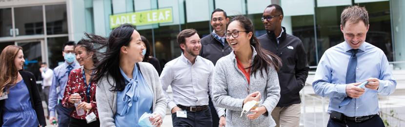
PEOPLE: EDUCATION
IMAGES 2021–2022 33
Incoming Class of 2025 residents on their first day at UCSF
THE CLASS OF 2025
Incoming Residents, July 2021
Alwalid Ashmeik, MD
Alwalid Ashmeik received his medical degree in 2020 from UCSF, graduating with distinction. He completed his internship (2021) at Highland Hospital in Oakland, CA. In 2019, he received the Yearlong Research Fellowship/TLI Predoctoral Fellowship from the UCSF School of Medicine (2019).
Ashmeik was a medical student researcher in UCSF Radiology’s Musculoskeletal and Quantitative Imaging Research Group (2016-2020) where he worked on studies of voriconazole-induced periostitis; the association of blood pressure with knee cartilage composition and structural knee anomalies; and the impact of type 2 diabetes and metabolic risk factors on knee joint health. He is an author on four journal articles and seven abstracts.
Ashmeik served as the Renal and Endocrinology small group facilitator at UCSF (2020) where he assisted in developing weekly review sessions and examinations for first-year medical students during their Renal, Endocrine, GI and Nutrition curriculum. He was also a mentor in UCSF’s Pre-Health Undergraduate Program, where he was paired with an underrepresented minority undergraduate student in a program that focuses on critically appraising medical literature, designing clinical and translational research studies, and encouraging longterm collaborations between UCSF graduate students and undergraduate students.
Ilan Benador-Shen, MD, PhD
Ilan Benador-Shen obtained his medical degree and PhD from Boston University School of Medicine in 2020. He completed an internship at St. Elizabeth Medical Center in Brighton, MA, in 2021.
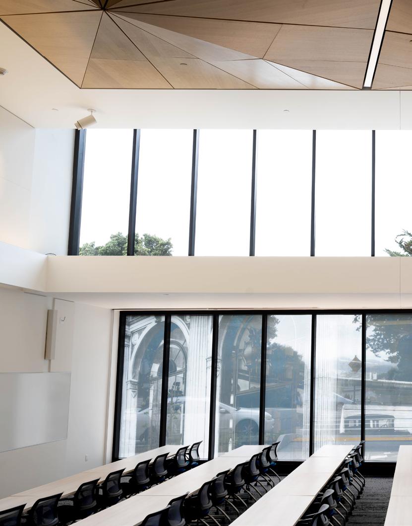
Benador-Shen served as a course developer for HarvardX cell biology and medical biochemistry courses (2015-2018) developing original lecture content and assessment questions.
Benador-Shen’s research at Boston Medical Center, Radiology (2019-2020), focused on using MR spectroscopy to characterize the metabolic profile of brain tumors using biopsy-proven brain tumors from the eTumour database. He served as a graduate research fellow at UC Los Angeles (2016-2018), where his work focused on brown adipose tissue, which is rich in
mitochondria and can promote weight loss. From 20182021, he was a reviewer for the Society for Redox Biology and Medicine in Boston, MA, reviewing original research manuscripts for a scientific journal focused on cell biology and metabolism. Benador is an author on more than 15 peer-reviewed journal articles and abstracts.
Benador-Shen was awarded a US patent for frozen mitochondria testing which was licensed to Enspire Bio. He is working with Enspire Bio CEO Dr. Amy Wang (in collaboration with Amgen Inc., UC San Diego and University Alabama Birmingham) to develop a new consumer test kit for frozen tissue biopsies, which will open the door to widespread clinical mitochondrial testing and work to address non-alcoholic fatty liver disease by identifying drugs that target peridroplet mitochondria in hepatocytes.
Luke W. Bonham, MD
Luke Bonham earned his medical degree in 2020 from The Johns Hopkins School of Medicine in Baltimore, MD. He completed an internship at the Virginia Mason Medical Center in Seattle, WA, in 2021.
He was the recipient of an RSNA Medical Student Research Award (2017), the Paul S. Wheeler Radiology Fund for Medical Students Scholarship (2018) and the Doyle, Brody and Asper Scholarships (2019). He received the Best Presentation in Public Health award at The Johns Hopkins Medical Student Research Symposium (2018).
Bonham gained experience as a medical student researcher at Johns Hopkins, Radiology, designing and implementing a study of MR-conditional needle heating during commonly used interventional MR procedures and at UCSF Radiology (2017-present) analyzing MRI/CT data to develop a multimodal imaging risk score for prediction of Alzheimer’s disease; and observing and assisting attending radiologists as they performed peripheral nerve blocks and ablations. Bonham is an author on more than 30 peer-reviewed publications.
Camilo Campo, MD
In 2020, Camilo Campo obtained his medical degree from Harvard Medical School in Boston, MA. He completed his internship at the Gunderson Health System in LaCrosse, WI in 2021.
Campos’ research at Brigham and Women’s Hospital, Emergency Radiology (2019-2020) focused on assessing the value of renal ultrasound for urolithiasis in the emergency department, and emergency applications of dual energy CT, including the use of color-coded dual energy CT to aid in the interpretation of CT venography of the head for emergent indications. Campo also performed research at the University of
34 IMAGES 2021–2022
Wisconsin-Madison’s Liver Imaging Research Program (2013-2016), where he focused on the development and validation of quantitative MRI biomarkers of chronic liver disease, and validation of new MR contrast agents. Campo is an author on seven peer-reviewed publications and was the recipient of a 2019 RSNA Medical Student Research Award.
Andrew Chang, MD, MSc
Andrew Chang completed his Master of Science degree in Molecular and Cellular Biology at The Johns Hopkins University in Baltimore, MD (2012). He completed his medical degree at the Warren Alpert Medical School of Brown University in Providence, RI (2020). Chang then completed a one-year internship at the California Pacific Medical Center in San Francisco, CA (2021).
Chang performed research for The Johns Hopkins School of Medicine, Department of Psychiatry as a laboratory technologist (2013-2016), investigating deep brain stimulation to treat autism-like behaviors in animal models. He obtained grant funding (2012-2016) leading to an R01 grant in 2017. He has published 27 original publications in peer-reviewed journals.
Chang served as a medical student advisor for the Office of Medical Education, Warren Alpert Medical School (20162018). He was the co-president and community outreach coordinator of the Asian Pacific American Medical Student Association (2016-2017) and volunteered in bimonthly health outreach events for Asian communities, establishing ties with Laotian and Vietnamese refugee populations, as well as obtaining funding for medical supplies.
He is the recipient of the Rhode Island Radiology Society Award (2020) and an RSNA Medical Student Research Travel award (2018), as well as travel awards from the Warren Alpert Medical School (2017-2019).
Paran Davari, MD
In 2020, Paran Davari earned her medical degree at the University of Kentucky College of Medicine in Lexington. She completed an internship at Highland Hospital in Oakland, CA (2021).
Davari was a tutor for the MD Tutoring program at the University of Kentucky (2017-2018) and served as vice president of the Student Ambassador Program where she organized and represented the medical school at events in rural areas throughout Kentucky (2017-2018).
Davari received the Professional Student Mentored Research Fellowship of the University of Kentucky College of Medicine. Her research in the Departments of Neurology and Pharmacology (2017-2020) focused on the
association between potentially inappropriate medication use and neuropathology in patients with clinically diagnosed Alzheimer’s disease, and the potential impact of inappropriate medication use on performance in tests of attention and executive function. She is an author on six peer-reviewed publications.
Eric A. Davis, MD
Eric Davis received his medical degree from Harvard Medical School in Boston, MA (2018). He completed an orthopedic internship at the University of North Carolina, Chapel Hill (2020).
Davis received a Harvard Medical School Research Grant (May 2016). He performed research at Mass General in Boston, MA on disparities in radiologic care (2020) and was one of two medical students chosen for a yearlong research and clinical fellowship which allowed weekly work with attendings in the clinic and operating room, as well as participation in a variety of research projects. He is an author on five peer-reviewed publications.
Davis was the student representative to the Massachusetts Medical Society Committee on Professional Liability (20172018). He tutored and mentored undergraduate students interested in applying to medical school for the Minority Association of Pre-Medical Students (2013-2018) and a tennis instructor for Volleys Against Violence (20162018), a free weekly tennis program for K-12 children from underserved communities in the greater Boston Area.
Christopher Hyde, MD
In 2020, Christopher Hyde received his medical degree from Baylor College of Medicine in Houston, TX. He completed his internship at Santa Barbara Cottage Hospital, CA in 2021.
While at Baylor, Hyde performed research in the departments of Radiology (2019-2020), Urology (20172019) and Pediatric Emergency Medicine (2017-2019). His projects included investigations into the utility of FDG/ PET-CT in managing relapsed recurrent seminomatous germ cell tumors, and PET-imaging of testicular cancer. He also demonstrated efficacy of near-peer teaching in Radiology during undergraduate medical education.
He was chair of the Baylor Honor Council (2019-2020), where he led a student cohort in advocating for and upholding professionalism campus-wide and presented at new student orientation on key components of the Honor Code and academic integrity. As president of the Radiology Interest Group (RIG) (2019-2020), he led oversight of all RIG programing, including student-run electives, activity fairs and mentoring events. In addition, he was an Emerging Leaders Committee member of the Muscular Dystrophy Association in Houston (2018-2020),
PEOPLE: EDUCATION IMAGES 2021–2022 35
collaborating with young professionals in activism and philanthropy benefiting those with muscular dystrophies.
Sera
Kim, MD
Sera Kim received her medical degree from Emory University School of Medicine in Atlanta, GA (2020). She completed an internship at Wellstar Kennestone Hospital in Marietta, GA (2021).
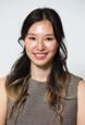
Kim’s research at Emory focused on prognosis and clinical management in patients with glioblastoma, using the Brain Tumor Reporting and Data System to conduct chart reviews to determine how scores correlate with patient prognosis (2018-2020) and imaging evaluation and treatment of spontaneous intracranial hypotension (2017-2019). She is an author of four peerreviewed publications.
Kim developed an educational program geared toward third-year medical students to raise their awareness of intimate partner violence and delivered lectures during the Psychiatry clerkship every six weeks (2019-2020). She also led a Narrative Reflection Workshop for third-year medical students, as well as organizing the Drawing with Stefan Tigges, MD, workshop to encourage students to reflect on their medical experiences through drawing (2019).
Kim was president of the Emory Medical Education Interest Group (2019-2020), and co-president of the Emory Radiology Interest Group (2019-2020). She also served as a student representative on the Emory Radiology Committee on Diversity and Inclusion (2019-2020). She was nominated by peers and selected as a member of the Gold Humanism Society, Atlanta, GA (2019).
Lohith
G. Kini, MD, PhD
In 2020, Lohith Kini completed a medical degree and a PhD in bioengineering at the University of Pennsylvania in Philadelphia. He also served as an HHMI-NIBIB Interfaces Scholar (2020). Kini did an internship at Sinai Hospital in Baltimore, MD (2021).
Kini’s research at Penn’s Department of Bioengineering (2013-2020) focused on translational neuroengineering. His research interests are in the areas of biomedical imaging, data science, machine learning and the interface between big data and medicine. One area of focus was analyzing 3T MRI and 7T MRI multi-modal images of research patients with epilepsy who underwent surgical resection and electrode implants after imaging. He built a knowledge-expert system that applied machine learning on multimodal neuroimaging in epilepsy patients to identify subtle epileptogenic lesions. He also validated and built computational models of virtual resection in epilepsy to optimize surgical targets. He received the ITMAT Prize for Clinical and Translational Research at Penn (2019) and a NAM T32 Training
Grant (Center for Neuroengineering and Therapeutics). Kini is an author on 13 peer-reviewed publications.
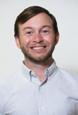
Kini has been a volunteer at EducationNow2, Inc, a nonprofit dedicated to advancing education in renewable energy technologies. As a board member, he helped set up an online presence to fund project workshops to teach students, locally and in Nepal, important STEM concepts and about renewable energy.
Joseph Knox, MD
Joseph Knox received his medical degree from UCSF School of Medicine in May 2020. He did an internship at Santa Clara Valley Medical Center, CA (2021).
While in medical school, Knox was a UCSF Department of Orthopaedic Surgery Heiman Research Fellow for two years, performing research focused on imaging cartilage and ACL-injuries. He served a yearlong research fellowship under Principal Investigator Dr. Daniel Cooke, investigating the impact of aortic arch anatomy on technical performance and clinical outcomes in stroke patients. He also used 3D-printed aneurysm modeling to assess angioplasty balloon performance. His work under Dr. Ryan Kohlbrenner studied clinical outcomes for cryoablation for small renal masses. Knox is an author on nine peerreviewed publications.
Knox was the social chair for the UCSF Associated Students of the School of Medicine (ASSM) in 2015-2016 & 2018-2019 and co-president of the ASSM from 2016-2017.
He was co-leader and co-creator of the UCSF Interventional Radiology Interest Group (2018-2020), working with faculty advisor Dr. Vishal Kumar. He served on several UCSF committees, including the UCSF Associate Dean of Students Search Committee (2016) and the Student Governance Committee (2017-2020). He also tutored UCSF medical students in pre-clinical curriculum (2018-2019). Knox received the Society of Interventional Radiology Medical Student Scholarship (2019) and the UCSF Long-Term Dean’s Prize in Research and Scholarship (2019).
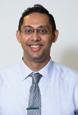

Harrison Ty-Sen Lee, MD, MBA
Harrison Lee received his MD and MBA from the University of Pennsylvania’s Perelman School of Medicine and Wharton School of Business (2020), respectively. He completed an Internship at Lankenau Medical Center, Wynnewood, PA (2021).
Lee was a summer consultant at the Boston Consulting Group, Philadelphia, PA (2019) and a medical Intern at the Center for Health Care Innovation in the University of Pennsylvania Health System (2016). As the lead medical coordinator for United Community Clinic (2015-2016), he
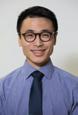
36 IMAGES 2021–2022
led a 25-person team running a free health clinic in West Philadelphia. Knox was awarded a Penn CAREs Grant to establish diabetes care at the clinic.
He is the recipient of the Marc Levine Radiology Research Prize (2020), awarded annually to a University of Pennsylvania medical student for outstanding research related to medical imaging, and the Wharton Healthcare Fellowship (2018), a scholarship for exceptional professional, academic and personal achievement.
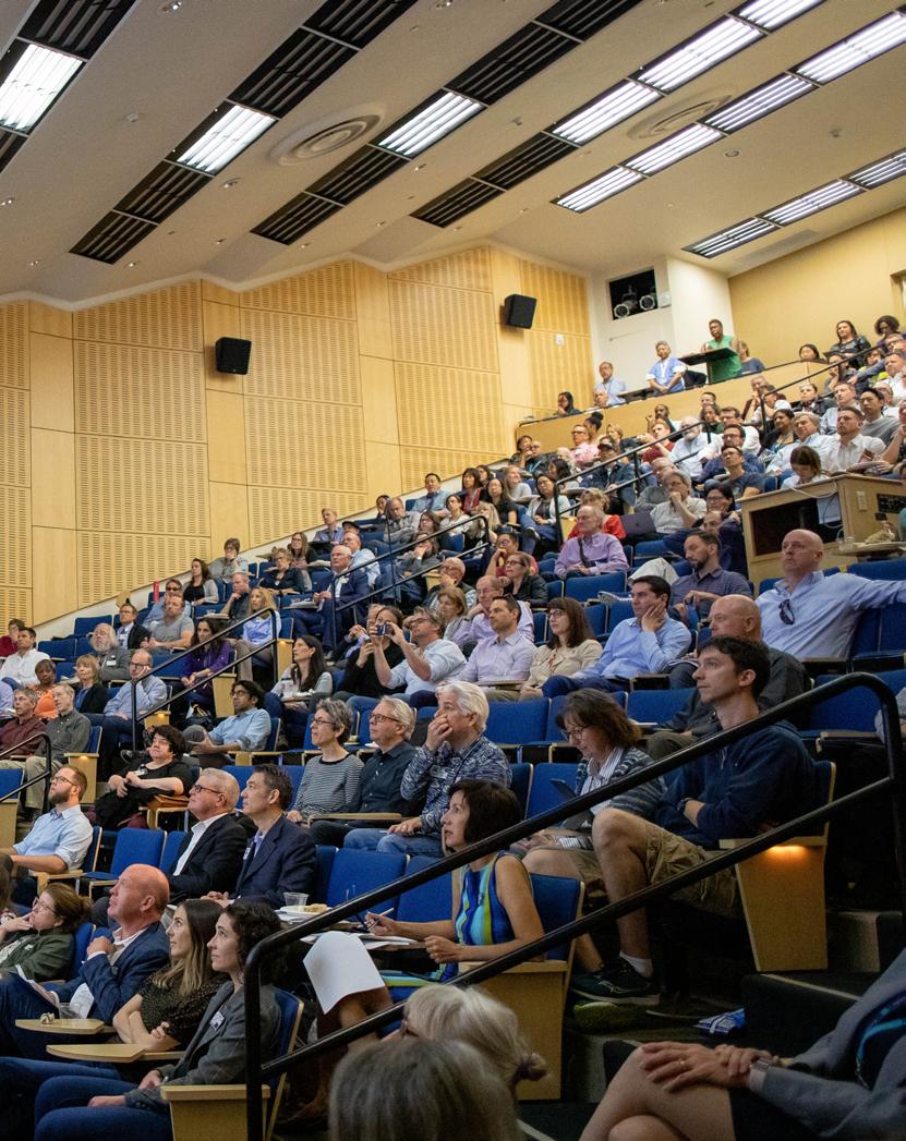
Alexia R. Tatem, MD, MPH
In 2020, Alexia Tatem received her medical degree from the University of Arizona College of Medicine in Phoenix. She completed her internship at the same institution in 2021.
Tatem has done research at Banner Health, Denver, CO, where she studied the utility of CT imaging in patients admitted for medical therapy for COVID (2020-2021). She was a co-investigator at Virtual Radiologic in Eden Prairie, MN, where she did a study evaluating early career radiologists’ performance in a teleradiology practice based on residency training style (2019-2020). At the University of Arizona (2017-2020), she performed research on an evidence-based lifestyle program and cognitive health, with the goal of evaluating the impact of a multi-component lifestyle program on cognitive health. This study was presented at the annual RSNA conference.
In 2020, Tatem received the Glasgow-Rubin Citation for Academic Achievement, awarded by the American Medical Women’s Association to women in the top ten percent of medical school graduating classes. She was also a recipient of a 2019 Scholastic Achievement Award from Mutual Insurance Company of Arizona, a scholarship awarded to three scholastically exceptional medical students in the state of Arizona.
She was a clinical volunteer at Wesley Clinic in Phoenix (20162020) and served as a clinical volunteer on a global health trip to San Pedro DeMacoris, Dominican Republic (2017).
Xiao Wu, MD
In 2020, Xiao Wu received her medical degree from the Yale School of Medicine in New Haven, CT. She completed her internship at UCSF in 2021.
At Yale School of Public Health, Wu served as a teaching assistant for a student class of 150 (2016-2017). She was the student leader of the Yale Radiology Artificial Intelligence Group (2018-2020) and the treasurer of the Yale Radiology Interest Group (2016-2017).
Wu’s did research in hepatocellular carcinoma at Stanford School of Medicine, Palo Alto, CA, (2019-2020). At Yale (2015-2020), her research focused on stroke, intracranial
aneurysms, cervical spine trauma and thunderclap headache. She is an author on more than 25 peerreviewed publications and the recipient of a research grant from the American College of Radiology for projects on stroke management.
Wu plans to continue her residency in the UCSF Integrated Interventional Radiology program following her diagnostic radiology training.
Alice Zhou, MD
Alice Zhou received her medical degree from Johns Hopkins University School of Medicine in 2020, where she served as president and co-president of the interventional and diagnostic radiology interest groups. She was a member of Hopkins Health Management Advisory Group, a student-led healthcare consultancy that solves quality improvement and healthcare management problems, and a teaching assistant in the Discover Hopkins Summer Program, a medical school intensive course designed to introduce high school students to scientific and clinical aspects of medicine. Her internship was completed at UCSF in 2021.
At Hopkins, Zhou performed research evaluating elements of a business proposal and model that contributed to successful funding and implementation of a joint Internal Medicine- Radiology centralized bedside procedure service at Johns Hopkins Hospital with the goal of empowering other hospital systems to develop similar services. Additional research at Hopkins retrospectively analyzed prophylactic antibiotic use and impact on infection rates following first-stick percutaneous transhepatic cholangiography in patients with pre-existing biliary stents. Zhou also performed research at UCLA focused on examinations of blood pressure and renal function outcomes after renal artery angioplasty and stenting for transplant renal artery stenosis. Zhou is an author on 6 peer reviewed research publications.
Minerva Zhou, MD
Minerva Zhou received her medical degree from Washington University School of Medicine in St. Louis, MO (2020). Her internship was at Mercy Hospital in St. Louis (2021).
Zhou was the recipient of a RSNA Medical Student Grant (2017) and a Society of Interventional Radiology (SIR) student travel scholarship (2018). In 2020 she received the Hugh M. Wilson Award for Research in Radiology and the American Medical Women’s Association Glasgow-Rubin Citation for Academic Achievement.
Zhou is an author of 10 peer-reviewed journal articles focused on various topics, including patient-centered outcomes in ischemic stroke, and multiple abstracts.
PEOPLE: EDUCATION IMAGES 2021–2022 37
THE YEAR IN THE YEAR IN PICTURES PICTURES
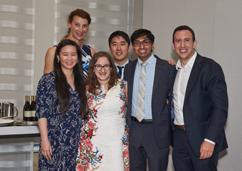
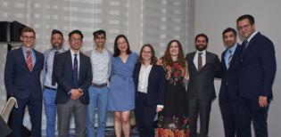
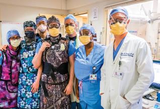
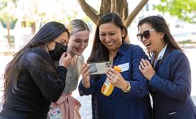
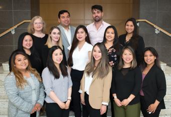
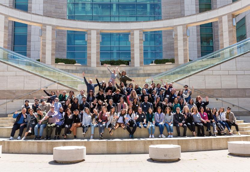
1 6 7 9 10 11 2 4 3 8 5
Radiology staff from across all radiology sites gathered on April 28th for the inaugural staff retreat.
1. The Parnassus team (l-r top row), Rita Gaber, Peter Cedillos, Roman Birko, (l-r middle row): Kristine Chooey, Janay Hansen, Apple Palad, Jocelyn Pulido, Amy Pradhan (l-r bottom row): Janelle Arquiza, Connie Jang, Andrea Fung, Lorna Kwok, Madonna Galliendes.
2. Technologists and Nurses at Mission Bay
3. Kim Semien and Evelyn Escobar at the Campus Life Services’ Wellness and Community Recess Time llama event
38 IMAGES 2021–2022
4. Drs. Matt Amans, Travis Caton, and Chris Dowd
5. The Abdominal Fellows graduate
6. Denisha Otis and Kadija Siddiqua, clinical research coordinators.
7. UCSF Technologists celebrate Halloween
8. Interventional Radiology Team (l-r) Henel Patel, MD, Dea Mochnick, RN, Jiwon Youm, MD, MS, John “Buck” Dryden, MD, Andrew Taylor, MD, PhD, Cindy Folefack, Miles Conrad, MD
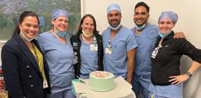
9. Members of the Cardiothoracic Section at Commencement 2022: (l-r) Drs. Katherine Cheng (fellowship graduate), Jamie Lee Twist Shroeder, Maya Vella, Jae Ho Sohn, Shravan Sridhar, (fellowship graduate), and Brian Haas.
10. (l-r) Jocelyn Pulido, Janay Hansen, Amy Pradhan, Hanh Ryan
11. T32 Program (l-r) Drs. Thomas Link, program director, Allen Ye, Jeanette Mathieu, Samantha Pisani Petrucci, T32 program trainees
12. Colleagues celebrate Dr. Ron Zagoria on his retirement
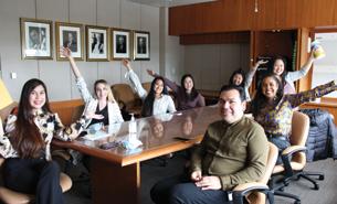
13. (l-r) Drs. Chris Hess, John Kurhanewicz, Dan Vigneron at the HMTRC workshop
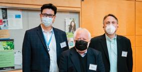
14. The Larson lab at Asilomar, (l-r) Abhejit Rajagopal, Peder Larson, Nikhil Deveshwar, Xiaoxi Liu, Sule Sahin, Jingwen Yao. (l-r) Drs. Chris Hess, John Kurhanewicz, Dan Vigneron at the HMTRC workshop
15. Christine Sarmiento signing the wellbeing wall at the staff retreat
16. Julia Carmona-Bozo, Cindy Cheng, Kevin Ng at the Bay Area Science Festival.
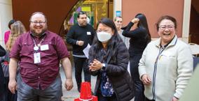
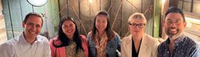
17. Teamwork at the staff retreat (foreground): Martin Rawlings-Fein, Susan Lin, Cecille Vicencio
18. David Gospe, RT, center, receives a UCSF PRIDE Experience Award for outstanding patient service.
19. Will Byrne, pre-clinical MRI/Spectroscopy core engineer, Will Mander, DNP-services, John Kurhanewicz and Renuka Sriram, Prostate Cancer Imaging lab.
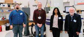
20. Chief residents Kevin Sweetwood and Andrew Taliaferro (not pictured Joelle Harwin)
21. Parnassus Staff
22. Colleagues celebrate Dr. Ron Zagoria on his retirement
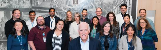
23. (l-r) Earl Miller Lecturer Dr. David Rosman with Abdominal Section Faculty Drs. Priyanka Jha, Hailey Choi, Liina Poder and Derek Sun
24. Dr. Michael Hoff explains magnets at the Bay Area Science Festival.
25. Renuka Sriram, PhD
16 14 17 20 23 21 19 18 22 12 15 25 24 13
IMAGES 2021–2022 39
THE CLASS OF 2026
Incoming Residents, July 2022
Venus Barlas, MD
Venus Barlas earned her medical degree in 2021 with honors from the University of New Mexico School of Medicine in Albuquerque, NM, and was awarded for outstanding narrative during her practicum Immersion Experience in rural New Mexico. At the University of New Mexico Department of Radiology, she served as a Student Technical Specialist. With the Refugee Well-Being Project in Albuquerque, she was awarded a research stipend for her project, “Mental Health Differences between Men and Women refuges from Afghanistan, Africa, and Iraq”. There, she was also an on-call interpreter for events and interviews conducted in Farsi or Dari. At the University of New Mexico, she was first author on “How to Produce Face Shields for Your Hospital Locally Using 3D-Printing: A Guide for Students and Practicing Physicians,” published in 2020 with the Journal of Community Medicine and Public Health Reports. She joined UCSF’s diagnostic radiology resident program in July 2022.
Anthony Finnay Blackburn, MD
Anthony Blackburn earned his medical degree in 2021 from Weill Cornell Medicine in New York, where he was awarded the Atkins Fellowship, the Tai Scholarship, and was a recipient of The Catherine Williams Wilen Memorial Fund. While at Cornell, he was the Primary Investigator for a study in a New AntiInflammatory Role of R-Spondin 3 in Adipose Tissue. During the height of the pandemic, Andrew provided concise summaries and critical analysis of the growing body of research on COVID-19 for clinicians caring for patients suspected or diagnosed with COVID-19. He also collected critical data from over fifty patients infected with COVID-19 at New York-Presbyterian to help practicing physicians make better informed clinical decisions. He completed a general surgery internship at UCSF in June 2022 and entered the diagnostic radiology residency program in July 2022.
Timothy L. Chen, MD
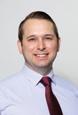
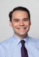
Timothy Chen earned his medical degree with honors in 2021 from University of Illinois College of Medicine at Peoria. There he received the CIRA Radiology Award (2021), Notable Peer Educator Service
(2021), and Outstanding Peer Educator Mentor awards (2020). His research with the UICOMP Department of Radiology investigated the use of MRS thermometry for mapping in vivo brain temperatures in normal vs. stroke patients. He is currently working with UCSF Big Data in radiology on natural language processing tasks such as creating domain specific word embeddings and using a BERT architecture to create a domain specific word appropriateness checker. He completed a yearlong internal medicine internship in June 2022 at the Santa Clara Valley Medical Center in San Jose, CA, before entering UCSF’s diagnostic radiology resident program in July 2022.
Brandon K.K. Fields, MD
Brandon Fields earned his medical degree in 2021 from the Keck School of Medicine at the University of Southern California in Los Angeles, where he graduated with highest distinction. There he was valedictorian with the award for Academic Excellence and was elected to the Order of Arête which is the highest honor accorded to USC graduate students, awarded in recognition of significant campus or community leadership.
While at USC, he led two research teams investigating differential death rates due to COVID-19 in various countries and the effects of SARS-CoV-2 infection in pregnant patients, respectively. Awarded two principal investigator grants from the Radiological Society of North America and five conference travel fellowships. In 2022, after completing his internship at Scripps Mercy Hospital San Diego, CA, he entered UCSF’s diagnostic radiology resident program in July 2022.
Neda
Ghassemi, MD
Neda Ghassemi earned her medical degree in 2021 at the University of California San Diego School of Medicine. There she received the UCSD School of Medicine Department of Radiology Senior Student Award. She conducted research to determine if fetal frontal horn morphology and their proximity to the CSP would assist in suspecting agenesis of the corpus callosum earlier than known indirect ultrasound findings. In June 2022, she completed a yearlong internal medicine internship at Kaiser Permanente Los Angeles Medical Center, before joining the UCSF diagnostic radiology residency program in July 2022.
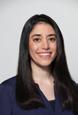
40 IMAGES 2021–2022
Bradley King, MD
Bradley King earned his medical degree with honors from the University of California San Diego School of Medicine in La Jolla. At UCSD he performed research determining outcomes of outpatient G-tube placement in head and neck cancer patients, as well as a study on complications and palliative benefits associated with tunneled peritoneal catheters in patients with refractory ascites. He has also investigated the impact of patient frailty on outcomes following transarterial chemoembolization at UCSF’s Taylor Lab. He presented “Frailty as a Prognostic Tool in Interventional Radiology” at the Society of Interventional Radiology annual meeting in 2021. He volunteered at Health Frontiers in Tijuana, which provides health care to vulnerable homeless, migrant, refugee, and transgender patients in a resource-limited setting. In July 2022, he joined the UCSF department of diagnostic radiology as a first-year resident.
Sean Lee, MD
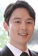
Sean Lee earned his medical degree in 2021 from the Warren Alpert Medical School of Brown University in Providence, RI. He was a primary investigator researching the impact of the economic climate on living organ donation rates in the USA, with findings published in the Journal of American College of Surgeons. He was also a PI on a study of Type 2 diabetes mellitus incidence risk based on obesity phenotype stratification in African American population in Jackson, MS. In 2022, Lee became a Diagnostic Radiology resident at UCSF after completing a one-year transitional internship at Signature Healthcare Brockton Hospital in Brockton, MA.
Eric John Lopez, MD
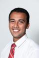
Eric John Lopez earned his medical degree from the University of California San Francisco School of Medicine, graduating with distinction in 2021. There he was a Health Policy Research Fellow for the Henry J. Kaiser Family Foundation, conducting health policy research focusing on Medicare provider payment policy and issues pertaining to the COVID-19 pandemic. He also independently conceived, designed, and completed a study of hospital characteristics that predict inappropriate utilization of outpatient brain CT scans, resulting in a sole-author publication in the Journal of the American College of Radiology (2020). He co-founded UCSF’s Psychiatry Interest Group, including an affiliated elective course for School of Medicine students. In June 2022, he completed
a preliminary internal medicine internship at the Kaiser Permanente Oakland Medical Center in Oakland, CA, before he became an incoming diagnostic radiology resident at UCSF in July 2022.
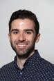
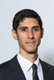
Rohil Malpani, MD
Rohil Malpani earned his medical degree in 2021 from Yale University School of Medicine in New Haven, CT. He received the Yale School of Medicine Medical Student Fellowship, and the Richard A. Moggio MD Student Research Fellowship in 2020. There he engineered a machine learning project in interventional oncology using a convolutional neural network to remove imaging artifacts in Cone Beam CT and localize lipiodol deposit for intra-procedure quantification of the efficacy of transarterial chemoembolization in hepatocellular carcinoma patients, which he presented at RSNA 2020, and published in JVIR. He co-authored a technologic innovation project detailing CompressionAssisted Needle Placement biopsy, also published in JVIR. He completed an internship at California Pacific Medical Center in San Francisco, in 2022, before joining the UCSF diagnostic radiology residency in July 2022.
Shan McBurney-Lin, MD, MBA
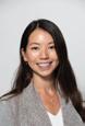
Shan McBurney-Lin earned her medical degree in 2021 from the Duke University School of Medicine in Durham, NC, along with her MBA in Health Sector management from Duke University’s Fuqua School of Business where she won the Fuqua Health Sector Management Fellow Excellence Award. There she served as Vice President and President of the American Medical Women’s Association, Duke Chapter. She conducted research with the Duke Department of Radiology evaluating the root cause, frequency, and cost of preventable Emergency Department visits by interventional radiology patients. With the Department of Ophthalmology, she analyzed outcomes of novel surgical tool utilization for treatment of cataracts, and in another project at the Marianas Eye Institute characterized a previously undescribed species of parasitic nematode causing uveitis. She became a diagnostic radiology resident at UCSF in July 2022.
PEOPLE: EDUCATION IMAGES 2021–2022 41
Paola M. Perez Rivera, MD
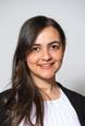
Paola Rivera earned her medical degree in 2021 from the University of California San Francisco School of Medicine. She is currently developing a family of novel endovascular filtration devices to remove drugs from the blood with the UCSF Interventional Radiology research lab. She completed a yearlong internship at Riverside Community Hospital/ University of California Riverside School of Medicine in June of 2022 and returned to UCSF to join the diagnostic radiology residency program in July 2022.
Michael
Romano, MD, PhD
Michael Romano earned his medical degree along with his PhD in Computational Neuroscience in 2021 from the Boston University School of Medicine. He received the Boston University Ignition Award for his Radiology Interventions, Safety, Quality and Complications e-curriculum where he developed a case-based e-curriculum in which interventional radiology trainees learn details of procedures, instrumentation, and potential pitfalls before operating. He completed a yearlong internal medicine internship at Saint Elizabeth’s Medical Center in Boston, MA, before beginning his diagnostic radiology residency at UCSF in July 2022
Carol Tran, MD
Carol Tran earned her medical degree from UCSF in 2021, where she received the UCSF Long-Term Dean’s Prize in Research as well as the RSNA Trainee Research Prize. She conducted research for a retrospective cohort study on the risks and benefits of inducting labor for the Department of Obstetrics, Gynecology & Reproductive services. She has recently developed and validated an automated deep learning-based method for segmentation and volumetric quantification of the fetal brain from single-shot fast spin echo T2-weighted fetal MRIs at UCSF.
Prior to joining the UCSF diagnostic radiology residency program, in June 2022 she completed an internship in internal medicine at the Alameda Health System’s Highland Hospital in Oakland, CA, and will complete advanced cardiovascular life support provider training in 2023.
Sarah Yu, MD
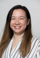
Sarah Yu earned her medical degree in 2021 from the Ohio State University College of Medicine in Columbus, OH, where she received the 2020 Outstanding Academic Achievement Scholarship Award. She has conducted research on detecting bone fractures and optimizing engagement for AI-driven image data curation. She is an author on six published peer-reviewed journal articles and has presented four times at the RSNA annual meeting. She became a diagnostic radiology resident at UCSF in July 2022.
Yannan Yu, MD
Yannan Yu earned her medical degree in 2016 from the Peking Union Medical College in Beijing, China. She was a postdoctoral fellow at Stanford University where she researched improving acute stroke triaging using deep learning, winning the Lucien Levy Best Research Article Award of 2021. In 2022, she completed an Internal Medicine internship at University of Massachusetts Memorial Medical Center in Worcester, MA, before joining UCSF as a diagnostic radiology resident in July 2022.
42 IMAGES 2021–2022
Congratulations to our outstanding 2021 graduates!
RESIDENCY PROGRAM GRADUATES, CLASS OF 2021
Justin Banaga, MD Fellowship, Breast Imaging, UCSF
Evan Calabrese, MD, PhD Fellowship, Neuroradiology, UCSF
Dylan Crebs, MD Fellowship, Musculoskeletal, UCSF
Tyler Gleason, MD Fellowship, Neuroradiology, UCSF
Karl Grunseich, MD Fellowship, Neuroradiology, UCSF
Matthew Kerchberger, MD Fellowship, Abdominal Imaging and Ultrasound, UCSF
Yoon-Jin Kim, MD Fellowship, Interventional Radiology, UCSF and OHSU
Sage Kramer, MD Fellowship, Abdominal Imaging and Ultrasound, UCSF
Olivia Linden, MD Fellowship, Breast Imaging, UCLA
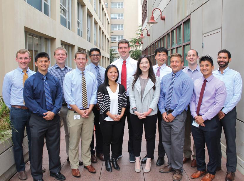
Milan Manchandria, MD Fellowship, Neuroradiology, UCSF
Matthew Mikhail, MD Fellowship, Abdominal Imaging and Ultrasound, UCSF
Jaehoon Shin, MD, PhD Fellowship, Independent Interventional Radiology, UCSF
Brian Trinh, MD Fellowship, Neuroradiology, UCSF
Mark Winkler, MD Fellowship, Abdominal Imaging and Body, Mayo Clinic, AZ
Left to right, back row: Matthew Kerchberger, MPH, MD, Evan Calabrese, MD, PhD, Milan Manchandia, MD, Tyler Gleason, MD, Jaehoon Shin, MD, PhD, Sage Kramer, MD, Matthew Mikhael, MD. Left to right, front row: Dylan Krebs, MD, Mark Winkler, MD, Yoon-Jin Kim, MD, Olivia Linden, MD, Karl Grunseich, MD, Justin Banaga, MD. Not pictured, Brian Trinh, MD.
IMAGES 2021–2022 43
RESIDENCY PROGRAM GRADUATES, CLASS OF 2022
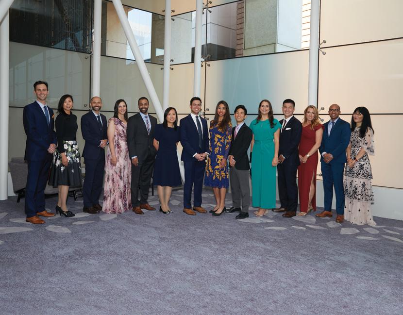
Maggie Chung, MD Fellowship, Breast Imaging, UCSF
Joelle Harwin, MD Fellowship, Abdominal Imaging and Ultrasound, UCSF
Omar Hassan, MD Fellowship, Abdominal Imaging and Ultrasound, UCSF
Jamie Holtz, MD Fellowship, Breast Imaging, UCSF
Eric Mastria, MD PGY6, Interventional Radiology, UCSF
Congratulations
Yan Li, MD Fellowship, Breast Imaging, UCSF
Jeanette Mathieu, MD Fellowship, Neuroradiology, UCSF
Alyssa McNamara, MD Fellowship, Abdominal Imaging and Ultrasound, UCSF
Samantha Pisani Petrucci, MD, PhD Fellowship, Neuroradiology, UCSF
Kevin Sweetwood, MD Fellowship, Musculoskeletal, UCSF
Andrew Taliaferro MD Fellowship, Breast Imaging, UCSF
Zak Walker, MD Fellowship, Abdominal Imaging and Ultrasound, UCSF
Allen Ye, MD, PhD Fellowship, Neuroradiology, UCSF
Yagi Yusuke, MD Fellowship, Abdominal Imaging and Ultrasound, UCSF
Left to right: Kevin Sweetwood, MD, Maggie Chung, MD, Eric Mastria, MD, Alyssa McNamara, MD, Zachary Walker, MD, Yan Li, MD, Omar Hassan, MD, Jeanette Mathieu, MD, Yusuke Yagi, MD, Jamie Holtz, MD, Allen Ye, MD, PhD, Samantha Pisani Petrucci, MD, PhD, Andrew Taliaferro, MD, Program Director Soonmee Cha, MD. Not pictured, Joelle Harwin, MD.
44 IMAGES 2021–2022
to our outstanding 2022 graduates!
The Department of Radiology & Biomedical Imaging is grateful to the many alumni who give back with a gift to the department.
tiny.ucsf.edu/radgift

IMAGES 2021–2022 45
CLINICAL FELLOWS & CLINICAL INSTRUCTORS 2021-22
ABDOMINAL IMAGING/ULTRASOUND
■ Jake Adkins, MD, MS
■ Colin Burke, MD
■ Sina Houshmand, MD
■ James Kerchberger, MD, MPH
■ Nandan Keshav, MD, MS
■ Pwint Khine, DO
■ Sage Kramer, MD
■ Matthew Mikhail, MD
■ Nguyen Stephanie Nguyen, MD, MSc
■ Sara Nobles, MD
■ Neel Patel, MD
■ Michael Shirazi, DO
■ Michael Weintraub, MD
BREAST IMAGING
■ Justin Banaga, MD
■ Stephany Barreto, MD
■ Eda Dou, MD
■ Bilal Khan, MD
■ Ryan Sieberg, MD
CARDIOTHORACIC IMAGING
■ Katherine Cheng, MD, MS
■ Shravan Sridhar, MD
INTERVENTIONAL RADIOLOGY
■ John Dryden, MD
■ Gorantla Varun Gorantla, MD
■ Oskar Kishner, MD
■ Henal Patel, MD
■ Jaehoon Shin, MD, PhD
■ Jiwon Youm, MD, MS
MUSCULOSKELETAL RADIOLOGY
■ Alex Bergman, MD
■ David Chen, MD
■ Dylan Crebs, MD
■ Jose Lopez, MD
NEURORADIOLOGY
■ Arafat Ali, DO
■ Evan Calabrese, MD, PhD
■ Mohammed Fakhri, MD, PhD
■ Tyler Gleason, MD
■ Karl Grunseich, MD
■ Milan Manchandia, MD
■ Edgar Perez, MD
■ Stephanie Teixeira, MD
■ Brian Trinh, MD
CLINICAL INSTRUCTORS IN NEURORADIOLOGY
■ Heather Early, MD
■ Ashwani Gore, MD
■ Ian Mark, MD
■ Amrutha Ramachandran, MBBS
■ Tiffany Tsai, MD
■ Jessica Zhang, MD
NEUROINTERVENTIONAL RADIOLOGY
■ Baker Amanda Baker, MD
■ Caton Michael (Travis) Caton, MD
■ Eric Smith, MD
46 IMAGES 2021–2022
CLINICAL FELLOWS & CLINICAL INSTRUCTORS 2022-23
ABDOMINAL IMAGING/ULTRASOUND
■ Kaci Dudley, MD
■ Pierre Halteh, MD
■ Tiffany Han, MD
■ Joelle Harwin, MD
■ Omar Hassan, MD
■ Alyssa Kirsch, MD
■ Rachel Latshaw, DO
■ Alyssa McNamara, MD
■ Ahmad Mousa, MD
■ Andrew Soroka, MD
■ Andrew Vinson, MD
■ Zachary Walker, MD
■ Yusuke Yagi, MD
BREAST IMAGING
■ Maggie Chung, MD
■ Jamie Holtz, MD
■ Manav Kumar, MD
■ Yan Li, MD, PhD
■ Andrew Taliaferro, MD
INTERVENTIONAL RADIOLOGY
■ Samyuktha Balabhadra, MD
■ Sara Dastmalchian, MD
■ Samir Haroon, MD, MPH
■ Oskar Kizhner, MD
MUSCULOSKELETAL
■ Redmond-Craig Anderson, MD
■ Sean Boone, MD
■ Jonathan Friedman, MD
■ Kevin Sweetwood, MD
■ NEURORADIOLOGY
■ Amanda Baker, MD
■ Ryan Fisicaro, MD
■ Vineet Gorolay, MD
■ Jeffrey Huang, MD
■ Jeannette Mathieu, MD
■ Samantha Petrucci, MD, PhD
■ Ngoc-Anh Tran, MD
■ Brian Tsui, MD
■ Allen Ye, MD, PhD
CLINICAL INSTRUCTORS IN NEURORADIOLOGY
■ Mohammed Fakhri, MD, PhD
■ Edgar Perez, MD
■ Stephanie Teixeira, MD
NEURO INTERVENTIONAL
■ Woody Han, MD
■ Raghav Mattay, MD
■ Eric Smith, MD
PEDIATRIC IMAGING
■ Tammy Kim, MD
PEOPLE: EDUCATION IMAGES 2021–2022 47
GOLDBERG CENTER
By Emma Webb, MD
The Henry I. Goldberg Center for Advanced Imaging Education is the headquarters for all medical student education in the UCSF Department of Radiology and Biomedical Imaging. The Center oversees radiology instruction in the pre-clinical curriculum, provides a course during clinical clerkships on the appropriate use of diagnostic tests, organizes lectures to senior students and a variety of radiology electives spanning both clinical applications of radiology and imaging research, and offers career advising and mentoring to UCSF medical students.
This year brought many long-awaited changes to medical education at UCSF. With the easing of pandemic-related restrictions, students returned to small group and clinical teaching in person. This meant we were able host a full complement of students in the reading rooms and interventional suites. We also returned to pre-clinical classrooms and skills training sessions to give students important early exposure to our field. For two years, ultrasound skills training was severely limited due to COVID-related restrictions. We are happy to report that it has resumed! We find this program is an excellent way to engage students in medical imaging early in their education. While remote and digital substitutes were a stop gap, it’s great to work directly with the students once again. Happily, our department’s many dedicated educators didn’t miss a beat, and were ready to meet the renewed demand.
During the pandemic, we reimagined our longstanding fourthyear classroom elective to be delivered virtually over Zoom. This remote format made the course even more accessible to many students. We no longer had to limit enrollment because of classroom size, and the course remains very popular. An added benefit is that the remote format makes teaching more accessible for many of our instructors, who no longer have to travel between hospital sites to lecture. This may be one of the lasting beneficial changes to our program from the pandemic, and for these reasons we have opted to keep the course remote for the near future.
Our Education Team
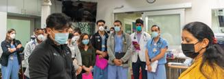
We were excited to have Maya Vella, MD, officially join our team as faculty after many years of contributing as a trainee in the department. She is now co-director for our Radiology Reading Room electives, where she will continue improving the student learning experience. Dr. Vella recently participated in the prestigious Harvard Macy program for future clinician educators, where her project addressed improving reading room experiences for medical students. Already a well-respected educator in our department, Dr. Vella brings her many talents and exciting ideas to this new leadership role.
Many faculty, fellows and resident members of the Medical Student Education Committee supervise the Goldberg Center’s medical student programs. We were fortunate to have such a dedicated group working in medical student education, which has helped us meet the many challenges of the rapidly shifting curriculum due to COVID interruptions. Emily Webb, MD, and Priyanka Jha, MD, served as committee co-chairs. Our trainee representatives include Jamie Holtz, MD, Kevin Sweetwood, MD, Adam Yen MD, and Amanda Liu, MD. Faculty committee members include Matt Amans, MD, Matt Barkovich, MD, Taylor Chung, MD, Miles Conrad, MD, Brett Elicker, MD, Vickie Feldstein, MD, Shital Gandhi, MD, Joe Leach, MD, Evan Lehrman, MD, Kevin Mc Gill, MD, Sujal Nanavati, MD, Rina Patel, MD, Elissa Price, MD, Leo Sugrue, MD, and Khai Vu, MD.
For the first time, there was a tie in the voting for the Outstanding Medical Student Educator Award for the Department of Radiology and Biomedical Imaging. The two immensely deserving recipients were Miles Conrad, MD, MPH, and Sujal Nanavati, MD, both of whom have taught interventional radiology to medical students for many years. Miles Conrad is course director for an interventional radiology elective for fourth-year students. He has long been innovating procedure skills sessions in the School of Medicine (SOM), and he serves as an SOM clinical coach and longitudinal mentor for a group of UCSF medical students. Sujal Nanavati is one of the core teachers in the interventional radiology elective at ZSFG and his impressive skills have been lauded with repeated nominations for this award. He was described by his nominator as an exemplary educator who is both technically exceptional and incredibly kind to his students.
For more information about the Goldberg Learning Center’s activities, please contact Samira Zebarjadian (Samira.Zebarjadian2@ucsf.edu) or visit radiology.ucsf.edu/education/medical-students.
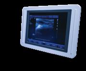
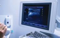
48 IMAGES 2021–2022
Medical students learn ultrasound from Radiology faculty.
MASTER OF SCIENCE IN BIOMEDICAL IMAGING
Graduate Studies in Medical Imaging
Typically, a cohort of 10 to 15 students from across the US and around the world enrolls in the Master of Science in Biomedical Imaging (MSBI) to spend a year learning the fundamentals of medical imaging at UCSF. The 2022 class was smaller—eight students, largely from the Bay Area—due, no doubt, to the challenges posed by the pandemic. The MSBI program is designed to bring students with diverse backgrounds rapidly up to speed on the scientific underpinnings of medical imaging technologies. Students entering the program typically have an undergraduate degree in engineering or the physical or biological sciences.
The MSBI program is led by Director of Graduate Studies Susan Noworolski, PhD, and Program Director Alastair Martin, PhD. In the past year we welcomed Mike Leon as the new program coordinator and thank Rukayah Abdolcader for her years of service to the program. MSBI faculty includes 13 PhD professors from the Department of Radiology and Biomedical Imaging. A wide range of clinical faculty contribute to the program by giving guest lectures and hosting MSBI students for job-shadowing or demonstration experiences. These real-world applications of medical imaging are particularly popular with the students.
A Breadth of Learning Experiences
Mandatory MSBI core courses deliver in-depth training in the principles of in-vivo imaging modalities, including MRI, CT, PET, SPECT and ultrasound. Students also choose from a wide set of electives related to imaging applications for major diseases and different organ systems.
Following their coursework, students bring the various elements of their learning together in applied research projects, working with faculty supervisors or as interns with industry partners. MSBI students benefit from the department’s wide range of state-of-the-art imaging facilities and consistently report that the hands-on activities and research opportunities are highlights of the MSBI program.
The pandemic drove a hybrid learning model again this year, but in-person activities increased and we are hoping for a fully in-person experience for 2022-23. The class was able to have a team-building experience this year and elected to attend the Harry Potter play as a group. Research activity, particularly that focused on computational imaging, remains a point of emphasis for MSBI students and is a high point of their time in the program. A high percentage of MSBI students remain
through the summer academic quarter to apply their newly honed skills on a wide range of research projects under the supervision of UCSF faculty. These projects culminate in the MSBI Imaging Symposium, held in late August, where the students’ research accomplishments are highlighted.
MBSI - Graduates of 2022
The MSBI class of 2022 has seven graduates and one student who will continue part-time for the 2022–2023 year. Many of this year’s class were interested in exploring job opportunities in industrial settings or academic labs. Others were heading off to, or aspired to, medical school or PhD programs. All will leave having experienced an intense and fulfilling year within our department.
PEOPLE: EDUCATION IMAGES 2021–2022 49
UCSF Master of Science in Biomedical Imaging Class of 2021-2022 with Program Director Alastair Martin, right.
RADIOLOGY CONTINUING MEDICAL EDUCATION
In-Person CME Courses Return
Elissa Price, MD, Post-Graduate Education Committee chair (20182022), exercised a steady hand, steering Continuing Medical Education (CME) through the initial unpredictable stages of the COVID-19 pandemic. Now, live CME programs are getting back on track despite the ever-morphing COVID landscape. “It was a challenge to determine when and how to resume live meetings,” Dr. Price said. “Between COVID variants, vaccination concerns, disruptions in the hotel and airline industries, and various travel restrictions, there was no obvious green light.”
Dr. Price was pleased to be able to transition from fully remote to in-person lectures. “The interest for live CME courses is there. We just want to make sure that we are making the meeting environment as safe as possible for our faculty and attendees.”
At the beginning of 2022, the department resumed its courses in Kona, Hawaii. Many attendees were grateful to meet in person, and to have the opportunity to ask questions of UCSF faculty as part of a live discussion.
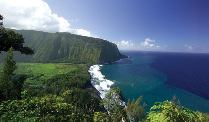
Brett Elicker, MD, succeeded Dr. Price as the PostGraduate Education Committee chair in fall, 2022.
2023 CME MEETINGS
Visit our website for more CME calendar events: radiology.ucsf.edu/cme
January 15 – 20: Abdominal & Thoracic Imaging......................................................................................... Kona, Hawaii
February 5 – 10: Neuro and Musculoskeletal Imaging
February 12 – 14: Thoracic Imaging
February 15 – 17: Abdominal & Pelvic Imaging ...........................................................................
February 26 – March 3: Breast Imaging
October 22 – 27: Imaging Update
Kona, Hawaii
Palm Springs, California
Palm Springs, California
Kona, Hawaii
Maui, Hawaii
Waipio Valley, Hawaii
50 IMAGES 2021–2022
Inaugural ER and Trauma Imaging Course
The department held a first-time course in September 2021 on ER and Trauma Imaging, headed by course co-chairs Soonmee Cha, MD, and Preethi Raghu, MD. This two-day joint effort, held virtually, featured two dozen faculty members from the Parnassus, Mission Bay, ZSFG and VAMC campuses. They covered abdominal, chest, musculoskeletal and neuro imaging, as well as emergent interventional procedures.
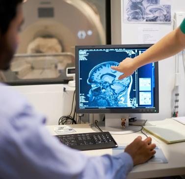
In focusing on the time-critical nature of ER-Trauma, Dr. Cha noted that, “Radiology
is often the first step in a collaborative diagnostic effort in the emergency and trauma setting. This course reinforced protocols for quick and accurate imaging assessment of the ER-trauma patient. The array of topics and speakers stressed the key role of radiology in the initial diagnosis and subsequent management of patients, in conjunction with emergency medicine, trauma surgery and neurosurgery.”
The course was clinically valuable for all medical professionals involved in the chain of treatment of the emergency-trauma patient, enabling their teams to recommend and perform appropriate imaging and procedures and ensure continuity of patient care.
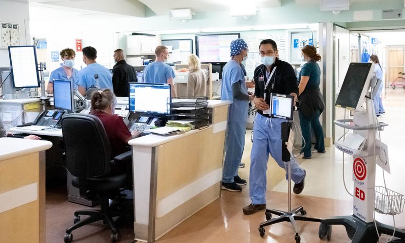
For further information, please contact: UCSF Radiology Continuing Medical Education Telephone: (415) 476-5731, E-mail: rad-cme@ucsf.edu
UCSF
SELF-STUDY
LEARNING
courses are available for CME credit through website: oakstone.com 325 UCSF Radiology Comprehensive Review 2021 ...................................................................................... 31.75 credits 467 UCSF Emergency and Trauma Imaging 2021 12.5 credits 436 UCSF Abdominal and Thoracic Imaging 2022 20.75 credits 442 UCSF Neuro and Musculoskeletal imaging 2022 ................................................................................... 22.25 credits 470 UCSF Breast Imaging 2022 20.0 credits
Soonmee Cha, MD
Preethi Raghu, MD
PEOPLE: EDUCATION IMAGES 2021–2022 51
HONORS AND AWARDS
ABDOMINAL IMAGING AND ULTRASOUND
■ Spencer Behr, MD, received the Clinical Faculty Excellence Award, June 2022.
■ Hailey Choi, MD, was named a fellow of the Society of Radiologists in Ultrasound, October 2022.
■ Priyanka Jha, MD, received the Society of Abdominal Radiology Scientific Merit Award, March 2022. Recipient, RSNA Cum Laude Award, November 2021.
■ Joseph Leach, MD, PhD, was awarded a VA CDA-2 Career Development Grant, February 2022.
■ John Mongan, MD, PhD, FSAR, was named a fellow of the Society of Abdominal Radiology, March 2022.
■ Tara Morgan, MD, received a RSNA Certificate of Merit, November 2021.
■ Liina Poder, MD, received the Clinical Faculty Excellence Award, June 2021.
■ Derek Sun, MD, received the Hideyo Minagi Outstanding Teacher Award, June 2022. He was a finalist for a UCSF Henry J. Kaiser Award for Excellence in Teaching, October 2022.
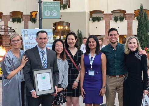
■ Z. Jane Wang, MD, was named the Society for Abdominal Radiology Lifelong Learning Portfolio Director, July 2021.
■ Emma Webb, MD, was a semifinalist for Most Effective Radiology Educator in the 2022 Minnies award campaign, AuntMinnie.com, August 2022.
■ Sean Woolen, MD, received an American Roentgen Ray Society Scholarship, April 2022, and the Society for Abdominal Radiology Howard Stern Research Grant, March 2022.
■ Ronald Zagoria, MD, received the Society of Abdominal Radiology Gold Medal, March 2022.
BREAST IMAGING
■ Heather Greenwood, MD, received the American Board of Radiology Volunteer Service Award, August 2021. She was named a Fellow of the Society of Breast Imaging, November 2022.
■ Bonnie N. Joe, MD, PhD, was a Visiting Professor in Breast Imaging at the Taipei Veterans Hospital Virtual Grand Rounds, May 2022.
■ Amie Lee, MD, was awarded a National Cancer Institute CRDF Global Grant, August 2021.
■ Elissa Price, MD, was a Visiting Professor in Breast Imaging at the Taipei Veterans Hospital Virtual Grand Rounds, May 2022.
■ Kimberly Ray, MD, was a Visiting Professor in Breast Imaging at the Taipei Veterans Hospital Virtual Grand Rounds, May 2022.
Dr. Sean Woolen received the Society for Abdominal Radiology’s Howard Stern Research Award for promising projects in the field of abdominal radiology. (l-r) Drs. Liina Poder, Sean Woolen, Hailey Choi, Stephanie Nguyen, Preethi Raghu, Omar Hassan, and Kat Zagoria.
52 IMAGES 2021–2022
Highlights from the Department
CARDIAC & PULMONARY IMAGING
■ Yoo Jin Lee, MD, received an RSNA Research & Education Foundation Research Scholar Grant, September 2022.
■ Shravan Sridhar, MD, MS, received Cum Laude for his RSNA Educational Exhibit, November 2022.
■ Maya Vella, MD received the Outstanding Fellow/ Clinical Instructor Teaching Award, June 2021.
INTERVENTIONAL RADIOLOGY
■ Jaehoon Shin, MD, PhD received the Rahul Desikan Award, December 2022
■ Rachelle Durand, DO, received a UCSF Benioff Children's Hospital PRIDE Award in the category of Professionalism, October 2022.
MOLECULAR IMAGING & THERAPEUTICS
■ Courtney Lawhn-Heath, MD, was named a Society of Nuclear Medicine in Medicine (SNMMI) Future Thought Leader in Nuclear Medicine and Molecular Imaging in the "Ones to Watch" under 30 list, July 2021.
MUSCULOSKELETAL IMAGING
■ Matthew Bucknor, MD, MFA, received his Master of Fine Arts in Creative Writing from the Warren Wilson College Program for Writers, July 2022.
■ Thomas Link, MD, PhD, received the UCSF Health Exceptional Physician Award, March 30, 2022.
■ Judong Pan, MD, received the Outstanding Clinical Faculty Award, June 10, 2022.
■ Rina Patel, MD, received an RSNA Certificate of Merit, November 2021.
■ Preeti Sukerkar, MD, was appointed a UCSF School of Medicine Coach, August 2021
NEURORADIOLOGY
■ Soonmee Cha, MD, received the UCSF Neurosurgery Diane D. Ralston Award, June 2022, and the Hideyo Minagi Outstanding Teacher Award, June 2021.
■ Christine Glastonbury, MBBS, received the Radiology Award for Outstanding Faculty Mentoring, June 2021, and Radiographics Editor's Recognition for Distinction in Reviewing, 2021, and 2022.
■ Orit Glenn, MD, received the American Society of Neuroradiology, Women in Neuroradiology Leadership Award, June 2022.
■ Yi Li, MD, was named Program Director of the Neuroradiology Fellowship, September 2021, awarded the American Society of Pediatric Neuroradiology Guerbet Grant, January 2022, and accepted to Academy for Radiology & Biomedical Imaging Research's Early Career Investigators in Imaging (CECI2) as the representative of the Society for Pediatric Radiology, March 2022.
■ Camilla Lindan, MD, received the Outstanding Clinical Faculty Award, June 2021, and the 20212022 Henry J. Kaiser Award for Excellence in Teaching for Volunteer Faculty, October 2022.
■ Andreas Rauschecker, MD, PhD, was named Associate Program Director, Neuroradiology Fellowship Program, September 2021, and received the Rahul Desikan Award, November 2021.
■ Bhavya Rehani, MD, was profiled in Superpowers for Good: The Skills You Can Master to Leave Your Mark on the World by Devin D. Thorp, September 2021.
■ Vinil Shah, MD received a RSNA Certificate of Merit, November 2021.
NEUROENDOVASCULAR SURGERY
■ Steven W. Hetts, MD, was named a Fellow of the American College of Radiology, May 2022.
Dr. Orit Glenn received the 2022 Women in Leadership Award. The award was established by the American Society of Neuroradiology (Foundation), American College of Radiology (ACR), and American Association for Women Radiologists (AAWR) to provide leadership opportunities for women in neuroradiology with demonstrated experience and promise for leadership.
HONORS
AND AWARDS
IMAGES 2021–2022 53
ZSFG
■ Miles Conrad, MD, MPH, received the UCSF Radiology and Biomedical Imaging Medical Student Teaching Award, June 2022.
■ Vishal Kumar, MD, was named a UCSF School of Medicine Bridges Coach, September 2022.
■ Sujal Nanavati, MD, received the UCSF Radiology & Biomedical Imaging Medical Student Teaching Award, June 2022.
■ Preethi Raghu, MD, was awarded Clinical Practice Group Strategic Use of Reserves Fund grants for Sonographer Education, Wellness, and Workflow improvement, December 2021, chaired the Inaugural Virtual Emergency & Trauma Radiology CME Course, September 2021, and was the recipient of a RSNA Cum Laude Award, November 2021.
■ Lori Strachowski, MD, received the RSNA Cum Laude Award, November 2021.
■ Mark W. Wilson, MD, chaired the UCSF School of Medicine John A. Watson Scholar Selection Committee, 2021-2022. He received a Volunteer Service Award from the American Board of Radiology, September 2022.
RESEARCH
■ Kondapa Naidu Bobba, PhD, received a Helen Diller Family Comprehensive Cancer Center—Radiology and Biomedical Imaging Pilot Award, August 2022.
■ Myriam Chaumeil, PhD, was an Invited Discovery Day Speaker at UCSF Alumni Week, April 2022.
■ Michael Evans, PhD, received the Helen Diller Family Comprehensive Cancer Center—Radiology and Biomedical Imaging Pilot, August 2022.
■ Nola Hylton, PhD, received the International Society of Magnetic Resonance in Medicine Gold Medal, May 2022, and was inducted into the National Academy of Engineering, February 2022.
■ Peder Larson, MD, received the UCSF Chancellor Award for Disability Service, October 2021.
■ Sharmila Majumdar, PhD, received the UCSF Academic Senate Service Award, October 2022, and was named one of the 2022 top female scientists in the U.S. by Research.com in December 2022.
■ Valentina Pedoia, PhD was the Robert H. Freiberger, MD, Honorary Lecturer in Musculoskeletal Radiology at the Hospital for Special Surgery, New York, June 2022.
■ Youngho Seo, PhD, received the MSBI Teaching Award, August 2021.
■ Duygu Tosun, PhD, was recognized as an Academy for Radiology & Biomedical Research Distinguished Investigator at RSNA, November 2022.
■ Henry VanBrocklin, PhD, received the Helen Diller Family Comprehensive Cancer Center—Radiology and Biomedical Imaging Pilot Award, August 2022. He received the MSBI Outstanding Teacher Award, August 2022.
■ Daniel Vigneron, PhD, received the Radiology Award for Outstanding Faculty Mentoring, June 2022.
■ Michael W. Weiner, MD, received the Alzheimer's Association Henry Wisniewski Lifetime Achievement Award, July 2021.
FELLOWS
■ James (Matt) Kerchberger, MD, received the Elmer Ng Outstanding Resident Award, June 2021 and the Outstanding Teaching Fellow/Clinical Instructor, June 2022.
Dr. Nola Hylton received the ISMRM Gold Medal, recognizing her major research contributions to the field of magnetic resonance, at the society's annual meeting in London. (l-r) ISMRM President Dr. Fernando
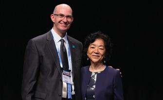
54 IMAGES 2021–2022
Calamante, PhD and Dr. Hylton.
■ Evan Calabrese, MD, was awarded the American Society of Neuroradiology (ASNR) Foundation Grant, June 2022, and received The Margulis Society Resident Research Award, 2021.
■ Stephanie Nguyen, MD, received the Society of Abdominal Radiology Best Trainee Exhibit Award, March 2022.
RESIDENTS
■ Maggie Chung, MD, received the Roentgen Resident/Fellow Research Award, RSNA, March 2022, the Elmer Ng Outstanding Resident Award and the Margulis Society Outstanding Resident Research Award, June 2022.
■ Masis Isikbay, MD, was an Education Committee Member for the American Society of Neuroradiology, 2021, and Radiology In Training Associate Editor, May 2021. He was selected as Chief Resident, June 2022, was an Investigative Radiology reviewer, 2021-2022, and a Communications Officer for the California Radiological Society, 2021-2022. He was appointed Radiology in Training associate editor, July 2022. He received the RSNA Educational Program Award, November 2022.
■ Olivia Linden, MD, received the Elmer Ng Outstanding Resident Award, June 2021.
■ Kevin Tereshima, MD was selected as chief resident, June 2022.
■ Xiao Wu, MD, received the UCSF CTSI Spring Resident Research Funding Award, May 2022. She received a Clinical & Translational Science Institute award, June 2022. She was awarded an RSNA Trainee Research Prize, October 2022.
■ Allen Ye, MD, received the UCSF Resident Research Fund Award December 2021.
■ Adam Yen, MD, was selected as Chief Resident, June 2022, and received a RSNA Cum Laude award, December 2021.

■ Alice Zhou, MD, received the ZSFG Krevans Award for Excellence in Patient Care, June 2022.
MEDICAL STUDENTS
■ Céline Taglang, received the Bruce Hasegawa, PhD Award, November 2021 and was a review editor for Frontiers in Oncology, 2021-2022 and an abstract reviewer for the ISMRM annual meeting, May 2022.
RESEARCH TRAINEES
■ Georgios Batsios, PhD, was elected as an International Society for Magnetic Resonance in Medicine Junior Fellow at the Joint Annual ISMRM-ESMRMB meeting, received the ISMRM Summa Cum Laude award and was awarded First Place in the Bruker MRI Competition, May 2022. He received the Bruce Hasegawa, PhD Award, December 2022.
■ Upasana Upadhyay Bharadwaj, MBBS, received a Certificate of Distinction from Skeletal Radiology, February 2022.
■ Kenneth Gao, was awarded Best Musculoskeletal Abstract, International Society for Magnetic Resonance in Medicine and received the ISMRM Summa Cum Laude Merit Award given to International Society for Magnetic Resonance in Medicine trainee members whose abstracts score in the top 5 percent, May 2022
■ Gurvant Chaudhari, received the RSNA Research medical Student Grant, November 2022.
HONORS AND AWARDS IMAGES 2021–2022 55
Dr. Soonmee Cha received the UCSF Department of Neurological Surgery’s Diane Ralston Award for her contributions in the training of neurosurgical residents. She is pictured with Neuro Surgery’s chief residents Drs. John Burke, Kunal Raygor, and Taemin Oh at the award ceremony.
■ Yangjie Huang, PhD, received a SNMMI Alavi-Mandell Award for an outstanding 2021 Journal of Nuclear Medicine paper, May 2022.
■ Kwang S. Kim, PhD received the Outstanding Poster Presentation Award at the UCSF Radiology Research Symposium, December, 2022.
■ Abhejit Rajagopal, PhD, received the Best Podium Presentation Award at the UCSF Radiology Research Symposium, December, 2022.
■ Saghi Sadoughi, PhD, received a UCSF Sandler Program Award for Breakthrough Biomedical Research, August 2021, and was awarded a UCSF Core Center for Musculoskeletal Biology and Medicine Grant, October 2021.
■ Jaewon Yang, PhD, received a SNMMI Alavi-Mandell Award for an outstanding 2021 Journal of Nuclear Medicine paper, May 2022.
■ Ning Zhao, PhD, received a SNMMI Alavi-Mandell Award for an outstanding 2021 Journal of Nuclear Medicine paper, May 2022, and was named a Society of Nuclear Medicine in Medicine (SNMMI) Future Thought Leader in Nuclear Medicine and Molecular Imaging in the "Ones to Watch" under 30 list, July 2021.
NURSES AND TECHNOLOGISTS
■ Charles Edgar Clary received the UCSF Medal for his work as a co-founder of the UCSF Black Caucus, 2022.
■ Reynaldo Estaniel, RT, received the Lanna Lee, RT Award, December 2022.
■ Jordan Fong received the Richard Sollitto, MD Award, December 2022.
■ Dave Gospe, RT, received a UCSF Health PRIDE Experience Award in the Clinical Specialist and Technologist Category.
■ Philip Hwang, RT, received the Lanna Lee RT Award, December 2021.
■ Chester Lim, RT, was acknowledged as an Outstanding Educator by the Joint Review Committee on Education in Radiologic Technology, April 2022.
■ Sueann Mark RN, PhD, was appointed to UCSF School of Nursing Faculty, August 2021.
■ David Poon, RT, was selected as a mentor for the American Society of Radiologic Technologists Diversity Leadership Development Program, May 2022.
STAFF
■ Kathy Brady-Porter received the Richard Sollitto, MD, Award for Outstanding Medical Center Employee, December 2021.
■ Jenny Che received the Cathy Garzio Award for Outstanding Staff Employee, December 2021.
■ Cindy Cheng received the Malena Ryan Award for Outstanding Teamwork, April 2022.
Dr. Sharmila Majumdar, was recognized for her leadership by the UCSF Academic Senate and was named a top female scientist by research.com
 Dr. Ron Zagoria, recipient of the 2022 Society for Abdominal Radiology Gold Medal. The award honors lifetime achievements of society members who have made exceptional and lasting contributions to the field of abdominal radiology.
56 IMAGES 2021–2022
PEOPLE ALUMNI
Alumnae Reflect on Their Radiology Training and Careers
We invited department alumnae to contribute reflections on their experience as trainees and faculty at UCSF, plus insights and advice for current and future radiologists. We hope you enjoy these glimpses into radiology as a career.
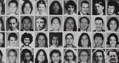
As of December 2022, our Department of Radiology & Biomedical Imaging has 143 faculty with 36 Imaging Scientists and 107 Radiologists, including ten emeritus faculty working on Recall. Of our total faculty, 59 (41%) of these faculty identify as women. Forty-three of the 107 Radiologists (40%) identify as women, compared to the national average of just 26% women Radiologists. Women
leaders include the Vice Chair of Research, Sharmila Majumdar, PhD, Vice Chair of Education, Soonmee Cha, MD, and Vice Chair for Academic Affairs, Christine Glastonbury, MBBS.
Z. Jane Wang, MD is the Associate Chair for Strategic Planning and Section Chief, Abdominal Imaging, and Bonnie Joe, MD is the Section Chief, Breast Imaging. Advisor to the Chair for Faculty Development is Susan Wall, MD. Kimberly Kallianos, MD and Liina Poder, MD are Modality Directors. Emma Webb, MD, and Maya Vella, MD, are the Medical Education co-directors. Tatiana Kelil, MD and Preethi Raghu, MD lead the Faculty DEI Committee.
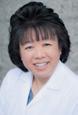 Miriam Bredella, MD, MBA Harvard Medical School, Professor and Vice Chair, Department of Radiology, Director, Center for Faculty Development, UCSF 2000-2004
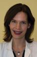
I was a resident in diagnostic radiology at UCSF and during that time had great women role models which prepared me for a career in academic radiology. I am now vice chair for faculty affairs and clinical operations at the Massachusetts General Hospital and a professor of radiology at Harvard Medical School. I also direct the MGH-wide Center for Faculty Development where I focus on advancing the careers of women faculty and trainees. I have kept in touch with my former mentors at UCSF and often contact them for advice or to collaborate to advance our faculty. UCSF is like a family to me.
Judy Yee,
MD, FACR University Chair of Radiology, Montefiore Health System, Professor of Radiology, Albert Einstein College of Medicine, UCSF 1992-2017
My experiences at UCSF helped me define what is needed for successful leadership. I always focus on doing my best and do not become daunted by the fact that I am working in a male-dominant field. As Chair of the Radiology Department at Montefiore Health System and the Albert Einstein College of Medicine, I have enjoyed using my leadership skills to have a broader impact on improving patient care in a timely manner. I have also found mentorship and sponsorship of faculty to be extremely gratifying both when I was at UCSF and in my current position. It has been so rewarding to help grow new leaders around the country! I hope to see more women in radiology leadership and fully support diversity, equity, and inclusion (DEI) efforts at multiple levels.
IMAGES 2021–2022 57
Lynne Steinbach, MD, FACR Professor Emeritus, Clinical Radiology and Orthopaedic Surgery, UCSF 1984-present
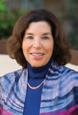
UCSF has always been my home and runs in my blood. I was born there and it was the site where my father, Howard Steinbach, served as Executive Vice Chair of the Department of Radiology. He was a wonderful role model for me with his brilliance, modesty and sense of humor. I think this gave me an extra incentive and layer of confidence when I arrived at UCSF as an Assistant Professor and new mother.
In my day, there was no such thing as maternity leave. I took three weeks of vacation during my fellowship and then went back to work with only one week of vacation left to do job interviews. Of course, this has changed over the years with new parental leave policies that reduce the stress that many of us had to endure. I am thankful for the support of my husband and mother as well as our live-in housekeeper for allowing me to balance career and family.
UCSF was such a stimulating place when I arrived and has continued to be that way through the decades. I immediately looked to mentors from inside and outside of
UCSF in radiology and other specialties of medicine who gave me support and excellent advice on how to navigate the waters. I did find my niche and ran with it. A focus on teaching lead to the annual department teaching award in my earlier career. Working with the American Association for Women Radiologists (AAWR) from the beginning in both a local group as well as the national society helped me to understand the problems facing women in a mostly male profession. I experienced harassment on several levels during my career at UCSF and quietly worked to overcome those challenges, taking the high road and succeeding each time. I became active in professional societies and eventually served as president of the AAWR and the International Skeletal Society. I learned ways to thrive and increased my networking and mentoring of others who are now prominent in radiology. I was also passionate about my field and enjoyed lecturing and doing research, writing papers and books with others.
My advice to both women and men is to be good natured and a good citizen, work on confidence and awareness, have family and mentors around for support, learn from your mistakes, find a niche that you are passionate about, and take advantage of and participate in the great resources for career growth in and outside of our department.
DATA SNAPSHOT: WOMEN IN RADIOLOGY
Looking back almost 40 years, 23% of all medical residents nationwide were women and 21% of radiology residents were women, according to data from the American Association for Women Radiologists (AAWR).
While the number of women entering medicine has more than doubled in the past 40 years, there has been only a modest increase in the number of women choosing radiology as their career.
40 Years (1982-2022)
Women in Medicine
Women in Radiology
1912
FIRST RADIOGRAPHY INSTRUCTOR AT UCSF
Dr. Anna Keith Prentis Davenport, an assistant professor of surgery in the Medical Department of the University of California, later UCSF, was noted as the first instructor for radiography.
58 IMAGES 2021–2022
Women
21 79 Men
50% Women Medical Students
In the U.S., the percentage of women medical students has hovered around 50% since 2009.
1939
DIVISION OF ROENTGENOLOGY WAS ESTABLISHED
Women
The Division of Roentgenology was established as separate from surgery, with a name change to Division of Radiology in 1941 to signify a broader scope than just the use of roentgen rays. Diagnostic and therapeutic radiology sections were established in 1943, with nuclear medicine added in 1964.
(23%)
23 77 Men (21%)
40 Years (1982-2022)
Women in Radiology
Katherine Andriole, PhD Director of Research Strategy and Operations, Center for Clinical Data Science, Associate Professor of Radiology, Director of Imaging Informatics, Brigham and Women’s Hospital and Harvard Medical School UCSF 1992-2004
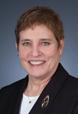
I was a fellow and subsequently faculty at UCSF at a time when imaging informatics was nascent. I investigated computed radiography (CR), a new modality for acquiring digital projection radiography and was involved in developing PACS before they were commercial entities. Under department chair Ron Arenson, MD, and faculty leaders David Avrin, MD, Bernie Huang, DSc and Bob Gould, DSc, the Laboratory for Radiological informatics (LRI) was built. It was a wonderful exciting time in which we did foundational work in moving PACS and digital radiology to clinical implementation. There were few women involved in imaging informatics at the time, but I had wonderful mentors in the LRI who have remained lifelong colleagues and friends.
In future issues we’d like to hear from you. Please email us at rad-news@ucsf.edu with your reflections on your time as a UCSF trainee or faculty member.
2020
WOMEN IN RADIOLOGY

46% of the residents in Accreditation Council for Graduate Medical Education (ACGME) accredited programs were women, yet among diagnostic radiology residents, only 27% are women. Among interventional radiology residents, 20% are women.
2021
UCSF ENTERING CLASS 52% WOMEN
In our department, the 2021 entering class of residents, including diagnostic radiology and integrated interventional radiology, was 37.5% women. Since the department established an integrated IR residency in 2018, three of four trainees in that program are women.
PEOPLE: ALUMNI
Rita Freimanis, Professor, Clinical Radiology
Imaging
UCSF Department of Radiology & Biomedical Imaging by Gender 59 82 Men Women (41%) National Average Radiologists by Gender 26 74 Men UCSF Department of Radiology & Biomedical Imaging Radiologists by Gender 43 64 Men Women (41%) Women (26%) IMAGES 2021–2022 59
UCSF Department of Radiology and Biomedical Imaging 36 107 Radiologists
Scientists
1967

Charles Gooding, MD, and Gretchen Gooding, MD, (1975), Mill Valley, CA, celebrated their sixty-first wedding anniversary with trip to Cabo San Lucas, Mexico.
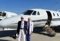
1985
David Steinberg, MD, Las Vegas, NV, writes that he met Mark Winkler, MD, (1985) in Golden Gate Park during a Residency event their first year at UCSF. They formed a friendship that has seen them through the last 40 years. After residency and a fellowship, Dr. Steinberg returned to Las Vegas, NV to practice with his father, Dr. Leon Steinberg. Dr. Winkler joined soon after and together, they founded Steinberg Diagnostic Medical Imaging (SDMI). The first SDMI office opened in February 1988, on a small piece of land adjacent to one of the city’s busiest hospitals. Their vision: a 100% outpatient radiology clinic focused on the founding philosophy “If you practice great medicine, patients will follow.” What began as a friendship in residency now encompasses 10 locations (with more on the way), more than 500 team members and 35 radiologists who perform hundreds of thousands of procedures annually.
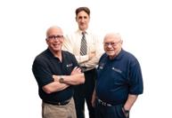
1991
Paul M. Radosevich, MD, PhD
1956-2022 Radiology Resident 1991, Interventional Radiology (IR) Fellowship 1993. Paul practiced at Kaiser Oakland/Richmond for his entire career, where he introduced many new IR procedures, established IR referral patterns throughout the Northern California Kaiser region, held a number of leadership positions and developed IR into a recognized clinical specialty. His personal interests included cycling, abalone diving, mushroom hunting, agriculture, fermentation, wine making and exploration of the natural world.
1992
Katherine P. Andriole, PhD, FACR, FSIIM, Branford, CT, received the Gold Medal of the Society for SIIM (Society for Imaging Informatics in Medicine) on June 10, 2022. This is the highest award the Society bestows and is awarded annually in recognition of outstanding accomplishments in research and/or implementation of imaging informatics applications. It is given to individuals who have made an exceptional contribution to the advancement of imaging informatics and to SIIM (SCAR/RISC).
ALUMNI NEWS
Photo: Drs. Charles and Gretchen Gooding
Photo: (l-r) Drs. David Steinberg, Mark Winkler and Leon Steinberg
60 IMAGES 2021–2022
Photo: Dr. Katherine P. Andriole
Howard A. Rowley, MD, Madison, WI, received the UCSF Radiology & Biomedical Imaging Outstanding Alumni Award
1996
Daniel Sze, MD, PhD, Menlo Park, CA, became the editor-in-chief of the Journal of Vascular and Interventional Radiology (JVIR) in 2021, taking over from UCSF alumnus Ziv Haskal, MD, who led the JVIR for 10 years. He notes that new and revitalized programs at the JVIR include emphasis on intuitive graphical and visual communication, multimedia offerings such as podcasts and a blog, translation of abstracts into Spanish, Portuguese, and Chinese, and outreach to previously underrepresented communities within IR. The editorial board continues to evolve, including the promotion of UCSF faculty member R. Peter Lokken, MD, to deputy editor. Dr. Sze writes that: “The JVIR ecosystem appreciates and looks forward to continued support from the UCSF IR and neuro IR teams, who have contributed their top-quality research and peer review to the success of the journal.”
2006
Colonel Michael Wirt, MD, PhD, San Antonio, TX, the Brooke Army Medical Center Chair of Radiology, represented the State of Texas in several road cycling events at the National Senior Games in Fort Lauderdale, FL, on May 18-22, 2022. Dr. Wirt won the Bronze Medal in the 10-km time trial and achieved two additional national top five podium finishes in the 5-km Time Trial
and the 40-km Road Race within his age group. Dr. Wirt is the 2022 Texas State Senior Games Cycling Champion in the 5-km and 10-km Time Trials and 20-km Road Race. He recently qualified for the 2023 National Senior Games in Pittsburg, PA.
2014
Anand Patel, MD, Manhattan Beach, CA, was promoted to chairman of Radiology Departments at Providence Little Company of Mary Hospitals in Torrance and San Pedro in 2022. He writes that “This past year, we have been blessed to have an addition to our family with our one-year-old son Noah Patel. My wife Sheena is expecting twins in the fall as well!”
2016
Lina Nayak, MD, has taken on the role of section chief of Breast Imaging at Kaiser Santa Clara. In this role, she leads a team of nine breast radiologists and 50 staff members and oversees 50,000 patient encounters annually.
In February 2022, she was selected by her peers for the prestigious Professional Staff Association Silver Scope Award. “This award goes to the physician whose expertise and thoughtful response to his/her colleagues is appreciated by all. This physician consistently displays a willingness and genuine enthusiasm to teach and share knowledge.”
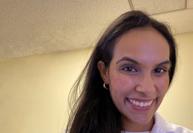
1993
Photo: Howard A. Rowley, MD
Photo: The Journal of Vascular and Interventional Radiology
Photo: Colonel Michael Wirt receives bronze medal
Photo: Dr. Anand Patel, Sheena and Noah Patel
PEOPLE: ALUMNI IMAGES 2021–2022 61
Photo: Dr. Lina Nayak
MARGULIS
SOCIETY 2021-2022
New Margulis Society Board President
Christopher Mutch, MD, PhD, became Margulis Society board president in July. Mutch completed a diagnostic radiology residency in the department in 2016, followed by a UCSF Neuroradiology fellowship and clinical instructorship. Mutch has been a physician at Bay Imaging Consultants since 2017. Outgoing board president Nick Costouros, MD, will continue to serve on the board. During his tenure, he led the board in implementing fundraising to create the Margulis Commons, a dedicated space for our residents that is used for conferences, PACS access and studying. He also oversaw two successful Galas and began the strategic planning effort for the Society.
MARGULIS SOCIETY BOARD OF DIRECTORS
President
Christopher Mutch, MD, PhD
Board Members
Alexander Adduci, MD, PhD
Avanti Ambekar, MD
Soonmee Cha, MD
Erik H.L. Gaensler, MD
Christine Glastonbury, MBBS
Christopher Hess, MD, PhD
Donna Hoghooghi, MD
Kimberly Kallianos, MD
Naveen Kumar, MD
Immediate Past-President
Nick G. Costouros, MD
Jafi Lipson, MD
Peter S. Moskowitz, MD
Derk D. Purcell, MD
Gautham P. Reddy, MD, MPH
Diego E. Ruiz, MD
Christopher J. Schultz, MD
Volney F. Van Dalsem, III, MD
Javier Villanueva-Meyer, MD
Chung Receives Research Award
Senior resident Maggie Chung, MD, received the 2022 Margulis Society
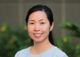
Outstanding Resident Research Award, which was presented at Commencement on June 10.
Highlights of Chung’s work include identifying ultrasound as the primary imaging modality for the evaluation of palpable breast masses in breastfeeding women, which culminated in a first-author publication in Radiology. This was featured as an “Editor’s Choice” article and highlighted by an editorial. The publication was also featured on Radiology’s YouTube podcast “Radiology in a Minute, AuntMinnie and Radiology Business.
Chung has also investigated the application of deep learning for patient care improvement. She demonstrated the feasibility of simulating contrast-enhanced breast MRI using deep learning, which has the potential to reduce risks and costs associated with gadolinium administration and extend the accessibility of breast MRI for breast cancer screening. She has also studied the use of deep learning to detect surgical sponges on intra-operative films to improve patient safety, as well as studying whole-body tumor volume segmentation using deep learning to improve metastatic lung cancer survival prediction.
The selection committee commented on Chung’s cutting-edge work, the high number of first author publications, her numerous poster presentations, and her productivity. In addition, it was noted that she has had a positive impact on patient care through the media and editorial coverage of her research.
Outstanding Alumnus Dr. Richard A. Barth Visits Department
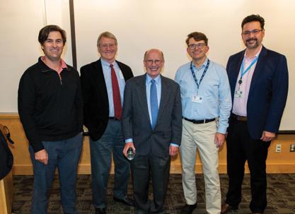
The Margulis Society was honored to host invited alumnus Richard A. Barth, MD, FACR, FAAP of the Lucile Packard Children’s Hospital, Stanford on November 10, 2022. Dr. Barth presented Microbubbles”: A New Era in Pediatric Ultrasound to an in-person and remote audience. The Margulis Society Alumnus lecture brings a distinguished alumnus of the department to UCSF to present their work and share their knowledge.
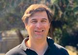 (l-r) Drs. Chris Mutch, Volney Van Dalsem, Richard Barth, Nick Costouros, and Chris Hess.
62 IMAGES 2021–2022
W. Richard “Rick” Webb, MD 1945-2022
It is with a heavy heart that we share the sad news that emeritus professor W. Richard “Rick” Webb, MD passed away on Friday, September 30, 2022, at his home in Novato after a long illness. A San Francisco native
and world-renowned radiologist who spent the greater part of his illustrious career at UCSF, Dr. Webb retired from the UCSF Department of Radiology & Biomedical Imaging in 2009 after serving for 37 years on the department’s faculty and transforming the practice of thoracic radiology.
Born October 26, 1945, in San Francisco, Dr. Webb grew up in Kentfield with his parents Wayne and Norma and his sister Judith. He attended Stanford University for his undergraduate degree and then UCSF for medical school, graduating in 1971. At UCSF, he also went on to complete his diagnostic radiology residency, where he served as Chief Resident, and his thoracic imaging fellowship. In the Air Force from 1976-78, Major Webb was stationed at Travis Air Force Base, and upon finishing his military service joined the UCSF radiology faculty. Dr. Webb served as Chief of Thoracic Imaging from 1995 – 2006, a formative period in the section’s rise to national and international eminence in the field.
Dr. Webb’s contributions to thoracic radiology cannot be overstated. His foundational work in high-resolution chest CT underpins the modern evaluation of diffuse lung disease. During his career, he authored or co-authored more than 200 manuscripts, 8 books issued and reissued in new editions and multiple languages, and no fewer than 100 book chapters and other publications. His textbook High-Resolution CT of the Lung, first published in 1992 and now in its sixth edition, is considered the definitive book on the subject.
Dr. Webb’s legacy in radiology includes his mentorship of many of the world’s leading thoracic radiologists in practice today and his genuine passion and penchant for teaching. Over the course of his career, he delivered lectures in more than 35 countries. He was an immensely popular lecturer and course chair in UCSF’s CME programs around the world. He was known by students, residents, fellows, and clinical colleagues at UCSF for his vast knowledge, incredible diagnostic acumen, and “salty” but endearing nature. He was approachable, efficient in highlighting key concepts and teaching points, and highly entertaining in his teaching style. In 2004, he was named the Hideyo Minagi Professor of Radiology at the San Francisco General Hospital.
Dr. Webb was President and Past President of the Society of Thoracic Radiology, a member of he Fleischner Society and a fellow of the Society of Computed Body Tomography, emeritus editor of the Journal of Thoracic Imaging, and served on numerous journal editorial boards in the US and abroad. In recognition of his lifetime contributions to thoracic radiology, he delivered the 2004 Fleischner Lecture and received the Fleischner Medal, the highest honors bestowed by the Fleischner Society. Among many other accolades, he was also recognized in 2013 as Gold Medalist in the Society of Thoracic Radiology, a distinction imparted for his stature and many contributions to the field.
Dr. Webb was a dedicated husband, father, and grandfather, and a bon vivant who loved the company of his colleagues and friends. We share our condolences with his wife of 52 years, Teresa, daughter Emma Webb, MD, professor of clinical radiology in our abdominal imaging section, sons Sonny and Andy, sister Judith, and grandchildren Cole, Jack, Teddy, and Cordelia. He is also survived by a large extended family of nieces, nephews, and cousins.
Share messages or stories about Rick Webb at: https://www.weremember.com/w-richard-webb/4u6o/memories
IMAGES 2021–2022 63
IN
MEMORIAM
PEOPLE: ALUMNI
STRATEGIC PLAN PORTFOLIO
Our department’s Strategic Plan 2020-25 guides our shared commitment to a culture of innovation through collaboration and growth in all areas of our mission. Work on the strategic plan began in 2019 with two department-wide retreats and a series of workgroup sessions. This resulted in a clear department vision – Leading Imaging Innovation to Improve Health –and five strategic aims—
Five Strategic Aims
In the larger context of the School of Medicine and UCSF Health, our department’s strategic plan creates synergy in efforts to advance our shared mission. For example—
■ Radiology plays a critical role in patient care, and so an important strategic goal for the Department is partnering with UCSF Health to maintain its leadership in complex care, expand services regionally, and bring UCSF expertise close to where the patients live.
■ Leading innovation is a core mission for both our department and the institution, and so one of our strategic aims is to channel investments into team science and moonshots to drive imaging innovation to transform patient care.
■ Central to our Department and the institution is our people, and a key strategic aim for the department is to develop and empower diverse talents and support our diverse community of faculty, staff, and trainees to do their best work.
The following stories are a selection of efforts in 202122 that show the breadth and depth of how we’re delivering on the strategic plan. These are some of the many efforts underway across the department. Please follow our blog and social media accounts to learn more as it happens.
■ radiology.ucsf.edu/blog
■ twitter.com/UCSFimaging
■ instagram.com/ucsf_imaging
■ facebook.com/UCSFImaging
Zhen Jane Wang, MD is associate chair for strategic planning, chief of abdominal imaging and ultrasound, and the associate director for the T32 clinician scientist training program in the Department of Radiology and Biomedical Imaging at the University of California, San Francisco.
Diversify Our Portfolio Expand Our Reach Team Science & “Moonshots”
Enhance Community & Professional Development
Strengthen Operations
64 IMAGES 2021–2022
EXPAND OUR REACH
New Digital Signage
A new digital display project for the China Basin waiting room flatscreens is now up and running, displaying faculty profiles and patient education information. This effort to connect with patients was originally conceived by the department Patient Experience Group including Orit Glenn, MD, Vicki Feldstein, MD, Sean Woolen, MD, and Heather Nichols, former director of process improvement. Many thanks also to our team of dedicated staff, including Mabel Zelaya, Francis Horan, and Rita Gaber who have brought the idea to fruition.
The digital display shows explanatory videos about imaging procedures and other patient resources that help alleviate patient anxiety about their exams and build trust between our patients and providers. Additionally, the digital display showcases our faculty photos and bios to engage and connect patients to the people in the department and their work. Our goal is to communicate the unique value UCSF Imaging brings to our patients and community.
Faculty Service
In the last several years, our faculty have had visible leadership roles in the health system, Academic Senate, School of Medicine, and systemwide committees, including service as Academic Senate Chair, and Chairs of SOM Faculty Council, Committee on Space, Academic Planning and Budget, Committee on Equal Opportunity, and Executive Council.
Faculty service data for Figure 1 and Table 1 was compiled by the Academic Affairs team.
Since FY19, our faculty have served on a broad cross-section of committees, increasing the external visibility of our department.
One of the new flatscreen displays in the China Basin waiting room.
2018-19 0 16 8 4 12 2 18 10 6 14 2019-20 # of faculty # of committees 2020-21 2021-22
Figure 1: Faculty and Committee Service
STRATEGIC PLAN IMAGES 2021–2022 65
Table 1: Faculty Committee Service
UCSF Academic Senate Committees
Assembly of the Academic Senate
Clinical Affairs
Academic Personnel
Academic Planning & Budget
Committee on Committees
Equal Opportunity
Faculty Welfare
Research
Rules & Jurisdiction
Space
COVID Faculty Support Committee
Executive Council
Graduate Council
School of Medicine Faculty Council
UCSF Committees
IT Governance
Standing Panel for Faculty Misconduct Investigations
Systemwide Committees
Academic Council
Academic Personnel
Affirmative Action, Diversity, and Equity Committees (UCOC)
Faculty Welfare / Future of UC Health Care Plans
Task Force on Agriculture & Natural Resources
UC Systemwide Committee Representatives
FY22 FY21 FY20 FY19 l l l l l l l l l l l l l l l l l l l l l l l l l l l l l l l l l l l l l l l l l l l l l l l l l l l l l l l l l l l l
66 IMAGES 2021–2022
SCIENCE & “MOONSHOTS”
Hyperpolarized MRI Technology Resource Center (HMTRC) –
Developing and disseminating new MRI technology to advance patient care
By Zhen Jane Wang, MD, and Daniel Vigneron, PhD
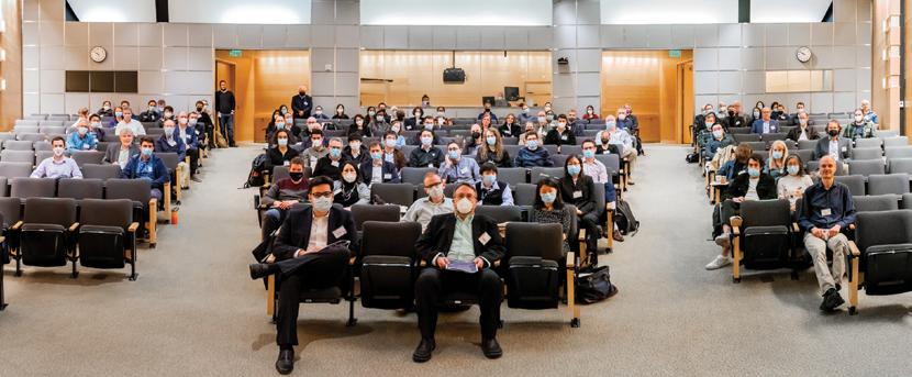
With funding from the National Institutes for Health, the HMTRC develops and disseminates hyperpolarized (HP) 13C MR techniques and instrumentation, specialized data acquisition methodology, and analysis software for biomedical research.
Hyperpolarized 13C MRI is an emerging molecular imaging technique that provides unprecedented tissue metabolic information. The overarching goal of the HMTRC is to advance precision molecular imaging and address unmet clinical needs in cancers and various other diseases.
Established in 2011, the HMTRC is the quintessential example of our department’s vision of Leading Imaging Innovation to Improve Health, and its strategic pillar of Team Science & Moonshot.
Hyperpolarized 13C MRI – a paradigm-shifting molecular imaging technology
Hyperpolarized 13C MRI is an emerging powerful molecular imaging strategy that allows safe, non-radioactive, real time, and pathway-specific investigation of dynamic metabolic and physiological processes that were previously inaccessible by imaging. This has been made possible by the recent development of the dynamic nuclear polarization (DNP) technique that can dramatically increase the signal of 13C labeled biomolecules by more than 50,000-fold. This technique has enabled rapid in vivo investigation of metabolism that is central to a variety of diseases, including cancer, cardiovascular disease, and metabolic disease of the liver and kidney.
One key advantage of HP 13C technology is the diverse array of probes that can be polarized. The most widely studied HP probe to date is 13C pyruvate, a highly biologically relevant probe as pyruvate lies at a critical branch point of multiple metabolic pathways, including glycolysis, tricarboxylic acid (TCA) cycle, and amino acid biosynthesis. This is the first HP probe that has been translated for clinical metabolic investigations here at UCSF, and has been shown to be safe and feasible. There are numerous other HP 13C probes, mainly comprised of
endogenous biomolecules, that have shown great promise for investigating physiology and metabolism relevant to human diseases. Examples include probes to measure perfusion, pH, tissue necrosis, and redox capacity.
Clinical translation of hyperpolarized 13C MRI to address unmet needs
The Center investigators pioneered many new HP 13C MRI techniques including the first-in-human clinical trial of HP 13C pyruvate MRI showing not only safety and feasibility, but also the ability to detect occult prostate cancer not visualized on standard-of-care MRI. These human studies, reported in 60 publications thus far, have demonstrated the value of this powerful molecular imaging technology to risk stratify disease and monitor response to therapy in cancers as well as other diseases (Figs 1 and 2). For example, quantitative HP 13C pyruvate measures of cellular energy metabolites are significantly increased in aggressive prostate, breast, brain, renal, and pancreatic cancers, and decreased with response to therapy.
Looking ahead, a key focus of the HMTRC in the next five years is accelerating the clinical translation of this emerging molecular imaging technology through a close collaboration between basic scientists and clinicians and through strong partnership with industry and government agencies. The projects are designed to have major scientific and clinical impact through novel HP technology development and new multi-site collaborations.
Additionally, a Radiologist Leadership Committee with Robert Flavell, MD, PhD; Michael Ohliger, MD; Javier Villanueva-Meyer, MD; Zhen Jane Wang, MD; and David Wilson, MD, PhD, has been established to lead clinical translation efforts and guide technology development to address unmet clinical needs for cancers of the prostate, brain, kidney, and pancreas; traumatic brain injury; liver disease; and cardiac disease. For example, HMTRC investigators are studying HP 13C pyruvate MRI for early response monitoring of prostate cancer metastases
TEAM
STRATEGIC PLAN
IMAGES 2021–2022 67
Figure 4. The March 2022 Technology Development Workshop attended by participants from around the world.
The Hyperpolarized MRI Technology Resource Center (HMTRC) is a unique national resource with beyond state-of-the-art equipment & facilities for creating innovative HP C-13 MRI technology. The Center supports high impact bench-to-bedside research and clinical translation including the first human studies using the technology published in 2013 with many new scientific and clinical investigations since then.
The HMTRC is a National Center for Biomedical Imaging and Bioengineering (NCBIB). This prestigious network of Centers is funded by the National Institute of Biomedical Imaging and Bioengineering (NIBIB) of the National Institutes of Health.
FUNDING
Established in 2011, the HMTRC has received $18M in grant funding, including a 2022 renewal for $6M
$5.3M in shared equipment grants
HMTRC has supported numerous UCSF investigators whose HP MRI work has secured $135M in funding in the last decade


220+ publications by HMTRC investigators
Technology developed & disseminated by HMTRC investigators is now at 14 clinical sites
42 clinical trials are underway at 14 different sites around the world
HMTRC TEAM
19 UCSF faculty + 43 staff & trainees
Dan Vigneron, PhD, Director
Zhen “Jane” Wang, MD, Medical Director
Jenny Che, Program Officer
Jenna Bernard, PhD, Data Scientist and Communication Manager
PUSH-PULL COLLABORATIONS FOR TR&D PROJECTS
In the new funding cycle, there are 13 independently funded Collaborative Projects (10 extramural) that involve 15 PIs nationally
The HMTRC’s technology resources are used by 12 Service Projects (10 extramural) that involve 10 PIs nationally
FUTURE DIRECTIONS
TR&D1 investigators will develop and test the next generation clinical polarizer. Expected improvements include increased reliability, speed of delivery, reduced cost, and higher polarization for robust clinical metabolic imaging. Additional focus on patient-centered design with attention to ergonomics and patient comfort.
TR&D2 investigators will pursue innovations in increased HP C-13 probe sensitivity and in vivo performance. Additional focus will be on clinical translation of new probes such as those to measure perfusion and pH, as well as optimizing and validating HP C-13 MRI biomarkers and methods.
TR&D3 investigators will develop next-generation imaging sequences & reconstruction methods that provide high spatial-temporal resolution, coverage, and sensitivity to metabolism. Additional focus will be on integrating HP MRI analyses with other imaging and clinical data, & standardized methods & protocols to enable multi-site comparison & trials.
3 TECHNOLOGY RESEARCH AND DEVELOPMENT (TR&D) PROJECTS
Dynamic nuclear polarization (DNP) & MR acquisition development
Project Leads
Dan Vigneron, PhD
Jeremy Gordon, PhD
Clinical Translation Lead
Michael Ohliger, MD, PhD
following immune therapy (Figure 2), and for evaluating brain metabolism (Figure 3), the alteration of which is implicated in various degenerative brain diseases.
Training and dissemination to increase impact
The HMTRC training and dissemination program already has and will continue to create a national and global impact on the advancement of this technology. This is accomplished through several different approaches. To date, the Center has provided focused hands-on training in equipment, acquisition techniques and analysis software to over 150 individuals including graduate students, postdoctoral fellows, radiology residents, and researchers from the Collaborative and Service Projects. Nineteen of these trainees thus far have become faculty at UCSF or at other institutions. Over 60 sites to date have visited the HMTRC to receive hands-on training in the technologies and resources developed by the Center. The training that takes place at the annual HMTRC Workshop (Figure 4) is particularly valuable to the push-pull development of these technologies as they provide training and interactions focused on the technical aspects of HP 13C MR preclinical and translational research and analysis. In addition, protocols, software, regulatory, clinical trial set-up & support are disseminated at these Workshops to provide experience on how this technology is implemented for clinical research.
To enable a broader dissemination of the HP 13C technology developed by the HMTRC, several webbased initiatives have been developed to benefit other researchers, and to enable multi-site studies. For example, despite the COVID-19 pandemic, the Center continues its dissemination effort virtually via the launch of a successful and well-received Hyperpolarized MRI Seminar Series in 2021-22.
Pre-clinical models, optimize & develop probes, validate HP MR
findings
Project Leads
John Kurhanewicz, PhD
Renuka Sriram, PhD
Clinical Translation Lead
Robert Flavell, MD, PhD
Open-source platform to reconstruct & interpret HP MR data
Project Leads
Duan Xu, PhD
Peder Larson, PhD
Clinical Translation Lead Javier Villanueva-Meyer, MD
To learn more: hyperpolarizedmri.ucsf.edu


In the last year, the Center has also launched a new website (http://hyperpolarizedmri.ucsf.edu) which is a key component in the training and dissemination of tools, resources and technologies developed by the Center. Content on the website includes over 100 “How-to” tutorials and training modules, recorded workshop and HP 13C seminar presentations, with over 12,400 total website views in the year of website launch. This platform for online resources accompanies the dissemination of Centerdeveloped technologies including analysis software, MRI sequences, and HP probe preparations. These resources have shown a broad reach in the research community, for example, with over 23,000 software downloads to date.
HMTRC – looking back and looking forward
The HMTRC was recently reviewed by the External Advisory Committee comprised of experts in the technology. Their review has concluded the following:
HYPERPOLARIZED MRI TECHNOLOGY RESOURCE CENTER
TR&D1 TR&D2 TR&D3
1HT2W Pyruvate Lactate Bicarbonate
68 IMAGES 2021–2022
PUBLICATION & DISSEMINATION
“The HMTRC has led the nation (and the world) in the development and translation of HP MRI through a unique combination of skills, expertise, instrumentation, environment, clinical engagement, and industry engagement, which simply does not exist elsewhere. The HMTRC will continue to serve as a critical national resource through the next funding cycle as the field of HP MRI expands and matures, particularly with the clinical translation of new HP agents, and more sites engaging in research involving human subjects. The restructured Center benefits from clear and enhanced synergy between all TR&Ds and Collaborative Projects that are more broadly geographically distributed. HMTRC programs for training and dissemination remain outstanding. Progress in the
engagement and stimulation of clinical interest is nothing short of remarkable, and crucial for the continuing growth and development of this field.”
Just as MRI advanced medical imaging by providing information beyond that from CT/X-ray, HP 13C MRI can provide clinically-needed metabolic biomarker information beyond what PET & SPECT can, and in a safe, non-radioactive two-minute addition to current MRI exams. Looking forward, the faculty, staff, and trainees in the HMTRC are energized by their work in advancing this innovative and powerful technology for improving disease diagnosis and response monitoring, and ultimately advancing patient care.
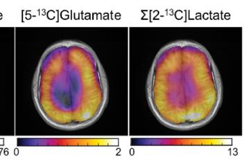

Figure 2. Hyperpolarized [1-13C] pyruvate MRI for metabolic assessment of prostate cancer osseous metastases following immune therapy. Assessment of prostate cancer osseous metastases response to therapy is a significant unmet clinical need. In this patient with prostate cancer metastatic to the bone, kPL, which measures the apparent enzymatic rate constants for pyruvateto-lactate conversion, was undetectable (u) in representative osseous metastasis after 11 weeks of immune therapy while tumor size remained largely unchanged. (Adapted from Figure in Hyperpolarized [1-13C] Pyruvate Magnetic Resonance Imaging Detects an Early Metabolic Response to Immune Checkpoint Inhibitor Therapy in Prostate Cancer, by De Kouchkovsky et al, European Radiology 2021)
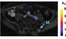 HP 13C Pyruvate MRI after 11 weeks of immune therapy
Baseline HP 13C Pyruvate MRI
HP 13C Pyruvate MRI after 11 weeks of immune therapy
Baseline HP 13C Pyruvate MRI
STRATEGIC PLAN IMAGES 2021–2022 69
Figure 3. Brain metabolism probed by hyperpolarized [2-13C] pyruvate MRI. The conversion from [2-13C] pyruvate to [5-13C] glutamate and [2-13C] lactate reflects oxidative and glycolytic metabolism, the alterations of which are implicated in various diseases of the brain.
Automated AI Coronary Artery Calcium Scoring is Live on all UCSF Non-Contrast Chest CTs
By John Mongan, MD, PhD, and Kimberly Kallianos, MD
John Mongan, MD, PhD
 Kimberly Kallianos, MD
Coronary artery disease (CAD) is the most common manifestation of cardiovascular disease and the most common cause of mortality in the U.S. Coronary artery calcium scoring (CAC) is an important predictor of risk for CAD. If patients can be identified as high risk, they can reduce that risk through medical interventions and lifestyle
changes. However, determining a CAC score requires a special, cardiac gated CT scan and is performed much less frequently than routine non-contrast chest CT. An artificial intelligence (AI) algorithm using deep learning can enable clinicians to estimate the CAC score on routine non-contrast chest CT, potentially allowing opportunistic early preventive interventions. A multi-center team (including UCSF) took part in a study and developed a fully automatic, end-to-end deep learning model for automated CAC scoring using routine non-gated unenhanced chest CT exams. The model was trained using traditional CAC scores based on gated CT as ground truth; the trained model was evaluated on multiple external datasets.
Investigators released labeled datasets of gated and nongated scans with annotations to potentially help fuel further efforts in this domain by other investigators. The result was good news.
The first part of the good news is that the model achieved good diagnostic performance in identifying patients with any CAC and CAC scores 100 across all sites (PPV ranges from 87 to 100 percent).
The second part of the good news is that here at UCSF, we are live in production with automated AI coronary artery calcium scoring on our non-contrast chest CTs. This is an important milestone for the UCSF Center for Intelligent Imaging (ci2) in that this is our first full-scale production deployment of an AI algorithm contributed to by UCSF investigators. With this news, UCSF becomes the first medical center to put this algorithm into production.
The project was led by Bhavik Patel, MD, then at Stanford University and currently Director of AI and an associate professor of radiology at the Mayo Clinic in Arizona, in conjunction with John Mongan, MD, PhD and Kimberly Kallianos, MD, UCSF ci2 investigators who validated the performance of the algorithm on UCSF data. The algorithm is deployed in conjunction with Bunkerhill Health; AI inference is performed on the Bunkerhill cloud platform.
More detailed information about the algorithm is available in a recently published paper in NPJ Digital Medicine https://doi.org/10.1038/s41746-021-00460-1 (open access)
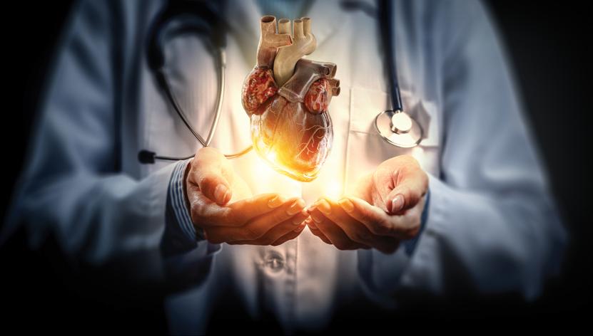
70 IMAGES 2021–2022
COMMUNITY & PROFESSIONAL DEVELOPMENT
Narrative Medicine and Well-Being Incorporated into New Ultrasound Procedure Simulation Course at UCSF
In May 2022, three department faculty members and a clinical fellow created and led a novel three-part ultrasound procedure course for senior medical students at the UCSF School of Medicine. Training focused on technique for central venous access, paracentesis, and thoracentesis paired with reflection and storytelling to contextualize procedural skills in a larger framework of patient experience and physician emotion.
Course faculty are Rachelle Durand, DO and Miles Conrad, MD, MPH from the Interventional Radiology section, and Preethi Raghu, MD, with contributions from Michelle Bui, UCSF medical student. In 2021-22 Clinical Fellow Stephanie Nguyen, MD, from the Abdominal Imaging and Ultrasound section participated. Dr. Conrad and Dr. Raghu are clinical faculty at Zuckerberg San Francisco General Hospital.
The three-hour elective took place at UCSF Kanbar Center for Simulation and Clinical Skills and was part of the career launch phase of the fourth-year curriculum. Drs. Conrad and Durand led part one of the course, a didactic session on procedural techniques emphasizing how to use procedural schemas or algorithms when unexpected complications arise.
Part two of the course was a hands-on simulation where all students were able to apply and practice their procedural skills on specialized gel phantoms at various ultrasound scanning stations. “Recognizing how to troubleshoot difficulties during procedures is essential to being confident while learning procedural technique,” said Dr. Durand.
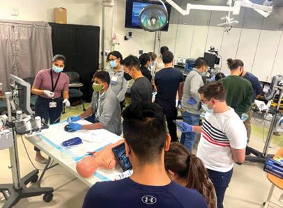
Part three was a narrative medicine session led by Dr. Raghu that integrated the storytelling aspect of a procedural experience using the perspectives of patients and physicians. “Performing procedures requires technical expertise. But absorbing the weight of a procedural error when feeling shame or anxiety requires emotional intelligence. Narrative medicine is a way to immerse trainees in the interplay between procedural errors, physician emotions, and patient experiences,” said Dr. Raghu. During the session, students learned real stories of procedural errors in radiology alongside a framework for reflecting upon and processing these errors. “I still remember being in their shoes, equally excited and nervous about entering internship. The course in total felt like a safe space to practice procedural skills, share
experiences and apprehensions, and normalize and plan for the challenges associated with performing procedures,” said Dr. Nguyen.
“Procedural errors can take a toll on doctor wellbeing” said Dr. Conrad. “Learning how to recover from procedural complications could be integrated more into medical student radiology education. The UCSF School of Medicine has taught students the concept of ‘second victim’ for medical errors. So, we are building on that. Procedural error can be distinct from other medical errors committed by teams as it is often a sudden jarring experience for only one or two operators, making the experience especially traumatizing and isolating. Additionally, these procedures are often characterized as being ‘basic’ or ‘minor,’ making trainees feel even more stigmatized by their errors.”
UCSF Radiology faculty look forward to holding the ultrasound procedure course again in the upcoming academic year. Our involvement in UCSF medical student education also includes the UCSF Bridges coaching program, designed to provide academic guidance to promote professional and personal development throughout the medical school experience. Read more about our participation in the Bridges coaching program on our blog
ENHANCE
STRATEGIC PLAN
IMAGES 2021–2022 71
Students engage in hands-on simulation with specialized gel phantoms at various ultrasound scanning stations.
Faculty and Staff DEI Committee Leadership
Faculty and Staff DEI Committee Leadership
Tatiana Kelil, MD and Preethi Raghu, MD, are the new co-chairs of the faculty Diversity, Equity, and Inclusion Committee in the UCSF Department of Radiology and Biomedical Imaging. Dr. Raghu is based at Zuckerberg San Francisco General Hospital where she specializes in abdominal imaging and ultrasound. Dr. Kelil works primarily at the UCSF Breast Care Center and specializes in breast imaging. Dr. Kelil said that “shared committee leadership, from within UCSF and the county hospital, is an opportunity to bring together a synergy of energy and perspectives.”
The new co-chairs step into their positions at a crucial time for women in medicine. While everyone has felt the burdens of the pandemic years, women have been disproportionately affected. Lockdowns and school closures led to sharply increased responsibilities in childcare and education falling heavily on women. Reports show that academic productivity among women was significantly lower during this period. The incoming chairs want to make sure such women are not left behind, and to provide support for progression up the academic ladder. Drs. Kelil and Raghu noted their interest in emphasizing the contributions of women radiologists and radiologists from less represented backgrounds.
Dr. Kelil noted that, “As a subspeciality, radiology has room for improvement. Out of 20 subspecialties, radiology was ranked about 17th in membership among women and 20th in underrepresented minorities.» Though the department here at UCSF is ahead of the pack and getting better in measures such as percentage of women residents, there are opportunities for improvement and both chairs look forward to working with our faculty to continue our department›s DEI leadership.
Dr. Raghu said, “It is our goal to make radiology a more accessible and equitable field to enter and practice in. I remember the apprehension I felt joining a field with such
a low percentage of female faculty, especially as someone with no family background in medicine and who was an immigrant myself. I am very grateful for the crucial support I received from mentors with DEI backgrounds and I now plan to use the DEI committee to ensure more people can follow my path.”

Dr Kelil has also focused her attention on the UCSF Health Equity Council’s goal of expanding healthcare access and providing all patients the same kind of treatment across the wide range of facilities. She said, “To ensure there is not any inequity to access or patient experience between health campuses or ethnicities, our organizations need to be proactive instead of reactive. Diverse representation is crucial to correcting the disparities in health outcomes and mortality rates we can see in our own backyard. Across medicine as a whole, Black women and Latina women accounted each for only 2 percent of professionals and the effects of that are something I will deal during my time as DEI chair.”
As for what’s ahead for the DEI committee, the new co-chairs will continue the existing initiatives such as the department’s Research Initiative to promote Diversity in Radiology (RIDR) and the Radiology Elective to promote Diversity in Radiology (REDR), a program designed to encourage medical students from diverse and underrepresented backgrounds in radiology to apply to the two-week fourth year clinical elective offered through the School of Medicine at UCSF. Dr. Raghu identifies RIDR as a “key way to reach out to high school, college, and medical students and promote diversity. It is something we will focus on continuing and improving.”
But even in these first early days they also want to expand. Dr. Kelil said, “The RIDR program is very successful, but is by its nature limited to students who already have an interest in research and have eight weeks to dedicate. Another program, perhaps shorter and more focused on clinical exposure to radiology could possibly promote the
72 IMAGES 2021–2022
Preethi Raghu, MD Matthew Bucknor, MD Martin Rawlings-Fein & Cindy Cheng Tatiana Kelil, MD
specialty where students might not otherwise have the time to consider it. A round of shadowing or preceptorship to see what radiology is like could spark a career aspiration in high school students who otherwise might not even be fully aware of radiology as a profession.”
While the department has effectively managed the necessities of pandemic distance, even the most effective online spaces are not quite the same. Many global health outreach efforts are still happening, as everything was converted to an online webinar, community and social health meetings are held on Zoom, and shadowing in person has already resumed. Looking ahead, Dr. Kelil confirmed that the DEI committee is planning on holding events in outdoor spaces to be present physically and socially as much as is feasible and reasonable.
As they step into the position of co-chairs, Dr. Kelil and Dr. Raghu extend their thanks to Matthew Bucknor, MD, the outgoing chair, for his inspired leadership. As Dr. Raghu said, “Dr. Bucknor built the RIDR summer internship program from the ground up to promote an interest in research and radiology in highschoolers, undergraduates, and medical students. It is one of the most successful DEI programs in the department. It even held strong through the complete conversion into an online experience.
Staff DEI Committee Fosters Discussion
The department’s Staff Diversity, Equity and Inclusion Committee currently has 13 members and is led by Martin Rawlings-Fein, audio-visual technician, and Cindy Cheng, communications and events manager. The committee strives to help staff make connections and share across different cultures, promote allyship and ensure that staff perspectives on DEI are incorporated into the department’s work and culture.
To accomplish these goals, the committee organizes events, nearly one every month for the past year, including the RAD Against Racism speaker series. Presentations and discussions have included Asian Sexuality and Gender: Representation and Stereotypes in American Film, a Beyond Land Acknowledgment Grand Rounds with Corrina Gould, Director of the Sogorea Te’ Land Trust, and Baking Accessibility into Zoom with UCSF accessibility expert Jill Wolters.
“These events are meant to bring us together. Our greatest accomplishment has been listening to people and bringing their desired events to life,” Martin observed. “We want to bring out the stories from underneath.” Cindy noted that, “The more we talk, the more we learn. Community requires participation.”
Diversity Inclusion Equity
STRATEGIC PLAN IMAGES 2021–2022 73
New Clinical Program: Emergency Radiology at ZSFG
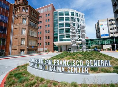
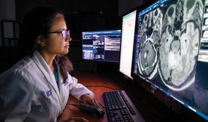 By Mark Wilson, MD, and Jason Talbott, MD, PhD
On July 1, 2022, UCSF and Zuckerberg San Francisco General Hospital (ZSFG) established a new Emergency Radiology section to provide after-hours faculty coverage for imaging exams conducted between 5 pm and midnight. With the leadership of Mark Wilson, MD, ZSFG Chief of Radiology, faculty members Jason Talbott, MD, PhD, Shital Gandhi, MBBS, and Amrutha Ramachandran, MBBS, staff this new endeavor along with 8 per-diem faculty.
Prior to establishing this new section, on-call radiology residents reviewed and provided a preliminary interpretation of after-hours studies, with the attending radiologist doing a final read the following day. Our data show that historical next-day attending call-back rates were approximately 2.7% for patients discharged from the emergency department (or 1-2 exams per day). While the overall rate of call-backs was relatively low, this presented particular challenges for ZSFG’s patient population, approximately 16% of whom do not have stable housing, which makes contact and follow-up care more difficult.
Between July and December 2022, faculty provided final reads for all ZSFG emergency department cross-sectional imaging studies (N = 5,000, approx) performed between 5pm and midnight. Comparing July – December 2021 with July - December 2022, this new model has reduced discrepant overreads by 55% for patients discharged from the emergency department. Comparing the same periods, the average turnaround time from wet read to final read dropped from 30 hours to just over 2 hours.
Preserving on-call resident autonomy and enhancing the resident learning experience are key elements of this new clinical program. On-call residents handle the protocol, interpretation, and initial wet reads for more than 95% of imaging exams at ZSFG. This independent call system creates a great learning environment in San Francisco’s only Level 1 trauma center, granting residents an autonomy that few other training programs provide. Our residents value this opportunity for independence, while the new Emergency Radiology section helps decrease the turnaround time for final interpretation of imaging studies.
In the program’s first six months, we achieved 96% faculty coverage from 5 pm – midnight. Our goal for 2023 is to achieve 100% coverage for 5 pm – midnight, while maintaining our focus on improved patient outcomes and ensuring that the trainee experience remains autonomous and distinctive.
 Celebrating the new Emergency Radiology section at ZSFG. Left to right: Vinil Shah, MD, Margaret Damiano, MBA, Shital Gandhi, MD, Jason Talbott, MD, PhD; Mark Wilson, MD: Joe Cuschieri, MD, Preethi Raghu, MD, Yusuke Yagi, MD
Shital Gandhi, MBBS, in the reading room
Celebrating the new Emergency Radiology section at ZSFG. Left to right: Vinil Shah, MD, Margaret Damiano, MBA, Shital Gandhi, MD, Jason Talbott, MD, PhD; Mark Wilson, MD: Joe Cuschieri, MD, Preethi Raghu, MD, Yusuke Yagi, MD
Shital Gandhi, MBBS, in the reading room
74 IMAGES 2021–2022
Zuckerberg San Francisco General Hospital
Wellbeing and Professional Climate: Connection, Communication, Our Future
By Matthew Bucknor, MD
mproving connection and communication has been the guiding theme for our work these past 18 months, with new and ongoing projects that continuously refine and improve how we deliver on our mission and strategic plan. With the easing of pandemic restrictions, we have enjoyed gathering in person for retreats, conferences, symposia, and celebrations that shape our professional culture and contribute to wellbeing while also maintaining the benefits of a hybrid work environment.
Matthew Bucknor, MD
Faculty Wellbeing efforts build on the results from the Health system’s annual Net Promoter Survey and our own internal climate survey to inform initiatives that foster connections, collaboration, and insight into how the department works. A new, interactive quarterly town hall series hosted an open discussion on Promoting Broad Input into Department Decisions, a revenue cycle presentation from our CFO Tom McElderry on Following the Dollar in Radiology, and a fireside chat exploring Workplace Culture: Moving from Good to Great with Chief Physician Experience Officers Diane Sliwka, MD (UCSF) and Starr Knight, MD (ZSFG). Building on the town hall series, department chair Chris Hess and I met individually with sections and section leaders to address challenges related to physician burnout while amplifying the myriad strengths of our people. Central to these efforts is implementing systems of continuous improvement to sustain positive change. These efforts have seeded a new initiative of monthly dinners with six faculty members and department leadership to further explore opportunities to enhance our professional climate.
Mentorship. Now going into its seventh year, the Research Initiative to Promote Diversity in Radiology (RIDR) brings high school, college, and medical students,
Ifrom groups that are disadvantaged or historically underrepresented in radiology, to participate in basic science, translational, or clinical research projects for eight weeks during the summer. Fourteen students explored radiology as a career choice in 2021, and 18 in 2022. In fall 2021, thanks to the generosity of our faculty and alumni donors, we completed a successful crowdfunding campaign that supported two RIDR students in summer 2022. Student projects cover the full spectrum of imaging research, including studies focused on cancer, abdominal imaging, musculoskeletal imaging, neurodegenerative disease, and health care disparities.
Book and Podcast Clubs foster a department culture of connection and learning. Research Administrator Stephanie Murphy leads a monthly staff podcast club that explores wellness, professional development, and work culture. I lead a department-wide book club that meets quarterly to discuss a wide range of works, from cognitive science to organizational culture, as well as fiction that helps us to reflect on the many dimensions of change that are reshaping society at a breathtaking pace.
Diversity, Equity, and Inclusion Committee leadership. Elsewhere in this issue, you will see that Tatiana Kelil, MD, and Preethi Raghu, MD, are the new co-chairs of our faculty DEI committee, bringing their unique perspectives and talents to this work. Looking ahead to our future, I am confident that with Tatiana’s and Preethi’s leadership, we will continue to build on our successes and define new paths for inclusion and wellbeing in our profession.
Matthew Bucknor, MD, is associate chair of wellbeing and professional climate, associate professor in residence in the musculoskeletal imaging section, and immediate past chair of the faculty Diversity, Equity, and Inclusion Committee in the Department of Radiology and Biomedical Imaging at the University of California, San Francisco. He also serves as Executive Sponsor of Differences Matter for the School of Medicine.
Learn more about Faculty and Staff DEI Committee Leadership on page 72.
“
It is critical that we bring people with diverse perspectives and backgrounds into medicine and cultivate a sense of belonging. Our medical teams must reflect the diversity of our communities in order for patients to have the best possible outcomes.”
STRATEGIC PLAN IMAGES 2021–2022 75
OPERATIONS
Successful Collaboration Between UC Radiology and IT Departments Solves a PACS Challenge
The success of this UCSF IT and Radiology collaboration is an excellent example of how technology enables the UCSF mission: advancing health worldwide
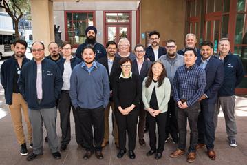
Most in radiology are familiar with Picture Archiving and Communication System (PACS), how most radiology departments store, distribute, and display imaging studies. Back in August 2019, Marc Kohli, MD and John Mongan, MD, PhD, UCSF Radiology imaging informatics experts, were faced with a challenge – replacing the Department’s ten-year-old PACS which would no longer be supported by the vendor. A similar challenge faced the other four UC radiology departments – UCLA, UC Irvine, UC San Diego and UC Davis. Based on our history of collaboration, informatics teams at all five campuses created one Request for Proposal (RFP) to find worklist and image display solutions. Nuance workflow orchestrator (WFO) was selected to provide the ability to launch multiple image display solutions supporting cross-UC and strategic growth opportunities. Visage Imaging was the unanimous selection for image display after several rounds of interviews, discussions, and a proof-ofconcept pilot using UC data on UC equipment. The Visage viewer provides excellent user experience for radiologists and IT professionals. In addition to the user experience, Visage capabilities enabled UC to consider moving PACS operations to the cloud.
The Team Involved
The typical complexity of a PACS replacement was compounded by cloud capabilities, which required strong multidisciplinary approach. The team consisted of 80 people from numerous disciplines from both the five UC campuses and Visage. At UCSF, collaboration between Information Technology (IT) and Imaging IT was key.
“Thanks to Joe Bengfort, UCSF Chief Information Officer and Mo Bagadi, Chief Technology Officer for prioritizing this project,” says Neil Singh, UCSF Radiology Director
of Clinical IT Operations. “The support and partnership empowered the UCSF Imaging IT team with a high degree of knowledge and experience, coupled with the advanced Visage tools, to lead and deliver a successful project.”
“I’m incredibly proud of what we produced and of the teams, people and collaboration,” adds Dr. Kohli. “There was a great relationship between central IT and department IT. Making everything work required new skills, and relationships with clear communication about shared responsibilities.” Dr. Mongan agreed, “A key to the success of the project were the efforts of representatives of each clinical section who served on the AIIMS Steering Committee. The ASC made sure that each section’s needs were represented in planning and implementation and kept the department involved and engaged throughout the process.”
Challenges Encountered
With any successful project, some challenges were encountered along the way. “When it comes to protected health information (PHI) and personal identifiable information (PII), encryption is important because we need to transmit images, and new technology presented challenges,” says Singh. “But thanks to the knowledge of network and security teams, we were able to find the right solution that did not impact performance while also finding ways to keep data more protected and safe.”
“One major hurdle for us was using encryption when transferring images such as MRIs and CT scans to the cloud. There are ways of doing this, but they’re not widely used. Coming up with solutions here was definitely a challenge,” says Jeff Block, UCSF Radiology director of infrastructure.
STRENGTHEN
76 IMAGES 2021–2022
Successful Results
As a result of this project, all five UC campuses will be using Visage as their single vendor for PACS through 2028. Implementation began with UCSF in late fall, early winter 2021 with the rest of the campuses following in 2022.
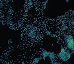
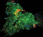
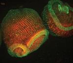
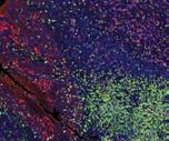
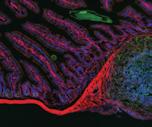
During the go-live radiologists and our trainees quickly adapted to their daily work using Visage. The system design ensures that the entire image history for a patient is immediately available, and radiologists no longer wait to retrieve prior exams from slow storage. Image display, and time spent switching between patients have dropped dramatically. The ASC champions created hanging protocols that save time for interpretation, allowing radiologists to focus more on images. Breast and ultrasound in particular saw improvements as they now have a single viewer for all their work.
Overall, patients and physicians will receive tests sooner, allowing them to determine the course of treatment in less time.
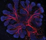

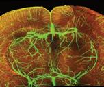
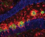
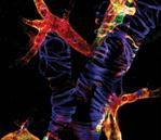
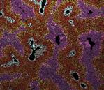
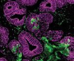
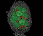
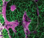
STRATEGIC PLAN IMAGES 2021–2022 77
UCSF Department of Radiology and Biomedical Imaging radiology.ucsf.edu
Art in Memory of Dr. Rahul Desikan
Quintessentially human. Brilliant. Humble. Loving.
These words describing the late Rahul Desikan, MD, are inscribed on the painting commissioned by the Department of Radiology in memory of a beloved colleague, mentor and friend to many in the department. The painting, pictured here, hangs in the department’s Goldberg Center.
Dr. Desikan began an assistant professorship at UCSF in 2016 and shared a lab with Dr. Leo Sugrue. He dedicated his scientific career to understanding the pathobiology of neurodegenerative illness and neurodevelopment. In 2017, Dr. Desikan was himself diagnosed with ALS and succumbed to the disease in 2019. He is survived by his wife, Dr. Maya Vijayaraghavan and their two sons.
In 2018, Dr. Christopher Hess, chair of the Department of Radiology, in response during an interview for a Washington Post story about Dr. Desikan, described the latter’s journey as “the most rapid trajectory of an academic that I think I’d ever seen …. I have met very few people who have true genius, and he would be at the top of the list.”
Rahul Desikan, MD, PhD (1978-2019)
Bibby Gignilliat (American, b. 1960), Many Colors of Rahul, 2021, 40 x 60 inches, acrylic mixed media on wood panel.
