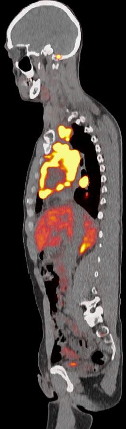
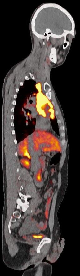
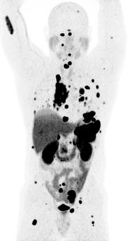
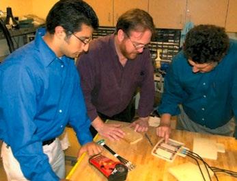
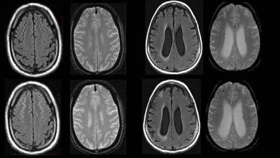
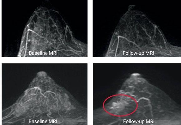
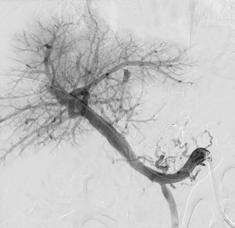
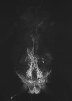
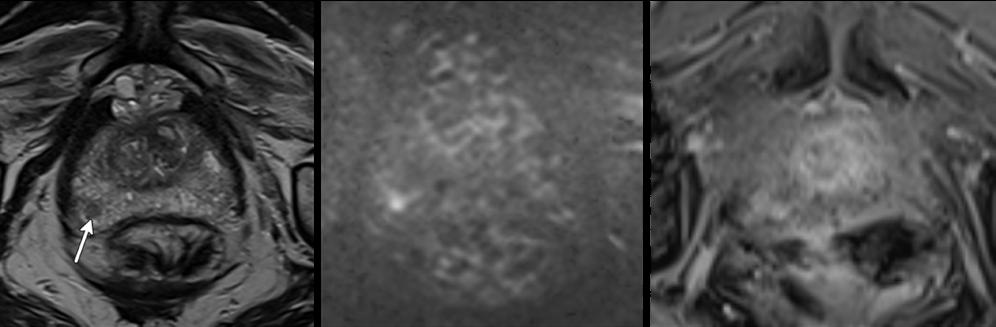
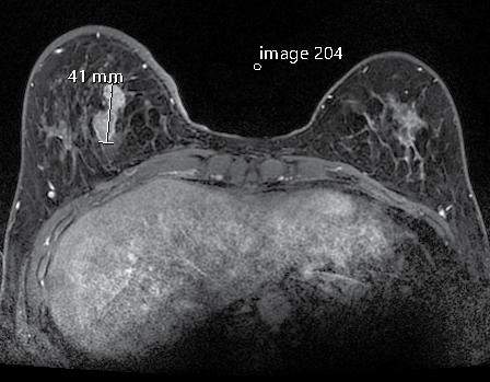
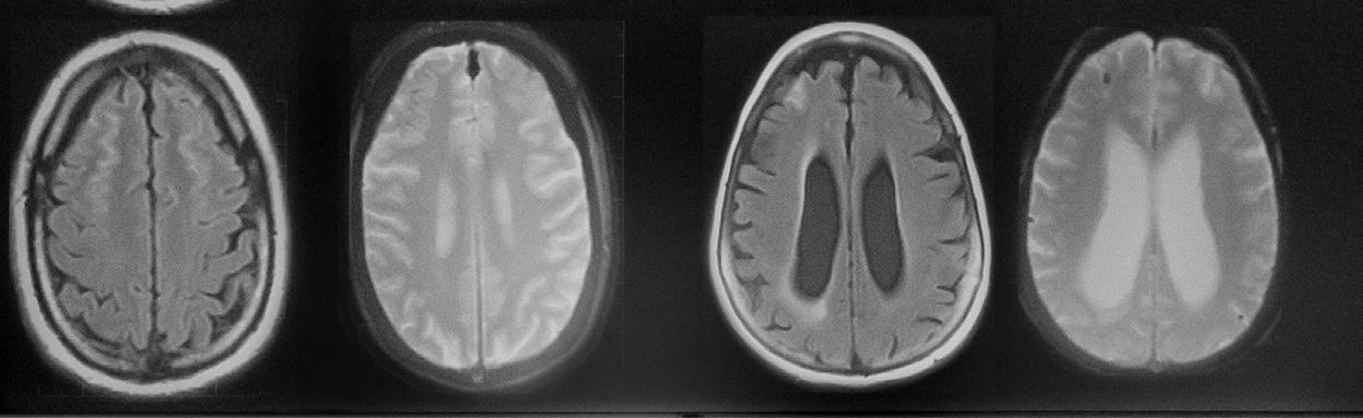
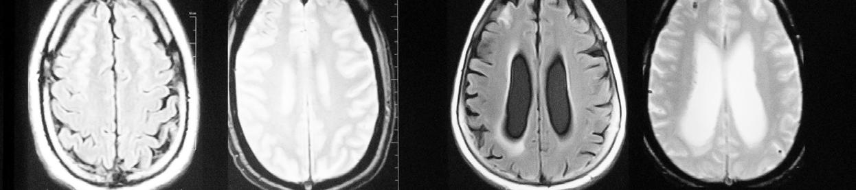
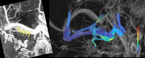
Maximum intensity projection (MIPs) images from DOTATATE PET/CTs before and after treatment with peptide receptor radionuclide therapy (PRRT). Baseline DOTATATE PET demonstrates numerous sites of markedly avid positive disease indicating the patient is a candidate for PRRT. Post-treatment imaging demonstrates improvement in the extent of disease, although extensive residual disease.
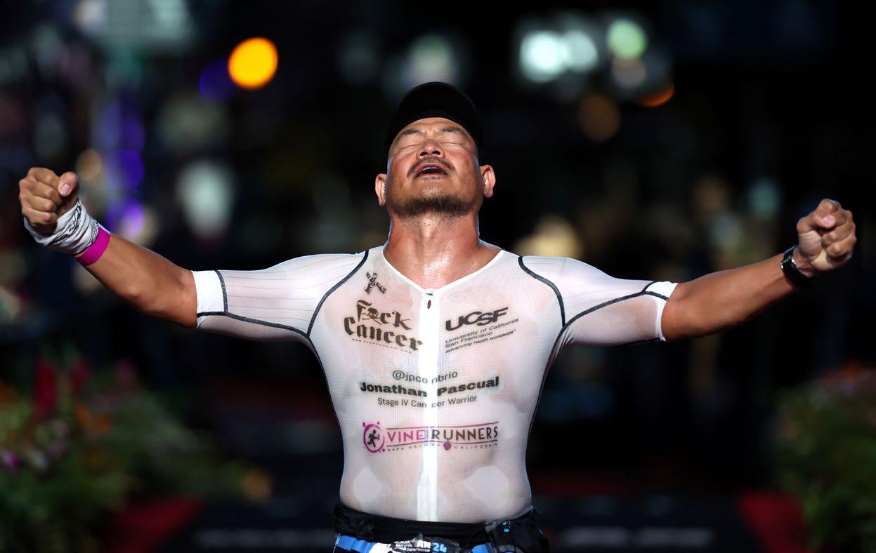
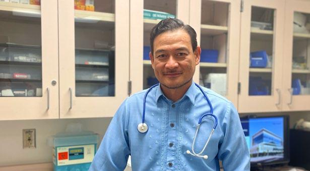
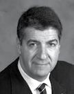
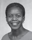
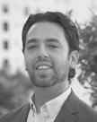
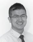
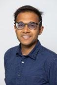
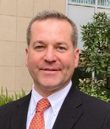
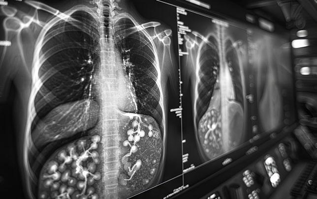
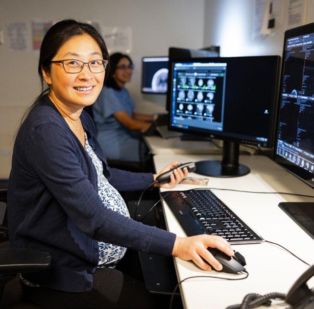
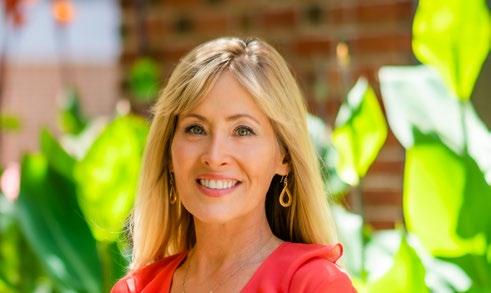
Front Cover: Images courtesy of Jonathan Pascual, NP
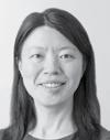
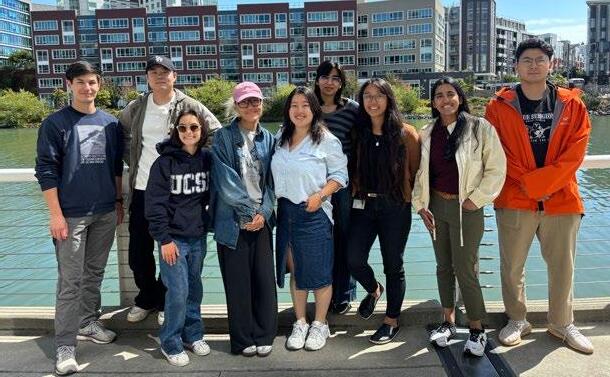
Back Cover: Resident Minerva Zhou, MD, '25
Executive Editor: Christopher Hess, MD, PhD
Managing Editor: Rita Gaber
Writers: Arleen Bandarrae, Francis Horan, Erin Sullivan
Photography: Elisabeth Fall, James Ramirez, Andrea Rowe, Marco Sanchez, Susan Merrell, Arleen Bandarrae
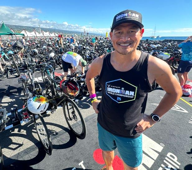
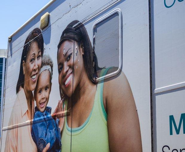
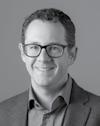
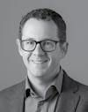
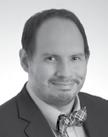
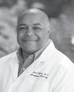
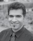
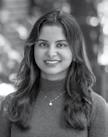
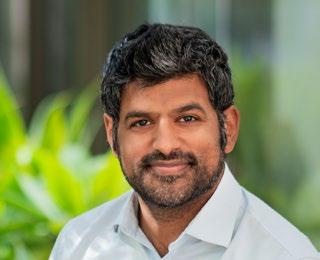
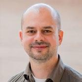
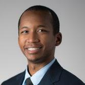
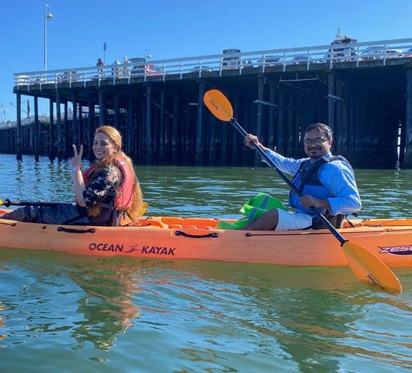
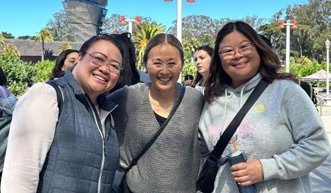
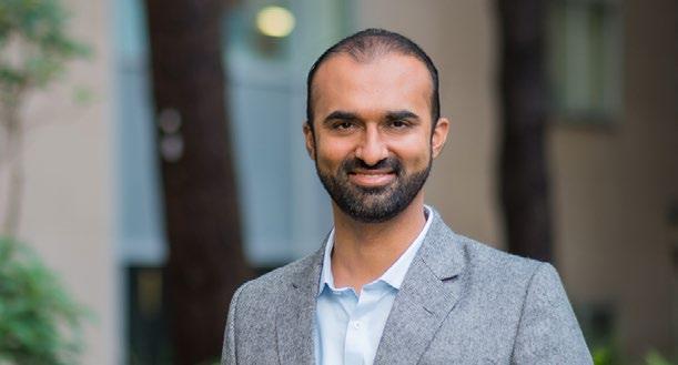
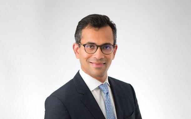
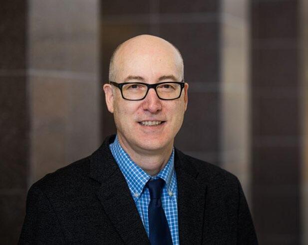
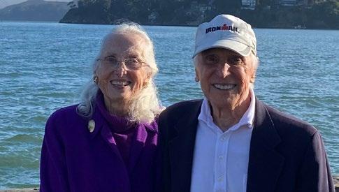
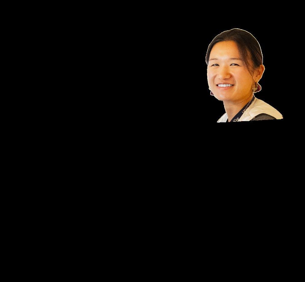
Jonathan Pascual, endurance athlete and cancer patient, is living well with PRRT.
4
CHAIR’S MESSAGE
Catalysts that are carrying us to the next equilibrium.
9
FEATURES
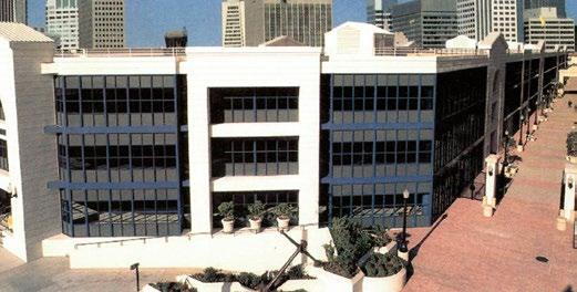
Faculty Philanthropy. Avon Breast Center at 20. Inspiring Educators. New Lexicon for 1st Trimester Ultrasound. Green Radiology. Ultra Processed Foods and Muscle Quality. Building a NextGen 7Tesla. Imaging Long COVID. A Look Back at CMFI.
27
ACADEMIC AFFAIRS
Faculty Achievements and Milestones.
52
EDUCATION
Training Academic Radiologists. CME destination courses.
63
HOW WE WORK Connection and Wellbeing. Partnerships that advance our mission.
84 CHIEF REPORTS
88 HONORS AND AWARDS
116 ALUMNI NOTES
Margulis Society. News from Alumni and Friends. Remembering Vincent McCormick.
MESSAGE FROM THE CHAIR
I’m grateful that we’re defining the future together.” “
Dear Friends,
As we welcome 2025, I hope this message finds you well and thriving.
Images magazine primarily reviews the events of 2024. But I would posit that it’s more important that we consider the long view when thinking about the dynamic nature of change in our field. The concept of punctuated equilibrium, borrowed from evolutionary biology, provides a fitting metaphor for how our practice, our culture, and our field evolve over longer periods of time. In a punctuated equilibrium, long periods of relative stability are interrupted by brief, transformative bursts of change.
This issue’s stories and reports highlight some pivotal advances in our clinical, educational, and research practices. The cover story features Jonathan Pascual, a Nurse Practitioner in UCSF’s Lung Transplant Service and an endurance athlete who is also a cancer patient living with metastatic disease. Thanks to radioligand therapy – a treatment strategy pioneered in our department –Jonathan is living well and fulfilling his dream of competing in an Ironman triathlon, continuing to work, and traveling. He is thriving. Jonathan’s inspiring story illustrates how radiopharmaceuticals are propelling us toward the next equilibrium point in cancer treatment.
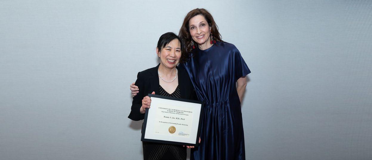
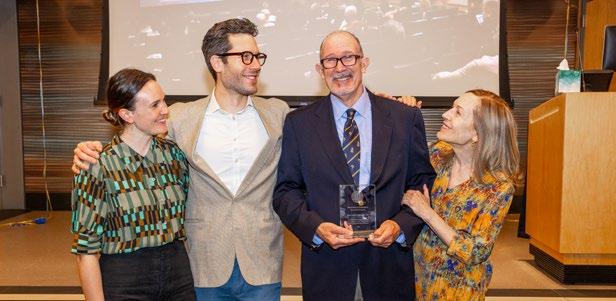
I am deeply grateful to Drs. Edward and Dale Sickles for endowing a distinguished professorship now held by Bonnie Joe, MD, PhD. Their generous gift invests in the next generation of clinician-scientists, ensuring our department remains a leading innovator while also honoring Ed’s groundbreaking contributions to breast imaging over his many decades of practice. In his typically humble manner, Dr. Sickles has expressed that he is honored to give back to the department that fostered his career success.
Christopher Hess, MD, PhD
As always, we celebrate the achievements of our people. This year is no exception:
■ Nola Hylton was elected to the National Academy of Medicine, one of the highest honors in medicine, for her pioneering work in breast MRI technology
■ Our diagnostic residency is ranked among the top in the nation by Doximity for the 11th consecutive year.
■ Our faculty serve as leaders in national and international professional and scientific societies while managing demanding clinical services and research laboratories.
■ Our research teams garnered more extramural funding than any other public-funded institution, fueling our ability to push the boundaries of imaging science and improve health.
■ Our administrative and clinical care teams partnered with our clinical faculty and trainees to enhance our capacity to teach, heal, and discover.
While we celebrate these accomplishments, it’s important that we recognize the profound forces that shape the future of clinical care, education, and research in diagnostic and interventional radiology and in imaging science. Many of you are acutely aware of what I consider the two most significant challenges: managing clinical growth that outpaces growth in research and education and mitigating the mounting threats to research resources. These headwinds are strong, but so are we. Our path forward is clear: we must grow the entirety of our tripartite mission by expanding our academic pursuits in proportion to our clinical practice.
Transforming Education
To recruit the world’s best radiologists and imaging scientists, we are launching the International Radiology Scholars Fellowship, led by Derek Sun, MD. This ambitious program involves navigating California licensing, the American Board of Radiology, and the U.S. visa system, and requires that we develop mechanisms to facilitate transition of the most promising international radiologists into a healthcare system very different than the ones to which they are accustomed. Our goal is to cultivate new academic leaders in our rapidly evolving discipline.
Advancing Clinical Care
In 2024, UCSF acquired Dignity Health’s St. Mary’s Medical Center and Saint Francis Memorial Hospital, adding nearly 600 beds to increase patient capacity ahead of the new Parnassus Heights hospital opening in 2030. To manage this expansion, we are developing innovative workforce models, including inviting radiologists without academic commitments to work alongside our academic radiologists. This approach will allow us to protect the academic mission of faculty radiologists while leveraging the talent of highly skilled clinical radiologists that have in the past practiced outside of traditional academic circles.
Pioneering Research
We continue to build our brand of excellence in research. The trajectory of innovation and discovery continues through our successful programs in molecular and
metabolic imaging, artificial intelligence, MRI, and multiple other areas in imaging sciences. One of our newest faculty members, Rajesh Shah, MD, as Director of Clinical Trials, is spearheading our efforts to bring new technologies and pharmaceuticals to patients through a coordinated clinical trials program. In January, we launched the Care Innovation Hub, a groundbreaking academic-industry collaboration with GE Healthcare. This initiative focuses on developing advanced imaging tools for diagnosing and treating neurodegenerative diseases and cancer, optimizing imaging fleet management, and delivering more seamless imaging care to patients.
Just as we go to press with this issue of Images, we are starting to recognize the potential impacts of many new policies articulated by the administration in Washington, DC. Some of these changes are crosscutting and may ultimately jeopardize federal funding for both research and healthcare delivery. While uncertainty across the University of California and other academic health systems is high, I remain confident that we will emerge stronger as long as we stay true to our values and mission. I am convinced that positive change and progress will come at the edge of our comfort zone in these uncertain times.
Looking back on this era, history will show UCSF as a consistent driving force behind the punctuated equilibrium over which radiology and biomedical imaging continue to evolve. I am profoundly grateful and proud that we are defining this future together.
Christopher Hess, MD, PhD
Alexander R. Margulis Distinguished Professor and Chair UCSF Department of Radiology and Biomedical Imaging
I want to shatter the misconception that stage IV cancer cancer is a death sentence.”
UCSF Health Nurse Practitioner and Endurance Athlete Shares His Cancer Journey
By Arleen Bandarrae
The art of dying is to live very well, Jonathan Pascual believes.
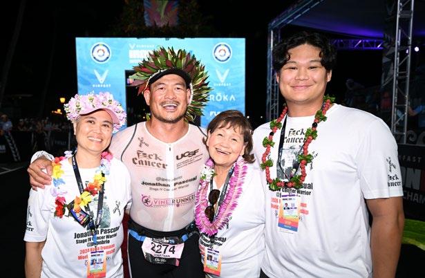
This belief fuels his passion for life and largely propelled him across the finish line of the Ironman World Championship race in Hawaii on October 26, 2024. Competing against the best triathletes from around the world, he accomplished his dream—swimming 2.4 miles in the ocean, cycling 112 miles, and running a 26.2-mile marathon—two years after being diagnosed with advanced cancer.
“I want to shatter the misconception that stage IV cancer is a death sentence,” he said.
In 2022, Pascual, a nurse practitioner at UCSF Health, was diagnosed with mediastinal paraganglioma, cancer of the autonomic nervous system. Doctors discovered a 9-centimeter tumor near his heart and tumors in his spine. His condition was categorized as stage IV when the cancer
metastasized, spreading to his lymph nodes, lungs, and throughout his body.
“I know I’m going to lose against cancer. There’s no cure. But to me, I win daily by doing the things that I love to do, and being with the people that I love most,” he said.
Pascual views his cancer as a chronic illness to manage. A 50-year-old endurance athlete, he refers to his pain as “the discomforts” while he trains, as he’s done for the last 20 years, to compete in triathlons and ultra marathons. He’s completed 15 Ironman competitions, which qualified him for entry into the prestigious championship event in Hawaii.
“As a healthcare worker, I’ve seen people go through the suffering of illness. Some we help get better but some we can’t, and then they transition to the other life,” he said. “It gives you a sense of clarity about what is really important and that takes practice.”
Jonathan Pascual crosses the finish line of the Ironman World Championship race in October 2024. Photo credit: Ironman/Getty Image
A Caregiver Becomes the Patient
Pascual, who joined UCSF Health in 2000, is currently a nurse practitioner at the UCSF Lung Transplant Program, where he cares for patients before and after lung transplantation. He’s worked with the lung transplant team for 10 years and is proud that the program grew to a record of 116 transplants last year. Previously, he helped build the Mechanical Circulatory Support (MCS) program at UCSF in 2004, which uses mechanical heart pumps to care for patients with advanced heart failure.
His personal health journey started in 2007, when he was diagnosed with a brain tumor. Cared for by his colleagues in Neuro Intensive Care, he had surgery to remove the tumor and recovered well. Within three months, his doctor gave him the green light to train for his third Ironman triathlon.
He felt healthy for many years until early 2022, when he experienced dizziness and shortness of breath—to the point of fainting upon standing on a few occasions.
“I thought I wasn’t training hard enough and told myself to get to work,” he said. “Then, I was going to go for a 22mile run and wondered, ‘why am I getting fitter and leaner, but I’m short of breath, my face is bloated, and the veins in my neck are thick as ropes?’”
He knew something was wrong and following the advice of his colleagues, he went to the emergency room at UCSF Medical Center on March 24, 2022, which led to a series of diagnostic imaging procedures.
“As soon as I saw the CT scan, I knew I had cancer—even before the doctor told me that what we’re seeing in the images was most likely cancer,” he said.
A Journey Toward Cancer Stability
“I had to look at my life daily and decide how I wanted to live it,” he said.
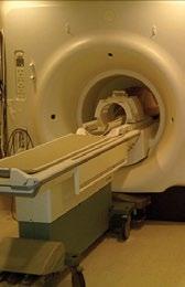
Working with a team of specialists at UCSF Health, he underwent treatment that included a combination of radiation therapy and nuclear medicine treatment.
“Our main goal was to stabilize the cancer and reduce the size of the tumors,” he said.
Using high-dose, precisely targeted X-rays in radiation therapy, his tumor cells shrunk with minimal damage to surrounding healthy tissues.
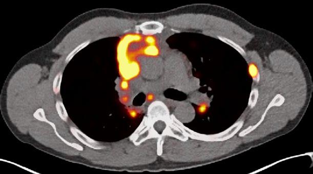
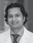
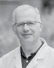
Pascual's care team included medical oncologist Claire Mulvey, MD, and radiologist Thomas Hope, MD, who worked closely together throughout the course of his treatment, which included Peptide Receptor Radionuclide Therapy (PRRT). PRRT is a molecular therapy often used to treat neuroendocrine tumors (NETs) using a radioactive substance that is attached to a peptide designed to bind to receptors on the surface of tumor cells.
“For Jonathan, although PRRT has not removed the majority of his tumors, stabilizing his disease has allowed him to do the things that he enjoys,” said Hope.
“I am amazed by the joy that Jonathan brings to life. He does not let his cancer define the road he will travel,” Hope added. “We often see our patients through the images we take, but there is no relationship between Jonathan’s smile and what one can only call extensive metastases on his imaging.”
“ I am amazed by the joy that Jonathan brings to life. He does not let his cancer define the road he will travel.”
~ Thomas Hope, MD
Chief of Neuroradiology Vinil Shah, MD, treated Pascual with kyphoplasty – a minimally invasive surgical procedure that stabilized vertebral compression fractures caused by the cancer in his spine – to reduce pain and restore the affected vertebrae for better mobility.
“It was overwhelming to be taken care of by my friends and colleagues. I felt so loved and supported,” he said. “So many people come together to take care of you. I’ve seen them in the hallway, and we’ve said good morning for so long, and now I’m on the other side, and it feels different. It’s a very humbling experience.”
He got through the most difficult days by staying focused on achieving cancer stability through his treatment at UCSF, through complementary medicine—including the barley tea his wife got from the Philippines—and with exercise and rest. And most importantly, he focused on building a support system that opened him to loving care from others.
Pascual still experiences the ill effects of both his cancer and his radiation treatments, including shortness of breath, swelling around his face and neck, and neuropathies that cause numbness and tingling in his arms and legs. To manage his shortness of breath, he must slow down his pace when running, swimming, and biking, but he doesn’t let that stop him.
“I get my mind busy. I go to work. I work out, and once I start doing other things, I’m no longer focused on all those discomforts,” he said.
A Celebration of Resilience
Reflecting on his cancer journey, it became important for Pascual to celebrate his resilience by bringing his community together. He created JP’s Backyard Ultra, a running and hiking event at the Skyline Wilderness Park in Napa, supporting the Fxck Cancer foundation, a nonprofit dedicated to cancer prevention, early detection, and providing support to those affected by cancer. He planned the event for his friends and their families, and designed the course as a series of loops, so that whether participants walked or ran, they started together. In fall 2024, more than 350 people joined, raising more than $14,000.
“That was such a fulfilling and gratifying day,” said Pascual. “To see my triathlon group, my running group, my cycling group, my family, my work family, and so many other people that have touched my life enjoying each other’s company. My goodness! What a celebration of life.”
Today, he continues to monitor his cancer with regular imaging. Depending on what the scans show, or if he becomes more symptomatic, he may resume cancer treatment in the future.
He continues to work at UCSF part-time and shares his inspiring story through speaking engagements. Recently he visited to his son’s former high school and spoke to students, many of whom have also been affected by cancer directly or through the experience of a family member or friend. He stresses that cancer can be a long journey, and he highlights the importance of reaching out to those affected, not just after a diagnosis, but in the months and years down the road.
“What really touched me is that I realize I’m carrying this terminal disease, but I have also been carried by my village to where I am now,” he said. “It’s my UCSF community, my caregivers, my family, my friends, and so many others, even strangers who have reached out to me—that is the most touching and heartwarming part of this journey.”
Peptide Receptor Radionuclide Therapy
Peptide Receptor Radionuclide Therapy (PRRT) is a targeted cancer treatment that uses a radioactive substance (radionuclide), attached to a peptide (DOTATATE), to deliver radiation directly to neuroendocrine tumors (NETs).
How PRRT works:
The peptide is designed to bind to somatostatin receptors found on the surface of certain cancer cells. The radionuclide, Lutetium-177, a radioactive isotope that emits radiation, when attached to the peptide, delivers radiation directly to a patient’s tumor cells to treat the tumor.
PRRT is commonly used to treat patients with advanced or metastatic neuroendocrine tumors to control tumor growth, reduce symptoms, and may improve quality of life for patients. There are no available FDA approved therapies to treat paraganglioma, but PRRT can be an effective therapy in a subset of these patients.
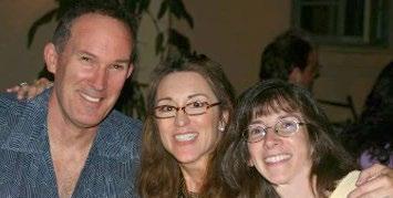
Jonathan Pascual with his wife Monette (left), his mother Betty (center right), and his son Ionakana “Iona” Pascual (right).
FEATURES
“ We’re delighted to be able to make this gift.”
Edward A. Sickles Distinguished Professorship in Radiology
For nearly 60 years Edward and Dale Sickles have shared a life dedicated to medicine. Their recent gift, to fund a distinguished professorship in breast imaging, will provide valuable time and resources to advance the field that Ed played a key role in shaping for more than 40 years.
Ed and Dale met in 1965, on their first day at Cornell Medical College in New York City and married during their second year of medical school. They came to San Francisco in 1973 when Ed began his radiology residency at UCSF after working on cancer research in the U.S. Public Health Service during the Vietnam War. Dale practiced as a pediatrician in Oakland and was the medical director of Alameda County California Children’s Services, a program for children with complex and chronic diseases.
Both are modest and soft-spoken, quick to smile and credit each other for insight. Describing the impetus for their philanthropy, Ed said, “This was an idea of Dale’s. She has carefully managed our finances over the years, and when she mentioned endowing a distinguished professorship it just clicked. Of course we should do this! Why didn’t I think of it?”
His career as breast radiologist began almost as a fluke. Before beginning the last year of his residency, two of the three radiologists who were assigned to interpret mammograms left UCSF, and the remaining radiologist didn’t want to be solely responsible for mammograms.
Alex Margulis, the department chair at the time, made Ed an offer he couldn’t refuse – he could join the faculty right away if he agreed to interpret all the mammograms. There was just one catch: Ed had never even seen a mammogram, so he took a one-week course offered by a UCLA radiologist, and from then on was self-taught.
Ed became a prolific contributor to breast imaging education and scientific literature, authoring more than 375 scientific publications, teaching hundreds of residents and breast imaging fellows at UCSF, and teaching thousands of practicing radiologists all over the U.S. and in many foreign countries. He developed an enlarged fine-detail type of “magnification” mammogram that allows earlier detection of cancer in some women and fewer biopsies for some women who do not have cancer. He proved that women with multiple similar findings in both breasts on a screening mammogram do not need any additional imaging or biopsy; and he developed the “probably benign” approach for mammograms that safely and effectively substitutes a 6-month follow-up mammogram for biopsy. Millions of women around the world have benefited from these advances.
With his customary modesty, reflecting on his partnership with Dale, Ed remarked, “This gift couldn’t have happened without her. My career couldn’t have happened without her. Giving back to UCSF is the natural thing to do, because my career happened here.”
When he learned that Bonnie Joe, MD, PhD, was named the inaugural Edward A. Sickles Distinguished Professor, Ed was thrilled, observing that, “Bonnie has a brilliant intellect. You give her a problem and she fixes it. She’s a wonderful person to work with, and she’s kind, calm, nurturing. That’s what has made the modern Breast Imaging Division what it is.”
Ed still reads mammograms four days a month as a recall faculty member and plans to continue as long as his vision remains 20/20. When he isn’t working, he and Dale enjoy spending time with their daughter, long daily walks, birdwatching, opera, art, and travel. When asked what they hoped their gift would accomplish, the Sickles agreed: “We want others at UCSF see that they too can do something like this. We’re delighted to be able to make this gift.”
By Author
ZSFG Celebrates 20 Years of Comprehensive Breast Care
By Arleen Bandarrae
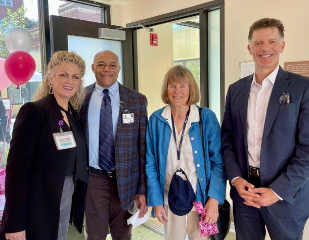
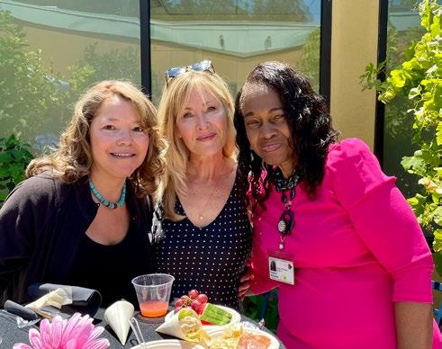
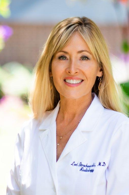
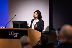
We have a lot to be proud of,” remarked Lori Strachowski, MD, a breast imaging specialist and co-founder of the Avon Comprehensive Breast Center at Zuckerburg San Francisco General Hospital (ZSFG). In July 2024, she addressed a crowd of doctors, nurses, technologists, and staff who gathered among the plants and flowers in the center’s courtyard to celebrate its 20-year anniversary.
Mary McGinty, Director of Imaging and Pathology at ZSFG, Mark Wilson, MD, Chief of Radiology, Judith Luce, Emeritus Clinical Professor of Medicine, and Grant Colfax, MD, Director of the San Francisco Department of Public Health at the Avon Comprehensive Breast Center’s 20th anniversary celebration in July 2024.
“This center is a gift to the underserved women of San Francisco. A testament that all patients are equally deserving of comprehensive and compassionate medical care,” said Strachowski.
When Strachowski joined the faculty in 1998, the breast imaging center did not exist. Screening and diagnostic mammogram services were housed in Building 5 of the San Francisco General Hospital, right next to the emergency room. The county hospital’s two mammogram machines were running seven days a week serving around 4,000 patients annually, but often with long wait times. The inconvenient location and wait time led many women to skip regular screenings, and as a result, breast imaging specialists often saw patients with advanced stages of cancer.
“
This center is a gift to the underserved women of San Francisco. A testament that all patients are equally deserving of comprehensive and compassionate medical care.”
~ Lori Strachowski, MD
Visionary Leadership
“Lori Strachowski and Judy Luce are the dynamic duo who had the foresight to apply for grant funding to build a center that’s a one-stop shop for women,” said UCSF Breast Imaging Division Chief Bonnie Joe, MD, PhD.
Strachowski and oncologist Judith Luce, MD, led a groundbreaking partnership with beauty products company Avon to envision a patient-focused facility with the potential to dramatically increase access to screening mammography and serve as many as 10,000 patients per year.
Avon began its crusade against breast cancer in 1992, and held the first 3-day walk in San Francisco to fight breast cancer in 1998, with proceeds donated to the local community. In 2000, Avon announced that San Francisco General Hospital (renamed Zuckerberg San Francisco General Hospital in 2016) would receive a $10 million grant to build a flagship breast cancer center. San Francisco General Hospital was selected because of its ability to provide a tertiary care center affiliated with the county hospital that could offer cancer care and research. UCSF also contributed $3.6 million for research projects.
The Avon Comprehensive Breast Center opened its doors in 2004 – a 5,000 square foot, modular building just outside the main hospital, at 22nd Street and Main Campus Drive. Designed by Tsang Architecture of San Francisco, the building features a healing garden in the center that was designed and donated by landscape designer Topher Delaney. The garden is named in honor of Glen Ellen resident Carolyn Stolman, who believed in the healing power of nature and died of breast cancer in February 2004.
Mary McGinty, Director of Imaging and Pathology at ZSFG, was one of the first mammographers at the center. Today she manages the center where she has worked for 20 years. The team ensures all patients are supported throughout their experience. Patient navigators — fluent in Cantonese, Mandarin and Spanish — provide critical care coordination and emotional support for patients, helping them overcome language barriers and offering information about the benefits of annual screening mammograms in their native languages.
Avon Comprehensive Breast Center
The Avon Comprehensive Breast Center is a vital safety net for uninsured, low-income women. Last year, more than 12,000 women were screened for breast cancer at the center, which serves a diverse patient population: 40% Asian, 30% Hispanic, 14% Caucasian, and 11% Black.
Breast cancer is the most common cancer among women and the second leading cause of cancer death after lung cancer, claiming about 40,000 lives annually. Early detection is key to survival, and regular screening mammograms are the best way to catch breast cancer early, when it is easier to treat successfully.
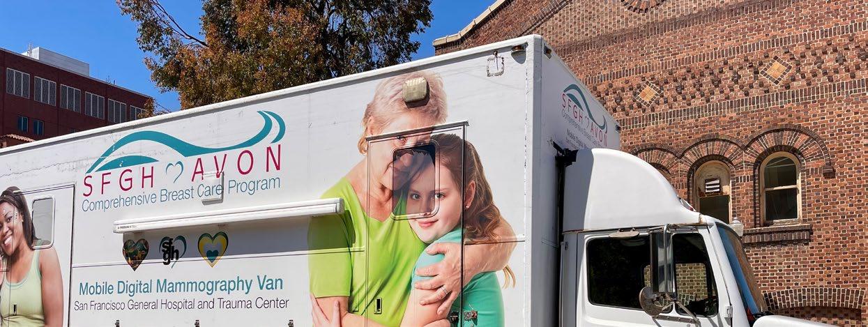
Dedicated physicians and technologists, equipped with state-of-the-art technology, provide the highest quality care to improve patient outcomes through early detection and treatment of breast cancer. To reach even more women, the team takes the center’s services on the road with the MammoVan, a van equipped with a mammography machine that functions as a mobile clinic and visits nine San Francisco community centers, making mammography accessible to women who are more likely to get screened in the comfort of their neighborhood.
The San Francisco General Hospital Foundation is currently leading a campaign to secure funding for the MammoVan program in 2025. The existing vehicle will be retired due to emissions standards, and new funding is needed to continue the program. For information about ways to donate, contact: development@sfghf.org
Digital Breast Tomosynthesis (DBT)
DBT creates a 3D breast image allowing radiologists to examine breast tissue layer by layer. This advanced imaging technique improves cancer detection through better visualization, especially for women with dense breast tissue. DBT is increasingly used for breast cancer screening and diagnostic procedures because it allows easier differentiation of benign and malignant lesions.
The Center plans to add contrast enhanced mammography services soon. The Center's TMIST trial (tomosynthesis mammographic imaging screening) sponsored by the National Cancer Institute, plays a crucial role in ensuring that Hispanic, Asian American, and African American populations are represented in research so that study findings are broadly applicable.
A Breast Imaging Center of Excellence
“As breast imagers, we’re passionate about early cancer detection and caring for patients, educating our trainees, and being mindful of translational research to improve the standard of care,” said Joe. She highlighted that the center, accredited by the American College of Radiology in Mammography, Breast Ultrasound, Stereotactic Biopsy, and Breast MR, was the first in San Francisco to deploy top-of-the-line digital mammography units, along with dedicated breast ultrasound machines for targeted breast ultrasounds and ultrasound guided biopsies.
“Just as it was the first site to get digital mammography capabilities, I’m proud to tell you it was also the first to get Digital Breast Tomosynthesis,” said Joe, who highlighted what’s next for the future of imaging services at the center.
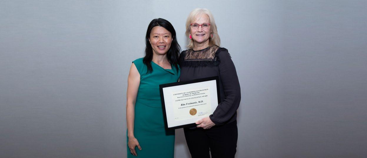
In 2025, Heather Greenwood, MD, will be the center’s new operations director, replacing Rita Freimanis who transitioned to Emeritus Professor in December 2024.
“We still haven’t met all patient needs,” said Joe. She is actively recruiting faculty and technologists and has plans to continue to develop a breast imaging nursing navigation program specific to the center.
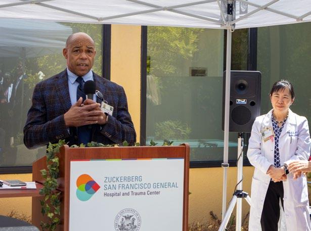
“Here’s to another 20 years of fantastic patient care at the Avon Center under Dr. Bonnie Joe’s leadership,” said Mark Wilson, MD, Chief Radiologist at ZSFG, at the anniversary celebration. He congratulated the co-founders on this remarkable achievement and expressed gratitude to the ZSFG radiologists, technologists, patient navigators, medical assistants, schedulers, oncologists, and surgeons who continue to deliver comprehensive patient care at the Avon Comprehensive Breast Center.
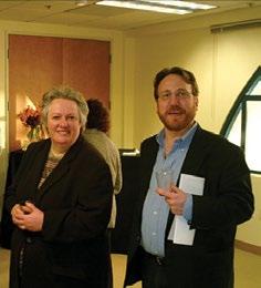
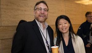
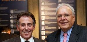
Lori Strachowski (center) celebrates with colleagues.
Bonnie Joe, MD, and colleague.
Unlocking Potential: Speaker Training Empowers Faculty
By Arleen Bandarrae
Giving lectures is an essential skill in academic medicine,” says Susan Wall, MD, the architect and lead instructor of the Speaker Training Course since 2009. This unique offering within Radiology’s Faculty Development program equips junior faculty with the skills to become compelling public speakers.
“We’ve seen, time and again, that effective public speaking is crucial to early career advancement,” emphasizes Chair Christopher Hess, MD, PhD, a notable member of the course’s inaugural cohort. An assistant professor at the time, he experienced the program’s transformative impact firsthand.
“I clearly remember the themes –focus your message, don’t reprise a textbook, articulate the most important tips and tricks of the trade, and develop your own distinctive style by learning from master teachers,” recalls Hess. “This guidance improved my public speaking immeasurably. I
go back to these basics often to hone my craft as an educator, clinician, and researcher.”
“ I'm proud that the course has thrived"
~ Susan Wall, MD
The “hands on” speaking course integrates pre-work, recorded practice talks, personalized coaching, and peer feedback. Core to the curriculum is developing a clear message and mastering effective lecture organization, delivery, and preparation. Through comprehensive training, participants cultivate skilled presentation habits, including engaging audiences with a focused message, effectively utilizing supporting visuals, and confidently handling Q&A.
“From the Speaker Course, our faculty and alumni often follow a familiar trajectory – present and chair CME courses, deliver invited lectures
at national and international meetings, participate in professional and academic committees and leadership, serve in elected office in professional societies,” says Hess.
“It’s remarkable and I’m proud that the course has thrived,” said Wall, who completed her 25th and final course as an instructor in July 2024. During her time as an instructor, she taught more than 100 class meetings over 15 years and engaged a total of 97 participants.
Future courses will be led by Elissa Price, MD, who has co-instructed alongside Wall for many years, and Xin Cynthia Wu, MD, who joined the teaching team in summer 2024.
The department extends a heartfelt thank you to Dr. Wall for her 15 years of vision and commitment to this very special and effective faculty development offering. We are delighted to welcome Drs. Price and Wu as the new course leads.
To learn more or register: Radiology.ucsf.edu/academic-affairs/faculty-development-courses.
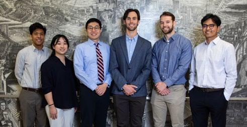
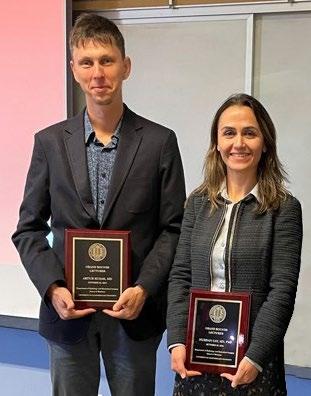
A recent Speaker Training Course cohort with Alyssa Kirsch, MD; instructors Drs. Susan Wall, Elissa Price, and Xin (Cynthia) Wu; Yoo Jin Lee, MD, Martin Rawlings-Fine (tech support), and Kang Wang, MD, PhD.
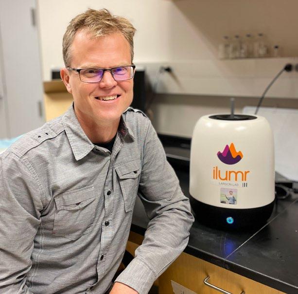
A Tabletop MRI Enhances In-Classroom Learning for MSBI Students
By Francis Horan
Students in the Master of Science in Biomedical Imaging (MSBI) program join Peder Larson, PhD, for the annual Principles of MRI course. The course illuminates the physics behind MRI scanners, and this year includes a new tool for hands-on learning. In Larson’s classroom on the second floor of Mission Hall, with Koret Quad visible out the window, students have on their table a machine that vaguely resembles a rice cooker or a speaker about to throw down some serious bass.
It's a tabletop MRI scanner, the Ilumr from Resonint, weighing about as much as a toddler, and it enables students to investigate the physics of MRI technology. Unlike MRI machines in clinical and research settings, which require special shielding, cooling systems, and weigh between five and 40 tons, this portable device offers an unparalleled opportunity for exploration. Students can freely manipulate imaging parameters, observe the effects of different techniques, and even induce artifacts to gain a deeper understanding of the underlying principles.
Larson's classes have taken on a playful yet informative tone, with everyday objects becoming subjects of MRI scans. From juicy blackberries to crunchy cauliflower florets, students image a variety of samples, learning about signal intensity, contrast, and spatial resolution. As the machine softly whirrs like a gentle air conditioner with occasional beeps, students witness the effects of motion artifacts by gently wiggling the sample tube during a scan, creating intriguing "ghost" images, or drop a metal contaminant, either steel or brass, into the sample tube to show how signal is destroyed and how artifacts pile up in the image.
Larson is thankful for the department’s investment in this important teaching resource, “This tabletop MRI provides hands-on experience in the classroom setting and fosters a deeper understanding of the technology and its underlying physics in our next generation of imaging specialists.”
Peder Larson, PhD, demonstrates the tabletop MRI scanner to MSBI students.
A New Lexicon for First Trimester Ultrasound
By Francis Horan
Everyone knows that it is possible to speak accurately and still say the wrong thing. Radiologists have a responsibility to communicate their findings to clinicians and patients clearly while managing expectations, and prenatal ultrasound specialists enter the story at a particularly sensitive time. There are profound emotional, and increasingly legal, stakes to the words used to describe these findings. Lori Strachowski, MD, of ZSFG and UCSF and Shuchi Rodgers, MD, of Thomas Jefferson University Hospitals in New Jersey helped lead The Society of Radiologists in Ultrasound in creating a recommended lexicon for First Trimester Ultrasound, published simultaneously in Radiology and the American Journal of Obstetrics & Gynecology, standardizing the terminology radiologists use to describe prenatal ultrasound findings.
Strachowski and the other committee members met over six months to achieve consensus on each standardized term. They identified both preferred terms to use and terms to avoid.
Patients are Listening
Traditionally, radiologists focused on communicating with other medical professionals through ultrasound reports. However, with increased patient access to medical records, the committee recognized the importance of how terminology impacts patients. The committee focused on using simple, easily understood language while maintaining scientific accuracy. They also prioritized choosing words that minimize anxiety and distress for patients and avoided terms that imply guarantees or outcomes that may not be certain.
The changes include substituting "concerning" for the potentially more alarming term "suspicious." The committee also discourages the use of "viable" in the
first trimester, as it can create unrealistic expectations of a successful pregnancy. Similarly, the term pregnancy “failure" should be avoided, as many patients find it insensitive and associated with blame resulting in guilt; instead they prefer "miscarriage" or “early pregnancy loss.”
The Lexicon also advises that radiologists avoid using the term “normal pregnancy” in the first trimester before it is possible to notice most defects.
Legal Implications of Ultrasound Findings
The legal landscape of the United States is changing in ways that must influence the practice of prenatal ultrasound. To avoid misinterpretations related to "heartbeat laws," the committee recommends using "cardiac activity" in place of any reference to the yet incompletely formed “heart” in the first trimester. Additionally, using terms like "live" or "living" in ectopic pregnancy situations can create legal and ethical challenges.
Simple Changes with Profound Impacts
This new lexicon emphasizes the importance of thoughtful communication beyond purely medical accuracy. By considering the emotional and legal implications of their words, radiologists can better support patients during a critical and often emotionally charged time.
Lori Strachowski, MD, and a committee of The Society of Radiologists in Ultrasound published a lexicon of terms for first trimester ultrasound that standardize the medical language, are mindful of patient emotions and expectations and address the current landscape of early pregnancy care in the USA.
To read paper: A Lexicon for First-Trimester US: Society of Radiologists in Ultrasound Consensus Conference Recommendations. Published Online: Aug 27, 2024, https://doi.org/10.1148/radiol.240122
Green Radiology: RSNA Sustainability Task Force Recommends Approaches to Sustainable Imaging
by Francis Horan
Sustainability and reducing carbon emissions are existential responsibilities for anyone who intends to live on the planet Earth. It is also an opportunity for institutions to reduce costs by eliminating wasteful practices. The healthcare sector constitutes 4.6% of global emissions and nearly 10% of US emissions, and imaging is a significant portion of that. An independent RSNA Sustainability Task Force, including Christopher Hess, MD, PhD, and Sean Woolen, MD, MS, announced its official recommendations at December’s annual conference.
As Chair of the RSNA Task Force, Hess said that cleaning up energy sources will be the key, as “turn it off” only goes so far, but radiology has many areas where we can take great strides. “We have moved past the ‘I can’t do anything myself’ instinct,” Hess said. In their talks, Hess and Woolen highlighted four areas for sustainability efforts to target.
Turn it Off, Set to Standby
Woolen led the research sub-group, collaborating with Charles Goh, FRCR, MBBS, MMed. They outlined the RSNA's role in promoting sustainability research, with a focus on standardizing terminology and evaluation metrics. As Woolen stated, "To solve a problem, we must first measure it and agree on its definition." His work in this area earned him the RSNA grant for Emerging Issues in Environmental Impact and Sustainability of Radiology. In 2021, UCSF and Siemens partnered to measure and mitigate energy usage. The energy date helped identify periods when imaging facilities were inactive but still consuming significant electricity. By comparing these "on but inactive" periods with scanner startup times, they
could optimize energy consumption by implementing lowpower standby mode or complete shutdowns.
Woolen said, “You can turn it off. Powering down equipment during long inactive time periods saves you money and the planet.”
Spot the Energy Efficiency
Hess and the committee also noted the progress made with the EPA's Energy Star Group. Energy Star, responsible for the energy efficiency ratings on appliances like refrigerators and washing machines, is now exploring the possibility of extending these ratings to medical imaging equipment, including MRI systems. The Energy Star Group has shown keen interest and is actively seeking more data from industry and healthcare partners to establish a robust framework for evaluating the energy efficiency of medical imaging devices.
Reimagine the Supply Chain
While the high energy consumption of imaging systems is a major contributor to radiology's environmental impact, Hess highlighted that the supply chain is, in fact, the primary source of CO2 emissions (CO2e). To address this, a systemic overhaul of radiology's procurement and transportation processes is necessary, requiring collaboration and innovation from all stakeholders. Hess shared UCSF's proactive approach, implementing agreements that mandate the use of sustainable air fuels for the delivery of new scanners.
Hess said, “This is a blue-horizon opportunity for healthcare and industry to work together.”
Remember What AI Costs
As healthcare professionals make significant progress in reducing emissions, new challenges are emerging. AI and other computational resources, which have potential to revolutionize some aspects of medical care, are incredibly energy intensive. This burgeoning demand for AI, which is projected to consume a substantial portion of the US's electricity, could undermine recent strides in energy efficiency. Radiology in particular must be mindful of its role in both leveraging AI's benefits and mitigating its environmental impact. Hess urged RSNA attendees to be cognizant of AI's hidden energy costs and to avoid unnecessary usage, much like the simple act of turning off lights to conserve energy.
Hess noted that “The task force convened virtually multiple times throughout its one-year charge to produce a prioritized recommendation report to the Board in December 2024. The task force’s work is divided into different strategic domains where the RSNA is anticipated to have the greatest impact and influence.” The task force’s recommendations addressed four key pillars: research, education, collaborations with imaging and industry, and assessing gaps and opportunities in the landscape of current sustainability efforts in medicine.
American Journal of Radiology (AJR) Podcast Series on Sustainability
Sean Woolen, MD, MS
Sean Woolen, MD, MS, hosts this podcast with AJR, launched in July 2024 as a forum for leaders in imaging sustainability to share ideas and practical strategies for mitigating radiology’s environmental impact.
Episode 1: Radiology’s Role in the Climate Crisis: Why It Matters
Kate Hanneman, MD, MPH, joins Dr. Woolen to survey the environmental impacts of radiology and discuss sustainable practices. They discuss planetary health, the effects of climate change on radiology, healthcare’s carbon footprint, and future investments to mitigate environmental impact.
Episode 2: The Role of Data in Radiology’s Green Transformation
Katherine Maturen, MD, MS, explores how data-driven approaches can advance sustainability, the importance of standardizing measurement outcomes, and how radiology organizations can support sustainability efforts.
Episode 3: Strategies for Climate-Resilient Imaging Services
Amanda Marrero-Gonzalez, MD, reports on extreme weather and how it affects healthcare delivery, challenges for radiology during hurricanes, and strategies to build climate-resilient health systems for the future.
Episode 4: The Power of Patient Perspectives
Reed Omary, MD, MS, and Elizabeth Schumacher, JD, discuss the role of patient preferences in sustainable healthcare, including how patient insights can shape radiology practices and promote environmentally responsible medicine.
Find the Sustainability Podcast here: tinyurl.com/SustainabilityPodcastAJR
Adiet high in ultra-processed foods is associated with higher amounts of fat stored inside thigh muscles, regardless of the amount of calories consumed or level of physical activity, according to a study presented by a team of UCSF researchers at the 2024 RSNA meeting. Higher amounts of intramuscular fat in the thigh could also increase the risk for knee osteoarthritis.
“The novelty of this study is that it investigates the impact of diet quality, specifically the role of ultra-processed foods in relation to intramuscular fat in the thigh muscles assessed by MRI,” said author Zehra Akkaya, MD, researcher and former Fulbright Scholar in the Department of Radiology and Biomedical Imaging at the University of California, San Francisco. “This is the first imaging study looking into the relationship between MRI-based skeletal muscle quality and quality of diet.”
The use of natural and minimally processed ingredients in many modern diets has decreased, more often being replaced with ingredients that have been industrially processed, artificially flavored, colored or chemically altered. Foods such as breakfast cereals, margarines/ spreads, packaged snacks, hot dogs, soft drinks and energy drinks, candies and desserts, frozen pizzas, ready-toeat meals, mass-produced packaged breads and buns, and more, include synthesized ingredients and are highly processed.
These ultra-processed foods usually have longer shelf lives and are highly appealing, as they are convenient and contain a combination of sugar, fat, salt and carbohydrates which affect the brain’s reward system, making it hard to stop eating.
For the study, researchers analyzed data from 666 individuals who participated in the Osteoarthritis
Initiative who were not yet affected by osteoarthritis, based on imaging. The Osteoarthritis Initiative is a nationwide research study, sponsored by the National Institutes of Health, that helps researchers better understand how to prevent and treat knee osteoarthritis.
“Research from our group and others has previously shown that quantitative and functional decline in thigh muscles is potentially associated with onset and progression of knee osteoarthritis,” Dr. Akkaya said. “On MRI images, this decline can be seen as fatty degeneration of the muscle, where streaks of fat replace muscle fibers.”
Of the 666 individuals, (455 men, 211 women) the average age was 60 years. On average, participants were overweight with a body mass index (BMI) of 27. Approximately 40% of the foods that they ate in the past year were ultra-processed.
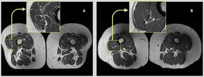
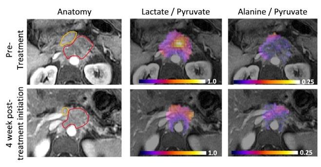
Figure 1. Axial T1-weighted bilateral thigh MR images and magnified frames providing a closer look at the areas in lateral aspects of quadriceps femoris muscles (knee extensors) from two obese, female participants, aged 58 (A) and 62 years (B), respectively. In A, the thigh muscles on both sides demonstrate abundant fatty streaks, consistent with a high Goutallier grade of 45 for this participant, whose diet from the past 12 months consisted 68% of ultra-processed foods. In B, the thigh muscles show fewer fatty streaks as highlighted in the magnified image, consistent with a low Goutallier grade of 17 for this participant, whose diet contained only 36% ultra-processed foods.
Zehra Akkaya, MD
The researchers found that the more ultra-processed foods people consumed, the more intramuscular fat they had in their thigh muscles, regardless of energy (caloric) intake.
“In an adult population at risk for but without knee or hip osteoarthritis, consuming ultra-processed foods is linked to increased fat within the thigh muscles,” Dr. Akkaya said. “These findings held true regardless of dietary energy content, BMI, sociodemographic factors or physical activity levels.”
Targeting modifiable lifestyle factors— mainly prevention of obesity via a healthy, balanced diet and adequate exercise—has been the mainstay of initial management for knee osteoarthritis, Dr. Akkaya noted.
“Osteoarthritis is an increasingly prevalent and costly global health issue. It is the largest contributor to non-cancer related health care costs in the U.S. and around the world,” Dr. Akkaya said. “Since this condition is highly linked to obesity and unhealthy lifestyle choices, there are potential avenues for lifestyle modification and disease management.”
By exploring how ultra-processed food consumption impacts muscle composition, this study provides valuable insights into dietary influences on muscle health.
“Understanding this relationship could have important clinical implications, as it offers a new perspective on how diet quality affects musculoskeletal health,” Dr. Akkaya said.
A series of New York Times articles in 2024 on ultra-processed foods elevated this public health topic for readers. This particular study by Drs. Akkaya and Ziegeler and their co-authors was #3 of the top 25 articles viewed on Aunt Minnie during RSNA 2024. The study was also picked up by the Financial Times, Newsweek, US News & World Report, Business Insider, and other media outlets.
Co-authors are Gabby B. Joseph, PhD, Katharina Ziegeler, MD, Wynton M. Sims, John A. Lynch, PhD, and Thomas M. Link, MD, PhD.
Reprinted with permission of the Radiological Society of North America.
Part A Part B
Mean Values (n=666) Results for the relationships between UPF consumption and thigh muscle Goutallier Grades Mean
Mean GG add (SD) (Range=0-24)
Part A presents the mean values for the predictor (UPF) and outcomes (Goutallier grades) for the study cohort. Part B presents the results from linear regression models for all thigh muscles (GG all), knee extensors (GG ext), knee flexors (GG flex) and thigh adductors (GG add). Beta coefficients represent the change in GG for 1 SD increase in UPF. Models were adusted for age, sex, race, body mass index, total daily calorie intake, education and income levels, physical activity scores and depression. Bold letters indicate statistically significant results. CI: Confidence interval; GG: Goutallier grade; SD: Standard deviation; UPF: Ultra-processed foods.
Refer to original article: tinyurl.com/UPFandMuscleQuality
ZSFG and UCSF Researchers
Shed Light on Long COVID with a Cover Story in Science
Translational Medicine
By Erin Sullivan
Long COVID (LC) has challenged the medical community with lingering questions about its mechanisms and symptoms. A team of researchers from ZSFG and UCSF’s Department of Radiology and Biomedical Imaging sheds light on this complex condition by exploring viral persistence and immune responses post-COVID-19.
The researchers report on their work in “Tissue-based T cell activation and viral RNA persist for up to 2 years after SARS-CoV-2 infection” published as the cover story in Science Translational Medicine (Vol. 16, No. 754).
First author Michael Peluso, MD, and senior authors Henry F. VanBrocklin, PhD, and Timothy Henrich, MD, in collaboration with a team of multidisciplinary researchers, used whole-body positron emission tomography (PET) imaging with the innovative radiopharmaceutical agent [18F]F-AraG from Cellsight Technologies. This agent is pivotal in mapping activated T lymphocytes in the body, providing a unique insight into immune responses following SARS-CoV-2 infection. The study included a cohort of 24 participants, imaged between 27 to 910 days post-infection, offering a broad perspective on the temporal dynamics of Long Covid.
Reflecting on the impetus for this study, Peluso noted that, “We have seen since April 2020 that many of our study participants were experiencing unexplained symptoms following COVID-19 that lasted for weeks, months, or longer. Some are very debilitated and almost all have experienced an effect on their quality of life. We are very motivated to get answers for the tens of millions of people suffering from this condition, which still has no approved diagnostic tests or treatments. With our background in virology and immunology, one of the first questions we always ask when we encounter a patient with unexplained symptoms is What could be going on with their immune system? We are fortunate at UCSF to have amazing researchers and resources who can help answer these questions with technologies that do not exist anywhere else.”
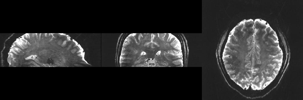
Imaging Long COVID. The cover shows severe acute respiratory syndrome coronavirus 2 (SARSCoV-2) spike protein–encoding single-stranded RNA (ssRNA, green) in rectosigmoid tissue collected from an individual with Long COVID nearly 2 years after their acute SARS-CoV-2 infection. Nuclei are shown in blue. Peluso et al. performed whole-body positron emission tomography imaging with a tracer that tags activated T cells in a cohort of 24 individuals up to 910 days after acute SARS-CoV-2 infection. The authors found that individuals with Long COVID symptoms had more tracer uptake than those without symptoms, including in the gut. Further, rectosigmoid tissue collected from five participants with Long COVID consistently harbored SARS-CoV-2 spike protein-encoding ssRNA. These data suggest that ongoing T cell activation and viral persistence may be drivers of Long COVID.
Credit: Peluso et al./Science Translational Medicine
The study results are compelling. Participants with postacute COVID-19 exhibited heightened [18F]F-AraG uptake compared to pre-pandemic controls in several anatomical regions, notably the brain stem, spinal cord, bone marrow, lymphoid tissues, cardiopulmonary areas, and the gut wall. This elevated uptake highlights increased T cell activation, a marker of immune system engagement.
Interestingly, T cell activation in the spinal cord and gut wall was closely linked with LC symptoms. Persistent pulmonary symptoms correlated with increased lung tissue activation. These observations were consistent even in participants without overt LC symptoms, indicating a broader immune response pattern post-infection.
In a subset of five participants with LC symptoms, colorectal tissue analysis revealed the presence of SARSCoV-2 RNA. Single-stranded spike protein-encoding RNA was detected in the rectosigmoid lamina propria tissue of all five participants, with double-stranded RNA found in three. This finding, observed up to 676 days post-infection, suggests a potential link between viral persistence and long-term immune disturbances.
Peluso said, “This was the first study to investigate immune activation on a total body level (previously almost
all had been in blood), and the results were shocking - a history of having had COVID-19, regardless of whether a person had Long COVID symptoms - seemed to result in ongoing inflammation for months or years. Even though this was more pronounced in people with Long COVID, it was present even in people who felt fine. We are only beginning to understand the implications of this observation, but it is possible that this ongoing inflammation is a reason for many of the negative health consequences of COVID-19 over the long term.”
This research represents a significant stride in Long COVID research, emphasizing the role of viral persistence and immune dysregulation. The innovative use of [18F]F-AraG PET imaging offers a noninvasive method to explore these mechanisms, paving the way for future investigations and potential therapeutic interventions. This research was supported by the PolyBio Foundation.
“This study provides strong evidence of persistent disease and may inform clinical trials to alleviate Long COVID symptoms. It also provides confirmation to patients that there is an underlying cause related to their symptoms and hope that treatment for their suffering may be possible,” adds Dr. VanBrocklin.
Reference
Michael J. Peluso et al. Tissue-based T cell activation and viral RNA persist for up to 2 years after SARS-CoV-2 infection. Sci. Transl. Med.16, eadk3295(2024). DOI:10.1126/scitranslmed.adk3295
First and Senior Authors
Michael Peluso, MD MPhil MHS DTM&H Assistant Professor of Medicine Division of HIV, Infectious Diseases, and Global Medicine Zuckerberg San Francisco General Hospital and University of California, San Francisco
Henry VanBrocklin, PhD Professor In Residence Director, Radiopharmaceutical Research, Center for Molecular and Functional Imaging (CMFI) University of California, San Francisco
Timothy Henrich, MD Professor In Residence University of California, San Francisco
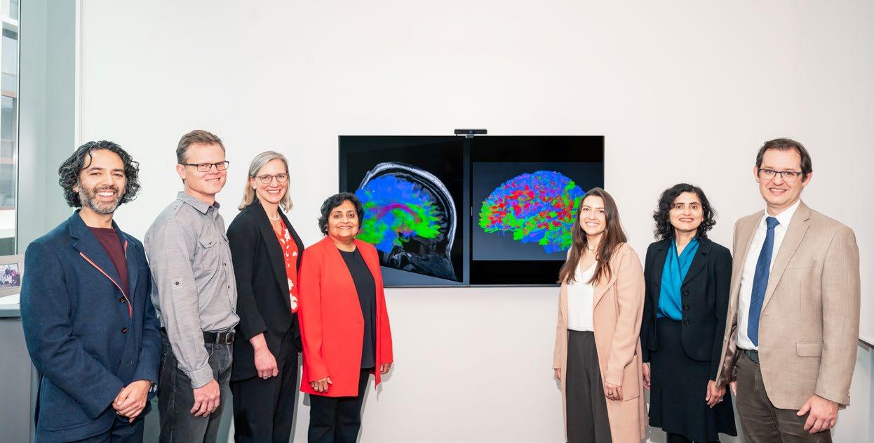
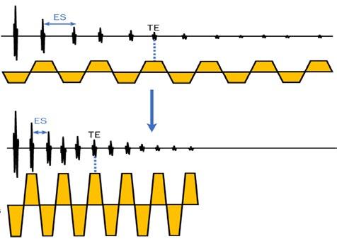
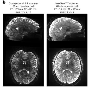
Next Gen 7T MRI Allows Neuroimaging with Unprecedented Precision
By Francis Horan
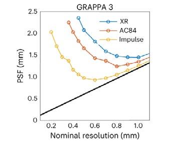
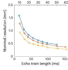
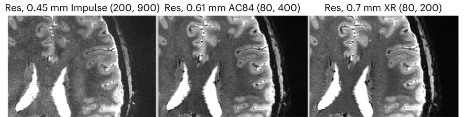
Conventional MRI scanners for clinical use generate images using magnets whose strength is measured at 1.5 Tesla (T) or 3T. At a limited number of locations across the world, clinicians and researchers have access to MRI scanners with much more powerful magnets operating at 7T. With the several dozen 7T machines located in the United States, imaging scientists can gain much higher resolution images, examining the tiny details that make up the architecture of the brain and body. However, there is still a ceiling to that resolution, as even with the most optimal scan parameters, features smaller than one to two millimeters vanish into the abstraction of the voxel, a three-dimensional pixel.
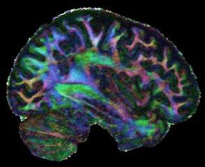
In December 2023, an international team of scientists including UCSF faculty member An (Joseph) Vu, PhD, broke that ceiling. Led by UC Berkeley professor and president of Advanced MRI Technologies, David Feinberg, PhD, MD, the team is a multi-institution collaboration with scientists from UCSF including Drs. An (Joseph) Vu and Pratik Mukherjee, UC Berkeley, Harvard, Siemens Healthineers (Erlangen, Germany), Advanced MRI Technologies (Sebastopol, CA), and MR CoilTech LTD (Glasgow, UK). Together, they constructed a next generation ultra-high resolution 7T MRI scanner, which achieves up to 10-fold increase in resolution over the current 7T standard, which is 50 times more detail than the hospital standard 3T scanners. With this new tool, functional images can now be captured with a voxel size of less than half a millimeter. This allows scientists to, for the first time, image functional clusters of neurons across
the entire brain, organized in cortical cell layers and cortical columns, opening up the study of a new realm of meso-scale local neurocircuitry.
Thanks to additional funding obtained by Drs. Vu, Feinberg, and Alexander Beckett, the NexGen 7T is now available to scientists across the Weill Neurohub consortium of UCSF, UC Berkeley, and the University of Washington, and as an international resource through the NIH BRAIN Initiative. The NexGen 7T MRI scanner is described in a Nature Methods article “Next-generation MRI scanner designed for ultra-high-resolution human brain imaging at 7 Tesla.”1
Over the years, the San Francisco Bay Area has had as many as four 7T scanners, each dedicated to state-ofthe-art imaging research at UCSF Mission Bay, the SF VA Medical Center, UC Berkeley, and Stanford. Since 2017, FDA-approved 7T scanners have become available, fueling a growing push to make this technology more readily accessible in clinical environments.
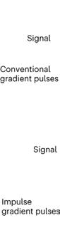
Although the NexGen 7T is not yet FDA approved, it has achieved remarkable breakthroughs in fast, high-resolution neuroimaging by virtue of several key advancements: an extremely powerful head gradient system, the first 128 channel receiver systems integrated into a 7T scanner, and several universally optimized RF pulse sequence protocol.
The new head-only magnetic gradient coil design that is an order of magnitude more powerful than those in
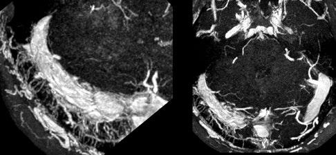
NexGen 7T 0.6 mm isotropic diffusion images (b=0 s/mm2) depicting fine anatomical structures including hippocampal layers and perivascular spaces.
commercially available 7T scanners. This advancement in gradient performance was achieved with a novel 3-layer wire winding design instead of only two layers. The faster and stronger the magnetic gradients are, the faster the MRI data can be encoded, fighting the clock of T2 signal decay and blurring. There were a lot of physics challenges to achieve such strong and fast gradients. In addition to the need to minimize peripheral nerve stimulation effects, the mechanical forces interacting with the field and the sound pressure levels both increase as the main magnetic field gets stronger. These challenges were detailed in “Acoustic noise reduction in the NexGen 7 T scanner.”2 For all these reasons, the scanner needed to be designed at a system-wide level, factoring in RF coil design, gradient coil design, and magnet design.
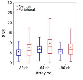
Another breakthrough came via the development of 64 channel and 96 channel receiver arrays coupled with the 128-channel receiver system, vastly improving on the standard 32 channel system in terms of SNR and the ability to accelerete the imaging. As the number of receiver channels increases, the size of the individual coil loops in the head coils become smaller, which in turn provides higher sensitivity and improved ability to accelerate data acquisition for fast, ultra-high resolution functional and structural MRI.
The improvements are not only related to hardware, as RF pulse sequence design has played a great role in these milestone achievements. In collaboration with Dr. Nicolas Boulant (CEA, NeuroSpin, France), the team has implemented precisely pre-calibrated universal pulses which can produce structural images of exquisite quality on almost any subject you place inside the scanner without the need for lengthy pre-scans or subject-specific calibrations. Furthermore, in two collaborations with Dr. Renzo Huber (NIMH, NIH) and with Dr. Suhyung Park (Chonnam National University, S. Korea), improvements in functional imaging pulse sequences more precisely identify neuronal activity in cortical layers, and similar resolution gains in collaboration by achieving unprecedented isotropic resolution in the 0.35mm – 0.6mm range.
Feinberg points out, “The NexGen 7T scanner achieves greatly improved precision in diffusion imaging of axonal fiber tracks from the cumulative gains of high signal from 7T and the much stronger gradient encoding, now possible. Secondly, the scanner’s ability to achieve
mesoscale functional imaging at depths in the cortex rather than averaging across the cortex provides more precise information to take new directions in neurocircuitry studies of different neurological disorders including depression, chronic pain, localization in epilepsy and revealing the underpinnings of many cognitive disorders.”
Vu is excited about the new avenues this next-gen 7T scanner will open up, explaining that “Traditionally, the RF pulse sequence optimization is a very involved process done on a per subject basis. One had to acquire calibration scans, model the head, and calculate how best to excite the whole brain. Not every 7T site has the time, expertise, and capability for such an optimized scan protocol. However, with the new universal pulses pre-calibrated technique the images come out very nice on any subject, right out of the box. In the past, some clinicians and collaborators have been hesitant to go to 7T because such technology was not readily available. But with these new NexGen 7T technologies, it removes the hesitancy bottleneck for wide-spread adoption into clinical neuroimaging and research. It is a game changer!”
References
1) Feinberg, D.A., Beckett, A.J.S., Vu, A.T. et al. Next-generation MRI scanner designed for ultra-high-resolution human brain imaging at 7 Tesla. Nat Methods 20, 2048–2057 (2023). https://doi.org/10.1038/s41592-023-02068-7
2) Boulant N, Ma S, Walker E, et al. Acoustic noise reduction in the NexGen 7 T scanner. Magn Reson Med. 2024; 92: 2261-2270. doi: 10.1002/mrm.30211
NexGen 7T 0.9 mm isotropic diffusion tensor images showing impressive anatomical quality and detail throughout the brain.
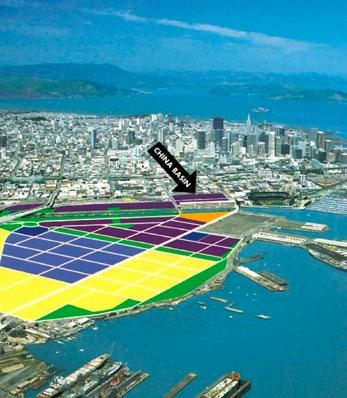
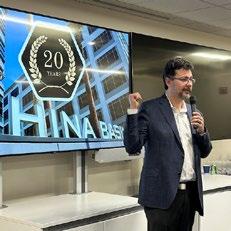
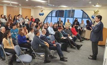
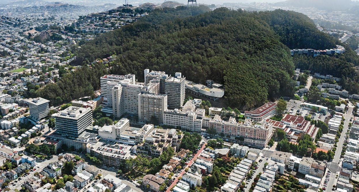
20 Years of Radiology at China Basin
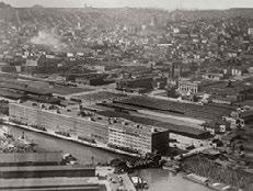
The Center for Molecular and Functional Imaging (CMFI) has been at the forefront of 3T research & major advances in theranostics & precision medicine for 20 years at China Basin Landing.
1922 - 2000
1922 China Basin Landing wharf-side building was built beside Southern Pacific Railroad as a United Fruit Company (now Chiquita) warehouse for import and export of fruit and vegetables.
2001 - 2004
2001-2002 An evaluation of all San Francisco zip codes by a vendor partner identified China Basin as a prime location for new UCSF clinical space.
2002 UCSF grants permission for off-campus location.
2003 Mission Bay Campus opens with construction still ongoing.
Bruce Hasegawa moves from Oyster Point to China Basin.
1990 Human Genome Project begins, sequencing DNA and setting the foundation for molecular imaging.
1991 China Basin Landing building two (Berry Street Building) opened as a three-story office facility, soon occupied by Bank of America computer facility.
A curious air conditioning system relied on pipes from a massive tub of ice and salt water in the basement. The metal delivery pipes began to disintegrate and leak, but the tub foundation
Sarah Nelson, PhD, and Dan Vigneron, PhD, appointed to leadership of the 3T Research Magnet Program.
Ron Arenson, MD, appointed as the acting
could later accommodate the heavy cyclotron and lead hot cells.
1992 Ron Arenson, MD, appointed Chairman of the Department of Radiology and Biomedical Imaging at UCSF, and with an eye to growth in research and clinical care began site evaluations for a cyclotron.
1995 Berkeley Lab installs its first cyclotron for production of PET radiopharmceuticals in the SF Bay area. UCSF Radiology starts imaging patients at the Berkeley Lab.
2000 San Francisco Giants open Pacific Bell Park and the dot-com bubble bursts, sending SF commercial real estate rents plummeting.
Recruitment begins for a permanent Director.
China Basin vivarium opens, staffed by LARC.
April 2003 Human Genome Project completes.
July 2003 Ben Franc, MD, MS, joins UCSF faculty. Later develops early PSMA imaging agents.
September 2003 Youngho Seo, PhD, joins UCSF.
December 2003
Center for Molecular and Functional Imaging (CMFI) opens at China Basin Landing.
China Basin Imaging Center opens on the first floor along with a 3T research MR scanner and a 16-slice research CT scanner. 140 faculty and staff relocate to the Center from sites all over city.
2005 - 2007
2005 Henry VanBrocklin, PhD, joins department as Director of the Radiopharmaceutical Research Program in CMFI.
Byers Hall opens on the Mission Bay campus, which provided new space for the Magnetic Resonance group which had been at China Basin.
February 2005 Siemens Biograph 16-slice PET/CT added to CMFI instruments.
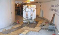
September 2005 UCSF’s first cyclotron installed over Labor Day weekend in the re-enforced former airconditioning room in China Basin Basement.
The 10-ton device fit down the parking garage ramp with one inch of clearance.
2006 Construction completed for radiopharmaceutical chemistry preclinical research lab (small animal imaging facility) on 1st floor of China Basin.
May 200 6 CMFI contracts with external radiopharmaceutical company to manage cyclotron.
Jim Slater, RPh, PhD, joins as the first director of radiopharmaceutical facility. Scott McClain joins as the first cyclotron engineer.
December 2006 Cyclotron begins producing clinical radiopharmaceuticals.
First micro-PET/CT scanner
September 2007 First new UCSF home-made compound synthesized, [18F]fluoropaciltaxel.
2008 - 2015
2008 Bill Mannone, BS, joins UCSF as senior cyclotron engineer.
The first external collaboration project between UCSF professor Ben Franc, MD, and Clifford Berkman, PhD, of SF State University, developing early PSMA imaging agents.
2013 Hyperpolarized-3T pyruvate research moves to Mission Bay.
UCSF now manages the cyclotron and hires the staff as UC employees.
Jim Slater appointed director of the radiopharmaceutical facility.
2015 UCSF Medical Center at Mission Bay opens.
2020 - 2023
2020 Robin Ippisch, PhD, joins the cyclotron staff. She is appointed director of the radiopharmaceutical facility in 2021.
2023 China Basin’s first micro-PET CT scanner replaced with a Mediso PET/CT.
New GE StarGuide SPECT/ CT opens.
Bill Mannone retires as senior cyclotron engineer.
December 2023 CMFI celebrates 20 years in operation.
PEOPLE
ACADEMIC AFFAIRS
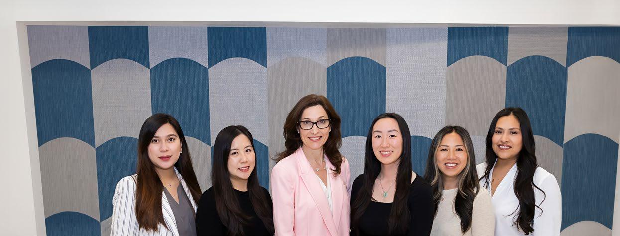
Meet the Academic Affairs Team!
By Christine Glastonbury, MBBS
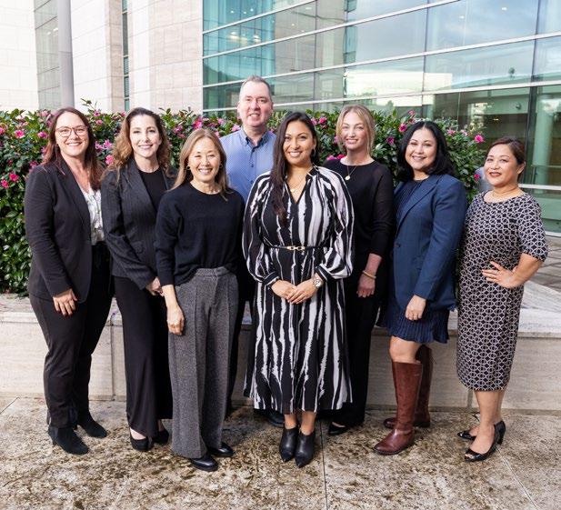
The Academic Affairs team has grown in number and in scope of work over the last year. This team of four dedicated administrative staff, Jocelyn Pulido, Connie Jang, Apple Palad and Selena Yan, under the leadership of Lorna Kwok, work to serve our faculty and promote their academic success.
FACULTY OF THE DEPARTMENT OF RADIOLOGY
EChair, Vice Chairs and Associate Chairs
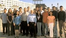
ach of the five Academic Affairs team members is responsible for a specific portfolio, but all work together and coordinate with other department administrators. The most visible portfolio, and the forum in which most clinical and research faculty directly interact with our team is through faculty recruitments, which has become a significantly larger energy investment for our team. In 2024, we opened 13 faculty searches and searched for two Endowed Professorships. The multiple search committees composed of department and nonradiology faculty interviewers have met with 27 candidates across almost every division. We have welcomed both UCSF-trained and external candidates to every campus and coordinated – for every candidate – lectures, group interviews, faculty dinners, and countless one-on-one interviews. We appreciate the dedication of the search chairs and the amazing UCSF faculty who give of their time to grow our department.
the UK and Germany. Apple Palad coordinates faculty onboarding and collaborates with Academic HR and the radiology administrative teams to streamline the many required credentialing steps, to ensure a seamless and timely transition to a faculty position at UCSF. Onboarding internationally trained faculty has greater complexity requiring US visa applications, specially designated California medical licenses (2113 or 2168 permits) and necessitates the ABR alternate pathway. Connie Jang, our Medical Privileges and Project Coordinator, has developed a comprehensive understanding of this process to ensure timely credentialing, medical licensure and visa issuance, working in close collaboration with the UCSF Office of Medical Affairs and Governance.
As of December 2024, we have 157 faculty in the department – 37 Imaging Scientists, 104 Radiologists, and 16 on recall. In 2024, we welcomed 11 new faculty members including internationally trained hires from
The department mentoring program supports 32 assistant professors. With much thanks to the more senior faculty who mentor, we celebrate January as National Mentoring Month and honor our faculty mentor-mentee pairs. This year the academic affairs team provided all faculty with desktop calendars and hosted a ‘Mentor-Mentee Mingle’ event at the grand opening of the newly remodeled M380 conference room.
Christopher Hess, MD, PhD Chair, Department of Radiology and Biomedical Imaging
During Mentoring Month in January, we gifted calendars to faculty and displayed posters at our various clinical & research locations.
January 2024
CELEBRATIONS 2024
From 2020-24 the Academic Affairs and Communications team partnered on an annual booklet celebrating faculty milestones. Writer Francis Horan prompt-engineered the AI images for this year's issue: tiny.ucsf.edu/2024Celebrations
Thomas Link, MD, PhD Chief, Musculoskeletal Radiology
Matthew Bucknor, MD Jonathan Friedman, MD
Kevin McGill, MD, MPH Daria Motamedi, MD
Alexandra Gersing, MD
Jyoti Narayanswami, MD
Kevin Sweetwood, MD Lynne Steinbach, MD Emeritus
K. Pallav Kolli, MD Chief, Interventional Radiology
Nicholas Fidelman, MD Ryan Kohlbrenner, MD Alexander Lam, MD Rajesh Shah, MD R. Peter Lokken, MD Jaehoon Shin, MD, PhD Andrew Taylor, MD, PhD
Susan Noworolski, PhD
Eugene Ozhinsky, PhD
Donna Peehl, PhD
Ashish Raj, PhD
Sabrina Ronen, PhD Emeritus
David Saloner, PhD
Youngho Seo, PhD
Renuka Sriram, PhD
Srikantan Nagarajan, PhD
Advancement Workshop held on April 8, 2024
Faculty Advancement is a year-long process working with each faculty member applying for merit or promotion steps and coordinating the paperwork and required components with Human Resources For the 2024 cycle, the department Merits & Promotions Committee reviewed 60 packets with 27 of these packets requiring special review by the Academic Senate Committee on Academic Personnel (CAP). The 27 CAP packets include 19 faculty promotions, and we congratulate the newly promoted faculty elsewhere in this issue of Images.
Together, Selena and Jocelyn have been managing NFA recruitment, USA visa processing, and all HR including funding and separations. Selena also partners with the department communications team to ensure our website is current and informative, so our faculty can more readily locate opportunities and resources our department offers.
The Academic Affairs team also manages the hiring and academic advancements of specialists and professional researchers. For the past 5 years, Jocelyn Pulido has coordinated the portfolios of Non-Faculty Academics (NFAs) and Volunteer Clinical Physicians (VCPs). Selena Yan joined our team this year and is transitioning into this role.
Christine Glastonbury, MBBS, has served as vice chair of academic affairs for five years and has focused on streamlining processes and identifying faculty opportunities and recognition. Under the leadership of the Chair, she has expanded the academic affairs team and the team portfolio, to ensure strong departmental support for our faculty, non-faculty academics, and volunteer clinical physicians.
Sujal Nanavati, MD
Jared Narvid, MD Michael Ohliger, MD, PhD Director MR
Preethi Raghu, MD
Alexander Rybkin, MD
Loretta Strachowski, MD Emeritus
Thienkhai Vu, MD, PhD Allen Ye, MD, PhD
Esther Yuh, MD, PhD
Yi Li, MD Kambiz Nael, MD Andreas Rauschecker, MD, PhD Leo Sugrue, MD, PhD Javier Villanueva-Meyer, MD David Wilson, MD, PhD Xin (Cynthia) Wu, MD
We are delighted to introduce clinical faculty members who are joining us in 2024. All of our faculty contribute to trainee education as well as new and ongoing initiatives that ensure our department’s reputation for clinical and research innovation.
As each new faculty member joins our UCSF community, we extend a warm welcome and our best wishes for successful, fulfilling careers.
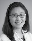
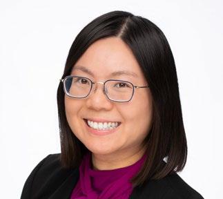
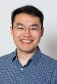
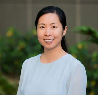
Shinn-Huey Shirley Chou, MD, MPH
Associate Professor of Clinical Radiology Breast Imaging
Dr. Shirley Chou is a breast imaging radiologist who evaluates breastrelated symptoms and detects and diagnoses breast cancer using imaging tools, including mammography, ultrasound, and MRI. She is a clinical expert at interpreting screening, diagnostic, and cancerstaging breast imaging examinations, and at performing breast imagingguided procedures, such as tissue sampling.
Her academic interests center around advancing biomedical imaging science to improve the health outcomes of those presenting with breast concerns, who are at elevated breast cancer risk, and who are diagnosed with breast cancer. Her academic passions also include recruiting and training future generations of clinically excellent radiologists.
Dr. Chou earned her medical degree from the Chicago Medical School at Rosalind Franklin University. She completed a diagnostic radiology residency at the University of Washington, followed by a breast imaging fellowship at the Brigham and Women’s Hospital in Boston. Upon completion of her fellowship, she became faculty of the Massachusetts General Hospital radiology department, during which time she earned an MPH in Clinical Effectiveness at the Harvard T.H. Chan School of Public Health and served in various leadership roles before joining the radiology faculty at UCSF.
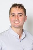
Sunit Davda, MBBS Assistant Professor of Clinical Radiology Pediatric Imaging
Sunit Davda, MBBS, received his medical degree with distinction at King’s College London in 2011 and became board certified as a Fellow of the Royal College of Radiologists, UK (FRCR) in 2017. Dr. Davda is also board certified in diagnostic
radiology by the Royal College of Physicians and Surgeons of Canada (FRCPC). Dr. Davda completed his residency at St. Bartholomew and the Royal London Hospital NHS Trust, followed by fellowships in Pediatric Diagnostic Radiology (2019) and Pediatric Interventional Radiology (2020) at the SickKids Hospital, University of Toronto. Dr. Davda served as a Consultant in Pediatric Interventional Radiology at London’s Great Ormond Street Hospital (2020-24).
Dr. Davda’s expertise is pediatric interventional radiology procedures ranging from neonates to young adults, as well as multimodality imaging of children. His subspecialty interests include complex conventional airway intervention, musculoskeletal pediatric intervention, gastrointestinal intervention, lymphatic intervention, and pediatric vascular access.
As a clinician, Dr. Davda has contributed to audit and quality improvement efforts, serving as the Audit and M&M lead for IR. Successful projects have included improving renal biopsy results and outcomes, improving access for patients lost to clinic follow-up during COVID, reducing infection rates in implantable venous ports, and improving the consent process in pediatric IR. He was an inaugural member of the Patient and Family Advisory Group for the imaging department at SickKids Hospital, advising on a range of family led improvements.
He has published book chapters on lymphatic malformations in children, and research includes pediatric lymphoma, imaging of peritoneal dialysis complications, and the spectrum of pulmonary aspergillosis. He is a co-investigator on the Multiomic Analysis of Paediatric Joint and Gut Inflammation (MAP-JAG) study. His international presentations include congenital tracheal anomalies, vascular access in children with complex anatomy, GI complications in pediatric intervention, chest intervention, bronchopleural fistulas in necrotic pneumonias, and complications of orbital sclerotherapy. Dr. Davda is also a reviewer for Pediatric Radiology
He was appointed a RadReach Mentor for widening participation by the Royal College of Radiologists. Passionate about people development, he has been a certified Executive Coach since 2022.
Alexandra Gersing, MD
Associate Professor In Residence Musculoskeletal Imaging
Dr. Gersing earned her medical degree in 2011 after studying at the University of Hamburg and the Technical University of Munich (TUM). She completed a radiology residency (2019), followed by a musculoskeletal imaging fellowship (2020), both at the University Hospital of the TUM. She was a postdoctoral research fellow in musculoskeletal imaging at UCSF from 2014 to 2016. Dr. Gersing has been a professor of radiology and Director of Magnetic Resonance
Imaging and AI Research at the University Hospital of Munich and the Ludwig Maximilians University Munich.
Dr. Gersing leads multidisciplinary teams applying MR imaging in patient care and research projects focusing on musculoskeletal imaging and AI. Her research is published in top journals such as Radiology, Osteoarthritis and Cartilage, American Journal of Sports Medicine, and European Radiology. In 2023, she was awarded the WilhelmConrad-Roentgen Prize from the German Society of Radiology and the Seed Grant of the International Skeletal Society. She is a member of the editorial board of several journals (e.g. European Journal of Radiology).
As an educator, Dr. Gersing mentors doctoral candidates, postdoctoral scholars, residents, and junior faculty. She has been an invited lecturer at several top-rated societies and institutions and serves as an elected executive committee member at the European Society of Skeletal Radiology and the German Society of Musculoskeletal Radiology.
Masis Isikbay, MD
Assistant Professor of Clinical Radiology ER/Acute Care
Masis Isikbay, MD, received his medical degree from Harvard Medical School in 2018. At UCSF, he completed his internship (2019), diagnostic radiology residency (2023) during which he served as
Chief Resident, and neuroradiology fellowship (2024).
Dr. Isikbay has taught trainees of all levels at UCSF School of Medicine, mentoring pre-med and medical students, leading symposia, and lecturing at regional, national, and international meetings. He created the medical education website Stepwards.com and accompanying YouTube channel, for which he received an RSNA education grant. Dr. Isikbay has contributed to quality improvement projects such as body and neuro IR post procedure order sets, first-year resident rotation guides, and primers for new trainees. He was recognized with the 2023 Elmer Ng Award for Outstanding Resident in Diagnostic Radiology.
Dr. Isikbay was a 2022-23 NIH T-32 scholar conducting research on improving clinical care including: the COVID-19 diagnostic pathway, machine learning mediated imaging post-processing, and diagnostic/procedural medical education. Dr. Isikbay worked with the UCSF Neuroendovascular Surgery service to create an updated classification system for shunting arteriovenous lesions of the spine. He was nominated as one of 2023’s most influential radiology researchers by AuntMinnie.com.
Dr. Isikbay is a deputy editor for the journal Radiology and was awarded the RSNA’s 2023-24 Olmstead Editorial Fellowship. Dr. Isikbay serves on the neuroradiology practice parameters committee for the ACR and on the education committee for the ASNR.
Nandan Keshav, MD, MS
Assistant Professor of Clinical Radiology
Abdominal Imaging
Dr. Keshav earned his medical degree at Eastern Virginia Medical School in Norfolk, VA, in 2016. He completed a diagnostic radiology residency in 2021 at University of New Mexico Health Sciences Center followed by a fellowship in abdominal imaging and ultrasound at UCSF. Since 2022 Dr Keshav has practiced at Kaiser Permanente in the East Bay where he helped implement an MRI endometriosis protocol, and worked with gynecologic surgeons in improving correlation between imaging and operative findings. He has also served as volunteer clinical faculty in the Abdominal Imaging and Ultrasound division at UCSF.
Dr. Keshav’s clinical and academic interests include hepatobiliary, pancreatic, and genitourinary imaging, with particular interest in the continued evolution and applicability of the Bosniak 2019 criteria for cystic renal disease. He has presented abstracts and educational exhibits at multiple imaging society meetings. He has also published educational review articles and served as a resident panelist for the Radiographics Genitourinary Panel in 2020.
Alyssa Kirsch, MD
Assistant Professor of Clinical Radiology
ZSFG and Abdominal Imaging
Dr. Kirsch earned her medical degree from the Oakland University William Beaumont School of Medicine in Rochester, Michigan in 2017. She completed a diagnostic radiology residency at Corewell Health University Hospital (formerly Beaumont Hospital), Royal Oak, Michigan in 2022, where she was chief resident. She completed her abdominal imaging fellowship at UCSF in December 2023, during which she focused on complicated examinations including posttransplant and post-operative imaging, trauma-based radiology, and diverse, rare pathology.
She has presented research at regional and national conferences on imaging blunt injuries to the bowel and mesentery, choroid plexus carcinoma, biopsy of suspicious osseous lesions, non-adnexal ectopic pregnancy, neuroimaging of acute viral encephalitis, and imaging of toxic and metabolic encephalopathy.
Dr. Kirsch enjoys teaching medical students whether in the reading room, via lectures, or in formal and informal mentoring relationships. During residency, Dr. Kirsch was recognized for her contributions to medical education (2020-22), and with neuroradiology Case of the Year awards.
Amanda Liu, MD
Assistant Professor of Clinical Radiology
Pediatric Radiology
Amanda Liu, MD, received her medical degree at the Warren Alpert Medical School of Brown University in Providence, RI, in 2018. Following an internship at the Tucson Hospitals Medical Education Program in Arizona, she completed a diagnostic radiology residency at UCSF in 2023 and received the Elmer Ng Outstanding Resident Award. She completed her pediatric radiology fellowship at the Children’s Hospital of Philadelphia in 2024.
Dr. Liu is passionate about radiology education. At UCSF, she served as chair of the Resident Curriculum Committee, spearheading the development of a new in-service exam, end-of-rotation quizzes, and monthly site-based case competitions. She also served as a CORE Curriculum class representative and Resident Liaison for Medical Student Education. In her final year of residency, she completed the UCSF GME Health Professions Education Pathway and created a set of pediatric musculoskeletal radiography modules with pre- and post-tests for first-year residents to improve their call preparedness. During her fellowship, she served as the fellow representative on the CHOP Radiology Education Committee and was selected as the fellow representative for the National Pediatric Radiology Fellowship Curriculum Editorial Board of the
Society for Pediatric Radiology. She has given multiple lectures to medical students and residents, and as a fellow, she was invited back to UCSF to lecture as the Residents’ Visiting Professor for CORE preparation.
Dr. Liu has conducted research on head ultrasound abnormalities in premature infants and ultrasound of pediatric umbilical disorders. In addition to clinical research, she has an interest in radiology education research. She is currently collaborating with faculty at CHOP on a multiinstitutional study implementing a machine learning-based educational application to support precision learning at the PACS workstation.
Yuntong (Lorin) Ma, MD, MPhil
Assistant Professor of Clinical Radiology
Musculoskeletal Imaging
SF VA Medical Center
Dr. Ma earned her medical degree at Washington University School of Medicine in St Louis. Dr. Ma completed a diagnostic radiology residency at Brigham and Women’s Hospital in Boston where she was a clinical fellow at Harvard Medical School. In 2023, Dr. Ma completed a fellowship in musculoskeletal imaging at Stanford, where she was also a clinical scholar. Dr. Ma holds an MPhil in Sociology from St. John’s College, Cambridge University, where she was a Gates Scholar.
During residency, Dr. Ma’s research examined radiologic-pathologic correlation for decision-making after
non-diagnostic CT-guided lung biopsies. She also investigated the diagnostic accuracy of radiographs for the detection of traumatic pelvic fractures in the elderly. During residency, Dr. Ma participated in the Mass General Brigham Data Science Pathway (DSP) to learn the fundamentals of artificial intelligence and machine learning in healthcare, and through this work developed a deep learning algorithm to differentiate rheumatoid arthritis from osteoarthritis on hand radiography. This work received the Senior Research Award.
Kambiz Nael, MD Professor In Residence Neuroradiology
Dr. Nael earned his medical degree from the Shiraz University Medical School in Shiraz, Iran, in 2000. At UCLA, he completed a diagnostic cardiovascular MRI post-doctoral fellowship (2006), a radiology residency (2011), and a neuroradiology fellowship (2012).
His faculty roles include neuroradiology and neuroscience at University of Arizona Medical Center (2012-15), the Icahn School of Medicine at Mount Sinai in New York (2015-19), and David Geffen School of Medicine at UCLA (2020-24). His leadership roles include Director of Neuroradiology MRI and Director of Stroke Imaging at University of Arizona, and Director of MRI, CT & Advanced Imaging and Director of Neuroimaging Advanced and Exploratory Lab at The Icahn School of Medicine.
Dr. Nael’s research investigates quantitative neuroimaging with a clinical focus on cerebrovascular diseases and stroke. His work on amplified MRI for coupled motion of the brain and blood flow was the winner of the American Society of Mechanical Engineers –Bioengineering Division Competition at the 2018 World Congress of Biomechanics, and the winner of the National Science Foundation visualization challenge. For ASNR, he is co-chair of the Research Committee and a member of the Educational Committee. He has won the Radiology Editor’s Recognition Award with Distinction for four consecutive years.
Rajesh Shah, MD
Professor of Clinical Radiology Interventional Radiology Director of Clinical Trials
Rajesh Shah, MD, earned his medical degree at The University of Chicago Pritzker School of Medicine in 2004, followed by a diagnostic radiology residency at University of Illinois Medical Center in Chicago (2009) and a vascular and interventional radiology fellowship at Stanford University Hospital in 2010. Since 2023 he has been Director of Interventional Radiology at the California Pacific Medical Center, Clinical Associate Professor at Stanford University, and an Interventional Radiologist at the VA Palo Alto Health Care System, where he has served in various leadership roles. Prior experience includes private practice and faculty roles at
Weill Cornell Medical College and Memorial Sloan-Kettering Cancer Center in New York City.
As an educator, Dr. Shah has mentored junior IR faculty at the VA Palo Alto, Stanford University, and at the California Pacific Medical Center. He has mentored trainees on research grants and created the VA resident rotation and “minifellowship” for IR-bound residents. Dr. Shah serves as the Society for Interventional Radiology (SIR) Division Councilor for Quality and Performance Improvement overseeing several committees dedicated to quality. In this role, he developed the Quality Improvement program for the SIR, and launched the VIRTEX Clinical Data Analytics Platform.
Dr. Shah was appointed as affiliated faculty at the Stanford Center for Artificial Intelligence in Medicine and Imaging (AIMI) and was awarded an AIMI grant to investigate machine learning in radiomics which has led to publications on machine learning in radiomics for lung cancer. He has published research on hepatocellular carcinoma, small-cell lung cancer, and embolic therapies. He is a Fellow of the Society of Interventional Radiology and active in the Society of Interventional Radiology and the American College of Radiology.
Thomas Yohannan, MD
Associate HS Clinical Professor Molecular Imaging & Therapeutics
Dr. Yohannan received his medical degree in 2011 from the University of Illinois. He completed a radiology residency in 2016 at Aurora St. Luke’s in Milwaukee where he won the Resident Teacher Award, followed by a nuclear medicine fellowship at UCSF (2017) and a clinical instructor year at Stanford (2018). Dr. Yohannan was most recently Chief of Nuclear Medicine and Radiation Safety Officer and Peer Review Liaison at Kaiser Permanente East Bay.
Dr. Yohannan’s research focuses on improving lung cancer diagnosis and staging with the aim of identifying why FDG PET has limited utility for characterizing subsolid lung nodules/ cancer and developing methods for better staging and characterization of T1 lung cancers. Previous research includes lymph node metastases in lung carcinoids using FDG-PET/CT to predict response to immunotherapy, evaluation of 68Ga-PSMA-11 PET in biochemical recurrence of prostate cancer, and use of 18 F-florbetaben whole-body PET/MRI for evaluation of systemic amyloid deposition.
At Kaiser, Dr. Yohannan led development of tools for imaging decision support, including creating an application to standardize physician messaging to thyroid cancer patients in multiple languages. In 2021, The Permanente Medical Group Leadership Academy honored this work.
Victoria Young, MD
Assistant Professor of Clinical Radiology Pediatric Radiology
Victoria Young, MD, earned her medical degree from the University of Miami Miller School of Medicine in 2013 followed by a diagnostic radiology residency (2018) and a vascular and interventional radiology fellowship (2019), at Northwestern University in Chicago. Dr. Young did a pediatric radiology fellowship (2020) and a clinical instructor year at Stanford. From 2021-24 she was a faculty member in pediatric/ interventional radiology at the Lucile Packard Children’s Hospital at Stanford.
Her translational and clinical research focuses on pediatric issues including MR neurography, liver transplantation, musculoskeletal MRI, and venous malformations. She has presented at conferences on tumor cryoablation, lymphangiography, and May-Thurner’s syndrome.
At Stanford, Dr. Young mentored women and underrepresented trainees and wrote for the university’s diversity newsletter. As the junior representative for the Society of Pediatric Interventional Radiology since 2022, she has implemented a mentor/mentee program. For three years, Dr. Young was co-lead of the Lucile Packard Children’s Hospital Radiology Peer Learning and Care Improvement committee (CIC).
Faculty Promotions
We are honored to announce that 15 faculty members – four new associate professors and 11 new full professors – received academic promotions effective July 1, 2024.
Our department and UCSF recognizes each promoted faculty member for their significant contributions to our mission: Advancing Health Worldwide. To do this well, each faculty member balances teaching, clinical practice, scientific inquiry and clinical translation, and service activities to create an outstanding portfolio of accomplishments. We are incredibly proud of each faculty member and their unique qualities as teachers and mentors; department and university leaders committed to Diversity, Equity and Inclusion; and physicians and imaging scientists known for their national or international stature in the field of radiology.
Matthew Amans, MD Professor of Clinical Radiology
Neuroendovascular Surgery
Jeremy Gordon, PhD
Associate Professor In Residence Research
Eugene Ozhinsky, PhD
Associate Adjunct Professor Research
Spencer Behr, MD Professor of Clinical Radiology Abdominal Imaging Molecular Imaging & Therapeutics
Jing Liu, PhD Adjunct Professor Research
Kimberly Ray, MD Professor of Clinical Radiology Breast Imaging
Matthew Bucknor, MD, MFA Professor In Residence Musculoskeletal Imaging
Kevin McGill, MD, MPH
Associate Professor of Clinical Radiology Musculoskeletal Imaging
Dorothy Shum, MD
HS Clinical Professor Abdominal Imaging
Hailey Choi, MD
Associate Professor of Clinical Radiology Abdominal Imaging
John Mongan, MD, PhD Professor of Clinical Radiology Abdominal Imaging
Jason Talbott, MD, PhD Professor of Clinical Radiology Emergency Radiology and Neuroradiology
Daniel Cooke, MD Professor In Residence Neuroendovascular Surgery
Michael Ohliger, MD, PhD Professor In Residence Abdominal Imaging
Andrew Taylor, MD, PhD Professor of Clinical Radiology Interventional Radiology
Faculty Named to Endowed Professorships
Christine Glastonbury, MBBS, Professor of Clinical Radiology and Vice Chair, Academic Affairs, has been appointed as the Elizabeth A. Guillaumin Endowed Chair in Neuroradiology. Dr. Glastonbury is a worldclass radiologist and renowned educator in head and neck imaging, a frequently invited and published speaker, and current president of the American Society of Head & Neck Radiology (ASHNR) and Vice President of the American Roentgen Ray Society (ARRS). At UCSF, Dr. Glastonbury has served as Chair of the Academic Senate Committee on Committees (COC) among other universitywide leadership roles. She is the first neuroradiologist to be appointed as a member of the National Comprehensive Cancer Network (NCCN) Head and Neck Expert Panel, and an ongoing member of the Head and Neck Expert Panel for the American Joint Committee on Cancer. Among many distinguished accolades that she has received, she is the 2021 recipient of the UCSF Radiology & Biomedical Imaging Outstanding Faculty Mentoring Award and has, for more than two decades, provided invaluable clarity for trainees and junior faculty on the unwritten rules of academia, especially for those who are underrepresented minorities and women and parents. Dr. Glastonbury’s wisdom and energetic creativity as a clinician, educator, and administrator are highly sought within and outside of UCSF.
Bonnie Joe, MD, PhD, is the inaugural Edward A. Sickles Distinguished Professor in Breast Imaging. Dr. Joe will also serve as Associate Chair, Faculty Mentoring. Dr. Joe exemplifies our department’s vision of the academic radiologist as a teacher, clinician, and researcher. During a period of exponential growth, Dr. Joe expanded breast imaging to serve outpatients in Berkeley, San Mateo, and on the Peninsula, and increased breast MRI access at China Basin. As co-Leader of the Breast Imaging Research Group, Dr. Joe is focused on imaging-based approaches to breast cancer diagnosis with goals to improve earlier detection, reduce disease recurrence, and improve survival. She is coleading exciting new multidisciplinary work in molecular breast imaging with dedicated breast PET (mammiPET). Dr. Joe is a Fellow of the Society of Breast Imaging (SBI), and the recipient of the UCSF Radiology & Biomedical Imaging Outstanding Faculty Mentoring Award in 2016 and the RSNA Honored Educator Award in 2023. With an international reputation for teaching and research, Dr. Joe serves on the SBI Board of Directors, co-chairs the 2025 ISMRM Breast MRI Workshop, serves as Deputy Editor for Radiology: Imaging Cancer, and recently began a second three-year term on the RSNA Research and Education Foundation Board of Trustees.
Outstanding Achievements
We congratulate four faculty members who received departmental awards at the June 2024 Commencement Ceremony. These awards honor faculty who demonstrate inspired teaching, clinical excellence, outstanding mentorship, and volunteer service.
In addition, the department honors an Outstanding Alumnus of our residency program.
Robert Barr, MD | Outstanding Alumnus
Presented to a graduate of the UCSF Department of Radiology & Biomedical Imaging residency program who has achieved a high level of professional accomplishment, who has provided outstanding service to the field of radiology through their leadership, and who has promoted the culture of our department through collaboration, professionalism and excellence. This year, Dr. Christopher Hess presented the award
Dr. Barr completed his training at UCSF – medical school, residency, and neuroradiology fellowship – 30 years ago, and has been in private practice in North Carolina since then, developing a highly regarded reputation as a clinician, researcher, and thought leader in neuroradiology and the wider profession. There are many ways to be an outstanding alumnus and an outstanding radiologist, and Bob exemplifies so many facets of this. He has served in executive leadership roles for national professional societies and is a member of the Harvey L. Neiman Health Policy Institute of the American College of Radiology where he has played a key role in federal policy work on the radiology revenue cycle.
In his remarks upon receiving the award, Dr. Barr noted that “UCSF is a special place and has given me a career that I have loved. My time at UCSF still has immediate relevance to me, as I gather for an annual dinner at RSNA with some of my co-residents. I still work with current and former faculty through the ABR, ACR, and ASNR, and several co-residents and co-fellows are still among my closest friends. You are an extraordinary group of people and I’ve spent my career trying to recreate something similar in my own practice that was inspired by my time here.”
His nominators made these comments:
“Bob has been a wonderful friend and supporter of UCSF over the years and is well-deserving of this recognition as an outstanding alumnus.”
“Despite a busy private practice, Dr. Barr has been president of the American Society of Neuroradiology (ASNR), past chair of the ASNR Health Policy Committee and ASNR Advisor to the AMA/Specialty Society Relative Value Scale Update Committee. He currently serves as president of the American Board of Radiology Board of Governors.”
Named in honor of emeritus faculty member Hideyo Minagi, MD, and presented by graduating residents since 1974, this award recognizes an outstanding faculty educator who most exemplifies the commitment, enthusiasm and dedication to teaching demonstrated by Dr. Hideyo Minagi over his long UCSF career.
In his remarks upon presenting Dr. Courtier’s award, senior resident Dr. Kevin Leu recalled their first meeting on July 1, 2020:
“The world was very different back then because of the pandemic. I stepped in the reading room on my first day with absolutely zero knowledge of radiology and a kind welcome from Dr. Courtier. I distinctly remember reading out my very first elbow X-ray, which had a periosteal reaction, which is a term I had never heard. Jesse patiently explained basic elbow anatomy and how healing fractures appear on radiographs. Then he looked at my report. I don’t remember exactly what I wrote, but what I can tell you with 100% certainty is that it was gibberish. Jesse read it and very kindly said, ‘Okay, let’s just change it a touch.’ He had to change my entire report. But more
importantly, I remember leaving with a sense that I had learned about elbow anatomy, periosteal reaction, and gained a skill that I built upon over the years.
“Jesse’s passion for education clearly shines. I have been challenged with numerous cases from his teaching files, cases tailored to the different levels of residents’ expertise. After having taken hundreds of cases over the years, I finally asked him this year: ‘Did I finish all your cases?’ To my astonishment, he said, ‘No, I still have 100 more cases!’
“Thanks to Jesse’s efforts – his teaching file cases and many lectures – we feel that we have a firm grasp on pediatric radiology concepts today because of this outstanding teacher.”
Rita Freimanis, MD | Clinical Faculty Excellence
The Clinical Faculty Excellence Award was established in 2021 to honor and celebrate our faculty who are distinguished by their dedication to clinical radiology, their superb skills in diagnostic and/or interventional radiology, and the exceptional care they provide to our patients. This honor is awarded annually. This year, Dr. Jane Wang made the award presentation.
Rita Freimanis, MD, is on the Breast Imaging faculty at ZSFG and she received nominations from within and outside the department for this award. She is described by colleagues and trainees as a physician with phenomenal clinical skills and exemplary commitment to her patients and colleagues. Upon receiving the award, Dr. Freimanis remarked, “Thank you to all my partners in breast imaging. You welcomed me here nine years ago. You taught me what I needed to know about how things get done around here and you became my friends because nobody does this alone, so it’s been my supreme pleasure. Thank you.”
Her nominators noted that –
“As a clinician, I strive to be like Rita Freimanis. She is the utmost professional who provides outstanding care to all her patients, including for our underserved populations. She is an amazing role model.”
“Rita is an outstanding radiologist who always goes above and beyond to provide excellent clinical service to her patients and colleagues. Her dedication to advancing the field of Radiology is demonstrated by her tireless commitment to the multi-institutional NIH supported TMIST trial for which she is the PI. She is also committed to advancing diversity, inclusion and equity as demonstrated by her achievement of recruiting the greatest number of diverse trial participants at ZSFG. She is an excellent teacher and mentor and an even better human being. She brings a lifetime worth of clinical experience and lived wisdom to our section and I can always count on her for guidance.”
“Dr Freimanis leads the Avon Breast Center and the TMIST trial. She has shown incredible dedication to this international clinical trial and to serving the community. She is an incredible doctor and colleague.”
Presented annually since 2015 to a faculty member who has provided extraordinary service to the department’s faculty through mentorship. This year, Dr. Christine Glastonbury presented the award.
Dr. Joe is distinguished by her commitment and selflessness in fostering the careers of junior colleagues and building on the extraordinary excellence of UCSF. In her two decades at UCSF, Dr. Joe consistently goes above and beyond to support her mentees, with kindness and devotion to their academic success.
One nominator remarked that “I am thrilled to nominate Bonnie Joe for the mentoring award. As an exceptional faculty mentor, Bonnie supports her mentees with tireless encouragement and guidance in navigating job opportunities, research, promotion, and grants. Bonnie has been instrumental in my research endeavors, always making time for me to share ideas with her, answer questions about methods, review my papers and grants, and provide insight into research directions.”
Another nominator noted that “At conferences, Dr. Joe is always looking out for her many mentees from across the country, actively seeking opportunities to connect her mentees with the right people and resources to propel them towards their goals. Bonnie embodies the true essence of mentorship. Her impact extends far beyond the academic realm, touching the lives of those fortunate enough to benefit from her kindness, guidance, and support.”
In her remarks on receiving the award, Dr. Joe noted, “I love our residents and trainees, and if someone has a question or an issue, I want to help if I can. I learned this from my own mentors and sponsors, many of whom are in the room tonight, and who are also my colleagues and friends. I’m really just trying to pay forward and honor the support of my own mentors over the years, and I ask that you all do the same. Thank you very much.”
Mark Mamlouk, MD | Outstanding Volunteer Clinical Physician
Our volunteer radiologists provide invaluable clinical care and education as expert members of our academic community. Since 1977, our department has conferred an annual award for outstanding volunteer clinical service. This year, Dr. Vinil Shah made the award presentation.
Dr. Mamlouk is a 2015 graduate of the UCSF neuroradiology fellowship, where he served as chief fellow. He works in a busy practice at Kaiser Santa Clara where he is the regional neuroradiology lead and director of the CSF leak program. He also regularly volunteers with our neuroradiology division, where he teaches residents and fellows, reads complex clinical cases, and participates in interdisciplinary conferences such as Birthmarks and Vascular Anomalies. He has remained academically engaged, publishes widely on many topics and is in leadership positions with many academic societies including the Board of Directors for the American Society of Pediatric Neuroradiology. Dr. Mamlouk is an amazing all-round, neuroradiologist as well as a friend and supportor of UCSF.
Dr. Mamlouk’s nominators noted that he is an “Amazing teacher, a highly motivated and engaged volunteer clinical faculty who loves coming to UCSF to teach, read clinical cases, and participate in conferences including the BVAC conference.”
“He has been exceptionally productive academically while at Kaiser, often collaborating with UCSF faculty on projects, and is a leader in multiple societies including ASNR, ASHNR, ASSR, and ASPNR.”
“Dr. Mamlouk is an outstanding neuroradiologist who is an extremely motivated and engaged volunteer clinical faculty.”
“Mark is a superb physician, educator and continues to be a scholar, publishing several seminal articles in our literature on CSF leak detection and treatment.”
Faculty Retirements
To our faculty members who officially retired during the 2023-24 academic year, we extend our congratulations for their accomplishments over the decades. We are delighted to thank and honor them here, for their service to patients, trainees, our department, and the wider radiology community.
Randall Higashida, MD
Professor Emeritus
Neurointerventional Radiology
Dr. Higashida earned his medical degree from the School of Medicine at Tulane University in New Orleans in 1980 and completed his residency and fellowship training at UCLA Medical Center in 1984. He joined the UCSF faculty in 1986 and served as Chief of Interventional Neurovascular Radiology from 1996-2022. Concurrently, Dr. Higashida is also Professor, Neurological Surgery, Neurology, and Anesthesiology at UCSF, and a faculty member at ZSFG and the San Francisco VA Medical Center. He specializes in minimally invasive treatment of complex cerebrovascular diseases and stroke.
Dr. Higashida developed one of the first intracranial stents for aneurysm treatment and UCSF pioneered the use of stents in patients with aneurysms that otherwise could not be treated endovascularly. He is a leading intellect in the ischemic stroke sphere, helping to develop the TICI scale by which success in intracranial revascularization is measured and leading some of the first trials for intra-arterial lytic agents in stroke treatment. He has trained numerous clinical fellows who have gone on to succeed him as presidents of the prestigious Society of NeuroInterventional Surgery (SNIS). His NIR trainees have founded and led many of the most highly regarded neurointerventional clinical programs around the world in the last three decades. He remains among the most highly sought-after opinion leaders in NIR worldwide. His basic science research studies new and innovative techniques to treat a variety of neurovascular disorders including aneurysms, vascular malformations, tumors, and vasospasm in animal models and in clinical practice.
A prolific scholar, Dr. Higashida has published more than 300 scientific articles on stroke and related topics, and is the author of more than 120 book chapters. His articles have appeared in the New England Journal of Medicine, Stroke, American Journal of Neuroradiology, Journal of Neurosurgery, Journal of Endovascular Surgery, Lancet, Neurology, and the Journal of the American Medical Association. He is on 10 scientific editorial boards and serves as a manuscript reviewer for 9 additional journals.
Lori M. Strachowski, MD
Professor Emeritus
Radiology and Obstetrics
Gynecology and Reproductive Sciences
ZSFG
Dr. Strachowski earned her medical degree from Rush Medical School, followed by residency in diagnostic radiology at Stanford and a fellowship in diagnostic ultrasound at UCSF. She joined the UCSF faculty in 1998. Dr. Strachowski’s concurrent leadership roles at ZSFG included Chief of Ultrasound, Chief of Breast Imaging, and Medical Director of the Avon Comprehensive Breast Care Center, which she co-founded.
At the Avon Center, Dr. Strachowski promoted high-quality and comprehensive services for women including genetic counseling and testing; education and outreach programs; and bilingual patient navigators to assure that every patient felt supported and empowered despite perceived cultural and linguistic differences. In 2006, Dr. Strachowski was instrumental in bringing the Mobile Mammography Van program to the Community Health Network of San Francisco. In 2010, she instituted a Breast MRI and MRI guided breast biopsy program to complete ZSFG’s spectrum of comprehensive breast imaging services.
Dr. Strachowski helped institute a Nuchal Translucency Screening Program at ZSFG for first trimester genetics screening and prenatal management for patients with complex fetal anomalies and complications of pregnancy. With a hands-on ultrasound philosophy, Dr. Strachowski frequently scanned patients herself, fervently promoting the same practice for residents and fellows. Over the years, she has strongly promoted radiologists’ expertise in obstetrical ultrasound by upholding the highest clinical standards, emphasizing compassionate and timely patient care, and providing enthusiastic teaching.
She has been recognized for her many contributions to clinical care and training over the years. At UCSF, she received the 2009 Excellence in Teaching Award from Haile T. Debas Academy of Medical Educators, the 2012 Hideyo Minagi Teacher of the Year Award from the Department of Radiology, the Outstanding Women’s Health Consultant of the Year Award from the Department of Obstetrics, Gynecology and Reproductive Sciences in 2014, 2018, and 2020. In 2019, she received the Anna S. Lev-Toaff Award from the Society of Radiology in Ultrasound.
Curious, Devoted, Nature-Lover: William Dillon, MD, Looks Backward and Forward
By Arleen Bandarrae and Rita Gaber
We sat down with Bill Dillon last summer after his official retirement, a hiking trip to Iceland and Norway, and just as he was beginning his next chapter as recall faculty. We asked him to describe himself in three words. He replied, “Oh, wow!” and then chose curious, devoted, and naturelover, which aptly summarize a storied career in research and clinical practice, scores of creative and satisfying professional relationships, and a lifetime of outdoor adventures with his family and friends.
It was 1979 and Bill Dillon was an intern at Virginia Mason Hospital in Seattle. He felt uneasy in internal medicine – a specialty he chose largely because he loved all of medicine and struggled to decide. Acting on a friend’s recommendation, he flew to Salt Lake City to explore a radiology residency position at the University of Utah. Following an excellent interview, the department chair suggested a hike in the Wasatch Mountains – a perfect backdrop for a nature lover to make the “unbelievably fortunate” decision that transformed his career in medicine.
“Intellectually, radiology seemed much more interesting in terms of problem solving and it touched all of medicine,” said Dillon. “Without really knowing much about radiology, and certainly nothing about the future of radiology as it turned out, I decided to give it a go.”
Four Decades of Accomplishments
“I feel unbelievably fortunate to have been in this specialty,” said Dillon. “We have this incredible reach. We touch literally every patient that comes into the hospital, and several hundred thousand people per year who go to our outpatient facilities. Imaging, especially in an academic setting, is the cutting-edge of innovation.”
Dillon credits his mother for his curiosity and love of reading, early habits of mind they cultivated together at the Hayward Public Library in Alameda County where he grew up in a large, extended Irish and Croatian family. Propelled by the freedom to explore his academic and clinical interests, Dillon’s expertise evolved over more than 40 years. His began with head and neck radiology, specifically bringing MR imaging to the field. Then he focused on diagnosing and treating patients with brain tumors who frequently sought care at UCSF Health for these complex conditions. Most recently, he has become renowned for treating spine issues in patients with pain or CSF leaks.
Driven by Curiosity and a Mazda RX7
Over the course of Dillon’s career, technology has transformed medical imaging and Dillon acknowledges that he was often in the right place at the right time. Magnetic resonance imaging (MRI) was developed in the 1970s, culminating in the first MR scan of the human body in 1977, with MR scanners becoming commercially available in the 1980s. During his fellowship at the VA in 1982, Dillon’s curiosity led him to drive patients in his twoseater sportscar from the Veterans Affairs Medical Center to a UCSF research facility at Oyster Point that was home to one of the first MRI scanners on the West Coast. (With a smile, Dillon notes that this was before HIPAA!)
“I realized MR technology would make a huge impact. If I left academics, it would be several years before I’d experience it in private practice,” he said, crediting MR technology and his mentors with keeping him in an academic career. “That was a dramatic change to be able to image with that kind of fidelity without radiation.”
For around a decade early in his career, Dillon specialized in head and neck radiology, drawing on expertise from his time at the University of Utah, where he was trained and mentored by Anthony Mancuso, MD, who was among the small group of neuroradiologists who developed the subspecialty in the 1960s and 1970s.
Dillon began to use CT imaging for spinal imaging as early as 1983. Developed in the late 1960s and early 1970s, computed tomography (CT) revolutionized medical imaging. As the resolution and speed of CT imaging advanced – the first CT scanners took several minutes to acquire a single image, compared to today’s CT scanners
that capture multiple images simultaneously in a fraction of a second – it became a vital tool in medical diagnostics, capable of providing detailed images of the body’s internal structures, including organs, blood vessels, and bones. In 1983, Dillon authored his first research paper on cranial CT. In 1984 his article, Magnetic Resonance Imaging of the Nasopharynx, was the first description of MR imaging in the field of head and neck radiology.
As CT technology accelerated, Dillon helped pioneer the replacement of invasive catheter angiography with CT angiography and perfusion CT, imaging techniques that use a contrast agent injected into the blood vessels to visualize and examine arteries, veins, and heart chambers to guide procedures and treat conditions such as stroke.
Reflecting on his service as chief of neuroradiology from 1992-2015 – the division was founded by his mentor T. Hans Newton – Dillon is quick to credit the group’s vibrant culture of innovation. Dillon noted that this leadership role prompted a natural shift to focus on complex brain tumors and related cancers, as significant numbers of patients were coming to UCSF seeking treatment for these conditions. For more than two decades, Dillon and his trainees partnered with research colleagues –including Sarah Nelson, PhD, and Dan Vigneron, PhD, who developed new tools for brain tumor imaging and characterization – to translate techniques developed in research labs into the clinic.
In the early 2000s, Dillon took a keen interested in spine imaging and pain management, including cerebrospinal fluid (CSF) leaks. Building upon his work in myelography and in partnership with Cynthia Chin, MD, Dillon developed new procedures that integrated high-field MRI and highresolution CT for diagnosis and treatment of spine and peripheral nerve problems. By locating the source of pain and then delivering pain medication with pinpoint accuracy, UCSF has been at the forefront of improving patient outcomes for complex spinal pain, in large part due to Dillon’s leadership in establishing the UCSF Precision Spine and Peripheral Nerve Center at China Basin.
Roadmap to Success: Navigate the Bumps, Have Fun Along the Way
In a recent presentation to the American Society of Neuroradiology, Dillon described a bumpy road as he recognized the struggles of early career academic radiologists in the audience who feel pulled in many different directions, in an environment where change is constant. From applying for grants, to receiving funding, Dillon understands the energy and focus required to launch new research projects, teaching and mentoring, while also managing high volume clinical workloads and carving out time for family.
As for the future of radiology and imaging science, Dillon notes promising research on the horizon in mental health, theranostics, HIFU, and new image-guided procedures. He’s personally interested in global education for radiologists through Health4TheWorld, a non-profit organization that he co-founded with Bhavya Rehani, MD. Regarding challenges, Dillon flags artificial intelligence, specifically its ethical and value-added use in healthcare, and the fact that federal funding for research has been essentially flat, indexed to inflation, for two decades, which stifles innovation and academic progress.
When pressed for advice, Dillon suggested that there’s no substitute for the synergy that develops when radiologists and imaging scientists collaborate in person. He elaborated: “Focus on one or two areas of interest and become an expert, then define your goals, and stick to them. Get the advice of others who are further along in their academic careers – they’re here for that. Then network like crazy and accumulate lots of mentors and friends!”
“You know your life goes by pretty quickly,” he said, reflecting on his career. “So, make sure that you’re having fun along the way!” He and his wife Irene, a former UCSF Radiology resident, enjoy travel and hiking in the mountains and have completed several classic hikes including the Tour d Mont Blanc, the Haut Route, and most recently hikes in Sardinia and Corsica.
William Dillon, MD, is professor emeritus and was the Elizabeth A. Guillaumin Professor of Radiology from 2004 – 2024. He joined the UCSF Radiology faculty in 1983. Dr. Dillon received his medical degree from UCLA in 1978 and completed his residency at the University of Utah in 1982, followed by a neuroradiology fellowship at UCSF in 1982-3. He is a co-founder of a non-profit, Health4theWorld.org.
Honors
■ Francis A. Sooy, MD, teaching award for Clinical Excellence, UCSF Otolaryngology, 1993
■ Gold Medal, American Society of Head and Neck Radiology, 2008
■ J. Elliott Royer award for contributions to clinical neurology, UCSF Psychiatry, 2011
■ Outstanding Researcher, American Society of Neuroradiology, 2014
■ Gold Medal, American Society of Neuroradiology, 2015
■ Outstanding Faculty Mentoring, UCSF Radiology and Biomedical Imaging, 2016
■ Gold Medal American Society of Spine Radiology, 2025
Leadership
■ Chief, Neuroradiology, San Francisco VA Medical Center, 1983-86
■ Chief, Neuroradiology, UCSF, 1992-2015
■ President, ASHNR, 1993
■ President, ASNR, 2000
■ Executive Medical Director for Ambulatory Imaging, UCSF Health, 2018-25
■ Executive Vice Chair, UCSF Radiology and Biomedical Imaging, 2007-24
Dr. Dillon was a senior editor for the American Journal of Neuroradiology (1998-2011) and has been a co-investigator in 25 funded projects and 10 clinical trials. He has published 300+ articles and 30+ book chapters.
Faculty Roster, Clinical and Research
January 1 - December 31, 2024
Abdominal Imaging
Zhen Jane Wang, MD (Chief) Professor In Residence
Spencer Behr, MD Professor, Clinical Radiology
Hailey Choi, MD Associate Professor, Clinical Radiology
Vickie Feldstein, MD Emeritus
Ruth Goldstein, MD Emeritus
Joelle Harwin, MD Assistant Professor, Clinical Radiology
Omar Hassan, MD Assistant Professor, Clinical Radiology
William Hong, MD Assistant Professor, Clinical Radiology
Sina Houshmand, MD
Assistant Professor, Clinical Radiology
Nandan Keshav, MD
Assistant Professor, Clinical Radiology
Marc Kohli, MD Professor, Clinical Radiology
Joseph Leach, MD, PhD Assistant Professor In Residence
John Mongan, MD, PhD Professor, Clinical Radiology
Liina Poder, MD Professor, Clinical Radiology
Dorothy Shum, MD HS Clinical Professor
Derek Sun, MD HS Clinical Associate Professor
Kang Wang, MD, PhD Assistant Professor In Residence
Emma Webb, MD Professor, Clinical Radiology
Sean Woolen, MD Assistant Professor, Clinical Radiology
Benjamin Yeh, MD Professor In Residence
Breast Imaging
Bonnie Joe, MD, PhD (Chief) Professor In Residence
Shinn-Huey Shirley Chou, MD, MPH Associate Professor, Clinical Radiology
Maggie Chung, MD Assistant Professor In Residence
Rita Freimanis, MD Professor, Clinical Radiology
Heather Greenwood, MD Associate Professor, Clinical Radiology
Jessica Hayward, MD Associate Professor, Clinical Radiology
Tatiana Kelil, MD Associate Professor, Clinical Radiology
Amie Lee, MD Associate Professor, Clinical Radiology
Elissa Price, MD Professor, Clinical Radiology
Kimberly Ray, MD Professor, Clinical Radiology
Edward Sickles, MD Emeritus
Cardiac & Pulmonary Imaging
Brett Elicker, MD (Chief) Professor, Clinical Radiology
Kimberly Kallianos, MD Associate Professor, Clinical Radiology
Yoo Jin Lee, MD Assistant Professor, Clinical Radiology
Jonathan Liu, MD Assistant Professor, Clinical Radiology
Jae Ho Sohn, MD, MS Assistant Professor In Residence
Shravan Sridhar, MD Assistant Professor, Clinical Radiology
Maya Vella, MD Assistant Professor, Clinical Radiology
ER/Acute Care
Jason Talbott, MD, PhD (Chief) Professor, Clinical Radiology
Shital Gandhi, MD HS Clinical Associate Professor
Masis Isikbay, MD Assistant Professor, Clinical Radiology
Amrutha Ramachandran, MD Assistant Professor, Clinical Radiology Imaging Scientists
Sharmila Majumdar, PhD (Vice Chair, Research) Professor
Robert Bok, MD, PhD Adjunct Professor
Linda Chao, PhD Adjunct Professor
Michael Evans, PhD Professor In Residence
Jeremy Gordon, PhD Associate Professor In Residence
Grant Gullberg, PhD Emeritus
Michael Hoff, PhD Associate Professor, Clinical Radiology
Nola Hylton, PhD Professor
Galateia Kazakia, PhD Professor In Residence
John Kurhanewicz, PhD Emeritus
Thomas Lang, PhD Emeritus
Peder Larson, PhD Professor In Residence
Yan Li, PhD Associate Professor In Residence
Jing Liu, PhD Adjunct Professor
Janine Lupo, PhD Professor In Residence
Alastair Martin, PhD Adjunct Professor
Dimitrios Mitsouras, PhD
Professor In Residence
Melanie Morrison, PhD Assistant Professor In Residence
Susanne Mueller, PhD Adjunct Professor
Srikantan Nagarajan, PhD Professor
Susan Noworolski, PhD Adjunct Professor
Eugene Ozhinsky, PhD Associate Adjunct Professor
Donna Peehl, PhD Adjunct Professor
Ashish Raj, PhD Professor In Residence
Sabrina Ronen, PhD Emeritus
David Saloner, PhD Professor In Residence
Youngho Seo, PhD Professor In Residence
Renuka Sriram, PhD Assistant Adjunct Professor
Duygu Tosun-Turgut, PhD Professor In Residence
Olga Tymofiyeva, PhD Associate Adjunct Professor
Henry Vanbrocklin, PhD Professor In Residence
Daniel Vigneron, PhD Professor
Pavithra Viswanath, PhD Associate Adjunct Professor
An (Joseph) Vu, PhD
Assistant Adjunct Professor
Michael Weiner, MD Emeritus
Duan Xu, PhD Professor In Residence
Yang Yang, PhD
Associate Professor In Residence
Daehyun Yoon, PhD
Assistant Adjunct Professor
Interventional Radiology
K. Pallav Kolli, MD (Chief) Professor, Clinical Radiology
Nicholas Fidelman, MD Professor, Clinical Radiology
Ryan Kohlbrenner, MD
Associate Professor, Clinical Radiology
Alexander Lam, MD Assistant Professor, Clinical Radiology
Evan Lehrman, MD Associate Professor, Clinical Radiology
R. Peter Lokken, MD Associate Professor, Clinical Radiology
Rajesh Shah, MD Professor, Clinical Radiology
Jaehoon Shin, MD, PhD Assistant Professor In Residence
Andrew Taylor, MD, PhD Professor, Clinical Radiology
Molecular Imaging and Therapeutics
Robert Flavell, MD, PhD (Chief) Associate Professor In Residence
Miguel Hernandez Pampaloni, MD, PhD Professor, Clinical Radiology
Thomas Hope, MD Professor In Residence
Roxanna Juarez, MD Assistant Professor, Clinical Radiology
Courtney Lawhn Heath, MD
Assistant Professor, Clinical Radiology
Yingbing Wang, MD Associate Professor, Clinical Radiology
Thomas Yohannan, MD
HS Clinical Associate Professor
Musculoskeletal Imaging
Thomas Link, MD, PhD (Chief) Professor In Residence
Matt Bucknor, MD Professor In Residence
Jonathan Friedman, MD Assistant Professor, Clinical Radiology
Alexandra Gersing, MD Associate Professor In Residence
Kevin McGill, MD, MPH Associate Professor, Clinical Radiology
Daria Motamedi, MD Associate Professor, Clinical Radiology
Rina Patel, MD Associate Professor, Clinical Radiology
Lynne Steinbach, MD Emeritus
Kevin Sweetwood, MD Assistant Professor, Clinical Radiology
Neuroendovascular Surgery
Steven Hetts, MD (Co-Chief) Professor In Residence
Matthew Amans, MD Professor, Clinical Radiology
Daniel Cooke, MD Professor In Residence
Christopher Dowd, MD Emeritus
Van Halbach, MD Emeritus
Randall Higashida, MD Emeritus
Kazim Narsinh, MD
Assistant Professor In Residence
Neuroradiology
Vinil Shah, MD (Chief) Associate Professor, Clinical Radiology
Matt Barkovich, MD
Assistant Professor In Residence
Soonmee Cha, MD Professor In Residence
Cynthia Chin, MD Emeritus
William Dillon, MD Emeritus
Elizabeth George, MBBS Assistant Professor, Clinical Radiology
Christine Glastonbury, MBBS Professor, Clinical Radiology
Orit Glenn, MD Professor, Clinical Radiology
Christopher Hess, MD, PhD
Professor
Yi Li, MD
Associate Professor, Clinical Radiology
Kambiz Nael, MD Professor In Residence
Andreas Rauschecker, MD, PhD
Assistant Professor In Residence
Leo Sugrue, MD, PhD
Associate Professor In Residence
Javier Villanueva-Meyer, MD
Associate Professor, Clinical Radiology
David Wilson, MD, PhD Professor In Residence
Xin (Cynthia) Wu, MD
Associate Professor, Clinical Radiology
Pediatric Radiology
Jesse Courtier, MD (Chief, BCH-SF) Professor, Clinical Radiology
Raymond Sze, MD, MAMS (Chief, BCHOakland) Professor, Clinical Radiology
Taylor Chung, MD
HS Clinical Professor
Pierre-Alain Cohen, MD
HS Clinical Professor
Kayla Cort, DO
HS Clinical Assistant Professor
Sunit Davda, MBBS
Assistant Professor, Clinical Radiology
Rachelle Durand, DO
Assistant Professor, Clinical Radiology
Bamidele Kammen, MD
HS Clinical Professor
Amanda Liu, MD
Assistant Professor, Clinical Radiology
Alexander Wai, MD
HS Clinical Professor
Victoria Young, MD
Assistant Professor, Clinical Radiology
Matthew Alan Zapala, MD, PhD
Associate Professor, Clinical Radiology
San Francisco VA
Michael Parsa, MD (Chief of Radiology, VA) Clinical Professor
Yuntong (Lorin) Ma, MD, MPhil
Assistant Professor, Clinical Radiology
Pratik Mukherjee, MD, PhD Professor In Residence
Rajiv Sawhney, MD
HS Clinical Professor
Zuckerbeg SF General
Mark Wilson, MD (Chief of Radiology, ZSFG) Professor In Residence
Christopher Brunson, MD
Assistant Professor, Clinical Radiology
Ellen Chang, MD, MS
HS Clinical Assistant Professor
Miles Conrad, MD, MPH Professor, Clinical Radiology
Brian Haas, MD
Associate Professor, Clinical Radiology
Alyssa Kirsch, MD
Assistant Professor, Clinical Radiology
Vishal Kumar, MD
Associate Professor, Clinical Radiology
Terry Lynch, MD
HS Clinical Professor
Weiya Mu, MD
Assistant Professor, Clinical Radiology
Sujal Nanavati, MD
HS Clinical Professor
Jared Narvid, MD
Associate Professor, Clinical Radiology
Michael Ohliger, MD, PhD Professor In Residence
Preethi Raghu, MD
Assistant Professor, Clinical Radiology
Alexander Rybkin, MD
HS Clinical Professor
Lori Strachowski, MD Emeritus
Thienkhai Vu, MD, PhD
HS Clinical Professor
Allen Ye, MD, PhD
Assistant Professor, Clinical Radiology
Esther Yuh, MD, PhD Professor In Residence
EDUCATION
RESIDENCY
Training Radiologists: Subspeciality Rotations, Research, Service
By Soonmee Cha, MD, Vice Chair for Education
We're so proud of our 2024 graduating residents! They joined us in July 2020, in the early days of the pandemic with a socially distanced and fully masked orientation. Congratulations to these outstanding graduates, pictured above at commencement with Melinda Parangan-Chu (far left) and Sandria Wong (far right).
Our diagnostic radiology residency program remains at the top of the national rankings by Doximity for the past 11 years in a row and carries on the tradition of providing a comprehensive, hands-on education in diagnostic and procedural imaging training, supported by experienced and dedicated faculty, advanced technology, and a collaborative and stimulating learning environment.
With an increase in case volume and variety, our residents are exposed to a wide range of cases and offered superb subspecialty rotations. High-level trauma care at Zuckerberg San Francisco General (ZSFG) offers invaluable experience with acute imaging critical decision-making supported by on-call and emergency radiology faculty. A mix of seasoned and innovative faculty with strong teaching and mentoring capabilities has further strengthened the
academic excellence. Ample research opportunities provide support for resident involvement in research, including funding, mentorship, and opportunities to present at national or international conferences.
We have created a new curriculum in healthcare administration and economics, interactive learning modules, and handson training for ultrasound and procedures. Our resident-led community service has helped with addressing local needs, building connections with neighborhood, and promoting personal growth through volunteering offers to build leadership, teamwork, problem-solving, and communication skills.
Our program excels at providing rigorous clinical training, robust academic and research opportunities, advanced imaging technology, and a supportive environment to ensure residents are well-prepared for successful careers.
Class of 2028
Incoming Residents, July 2024
Sin Mei Chan, MD
Sin Mei Chan earned her medical degree from the Yale University School of Medicine in 2023. There, with the Division of Vascular Surgery, she reviewed applications of high intensity focused ultrasound for atherosclerotic disease to precede to clinical trial and authored a review on biologic sex differences in inflammation during venous remodeling in arteriovenous fistula maturation. She was first author on Image-guided Percutaneous Strategies to Improve the Resectability of HCC
Since 2022, she has been the chair of the Women in IR Committee at the Society of Interventional Radiology, Medical Student Council, and an International Education Sub-Committee Member. She joined UCSF as a general surgery resident in 2023, before becoming an integrated interventional radiology resident in July 2024.
Richard Wang, MD
Richard Wang earned his medical degree from the Johns Hopkins University School of Medicine at Baltimore, MD in 2023, where he was a board member of the Distinguished Teaching Society. At Johns Hopkins Hospital Department of Radiology and Radiological Sciences, he conducted research on imaging characteristics and outcome
predictors of acute neurological injuries, and led the creation of several master datasets on stroke patients. He was a member of the Johns Hopkins COVID-19 EM Investigator Team with the JHH Department of Emergency Medicine. He also led a project determining the true burden of HBV in the ED setting, discovering that nearly 50% of all HBV cases go undiagnosed. Following completion of his preliminary internship at University of Maryland Medical Center in Baltimore, MD, he joined the UCSF diagnostic radiology residency in July 2024.
Teguru Tembo, MD
Teguro Tembo earned his medical degree from the SUNY Downstate College of Medicine in Brooklyn, NY, during which time he was elected to the AΩA Medical Honor Society. He was the E-Board Outreach Leader for the Downstate Street Medicine Outreach Association.
He conducted preliminary biological validations of tumor immune microenvironment phenotypes at Memorial Sloan Kettering Cancer Center in New York City. He researched the association of finasteride use with high grade prostate cancer with the Department of Urology at SUNY Downstate in NYC. After completing an internal medicine internship at Mount Sinai Morningside West in New York, NY, he joined the UCSF Early Specialization in Interventional Radiology program in July 2024.
Daniela Bermudez Garolera, MD
Daniela Garolera earned her medical degree in 2023 from the New York University Grossman School of Medicine in New York, NY.
At NYU she researched ultrafast GRASP breast MRI with the Department of Radiology, IOPTH dynamics in primary hyperparathyroidism with the Department of Surgery, and predictors of hip dysplasia with the Department of Orthopedic Surgery. She also researched the impact of acute toxicity in breast radiation therapy at the Tampa Bay Oncology Center in Largo, FL.
In July 2024, she became a diagnostic radiology resident at UCSF after completing a one-year transitional residency at HCA St. Petersburg Hospital in St. Petersburg, FL.
Christopher Song, MD
Christopher Song earned his medical degree from the Johns Hopkins University School of Medicine in Baltimore, MD, in 2023. He was first author of MRI With Gadolinium as a Measure of Blood-Labyrinth Barrier Integrity in Patients With Inner Ear Symptoms. He has conducted research on the patterns of signal intensity in CISS MRI of the inner ear and eye, as well as a systemic review of taste receptors in chronic rhinosinusitis. His interest in education led him to study resident instruction in ambulatory surgical centers.
After a preliminary year Internal Medicine residency at Mercy Medical Center in Baltimore, MD, he joined the UCSF department of diagnostic radiology as a first-year resident in July 2023.
John Alexander Rincón-Hekking, MD, MHI
John Rincón-Hekking earned his medical degree in 2023 from Harvard Medical School, following his Master’s in Health Informatics at the University of Michigan in 2018.
While in Boston, MA, he was project lead with nference Inc. for the creation of a deep-learning algorithm in collaboration with the Mayo Clinic to screen for myocarditis using a standard 12-lead ECG, and created software to predict normalized cost-effectiveness for patient encounters using raw EHR data. In 2020, he founded contraCOVID.com, a website dedicated to helping non-english speaking immigrants access important information related to COVID-19 and social assistance programs.
In June 2024, he completed a transitional internship at Cambridge Health Alliance in Cambridge, MA, before he became a diagnostic radiology resident at UCSF in July 2024.
Nikki Okwelogu, MD
Nikki Okwelogu earned her medical degree at UCSF in 2023. At UCSF she has conducted research on Ultrasound-guided bursal injections, as well as the treatment of pancreatic neuroendocrine tumors. Her research interests include the association between classic metaphyseal lesions and nonaccidental trauma in infants, diagnostic accuracy of FNA and CNB on indeterminate lipomatous lesions, and abdominal CT imaging. She presented at the UCSF Health Systems Improvement Symposium on her quality improvement work with the Department of Cardiology’s Division of Non-invasive Stress Testing.
After her internal medicine internship at Kaiser Permanente San Francisco, she joined the UCSF diagnostic radiology residency in July 2024.
Kathleen Marsh, MD
Kathleen Marsh earned her medical degree in 2023 at the Wake Forest School of Medicine in Winston-Salem, NC. There she was vice president of the Wake Forest Association of Women Surgeons Chapter, and developed a medical-legal partnership with Wake Forest Law School to address local advocacy projects. She served as Health and Wellness Coordinator at Shalom Clinic in Winston-Salem. Her research interests include adolescent idiopathic scoliosis, postoperative complications following hand surgery, carpometacarpal osteoarthritis, and anterior cervical discectomy and fusion.
She completed a transitional year residency at the University of North Dakota in June 2024, and became a diagnostic radiology resident at UCSF in July 2024.
Priyanka Garigipati, MD
Priya Garigipati earned her medical degree from the University of Texas Southwestern Medical School in 2023, where she was Community Engagement Committee Chair of the Gold Humanism Honor Society. Previously, she received a Fulbright where she collaborated in English education in Taitung, Taiwan.
At UT, she conducted research on MRI guided prostate biopsy, and diffusion tensor imaging tractography. She served as the lead research coordinator at the UT Southwestern Calvert Place Smoking Cessation Clinic.
She joined UCSF’s diagnostic radiology program in July 2024, after completing transitional residency at Santa Clara Valley Medical Center in San Jose, CA.
Liang Yen Liu, MD
Larry Liu earned his medical degree in 2023 from the Mayo Clinic Alix School of Medicine in Rochester, MN. As part of his interest in education, he served as a teaching assistant for Biochemistry & Genetics, Pathology & Immunology, and the USMLE Step 1 Board Review Course.
In 2023, he was awarded the Louis A. Gilula, MD Mentor Award by the American Society of Spine Radiology. While working at AbbVie Biotherapeutics, he designed flow cytometry staining panels and developed protocols for the cell banking group. He has delivered national presentations on the treatment of cerebrospinal venous fistulas, and deep learning in upper gastrointestinal endoscopy, for which he was co-first author.
In July 2024, he became a Diagnostic Radiology Resident at UCSF after completing a one-year transitional internship at Scrips Mercy Hospital in San Diego.
Matthew Lamberti, MD
Matthew Lamberti earned his medical degree from the Georgetown University School of Medicine in Washington, DC, where he co-led the Healthcare Leadership Track Healthcare Policy & Economics concentration and served on the Racial Justice Committee for Change.
He researched the impact of racism in the Covid public health crisis with the Department of Family Medicine, and sole-authored a manuscript of his results. He has presented on bronchial artery embolization, and prostatic artery embolization.
In June 2024, he completed a General Surgery Residency at Swedish First Hill Medical Center in Seattle, WA, before he became a diagnostic radiology resident at UCSF in July 2024.
Jocelyn Cheng, MD
Jocelyn Cheng earned her medical degree in 2023 from the Alpert Medical School of Brown University in Providence, RI, winning the Radiological Society of Rhode Island Prize. She served as a Medical School Tutor, as well as president of both the Imaging Interest Group and the Diagnostic Radiology Preclinical Elective.
She received the Alavi-Mandell Award from the Society of Nuclear Medicine and Medical Imaging for her article on DEI in academic nuclear medicine. She has firstauthored multiple presentations on AI algorithms, pediatric anesthesia, and breast cancer risk assessment.
In July 2024, she joined the diagnostic radiology class at UCSF.
Devan Diwanji, MD, PhD
In 2021, Devan Diwanji earned his PhD in Biomedical Science, and in 2023 medical degree, both from UCSF. He was the co-founder of Brain Camp@UCSF, a free camp for underrepresented-in-medicine high-school students. He led curriculum and mentorship teams for UCSF professional student coordinators and counselors. He was team lead for the UCSF QBI Coronavirus Research Consortium.
He has first authored or co-first authored 13 publications on topics including breast cancer, prostate cancer, and structural dynamics of the active HER4 and HER2/HER4 complexes. He directly contributed 12 published structures deposited in the Protein Data Bank, and won the best poster award at the 36th Protein Society International Conference.
He became a diagnostic radiology resident at UCSF in July 2024, following the completion of his preliminary medical internship at California Pacific Medical Center in June 2023.
Chief Residents at Commencement
2023-24
Chief Residents
2024-25 Rising Chief Residents
T32 Program
Funded by a grant from the National Institute of Biomedical Imaging and Bioengineering, the program is designed to give early career radiologists and nuclear medicine physicians the skills required to become independent investigators and leaders in academic biomedical imaging. The T32 curriculum includes focused mentoring and formal training in clinical research, biostatistical methods, and grant writing.
T32 leaders are program director Thomas Link, MD, PhD (Musculoskeletal Imaging), and associate program directors Z. Jane Wang, MD (Abdominal Imaging), and David Wilson, MD, PhD (Neuroimaging).
2023-24
2024-25
Madhavi Duvvuri, Stephen Wahlig, and Katie Cecil.
Lexi Tatem, Outstanding Clinical Fellow and Instructor Awardee Adam Yen, Eric Davis, and Andrew Chan
Thomas Link, Lohith Kini, MD, PhD, Eric Davis, MD, Luke Bonham, MD, David Wilson, and Zhen Jane Wang.
Thomas Link, Charlie Wang, MD, PhD, Madhavi Duvvuri, MD, MPhil, and Joe Baal, MD.
Clinical Fellows & Clinical Instructors of 2024-25
BODY IMAGING
■ Adish Parikh
■ Alexandra Silver
■ Aren Vierra
■ Christopher Murphy
■ Connie Lu
■ Emily Huang
■ Hamed Kordbacheh
■ Ivy Ewald
■ Joshua Barnaby
■ Joshua Rabang
■ Justin Caskey
■ Kali Xu
■ Katherine Cecil
■ Leo Jia
■ Marisa Martin
■ Reema Agarwal
■ Rishabh Agarwal
■ Stephen Wahlig
■ Swetha Aribindi
BREAST IMAGING
■ Aren Vierra
■ Connie Lu
■ Emily Huang
■ Ivy Ewald
■ Kali Xu
■ Katherine Cecil
INTERVENTIONAL RADIOLOGY
■ Jason Kim
■ Jeffrey Worthley
MUSCULOSKELETAL
■ Alex Ward
■ Brian Lee
■ Joe Baal
■ Kalpana Manral
■ Kenan Alkhalili
■ Michelle Lam
■ Robert Dionisio
NEURORADIOLOGY
■ Benjamin Voss
■ Bryce David Beutler
■ Charlie Wang
■ Christopher Lee
■ Dror Suhami
■ Elianna Goldstein
■ Joel McLouth
■ Justin Remer
■ Kevin Leu
■ Madhavi Duvvuri
■ Michael Thomas
■ Nicholas Hoehnle
■ Nour Dababo
■ Phuong Nguyen
■ Sartaaj Walia
NEURO INTERVENTIONAL
■ Edgar Perez
■ Jorge Moreno
■ Kafi Hemphill
Masters of Science in Biomedical Imaging Program
On Tuesday, August 20, nine students presented their theses in the annual Masters of Science in Biomedical Imaging (MSBI) Symposium at Genentech Hall on the UCSF Mission Bay campus. Celebrating with the MSBI students were eight faculty advisors and department researchers and staff. The graduating students did a fantastic job discussing their projects, which investigated a variety of advanced imaging topics.
Congratulations to 2023-24 MSBI Students
MSBI Students, 2024-25
2023-24 Thesis Symposium
The MSBI students selected Peder Larson, PhD, Professor and Director of the Body Research Interest Group, for this year’s MSBI Outstanding Teaching Award.
Thank you to MSBI leadership, Youngho Seo, PhD, MSBI Program Director, and Susan Noworolski, PhD, MSBI Director of Graduate Studies, for another successful symposium. Since the MSBI program was initiated in 2011, graduates have gone on to medical schools, PhD programs, research positions within academic labs, and a wide range of occupations in industry.
MSBI Students and Their Projects, 2023-24
■ Ellis Mayne: Evaluation of Efficacy of Combined CD46-Targeted 225-Ac-Radiopharmaceutical Therapy and FOR46 Antibody Drug Conjugate in Multiple Myeloma, Advisor: Robert Flavell, MD, PhD
■ Jo Veres: The Separate and Competing Impacts of Proteoglycan Fragment Size and Concentration on T1rho Relaxation Times, Advisor: Aaron Fields, PhD
■ Duc Huy Doan: Application of convolutional neural networks in multiparametric MR imaging to predict prostate cancer progression, Advisor: Susan Noworolski, PhD
■ Radhika Bhalerao: Predicting Meningioma Genetic Mutations from Magnetic Resonance Imaging using Machine Learning, Advisor: Andreas Rauschecker, MD, PhD
■ Tianrun Xiao: Optimization of MR-guided Focused Ultrasound Induced Blood-Brain Barrier Opening Protocol in a Rat Model, Advisor: Eugene Ozhinsky, PhD
■ Tangran Dong: Optimizing Diffusion Weighted Imaging for Breast Cancer Evaluation: A Study on Correcting EPI Distortions and Gradient Nonlinearity, Advisor: Nola Hylton, PhD
■ Chase Fitch: Dorsal Root Ganglia Diffusion Metrics in Patients with Lumbar Radiculopathy Undergoing Injection, Advisor: Sharmila Majumdar, PhD
■ Siddharthasiva Anbu Rajan: MRI-based Measures of Metabolic Health in the Assessment of Patients with Chronic Inflammatory States, Advisor: Susan Noworolski, PhD
■ Nadezhda Urmanov: Improved chemoenzymatic radiosynthesis of fluorine-18 labeled Sakebiose with microPET-CT imaging of Staphylococcus Aureus, Advisor: David Wilson, MD, PhD
Nicholas Finke, Jiuyi Zhang, Chili Phaler, Cheng Xue, Alexandra Wu, Bhavna Gundamaraju, Rocelle Evangelista, Bhakti Kulkarni, Zelun Kang. Not pictured: Brian Tuan and Alex Gao.
Ellis Mayne, Tangran Dong, Siddharthasiva Rajan Anbu, Jo Veres, Chase Fitch, Nadia Urmanov, Radhika Bhalerao, Duc Huy Doan, Tianrun Xiao, and Brian Tuan.
Continuing Education, CME Course Calendar
2025
February
September
Top Teachers in Neuro and Musculoskeletal Imaging
Hawaii Big Island, Kona, Hawaii
Sunday, February 9 to Friday, February 14, 2025
Course Director: Matthew Bucknor MD; Vinil Shah MD
CME Credits: 25
UCSF Top Teachers in Breast Imaging
Hawaii Big Island, Kona, Hawaii
Sunday, February 23 to Friday, February 28, 2025
Course Director: Elissa Price MD
CME Credits: 25
March
2nd Annual Neurovascular Symposium
Hyatt Indian Wells Resort & Spa, Palm Springs, CA
Wednesday, March 19 to Saturday, March 22, 2025
Course Director: Steven Hetts MD; Daniel Raper MBBS CME Credits: 15.0
Practical On-Call Radiology
Hyatt Indian Wells Resort & Spa, Palm Springs, CA
Thursday, March 20 to Saturday, March 22, 2025
Course Director: Brett Elicker MD
CME Credits: 15
Collaborative OB/Gyn Imaging: A Wine Country Retreat
Alila Resort & Spa (Hyatt Property), St. Helena, CA
Sunday, March 23 to Wednesday, March 26, 2025
Course Director: Lori Strachowski MD; Vickie Feldstein MD; Liina Poder MD
CME Credits: 19.25
April
Radiology Annual Review: Comprehensive Imaging
Webinar Live,
Monday, April 28 to Friday, May 2, 2025
Course Director: Soonmee Cha MD
CME Credits: 36
July
Imaging in Alaska
Alyeska Resort, Anchorage, Alaska
Monday, July 21 to Friday, July 25, 2025
Course Director: Joseph Leach MD PhD
CME Credits: 25
Imaging Update in Sorrento
Hilton Sorrento Palace, Sorrento, Italy
Monday, September 8 to Saturday, Sept. 13, 2025
Course Director: Elissa Price MD
CME Credits: 25
October
Imaging Update on Maui
Hyatt Regency Maui Resort & Spa, Lahaina, Maui
Sunday, October 19 to Friday, October 24, 2025
Course Director: Kimberly Kallianos MD
CME Credits: 25
November
Updates in IBD
St. Regis Hotel, San Francisco, CA
Saturday, November 8 to Sunday, November 9, 2025
December
Imaging in Puerto Rico
Fairmont El San Juan Hotel, San Juan, Puerto Rico
Monday, December 8 to Friday, December 12, 2025
2026
January
Body Imaging: Abdominal & Thoracic
Fairmont Orchid Resort & Spa, Kona, Hawaii
Sunday, January 18 to Friday, January 23, 2026
February
Neuro and Musculoskeletal Imaging
Fairmont Orchid Resort & Spa, Kona, Hawaii
Sunday, February 15 to Friday, February 20, 2026
October
Imaging on Kauai
Grand Hyatt Kauai Resort, Koloa, Kauai
Sunday, October 18 to Friday, October 23, 2026
ADMIN STAFF
RESEARCH CONFERENCE
Connection, Community, Flexibility
Matthew Bucknor, MD, MFA Associate Chair, Wellbeing and Professional Climate
To guide our wellbeing and climate work, we are using Surgeon General Vivek Murthy’s framework on the five essentials of workplace wellbeing: work-life harmony, mattering at work, connection and community, opportunity for growth, and protection from harm.
Promoting flexibility in work options for faculty and staff, while maintaining a strong community, is a key driver of work-life harmony. For clinical faculty, division chiefs have established standard rotations of remote work that allow on-site faculty to decompress worklists in the reading rooms in a way that doesn’t compromise education. The practice coordinator team was hired in 2024 to meet a long-standing faculty need for fewer interruptions in the reading rooms.
Our emphasis on connection and community remains a constant in the social life of our department. This year we read and discussed books, went bowling, picnicked
in Golden Gate Park, joined service projects, organized creative team building events, attended a Club Fugazi show, and watched the SF Giants play at PacBell Park. We celebrate holidays, birthdays, promotions, anniversaries, and new babies. Department Chair Chris Hess invites staff and faculty to join him for lunch and happy hours at our various sites.
While many of our events are planned weeks or months in advance, our spontaneous and informal connections –getting lunch in the hospital cafeteria, hosting a game night, or sharing a taxi from the airport to a conference hotel –speak volumes about the ‘mattering at work’ relationships we aim to foster.
Opportunities for growth are infused throughout our programming. At the spring research conference, Dr. Joseph Vu invited Pierre Khawand to present a wellreceived session on Mindful Priorities Management: Transforming Chaos into Calm in a Demanding World UCSF RadWomen invited Elizabeth Kagan Arleo, MD, from NY Presbyterian / Weill Cornell to deliver a Grand Rounds on Professional Guidance for Success in Radiology and Beyond. The DEI Staff Committee, co-chaired by Martin Rawlings-Fein and Cindy Cheng, hosted Ilana Kaufman for a lunch and learn presentation on Jews of Color: Beyond the Count & On the Horizon.
Although 2025 is beginning with uncertainty at the federal level about funding for scientific and medical research, our values related to belonging, inclusion, diversity, and excellence are more certain than ever. I look forward to seeing how our people thrive in the coming year.
In June we hosted the department’s first ever Family Book Club and picnic at Lindley Meadow in Golden Gate Park. It was a wonderful afternoon with perfect weather, and lots of fun and games inspired by the magic of Dave Eggers’s novel The Eyes and The Impossible.
TUpdate on Capital Projects
By Alastair Martin, PhD
he Department of Radiology and Biomedical Imaging continues to expand its portfolio of imaging systems while striving to replenish aging systems in our more mature facilities. With well over one hundred fixed installations that require design and construction services to replace, it can be very time consuming and expensive to keep our systems state-of-the-art. San Francisco is the most expensive place in the world to build and we feel that each time we replace a system. We have been working with UCSF Health leadership on a 10-year capital plan that would provide a broad refresh of our fleet and hope to have agreement on that in the near term. Important projects that will complete in 2025 include the Parnassus Emergency Department CT scanner, which is the most heavily used CT scanner at UCSF. The replacement will be our first photon counting CT (Siemens Naeotom Alpha), which has unparalleled imaging capabilities. We’ll also see a refresh of our UCIC facility (lower level of Medical Building 1), including the addition of a 3T MRI with impressive gradient performance (Siemens Cima.X).
Funding the required replacement cycle, while also building the new Helen Diller Hospital (HDH) at Parnassus, presents some challenges, but collectively offers a unique opportunity to enhance the value our vendor partnerships offer. Planned to open in 2030, HDH will feature five MRI, four CT and seven interventional radiology suites amongst its imaging systems. Vendor commitments for this equipment will occur in early 2025 to allow the HDH team to finalize the hospital design. Given the long lead time, the specific equipment to be installed is likely to be revised closer to the go-live date of the new hospital. Faculty and staff have been busy over the past year exploring vendor roadmaps to assure that the selected vendors will offer the best imaging solutions at the time of HDH opening.
Dignity Health’s San Francisco hospitals, including Saint Francis Memorial Hospital and St. Mary’s Medical Center, were formally acquired by UCSF on August 1, 2024. These facilities address a key strategic need for UCSF, and both contain substantial radiology resources. However, many of these imaging systems are outdated and in need of replacement. There is a critical need to shore up CT services at St Mary’s, where a Siemens X.Ceed CT scanner will be installed. Other projects will follow and their prioritization will be driven by the longer term vision for these facilities, which is presently in its formative stages.
The planned expansion of our outpatient imaging services into the community took another step forward in 2024 with the Bayfront Medical Building. Bayfront opened for patient care in August and features imaging resources (CT, XR, US) that support the urgent care, ambulatory surgery center, and specialty clinics at the site. 2025 will see a major expansion of outpatient services into the community with the opening of the Peninsula Outpatient Center (POPC) in Burlingame. This site will have a broad range of imaging, including MR, CT, PET/CT and mammography. Plans for additional outpatient centers are in the works, with the aim of improving our patients access to UCSF imaging resources.
Finally, we recently opened the Neuroimaging Technology Research Center (NTRC) in the Department of Psychiatry and Behavioral Science’s outpatient building at Mission Bay. Radiology, Psychiatry, Neurology, and Neurosurgery all collaborated on this donor funded research initiative, which aims to improve the diagnosis and treatment of neuropsychiatric disease. The NTRC features two state of the art MRI (Siemens Cima.X) and is led by Leo Sugrue, MD, PhD.
UCSF Achieved Goal of Carbon Neutral MRI Service in 2022
By Alastair Martin, PhD, Sean Woolen, MD, and Chris Hess, MD, PhD
MRI systems are known to consume substantial electrical power that can contribute to carbon emissions. UCSF’s Department of Radiology and Biomedical Imaging hosts over 20 MRI systems across its various campuses. The majority of these scanners operate on inherently carbon free clean energy, but some systems are powered from methane gas and have a carbon footprint.
Electrical power meters have been placed on 50% of our MRI systems and have allowed us to understand their power use characteristics. We used this information, along with an MRI instrument’s specific hours of operation and number of patients scanned, to project the energy use of all our scanners not employing green energy. This projection concluded that MRI instruments that utilized methane gas power sources consumed 738MWh of electricity in 2022. This correlates to 226 metric tons of carbon emissions associated with the operation of these instruments.
The American Carbon Registry (ACR) is a global carbon crediting program that seeks to catalyze carbon emission reductions. It issues cardon credits that are vetted to ensure the originating offset contributes to real emission reductions and has been utilized by UCOP. The Department of Radiology and Biomedical Imaging, via UCOP, retired ACR Carbon Credits that match our calculated carbon emissions from MRI instruments in 2022. This purchase allowed us to realize our 2022 goal of achieving a carbon neutral MRI service at UCSF.
In 2023 the University of California shifted its focus from achieving carbon neutrality by 2025, to a longer-term goal that would reduce electricity produced by methane gas by 90% by 2045. That switch reflected challenges in the carbon offset/credit marketplace that make it difficult to assure the purchased credits meet the desired goal. The department is similarly shifting its strategy and has begun exploring methods of reducing MRI energy use and improving the amount of energy required to perform an MRI examination. These efforts include utilizing low power modes when scanners aren’t active and employing acceleration methods and deep learning reconstruction methods to reduce scan times. Thus, while the university shifts to cleaner energy sources, we hope to moderate the power demands of our growing MRI service. Collectively these efforts reflect the universities commitment to a more sustainable future.
To learn more about University of California’s sustainability goals, please visit https://sustainabilityreport.ucop.edu/2024/locations/uc-san-francisco/
Informatics
On July 1, 2024, Hailey Choi, MD, was appointed Medical Director for Apex Imaging Informatics.
Dr. Choi’s appointment followed a four-month search during which a diverse, multidisciplinary committee interviewed a number of highly qualified internal clinical faculty candidates. Dr. Choi is an assistant professor in our Abdominal Imaging division, on service for both UCSF Health and ZSFG.
As the Medical Director for Apex Imaging Informatics, Dr. Choi has primary ownership for imaging-related tasks in Epic (e.g. orders, protocols, scheduling, etc.) including chairing the Radiology Apex Clinical Content Committee (RAC3). Other key projects will include integration of the Dignity hospitals and support for ongoing expansion of Radiology within UCSF Health. This role reports to Russ Cucina, MD, MS, Chief Health Information Officer, with a dotted line report to Marc Kohli, MD, Associate Chair for Clinical Informatics in Radiology and Biomedical Imaging.
Dr. Choi is well-positioned to lead us into the future, with her depth and breadth of experience in imaging informatics. Since her residency at Georgetown and throughout her early faculty career, Dr. Choi has survived six major information system transitions (EHR or PACS), leading to keen knowledge of what’s required for success in large health systems. In 2021, Dr. Choi volunteered to lead our department’s improvements to Apex ordering, addressing challenges highlighted by respondents to our referring provider survey. As a co-founder of RAC3, Dr. Choi was instrumental in creating standardized naming conventions for orders to improve communication, ensure appropriate billing, and harmonize orders across divisions and sites. With experience on clinical service at both ZSFG and UCSF Health, Dr. Choi has a unique perspective on a variety of Epic builds, including providing subject matter expertise for ZSFG Epic.
Administrator & Faculty Partnerships
Robin
Across our large and growing department, faculty and administrative teams partner on initiatives that support our vision for compassionate care, world-class education, and innovative research.
I am incredibly proud of what we accomplished together in 2024. Whether the success was day-to-day admin and finance, starting new programs, migrating and upgrading systems, research development, or streamlining operations – sustained excellence is the cumulative effect.
Though by no means an exhaustive list, here are some highlights from the past year. We look forward to continuing our work together in 2025.
Amy Pradhan, MPH, MS Vice Chair for Administration & CAO
Neuroimaging Technology Research Center
Mabel Zelaya
Site Director, Mission Bay and China Basin
A significant effort in 2024 was opening the multidisciplinary Neuroimaging Technology Research Center (NTRC) housed in the Nancy Friend Pritzker Psychiatry Building. Leo Sugrue, MD, PhD, is the medical director. In addition to Psychiatry and Radiology, NTRC collaborators include the departments of Neurology and Neurosurgery. The NTRC’s mission is to be a model for the application of advanced imaging and image-guided therapy in the diagnosis and treatment of neuropsychiatric disease.
Finance team members Carolyn Yu, Christine Chan, and Tom McElderry developed a pro forma and recharge rates for NTRC finances, working with Leo Sugrue, Sharmila Majumdar, Robin Ippisch, and partners in Neurology and Psychiatry. Robin Ippisch bridged the scientific and operational components of the NTRC, and supported Dr. Sugrue in hiring scientific staff.
Radiopharmaceutical Facility (RPF)
Robin Ippisch, PhD Director of Research Development
The RPF develops and produces radiopharmaceuticals for clinical and research purposes. Our team works on various, simultaneous projects for faculty PIs. Some 2024 highlights:
■ Mike Evans Lab. Approval of an Investigational New Drug, 18F-TRX, which was developed in-house by the Evans lab and translated into human use by the RPF team. Improved synthesis for research radiopharmaceutical 64Cu-GRIPB, to minimize dose failures.
■ Rob Flavell Lab. Extension of expiry for radiopharmaceutical 89Zr-YS5, which allows for more efficient patient recruitment and imaging.
■ Henry VanBrocklin Lab. Improved synthesis of radiopharmaceutical 18F-RP115 to enable better images for clinical trial work.
■ New Drug Application (NDA) for 18F-FET.
Collaborated with Drs. Tom Hope and Javier VillanuevaMeyer on submitting this NDA; validated drug production and quality control methods. If approved, 18F-FET would be the first radiopharmaceutical for glioblastoma multiforme imaging.
The Clinical Research Coordinator (CRC) Core has supported the 18F-FET clinical trial since it began in 2018, including study management, enrolling 143 patients, and ongoing regulatory support for NDA submission and FDA review.
New teleradiology contract for Benioff Children’s Hospital-Oakland. With BCH-Oakland’s Radiologistin-Chief Ray Sze, Director of Clinical IT Operations Neil Singh, Executive Director of Finance Laura Brotherton, and UCSF Senior Counsel we negotiated a pediatric teleradiology contract with Radia, Inc. to replace a company that ceased providing teleradiology service in California. After IT integration, Radia went live at BCH-Oakland in December 2024.
Finance model for new Emergency Radiology Division. Christine Chan, Faye Hoang, Sonam Shrestha, and Joey Kwong modeled the finances of the new Emergency Radiology (ER) division led by Jason Talbott, MD, PhD.
The pro forma P&L statement they produced provided valuable insights to Dr. Talbott about ER case volumes by day and modality, allowing him to plan coverage. It also ensured that clinical revenue from both Health and ZSFG are sufficient to cover the costs of the division and provide reasonable incentive payments, a necessary precursor to additional faculty recruitment.
“Araceli, Flora, and Mark have greatly facilitated contacting our providers to communicate urgent findings, something that tremendously improved the efficiency of our service.”
–Thomas Link, MD, PhD Chief, MSK
Clinical Affairs Unit
Elizabeth Odell
Director
of Clinical Affairs
This year we hired three practice coordinators – Araceli Zavala-Sheridan, Flora Stewart, and Mark Antonio – who support reading rooms for MSK, Abdominal, Neuro, Chest, MI&T, Peds, and Breast. They cover after-hours call Monday through Friday, 8 am - 8 pm.
Zavala-Sheridan, Flora Stewart & Mark Antonio
“ The interruptions to our workflow have decreased, and the efficiency has improved. We greatly appreciate their help.”
I'm impressed at how seamlessly the practice coordinators adapted to our workflow. When the chief residents created the on-call script, we expected it to take some time for Flora, Mark, and Araceli to adjust to the chaos of on-call service. From day one they have made a positive impact. I am very grateful to have them on board.”
–Katie Cecil, MD Fellow, Breast Imaging
“ Since the reading room practice coordinators began their expanded hours, we have received overwhelmingly positive feedback from our residents. They have proven to be an invaluable source of support in triaging requests and reducing frequent phone call interruptions, allowing our trainees to focus on providing exceptional patient care.”
–Lexi Tatum, MD, MPH Resident, '25
Araceli
UCSF Health Appoints Inaugural Chief Imaging Officer
New Role to Lead Imaging Operations
By Arleen Bandarrae
As the first Chief Imaging Officer (CIO) for UCSF Health, Jonathan Andrews leads comprehensive imaging operations across all hospitals and outpatient facilities in this inaugural position. Andrews will foster the continued growth and regional expansion of radiology while managing modality and site leadership for all imaging business units.
“We have a unique opportunity to redesign our operations infrastructure to drive innovation and efficiency. Our goals are to enhance the patient experience and meet the growing demand across all imaging modalities,” says Andrews, who came to UCSF from Kaiser Permanente. “We provide imaging for a very high volume of patients, and accommodate them with world-class care through a variety of modalities—from screening to diagnostic imaging, therapeutic imaging, and interventional radiology.”
At Kaiser Permanente, Andrews led imaging operations for multiple Northern California medical centers and outpatient facilities for five years and previously served for two years as Kaiser Permanente’s Regional Director of imaging services operations for the northwestern U.S. Before relocating to California, Andrews led imaging services at Johns Hopkins Health System (Bayview Medical Center), in Baltimore, Maryland. He began his career in radiology as a nuclear medicine technologist, first at Duke University Medical Center in Durham, North Carolina, then at Massachusetts General Hospital, the largest teaching hospital of Harvard Medical School, in his hometown of Boston, where his career progressed to include supervisory and managerial responsibilities.
We have a unique opportunity to redesign our operations infrastructure to drive innovation and efficiency. Our goals are to enhance the patient experience and meet the growing demand across all imaging modalities.”
~ Jonathan Andrews
As Chief Imaging Officer, Andrews is focused on strengthening dyad partnerships that leverage UCSF’s clinical expertise with operational acumen to improve patient care. His direct reports include Director of Imaging Services for each of the established business units in the system such as Ambulatory Services, Adult Care Services (hospitals), Pediatrics, Quality and Technology, Radiology Nursing, and a Community Hospitals Division Director. Additional director-level positions to be filled on Andrews’ leadership team include regulatory compliance and business operations.
Andrews is overseeing expansion to the new Peninsula Outpatient Center (POPC) offering MRI, PET/CT, CT, X-ray and Mammography services, and support for POPC’s ambulatory surgery center, oncology clinic, and infusion center. He is also directing resource and high-tech equipment planning for leading-edge radiology services at the new Helen Diller Hospital at Parnassus Heights, set to open in 2030.
Within Andrews’ first three months, new operationally specific dashboards went live. Built on reliable data, the dashboards pinpoint real-time metrics on access, capacity management, and productivity. With strong data to guide decision-making, Andrews envisions his leadership team picking up speed, leveraging efficiency, feeling connected to one another, and happy with their collective results.
“We’re together on this. Leadership isn’t about telling people what to do. It’s about inspiring people to know what they’re capable of,” says Andrews. “I am excited to join this incredible team to help guide operations in a department that has grown very big, very quickly.”
To strengthen Radiology’s large and geographically diverse team, Andrews is prioritizing leadership presence by blending modality specific oversight with site specific responsibility. He will also guide the refresh of standardized policies, protocols and procedures for all imaging services, both clinically and operationally.
From Military to Medicine
Growing up in Boston, Andrews never dreamed of a career in medicine.
After high school, he left home to serve in the U.S. Marine Corps. After completing his service, unsure of what civilian life would entail, he took a job as a security guard in a rural hospital in upstate New York, where his mother worked as an Executive Assistant.
Bolstered by the encouragement of colleagues, he journeyed from security officer to emergency room technician, taking on many other roles and responsibilities along his path. With hands-on exposure to emergency room trauma and the life-saving care that was being provided by everyone around him, he fell in love with medicine. “I’ve worked in just about every job you can think of in healthcare,” he says. “From security guard to Patient Registrar and Unit Secretary, to Athletic Trainer, Technologist, Imaging Manager and VP of Clinical Support.”
While exploring his career path in healthcare, Andrews suffered the loss of his father, who endured multiple bouts with lung cancer, which eventually metastasized, spreading throughout his body. During his father’s treatment, Andrews was introduced to the field of nuclear medicine.
“Nuclear medicine is a fascinating modality. Most imaging modalities are used to visualize anatomical structure. But nuclear medicine is more about visualizing physiologic function—you introduce a radioactive imaging agent and observe it passing through targeted body systems that you need to see and assess,” explains Andrews, who decided
to pursue a career in nuclear medicine after losing his father. He enrolled at the Rochester Institute of Technology in New York and graduated with a degree in nuclear medicine technology in 1999.
“Thank goodness I found this incredibly interesting work that I loved to do. It blends direct patient care with everadvancing technology, a best-of-both-worlds scenario for a career in medicine. I also found a deep connection to the academic environment in my later roles at John Hopkins, Massachusetts General, and Duke.”
Leading with Heart
“There’s so much value in being a good human first,” says Andrews, highlighting that although leadership is multifaceted, he strives to inspire positivity, kindness and acceptance, while guiding his teams toward operational performance and accountability. “It’s caring about who people are, first and foremost, no matter what their role is.”
Andrews is intentional in establishing connections with his team and an authentic understanding of those around him. He attributes a lot of his character to the way he was raised by his hard-working mom and English grandmother.
“My grandmother was a wonderful combination of strict and incredibly loving and supportive,” he says. “I didn’t come from much, but I came from love.”
When he’s not at work, Andrews spends time with his wife and sons, ages seven and eleven. Playing music for his boys since they were babies, he’s instilled his love of music, leading them to take pleasure in music lessons and learning to play the drums and guitar. Andrews grew up playing the tuba—one instrument his elementary school was happy to lend out. At the age of eight, his mom sold him on the tuba by saying “it drives the beat of the band.” He was hooked, played throughout his school years, and won several state and region-wide competitions.
Andrews’ boys also follow in their father’s footsteps when it comes to sports, playing seasonal league sports including baseball, soccer and basketball. As captain of many of his sports teams in high school, Andrews began developing his leadership skills early on.
Today, as CIO, Andrews is excited to drive the beat of imaging operations, tackle challenges with compassionate leadership, and set a vision that strengthens UCSF’s unparalleled reputation, both clinically and operationally.
“It’s an incredibly dedicated team with considerable tenure. The people I’m working with have wonderful things to say about this organization and about how they feel good being part of it. I’m really taken with that. It’s an incredible place to start!”
Announcing New Health Radiology Leaders
We're pleased to announce three new leadership roles at UCSF. These roles report directly to Chief Imaging Officer Jonathan Andrews and are key to driving excellence in patient care, operational performance, and team collaboration.
Curtis Abercrombie, Director of Imaging Services | Ambulatory
Curtis Abercrombie will join UCSF as Director of Imaging Services – Ambulatory, bringing extensive experience from his leadership roles in both medical centers and outpatient settings. Curtis began his radiology career over 20 years ago in Akron, Ohio, and has since led teams at organizations like Stanford Healthcare and Kaiser Permanente.
At Kaiser Permanente, he successfully improved access, efficiency, and regulatory readiness across ambulatory imaging centers and hospital departments. Curtis's focus on safety, quality, and employee engagement has consistently delivered measurable results. Starting in January 2025, Curtis will oversee UCSF's Ambulatory Imaging Services, focusing on optimizing care delivery and enhancing patient and team experiences.
Jeff Geiger, Director of Imaging Services | Pediatrics
Jeff Geiger assumes the role of Director of Imaging Services – Pediatrics, overseeing imaging operations at UCSF Benioff Children's Hospitals. With more than 30 years of clinical experience, Jeff's expertise spans adult and pediatric imaging, patient safety, and process improvement. Since joining UCSF in 2006, he has played a pivotal role in projects such as the Mission Bay Hospital and Precision Cancer Medical Building, among others.
His strategic leadership and commitment to advancing patient care will be instrumental as he takes on this new responsibility. Jeff's ability to integrate clinical excellence with operational improvements ensures continued growth and impact in pediatric imaging services.
Pfannenstiel, Director of Imaging Services | Quality and Technology
With over 21 years of dedication to UCSF Health, Jessica Pfannenstiel transitions into her new role as Director of Imaging Services, Quality and Technology. Jessica began her career as a CT technologist in 2003, later advancing to CT Manager in 2016, where she oversaw operations across multiple locations. She earned her Associate and Bachelor of Science degrees in Radiologic Technologies from Fort Hays State University and recently completed a Master's degree from California State University.
Jessica's leadership has driven improvements in operational efficiency, staff development, and project success, all while fostering a culture of collaboration and patient-centered care. In her new position, she will continue working with faculty and operational leaders to enhance technical quality, safety, and satisfaction for both staff and patients.
Enzo Poretta MBA, CRA, Radiology Director | Benioff Children’s Hospital-Oakland
In December, Enzo Poretta joined our Benioff Children’s Hospital-Oakland Pediatric Radiology team. Enzo will focus his oversight on several key areas within the department. Enzo has been rounding and meeting with our staff members to understand our current/ideal state. Enzo comes to us with nearly 20 years of Diagnostic Imaging experience, including his most recent experience at the Children's Hospital of Philadelphia. Enzo looks forward to advancing imaging services for our pediatric patients in Oakland.
Jessica
CLINICAL CARE
CLINICAL CARE
Right Time, Right Place: Radiology Makes the Difference for Patients in Emergency, Acute Care, and Surgery Settings
By Francis Horan
Emergency Radiology and Interventional Radiology bring the service of imaging to the patient, both when and where it is needed. Meeting this need through both capacity and chronology, Mark Wilson, MD, ZSFG Radiologist-in-Chief and Jason Talbott, MD, PhD, Chief of Emergency Radiology are focused on improving patient care by strengthening our ability to handle multiple simultaneous emergencies, and by increasing coverage during late and overnight hours.
The Emergency Radiology division has vastly reduced turnaround times on late night imaging studies, while Interventional Radiology’s collaboration with Trauma surgery, the Neurovascular service, and the emergency department at ZSFG has eliminated dangerous delays between intake and treatment for life-threatening crises. Their evolution represents the Department of Radiology and Biomedical Imaging’s ongoing commitment to patient care and safety.
Emergency Radiology: When They’re Needed
Dr. Talbott leads the Emergency Radiology division at ZSFG and has recently expanded faculty coverage to UCSF’s Parnassus location as they strengthen ties with the pediatric emergency rooms at Benioff Children’s Hospital at Mission Bay and Oakland. He was part of the group who set out to improve UCSF’s ability to supply timely image readings after hours and overnight, while maintaining the autonomy of the call experience for UCSF residents, which is a critical component of their training. With the leadership of Mark Wilson, MD, ZSFG Chief of Radiology, faculty members Jason Talbott, MD, PhD, Shital Gandhi, MBBS, and Amrutha Ramachandran, MBBS, staff this new division along with eight per-diem physicians, and
newly hired faculty member Masis Isikbay, MD, who joined the group in September 2024 to cover the Parnassus site.
Dr. Christopher Murphy, a recent graduate of the UCSF residency and current abdominal imaging fellow at UCSF, was recently hired and will join the Emergency Radiology division in September of 2025. Talbott is recruiting for one to two additional open positions to cover Parnassus acute care and the emergency department.
Talbott emphasized that resident-led after hours and overnight call shifts are a valuable educational resource, “On-call residents handle the protocol, interpretation and initial wet reads for virtually all of the on-call imaging exams at ZSFG. It’s a great learning environment and something that our resident trainees value greatly. Diagnostic and treatment decisions are made quickly in an emergency department environment, which builds trainees’ expertise, efficiency, and confidence.”
In the absence of Emergency Radiology services, a radiology attending provides a final read for off-hour studies the next morning. With the ever-growing volume and complexity of on-call CT and MRI studies, the need for real-time radiology attending review of these exams has increased. Delayed attending review of ED studies can present a challenge for continuity of care. Talbott says, “Approximately sixteen percent of our patients are undomiciled or unstably housed. So, we often can’t call them back or count on follow up once they have been discharged from the ED.”
UCSF residents do an incredible job on-call and the overall rate of significant call-backs (i.e. cases where the final attending read the next day is discrepant from the on-call resident-provided wet read) is relatively low. Nevertheless, these call-backs can lead to delayed diagnosis and are an issue of health equity that Talbott has worked to solve by developing the Emergency Radiology Division to provide real-time attending reads for the very busy period from 5:00 pm to midnight, rather than requiring patients and providers to wait until the following morning for final reads.
Talbott said, “We had the chance to learn from the challenges of the pandemic. Remote attending work enables flexibility, limits interference with on-call resident autonomy, and allows residents to maintain ownership of call responsibilities. In the future, as we look to expand coverage beyond midnight, it also allows us to leverage time zone differences across the USA to our advantage and more efficiently staff late night and early morning hours.”
The growing ER division provides 100% evening coverage at ZSFG and currently provides coverage with final reads two nights a week for all emergency department crosssectional imaging studies at Parnassus and Mission Bay campuses. This model has reduced discrepant overreads by more than 55% for patients discharged from the emergency department at ZSFG. Comparing the same periods, the average turnaround time from wet read to final read dropped from nearly 30 hours to approximately 45 minutes. The turnaround time is targeted to be less than 60 minutes.
Interventional Radiology at ZSFG: Where They’re Needed
At a large safety-net hospital and trauma center like Zuckerberg San Francisco General, sometimes everything happens at once. During the summer of 2023, the Interventional Radiology team at ZSFG hospital was faced with the challenge of caring for four patients simultaneously who were experiencing life-threatening stroke, brain trauma, or heart attack, and all in need of immediate care. Fortunately, Mark Wilson, MD, and the radiology and interventional radiology teams at ZSFG had planned to meet such a challenge.
Dr. Wilson took the position as Chief of Radiology and Chief of Interventional Radiology at San Francisco General Hospital in 2008, the year before the city decided to construct a new hospital building for the historic medical campus on Potrero Hill. Work designing the titanic new project began almost immediately, and Wilson was involved in the design process from the beginning.
The mandate for this new hospital was to “bring the service to the patient”, and interventional radiology stood to play a major role. The hospital reflected 150 years of organic growth, as divisions filled the space available based on history rather than a workflow pattern. A patient heading to the operating room and in need of interventional radiology or cardiology might be wheeled off to three different floors.
The new construction represented a chance to position San Francisco General Hospital in the best practices of hospital design and organization. Imaging is now at the forefront, with imaging machines present in the emergency ward, integrated into intake and positioned merely a few direct doors away from the ambulance bays. Wilson also helped with the decision to locate Interventional Radiology within the same space as the operating rooms.
During the design process, Wilson teamed up with James Marks, MD, PhD, Chief of the Medical Staff and Chief of Performance Excellence at ZSFG, to create an environment that integrated the possibilities of interventional radiology with the operating rooms. The primary IR suites are directly across a single hallway from the operating rooms, and one of the primary operating rooms is a hybrid IR room equipped with integrated bi-plane CT machines, retreating to the corners on robotic arms when not needed at the operating table.
The primary IR suite itself is a beautiful arrangement; a symmetrical butterfly of multiple rooms, each half centered around a reading room with broad windows into the arced control room, which looks out into two different procedure
rooms. The effect is a reassuring display of technology, like stepping onto the bridge of a starship. Here, patients are easily transferred from biplane imaging to MRI by their bed passing through a single door, crucial when time is of the essence such as in stroke, all observed without break by the technologists and radiologists.
Improving Imaging Workflow for Emergency Radiology and Acute Care Settings
Masis Isikbay, MD, the most recent addition to the Emergency Radiology team, is keen to optimize imaging workflows in the emergency department and urgent care settings.
By further refining clinical guidelines for when it is appropriate to order imaging studies, Isikbay aims to ensure that patients receive the right tests at the right time. The goal of this work is to triage patients appropriately so that those with serious diagnosis receive the appropriate care (with minimal delay).
In urgent/emergency care settings where moments matter, imaging workflow optimization can help providers be confident they are working up the patient appropriately while making sure the correct patients are admitted to the hospital for further evaluation. Isikbay emphasizes, “You cannot do everything all the time because our medical resources are finite. We can, however, leverage clinical data to help refine our approach to make sure we keep our patients safe and minimize the possibility of missed findings.”
Take dizziness, for example. Patients presenting with dizziness or vertigo can have a wide range of underlying causes. These can range from simple dehydration to life-threatening conditions like stroke / vertebral arterial dissection which are dangerous possibilities, but also low probability.
Care providers must strike a balance based on clinical judgement and would benefit from targeted research that can help identify when an imaging study should be ordered and specifically what test would best answer the clinical question. In instances where the imaging workflow is not clearly established, imaging volumes can be elevated unnecessarily, which can contribute to provider/radiologist burnout (without helping drive the patient’s care forward).
Isikbay’s approach involves carefully assessing each patient’s symptoms and medical history to determine the most appropriate imaging tests. He has developed a clinical research workflow which pulls clinical data from
Vertigo Workup
For the workup of vertigo, Isikbay is developing clinical screening questions with the help of urgent care providers, emergency physicians, and neurologists to help identify those patients who should be imaged for vertebral artery dissection (with a CT angiogram of the head/neck) and those who may have a stroke (a focused stroke protocol MRI of the brain without contrast). While preliminary and pending validation in both internal and external databases, examples of symptoms that likely indicate further workup include:
1. Acute rapid onset of vertigo
2. Persistent vertigo that does not come and go
3. Presence of double vision which is persistent
4. Difficulty swallowing or difficulty speaking
5. Additional focal neurological deficits (such as new acute limb weakness)
Patients who have some or all of the above symptoms would more likely benefit from additional imaging, while patients with more benign causes of vertigo should, generally, not present with these findings.
APeX and merges it with results from radiology reports in mPower to create a study database. This allows him to more easily study uncommon and dangerous causes – like vertebral artery dissection – for common presentations such as vertigo.
Isikbay uses these databases to identify presentation patterns, diagnostic efficacy of imaging studies and modalities, and glean patient outcomes. With this information, he plans to provide clinical support tools for urgent care/emergency department providers such as clinical screening questionnaires to help identify which patients would most benefit from imaging workup. Additionally, he aims to better establish which imaging study would best workup the patient appropriately.
Isikbay hopes these efforts will help patients feel more comfortable as they understand their care and provide useful guidance for ED physicians. “We can communicate to patients that we know what to look for. If a patient does not have red-flag symptoms, we can better contextualize the urgency of the workup they need. For patients who do have red-flag symptoms, we intend for these support tools to help ED physicians quickly order the correct studies to diagnose and treat truly emergent conditions.”
The Late-Night Teaching Files of ZSFG: Resident Initiative to Improve Education Tools
By Francis Horan
When the Emergency Radiology (ER) division launched in July 2022, implementing attending call coverage from 5 pm to midnight at Zuckerburg San Francisco General Hospital (ZSFG), this required changes to the education program to ensure that resident training offered both high quality instruction and autonomy. Two residents, Yannan Yu, MD, (’26) and Alice Zhou, MD, (’25) designed a system to use existing resources that the prior independent call system had provided through overnight primary case reads by a senior resident, then a review and final read by a junior resident with their attendings the next day.
Yu and Zhou noticed that the increased overnight efficiency eliminated some education opportunities for residents. Prior to the establishment of the ER/Acute Care division, senior residents staffed the overnight shifts at ZSFG as part of UCSF’s independent call structure. The ER division succeeded in vastly reducing the volume of studies remaining in the queue each morning. But that left first-year residents with fewer real-life examples to observe and learn from.
As Yu explained, “Attendings being there at night increases safety and efficacy, but we lose some learning the next day. We have to thoughtfully balance clinical quality and education.”
Yu and Zhou knew the benefit of having junior residents read overnight studies with attendings, illustrating and discussing pathologies that are less common during daytime imaging that tends to be more routine. This training alongside attendings is valuable for call preparation, so there had to be another way to ensure that residents could see the variety of emergency cases that the overnight shift encountered.
Zhou said, “As the ER division began its work, the list of overnight studies shrunk by over 50%. By simple chance, I did not get to see any MRs for appendicitis during my entire pediatrics rotation, all three years. My first time performing a reading was when I was on call.”
Yu and Zhou realized that ZSFG’s existing Teaching File database could help restore those morning educational moments. The ZSFG Teaching File is integrated with PACs and enables imaging studies to be saved in an anonymized format, stripped of all protected health info, for education use. Yu and Zhou, as part of the REFLECT program, a UCSF-wide fund to get residents involved in quality improvement efforts, pitched reinvigorating this underused system to address the changes brought by the ER division. The department was quick to support Yu and Zhou’s idea.
Now, ER attendings can save good teaching cases to an overnight conference list. Daytime residents then review these cases and save them to the ZSFG Teaching File to create a repository of cases for all residents to benefit from. That could be a very classic presentation of a common condition, or it could be a rare manifestation that a resident might not otherwise see. Over the last year more than 200 cases were saved to the teaching file, an effort the department has supported by providing a financial incentive. Approximately 20 cases are saved to the Teaching File every month, which are reviewed monthly at the Interesting Case Conference, a trainee-led program attended by all radiologists at ZSFG every Friday afternoon.
Zhou said, “The teaching file represents the apprenticeship way of learning that radiology often has. I appreciated that the department was very willing to make changes to the curriculum, to support the ongoing learning and the experience of residents. It gave me confidence in the way that we learn in this residency program.”
Yu and Zhou worked with chief resident Stephen Wahlig, MD, to design a tool for first-year residents to check their knowledge of common conditions using the ZSFG teaching files. These “call prep quizzes” had specific learning objectives for each modality rotation, showing the residents common images in on-call shifts, with checkpoints each quarter.
Rather than cherry-picked slides in a textbook, the quiz simulates the full reading pattern and allows residents to practice making a full report. Over the first year of this system, the residency class demonstrated a 25% increase in their quiz scores corresponding to improved call preparedness.
Zhou noted, “There has been a very good response. Though we probably shouldn’t have called them quizzes! That made the first-year residents a little nervous.”
Yannan Yu, MD Alice Zhou, MD
Call Prep Tool
Heather Greenwood, MD, Associate Professor, Breast Imaging
Detecting When DCIS will Progress to IDC
By Francis Horan
Ductal carcinoma in situ (DCIS) is a non-invasive form of breast cancer that can potentially progress to invasive cancer if left untreated. Early detection and treatment are therefore crucial.
In a retrospective study of patients with DCIS who declined surgery and received endocrine therapy, published in njp: Breast Cancer, researchers highlighted the importance of MRI in assessing DCIS risk and guiding treatment decisions in women who elect not to undergo standard surgical management and instead opt for endocrine treatment and imaging surveillance. The retrospective study identified MRI features in patients on endocrine therapy undergoing active surveillance that may predict a high risk for subsequent invasive cancer versus low risk for invasive cancer.
The study by lead author Heather Greenwood, MD, along with Rita Freimanis, MD, and Nola Hylton, PhD, and colleagues from the UCSF departments of surgery, medicine, and pathology, found that when patients were actively monitored while undergoing endocrine therapy radiologists can help identify the lesions at higher risk of progressing to invasive cancer for whom surgery may be the best treatment and those with lesions at lower risk who may safely stay on active surveillance with MRI follow-up.
The data analysis identified specific danger indicators and demonstrated how MRI can be used to track changes in DCIS lesions and assess the effectiveness of treatment.
This allows providers to tailor treatment plans for individual patients with DCIS. The presence of a distinct lesion, especially if it persists or becomes more focal, as well as changes in background parenchymal enhancement (BPE), are risk factors for invasive cancer. Patients with low-risk DCIS may benefit from endocrine risk reduction rather than surgery, and the data showed that endocrine therapy often caused these low-risk lesions to shrink or disappear.
While screening mammography is effective in detecting DCIS and targeting lesions for removal, this standard practice hasn't led to a decrease in invasive cancer incidence. This suggests that the majority of DCIS lesions are low-risk and amenable to non-surgical treatment strategies. Greenwood and her team demonstrated that MRI features after short-term endocrine therapy can help identify DCIS lesions with high, low, or intermediate risk of progressing to invasive disease.
The study found that active monitoring of patients undergoing endocrine therapy allows radiologists to identify high-risk lesions. This approach is safe and provides valuable information for patients and clinicians, as many at-risk patients already receive six months of neoadjuvant endocrine therapy.
Greenwood and her team hope that the use of MRI imaging will contribute to better personalized therapy and reduce the risk of surgical overtreatment for women with ductal carcinoma in situ.
Currently we do not have the ability to predict which DCIS lesions will progress to invasive cancer and which will not. Our research is aiming to see how we may be able to use specific MR imaging features to help determine risk of DCIS lesions, in order to better personalize care.” “
~ Heather Greenwood, MD
After Lung Cancer Treatment, Radiation Pneumonitis Can Mimic Tumor Growth
By Francis Horan
The treatment for lung cancer with the “Cyber Knife” (thoracic stereotactic body radiation therapy) went well, but six months later the patient notices something wrong with her breathing, and a new round of imaging shows a growing mass at the tumor site. Instead of cancer recurrence, close examination of the new imaging studies shows that the mass is acute radiation pneumonitis, characterized by acutely inflamed tissue around the treatment site.
Kimberly Kallianos, MD, presented her findings on oncologic therapy-induced lung disease at the RSNA 2024 annual meeting. She explained that acute radiation pneumonitis and the associated chronic fibrosis in the lungs can manifest six to 12 months after SBRT treatment and can evolve for about one year. This condition, though distressing, can often be resolved or improved with steroid treatment.
This diagnosis is often challenging, as sometimes radiation pneumonitis or fibrosis can appear in the lungs even when a patient has not had prior treatment for lung cancer. After radiation therapy for head and neck cancer or breast cancer, lung tissue may still respond to off-target radiation damage with inflammation that looks like new lung cancer or metastasized tumors.
Pulmonary parenchymal effects can also appear in response to drug toxicity; immunotherapy for lung cancer
produces high rates of pneumonitis, which generally appears around two to four months after treatment begins. Even mild existing interstitial lung disease increases odds of this reaction. Clinicians are often alarmed by the change in lung scans, especially in sensitive, immunesuppressed patients, as it can mimic new cancer signs. However, withdrawing the drug which caused the reaction, accompanied by steroid treatment, can resolve many manifestations of pulmonary drug toxicity. There are concerns around resuming drug treatment after the fibrosis, as 50% of patients have a second reaction after the drug is resumed, but in the meantime the patient will breathe easier.
Radiation, though a great tool in fighting cancer, can cause secondary tissue damage. Understanding the potential signs of these after-effects can save patients and referring clinicians the stress and anxiety of fearing a cancer recurrence and reduce the chance of overtreatment.
Kimberly Kallianos, MD, Associate Professor
Drug Delivery Across the Blood Brain Barrier
By Francis Horan
In “Strategies to Improve Drug Delivery Across the BloodBrain Barrier for Glioblastoma,” UCSF’s Kazim Narsinh, MD, John de Groot, MD, and collaborators in UCSF’s Helen Diller Cancer Center discussed the challenges of treating glioblastoma, with an emphasis on novel drugs and alternative methods of drug delivery. They explain the ongoing challenges of drug design, positing combination therapy and novel trial designs with early integration of on-treatment tumor tissue analysis and imaging biomarkers as optimal strategies for early trial designs moving forward.
Glioblastoma (GBM) is the most common and aggressive malignant brain tumor in adults. Despite some advancements in treatment, the prognosis for GBM remains poor, with a median overall survival of only 6.8%. The current standard treatment for GBM consists of surgery, radiation, and Temozolomide chemotherapy. While this treatment can extend life expectancy, it is not curative.
There are many challenges to treatment as GBM tumors are highly variable making it difficult to target all cancer cells, the tumors suppress the immune system, and the blood-brain-barrier (BBB) restricts the delivery of drugs to the brain. New approaches to drug delivery are needed to counter these obstacles and Narsinh then elaborates on possibilities.
There are treatments in development to overcome those barriers, including novel drug designs such as nanoparticles and antibody–drug conjugates, new methods of drug delivery such as convection-enhanced and intra-arterial delivery, and methods to enhance drug penetration such as blood–brain barrier disruption by MRI-guided focused ultrasound and laser interstitial thermal therapy. MRI-guided focused ultrasound holds tremendous promise as a versatile tool for improving drug delivery and favorably altering the tumor microenvironment.
Some drugs can be chemically modified to more easily pass the blood-brain barrier, mixing antineoplastic effects with something that increases its solubility or cell permeability, or by exploiting existing mechanisms of barrier entry. A diverse range of nanoparticles can be delivered across the BBB by passive accumulation of plain nanocarriers or by imitating biological entities.
Drug delivery mechanisms for GBM treatment can be broadly categorized into diffuse delivery and targeted delivery. These methods can be utilized to deliver traditional chemotherapeutic agents as well as novel drugs, such as nanoparticles, antibody–drug conjugates, or radioimmunotherapy. Focused ultrasound can be utilized to disrupt the blood–brain barrier and ultimately increase delivery of these molecules.
Reference
Narsinh KH, Perez E, Haddad AF, Young JS, Savastano L, Villanueva-Meyer JE, Winkler E, de Groot J. Strategies to Improve Drug Delivery Across the Blood-Brain Barrier for Glioblastoma. Curr Neurol Neurosci Rep. 2024 May; 24(5):123-139. doi: 10.1007/s11910-024-01338-x. PMID: 38578405; PMCID: PMC11016125.
Kazim Narsinh, MD, Assistant Professor
Rachelle Durand, DO
Treating Pediatric Portal Hypertension
By Francis Horan
Pediatric interventional radiologists navigate the unique challenges of treating small vessels and developing bodies, adapting techniques originally developed for adults to pediatric anatomy. These clinical challenges, in turn, inspire research questions about device design, imaging techniques, and treatment approaches. Rachelle Durand, DO, finds great satisfaction in bringing research findings back to the bedside, in the form of refined procedures and improved outcomes for our youngest patients.
In her work with the UCSF Pediatric Liver Center of Excellence, Durand treats approximately 40 new liver transplant patients annually as well as patients with various liver conditions. A condition of particular interest to Durand is portal hypertension, characterized by abnormally high blood pressure in the supplying veins of the liver, which poses significant challenges to pediatric patients.
Durand said, “I’m interested in better understanding the timing, efficiency, and durability of procedures we perform at the Liver Center, so we can counsel our patients as to when is the right time to act.”
Treatment for portal hypertension often involves procedures to widen constricted or occluded blood vessels. However, maintaining these widened passages, especially in children, can be challenging due to growth and the potential for restenosis. In collaboration with Pallav Kohli, MD, chief of Body Interventional Radiology, Durand leverages her expertise in pediatric and adult liver cases to guide her research in personalizing treatment plans.
Durand often sees her patients over an extended period and loves the spirit of long-term collaboration it brings to the team of specialists working towards a common goal. She noted, “Portal hypertension is a lifelong disease, so we are always involved in periodic surveillance. It’s truly multidisciplinary from the start as we work with the hepatologists and the liver team to develop a combined recommendation.”
Researching treatments for this long-term disease requires many imaging modalities. Durand leverages the experience of her fellow pediatric radiologists Sunit Davda, MBBS, and Victoria Young, MD, as well as Jesse Courtier, MD, Chief of Pediatric Radiology at UCSF Benioff Children’s Hospital. Courtier is an expert in using 4D MRI to assess how blood moves through the vessels over time. Courtier’s visualizations aid Durand’s research by both establishing a baseline for normal blood flow and observing how that changes as the veins narrow. Durand is quick to note that proper imaging techniques can also change during treatment: for example, after stents are implanted, CT becomes the preferred modality as metal distorts magnetic imaging.
Durand emphasizes the importance of regular imaging assessments to answer crucial questions. She observes that the care teams are always asking specific questions to get at the key clinical questions for each patient: “Is the patient’s condition improving or worsening? Has the treated vein renarrowed? How can we minimize the number of necessary surgical interventions?”
2024 has been a year of growth and challenges overcome for our Pediatric Radiology Division!
Pediatric IR. We are progressively realizing the goal of a full service Pediatric Interventional Radiology (IR) program. This year we hired two new faculty members –Victoria Young, MD, and Sunit Davda, MBBS – who bring substantial expertise in clinical procedures, education, and research.
In partnership with our UCSF IR colleagues, we were able to provide 24/7/365 trauma IR coverage, enabling Benioff Children’s Hospital-Oakland to pass the Level I trauma recertification site visit with flying colors. We are actively planning the siting and installation of a modern biplane angiography suite to meet the needs of our IR, neurointerventional, and cardiac catheterization colleagues.
Pediatric Diagnostic Imaging. Our new 3.0T magnet will go live in May 2025, and we will have a temporary 1.5T trailer magnet to maintain two on-site MRIs. The “Courtyard” 3.0T magnet will be moved to Walnut Creek (there will be a trailer there, too). We are actively planning a comprehensive remodel of the current radiology footprint to optimize the space within the historic main building as the new Hospital Tower, planned to open in 5-8 years, does not include space for imaging.
When Envision left California, the Oakland pediatric radiologists were tasked with covering first line overnight call to handle our busy trauma service. This was a significant challenge. We now have a contract with Radia to handle overnight teleradiology. Thank you to administrative leadership for negotiating and finalizing the contract, to our Informatics team for building the IT bridges and workflows, and to our UCSF neuroradiology colleagues for covering our call shifts when our Mission Bay pediatric radiology colleagues were on weekend call. I am exceptionally grateful to our heroic Oakland Children’s pediatric radiologists who provided uninterrupted care to our patients and community during this challenging period.
SPR’s Pioneer Award to Taylor Chung. Finally, we are extremely proud to recognize and celebrate Dr. Taylor Chung, who will be awarded one of the Society of Pediatric Radiology’s (SPR) highest honors at the annual meeting in April 2025. The Pioneer Award recognizes individuals who have made foundational advances to the field of Pediatric Radiology. Dr. Chung helped develop and continues to advance and advocate for field of pediatric Cardiac CT and MRI. Over the past two decades, he has also helped train several generations of pediatric cardiac imagers by founding and continuing to teach at the SPR Basic and Advanced Cardiac Imaging Courses.
Abdominal Imaging
By Zhen Jane Wang, MD
The Abdominal Imaging Division provides highvalue, advanced imaging to diagnose and aid in the management of various diseases of the liver, pancreas, gastrointestinal tract, uterus, ovary, prostate and bladder. Utilizing state-of-the art MRI, CT, Ultrasound and PET, our expert radiologists collaborate to support personalized care and improve patient outcomes. Below are two examples of our specialized services.
Clinical Prostate Imaging. We are a leader in advanced imaging for prostate cancer, performing over 1,900 clinical prostate MRIs annually. With cutting-edge multiparametric MRI, we provide highly detailed scans that improve the detection of clinically significant cancer while reducing unnecessary biopsies. Division faculty collaborate with urology colleagues to improve targeted biopsy using MRIultrasound fusion technologies to enhance diagnostic accuracy and personalized treatment planning. Our
faculty are also active in prostate imaging research and innovation, including advanced diffusion and metabolic imaging for prostate cancer detection and therapy response assessment. Through such work, we continue to set the standard in prostate imaging, offering patients state-of-the-art technology and improved outcomes.
MAPS for Placenta Accreta Spectrum Diagnosis and Treatment. Abdominal Imaging faculty play a key role in the UCSF Multidisciplinary Approach to the Placenta Service (MAPS), collaborating with other specialists to diagnose and manage pregnancies affected by placenta accreta spectrum (PAS) disorders. Our skilled radiologists interpret obstetric ultrasounds and MRI of the uterus and placenta, ensuring accurate diagnosis, personalized care planning through the postpartum period, and optimized outcomes for mothers and babies. We are seeing annual increases in referrals and caseload, and our growing experience has enhanced our ability to confidently recognize PAS and provide prognosis. Our faculty conduct research in this area and do outreach and teaching to aid other radiologists in understanding and correctly diagnosing this high-risk condition.
Recent publication highlights
NEJM article on treatment of neuroendocrine tumors includes Spencer Behr, MD as co-author and the radiology lead of the study. The trial shows Cabozantinib, a tyrosine kinase inhibitor, significantly improved progression-free survival in patients with previously treated, progressive advanced extrapancreatic or pancreatic neuroendocrine tumors.
Consensus on Routine Pelvic US for Endometriosis in Radiology includes co-author Liina Poder, MD. The statement provides recommendations for augmenting routine pelvic US examinations through additional maneuvers and imaging to improve diagnosis of deep infiltrative endometriosis.
Multiparametric MRI of prostate cancer demonstrates low T2 signal (left), restricted diffusion (middle), and increased enhancement (right).
Neuroradiology
By Vinil Shah, MD
Our division welcomed Kambiz Nael, MD as a new faculty member in March, and we celebrated Bill Dillon’s retirement with a festschrift in October.
International Outreach Professor
Xin (Cynthia) Wu, MD, served as Anne G. Osborn ASNR International Outreach Professor at the Kilimanjaro Christian Medical Centre in the foothills of Tanzania. The program aims to facilitate the exchange of knowledge and techniques, and to teach.
BRAIN Lab Projects
Andreas Rauschecker’s BRAIN lab published a fully automated computational package – pyALFE –for producing quantitative descriptions of clinical brain MRIs, available at reghbali.github.io/pyalfe/intro.html. For the tech-savvy folks out there, feel free to try it yourself!
The article was published in Neuroinformatics in January 2025, and is entitled “Automated Lesion and Feature Extraction Pipeline for Brain MRIs with Interpretability.”
The lab, in collaboration with the Computational Core and Clinical Deployment Teams of the Center for Intelligent Imaging, has implemented an automated AI-based brain metastasis detection system that is directly accessible from our clinical PACS. A clinical trial comparing brain MRIs read with or without AI model assistance will begin in January. A second AI model for longitudinal volumetric tracking of meningiomas is also set to make its debut in the reading room early 2025.
UCSF Neurorecovery Clinic
Pratik Mukherjee, MD, PhD, began a collaboration with Neurology, Neurosurgery and Rehabilitation Medicine at the newly established UCSF Neurorecovery Clinic at the Weill Institute of Sciences. The clinic provides consultations and care for patients recovering from or adapting to acquired neurological injuries such as concussion, TBI, stroke, and spinal cord injuries.
Dr. Mukherjee also joined the inaugural Core Faculty of the new UCSF/UC Berkeley Joint Graduate Group in Computational Precision Health.
Dr. Wu with the radiology residents she taught in Tanzania.
Just recently, research presented at RSNA 2024 was featured in Financial Times and Newsweek among others. The study demonstrated, using data from the Osteoarthritis Initiative, that consuming ultra-processed food was associated with higher intramuscular fat infiltration of the thighs in people without radiographic osteoarthritis or frequent pain in the knee and hip. Dr. Zehra Akkaya, a visiting Fulbright scholar from Ankara, Turkey, performed this research with division members.
Our division hosted the inaugural Resnick fellowship sponsored by the International Skeletal Society (ISS). This highly selective fellowship supports two academic rising stars: Dr. Nurdan Cay from Ankara, Turkey, and Dr. Artur Kusak from Lodz, Poland, who spent two weeks with the MSK Divisions of UCSF and UCSD lecturing, participating in teaching and research, and observing clinical service.
Additional honors and awards for MSK faculty included promotion to Associate Professor for Dr. Kevin McGill and Full Professor for Dr. Matt Bucknor. Dr. Kevin Sweetwood is the new Fellowship Program Director and - more news to celebratewelcomed twin daughters, Lily and Vivienne.
Musculoskeletal Imaging
By Thomas Link, MD, PhD
2024 was a challenging year for the MSK Division but also one filled with many highlights and success stories.
Dr. Alex Gersing, a new faculty member, received the prestigious seed grant award of the International Skeletal Society, which she presented at the 2024 ISS meeting in Montreal. Photo courtesy of DRG / Thomas Ralafzyk.
Jan Fritz, MD, from NYU presented MRI in the Era of Precision Medicine and AI with a Focus on MSK Imaging for the Howard Steinbach Memorial Lecture. Pictured here with Lynne Steinbach, MD.
Breast Imaging
by Bonnie Joe, MD, PhD
Our division provides breast imaging services across UCSF and all affiliated campuses, with most of our practice at the Precision Cancer Medical Building (PCMB) at Mission Bay. One-third of our practice time is devoted to the Avon Breast Center at ZSFG. Our faculty experts collaborate with referring providers in oncology and breast surgery.
This year we welcomed Shirley (Shinn-Huey) Chou, MD, MPH to our faculty, and Lori Strachowski is on recall after retirement. New operations directors include Tatiana Kelil, MD for PCMB, Heather Greenwood, MD, at ZSFG, and Kim Ray, MD at Berkeley Outpatient Center (BOPC). We are actively recruiting faculty for expansion to the new Peninsula Outpatient Center, where screening mammography will be the initial offering. Three outstanding alumni volunteer as clinical physicians: Ying Fung, MD, Mai Le, MD, and Yan Li, MD, PhD. New clinical services include contrast-enhanced mammography (CEM) which is a vascular imaging option for patients with dense breasts who want supplemental screening and don’t meet criteria for breast MRI screening or for those with contraindication to breast MRI.
The Avon Breast Center at ZSFG provides the same high level of care as UCSF, including screening and diagnostic mammography, breast US, MRI, and all procedures. Planned for 2025 is upgrading to digital breast tomosynthesis (‘3D’) mammography for all mammography units. Based at the Avon Center, the long-running international TMIST, multi-center trial of 3D Mammo compared to 2D Mammo ensures that diverse patients have access to a major research trial, so that study results are generalizable and relevant to all populations.
We continue to do exciting research in breast MRI as an imaging biomarker for neoadjuvant treatment trials in breast cancer, breast molecular imaging using dedicated breast PET (MammiPET), and are in the process of joining a national multi-center contrast enhanced mammography (CEM) trial. Use of artificial Intelligence tools to better predict cancer risk, streamline workflows or improve MRI sequences in breast imaging are active areas of research.
Maggie Chung, MD and Adam Yala, PhD are advancing research on the clinical translation of Mirai, an AI algorithm that can analyze a mammogram in seconds and predict future risk of developing breast cancer. Chung and Yala are Co-Principal Investigators for a large multi-center trial using AI models to select patients whose screening mammograms read as normal but are assessed by the AI model as high-risk. Once identified, high-risk patients can then be offered supplemental MRI screening to improve early detection and outcomes.
A phase 2 clinical trial published in Nature Medicine reported on the effectiveness of novel drugs such as pembrolizumab for patients with high-risk, earlystage breast cancer. The algorithm is used to make recommendations in the ongoing I-SPY 2.2. Contributors include Wen Li, PhD, Natsuko Onishi Yamashita, MD, PhD, Elissa Price, MD, and Nola Hylton, PhD, with Laura Esserman, MD, MBA, from UCSF Surgery and colleagues at 27 other universities and hospitals.
In January, I assumed the role of Associate Chair for Mentoring stepping down as Chief after 17+ years. I am delighted that Tatiana Kelil, MD, is serving as Interim Chief and I look forward to seeing the division thrive under her stewardship.
HONORS AND AWARDS
Abdominal Imaging
■ Hailey Choi, MD, received the RadioGraphics Editor’s Recognition Award with Distinction and the AAMC Early Career Women Faculty Leadership Development Seminar Award.
■ William Hong, MD, received a research award from the Society of Abdominal Radiology research for his project The Use of Whole-Body DiffusionWeighted Imaging PSMA PET-MRI for Evaluating Candidates for Radioligand Therapy in Patients with Metastatic Castration-Resistant Cancer.
■ Marc Kohli, MD, received the Educator Award at SIIM 2024 for his work as a founder of the SIIM Hackathon.
■ Michael Ohliger, MD, PhD, was named a Fellow of the Society of Abdominal Radiology.
■ Sean Woolen, MD, received an RSNA Emerging Issues grant for his project Identifying MRI Scanning and Cooling Approaches for Energy Reduction in Radiology: A Path to Decarbonizing MRI Operations.
■ Ben Yeh, MD, delivered the 2024 Society of Advanced Body Imaging (SABI) Innovations Lecture on Future CT Contrast Agents, and received the Anders Gustaf Ekeberg Prize for excellence in tantalum research and innovation from the Tantalum Niobium International Study Center.
Breast Imaging
■ Maggie Chung, MD, was named one of the 2024 Spark Award cohort as selected by the Bakar Fellows Program. She received the RSNA Research Scholar Grant for Deep-learning Based Simulated Contrast Breast MRI for Supplemental Breast Cancer Screening.
■ Rita Freimanis, MD, received the Clinical Faculty Excellence Award at commencement.
■ Bonnie Joe, MD, PhD, was named Directorat-Large of the Society of Breast Imaging. She received the Outstanding Faculty Mentoring Award at commencement. She was named an Evens Society Honoree by the Mallinckrodt Institute of Radiology at Washington University in St. Louis.
Interventional Radiology
■ Alexander Lam, MD, won the UCSF Health Q2 PRIDE Experience Award in the Physician category.
■ Jaehoon Shin, MD, PhD, received the RSNA Research Scholar Grant for A Novel Strategy for Locoregional engineered T cell Therapy.
Molecular Imaging and Therapeutics
■ Thomas Hope, MD, received the inaugural Richard L. Wahl Mid-Career Leadership Award from the Education and Research Foundation for Nuclear Medicine and Molecular Imaging (ERF) and SNMMI.
Musculoskeletal Radiology
■ Alexandra Gersing, MD, received the prestigious seed grant award of the International Skeletal Society, which she presented at the 2024 ISS meeting in Montreal.
■ Thomas Link, MD, PhD, continued his service as Editor-in-Chief for Skeletal Radiology and was elected to the Osteoarthritis Research Society International Board of Directors.
Neuroradiology
■ Elisabeth George, MBBS, received the RadioGraphics Editor’s Recognition Award with Special Distinction.
■ Christine Glastonbury, MBBS, received the RadioGraphics Editor’s Recognition Award with Distinction. She received the ASNR Outstanding Contributions in Neuroradiology Education Award. She became Vice President of the American Roentgen Ray Society.
■ Yi Li, MD, serves on the Board of Directors for the Society for Pediatric Radiology Research and Education Foundation and the American Society of Pediatric Neuroradiology. She chairs the RSNA Resident and Fellow Grant Study Section
■ Pratik Mukherjee, MD, PhD, was awarded one of the first Pilot Awards of the newly established National Artificial Intelligence Research Resource (NAIRR) initiative funded by the National Science Foundation.
■ Andreas Rauschecker, MD, PhD, received the National Comprehensive Cancer Network (NCCN) Foundation Young Investigator Award Honoring Future Leaders of Cancer Research, April 2024. He was awarded ASNR’s Clinical Utility of AIEnabled Quantitative Meningioma Monitoring Grant, from the Foundation of ASNR Grant Program.
■ Xin (Cynthia) Wu, MD, served as junior member-atlarge on the ASHNR executive committee.
Pediatric Radiology
■ Jesse Courtier, MD, was named an Exceptional Physician for the 2023-24 academic year. He received the Hideyo Minagi Outstanding Teacher Award at commencement.
■ Bamidele Kammen, MD, received the RadioGraphics Editor’s Recognition Award with Distinction.
ZSFG
■ Miles Conrad, MD, MPH, was appointed to the Haile T. Debas Academy of Medical Educators.
■ Vishal Kumar, MD, was appointed to the Haile T. Debas Academy of Medical Educators.
■ Michael Ohliger, MD, PhD, received the RadioGraphics Editor’s Recognition Award with Special Distinction.
■ Preethi Raghu, MD, received funding for her proposal to establish a narrative medicine program for UCSF medical students. Funding was awarded by the UCSF Academy of Medical Educators and the Academy Chair for Education in Radiology and Biomedical Imaging.
Research Faculty
■ Michael Evans, PhD, received the inaugural Sam Gambhir, MD, Trailblazer Award from the SNMMI for outstanding achievement and excellence in transformative research and exceptional mentorship for mid-career professionals.
■ Nola Hylton, PhD was inducted into the National Academy of Medicine.
■ Peder Larson, PhD, received the MSBI Outstanding Teacher Award.
■ Eugene Ozhinsky, PhD, received a Pilot for Early Career Investigators in the Spring UCSF RAP award cycle for his project Development of a Combined Structural/Functional MRI Pipeline for Evaluation of Patients with Cervical Spine Degenerative Disorders.
■ Youngho Seo, PhD, was selected to be an at-large member of the systemwide Academic Council’s Special Committee on Lab Issues (ACSCOLI).
■ Pavithra Viswanath, PhD, received a GameChanger Grant from ChadTough Defeat DIPG Foundation. She received the American Brain Tumor Association Grant.
Researchers
■ Andrew Burghardt won the podium presentation category at the UCSF Imaging Research Symposium.
■ Rupsa Bhattacharjee, PhD, was selected as a Junior Fellow at ISMRM and received an ISMRM Magna Cum Laude Merit Award. She received the Bruce Hasegawa Award at the UCSF Imaging Research Symposium.
■ Kondapa Naidu Bobba, PhD, was named one of SNMMI’s Ones to Watch.
■ Gabrielle Hoyer, PhD, received an ISMRM Magna Cum Laude Merit Award.
■ Yaewon Kim, PhD, received an ISMRM Magna Cum Laude Merit Award.
■ Brendan Mitchell, PhD student, was selected to receive a National Science Foundation (NSF) Graduate Research Fellowship.
■ Tician Schnitzler, MD, visiting post-doc and fourth year radiology resident from Germany/ Switzerland, was selected for the RSNA fellow grant for Qualitative and Quantitative Imaging Biomarkers of Interstitial Lung Abnormality (ILA) That Predict Progression to Idiopathic Pulmonary Fibrosis (IPF).
■ Avantika Sinha won the podium presentation category at the UCSF Imaging Research Symposium.
■ Zheren Zhu received an ISMRM Magna Cum Laude Merit Award.
Medical Students
■ Cindy Folefack, UCSF Med, ’25, received the RSNA Research Medical Student Grant for Quantification of Pleural Loculations with MRI as a Model for Future Study of Pleural Fibrinolytics Efficacy.
■ Ibukunoluwa Ibrahim, UCSF Med, ’26, received the RSNA Research Medical Student Grant for Outcomes of Combined TACE and Thermal Ablation vs Radioembolization for Early-Stage HCC: A Propensity Matched Retrospective Analysis He also received a SIR Foundation grant.
■ Jonathan Shih, UCSF Med, ’26, received the RSNA Research Medical Student Grant for MRI Brain after Pediatric Cardiac Arrest: Descriptive and Quantitative Patterns of Injury in Association with Neurologic Outcome.
Residents
■ Eric Davis, MD, Jocelyn Cheng, MD, Brandon K.K. Fields, MD, Lexi Tatum, MD, Justin Yoon, MD, and Stephen Wahlig, MD, were a part of the team that won the RSNA Resident Imaging Competition.
■ Joe Baal, MD, received the Margulis Society Resident Research Award at commencement.
■ Luke Bonham, MD, received an RSNA Research Resident/Fellow Grant for Evaluation of Glutamatergic Physiology in Frontotemporal Dementia Using Astrocytic Glutamate Transporter (EAAT2) PET and Glutamate Proton Magnetic Resonance Spectroscopy. He received the Rahul Desikan Award at the UCSF Imaging Research Symposium.
■ Brandon K.K. Fields, MD, was appointed to the RSNA’s Education Council. He was named Chair of the RSNA Resident and Fellow Committee.
■ Lohith Kini, MD, PhD, received the RSNA Resident Grant for Guiding Lesional and Non-Lesional Epilepsy Surgery Through a Multi-Modal, Network Approach.
■ Jose Mendez, MD, received the Resident-inTraining Award at the Society for Interventional Radiology annual meeting.
■ Michael Romano, MD, PhD, received a Certificate of Merit for Educational Exhibit at ASNR.
■ Stephen Wahlig, MD, received the Elmer Ng Outstanding Resident Award at commencement.
■ Xiao Wu, MD, received the Research Award and Peer’s Choice Award at the Society for Interventional Radiology annual meeting.
■ Justin Yoon, MD, received the ZSFG Krevans Award.
■ Yannan Yu, MD, received the Outstanding Presentation Award at ASNR.
■ Minerva Zhou, MD, received a 2024 Trainee Research Grant in Neuroradiology from The Foundation of the ASNR for her project, Automated Volumetric Measurements for Early Identification of Fetal Hydrocephalus Requiring Intervention.
Fellows & Clinical Instructors
■ Masis Isikbay, MD, received the RadioGraphics Editor’s Recognition Award with Special Distinction.
■ Brandon Tsui, MD, received the Outstanding Presentation Award at ASNR.
■ Adam Yen, MD, received the Outstanding Fellow/ Instructor Teaching Award at commencement.
MSBI Students
■ Radhika Bhalerao had her first author abstract selected as the Best Scientific Oral Presentation at the ASPNR annual meeting, out of 140 accepted abstracts.
Campus Staff
■ Kristine Chooey received the Malena Ryan Award at the annual staff retreat.
■ Sean Kirklen won the title Meeting Professional of the Year from Smartmeetings.com.
■ Melinda Parangan-Chu and Sandria Wong were recognized by the Association of University Radiologists for more than 15 years of service.
■ Connie Jang received the Cathy Garzio Award.
Health Staff
■ Christopher Pak received the PRIDE award from UCSF.
■ George Glover received the Lanna Lee Award.
■ Sumila Shakya received the Richard Solitto Award.
Congratulations to UCSF Radiology’s RSNA Grant Recipients!
Each year, we are proud to recognize department members who receive research funding from the Radiological Society of North America (RSNA). This year’s grant recipients include three medical students, two residents, one postdoctoral fellow, and three faculty members.
2024 Research Medical Student Grant
The Research Medical Student Grant offers early exposure to radiology research with the aim of igniting a passion for the specialty.
With support of the UCSF radiology community and a network of mentors, a summer project has the potential to become a career-long pursuit of research and discovery. Each grant is $3,000, matched by UCSF Radiology & Biomedical Imaging. This year’s medical student grantees:
Jonathan Shih, UCSF Med, ’26 – MRI Brain after Pediatric Cardiac Arrest: Descriptive and Quantitative Patterns of Injury in Association with Neurologic Outcome, PI and Mentor: Yi Li, MD, Co-Investigators: Duan Xu, PhD and Rachel Vassar, MD (Neurology)
Emerging Issues Grant
The RSNA’s Emerging Issues Grant provides up to $100,000 for projects that enable the current and next generation of radiologists to stay abreast of a continuously evolving profession and help achieve equitable care for all. These grants address urgent topics that threaten the health and well-being of disparate populations. Awarded funding for the Environmental Impact and Sustainability of Radiology:
Energy Reduction in Radiology: A Path to Decarbonizing MRI Operations
Research Scholar Grant
The RSNA’s prestigious two-year Research Scholar Grant in the amount of $200,000 provides protected research time to conduct complex projects with the guidance of a mentor and scientific advisor in preparation for NIH funding. Faculty recipients:
Chung, MD – Deep-learning Based Simulated Contrast Breast MRI for Supplemental Breast Cancer Screening, Scientific Advisors: Bonnie Joe, MD, PhD, and Amie Lee, MD
Shin, MD, PhD – A Novel Strategy for Locoregional engineered T cell Therapy, Scientific Advisors: David Wilson, MD, PhD, and Kole Roybal, PhD (Immunology)
Maggie
Jaehoon
Sean Woolen, MD – Identifying MRI Scanning and Cooling Approaches for
Research Resident/Fellow Grant
The Research Resident/Fellow Grant offers trainees the opportunity to pursue research at a critical point early in their radiology careers. Working alongside an experienced advisor, trainees gain insight in research methods and techniques. This two-year grant awards a maximum of $50,000/$75,000. Trainee recipients:
Luke Bonham, MD, PGY5
– Evaluation of Glutamatergic Physiology in Frontotemporal Dementia Using Astrocytic Glutamate Transporter (EAAT2) PET and Glutamate Proton Magnetic Resonance Spectroscopy
Advisor: Leo Sugrue, MD, PhD
RIDR Alumni
Lohith Kini, MD, PhD, PGY5 –Guiding Lesional and Non-Lesional Epilepsy Surgery Through a MultiModal, Network Approach, Advisor: Andreas Rauschecker, MD, PhD, Co-Investigator: Dr. Ankit Khambhati, PhD (Neurosurgery)
We’re exceptionally proud of two recent RDIR alumni who received RSNA Research Medical Student Grants.
Cindy Folefack, UCSF Med, ’25 –Quantification of Pleural Loculations with MRI as a Model for Future Study of Pleural Fibrinolytics Efficacy
PI: Miles Conrad, MD, Mentor: Thienkhai Vu, MD
Ibukunoluwa Ibrahim, UCSF Med, ’26 – Outcomes of Combined TACE and Thermal Ablation vs Radioembolization for Early-Stage HCC: A Propensity Matched Retrospective Analysis, PI and Mentor: Christopher Brunson, MD, Advisors: Vishal Kumar, MD, and Mark Wilson, MD
Tician Schnitzler, MD, Postdoctoral Fellow – Qualitative and Quantitative Imaging Biomarkers of Interstitial Lung Abnormality (ILA) That Predict Progression to Idiopathic Pulmonary Fibrosis (IPF)
Advisor: Jae Ho Sohn, MD, MS
Nola Hylton, PhD, Elected to the National Academy of Medicine
One of the highest honors in the field of health and medicine, the National Academy of Medicine recognized Nola Hylton’s work in the early development of MRI imaging for the detection, diagnosis and staging of breast cancer, and for improving the health of millions of women globally.
As director of UCSF’s Breast Imaging Research Group, Hylton’s laboratory collaborates with radiologists, surgeons, oncologists and researchers around the world to optimize MRI techniques for breast cancer patients.
In a recent interview, Dr. Hylton answered a few questions about her career –
What inspired your work in MRI technology?
During my PhD training, MRI was moving from a lab tool into the clinical setting. My Stanford advisor Al Macovski was going on a year-long sabbatical to Europe, but I needed to get traction on my research, so he arranged for me to work in this incredible UCSF lab at Oyster Point where they were building an MRI system, all the electronics, the software, everything. I was lucky to be part of that, and it helped me understand fundamentally how the machine produced pictures. I would sign-up for scanner time on a Wednesday, and I had the machine to myself all day. Unheard of now!
The practicality here is that the major manufacturers of MRI equipment saw a market, and they started to build radio frequency coils, which are designed anatomically for the head, leg, ankles. The breast was an obvious focus because of the prevalence of breast cancer. But the design of the coil was challenging.
When breast coils did become available our department chair at the time, Dr. Alex Margulis, said “We’re getting a breast coil. You should start doing this work!”
How did you become a breast cancer researcher?
My training up to this point was in male dominated fields – chemical engineering at MIT and applied physics at Stanford. When I moved to breast cancer research, the patients and most of the treating physicians were women. It was remarkable to be in an environment where a technology that I really enjoyed was suddenly much more relevant to my own life.
Within a few years, there was an international effort to develop MRI technology. In 1990, the NIH established the
Office of Research on Women’s Health and Alex Margulis sent me to one of their meetings. When I look back on it, there were luminaries from around the world collaborating on a multicenter trial. Had I not been exposed to these investigators so early in my career – many of whom are my colleagues and friends decades later – I don’t know that I would be working in clinical trials so strongly now.
The group was tackling questions like What should the equipment be? What should be the standards for acquiring data, for measuring, for interpreting? We are all highly opinionated people who debate and disagree. But I don’t really see that as a nonstarter because, in fact, we all want this technology to be applicable and we all care about patients and doing good science.
What are your thoughts on the future of MRI in breast imaging?
While my area is imaging and MRI, I think of myself first as a breast cancer researcher interested in breast cancer risk factors, who develops breast cancer, and the likelihood that a new technology will be meaningful in the setting of breast cancer.
There are so many clever things we can do with MRI. It’s attractive from the physics point of view. But I’m painfully aware of the fact that what we do is not available to most women with breast cancer who don’t have access to academic centers or insurance coverage for MRIs during their oncology care.
We might make breast MRI more deliverable and miniature, and probably use AI to transform the technology into something much more feasible and inexpensive – there are portable, low-cost machines now. It sounds futuristic, but I see potential for handhelds with the ability to scan using an MRI phenomenon. I don’t think we’re tied to MRI, I think we’re tied to a technology that tells us about the amount of response a tumor is having. Many options are available now -- liquid biopsies, contrast-enhanced mammography, blood markers.
As a field, we must figure out what’s the best way to monitor treatment. You don’t always need a whole MRI exam. And then there are times when we really do need the exquisite picture of what’s there before surgery, where things are, and what needs to be taken out.
NAM recognizes individuals who have demonstrated outstanding professional achievements and commitment to service in the medical sciences, health care and public health. The academy elects no more than 100 members a year.
RESEARCH
RESEARCH
RESEARCH ACHIEVEMENTS AND HIGHLIGHTS
Iam exceedingly proud of our researchers’ outstanding success during the past year, continuing our history of significant NIH, non-profit, and industry grant funding; high-impact publications; prestigious awards; and leadership in national and international professional societies. We continue our success in attracting NIH funding and ranked second in the nation at $27,227,144 for the recent fiscal year.
We currently support 46 postdoctoral fellowships who work with 24 PIs, and our Center for Intelligent Imaging (ci2) internship program sponsored four summer and several half-year internships in 2024.
Our theme for 2024 has been investment in our research infrastructure, which will pave the way for groundbreaking discoveries and innovations in basic, clinical, and translational research. Upgrades and expansions that are complete or in progress include an upgrade of the MicroPET/CT instrument, the expansion of the chemistry lab, and upgrade to the cyclotron at our China Basin facility.
Vice Chair's Overview
Sharmila Majumdar, PhD Margaret Hart Surbeck Distinguished Professor
The new microPET/CT (Mediso NanoScan S123S/ CT1512) comes with a significant custom-made upgrade that includes industry-leading sensitivity, over 10% (i.e., over 10% of photons emitted from radiopharmaceuticals are used for image generation), unprecedented spatial resolution, and ease of multi-animal imaging using a dedicated animal bed made for this new scanner. In addition, with an upgraded technical capability of CT which offers industry-standard helical CT scanning at a much faster imaging than the CT of our previous microPET/ CT scanner, we have been able to perform many highthroughput studies that helped our investigators to achieve a large sample size essential for high-quality research outputs.
Renovation of the chemistry lab and creation of additional wet lab spaces on the third floor of our China Basin facility will expand our radiochemistry research capabilities. Current investigators will have more space to perform their work, and new radiochemistry faculty will have their own dedicated space. Additional investments in infrastructure at
China Basin include a new quality control lab on third floor and the remodeling of the cyclotron facility located in the basement. The investments in the radiochemistry research infrastructure will support our expansion of theranostics.
Neuroimaging Technology Resource Center (NTRC) at Pritzker Psychiatry building
Leo Sugrue, MD, PhD, will lead imaging research at the new Neuroimaging Technology Research Center (NTRC) opening in the new Nancy Friend Pritzker Psychiatric Building. The NTRC is a unique center that combines outpatient mental health care for people of all ages with collaborative research across psychiatry, psychology, neurology, neurosurgery, radiology, pediatrics, anesthesiology, and obstetrics/ gynecology – all under one roof.
At NTRC, our researchers and clinicians will pursue multiple, new neuromodulation technologies like focused ultrasound as novel therapies to treat psychiatric disorders. These emerging technologies will rely on advanced imaging to map the structural and functional connections in individual patients and personalize the targeting of neuromodulation to measure its effects. We’ll develop new, noninvasive personalized treatments for psychiatric disorders such as depression, OCD, chronic pain, and addiction.
The NTRC will feature two next generation 3T Siemens Cima.X MRI scanners with gradients optimized for advanced neuroimaging applications and neuromodulation suites for both focused ultrasound and transcranial magnetic stimulation. Image-guided neuromodulation is an exciting, growing multidisciplinary program that combines neuroradiology specialists with experts in psychiatry, neurosurgery and neurology across UCSF and the UC Medical Center. The NTRC has the potential to transform the treatment of psychiatric disease as we know it.
Three Major Upgrades of Surbeck Laboratory for Advanced Imaging
The Surbeck Laboratory at Byers Hall on the Mission Bay Campus aims to create an optimal environment to develop and optimize new imaging technologies for diagnosis and treatment.
We are pleased to have implemented a major upgrade of the Byers Hall 3T MR scanner that supports 40+ funded research grant projects led by PIs from ten UCSF departments totaling over $30M in annual total costs. Research scans with a clinical component generate ~$150,000/year in clinical revenues. This MR was last upgraded in 2017 and was no longer state-of-the-art. To improve our studies including academic-industry research with GE, the 3T was upgraded by GE at cost to their current state-of-the art Premier Platform with new hardware and software including new coils, gradients, amplifiers and multi-nuclear MR capabilities. This has greatly improved our capacity for the clinical research.
The second upgrade is to a clinical hyperpolarizer for the $2M next generation GE SPINlab polarizer funded with an NIH Shared Instrumentation Grant led by PI Dan Vigneron, PhD. This is ramp-able from 5T to 7T for much higher quality hyperpolarized C-13 MRI.
The third major update is to the Byers Hall 7T MR Scanner (installed 20 years ago) to a new compact, zero heliumboil-off, GE 7T scanner with greatly improved gradients, much smaller size, and actively shielded for improved siting. Funded by ~$14M NIH Brain Initiative Grant with PI Duan Xu, PhD. The prior 7T magnet was removed recently and the new scanner is expected to be installed in 2025.
UCSF and GE HealthCare Launch Care Innovation Hub
On January 15, 2025, UCSF Department of Radiology & Biomedical Imaging and GE HealthCare announced the launch of a Care Innovation Hub, a joint research collaboration that aims to address meaningful clinical challenges in three key areas: accessibility to advanced medical imaging, noninvasive diagnosis and management of neurological and neurodegenerative disease, and precision oncology.
The Care Innovation Hub leverages the strengths of academia and industry to create, evaluate and translate novel technology into a clinical setting with the goals of advancing diagnosis and treatment of disease, improving hospital operations and driving more equitable access to care.
The Care Innovation Hub builds on a decades-long history between UCSF and GE HealthCare, bringing research focus areas under one framework. It integrates the research and clinical expertise of UCSF in critical care areas such as brain health, neurodegenerative disease and oncology, with GE HealthCare’s deep knowledge in research and product development.
“Our collaboration with GE HealthCare brings a practical focus on addressing well-defined clinical objectives,” said Sharmila Majumdar, PhD, Research Vice Chair in the UCSF Department of Radiology & Biomedical Imaging. “Together, we’re accelerating innovation in ways that will improve access to care and outcomes across healthcare settings.”
■ This new joint research program combines UCSF’s advanced clinical and research teams with GE HealthCare’s technical and engineering expertise to develop solutions that directly impact patient care.
■ The collaboration aims to address two of the most prevalent disease states worldwide, neurodegenerative disease and cancer, by increasing accessibility of imaging, driving clinical impact and translating techniques to diagnose and treat these critical illnesses.
The Care Innovation Hub centers on three focus areas designed to address impactful clinical goals and answer critical questions in the field of medical imaging and treatment:
■ Building an imaging service line of the future
■ Advancing solutions for neurodegenerative disease
■ Driving accessible precision oncology
“We’re honored to collaborate with UCSF on this important work, which has the potential to significantly improve patient outcomes and address life-threatening diseases like Alzheimer’s disease and prostate cancer worldwide,” said Erin Angel, PhD, GE HealthCare Global Vice President, Research and Scientific Affairs. “By combining our
strengths, we’re taking steps toward solutions that meet real clinical needs. Together, we’re building something we hope will make a meaningful difference for patients and the future of healthcare.”
Building an imaging service line of the future
UCSF and GE HealthCare aim to dramatically improve medical imaging services by developing more automated imaging methods, such as patient-specific magnetic resonance imaging (MRI) techniques that could adapt to patient needs in real time. This aims to increase efficiency and accessibility to create more consistent, high quality, and personalized care. Projects within this focus area concentrate on advancing quantitative imaging for cardiac and musculoskeletal disease and developing methods to enable high-quality remote scanning.
This focus area aims to answer the question: Can we build a fully automated imaging service line that delivers exceptional care, minimizes inefficiencies and adapts to patients in real time?
Advancing solutions for brain health and neurodegenerative disease
UCSF and GE HealthCare aim to expand understanding of brain functions using advanced imaging. The team aims to explore the links between white matter injury, vascular
disease, and Alzheimer’s disease, and identify ways to predict treatment efficacy for brain health interventions.
This focus area aims to answer the question: Can we leverage advanced imaging to evaluate aging and biomarkers to better understand neurodegenerative disease, including Alzheimer’s disease?
Driving Accessible Precision Oncology
UCSF and GE HealthCare hope to develop quantitative imaging methods to monitor patient response to radiopharmaceutical therapies (RPTs) and create protocols to expand access to these emerging treatments. The team aims to standardize processes for new approaches, such as visualization of alpha-emitting radiopharmaceuticals. Through this work, the team aims to establish quantitative methods to assess how a patient is responding to treatment, and build, evaluate, and translate novel diagnostic innovations to patient care.
This focus area aims to answer the question: Can we create new methods to assess how a patient is responding to RPT and expand access to theranostics to clinicians and patients in their own communities?
The collaboration research activities will take place at University of California, San Francisco facilities in the San Francisco Bay Area, California.
Intelligent Imaging in the Operating Room
By Arleen Bandarrae
Dressed head-to-toe in sterile scrubs, Vanitha Sankaranarayanan, MS, stood in awe in the operating room. She observed as a neurosurgeon performed brain surgery guided by critical information from a diffusion tenser imaging (DTI) brain scan displayed on a digital screen. Through sensors on the patient’s physical brain, the surgeon could see the brain’s essential motor pathways colorfully illuminated on-screen, and where they were in relation to the tumor and how best to avoid them during surgery.
“It was inspiring to see that what I am doing is crucial,” says Sankaranarayanan. “I was nervous, but it was really cool to see how the surgeons operate.”
Observing this surgery shortly after she joined UCSF’s 3DLab in 2009 has influenced her work ever since. This experience gave her an opportunity to see how DTI tractography – the elegant neuronal pathways she was hired to create through post-processing of MRI images –were being used in a neurosurgical procedure.
Starting at its inception, Sankaranarayanan was the third full-time employee at the 3DLab, a resource within UCSF’s Center for Intelligent Imaging (ci2) that provides a range of advanced image processing. UCSF physicians use 3DLab services to enhance clinical diagnosis, surgical planning, treatment planning and response, intra–operative navigation, and patient education. During the past 15 years, her work as an image analysis and visualization lead has grown alongside the 3DLab. Early on, she created about 10 DTI brain tractography studies each month.
Today, she produces as many as 65-70 per month, and her 3DLab colleagues have increased to 10. She’s also a leader of the lab’s Engineering and Translational Research group, which is responsible for translating departmental research for clinical use. In addition, she creates T1rho/T2 maps that indicate cartilage
degeneration in the knee and hip, among other pipeline projects deploying soon.
“I love what I’m doing, and I am fortunate to get to work with the team who developed the algorithm,” says Sankaranarayanan, referring to the technology behind DTI, developed by UCSF researchers and transitioned to the clinical environment to assist neurosurgeons.
From Engineering to Biomedical Imaging
At the start of her UCSF career, Sankaranarayanan knew more about technology and computer algorithms than brain anatomy.
Sankaranarayanan grew up in Southern India and obtained a bachelor of science degree in electronics and communication engineering. When she came to the United States, she began working in the software industry, and although she could envision a solid career in IT programming, she believed it would feel too routine. Instead, she followed her heart to pursue a career she was convinced would make a more meaningful difference in people’s lives.
Arising from exposure to a biomedical instrumentation undergraduate elective course, she explored biomedical engineering. She enrolled in UCLA’s graduate biomedical program and earned her master’s degree, learning anatomy, medical imaging, and all about MRI and CT technology.
“I was fortunate that the neuroradiologists were just down the hall,” says Sankaranarayanan from her China Basin office, recalling her first days at UCSF. She didn’t want to make mistakes, so she’d walk down the hall and sit with the neuroradiology fellows to ask questions or get advice on how particular tumors affect the brain. In turn, they shared their latest research and papers of interest.
“Today, I know more brain anatomy than the technology behind it,” she says, amused by how her expertise has shifted over time.
In the beginning, Sankaranarayanan’s work focused on interior brain tractography of motor pathways. Then neurosurgeons requested to see optic and language pathways too, which Sankaranarayanan now includes, using the same DTI techniques that involve seeding a region of interest on the MRI and then working with an algorithm to generate the pathways. Advances in technology and computing power have dramatically reduced the processing time for brain tractography, increasingly used when tumors are located close to motor, optic, and language pathways. Now, DTI imaging is also being used for arteriovenous malformation (AVM) and epilepsy pre-op cases.
Predicting Pancreatic Cancer Response to Systemic Therapies
by Francis Horan
Jeremy Gordon, PhD
Anew study led by Jeremy Gordon, PhD, and Zhen Jane Wang, MD, funded by the National Cancer Institute (NCI), aims to improve management of pancreatic ductal adenocarcinoma (PDA) by leveraging the power of hyperpolarized (HP) 13C pyruvate magnetic resonance imaging (MRI). PDA remains a formidable challenge, with limited treatment options and a high mortality rate. Current methods for assessing treatment response, such as conventional imaging and tumor markers, often fail to provide timely and accurate information, hindering the ability to adjust therapies effectively.
HP 13C pyruvate MRI provides a unique window into the metabolic activity of tumors. By tracking the fate of pyruvate within cancer cells, this innovative imaging technique allows real-time observation of the metabolic reprogramming that drives tumor growth and resistance to therapy. Dr. Gordon notes, “Our preliminary data have demonstrated the ability of HP 13C pyruvate MRI to distinguish between normal pancreatic tissue and PDA, suggesting its potential to characterize tumor metabolism with high specificity.”
Building upon these initial findings, Drs. Gordon and Wang and their team will refine HP 13C pyruvate MRI acquisition and analysis protocols specifically for PDA. This crucial step will ensure optimal image quality and accurate quantification of metabolic parameters, maximizing
the clinical utility of this emerging imaging modality. The project will then investigate the ability of HP 13C pyruvate MRI to monitor early therapy response in patients with advanced, nonresectable PDA. Drs. Gordon and Wang explain, “By tracking changes in tumor metabolism during the early stages of treatment, we aim to determine if this imaging technique can provide valuable information for guiding treatment decisions and potentially identifying patients who may benefit from alternative therapies.”
Neoadjuvant therapy (NAT), which combines chemotherapy and/or radiotherapy, followed by surgery, offer a potential improvement in patient outcomes for some individuals with localized and potentially resectable disease. However, Drs. Gordon and Wang note that determining which patients have
Zhen Jane Wang, MD
responded to NAT and benefit from a subsequent potentially highly morbid surgery remains a significant clinical challenge. To help address this unmet clinical need, this study will also investigate whether metabolic information obtained through HP 13C pyruvate MRI can identify patients who have responded to NAT by comparing to response at pathology in those patients who undergo subsequent surgical resection.
By providing a more accurate and timely assessment of treatment response, HP 13C pyruvate MRI may eventually be able to empower clinicians to promptly discontinue ineffective therapies and switch to more promising treatment options. Additionally, it may enable clinicians to select the most appropriate patients for surgery after NAT, optimizing treatment strategies and reducing unnecessary surgical intervention.
HP 13C pyruvate MRI from a patient with locally advanced PDA who demonstrated early metabolic response.
Neuroimaging Research
Neuroimaging Research Group
Sri Nagarajan, PhD
The Neuroimaging Research Group includes basic science and clinical faculty within the Department of Radiology and Biomedical Imaging with diverse interests in brain imaging and its applications. The group develops state-of-theart computational and informatics tools for multimodal structural and functional brain imaging. We’re sharing here highlights and excerpted abstracts of eight recent peer-reviewed publications.
Neuroimaging Research Group Vision
■ Understand the relationship between brain and behavior in health and disease
■ Integrate information between molecules and mind
■ Translate neuroimaging advances to the clinic
Brain Imaging at 7 Tesla
Joseph Vu, PhD
Dr. Joseph Vu’s work on next-generation MRI scanners for ultra-high-resolution human brain imaging at 7 Tesla was published in Nature Methods in 2023. In this publication, to increase granularity in human neuroimaging science, they designed and built a next-generation 7 Tesla magnetic resonance imaging scanner to reach ultra-high resolution by implementing several advances in hardware.
To improve spatial encoding and increase the image signal-to-noise ratio, they developed a head-only asymmetric gradient coil (200 mT m−1, 900 T m−1s−1) with an additional third layer of windings, which was integrated a 128-channel receiver system with 64- and 96-channel receiver coil arrays to boost signal in the cerebral cortex while reducing g-factor noise to enable higher accelerations. Additionally, a 16-channel transmit system reduced power deposition and improved image uniformity. This scanner routinely performs functional imaging studies at 0.35–0.45 mm isotropic spatial resolution to reveal cortical layer functional activity, achieves high angular resolution in diffusion imaging and reduces acquisition time for both functional and structural imaging.
Figure 1. a, EPI pulse sequence diagram shows readout gradient pulses with conventional versus higher amplitude and faster SR that reduces ES. b, Comparison of EPI image quality at 0.6 mm isotropic resolution covering the brain on the conventional 7 T scanner (MAGNETOM 7 T Plus) and the NexGen 7 T scanner with the Impulse head gradient coil using the same acquisition parameters: GRAPPA × SMS = 4 × 3, partial Fourier 6/8, 216 slices, matrix size 320 × 320. The left shows the conventional 7 T (80 mT m−1, 200 T m−1 s−1, 32-ch Rx, 8-ch Tx). The right shows the NexGen 7 T (200 mT m−1, 900 T m−1 s−1, 64-ch Rx, 8-ch Tx coil). c, Box plot of temporal SNR within a central and peripheral ROI (n = 3,333,301 and 3,980,755 voxels, respectively). Boxes show median, 25th and 75th percentile values. Circles show mean values. Whiskers show 1.5 times the interquartile range. d, PSF on EPI image phase-encoded axis due to T2* decay for a given resolution achievable at three different gradient coil performances using GRAPPA acceleration of 3. e, Achievable nominal resolution at a given TE with same echo train duration with the same T2* signal decay for three different gradient coil performances using GRAPPA acceleration of 4 and 6/8 partial Fourier. f, EPI images, at maximum achievable resolution (Res) at TE 26 ms for three different gradient coils. The highest achievable isotropic volumetric resolutions are 0.09 μl (0.45 mm isotropic voxel), 0.23 μl (0.61 mm isotropic) and 0.343 μl (0.7 mm isotropic). Differences in Gmax and SR are noted (mT m−1, T m−1 s−1) for different gradient coils.
Reference
Next Generation MRI scanner designed for ultra-high resolution human brain imaging at 7-Tesla, Nature Methods November 2023. David A. Feinberg, Alexander J.S. Beckett, An T. Vu, et al. https://doi.org/10.1038/s41592-023-02068-7
Deep Brain Stimulation (DBS) for Obsessive Compulsive Disorders (OCD)
Melanie Morrison, PhD
Dr. Melanie Morrison’s lab, with colleagues from the Weill Institute for Neurosciences, examined the therapeutic effects of DBS for obsessive compulsive disorders, published in Human Brain Mapping. DBS of the anterior limb of the internal capsule (ALIC) is an emerging treatment for severe, refractory OCD. The therapeutic effects of DBS are hypothesized to be mediated by direct modulation of a distributed cortico-striato-thalmo-cortical network underlying OCD symptoms. However, the exact underlying mechanism by which DBS exerts its therapeutic effects remains unclear. In five participants receiving DBS for severe, refractory OCD (3 responders, 2 nonresponders), they conducted a DBS On/Off cycling paradigm during the acquisition of functional MRI to determine the network effects of stimulation across a variety of bipolar configurations. They also performed tractography using diffusion-weighted imaging (DWI) to relate the functional impact of DBS to the underlying structural connectivity between active stimulation contacts and functional brain networks.
They found that therapeutic DBS had a distributed effect, suppressing BOLD activity within regions such as the orbitofrontal cortex, dorsomedial prefrontal cortex, and subthalamic nuclei compared to non-therapeutic configurations. Many of the regions suppressed by therapeutic DBS were components of the default mode network (DMN). Moreover, the estimated stimulation field from the therapeutic configurations exhibited significant structural connectivity to core nodes of the DMN. Therefore, therapeutic DBS for OCD suppresses BOLD activity within a distributed set of regions within the DMN relative to non-therapeutic configurations and these effects may be mediated by interruption of communication through structural white matter connections surrounding the DBS active contacts.
Reference
Therapeutic DBS for OCD Suppresses the Default Mode Network. Hum Brain Mapp. 2024 Dec 25; 45(18):e70106. Slepneva N, Basich-Pease G, Reid L, Frank AC, Norbu T, Krystal AD, Sugrue LP, Motzkin JC, Larson PS, Starr PA, Morrison MA, Lee AM. https://doi.org/10.1002/ hbm.70106
Neurophysiological Trajectories in Alzheimer’s Progression
Sri Nagarajan, PhD and Kamalini Ranasinghe, MBBS, PhD
In a recent publication in the journal eLife, Drs. Kamalini Ranasinghe and Sri Nagarajan determined the neurophysiological trajectories in Alzheimer’s disease progression. Alzheimer’s disease (AD) is characterized by the accumulation of amyloid-β and misfolded tau proteins causing synaptic dysfunction, and progressive neurodegeneration and cognitive decline. Altered neural oscillations have been consistently demonstrated in AD. However, the trajectories of abnormal neural oscillations in AD progression and their relationship to neurodegeneration and cognitive decline are unknown. In this manuscript, they deployed robust event-based sequencing models (EBMs) to investigate the trajectories of long-range and local neural synchrony across AD stages, estimated from resting-state magnetoencephalography.
The increases in neural synchrony in the delta-theta band and the decreases in the alpha and beta bands showed progressive changes throughout the stages of the EBM. Decreases in alpha and beta band synchrony preceded both neurodegeneration and cognitive decline, indicating that frequency-specific neuronal synchrony abnormalities are early manifestations of AD pathophysiology. The long-range synchrony effects were greater than the local synchrony, indicating a greater sensitivity of connectivity metrics involving multiple regions of the brain. These results demonstrate the evolution of functional neuronal deficits along the sequence of AD progression.
Figure 2. E. The ratio of subjects classified to each stage. The ratio was evaluated on the basis of the probabilities that each subject will be assigned to each of the ten stages. F. Alpha-band trajectories of long-range synchrony, parahippocampal gyrus (PHG) volume loss, and mini-mental state examination (MMSE) score as a function of the ten stages, showing probability-based weighted means (± SE). The asterisks (*q < 0.05 and ***q < 0.001, false discovery rate - FDR corrected) denote statistical significance in comparisons between stages 5 vs 1. G. Regional alpha-band amplitude-envelope correlation (AEC) along the stages. The deviations from the regional patterns of the control group are shown. The regional patterns at the onset of the mild cognitive impairment (MCI) stage were circled with dotted lines. H. Changes in regional patterns during the preclinical stages. Regional comparisons between two stages are shown based on non-parametric tests of weighted mean differences. Differences exceeding threshold (q<0.05q<0.05, FDR corrected) are displayed.
A collaboration between Drs. Raj and Nagarajan and spearheaded by PhD student, Ben Sipes, led to discovery of novel properties of the structural connectome, published in Nature Communications Biology. Unifying integration and segregation in the brain has been a fundamental puzzle in neuroscience ever since the conception of the “binding problem.”
This publication introduces a framework that places integration and segregation within a continuum based on a fundamental property of the brain–its structural connectivity graph, Laplacian harmonics and a new feature we term the gapspectrum. This framework organizes harmonics into three regimes – integrative, segregative, and degenerate – that together account for various group-level properties.
Integrative and segregative harmonics occupy the ends of the continuum, and they share properties such as reproducibility across individuals, stability to perturbation, and involve “bottom-up” sensory networks. Degenerate harmonics are in the middle of the continuum, and they are subject-specific, flexible, and involve “top-down” networks. The proposed framework accommodates inter-subject variation, sensitivity to changes, and structurefunction coupling in ways that offer promising avenues for studying cognition and consciousness in the brain.
Figure 3 A . Projection of the resting-state functional MRI time series onto the harmonic power vectors and show the regime-averaged projection for a single subject. B. The projection energy (L2-norm across time) across all SC harmonics. The blue triangle indicates the previously reported divide between coupled/decoupled harmonics. C. The projection energy is mapped back to region space, showing region-wise resting-state functional participation in each regime. D. Quantifying the regime-wise projection energy showed that the integrative harmonics have the most energy per harmonic, followed by the degenerate harmonics, and least for the segregative harmonics. E. Quantifying the harmonic signal’s temporal diversity (i.e., sample entropy) showed the opposite pattern, with segregative harmonics carrying the most bits per harmonic, followed by degenerate and integrative regimes. F. Quantifying the harmonic diversity of each regime in the functional time series showed that many degenerate harmonics are co-active, while integrative and segregative harmonics are more specific in time. G. A ternary plot shows that the brain spends most of its time in a state of 46.5% integration, 33.6% degenerate, and 19.9% segregation. The upper-right inset shows the first principal component of this distribution, indicating that most functional variation is along the degenerate axis. (*:p < 0.05, **:p < 0.005, ***:p < < 0.001).
Reference
Integrative, segregative, and degenerate harmonics of the structural connectome. Commun Biol. 2024 Aug 14; 7(1):986. Sipes BS, Nagarajan SS, Raj A. https://doi.org/10.1038/s42003-024-06669-6
Gene-Exposure Interactions in Gulf War Illness
Linda Chao, PhD
In a recent study of Gulf War Illness by Dr. Linda Chao, published in the International Journal of Environment Research and Public Health, the research team examined gene-exposure interactions. Deployment-related neurotoxicant exposures are implicated in the etiology of Gulf War illness (GWI), the multisymptom condition associated with military service in the 1990–1991 Gulf War (GW). A Q/R polymorphism at position 192 of the paraoxonase (PON)-1 enzyme produce PON1192 variants with different capacities for neutralizing specific chemicals, including certain acetylcholinesterase inhibitors. They evaluated PON1192 status and GW exposures in 295 GWI cases and 103 GW veteran controls.
Multivariable logistic regression determined independent associations of GWI with GW exposures overall and in PON1192 subgroups. Exact logistic regression explored effects of exposure combinations in PON1192 subgroups.
Hearing chemical alarms (proxy for possible nerve agent exposure) was associated with GWI only among RR status veterans (OR = 8.60, p = 0.014). Deployment-related skin pesticide use was associated with GWI only among QQ (OR = 3.30, p = 0.010) and QR (OR = 4.22, p < 0.001) status veterans. Exploratory assessments indicated that chemical alarms were associated with GWI in the subgroup of RR status veterans who took pyridostigmine bromide (PB) (exact OR = 19.02, p = 0.009) but not RR veterans who did not take PB (exact OR = 0.97, p = 1.00). Similarly, skin pesticide use was associated with GWI among QQ status veterans who took PB (exact OR = 6.34, p = 0.001) but not QQ veterans who did not take PB (exact OR = 0.59, p = 0.782).
These results suggest a complex pattern of PON1192 exposures and exposure–exposure interactions in the development of GWI.
Reference
PON1 Status in Relation to Gulf War Illness: Evidence of Gene-Exposure Interactions from a Multisite Case-Control Study of 1990-1991 Gulf War Veterans. Int J Environ Res Public Health. 2024 Jul 24; 21(8). Steele L, Furlong CE, Richter RJ, Marsillach J, Janulewicz PA, Krengel MH, Klimas NG, Sullivan K, Chao LL. PMID: 39200575; PMCID: PMC11353671. https://doi.org/10.3390/ijerph21080964
Alpha-synuclein Seed Amplification Assay and its Association with Alzheimer’s Disease
Duygu Tosun, PhD
In two recent studies published in the journal Alzheimer’s & Dementia, Dr. Duygu Tosun examined the effects of alpha-synuclein seed amplification assay and its association with Alzheimer’s disease biomarkers, disease progression and cognitive decline in the ADNI dataset.
Cerebrospinal fluid (CSF) α-synuclein (α-syn) seed amplification assay (SAA) is a sensitive and specific tool for detecting Lewy body co-pathology in Alzheimer’s disease. A total of 1637 cross-sectional and 407 longitudinal CSF samples from the Alzheimer’s Disease Neuroimaging Initiative (ADNI) were tested with SAA. They examined longitudinal dynamics of amyloid beta (A β), α-syn seeds, and phosphorylated tau181 (p-tau181), along with global and domain-specific cognition in stable SAA+, stable SAA−, and those who converted to SAA+ from SAA−. SAA+ individuals had faster cognitive decline than SAA−, notably in mild cognitive impairment, and presented with earlier symptom onset. SAA+ conversion was associated with CSF A β42 positivity but did not impact the progression of either CSF A β42 or CSF p-tau181 status. CSF A β42, p-tau181, and α-syn SAA were all strong predictors of clinical progression, particularly CSF A β42. In vitro, CSF α-syn SAA kinetic parameters were associated with participant demographics, clinical profiles, and cognitive decline.
These results highlight the interplay between amyloid and α-syn and their association with disease progression.
References
Timing of Alzheimer’s disease biomarker progressions: A two-decade observational study from the Alzheimer’s Disease Neuroimaging Initiative (ADNI). Alzheimers Dement. 2024 Oct 21. Schaap T, Thropp P, Tosun D; Alzheimer’s Disease Neuroimaging Initiative. doi: 10.1002/ alz.14306. PMID: 39428963.
Association of CSF α -synuclein seed amplification assay positivity with disease progression and cognitive decline: A longitudinal Alzheimer’s Disease Neuroimaging Initiative study. Alzheimers Dement. 2024 Oct 20. Tosun D, Hausle Z, Thropp P, Concha-Marambio L, Lamoureux J, Lebovitz R, Shaw LM, Singleton AB, Weiner MW; Alzheimer’s Disease Neuroimaging Initiative; Blauwendraat C. doi: 10.1002/alz.14276. PMID: 39428831.
Prenatal Exposure to Hypoxic Risk Conditions in Autistic and Neurotypical Youth
Yan Li, PhD and Carly Demopoulous, PhD
In a recent study of prenatal exposure to hypoxic risk conditions in autistic and neurotypical youth, Drs. Carly Demopoulos and Yan Li examined associations between ventricular differences, sleep disturbance and sensory processing.
A growing body of research suggests conditions during the period of pregnancy and birth can affect how autism spectrum disorder (ASD) presents itself. This study aimed to investigate the incidence of oxygen deprivation during this period known as prenatal and perinatal hypoxic risk (HR) conditions in ASD compared with neurotypical control (NTC) youth. They examined ventricular morphology variations associated with HR exposure, and evaluated associations with clinical symptoms. Results from a cohort of 104 youth revealed a higher incidence of exposure to prenatal hypoxic conditions in the ASD group. Additionally, ASD individuals with prenatal hypoxic exposure (ASD + HR) demonstrated larger third ventricle volumes compared with both ASD and NTC individuals without such exposure (ASD-HR and NTC-HR, respectively). Furthermore, associations were identified between prenatal hypoxic exposure, third ventricle volume, sensory dysfunction, and severity of sleep disturbances.
These findings suggest exposure to prenatal hypoxic risk conditions may exacerbate or modify the neurodevelopmental trajectory and symptom severity in ASD, emphasizing the need for better prenatal care and specific interventions to reduce these risks.
Reference
Prenatal exposure to hypoxic risk conditions in autistic and neurotypical youth: Associated ventricular differences, sleep disturbance, and sensory processing. Autism Res. 2024 Oct 16. Preciado C, Baida M, Li Y, Li Y, Demopoulos C. https://doi.org/10.1002/aur.3250. PMID: 39411851.
Alzheimer’s Disease Neuroimaging Initiative (ADNI): Standardized Statistical Framework for Comparison of Biomarkers
Michael Weiner, PhD
The journal Alzheimer’s and Dementia published a special issue in October 2024 on the progress made by the Alzheimer’s Disease Neuroimaging Initiative (ADNI) spearheaded by Dr. Michael Weiner. ADNI aims to improve Alzheimer’s disease (AD) clinical trials, and since 2006 has shared clinical, neuroimaging, and cognitive data, and biofluid samples.
Conventional search methods identified 1,459 publications from 2021 to 2022 using ADNI data/samples and 291 impactful studies were recently reviewed by Dr. Weiner and colleagues. This review details how ADNI studies improved disease progression understanding and clinical trial efficiency. Advances in subject selection, detection of treatment effects, harmonization, and modeling improved clinical trials and plasma biomarkers like phosphorylated tau showed promise for clinical use. Biomarkers of amyloid beta, tau, neurodegeneration, inflammation, and others were prognostic with individualized prediction algorithms available online.
Studies supported the amyloid cascade, emphasized the importance of neuroinflammation, and detailed widespread heterogeneity in disease, linked to genetic and vascular risk, co-pathologies, sex, and resilience. Biological subtypes were consistently observed. Generalizability of ADNI results is limited by lack of cohort diversity, an issue ADNI-4 aims to address by enrolling a diverse cohort.
Reference
Standardized statistical framework for comparison of biomarkers: Techniques from ADNI. Alzheimer’s & Dementia, Oct 2024. Danielle J. Harvey, Duygu Tosun, Clifford R. Jack Jr, Michael Weiner, Laurel A. Beckett, for the Alzheimer’s Disease Neuroimaging Initiative. https://doi. org/10.1002/alz.14160
Vascular and Cardiac Imaging Research Group
By David Saloner, PhD and Kambiz Nael, MD
The Vascular and Cardiac Imaging Research Group (VCIRG) has a wide range of funded projects directed at utilizing advanced imaging capabilities to elucidate the physiology underlying the evolution of vascular disease, and importantly to provide our clinicians with tools needed to better manage patients with vascular and cardiac complications.
Assessment of hemodynamics with noninvasive imaging
The VCIRG team has implemented 4D Flow methodologies in a broad range of vascular disorders. As these acquisitions are inherently slow given the need to achieve good coverage with high spatial and temporal resolution, our team has devoted substantial effort to implementing accelerated undersampled techniques and have demonstrated high image quality with acceleration factors as high as 10. This provides clinically acceptable imaging times that are of the order of around 6 minutes. Coupled with the use of ultrasmall paramagnetic iron oxides (e.g. Ferumoxytol) which have an intravascular half life of 14 hours, it is possible to obtain high quality images delineating previously unidentified physiological flow conditions. Figure 1 is an example of a 4D Flow study acquired in a patient with pulsatile tinnitus. The MR angiographic image, obtained at 0.5mm isotropic resolution shows features that have previously received little attention, namely septations or trabeculations in the transverse sinus. Additionally, the 4D Flow image clearly demonstrates the preferential channel for blood flow in this subject. This information is helpful in informing the interventional Neuroradiologists as to what anatomy they might be encountering when catheterizing the patient for treatment.
High resolution cross-sectional vascular imaging
The VCIRG team has pursued the acquisition of new insights into extremely small structures at the capillary level in many organ systems throughout the body, also using Ferumoxytol. One example is in patients with hereditary hemorrhagic telangiectasia (HHT) where individuals can present with lesions in the brain, lungs, and abdominal organs. Current clinical workup requires separate imaging sessions, typically over many days, which are expensive and exhausting for our patients, Our team is exploring whether we can utilize the long half life of the uspio’s to perform a one session, multi-organ evaluation. Using
acquisitions with 0.5mm isotropic resolution, dilation of the capillary bed can be detected. Similar methodologies have shown high promise in providing visualization of the spider web of fistulas that connect arteries to the veins in dural arteriovenous fistulas (DAVF). This is demonstrated in Figure 2 in a patient with a DAVF that presents a risk of rupture with potentially devastating outcomes.
MR-derived oxygen extraction fraction (OEF) in defining acute ischemic core
While currently reduced diffusion is considered one the most accurate and sensitive techniques to determine acute ischemic core, reversal of diffusion signal following reperfusion therapies has been reported. Our team has explored the potential of advanced metabolic MRI techniques to redefine tissue viability assessment in patients with Acute Ischemic Stroke (AIS).
The results of our research have demonstrated that elevation of Oxygen Extraction Fraction (OEF) which is
Figure 1. Left: Post ferumoxytol CE-MRA of transverse sinus showing septation (yellow arrows). Right 4D Flow showing preferential flow channel
Figure 2. Left: Magnification image of post ferumoxytol CE-MRA showing artery-vein fistula connections on the patient’s right transverse sinus. Right: The contralateral transverse sinus is devoid of those features.
derived from routinely performed Dynamic Susceptibility
Contrast (DSC) perfusion MRI, can predict diffusion reversal following successful reperfusion in patients with AIS. We have shown that OEF provides valuable insights into brain tissue viability despite the presence of reduced diffusion and could potentially improve the accuracy of ischemic core estimations and guide more effective reperfusion strategies. This emerging paradigm offers a transformative opportunity in stroke management, enabling more personalized and effective treatments tailored to individual patient needs. The results of this research will be presented at the 2025 International Stroke Conference. Figure 3 illustrates a representative case of diffusion reversal in the region with elevated OEF.
Vascular Flow Reserve
Together with collaborators in Neurology, our team has worked on methods to perform intracranial cerebrovascular resistance testing using 4D Flow and compressed sensing (Figure 4). Participants are administered IV Acetazolamide (Diamox) which induces increased cerebral blood flow. Changes in flow can be measured across individual vessels and correlated with plaque burden and degree of stenosis. These methods have been distributed to University of North Carolina and University of Pittsburg as part of a multicenter study of people living with HIV as part of the national HIV Combined Clinical Study.
Outlook
VCIRG looks forward to expanding efforts to obtain novel insights into vascular and cardiac geometric morphology and physiologic function with multiple disciplinary collaborations at UCSF, domestically, and internationally, with studies designed to validate the generalizability of the novel imaging and analysis methods.
corresponding
Figure 3. Example of diffusion reversal following successful reperfusion. Representative images from pretreatment MRI demonstrating ischemic core on B1000/ADC images with a larger region of penumbra illustrated on Tmax maps. OEF map shows significant elevation of OEF associated with the ischemic bed. On 24-hr follow up MRI after successful reperfusion, there is reversal of reduced diffusion in most of the ischemic bed and in regions with
elevated OEF on pretreatment MRI. 4D Flow measurements across the Circle of Willis before and after IV administration of Diamox.
Figure 4. 4D Flow measurements across the Circle of Willis before and after IV administration of Diamox.
Advanced Imaging Technologies SRG
Hyperpolarized MRI Technology Resource Center (HMTRC)
Technological Advancements in HP C-13 MRI
Probe development. A key benefit of HP 13C technology is the diverse array of probes that can be polarized to image cellular metabolism and physiology. The Center has translated five HP probes [1-13C]pyruvate, [2-13C]pyruvate, urea+pyruvate, and 13C-glycerolcarbonate and HP [13C,15N2] urea for use in human studies to investigate their clinical value in disease detection, characterization, and monitoring treatment response.
In 2024, the HMTRC performed its first ever human study utilizing the two new probes, 13C-glycerolcarbonate and HP [13C,15N2]urea.
The Center continues to develop new methods for faster, more robust production of sterile HP agents for human studies by investigating D20 in probe preparations and the development of the NEPTIS system. Research on D20 shows a significant increase in T1 relaxation time for HP [1-13C]pyruvate, [2-13C]pyruvate which demonstrates a potential benefit in polarization enhancement. The NEPTIS system enables multiple improvements which significantly increases sensitivity (signal-to-noise ratio) and reliability of our HP C-13 MRI studies: 1) higher probe concentrations; 2) removes residual EPA; 3) larger final product volumes; and 4) faster delivery time to patient.
Publication highlights
Several milestone studies have moved the technology of HP C-13 MRI forward in early disease detection and for monitoring treatment responses.
HP C-13 consensus efforts. In 2024, a pivotal accomplishment was publication of a consensus paper on executing hyperpolarized 13C MRI studies that will increase reproducibility and enable multicenter studies.
A questionnaire of 263 statements was sent to 29 panelists representing the 13 sites currently performing [1-13C] pyruvate HP research. Panelists shared expertise on four major topics:
1. Hyperpolarized C-13 Pyruvate Preparation
2. MRI System Set-up, Calibration & Phantoms
3. Acquisition & Reconstruction
4. Data Analysis and Quantification
Areas with strong consensus supported by experimental results were identified along with best practice recommendations for conducting multi-center studies with HP 13C-pyruvate. This study also elucidated areas where there is a lack of consensus and helps researchers consider options in planning multi-center human studies.1
Pancreatic cancer. In research funded by a recent NIH R01, Gordon et al., measured changes in pancreatic ductal adenocarcinoma metabolism following systemic chemotherapy through use of hyperpolarized (HP) [1-13C] pyruvate MRI. Results demonstrated the ability of this HP C-13 MRI technology to detect for the first time early changes in tumor metabolism as promising biomarkers for predicting treatment response in these patients.2
Prostate cancer. de Kouchkovsky et al. performed first simultaneous dual HP [1-13C]pyruvate and [13C]urea MRI with correlations to histopathologic findings in a patient with localized PC scheduled for radical prostatectomy. This demonstrated the feasibility of dual-agent HP MRI in PC patients and its potential to detect aggressive disease.3
Kidney transplant. Liu et al. investigated the feasibility of hyperpolarized (HP) [1-13C]pyruvate MRI for assessing kidney allograft metabolism. Data from this study show promising results for the use of HP C-13 MRI technology
for the monitoring kidney allograft pyruvate metabolism which may improve the early detection of injury and guide appropriate biopsies.4
Spatiotemporal denoising. Nickles et al. demonstrated that post-processing spatiotemporal denoising greatly improves visualization of low SNR metabolites in the human abdomen. This improvement in spatial coverage of [1-13C]pyruvate MRI may improve clinical research of patients with abdominal cancers including PDA, HCC, RCC, and metastatic cancers.5
Acquisition and analysis models. Sahin et al proposed and defined a MS-bSSFP PK model to use for fitting conversion rate constants, kPL and kPB, for dynamic HP C-13 studies. This model provides a strong backbone for MS-bSSFP HP 13C studies, which are increasing in popularity, and can be adjusted for a variety of specific applications or acquisition parameter sets.6
Preclinical publication highlights. Diaz et al. describe best data storage practices and demonstrate pipelines and methods that utilize the Digital Imaging and Communications in Medicine (DICOM) standard. From this work, a pythonbased method to efficiently modify DICOM objects was investigated. The best practices recommended in this article will support future multi-site trials.7
HMTRC Publication Summary, 2011-24
■ 286 total
■ 16 in 2024 including a multi-site position paper
■ 107+ Human Research publications since 2013, UCSF authors contributed to 55
Equipment and Facilities Upgrades
For clinical scanning, the 3T Scanner at Byers Hall was upgraded to the GE Premier in March 2024 to provide higher gradient strengths, larger 70cm bore, and improved RF electronics and software.
In the preclinical lab, the Bruker BioSpec® 9.4T was funded by NIH S10 grant OD03025. This two-channel system will optimize research of murine and rat imaging as it is fully equipped with 1H and 1H/2H body and head coils. In addition, a new Bruker prototype HP polarizer is planned for installation in the preclinical lab in 2025.
Advancing HP C-13 Development World-wide
UCSF leads clinical human studies for C-13 pyruvate. From December 2018 to December 2023, more than 15 sites world-wide have conducted 1,650 studies on 1,112 patients using C-13 pyruvate. The UCSF HMTRC leads with 680 studies (40%) completed as of December 2023.
Training and dissemination. The Center offers a website, hands-on training, seminar series, monthly newsletters, and an annual workshop.
On April 4-5, 2024, the HMTRC celebrated its 12th year with record-breaking attendance at this annual workshop: 246 participants from 44 institutions located in 10+ countries. Attendees represented academia, industry, and government organizations.
Looking Ahead, Future Directions
The HMTRC will continue serving as a critical world-wide resource in the development and translation of HP molecular imaging. Several new studies will advance this aim.
Peder Larson, PhD (Radiology and Biomedical Imaging) and Maria Roselle Abraham, MD (Cardiology) are PIs for a recently funded R01, Hyperpolarized 13C MRI of Cardiac Metabolic Remodeling in Heart Disease. The goal of this project is to develop a metabolic imaging method, hyperpolarized carbon-13 MRI, to measure key processes in glucose metabolism that are altered in heart disease, which cannot be measured by any existing technique. The research team will develop techniques for hyperpolarized carbon-13 MRI scanning and image analysis and apply those to hypertrophic cardiomyopathy (HCM) patient studies to observe regional and global changes in glucose metabolism that correlate with clinical phenotype and are modulated by cutting-edge treatment strategies.
Through this P41 center, Administrative Supplement for the Optimization, Calibration, and Dissemination of Carbon-13 Imaging Phantoms was funded to benefit the HMTRC’s dissemination and training efforts, especially for “beyond state-of-the-art” polarizer and MRI hardware technologies for all HP 13C MRI studies performed and supported by HMTRC.
Jeremy Gordon, PhD and Zhen Jane Wang, MD, both from our department, are PIs for an NIH R01 grant, Translating Hyperpolarized 13C MRI as a Novel Tool to Predict Treatment Response in Pancreatic Cancer. This project will focus on patients with pancreatic ductal adenocarcinoma. The data from this first study will inform future investigations incorporating metabolic imaging biomarkers for defining clinically relevant thresholds, enable timely therapy switch in non-responders, and guide high-stake decisions about the benefit of a highly morbid surgery, with the overarching goal of better guiding treatment decisions to improve survival of patients with this deadly disease.
Website Engagement
■ 104,200+ views
■ 33 videos added in 2024
■ 197+ video talks and tutorials
■ 63.5% of site traffic originates outside California
References
1) Larson PEZ, Bernard JML, Bankson JA, Bøgh N, Bok RA, Chen AP, Cunningham CH, Gordon JW, Hövener JB, Laustsen C, Mayer D, McLean MA, Schilling F, Slater JB, Vanderheyden JL, von Morze C, Vigneron DB, Xu D; HP 13C MRI Consensus Group. Current methods for hyperpolarized [1-13C]pyruvate MRI human studies. Magn Reson Med. 2024 Jun; 91(6):2204-2228. doi: 10.1002/mrm.29875. Epub 2024 Mar 5. PMID: 38441968; PMCID: PMC10997462.
2) Gordon JW, Chen HY, Nickles T, Lee PM, Bok R, Ohliger MA, Okamoto K, Ko AH, Larson PEZ, Wang ZJ. Hyperpolarized 13C Metabolic MRI of Patients with Pancreatic Ductal Adenocarcinoma. J Magn Reson Imaging. 2024 Aug;60(2):741-749. doi: 10.1002/jmri.29162. Epub 2023 Dec 2. PMID: 38041836; PMCID: PMC11144260.
3) Ivan de Kouchkovsky, Hao Nguyen, Hsin-Yu Chen, Xiaoxi Liu, Hecong Qin, Bradley A. Stohr, Romelyn Delos Santos, Michael A. Ohliger, Zhen Jane Wang, Robert A. Bok, Jeremy W. Gordon, Peder E.Z. Larson, Mary Frost, Kimberly Okamoto, Daniel Gebrezgiabhier, Matthew Cooperberg, Daniel B. Vigneron, John Kurhanewicz, Rahul Aggarwal. Dual hyperpolarized [1-13C]pyruvate and [13C]urea magnetic resonance imaging of prostate cancer, Journal of Magnetic Resonance Open, Volume 21, 2024, 100165, ISSN 2666-4410, https://doi.org/10.1016/j.jmro.2024.100165.
4) Liu X, Lai YC, Cui D, Kung SC, Park M, Laszik Z, Larson PEZ, Wang ZJ. Initial Experience of Metabolic Imaging With Hyperpolarized [1-13C] pyruvate MRI in Kidney Transplant Patients. J Magn Reson Imaging. 2024 Sep 6. doi: 10.1002/jmri.29580. PMID: 39239784.
5) Nickles TM, Kim Y, Lee PM, Chen HY, Ohliger M, Bok RA, Wang ZJ, Larson PEZ, Vigneron DB, Gordon JW. Hyperpolarized 13 C metabolic imaging of the human abdomen with spatiotemporal denoising. Magn Reson Med. 2024 May;91(5):2153-2161. doi: 10.1002/mrm.29985. Epub 2024 Jan 9. PMID: 38193310; PMCID: PMC10950515.
6) Sahin S, Garnæs MF, Bennett A, Dwork N, Tang S, Liu X, Vaidya M, Wang ZJ, Larson PEZ. A pharmacokinetic model for hyperpolarized 13C-pyruvate MRI when using metabolite-specific bSSFP sequences. Magn Reson Med. 2024 Oct;92(4):1698-1713. doi: 10.1002/ mrm.30142. Epub 2024 May 22. PMID: 38775035; PMCID: PMC11262974.
7) Diaz E, Sriram R, Gordon JW, Sinha A, Liu X, Sahin SI, Crane JC, Olson MP, Chen HY, Bernard JML, Vigneron DB, Wang ZJ, Xu D, Larson PEZ. Data Format Standardization and DICOM Integration for Hyperpolarized 13C MRI. J Imaging Inform Med. 2024 Oct;37(5):2627-2634. doi: 10.1007/ s10278-024-01100-2. Epub 2024 May 6. PMID: 38710970; PMCID: PMC11522264.
Visit us online: hyperpolarizedmri.ucsf.edu
ALUMNI NEWS
1960s
Drs. Charles and Gretchen Gooding had much to celebrate in 2024: their 63rd wedding anniversary, Gretchen’s 89th birthday, and their 15th year of retirement from UCSF.
1970s
Herbert Y. Kressel, MD, shared this news: “This year I was made an Honorary Fellow of the Royal College of Radiologists in the UK. I was also made an Honorary Member of the Chinese Society of Radiology. In January 2025, I will step down as the Miriam H. Stoneman Professor of Radiology at Harvard Medical School and will become an Emeritus Professor there.” Dr. Kressel completed his radiology residency at UCSF, where he also completed a National Institutes of Health diagnostic radiology fellowship and served as a clinical instructor, 1973-1977.
1980s
Hans Ringertz, MD, PhD (adjunct professor, 1982-2009) shares this photo of his wife, daughter, grand-daughter, and great-grandchild. He is proud to note, “There are three MDs in the picture!”
1990s
Suzanne Anderson, an alum of our MSK Fellowship, with son Salomon Anderson-Sembach at his UCL University College of London graduation in September 2024.
After 18 years at Mayo Clinic in Rochester, MN, the last four as Division Chair, David Levin is returning to California as Clinical Professor, Thoracic Imaging, at Stanford University School of Medicine. David completed Radiology Residency in 1995 and was a Thoracic Imaging Fellow in 1996.
Dr. Kressel was our 2012 Margulis Alumnus Lecturer. Pictured here with Robert Brasch, MD, a fellow graduate of our 1976 Residency class.
Elizabeth (Beth) Burnside, MD, MPH, MS, FACR, accepted the Gold Medal from the Radiological Society of North America (RSNA) at its annual meeting in Chicago in December 2024. The Gold Medal is the RSNA’s highest honor, awarded to members who have rendered unusual service to the science of radiology. Burnside became the first radiologist from the University of Wisconsin School of Medicine and Public Health to receive this honor, and only the 22nd woman RSNA Gold Medal awardee in 105 years. Dr. Burnside noted that she is proud to follow in the footsteps of UCSF radiologists like Dr. Ron Arenson, an early mentor who influenced her to pursue biomedical informatics, which became a major career focus. Burnside emphasizes the value of her training at UCSF, where she was surrounded by extraordinary, inspiring people. Dr. Burnside is an alum of our Residency (1993-1998) and Breast Imaging Fellow (2000-2001).
Judy Yee, MD, was awarded the 2024 Society of Advanced Body Imaging (SABI) Gold Medal, given annually to individuals who have made outstanding contributions to the field of body imaging and have demonstrated exceptional leadership, innovation and dedication to the field. Dr. Yee completed her UCSF Abdominal Imaging Fellowship in 1993.
2000s
Amita Kamath, MD, was recently named Vice Chair of Education for the Department of Radiology of the Mount Sinai Health System. She continues to serve as Program Director for the Icahn School of Medicine at Mount Sinai West in New York City. Dr. Kamath is graduated from our residency in 2009.
Lorenzo Nardo, MD, PhD, faculty member at UC Davis and alum of our residency (2016) and MSK fellowship (2013), presented Long Axial Field of View PET/CT: Current State at the Stanford Cancer Institute Breakthroughs in Cancer seminar series.
Watch Youtube video of Stanford Cancer Institute Breakthroughs in Cancer: Lorenzo Nardo, MD, PhD
Bill Dillon, MD, writes that he “will receive the gold medal from the Americans Society of Spine radiology this February 2025.” Dr. Dillon is now an emeritus faculty member at UCSF, with a career retrospective elsewhere in this issue.
Orit Glenn, MD, was accepted into the 2023-24 SCARD-GE Healthcare Leading Empowering and Disrupting (LEAD) Program. Dr. Glenn is Professor and Director of Pediatric Neuroradiology at UCSF. She completed her UCSF residency in 1999 and fellowship in 2001.
Rocky Saenz, DO, writes that he published his fifth book in February 2024, CT for the non-Radiologist: The Essential CT Study Guide. Dr. Saenz completed an MRI fellowship at UCSF in 2006.
Lori Strachowski, MD, gave the third annual Deborah Levine Lecture for Beth Israel Deaconess and co-chaired the SRU Consensus Conference on a Lexicon for First Trimester Ultrasound (see story elsewhere in this issue).
Last year, Dr. Strachowski was named co-chair of American College of Radiology O-RADS, and in June 2025 will become the sole chair.
Current UCSF Faculty
In Memorium
Vincent D. McCormick, MD June 24, 1941 – March 23, 2024
Department alumnus and friend Vincent D. McCormick, MD, passed away on Saturday, March 23, 2024, after a period of declining health.
Vince earned his medical degree from the St. Louis School of Medicine in 1966, followed by an internship at University of Michigan Medical Center. Before residency, Vince was a medical officer with the U.S. Public Health Service for two years. He completed his residency in our department in 1972, after which he worked as a radiologist in Modesto for nearly 20 years while commuting monthly to San Francisco to teach at the General Hospital. Vince volunteered as a UCSF clinical instructor and faculty member from 1979-1991, and was a clinical professor from 1991-2006, inspiring and mentoring UCSF trainees with his knowledge of musculoskeletal and abdominal imaging and ultrasound.
A passionate educator, Vince directed the multimodality imaging fellowship at San Francisco General Hospital and served on the Residency Selection Committee for nearly a decade. He also served on the Postgraduate Education and Teaching Committees and was a dedicated
CME lecturer, teaching 23 courses in the five years before he retired from full-time practice. A champion for trainee education, Vince served as a Margulis Society board member from 2001-06. As a testament to his skill as a teacher, Vince was recognized in 1987 with our department’s Outstanding Clinical Faculty Teaching Award and in 2003 with the Hideyo Minagi Outstanding Teaching Award.
Vince’s passing is a great loss to our community. He was an amazing teacher, a fantastic radiologist and full of wit, perspective and life experience that he shared to the joy of residents and fellows that worked with him. Friends and colleagues will remember Vince for the great pleasure he took in windsurfing on the San Francisco Bay just off Chrissy Field, downhill telemark skiing, cooking, and gardening with friends in Dahlia Dell in Golden Gate Park.
Vince is survived by his wife Saundra and their children: Kelley, Kevin and Brian; his grandchildren: Nathaniel, Charlotte, Stefano, Jack, Elle and Finley; his siblings: Judith, Timothy and Sheila; as well as many nieces, nephews and friends.
Margulis Society Board of Directors
■ Christopher Mutch, MD, PhD, President
■ Nick Costouros, MD, Immediate Past President
■ Alexander Adduci, MD, PhD
■ Avanti Ambekar, MD
■ Soonmee Cha, MD
■ Erik Gaensler, MD
■ Christopher Hess, MD, PhD
■ Kimberly Kallianos, MD
■ Naveen Kumar, MD
■ Jafi Lipson, MD
■ Peter Moskowitz, MD
■ Derk Purcell, MD
■ Gautham Reddy, MD, MPH
■ Diego E. Ruiz, MD
■ Christopher J. Schultz, MD
■ Volney F. Van Dalsem, III, MD
■ Javier Villanueva-Meyer, MD
Resident & Fellow Representatives
■ Class of 2023: Adam Yen, MD
■ Class of 2024: Alex Ward, MD
■ PGY-5: Christopher Hyde, MD
■ PGY-4: Shan McBurney-Lin, MD, MBA
■ PGY-2: John Rincon-Hekking, MD
■ PGY-2: Devan Diwanji, MD
Joe Baal, MD, now a fellow in our MSK Division, received the 2024 Margulis Society Resident Research Award at commencement, presented by Christopher Mutch, MD, PhD.
Margulis Society
The Margulis Society Board is planning an inaugural Alumni Weekend October 3-4, 2025, in San Francisco, including a casual soirée on Friday evening, with CME on Saturday followed by an evening Gala. Please plan to attend! You should have received a Save the Date postcard, with more details to follow this spring.
As we have done for several years, we hosted a Career Evening in the fall to foster relationships within the radiology community and between trainees and alumni. Erik Gaensler, MD, of the Bay Area Imaging Radiology Group, moderated the Career Evening panel which included Drs. Soonmee Cha, Spencer Behr, Nazia Jafri, Kimberly Kallianos, Amie Lee, Howard A. Rowley, Chris Sonne, and Christopher Mutch. Trainees had the opportunity to discuss career paths and learn the nuances of private practice, academic medicine, and industry.
The Margulis Society members contribute to trainee attendance at the ACR Institute for Radiologic Pathology (AIRP) course in Washington, DC. This year, all 16 R3 residents from 2023-24 participated, with one resident in-person and 15 virtually.
Celebrate with us...
Inaugural UCSF Radiology Alumni Weekend
October 3-4, 2025 Connection. Education. Celebration. Join Your People Friday, October 3
• Casual Soirée Saturday, October 4
• Continuing Medical Education Course (day)
• Margulis Society Gala (evening) Scan
UCSF Department of Radiology and Biomedical Imaging
radiology.ucsf.edu
“
Your gift can ignite discovery and inspire cures.”