22, Shreeji Bhavan, 275-279, Samuel Street, Masjid Bunder (W), Mumbai-4000 03, INDIA.
EMAIL: theaestheticiansjournalindia@gmail.com
Website: theaestheticiansjournal.com
Printed, Published, Edited and Owned by Dom Daniel Printed at Swastik Printer, Gala No.9 & 10, Vishal Industrial Estate, Bhandup (West), Mumbai- 400078. Published at 22 Shreeji Bhavan, 275/279, Samuel Street, Masjid Bunder (West), Mumbai - 400003. India.
“The Aestheticians Journal” takes no responsibility for unsolicited photographs or material ALL PHOTOGRAPHS, UNLESS OTHERWISE INDICATED, ARE USED FOR ILLUSTRATIVE PURPOSE ONLY.
Views expressed in this Journal are those of the contributors and not of the publisher. Reproduction in whole or in parts of texts or photography is prohibited. Manuscripts, Photographs and art are selected at the discretion of the publisher free of charge (advertising excluded). Whether published or not, no material will be returned and remains the property of the publishing house, which may make use of it as seen fit. This may include the withdrawal of publication rights to other publishing houses.
All rights reserved. Reproducing in any manner without prior written permission prohibited.
Published for the period of May -2025
Seasonal Shifts: Dermatology in the Summer Spotlight
As the warmth of summer embraces us, our focus naturally shifts to protecting and enhancing skin health during this vibrant season. With longer days, increased sun exposure, and rising temperatures come a unique set of dermatological challenges—and exciting opportunities for innovation.
In this issue, we delve into “Dermatological Insights into Summer Skin Disorders and Their Management”, and “Glowing Through Summer: Skincare Starts with Sunscreen,” both emphasizing practical, evidence-based approaches to addressing common summer skin concerns while reinforcing the importance of preventive care and patient education.
We also explore trending and in-demand treatments that align with seasonal needs. From sun protection strategies, pigmentation management, and non-invasive rejuvenation techniques, to dermal fillers as the preferred solution for lip enhancement, and the use of hydroquinone-based depigmenting regimens in melasma and post-inflammatory hyperpigmentation. This edition is packed with clinical insights and practical updates to keep your practice aligned with the evolving needs of the season. Additionally, we spotlight the evolving role of intravenous glutathione in addressing concerns such as neck darkening, a condition gaining increasing attention.
Wishing you a season filled with glowing skin, professional growth, and revitalized energy.
HOPE YOU HAVE A GREAT READ
Thanks & Cheers
- Dom Daniel Executive Editor & Publisher
Editorial Board
Dr. Rajat Kandhari
MD, Msc- Non Surgical
Facial Aesthetics
Consultant Dermatologist, Specialist in Lasers and Aesthetic Procedures
Delhi
Dr. Mikki Singh
MBBS, DDVL
Founder of Bodycraft Clinic
Dermatologist & Cosmetologist
Bangalore, Karnataka
Dr. Sweta Nakhawa
MD (Dermatology)
Founder & Director of HL Aesthetic
Skincare Clinic
Dermatologist, Cosmetologist and Aesthetic Physician
Mumbai
Dr. Ravindra Bhite
MBBS, DVD
Dermatologist
Dr. Bhite Skin Cosmetology
Laser Clinic
Shrirampur, Maharashtra
Dr. Jyoti Aneja
MD
Founder & Medical Director
La Grace Luxury Skin Clinic
Mumbai
Dr. Siddhi Chavan
MBBS, DDVL
Consultant Dermatologist and Aesthetic Physician
Mumbai
A Synergistic Multi-Ingredient Approach in Recalcitrant Melasma: A Case Study
Dr. Rajat Kandhari MD, Msc- Non Surgical Facial Aesthetics
Consultant Dermatologist, Specialist in Lasers and Aesthetic Procedures
Delhi Introduction
Melasma is a common, acquired pigmentary disorder characterized by symmetrical, well-demarcated light to dark brown macules and patches, predominantly affecting the face. Occasionally, it may also appear on sun-exposed areas such as the neck and forearms. The condition is more prevalent in individuals with darker skin phototypes (Fitzpatrick skin types III–V) and is particularly common among Asian, Latin American, and Hispanic populations. Although melasma poses no physical harm, it can cause significant cosmetic disfigurement and emotional distress, severely impacting patients' quality of life. From a dermatologist's perspective, the primary challenge lies in its therapeutic resistance and frequent recurrence. Therefore, an in-depth understanding of its multifactorial etiology and complex pathogenesis is essential for effective and sustained management. The pathogenesis of melasma involves not only hyperfunctional melanocytes but also the
interplay of various dermal and epidermal cells, including keratinocytes, fibroblasts, mast cells, sebocytes, and endothelial cells. Chronic ultraviolet radiation exposure, hormonal influences, genetic predisposition, and inflammation-induced skin damage all contribute to its onset and persistence. Therapeutic strategies, thus, need to address melanocyte activity as well as the broader cutaneous microenvironment and underlying photoaging processes.1,2
Several treatment options have been explored, with photoprotection being foundational. Topical therapies remain the mainstay, with hydroquinone considered the gold standard for depigmentation. Other agents such as kojic acid, azelaic acid, niacinamide, resveratrol, 4-n-butylresorcinol, and ascorbic acid have shown varying degrees of efficacy. The FDAapproved triple combination of hydroquinone 4%, tretinoin 0.05%, and fluocinolone acetonide 0.01% remains a widely used benchmark, offering
synergistic depigmenting, anti-inflammatory, and skinrenewing effects. In addition to pharmacologic therapies, procedural interventions such as chemical peels, microneedling, laser therapy, and light-based modalities are increasingly employed to enhance clinical outcomes. Despite these advances, melasma continues to be a therapeutic challenge, necessitating individualized treatment plans and the exploration of novel combinations.1,2
In this context, we report the case of a 30-year-old female with recalcitrant melasma, successfully managed using a novel depigmenting topical formulation possessing skin-brightening and lightening properties. The formulation comprised Glycolic Acid, Kojic Acid, Niacinamide, Licorice Extract, and Tetrahydrocurcumin. Remarkable clinical improvement was observed within 12 weeks, including a significant reduction in pigmentation, improvement in overall skin tone, and enhanced cosmetic acceptability. This case underscores the synergistic efficacy of a multi-ingredient approach in the effective management of difficult-to-treat melasma.
Case presentation & History
A 30-year-old female patient presented to our dermatology outpatient clinic with complaints of progressively worsening facial pigmentation over the past three years. She reported the gradual onset of light brown to dark brown patches over both cheeks, which had progressively darkened and expanded
in size. The pigmentation was asymptomatic, but she expressed significant psychological distress and cosmetic concern, noting that the lesions were becoming increasingly noticeable despite the use of over-thecounter fairness creams and sunscreens. Her medical history was unremarkable, with no prior systemic illness or history of hormonal therapy. She was otherwise healthy and not on any long-term medications. There was no family history of similar pigmentation disorders. On further inquiry, the patient reported a history of chronic sun exposure related to her daily commute and outdoor activities. She denied the use of hormonal contraceptives or recent pregnancy, though she recalled an initial onset of pigmentation around the time of her second pregnancy.
Clinical Examination
On clinical examination, she had symmetrical, ill-defined, brownish macules distributed predominantly over the malar regions of the face. There was no evidence of erythema, scaling, or atrophy. The pigmentation was distributed symmetrically over the malar regions. The lesions were well-demarcated, patchy, and light to dark brown in color. The patient reported no prior response to over-the-counter lightening creams or sunscreen alone. The forehead, upper lip, and mandibular regions were spared. The pigmentation was accentuated under Wood’s lamp examination, indicating a predominantly epidermal type of melasma. No signs of dermal involvement or mixed-pattern pigmentation were noted.
Diagnosis
A diagnosis of epidermal melasma was made based on the classical clinical pattern and Wood’s lamp findings. The patient was counseled regarding the chronicity of the condition, its relapsing nature, and the importance of photoprotection as a cornerstone of therapy. A treatment plan was formulated with a focus on topical depigmentation using a multi-ingredient formulation and strict sun avoidance measures.
Therapeutic Intervention
The patient was initiated on a topical depigmenting formulation with skin-lightening and brightening properties, comprising:
• Glycolic Acid (3%) – An alpha-hydroxy acid (AHA) that promotes gentle exfoliation, enhances cell turnover, and improves the penetration of other active ingredients.
• Kojic Acid (2.5%) – A natural tyrosinase inhibitor that helps reduce melanin synthesis, effectively targeting hyperpigmentation.
• Alpha Arbutin (1.5%)
– A stable and safe skinbrightening agent that reduces melanin formation by inhibiting tyrosinase, offering a gentler alternative to hydroquinone.
• Niacinamide (4%) – A multifunctional agent that inhibits melanosome transfer, strengthens the skin barrier, and provides anti-inflammatory and antioxidant benefits.
• Licorice Extract – A botanical depigmenting agent rich in glabridin, which inhibits tyrosinase activity and helps soothe inflammation while reducing pigmentation.
• Tetrahydrocurcumin – A powerful antioxidant and antiinflammatory compound derived from curcumin, known for its potent skin-lightening and free radical scavenging effects.
The formulation was applied twice daily. The patient was also advised strict photoprotection with the regular use of a broadspectrum sunscreen (SPF 50+), applied twice daily.
Additionally, oral supplementation with Vitamin C (500 mg daily) was initiated as part of the nutritional support regimen. Vitamin C is a potent antioxidant that helps combat oxidative stress, supports collagen synthesis, and enhances the photoprotective and depigmenting effects of topical therapies. The combination of topical agents and systemic antioxidant support was aimed at improving overall skin tone, reducing oxidative injury, and maintaining long-term treatment benefits.
Follow-Up and Outcomes
Clinical photographs were taken at baseline and at 12 weeks.
• At 4 weeks: Mild improvement in pigmentation was observed. No irritation or side effects reported.
• At 8 weeks: Significant reduction in pigmentation intensity and area of involvement.
• At 12 weeks: Marked improvement in skin tone uniformity, with >70% clinical reduction in pigmentation.
Result
The patient reported high satisfaction due to visible improvement and no adverse effects such as erythema, burning, or rebound hyperpigmentation.
Before treatment
After treatment
Figure 1: After 3 months of treatment, significant reduction in pigmentation and improvement in skin tone uniformity
Discussion
Melasma is a complex and chronic pigmentary disorder with a multifactorial pathogenesis involving enhanced melanogenesis, oxidative stress, inflammation, and environmental triggers such as ultraviolet (UV) radiation and pollution.3 These diverse pathogenic mechanisms necessitate a multimodal therapeutic approach for effective and sustained management. In this case, a combination topical formulation was employed, comprising glycolic acid, kojic acid, niacinamide, licorice extract, and tetrahydrocurcumin—each contributing through distinct yet complementary mechanisms.
Glycolic acid, an alpha-hydroxy acid (AHA), facilitates gentle exfoliation by disrupting corneocyte adhesion and promotes epidermal turnover. It also enhances transdermal penetration of coapplied agents and inhibits tyrosinase, thereby suppressing melanin synthesis. Due to its dual action on exfoliation and melanogenesis, glycolic acid is a valuable component in depigmenting regimens, especially as an adjunct in maintenance therapy with a favorable safety profile.4 Kojic acid, a natural fungal metabolite, is a wellrecognized tyrosinase inhibitor. It also acts as a free radical scavenger and chelates divalent metal ions involved in melanin production. In-vitro and clinical studies support its effectiveness as a depigmenting agent, either as monotherapy or in synergistic combinations with other agents. Its additional anti-inflammatory, photo-protective, and antimicrobial properties further contribute to its therapeutic potential in melasma.5,6 Niacinamide (Vitamin B3) has emerged as a safe and multifunctional depigmenting agent. It reduces pigmentation primarily by inhibiting melanosome transfer from melanocytes to keratinocytes. Moreover, it enhances skin barrier function, possesses anti-inflammatory effects, and may reduce solar elastosis and mast cell infiltrate—pathological features often observed in melasma. These combined actions support its role in both active treatment and long-term maintenance.7 Licorice extract, particularly its active constituents such as glabridin and liquiritin, offers diverse depigmenting effects. Glabridin inhibits tyrosinase without affecting DNA synthesis, while liquiritin helps
disperse and remove melanin. Licorice extract also provides potent anti-inflammatory action, making it suitable even for sensitive or irritated skin. Its multifaceted mechanism, involving both tyrosinasedependent and -independent pathways, enhances its effect iveness and safety in treating melasma.8 Tetrahydrocurcumin (THC), a hydrogenated derivative of curcumin, is known for its superior antioxidant capacity. It scavenges reactive oxygen species and mitigates UVinduced oxidative stress, thereby preventing the activation of melanogenesis and dermal inflammation. These actions position THC as a supportive agent in reducing oxidative damage and sustaining the effects of depigmenting therapies.9 Vitamin C is a widely utilized cosmeceutical and essential micronutrient naturally found in high concentrations in healthy skin. It plays a pivotal role in protecting against ultraviolet (UV)-induced pigmentation, regulating collagen synthesis, and improving overall skin texture. Clinically, it has
References
1. Aishwarya K, Bhagwat PV, John N. Current concepts in melasma - A review article. J Skin Sex Transm Dis 2020;2(1):13-7.
2. Ghasemiyeh P, Fazlinejad R, Kiafar MR, Rasekh S, Mokhtarzadegan M and Mohammadi-Samani S (2024), Different therapeutic approaches in melasma: advances and limitations. Front. Pharmacol. 15:1337282. doi: 10.3389/ fphar.2024.1337282
4. González-Molina V, Martí-Pineda A, González N. Topical Treatments for
demonstrated efficacy in addressing uneven skin tone, fine lines, and pigmentation. However, consistent and long-term use is often required to achieve visibly appreciable results.10
Collectively, this synergistic multi-ingredient combination demonstrated significant clinical improvement in pigmentation with excellent tolerability, a key requirement for chronic conditions like melasma. Furthermore, the importance of strict and continuous photoprotection using broad-spectrum sunscreens cannot be overstated, as it is critical for preventing recurrence and enhancing therapeutic efficacy. This case reinforces the growing evidence supporting the use of safe, effective, and well-tolerated combination therapies in melasma, especially formulations that target multiple pathogenic pathways while minimizing the risk of adverse effects. Such approaches are not only effective for the active phase but also hold promise for maintenance therapy, ensuring long-term pigment control and patient satisfaction.
Conclusion
Melasma remains a therapeutically challenging pigmentary disorder due to its chronicity, multifactorial pathogenesis, and high recurrence rates. This case highlights the efficacy and safety of a comprehensive, multi-ingredient topical formulation incorporating glycolic acid, kojic acid, niacinamide, licorice extract, and tetrahy drocurcumin. By targeting key pathogenic mechanisms such as melanogenesis, inflammation, oxidative stress, and impaired barrier function, the combination approach yielded significant clinical improvement in epidermal melasma without adverse effects. The observed results underscore the importance of synergistic therapies that are both effective and well-tolerated, particularly for long-term management. Additionally, the critical role of consistent photoprotection in optimizing treatment outcomes cannot be overstated. This case reinforces the potential of non-hydroquinonebased regimens in addressing recalcitrant melasma and offers valuable insight into tailored, multimodal treatment strategies.
Melasma and Their Mechanism of Action. J Clin Aesthet Dermatol. 2022;15(5):19-28.
5. Kanthraj GR. Skin-lightening agents: New chemical and plant extracts -ongoing search for the holy grail!. Indian J Dermatol Venereol Leprol 2010;76:3-6
6. Liyanage A, Liyanage G, Sirimanna G, Schürer N. A review of depigmenting agents used in melasma treatment. J Clin Aesthet Dermatol. 2022;15(2):12–17.
7. Navarrete-Solís J, Castanedo-Cázares JP, Torres-Álvarez B, et al. A DoubleBlind, Randomized Clinical Trial of Niacinamide 4% versus Hydroquinone 4% in the Treatment of Melasma. Dermatol Res Pract. 2011;2011:379173. doi:10.1155/2011/379173
8. Toossi P, Esmaili-Azad M, Saeedi M. Evaluation of licorice efficacy on melasma. Iran J Dermatol. 2013;16:118–119.
9. Tang X, Dong Q, Li J, et al. AntiMelanogenic Mechanism of Tetrahydrocurcumin and Enhancing Its Topical Delivery Efficacy Using a LecithinBased Nanoemulsion. Pharmaceutics. 2021;13(8):1185. Published 2021 Jul 31. doi:10.3390/pharmaceutics13081185
10. Correia G, Magina S. Efficacy of topical vitamin C in melasma and photoaging: A systematic review. J Cosmet Dermatol. 2023;22(7):1938-1945. doi:10.1111/ jocd.15748
Clinical Evidence Supports Efficacy of Facial Acupuncture in Frown Lines Reduction.
Facial rejuvenation continues to gain prominence as both an aesthetic and psychosocial intervention, propelled by increasing life expectancy and a cultural preference for youthful appearance. While traditional approaches such as cosmetic surgery and botulinum toxin injections remain widely used, there is growing interest in non-invasive modalities like acupuncture, valued for their low-risk profile and systemic health benefits. A recent randomized, waitlist-controlled trial explored the efficacy of combined facial and body acupuncture in reducing glabellar frown lines in women aged 30 to 59. Conducted at a single centre, the intervention group received biweekly acupuncture over six weeks, targeting standardized facial and body points along with intradermal needling into the glabellar area. Results showed significant reductions in wrinkle severity both at rest and during frowning, with improvements maintained at follow-up. Subjective measures—including aesthetic improvement, patient satisfaction, and quality of life—particularly in physical discomfort and social functioning, also improved. The intervention was well-tolerated, with only minor adverse effects such as transient bruising and minimal bleeding. Although some improvements lessened over time, the sustained social benefit and absence of serious side effects support acupuncture as a safe, moderately effective, and patient-accepted alternative to conventional aesthetic treatments.
Minoxidil Pills: The Cutting-Edge Oral Hair Loss Treatment Gaining Popularity among Dermatologists
Dermatologists are increasingly prescribing low-dose minoxidil pills for hair loss, finding them potentially more effective than topical solutions. Oral minoxidil offers key benefits for individuals experiencing hair thinning or balding. It is more convenient than topical formulations, requiring just a once-daily pill, which improves patient adherence and longterm compliance. The oral form allows for better absorption and may deliver more consistent results, often matching or surpassing the effectiveness of topical solutions. It eliminates the mess and hassle of scalp application, making it especially appealing for patients with busy lifestyles. Prescribed in low doses, it has a lower risk of side effects, and its affordability makes it accessible to a wide range of patients. Additionally, it can be easily prescribed through telehealth platforms, broadening access to treatment while maintaining its reputation among dermatologists as a reliable off-label option for hair regrowth.
Glow-Up: The Science behind IV Glutathione and Hyaluronic Acid
Dr. Mikki Singh
MBBS, DDVL
Founder of Bodycraft Clinic
Dermatologist &
Cosmetologist
Bangalore, Karnataka
Introduction
Glutathione, a naturally occurring antioxidant in the human body, has garnered increasing attention in dermatology due to its potential skin-brightening and anti-aging properties. Composed of three amino acids—glutamate, cysteine, and glycine—glutathione exists in two forms: reduced (GSH) and oxidized (GSSG), with GSH being the biologically active form. As a powerful antioxidant, glutathione plays a crucial role in neutralizing free radicals, regulating oxidative stress, and maintaining cellular redox balance.1,2,3,4
In recent years, glutathione has emerged as one of the most popular systemic skinbrightening agents in the cosmetic and dermatological industries. Its applications extend beyond depigmentation, offering benefits such as improved skin elasticity, reduced wrinkles, and overall enhancement of skin tone.1,2,3,4
The skin-brightening effect of glutathione is primarily attributed to its antimelanogenic mechanisms, which include direct inhibition
of tyrosinase—a key enzyme involved in melanin synthesis— through binding to its coppercontaining active site, as well as indirect inhibition via its antioxidative action that reduces oxidative stress and free radical formation. Additionally, glutathione shifts melanin production from the darker eumelanin to the lighter pheomelanin, contributing to a visibly brighter skin tone.1,2,3,4
Available in topical, oral, and injectable formulations, glutathione has been widely marketed for cosmetic skin brightening. Topical and oral routes are generally considered safe and have shown variable but favourable outcomes in reducing melanin content and improving skin quality. Topical formulations have demonstrated melanin-reducing effects and skin texture improvements, although sustainability of results remains a concern. Oral glutathione has shown appreciable yet reversible skin-brightening benefits with minimal adverse effects.1,2,3,4
The intravenous (IV) route, while popularized for its
rapid results, remains highly controversial. Although some studies, such as one by Zubair et al., suggest transient skin-brightening benefits with IV glutathione, the effects were short-lived and often reversed after cessation. More importantly, concerns regarding safety have been significant, with reported adverse effects including liver dysfunction and even cases of anaphylaxis. Current evidence from IV glutathione studies is limited.1,2,3,4
Beyond aesthetic uses, glutathione has a broader therapeutic role in managing conditions associated with oxidative stress, including liver diseases, neurodegenerative disorders, and chronic inflammatory states. Its ability to modulate melanogenesis and mitigate pigmentation disorders makes it a molecule of dual interest—both as a therapeutic antioxidant and a cosmetic depigmenting agent.1,2,3,4
Despite its popularity, several questions remain unanswered regarding the optimal dosage, duration of treatment, route of administration, and long-term effects of glutathione for skin brightening. This case study aims to review and critically analyze the current clinical evidence on glutathione’s role in skin brightening, while addressing ongoing concerns and recent insights into its mechanisms of action.1,2,3,4
In the presented case, we explored the use of IV glutathione in combination with hyaluronic acid for skin
brightening. The skin on the neck is particularly delicate and sensitive, and over time, factors such as sun exposure, genetics, and aging contribute to increased pigmentation, dehydration, and visible signs of aging. These challenges make targeted skin-brightening treatments essential. Unlike facial skin, the neck has fewer oil glands and is structurally thinner, making it more prone to fine lines, dryness, and loss of firmness. As a result, the skin may appear dull, uneven, and less supple. Incorporating specific agents like glutathione and hyaluronic acid can help counteract these effects by not only reducing pigmentation but also restoring moisture, improving texture, and enhancing overall skin tone. With the right formulation and approach, it's possible to rejuvenate the neck area—reviving its radiance and helping it mirror the youthful glow often focused on the face.
Case report
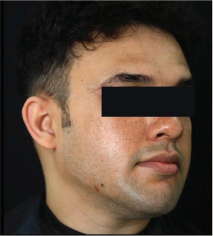
A 36-year-old male with Fitzpatrick Skin Type IV presented with the chief concern of dull and uneven skin tone localized to the anterior and lateral neck region. He expressed a desire to improve overall skin quality and brightness. The patient reported no significant history of dermatological or systemic illness but mentioned outdoor occupational exposure with minimal sun protection, contributing to progressive tanning and pigmentation. Clinical examination revealed visibly uneven skin tone
across the face and neck with slightly coarse texture, mild follicular prominence, and a few open pores. No signs of active dermatoses or post-inflammatory hyperpigmentation were observed. Based on clinical findings, a diagnosis of extrinsic photo-induced pigmentation with associated dullness of the neck region, along with early signs of skin aging and dehydration, was made.
The patient was initiated on a combination intravenous therapy comprising glutathione (1200 mg) and Vitamin C (100 mg), administered weekly in a single infusion. Glutathione was used for its antioxidant and depigmenting properties. Hyaluronic acid was administered intradermally and was intended to enhance skin hydration, plumpness, and overall smoothness. The treatment protocol spanned six weeks with a total of six sessions for Glutathione injections and 1 session of injectable Hyaluronic acid. The ‘before’ image documented a relatively dull and tanned skin with uneven pigmentation extending to the jawline. The ‘after’ image, captured post-therapy, showed marked improvement in skin tone, with visibly brighter, more hydrated, and even-toned skin. The patient also reported subjective enhancement in skin texture and glow, with no adverse effects or allergic reactions during the course of treatment.
Before treatment
After treatment
Figure 1: After six weeks of treatment marked improvement in skin tone, with visibly brighter
Mechanism of Action
Glutathione exerts its skin-brightening effects through multiple, well-characterized pathways. The primary mechanism involves the inhibition of tyrosinase, a copper-dependent enzyme that plays a central role in melanin synthesis. Glutathione directly binds to the copper ions at the active site of tyrosinase, thereby reducing the enzyme’s activity and slowing down the production of eumelanin, the darker form of melanin. Additionally, glutathione facilitates the conversion of eumelanin to pheomelanin—a lighter, yellowred pigment—by promoting the conjugation of cysteine with dopaquinone. This shift in melanin production from dark to light pigments contributes significantly to the overall skin-brightening effect.
Moreover, glutathione modulates melanocyte function through downregulation of microphthalmia-associated transcription factor (MITF), which controls both tyrosinase expression and melanocyte proliferation. By inhibiting MITF, glutathione helps maintain melanocyte homeostasis and suppresses excessive melanin production. Beyond its direct impact on melanin synthesis, glutathione’s antioxidant and anti-inflammatory properties further enhance its depigmenting potential. It neutralizes reactive oxygen species (ROS) that contribute to melanogenesis, particularly under UV exposure, and inhibits inflammatory mediators such as interleukin-6 (IL-6) and prostaglandins that can exacerbate post-inflammatory hyperpigmentation. When compared to conventional depigmenting agents like hydroquinone and kojic acid—which primarily act through tyrosinase inhibition—glutathione offers a broader spectrum of activity, combining antioxidant defense, anti-inflammatory action, and melanogenesis regulation. These multifaceted effects make it a compelling candidate in the management of pigmentation disorders, especially in individuals prone to inflammation-induced hyperpigmentation.1,2,3,4,5
Discussion
Glutathione, a naturally occurring thiol-tripeptide, plays a central
role in maintaining cellular redox homeostasis and has garnered attention for its potential as a systemic skin-brightening agent. Its use in cosmetic dermatology has been popularized, particularly in certain ethnic populations, due to its perceived antimelanogenic properties. However, there remains a disparity between marketing claims and the current scientific evidence supporting its efficacy and safety. While oral and topical formulations of glutathione have demonstrated favorable safety profiles and some degree of efficacy in reducing pigmentation, the intravenous (IV) route, though widely used, remains controversial due to safety concerns and limited clinical validation. Several randomized controlled trials (RCTs) have demonstrated the potential of oral and topical glutathione for modest skinbrightening effects. Arjinpathana et al. and Handog et al. reported significant reductions in melanin indices in Thai and Filipino women, respectively, using oral and buccal forms of glutathione. Topical formulations, such as 2% oxidized glutathione (GSSG), have also shown beneficial outcomes in reducing pigmentation over short-term applications. These studies, although promising, suffer from limitations including small sample sizes, short followup durations, and lack of biochemical monitoring of systemic glutathione levels.1,2,3,4,5 While exact standardized doses are yet to take shape, glutathione continues to gain popularity due to increasing demand for “safer” alternatives to traditional depigmenting
agents like hydroquinone and kojic acid. Nevertheless, there is a pressing need for largescale, double-blind, placebocontrolled trials with long-term follow-up to determine its true efficacy, safety, optimal dosage, and maintenance protocols. Until such evidence becomes available, the use of IV glutathione for cosmetic purposes should be approached with caution and limited to closely monitored clinical environments.1,2,3,4,5
Safety and Limitations of Glutathione
Safety Status:
Oral glutathione (GSH) supplements are generally recognized as safe (GRAS) under Section 201(s) of the Federal Food, Drug, and Cosmetic Act by the United States Food and Drug Administration (USFDA). In countries such as the United States, the Philippines, and Japan, there are no regulatory restrictions on the oral use of glutathione. In India and several other Asian countries, oral glutathione is available as an over-thecounter (OTC) supplement. While considered safe for
References
1. Alzahrani T F, Alotaibi S M, Alzahrani A A, et al. (January 27, 2025) Exploring the Safety and Efficacy of Glutathione Supplementation for Skin Lightening: A Narrative Review. Cureus 17(1): e78045. doi:10.7759/cureus.78045
2. Sitohang IBS, Ninditya S. Systemic Glutathione as a Skin-Whitening Agent in Adult. Dermatol Res Pract. 2020;2020:8547960. Published 2020 Apr 24. doi:10.1155/2020/8547960
general use, oral glutathione has low bioavailability, limiting its effectiveness. To achieve faster and more noticeable skinbrightening results, intravenous (IV) administration is often preferred, as it allows for higher systemic concentrations. The commonly recommended IV dose ranges between 600 to 1200 mg, administered once or twice weekly. However, there is currently limited robust scientific evidence supporting the clinical efficacy of IV glutathione, particularly for skin brightening.5
Limitations:
Despite its popularity, the use of intravenous glutathione comes with several limitations. One significant drawback is the high cost of IV formulations, which may limit accessibility. Furthermore, there is a lack of standardized protocols regarding optimal dosage, frequency, and duration of treatment. The mechanism of action for skin brightening through IV glutathione remains poorly understood. Importantly, IV glutathione has not been approved by the US-FDA for skin brightening purposes, and health authorities such as the
FDA of the Philippines have issued cautionary advisories against its use due to potential risks. Reported adverse effects, although relatively rare, include skin rashes, abdominal pain, and in some cases, serious hypersensitivity reactions. The absence of large-scale, wellcontrolled clinical trials further underscores the need for caution and more research to establish both the efficacy and long-term safety of intravenous glutathione.5
Conclusion
In conclusion, the combination of intravenous glutathione and hyaluronic acid proved effective and well-tolerated for cosmetic face brightening in this case. The synergistic benefits of antioxidant defense and dermal hydration likely contributed to the observed aesthetic improvements. For long-term maintenance, monthly IV infusions of glutathione and hyaluronic acid were recommended, along with the regular use of broad-spectrum sunscreen (SPF 50+) on the face and other exposed areas.
3. Sonthalia S, Jha AK, Lallas A, Jain G, Jakhar D. Glutathione for skin lightening: a regnant myth or evidence-based verity? Dermatol Pract Concept. 2018;8(1):1521. DOI: https://doi.org/10.5826/ dpc.0801a04
4. Mohan S, Mohan L, Sangal R, Singh N. Glutathione for skin lightening for dermatologists and cosmetologists. Int J Res Dermatol 2020;6:284-7.
5. Mahmood, Methaq. (2022). The Effectiveness of Glutathione on Skin Lightening: A Review.
Kesty Redness Scale Demonstrated to Improve Clinical Assessment of Facial Erythema,
Study Finds
A recent study introduced and validated the Kesty Redness Scale (KRS), an ordinal clinical assessment tool developed to objectively quantify facial erythema across a continuum of severity, ranging from clear skin to marked, feature-obscuring redness. Aimed at addressing the lack of a standardized, userfriendly instrument suitable for both aesthetic and general dermatologic practice—where diseasespecific grading systems often lack applicability—the KRS was evaluated through a prospective observational study. A diverse array of facial images was assessed by a panel of clinicians, including Dermatologists and plastic surgeons. The scale demonstrated excellent inter-rater reliability, with high statistical agreement observed across multiple metrics. Bland–Altman analysis revealed minimal systemic bias, reinforcing the scale’s validity. Participants uniformly found the KRS to be intuitive, practical, and readily integrable into clinical workflows. The study concludes that the KRS is a robust and accessible tool for the standardized assessment of facial erythema, with promising utility in both clinical and research environments. Future directions include the integration of artificial intelligence for automated image-based scoring, which may reduce inter-observer variability and enhance efficiency.
An innovative handpiece that combines radiofrequency (RF) and vacuum technology has been shown to be effective for facial contouring.
A novel handpiece combining radiofrequency (RF) and vacuum technology has demonstrated safety and efficacy in nonsurgical facial contouring. The device utilizes bipolar, channeling-optimized RF energy (CORE technology) with vacuum suction to target various tissue depths, promoting fat reduction, collagen stimulation, and improved circulation. A multicenter retrospective study involving female participants with varying degrees of skin laxity and localized fat showed noticeable improvements in facial contour and skin firmness after multiple treatment sessions. A blinded evaluator confirmed
visible improvements in the majority of before-and-after images. Histological analysis revealed increased fibroblast activity and no signs of thermal injury or adverse effects. Mild erythema and transient edema were the only reported side effects. The device was well-tolerated across diverse skin types and body compositions, with consistent outcomes regardless of age or BMI. Although limited by sample size and follow-up duration, the findings support the clinical value of this technology in aesthetic dermatology.
Microneedle Technology Shows Promise for Treating Nodular Acne
Nodular acne presents challenges for conventional treatments due to limited drug penetration and systemic side effects. Research has explored the use of dissolvable microneedles (MNs) for targeted clindamycin delivery, aiming to overcome the limitations of topical and systemic therapies. This approach localizes the antibiotic directly to the sebaceous glands, improving efficacy while minimizing systemic exposure. Using a chitosan, PVP, and PVA-based formulation, the researchers developed microneedles with robust mechanical properties and optimal penetration capacity. In vitro testing demonstrated the microneedles’ ability to penetrate the dermis and release clindamycin rapidly, achieving a complete release in three minutes. The study suggests that PVA-based dissolvable MNs offer a promising, localized treatment for nodular acne, with potential advantages over traditional therapies. However, further studies are required to confirm these findings and assess clinical applicability.
Genetic Insights Reveal Potential Therapeutic Target for Vitiligo
A recent Mendelian randomization (MR) study identified a potential protective effect of proprotein convertase subtilisin/kexin type 9 (PCSK9) inhibition against vitiligo. Genetic variants mimicking the pharmacologic action of PCSK9 inhibitors were associated with reduced risk of developing vitiligo. This association was not observed with 3-hydroxy-3-methylglutaryl-coenzyme A reductase (HMGCR) or Niemann–Pick C1-like 1 (NPC1L1) inhibition and appeared to be independent of lipid parameters such as low-density lipoprotein cholesterol (LDL-C) and triglycerides—suggesting a non-lipid-mediated mechanism. Proteomic analysis identified differential expression of proteins potentially mediating this effect. Elevated levels of C-X-C motif chemokine ligand 12 (CXCL12), cellular communication network factor 5 (CCN5), Fc receptor–like protein 1 (FCRL1), and legumain (LGMN) were linked to increased vitiligo risk. In contrast, fibroblast growth factor 2 (FGF2), which supports melanocyte survival, was suppressed by PCSK9 activity and associated with reduced risk when upregulated. These findings suggest PCSK9 inhibitors may hold promise for vitiligo therapy through immune pathway modulation. Further clinical and mechanistic studies are warranted.
Dermal Fillers: The Preferred Solution for Lip Enhancement
Dr. Jyoti Aneja MD
Founder & Medical Director
La Grace Luxury Skin Clinic Mumbai
Introduction
The lips are a central component of facial aesthetics, contributing significantly to perceived attractiveness and emotional expression. Alongside the eyes, they are regarded as one of the most visually captivating and defining features in both men and women. Lip morphology— including projection, proportion and volume—plays a pivotal role in facial harmony. A fuller vermilion display and gently projected lips are often associated with youth, vitality and sensuality, particularly in women. In classically attractive facial profiles, the upper lip typically exhibits slightly more projection than the lower lip, independent of gender. With age, however, the lips and perioral region undergo progressive anatomical changes. Intrinsic factors such as genetics and extrinsic elements like ultraviolet exposure and smoking accelerate collagen degradation, leading to volume depletion, diminished vermilion show and philtral elongation. These alterations contribute to a flattened lip contour, deepening of the mentolabial folds and the formation of perioral rhytides (fine perioral lines).
Furthermore, individuals with inherently thin lips or structural asymmetries frequently seek aesthetic enhancement to achieve balanced, harmonious proportions and a more youthful appearance.¹,²
These changes often lead to altered facial contours, primarily due to volume loss.
In recent years, the demand for minimally invasive cosmetic procedures has surged, driven by the desire to enhance attractiveness with minimal downtime. Advances in aesthetic techniques and the development of safe dermal fillers have enabled subtle yet effective modifications to facial contours, delivering natural-looking results. Lip augmentation, in particular, is primarily sought for beautification rather than rejuvenation. Achieving optimal results in perioral rejuvenation requires a sophisticated approach that combines multiple technologies and injectable treatments to restore volume and redefine lip contours.1,2
A variety of fillers, both temporary and permanent, have been utilized for lip augmentation with largely satisfying results.
However, complications such as granulomas have been reported, particularly with permanent fillers. Among the available options, hyaluronic acid (HA) and polyacrylamide (PA) are widely known fillers. HA has become the preferred choice due to its biodegradable nature, high patient satisfaction, longer-lasting effects, and lower risk of complications.1,2
Extensive global use and published clinical studies have confirmed the efficacy and safety of HA fillers, making them a cornerstone
in modern lip augmentation. They are now widely regarded as a safe, predictable and minimally invasive option for lip enhancement, allowing individuals to achieve youthful and aesthetically pleasing lips while maintaining natural expressions.1,2
Lip Anatomy3
Injection Technique for Lip Augmentation2,4
Anesthesia and Preparation: Lip augmentation begins with topical anesthesia, typically using a eutectic mixture of lidocaine and prilocaine or regional block. It is important to avoid distorting the shape of the lips during the procedure. Premixed HA fillers with lidocaine can help minimize discomfort. Swelling and bruising are common postprocedure effects, requiring cold compresses, NSAIDs (avoided in the first six hours to prevent masking vascular compromise), or even prednisolone for relief. Injection Technique: HA
is commonly used for lip augmentation due to its safety and effectiveness. HA with a 3/6 cross-linking ratio and a concentration of 25 mg/g is used. For volume restoration, HA is injected into the vermilion border at a 30-degree angle, no deeper than 2.5 mm, to avoid blood vessels. Small boluses (0.05–0.1 ml) are injected at each point, with a total of 0.5-1 ml for both lips per session. The injection technique follows a retrograde linear threading approach.
For shaping the lips, HA is injected into the vermilion border, avoiding the white roll to prevent a blunted lip margin. A smaller amount (0.02–0.4 ml) is injected slowly using the same retrograde linear threading technique. Lips should be assessed for asymmetry, and 0.05–0.1 ml of HA can be injected in specific areas to correct unevenness.
Lip Categorization for Augmentation:
1. Normal-Volume Lips: Patients seeking more projection will have HA injected into the vermilion (0.5ml). After 15 days, reassess and add more volume if needed.
2. Thin Lips: For atrophic lips, inject 0.5–1 ml of HA into the thinner lip to achieve the desired ratio. After ward, define the lips by injecting
Dermal
0.02–0.4 ml into the vermilion border. Sessions should be spaced 30 days apart.
3. Aged Thin Lips: With volume loss and vertical lines, inject HA into the vermilion to restore volume and into the vermilion border for definition. Use 0.5–1 ml per session with intervals of 30 days.
Post-Procedure Care:
• Lightly massage the treated area.
• Cleanse the skin with saline (0.9%).
• Apply ice packs to reduce pain, swelling and bruising.
• Avoid using a straw, kissing, strenuous exercise, hot temperatures, steam, sauna and hot drinks for 24 hours.
Follow-up and Complications:
A follow-up session is scheduled 15 days after the procedure, with a third session if necessary after 30 days. If signs of intravascular complication (such as blanching or pain) occur, treat with hyaluronidase, massage, warm compresses, and aspirin until capillary refill normalizes.
Complications: Expected post-procedure effects include edema, bruising and ecchymosis. While rare, complications like nodules, lumps or vascular compromise can occur. These are generally managed with massage, hyaluronidase injections, warm compresses and topical nitroglycerine. To prevent herpetic reactivation, oral antivirals (acyclovir, famciclovir or valaciclovir) may be prescribed.
Technique Precision: Proper technique is essential to avoid complications. Poor product placement or uneven distribution
can lead to lumps, nodules, or beading. Overcorrection should be avoided. Simultaneously augmenting both the cupids bow and the vermilion border ensures a balanced result. Techniques such as serial puncture and linear threading (antegrade or retrograde) are chosen based on patient needs and aesthetic goals. The use of cannulas has reduced downtime and improved patient satisfaction, allowing for a single entry point at each oral commissure to access both lips. Beginners are advised to start with needles before transitioning to cannulas for more refined results.
Patient 1 Patient 2
Dermal
Before treatment
After treatment
References
1. de Queiroz Hernandez, P.M., Cotrin, P., Valarelli, F.P. et al. Evaluation of the attractiveness of lips with different volumes after filling with hyaluronic acid. Sci Rep 13, 4589 (2023). https://doi. org/10.1038/s41598-023-31332-1
2. Luthra, Amit. “Shaping Lips with Fillers.” Journal of cutaneous and aesthetic surgery vol. 8,3 (2015): 139-42. doi:10.4103/0974-2077.167269
3. Cooper H, Gray T, Fronek L, et al. Lip augmentation with hyaluronic acid fillers: A review of considerations and techniques. J Drugs Dermatol. 2023;22(1):23-29. doi: 10.36849/JDD.6304.
4. Keramidas E, Rodopoulou S, Gavala MI. A Safe and Effective Lip Augmentation Method: The Step-by-Step Φ (Phi) Technique. Plast Reconstr Surg Glob Open. 2021 Feb 2;9(2):e3332. doi: 10.1097/
Conclusion
Lip augmentation using injectable fillers, particularly hyaluronic acid (HA), offers quick results with minimal downtime. HA is widely used due to its ease of use, availability, and public acceptance. The procedure allows for controlled and predictable results by targeting specific anatomical areas of the lip. While treatment decisions may sometimes be influenced by budget, it’s important to avoid compromising optimal results. Achieving the best aesthetic outcome requires skilled injection techniques, knowledge of lip anatomy, and an individualized approach tailored to each patient’s needs.
Glowing Through Summer: Skincare Starts with Sunscreen
Dr. Siddhi Chavan
MBBS, DDVL
Consultant Dermatologist and Aesthetic Physician
Mumbai
Introduction
Sunscreens are vital in dermatology, offering protection against both
immediate and long-term effects of ultraviolet (UV) radiation. They help prevent conditions such as sunburn, freckling, discoloration, photoaging, and significantly reduce the risk of skin cancer. Additionally, sunscreens are crucial in managing phototoxic and photoallergic reactions, as well as photosensitivity disorders like polymorphous light eruption, solar urticaria, chronic actinic dermatitis, persistent light reaction, lupus erythematosus, xeroderma pigmentosum, and albinism. They also play a significant role in mitigating photoaggravated dermatoses and addressing post-inflammatory hyperpigmentation, particularly following dermatological procedures. Incorporating sunscreens into daily skincare routines ensures effective photoprotection and supports optimal therapeutic outcomes.
The effects of UV radiation on the skin are influenced by factors
such as the intensity of the source, duration of exposure, UV wavelength, and skin pigmentation levels. Sunlight comprises approximately 95% UVA (315–400 nm) and 5% UVB (280–315 nm) radiation. UVB is more effective at causing erythema (sunburn), while UVA primarily contributes to skin photoaging. Both UVA and UVB have been implicated in skin cancer development, with individuals having lower melanin levels being more vulnerable.
Sunscreens are available in various forms, including lotions, creams, sprays, and sticks, and can be categorized based on their active ingredients as either physical blockers or chemical absorbers. Physical blockers, such as titanium dioxide (TiO2) and zinc oxide (ZnO), reflect or scatter UV radiation. Chemical absorbers, including avobenzone, octyl salicylate, oxybenzone (BP3), and octinoxate (OMC), absorb UV radiation energy and release it as heat. Some formulations combine
both types of ingredients for enhanced protection.
The Sun Protection Factor (SPF) measures the level of UVB protection a sunscreen offers. Dermatologists recommend using a sunscreen with an SPF of at least 30, which blocks approximately 97% of the sun's UVB rays. Higher-number SPFs block slightly more UVB rays, but no sunscreen can block 100% of them. It's important to note that SPF pertains primarily to
UVB protection, and consumers should look for broad-spectrum sunscreens that also protect against UVA radiation.
Effective sun protection involves more than just sunscreen application. Health agencies recommend multiple protective measures, including wearing protective clothing, hats, and sunglasses, seeking shade, and avoiding outdoor exposure when the UV Index is 3 or higher. Sunscreen serves as an additional protective measure in situations where other options are impractical.
Understanding SPF and other key aspects of sunscreen formulation can help individuals make informed choices for optimal skin health. Incorporating sunscreens into daily skincare routines, alongside other protective measures, ensures comprehensive photoprotection and supports overall dermatological well-being.
Glowing Through Summer: Skincare Starts with Sunscreen
Understanding SPF: What Does It Really Mean?
Sun Protection Factor (SPF) measures a sunscreen's ability to protect the skin from UVB rays, which are primarily responsible for sunburn and contribute to skin cancer. The SPF number indicates how much longer it takes for untanned skin to start to redden with sunscreen applied compared to how long it takes to start reddening without it. For example, an SPF 30 sunscreen theoretically allows a person to stay in the sun 30 times longer than if they were not wearing sunscreen. However, it's important to note that no sunscreen can block 100% of UVB rays; SPF 15 blocks approximately 93% of UVB radiation, SPF 30 blocks 97%, SPF 50 blocks 98%, and SPF 100 stops 99% of UVB rays from reaching your skin. Additionally, SPF does not measure protection against UVA rays, which can also contribute to skin damage and aging. Therefore, for comprehensive protection, it's essential to use a broadspectrum sunscreen that guards against both UVA and UVB radiation.
SPF Ratings and Protection
Levels:
• SPF 15: Blocks about 93% of UVB rays
• SPF 30: Blocks about 97% of UVB rays
• SPF 50: Blocks about 98% of UVB rays
• SPF 100: Blocks about 99% of UVB rays
While higher SPF values provide slightly more protection,
no sunscreen can block 100% of UV rays. Additionally, a higher SPF does not mean the protection lasts longer; reapplication is still necessary.
While SPF measures protection against UVB rays, UVA rays also pose significant risks. UVA rays penetrate deeper into the skin, leading to premature aging, DNA damage, and an increased risk of skin cancer. Unlike UVB rays, UVA is present throughout the day and can pass through glass and clouds. Broad-spectrum sunscreens protect against both UVA and UVB rays, ensuring comprehensive skin defense.
Chemical vs. Physical Sunscreens: Which One to Choose?
Sunscreens are vital in protecting the skin from the harmful effects of ultraviolet (UV) radiation, which can lead to sunburn, premature aging, and skin cancer. They are broadly categorized into two types: chemical (organic) and physical (mineral) sunscreens, each with distinct mechanisms of action, benefits, and considerations.
UVA vs. UVB Protection: Why Broad-Spectrum Sunscreens Matter
Chemical Sunscreens
Chemical sunscreens contain organic compounds such as oxybenzone, avobenzone, octisalate, and octinoxate. These ingredients absorb UV radiation, convert it into heat, and release it from the skin. This process helps prevent UV rays from penetrating deeper into the skin layers.
Pros:
• Typically have a lightweight consistency, making them easy to apply and suitable for daily use.
• Blend seamlessly into the skin without leaving a visible residue, which is advantageous for those seeking an invisible finish.
• Often formulated to be water-resistant, providing sustained protection during activities like swimming or sweating.
Cons:
• May cause skin irritation or allergic reactions, especially in individuals with sensitive skin.
• Some chemical ingredients, such as oxybenzone, have been associated with environmental concerns, particularly regarding coral reef health.
• Require approximately 15 to 30 minutes after application to become effective, necessitating advance planning before sun exposure.
Physical (Mineral) Sunscreens
Physical sunscreens utilize mineral-based ingredients like zinc oxide and titanium dioxide to create a protective barrier on the skin's surface. This barrier reflects and scatters both UVA and UVB rays, preventing them from penetrating the skin.
Pros:
• Provide immediate protection upon application, eliminating the need to wait before sun exposure.
• Less likely to cause skin irritation, making them suitable for sensitive skin, including individuals with conditions like rosacea.
• Offer broad-spectrum protection against both UVA and UVB rays, ensuring comprehensive defense against sun damage.
Cons:
• Often have a thicker texture, which can leave a visible white cast on
the skin, particularly noticeable on darker skin tones.
• May be less water-resistant compared to chemical sunscreens, requiring more frequent reapplication during water activities.
• Can be more challenging to spread evenly over the skin, potentially leading to areas with inadequate coverage.
Choosing the Right Sunscreen
The choice between chemical and physical sunscreens depends on individual preferences, skin type, and specific needs:
• Skin Sensitivity: Individuals with sensitive skin or conditions like eczema may prefer physical sunscreens due to their lower likelihood of causing irritation.
• Cosmetic Preferences: Those seeking a lightweight, invisible finish might opt for chemical sunscreens, while being mindful of potential skin reactions.
• Environmental Considerations: If environmental impact is a concern, selecting sunscreens free from certain chemical ingredients known to harm marine ecosystems is advisable.
• Activity Level: For waterbased activities, choosing a water-resistant formula is crucial, regardless of the type.
Regardless of the type chosen, applying sunscreen generously and reapplying every two hours, or more frequently if swimming or sweating, is essential for effective protection. Additionally, incorporating other sun safety measures, such as wearing protective clothing, seeking shade, and avoiding peak sun hours, enhances overall skin protection.
Glowing Through Summer: Skincare Starts with Sunscreen
Water Resistance and Reapplication Guidelines
Water-resistant sunscreens are essential for maintaining skin protection during activities involving water exposure or excessive sweating. The U.S. Food and Drug Administration (FDA) mandates that sunscreens claiming water resistance must specify on their labels whether they remain effective for 40 minutes or 80 minutes while swimming or sweating, based on standardized testing procedures. To ensure continuous protection, it's crucial to reapply sunscreen at appropriate intervals. For those wearing makeup, reapplying sunscreen can be challenging. Dermatologists suggest using mineralbased powder sunscreens designed to be applied over makeup, providing an easy way to maintain protection without disrupting cosmetic application.
Common Myths about Sunscreen
1. Myth: Higher SPF means all-day protection.
• Reality: Even SPF 100 needs reapplication every two hours.
2. Myth: Darker skin tones don’t need sunscreen.
• Reality: While melanin provides some natural protection, it is not enough to prevent UV damage or skin cancer.
3. Myth: Sunscreen is unnecessary on cloudy days.
• Reality: Up to 80% of UV rays can penetrate clouds, causing skin damage even when the sun is not visible.
4. Myth: Makeup with SPF is enough.
• Reality: The SPF in makeup is often too low and not applied in sufficient quantity to provide adequate protection.
The Environmental Impact of Sunscreens
Certain sunscreen ingredients, notably oxybenzone and octinoxate, have been linked to coral reef damage. These chemicals can cause coral bleaching, disrupt coral reproduction, and damage coral DNA. Reef-safe sunscreens are formulated with non-nano zinc oxide and titanium dioxide as environmentally friendly alternatives. The term "reef safe" typically indicates that the sunscreen contains only mineral UV-blocking ingredients like zinc oxide and titanium dioxide. Non-nano particles are larger than 100 nanometers, reducing the likelihood of ingestion by marine life. When selecting a reef-safe sunscreen, it's advisable to choose products labeled as containing non-nano zinc oxide or titanium dioxide and free from harmful chemicals like oxybenzone and octinoxate. This choice helps protect both our skin and the delicate ecosystems of coral reefs.
Sunscreen and Vitamin D: Striking a Balance
There is concern that excessive sunscreen use may contribute to vitamin D deficiency. However, studies suggest that typical sunscreen application does not completely block vitamin D synthesis. To maintain healthy levels, individuals can consume vitamin D-rich foods, take supplements, or get limited sun exposure under medical guidance.
Conclusion: Making Sunscreen a Daily Habit
Choosing the right sunscreen goes beyond just SPF— it involves considering UVA protection, skin type, and lifestyle factors. The key to
effective sun protection is consistent use and proper application. Whether opting for a chemical or physical formula, broad-spectrum coverage with at least SPF 30 is ideal for daily wear. By integrating sunscreen into a daily skincare routine, individuals can significantly reduce the risks of sun damage, premature aging, and skin cancer while maintaining healthy, radiant skin.
Glowing Through Summer: Skincare Starts with Sunscreen
Dermatological Insights into Summer Skin Disorders and Their Management
Dr. Sweta Nakhawa MD (Dermatology)
Founder & Director of HL Aesthetic Skincare Clinic
Dermatologist, Cosmetologist and Aesthetic Physician
Mumbai
Introduction
Summer brings high temperatures, humidity, and prolonged sun exposure, which can lead to various skin conditions. Dermatologists frequently encounter cases of melasma, heat rash, sunburn, fungal infections, and allergic reactions during this season. Understanding their causes and implementing effective treatment strategies is essential for optimal patient care.
Understanding Melasma and Its Summer Exacerbation Summer is a challenging season for individuals prone to melasma, as increased ultraviolet (UV) radiation intensifies melanin production. Even brief sun exposure can lead to pigmentation flare-ups, making diligent sun protection a crucial preventive strategy. Additionally, hormonal fluctuations due to pregnancy, menopause, or contraceptive use can further contribute to melasma’s persistence. Melasma is a common pigmentation disorder presenting as brown or grayish patches, primarily on sunexposed areas like the forehead, cheeks, nose, and upper lip.
While not harmful or contagious, melasma can cause significant cosmetic distress, particularly in summer when its severity often worsens. The primary culprit behind melasma is excessive melanocyte activity, triggered by factors such as prolonged sun exposure, hormonal changes, genetic predisposition, and certain medications or skincare treatments.
Preventing and Managing Melasma During Summer
A proactive and comprehensive approach is essential to prevent and manage melasma effectively during summer. Key strategies include:
1. Sun Protection:
• Apply a broad-spectrum sunscreen (SPF 30+ or higher) daily, with reapplication every two hours when outdoors.
• Use physical sunscreens containing zinc oxide or titanium dioxide for enhanced protection.
• Wear wide-brimmed hats, UV-blocking sunglasses, and protective clothing to minimize direct sun exposure.
• Seek shade whenever possible, especially during peak sun hours (10 AM – 4 PM).
2. Skin-Friendly Summer Skincare:
• Incorporate antioxidants like vitamin C and E to neutralize free radicals and prevent oxidative damage.
• Use gentle cleansers, hydrating serums, and non-comedogenic moisturizers to maintain skin barrier integrity.
• Avoid harsh chemical peels or aggressive exfoliation, which can worsen pigmentation and cause irritation.
3. Treatment Options for Persistent Melasma:
• Topical Agents: Hydroquinone, kojic acid, azelaic acid, and niacinamide help reduce pigmentation and prevent further melanin production.
• Advanced Dermatological Treatments: For resistant cases, dermatological interventions like chemical peels, microneedling, laser therapies and microdermabrasion can accelerate skin renewal and pigment reduction.
A holistic approach combining rigorous sun protection, a wellbalanced skincare routine, and professional treatments ensures optimal melasma control during the summer months. By adopting these measures, individuals can maintain clearer, more even-toned skin despite seasonal challenges.
Other Summer Skin Conditions
• Heat rash
One of the most common summer skin issues is heat rash, also known as miliaria. It occurs when sweat ducts become blocked due to excessive sweating in hot and humid conditions, leading to irritation. This condition is particularly prevalent among infants, athletes, and individuals who wear occlusive clothing. The symptoms vary depending on the severity, ranging from small, red, itchy papules (miliaria rubra) to clear, fluid-filled vesicles (miliaria crystallina) and even pustular lesions in severe cases (miliaria pustulosa). Treatment involves cooling the skin by staying in air-conditioned environments and wearing loose, breathable clothing. Patients can benefit from topical agents like calamine lotion and mild c orticosteroids, while antibacterial agents may be needed in cases of secondary infections. Preventing excessive sweating through the use of moisture-wicking fabrics and frequent cool showers is also essential to avoid recurrence.
• Sunburn
Another prevalent summer skin concern is sunburn, which results from over exposure to ultraviolet (UV) radiation. Sunburn leads to skin inflammation and DNA damage, manifesting as redness, pain, and peeling of the skin. In severe cases, patients may experience blistering and systemic symptoms such as fever. Immediate care involves using cold compresses, applying aloe vera gel, and taking anti-inflammatory agents like ibuprofen to alleviate pain and inflammation. Maintaining adequate hydration by drinking plenty of fluids and using topical moisturizers helps restore the skin barrier. Prevention remains the most effective strategy, and dermatologists should emphasize the importance of broadspectrum sunscreens with SPF 50+, wearing protective clothing, and limiting sun exposure during peak hours to re duce the risk of sun damage.
• Fungal infections
Fungal infections, particularly tinea and candidiasis, are also common in summer due to warm and humid conditions that promote fungal overgrowth in occluded areas such as the skin folds, feet, and groin. These infections typically present as itchy, red, scaly patches in tinea cases or macerated, white patches with discomfort in candidiasis. Treatment involves the application of topical antifungal agents such as clotrimazole, terbinafine, or ketoconazole, while oral antifungals like fluconazole or itraconazole may be necessary for more widespread infections. Preventative measures such as keeping the skin dry, wearing loose-fitting clothing, and using antifungal powders can significantly reduce the risk of developing these infections.
• Allergic reactions
Polymorphous light eruption (PMLE) is another common skin reaction seen during summer. It is caused by a delayed hypersensitivity reaction to UV radiation, primarily affecting individuals with sun sensitivity. Patients present with red, itchy papules, vesicles, or plaques on sun-exposed areas. The best way to manage PMLE is through strict sun avoidance, including the use of protective clothing and broad-spectrum sunscreen. For mild cases, topical steroids help reduce inflammation, while oral antihistamines can alleviate itching and discomfort associated with the condition.
• Heat dermatitis and contact dermatitis
Another summer-related dermatological issue is prickly heat dermatitis and contact dermatitis, which are triggered by excessive sweating, chemical-laden sunscreens, or insect repellents. These conditions present as itchy, red rashes, sometimes accompanied by blistering or oozing. Management focuses on identifying and avoiding irritants, recommending fragrance-free skincare products, and choosing breathable fabrics. Topical corticosteroids may be used to control inflammation, while cool compresses provide relief from irritation and discomfort.
• Summary
In summary, heat rash and other summer-related skin conditions can significantly impact a patient’s quality of life. Dermatologists should focus on preventive measures, recommend appropriate skincare routines, and implement targeted treatment approaches to manage these conditions effectively. Emphasizing the importance of sun protection, proper hygiene, and breathable clothing can help prevent summer skin ailments and ensure overall skin health during hot weather.
"Professional treatment advice emphasizes the importance of preventative and maintenance measures for lasting results and overall well-being."
CO2 Lasers in Skin Resurfacing: Key Applications in Aesthetic Dermatology
Dr. Ravindra Bhite MBBS, DVD Dermatologist
Dr. Bhite Skin Cosmetology Laser Clinic
Shrirampur, Maharashtra
Introduction
In dermatology, CO2 is most commonly utilized in the form of a laser—CO2 laser therapy—which has become an invaluable tool for a wide range of skin treatments. The CO2 laser emits an infrared wavelength that is preferentially absorbed by water in the skin, facilitating highly precise tissue ablation and vaporization. This unique property makes it particularly effective for targeting the skin’s surface layers and deeper dermal structures, making it ideal for both cosmetic and medical Dermatologic procedures. The CO2 laser operates at a wavelength of approximately 10,600 nanometres, which is efficiently absorbed by water molecules present in the skin. This absorption results in the vaporization of the targeted tissue, making it useful for procedures requiring the removal of skin lesions or resurfacing of damaged skin. Due to its ability to cut, coagulate, and vaporize tissue, CO2 laser therapy is regarded as one of the most versatile tools in modern Dermatology.1
The CO2 laser functions by delivering concentrated light energy to targeted areas of the
skin, enabling precise thermal effects for tissue ablation and regeneration. When this light is absorbed by the water in the skin's cells, it converts into heat, which causes the cells to vaporize. The precision of the CO2 laser allows for selective tissue removal without affecting the surrounding healthy skin. This feature is particularly valuable in Dermatological treatments where minimal damage to adjacent tissues is desired. Additionally, the thermal energy generated by the CO2 laser stimulates collagen production in the deeper layers of the skin, promoting skin tightening and rejuvenation.
The two primary modes of CO2 laser operation—ablative and fractional—offer different benefits depending on the specific needs of the patient and the skin condition being treated. In the ablative mode, the laser removes entire layers of skin, making it highly effective for deep skin resurfacing and the treatment of significant skin imperfections such as deep wrinkles, scars, and severe sun damage. In contrast, fractional CO2 lasers treat the skin in a grid pattern, leaving micro-columns
of untreated skin between treated areas, promoting faster healing and less downtime while still delivering effective results for conditions like mild to moderate photo-aging and acne scarring.1, 2
CO2 lasers have been used in medicine for several decades, with Dermatology being one of the first fields to adopt this technology. Initially, CO2 lasers were primarily employed for surgical procedures due to their precision and ability to cut tissue with minimal bleeding. As technology progressed, Dermatologists recognized the versatility of CO2 lasers in addressing various skin conditions including wrinkles, acne scars, age spots, seborrheic keratosis, and other benign neoplasms. The laser’s ability to precisely ablate abnormal tissue has made it effective in the excision of precancerous lesions, such as actinic keratosis, and benign growths, including warts and skin tags. Additionally, CO2 laser therapy plays a pivotal role in non-surgical skin tightening, offering an effective alternative to facelifts for patients seeking improvement in skin laxity and elasticity, particularly in areas such as the jawline, neck, and periocular regions.2
The development of fractional CO2 laser technology has significantly expanded the clinical applications of CO2 lasers, offering a non-invasive option for patients seeking cosmetic improvements with minimal downtime. This innovation enables controlled micro-ablative treatments that target small columns of tissue while leaving the surrounding
skin intact. As a result, fractional CO2 lasers promote faster recovery and minimize patient downtime, all while still achieving significant clinical outcomes. By allowing for more precise tissue targeting, fractional CO2 technology has broadened the range of conditions that can be effectively treated with minimal invasiveness, making it an ideal choice for patients seeking both cosmetic and therapeutic benefits.3
Overall, CO2 laser therapy has become a cornerstone in Dermatology, offering a wide range of therapeutic and cosmetic benefits, from improving skin texture and tone to removing lesions and scars, with minimal invasiveness and effective results.
Procedure of CO2 laser
CO2 laser therapy is a highly effective treatment. The procedure involves the use of a carbon dioxide laser to precisely remove layers of skin, stimulating collagen production and encouraging the growth of new, healthy skin. Below is a detailed breakdown of the CO2 laser therapy procedure:
1. Pre-Treatment Preparation
The treatment area is meticulously cleansed to eliminate surface contaminants, ensuring precise targeting of the skin by the laser. A numbing cream is applied 30 to 60 minutes before the procedure, with a local anaesthetic or sedation used for larger treatments to ensure comfort. Both the patient and the provider wear protective eyewear to shield the eyes from the laser's intense light during the procedure.4
CO2 Fractional Laser Machine
CO2 Lasers in Skin Resurfacing: Key Applications in Aesthetic Dermatology
2. Laser Treatment
The CO2 laser device is adjusted based on the patient's skin type, treatment area, and desired outcome, with the practitioner setting the energy levels, pulse durations, and treatment depth accordingly. The laser emits a focused beam of light that vaporizes damaged tissue layer by layer through a technique called ablation, stimulating the body's natural healing response and collagen production. Depending on the condition, the practitioner may use a fractional CO2 laser, which targets small micro-columns of skin for faster healing, or a full-field CO2 laser, which removes larger areas of skin for more severe conditions like deep wrinkles or scarring. The depth of laser penetration is tailored to the skin concern, with superficial treatments for fine lines requiring less aggressive settings and deeper treatments for scars or wrinkles needing more intense settings. The entire procedure typically lasts between 20 minutes to an hour, with the duration depending on the size of the treatment area; smaller areas, like around the eyes or mouth, take less time, while full-face treatments may take longer.4
3. Post-Treatment Care
After the CO2 laser treatment, the skin is typically cooled with gel, ice packs, or a cooling device to reduce swelling and discomfort. A moisturizing ointment or cream is then applied to promote healing, prevent dryness, and protect the skin during recovery. In more extensive treatments, bandages may be used to shield the area and prevent contamination. It is common for patients to experience erythema
(redness) and edema (swelling), which generally subside within a few hours to a few days. The practitioner may recommend overthe-counter anti-inflammatory medications, such as ibuprofen, to alleviate these effects and enhance comfort during the healing process.4
Result
Before treatment
After treatment
Figure 1: Effective result after using CO2 lasers for skin resurfacing
Application of CO2 in Dermatology
Carbon dioxide (CO2) laser therapy has become a cornerstone of modern Dermatology, providing precise and highly effective solutions for a broad spectrum of both aesthetic and medical dermatologic conditions. The CO2 laser delivers infrared light at a wavelength of 10,600 nanometres, which is absorbed efficiently by water in the skin tissues. This property allows the laser to vaporize targeted tissues selectively, making it particularly advantageous in procedures such as skin resurfacing, excision of benign lesions, and treatment of more complex dermatologic concerns.5
CO2 Laser in Skin Resurfacing
CO2 lasers are extensively employed for ablative skin resurfacing, where focused laser energy is directed at the skin’s surface to remove damaged tissue and stimulate dermal collagen production. CO2 lasers function in two primary modalities: ablative, which involves direct tissue vaporization, and fractional, which delivers energy in micro-beams to target specific areas of tissue while leaving surrounding tissue intact for faster healing. Conversely, fractional CO2 lasers treat the skin in a grid pattern, leaving untreated microcolumns of tissue between the treated areas. This mode promotes faster healing and shorter downtime while still achieving noticeable improvements in the appearance of the skin.6
1. Treatment of Benign Skin Lesions
CO2 lasers are commonly used for the excision and ablation of
benign skin lesions, including seborrheic keratosis, warts, and skin tags. The precision of the CO2 laser allows for selective destruction of the lesion with minimal collateral damage to surrounding healthy tissue, making it particularly useful in delicate anatomical regions such as the face and periorbital areas. Additionally, CO2 lasers are effective in the management of actinic keratosis, by removing such lesions with minimal scarring, thereby providing a safer alternative to traditional therapies like cryotherapy or surgical excision.7
2. Scar Revision
CO2 laser therapy is an important modality for the treatment of both hypertrophic and keloid scars. By promoting collagen remodelling and reducing the production of excessive scar tissue, CO2 lasers help to improve the appearance of these scars, flatten raised lesions, and restore a more natural skin texture. This technique is particularly beneficial for scars resulting from trauma, burns, or surgical procedures. The laser's ability to penetrate deeper dermal layers facilitates optimal healing and enhances aesthetic outcomes in scar revision procedures.5
3. Acne Scar Treatment
Acne scarring, particularly in patients with cystic acne, is a common and often challenging dermatologic issue. CO2 laser resurfacing plays a critical role in the management of acne scars by targeting deeper skin layers, stimulating collagen production, and promoting skin regeneration. Fractional CO2 lasers are frequently preferred for this indication as they provide
controlled micro-injuries to the skin, encouraging faster healing while still achieving consistent and satisfactory results in scar reduction.6
4. Skin Tightening and Rejuvenation
In addition to its resurfacing and scar revision benefits, CO2 laser therapy is utilized for nonsurgical skin tightening. The CO2 laser stimulates the production of collagen and elastin fibres in the dermis, which helps to improve skin elasticity and firmness. This application is particularly useful for treating areas with skin laxity and sagging, such as the jawline, neck, and periocular region. As collagen production increases over time, patients experience firmer, smoother skin and an overall more youthful appearance.7
Precautions and Considerations
While CO2 laser therapy offers significant therapeutic benefits, it must be employed judiciously, particularly in patients with darker skin types, due to the risk of hyperpigmentation or hypopigmentation. In addition, ablative CO2 resurfacing typically requires a longer recovery period and may carry increased risks of complications such as infection, scarring, or delayed wound healing. A comprehensive consultation with a qualified Dermatologist or laser specialist is necessary to assess each patient’s suitability for treatment and to determine the most appropriate course of action.6
Discussion
Benign skin lesions, scars, acne scars, and skin laxity are common dermatological concerns that
affect both physical appearance and psychological well-being. Benign lesions like seborrheic keratosis and warts can cause cosmetic distress, particularly in visible areas. Scars, including hypertrophic and keloid, may result from trauma or acne, leading to aesthetic and sometimes functional issues. Acne scars, especially in adolescents and young adults, often cause emotional and social distress. Skin laxity, linked to aging or weight loss, leads to sagging in areas like the face and neck, impacting a youthful appearance and selfconfidence. While conventional treatments such as topical therapies, cryotherapy, surgical excision, and chemical peels offer some relief, they often fall short in addressing deeper skin issues or require extended recovery periods. CO2 laser therapy, however, provides a more targeted, minimally invasive solution by vaporizing damaged tissue and stimulating collagen production. This approach not only improves the appearance of scars and lesions but also promotes skin tightening and rejuvenation, resulting in smoother skin texture and enhanced elasticity. Furthermore, CO2 laser treatments are highly customizable, with fractional modalities offering reduced recovery time, making them an ideal option for patients seeking effective, non-surgical solutions with minimal downtime.2, 3
The CO2 laser offers several advantages in dermatology, including precision and selectivity, which allows for highly targeted tissue removal with minimal damage to surrounding healthy areas, making it ideal
CO2 Lasers in Skin Resurfacing:
for delicate regions like the face and eyelids. Its ability to stimulate collagen production in the dermis promotes skin tightening and rejuvenation, addressing issues like aging and improving skin elasticity. The laser’s versatility makes it suitable for a wide range of conditions, such as acne scars, wrinkles, pigmentation problems, and benign lesions, while also minimizing bleeding due to its coagulative properties. Additionally, it provides a non-surgical option for skin tightening, offering a noninvasive alternative to facelifts. However, CO2 laser treatment has some disadvantages, including longer recovery times for ablative procedures, the risk of hyperpigmentation or hypopigmentation, potential scarring with improper
References
1. Omi T, Numano K. The Role of the CO2 Laser and Fractional CO2 Laser in Dermatology. Laser Ther. 2014 Mar 27; 23(1):49-60. doi: 10.5978/islsm.14RE-01. PMID: 24771971; PMCID: PMC3999431.
2. Madan V. Dermatological applications of carbon dioxide laser. J Cutan Aesthet Surg. 2013; 6(4):175-177. doi:10.4103/0974-2077.123393.
3. Conforti C, Vezzoni R, Giuffrida R, Fai A, Fadda S, Marangi GF, Persichetti P, Piccolo D, Segreto F, Zalaudek I, Dianzani C. An overview on the role of CO2 laser in general dermatology. Dermatol Ther.
technique, and certain contraindications, such as active skin infections, pregnancy, and dark skin types. For physicians, the CO2 laser offers enhanced precision and control, making it ideal for treating small, difficult-to-reach areas. It also helps reduce the risk of infection by sealing blood vessels, leading to a cleaner surgical site. The wide range of applications—from cosmetic to medical— makes it a versatile tool in Dermatology. Finally, the ability to provide effective, minimally invasive treatments ensures high patient satisfaction and a positive reputation for Dermatologists using this advanced technology.5, 6
Conclusion
The CO2 laser remains a cornerstone in dermatology, offering diverse therapeutic benefits such as skin resurfacing, scar revision, benign lesion excision, and skin rejuvenation. Advancements in CO2 laser technology, particularly the development of fractional modalities, have enhanced its safety, efficiency, and minimally invasive nature, allowing for superior results with reduced risk and downtime. CO2 laser therapy addresses both cosmetic concerns, including photo-aging, acne scars, and skin texture irregularities, and medical conditions, such as benign lesions and precancerous growths. Effective patient selection, precision in technique, and appropriate post-treatment care are essential for optimal outcomes. With ongoing advancements, CO2 lasers remain one of the most versatile tools in modern dermatology.
4. Omi T, Numano K. The Role of the CO2 Laser and Fractional CO2 Laser in Dermatology. Laser Ther. 2014; 23(1):4960. doi:10.5978/islsm.14-RE-01.
5. Kim C, GAO JC, Moy J, Lee HS. Fractional CO2 laser and adjunctive therapies in skin of colour melasma patients. JAAD Int. 2022 Jul 20; 8:118-123. doi: 10.1016/j. jdin.2022.02.010. PMID: 35875396; PMCID: PMC9305318.
6. Krupa Shankar D, Chakravarthi M, Shilpakar R. Carbon dioxide laser guidelines. J Cutan Aesthet Surg. 2009; 2(2):72-80. doi:10.4103/09742077.58519.
7. Fairhurst MV, Roenigk RK, Brodland DG. Carbon dioxide laser surgery for skin disease. Mayo Clin Proc. 1992 Jan; 67(1):49-58. doi: 10.1016/s00256196(12)60279-1. PMID: 1531079.
Advancement in Wearable Health Technology: Self-Healing Electronic Skin
A newly developed rapid self-healing electronic skin (E-Skin) patch offers a breakthrough in noninvasive, realtime electrophysiological and jointmovement monitoring. Constructed from stretchable PTMEG-modified thermoplastic polyurethane (TPU) integrated with a silver nanowire network, the E-Skin maintains intimate skin contact and self-heals within 10 seconds without external stimuli. This capability, achieved through the incorporation of bis (4-hydroxyphenyl) disulfide and enhanced by the inclusion of isophorone diisocyanate (IPDI), enables the patch to recover from mechanical damage caused by dynamic skin movements and environmental factors, functioning across a broad temperature range. The device integrates wireless electronics and AI-powered algorithms for continuous monitoring of muscle fatigue, providing real-time feedback on muscular health and injury risk. Ongoing research aims to optimize electrode materials, extend operational duration, miniaturize the device, and conduct large-scale validation studies to ensure its reliability across diverse populations. With applications in sports, occupational health, and rehabilitation, this self-healing E-Skin patch has the potential to revolutionize injury prevention, performance optimization, and health monitoring in real-world settings.
AI-Driven Wearable Device Reduces Nocturnal Scratching in Atopic Dermatitis
A recent study explored the efficacy of an AI-enabled wearable sensor in reducing nocturnal scratching in patients with atopic dermatitis (AD). AD, a common skin condition marked by dry, inflamed skin and intense itching, often leads to a scratch-itch cycle that exacerbates inflammation and disrupts sleep. The study involved a wearable sensor mounted on the dorsal hand, which provided haptic feedback when the AI algorithm detected scratching during sleep. The system demonstrated high accuracy, sensitivity, and specificity, with participants showing significant reductions in total scratch events and scratch duration, particularly with haptic feedback. The results indicated a decrease in the number of scratching events per night and a reduction in total scratch duration. The device was well-received by participants, with many expressing a preference for nonpharmacologic interventions. While the small sample size limits generalizability, the findings suggest that the AI-driven wearable sensor could offer a valuable, non-systemic treatment option for managing nocturnal scratching in mild AD, potentially reducing the need for topical corticosteroids or systemic therapy. Future studies with larger cohorts, including pediatric populations, are needed to further assess the device's effectiveness and broader applications.