Case Study of Tinea Corporis in A 45 Year Old Female Patient
EXECUTIVE EDITOR & PUBLISHER
Dom Daniel CORPORATE OFFICE
22, Shreeji Bhavan, 275-279, Samuel Street, Masjid Bunder (W), Mumbai-4000 03, INDIA.
EMAIL: info@residerm.com
TEL: + 91 22 2345 1404
Printed, Published, Edited and Owned by Dom Daniel Printed at Swastik Printer, Gala No.9 & 10, Vishal Industrial Estate, Bhandup (West), Mumbai- 400078. Published at 22 Shreeji Bhavan, 275/279, Samuel Street, Masjid Bunder (West), Mumbai - 400003. India.
“Residerm ” takes no responsibility for unsolicited photographs or material
ALL PHOTOGRAPHS, UNLESS OTHERWISE INDICATED, ARE USED FOR ILLUSTRATIVE PURPOSE ONLY.
Views expressed in this Journal are those of the contributors and not of the publisher. Reproduction in whole or in parts of texts or photography is prohibited. Manuscripts, Photographs and art are selected at the discretion of the publisher free of charge (advertising excluded). Whether published or not, no material will be returned and remains the property of the publishing house, which may make use of it as seen fit. This may include the withdrawal of publication rights to other publishing houses.
All rights reserved. Reproducing in any manner without prior written permission prohibited.
Published for the period of September -2025
FROM RARE CONDITIONS TO EVERYDAY PRACTICE: INSIGHTS IN DERMATOLOGY
Welcome to the September issue of RESIDERM, where we reaffirm our dedication to presenting clinically relevant insights and contributing to the advancement of dermatological science. Each issue is carefully curated to bring you practical knowledge, innovative approaches, and real-world experiences that can enhance patient care.
In this edition, we feature two noteworthy casebased articles that reflect both the diversity and complexity of dermatological practice. The first, “Scrotal Lymphangiectasia Successfully Treated With Carbon Dioxide Laser – A Case Report”, explores the successful management of a rare and often distressing condition using CO₂ laser therapy. This article not only demonstrates the therapeutic potential of laser technology but also provides a clear, evidence-based approach for addressing a challenging clinical scenario.
The second article, “A Case Study of Tinea Corporis” offers an in-depth examination of the diagnostic process and treatment strategies for one of the most common yet sometimes persistent fungal infections encountered in daily practice. The detailed clinical observations and management insights shared in this report serve as a valuable resource for dermatologists navigating similar cases.
We believe these articles will deepen your clinical understanding, foster informed decision-making, and encourage continued dialogue within our professional community.
We look forward to your contributions for the next edition.
Hope you have a great read!
Thanks & Cheers
- Dom Daniel Executive Editor & Publisher
Scrotal Lymphangiectasia Successfully Treated With Carbon Dioxide LaserA Case Report
Dr. Priyanka Patil
3rd Year Resident
Department of Dermatology, Venerology and Leprosy
Smt. Kashibai Navale Medical College, Pune
Dr. Chandrakant Poulkar
MD
Professor
Department of Dermatology, Venerology and Leprosy
Smt. Kashibai Navale Medical College, Pune
11
A Case Study of Tinea Corporis in A 45 Year Old Female Patient
Dr. G. Harish
MBBS, DDVL
Consultant Dermatologist
Khammam, Telangana
Scrotal Lymphangiectasia Successfully Treated With Carbon Dioxide Laser - A Case Report
Scrotal Lymphangiectasia Successfully Treated With Carbon Dioxide Laser -
A Case Report
Dr. Priyanka Patil
3rd Year Resident
Department of Dermatology, Venerology and Leprosy
Smt. Kashibai Navale Medical College, Pune
Dr. Chandrakant Poulkar
MD
Professor
Department of Dermatology, Venerology and Leprosy
Smt. Kashibai Navale Medical College, Pune
Introduction
Lymphangiectasia ........... in dermatology is an uncommon presentation. The condition clinically manifests as superficial vesicles or papules, typically presenting as numerous fluid-filled lesions in areas affected by chronic lymphedema, often following surgery, particularly in individuals with a history of malignancy. Although rare, lymphangiectasia has also been reported following varicocele surgery. The
cutaneous lesions can range from clear, fluidfilled blisters to smooth, flesh-colored nodules, commonly appearing along incisional scars. Coexisting lymphedema is present in the majority of cases of acquired lymphangioma. The lesions are typically translucent with thick walls and can lead to significant distress due to associated symptoms such as itching, pain, and swelling. The affected area may also demonstrate signs of
lymphedema, resulting from the accumulation of lymphatic fluid within the tissues. Localized wetness or profuse drainage of clear or milky fluid from ruptured vesicles is frequently observed, contributing to skin irritation and increasing the risk of secondary bacterial infections.1, 2
Acquired scrotal lymphangiectasia (AL) is an uncommon condition that typically arises as a consequence of factors such as radiation, surgery, malignancy, infections (including filariasis, tuberculosis, and lymphogranuloma venereum), trauma, or pregnancy. Surgical procedures, including radical mastectomy and vasectomy, may impair lymphatic drainage, thereby contributing to the onset of acquired lymphangiectasia. Additionally, systemic disorders such as scleroderma and crohn's disease have been implicated in its pathogenesis. AL is frequently observed in patients with chronic lymphedema, particularly following surgical or therapeutic procedures that disrupt normal lymphatic function.1, 2
Acquired scrotal
Scrotal Lymphangiectasia Successfully Treated With Carbon Dioxide Laser - A Case Report
lymphangiectasia results from lymphatic dysfunction, leading to the dilation of lymphatic vessels in the scrotal region. Impaired lymphatic drainage, often due to surgical trauma or radiation, increases lymphatic pressure and disrupts key signaling pathways like VEGFR-3 and LYVE-1. This dysfunction causes backflow of lymphatic fluid, vessel dilation, and impaired lymph transport. Accumulated fluid triggers an inflammatory response, releasing cytokines such as TNF-α, IL-1, and IL-6, which increase vascular permeability and promote edema. Chronic inflammation drives extracellular matrix remodeling, fibroblast activation, and vessel wall thickening, further compromising lymphatic flow. Dysregulated lymphangiogenesis .......... mediated by VEGF-C and VEGF-D creates abnormal, leaky vessels, exacerbating fluid accumulation and skin irritation. This creates a risk for secondary bacterial infections, with elevated hyaluronan levels contributing to tissue swelling. The condition is marked by dilated, dysfunctional lymphatic vessels, persistent fluid
retention, and tissue damage.1
Case report
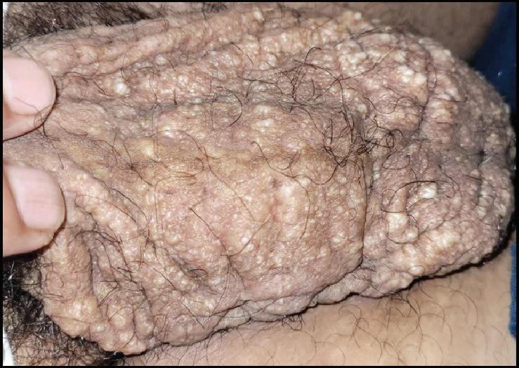
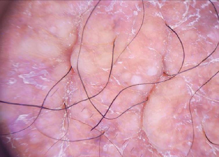
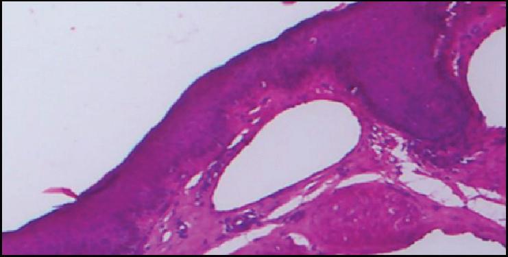
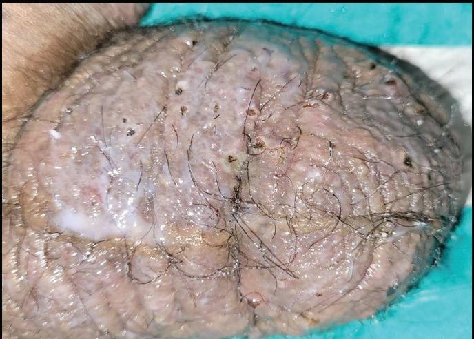
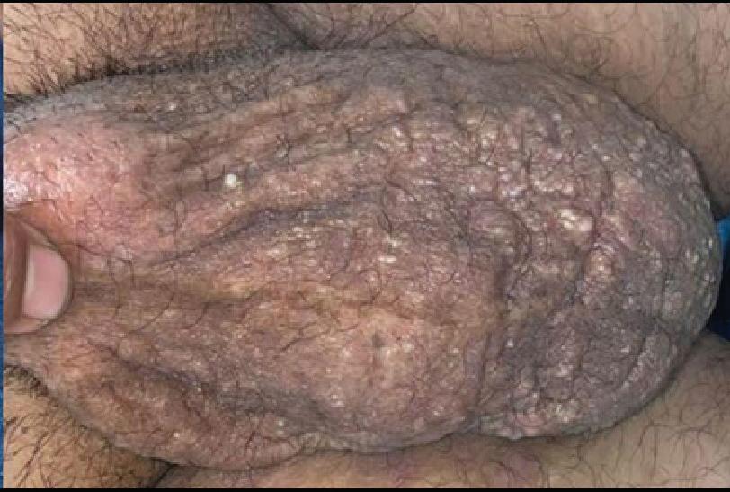
A 40-year-old male patient presents with a complaint of multiple raised lesions over the scrotum since 4-5 years. The lesions are associated with copious milky discharge, leading to wetting of undergarments. The patient has a history of varicocele surgery 5 years ago. There is no history of chronic cough, fever, malignancy, trauma, or painful swelling in the groin. On examination, multiple well-defined, translucent, thick-walled, fluid-filled vesicles and papules, ranging in size from 2-10 mm, are observed over the scrotum. Dermoscopy reveals multiple round to oval yellowishwhite lacunae surrounded by pale septa. On histopathological examination (HPE), multiple dilated dermal papillae reveal several markedly dilated lymphatic channels incompletely lined by a single layer of endothelium. These channels contain lymph with a slight admixture of red blood cells. Ultra-Sonography local examination of the scrotum shows a thickened and heterogeneous scrotal wall.
1: Multiple raised lesions over the scrotum (before Procedure)
Figure 2: Dermoscopy showed multiple round to oval yellowish white lacunae surrounded by pale septa
3: On histopathological examination
showed multiple dilated dermal papillae were observed several markedly dilated lymphatic channels incompletely lined by a single layer of endothelium.
Diagnosis
The diagnosis of acquired scrotal lymphangiectasia (AL) starts with a thorough history and physical exam, noting fluid-filled lesions in the scrotal area, often linked to prior surgeries. Dermoscopy helps visualize characteristic translucent, thick-walled vesicles and distinguishes AL from other conditions. Ultrasound identifies dilated lymphatic vessels, fluid accumulation, and related complications. Histopathology shows dilated vessels with thickened walls, fibrosis, edema, and chronic inflammation, confirming AL and differentiating it from other conditions.3,4,5
Treatment Method
The patient underwent a treatment regimen comprising three sessions of carbon dioxide (CO₂) laser therapy, performed at monthly intervals. The laser used was a 10,600 nm superpulsed CO₂ laser in surgical mode, with energy levels incrementally increased at each subsequent session to
Figure
Figure
optimize efficacy. During each session, most of the dilated lymphatic vessels were carefully ablated using the laser. The procedure was carried out under topical anesthesia to ensure patient comfort. Following the laser ablation, a post-procedure care regimen was initiated, including the application of a course of antibiotics to prevent secondary bacterial infections and promote healing. The treated area was also dressed with coconut dressing for three days. The patient tolerated the procedure well, with no significant adverse events reported during or after the sessions. At the end of the three-month treatment period, the patient showed marked improvement, with a significant reduction in lymphatic discharge. The outcomes indicate that the CO₂ laser therapy effectively reduced the burden of dilated lymphatics, minimized fluid accumulation, and improved the overall quality of life for the patient. This approach highlights the utility of laser therapy as a minimally invasive and well-tolerated option for managing acquired scrotal lymphangiectasia.6
Other treatment options for acquired scrotal lymphangiectasia (AL) include electrodessication, sclerotherapy, cryotherapy, and radiofrequency ablation (RFA). Electrodessication uses electrical currents to coagulate and ablate dilated lymphatic vessels, effectively reducing swelling, fluid leakage, and irritation, particularly for localized lesions when conservative treatments are ineffective. Sclerotherapy involves the injection of a sclerosing agent into the dilated vessels, promoting fibrosis and vessel closure, which helps reduce lymphatic fluid accumulation, edema, and discharge. Cryotherapy utilizes extreme cold to necrotize superficial dilated vessels, minimizing fluid leakage and irritation, and is most effective for localized lesions, though less suitable for extensive involvement or significant fibrosis. Radiofrequency ablation (RFA) applies thermal energy to ablate dilated vessels, reducing fluid buildup and alleviating symptoms. This minimally invasive technique offers faster recovery, superior cosmetic outcomes, and is especially beneficial for persistent or recurrent cases that have not responded well to conservative management.7
Result
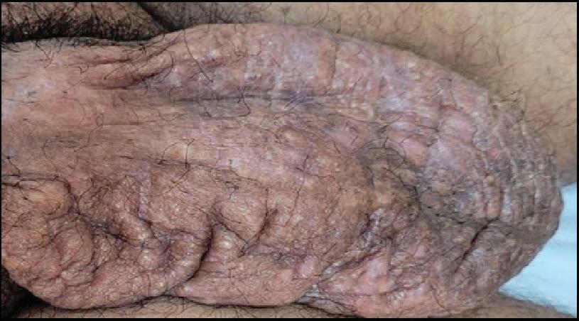
Figure 4: Intra-Procedure with carbon dioxide laser therapy
Discussion
Acquired scrotal lymphangiectasia (AL) significantly affects quality of life due to chronic discomfort, localized edema, erythema, and tenderness, interfering with daily activities like walking, sitting, and wearing restrictive clothing. The persistent leakage of lymphatic fluid leads to dermal irritation, maceration, and a higher risk of secondary bacterial infections. Recurrent flare-ups worsen symptoms, requiring frequent medical interventions. Additionally, the visible appearance of the lesions can cause psychological distress, impacting body image, self-esteem, and leading to social withdrawal and anxiety, particularly in intimate situations.2
The differential diagnosis of acquired scrotal
lymphangiectasia (AL) includes conditions with scrotal swelling or lymphatic abnormalities, such as cutaneous melanoma (metastatic), dermatitis herpetiformis, herpes simplex infections, metastatic carcinomas (e.g., breast, lung, prostate), genital and nongenital warts, herpes zoster, lymphangiomas, and Stewart-Treves syndrome (angiosarcoma in chronic lymphedema). Accurate diagnosis is crucial for appropriate treatment. Complications of AL include chronic discharge and lymphangioma circumscriptum, which present similarly with fluid-filled vesicles, dermal irritation, and persistent discharge, making differentiation challenging.5,8, 9
The etiology of acquired lymphangiectasia is rarely unknownand may be multifactorial. However, it has been documented to occur on the penis and scrotum following sacrococcygeal tumor excision and hernia repair procedures. Additionally, lymphangiectasia can develop on the vulva and inner thigh after surgical treatment for cervical or other pelvic malignancies,
Figure 6: After the 3rd sitting, there was a significant reduction in lymphatic discharge
Figure 5: After 2nd sitting
with acquired vulvar lymphangiomas resulting from pelvic lymphatic obstruction. Scrotal lymphangiectasia has also been observed in association with conditions such as scrofuloderma, lymphatic filariasis, and lymphogranuloma venereum. Therapy is challenging due to the high recurrence rate, underlying lymphedema, risk for infections, and prolonged healing time. Treatment options include complete surgical excision and grafting, cryotherapy, and CO2 laser vaporization. CO₂ laser therapy for acquired scrotal lymphangiectasia presents several clinical advantages, making it an optimal treatment modality for targeted lesions. The high precision of the 10600nm wavelength allows for selective treatment of affected lymphatic vessels while minimizing damage to surrounding tissue. This minimally invasive approach offers a significant reduction in recovery times and minimizes the risk of complications such as scarring and infection, as compared to surgical excision. The thermal effect of the laser effectively coagulates blood vessels, reducing intraoperative
Scrotal
bleeding and the need for postoperative intervention. Additionally, the procedure generally results in superior cosmetic outcomes, with less visible scarring, making it particularly advantageous in aesthetically sensitive areas like the scrotum. CO₂ laser treatment has proven to be highly effective in managing lymphangiectasia, providing symptomatic relief from fluid leakage, edema, and skin irritation. The only disadvantage is the high cost and unavailability in resource-poor settings. The procedure is associated with a rapid recovery period, with patients experiencing minimal postoperative discomfort and resuming normal activities within a short duration.2, 6
Conclusion
CO₂ laser treatment has proven to be an effective modality for acquired scrotal lymphangiectasia, yielding favorable cosmetic outcomes and significant improvements in patients' quality of life. It is less destructive and easy to operate with minimal post procedure pain. Given its effectiveness and advantageous cosmetic results, CO₂ laser therapy remains a preferred treatment option, particularly
for localized lesions and in patients seeking less invasive alternatives. Future advancements in management may focus on optimizing laser parameters, exploring combination therapies with modalities such as sclerotherapy or radiofrequency ablation, and enhancing its accessibility in resource-constrained settings. Additionally, further studies are needed to assess the long-term outcomes and recurrence rates to refine its clinical application.
References
1. Ji RC. Lymphatic endothelial cells, lymphedematous lymphangiogenesis, and molecular control of edema formation. Lymphat Res Biol. 2008; 6(3-4):123-137. doi:10.1089/lrb.2008.1005
2. Khadka DK, Pathak R, Agrawal S, Pokharel S. Acquired Lymphangiectasia of the Scrotum Successfully Treated with Radiofrequency Ablation: A Case Report with Dermoscopic Review. Case Rep Dermatol Med. 2023; 2023:7111912. Published 2023 Jan 10. doi:10.1155/2023/7111912.
3. Silvestre-Torner N, ImbernónMoya A, Martínez-García M, Burgos-Lázaro F. Acquired Cutaneous Lymphangiectasia: Dermoscopic Evidence from White-Yellowish Lacunae.
Scrotal Lymphangiectasia Successfully Treated With Carbon Dioxide Laser - A Case Report
DermatolPract Concept. 2021; 11(3):e2021062. Published 2021 Jul 8. doi:10.5826/dpc.1103a62.
4. Vishwanath T, Nagpal A, Ghate S, Sharma A. Scrotal Lymphangiectasia with Penile Elephantiasis in Underlying Lymphatic Filariasis-Challenging the Diagnostic Mind! A Case Report. Dermatopathology (Basel). 2021; 8(1):10-16. Published 2021 Jan 1. Doi: 10.3390/ dermatopathology8010002.
5. Lymphangiectasia Clinical PresentationUpdated: Apr 09, 2021 Author: Robert A Schwartz, MD, MPH; Chief Editor: Dirk M Elston, MD.
6. Binitha MP, Khader A, Sherjeena PB, Rini MR. Acquired Cutaneous Lymphangiectasia of the Scrotum Secondary to Filarial Lymphoedema. Kerala Medical Journal. 2015 Nov 27; 8(4):29–31.
7. Luo Y, Chen W, Xu D, Chen S, Ma S. Acquired Vulvar Lymphangioma Following CO2 Laser Treatment for Sebaceous Gland Nevus in a 10-YearOld Girl: A Case Report. ClinCosmetInvestigDermatol. 2025; 18:77-80. Published 2025 Jan 10. doi:10.2147/CCID. S503724.
8. Hamida MB, Baccouche D, El Fekih N, Fazaa B, Kamoun R. Lymphangiectasia of the vulva, treatment with CO 2 laser. Indian J DermatolVenereolLeprol 2012; 78:122.
9. Haneef NS, Ramachandra S, Metta AK, Haritha K. Lymphangiectasias of vulva. Indian Dermatol Online J. 2011; 2(1):40-42. doi:10.4103/22295178.79854
A Case Study of Tinea Corporis in A 45 Year Old Female Patient
A Case Study of Tinea Corporis in A 45 Year Old Female Patient
Dr. G. Harish
MBBS, DDVL
Consultant Dermatologist
Khammam, Telangana
Introduction
Tinea corporis, also known as ringworm of the body, is a common superficial fungal infection that affects the skin. It is caused by dermatophytes, which are fungi that feed on keratin found in the outer layer of the skin, hair, and nails. These fungi thrive in warm, moist environments and can infect humans and animals alike. Tinea corporis presents as circular or oval-shaped lesions on various parts of the body, including the trunk, arms, legs, and face, though it spares the palms, soles, and mucous membranes. This infection is highly contagious and can spread through direct contact with infected individuals, animals, or contaminated objects.1 Tinea corporis typically presents as a well-
defined, circular or ovalshaped, mildly erythematous patch or plaque with a raised, advancing edge. Initially, it appears as a flat, scaly lesion that spreads outward in a centrifugal pattern, creating an annular appearance commonly referred to as "ringworm." As the infection progresses, the central area may clear and become hypopigmented or brown, with reduced scaliness compared to the advancing border, which often exhibits an irregular or annular shape. Occasionally, the border may present with papules, vesicles, or pustules. Lesions can vary in shape, such as circinate or arcuate, and tend to be asymmetrically distributed, often coalescing into polycyclic patterns when multiple lesions
A Case Study of Tinea Corporis in
are present.2 In adults, tinea corporis frequently affects exposed skin areas, while in children and adolescents, the trunk is commonly involved. Bullous tinea corporis, a rare variant, manifests with vesicles or bullae primarily at the periphery of an erythematous, scaly plaque. Rupture of these vesicles or bullae can lead to erosions and crust formation over the erythematous background. In immunocompromised individuals, tinea corporis may present as a disseminated skin infection or even involve deeper layers, forming subcutaneous or deep abscesses. Rarely, it may present with purpuric macules, known as tinea corporis purpurica, indicating a more severe and potentially systemic fungal involvement. Recognition of these clinical presentations is crucial for accurate diagnosis and appropriate management of tinea corporis, ensuring effective treatment and prevention of complications in affected individuals.2
The pathophysiology of tinea corporis involves several mechanisms driven by dermatophytes, such as T. rubrum, that enable the fungus to establish
and propagate within the skin. Mannans present in the cell walls of these dermatophytes possess immune-suppressive properties, aiding in adherence to the skin and preventing premature shedding before initiating invasion. The fungi secrete enzymes including proteases, serine-subtilisins, and keratinases. Proteases facilitate the breakdown of keratin, serine-subtilisins initiate protein degradation through nucleophilic attacks on serine residues, and keratinases enable penetration of keratinized tissues, facilitating the spread of the fungus within the outer, nonliving, cornified layers of the skin. Infections are predominantly cutaneous, remaining confined to the superficial layers due to robust immune defenses in immunocompetent individuals. These defenses include the activation of serum inhibitory factors, polymorphonuclear leukocytes, and complement proteins, which collectively inhibit deeper tissue penetration by the fungus. The characteristic scaling observed at the active borders of lesions is attributed to heightened proliferation of epidermal
cells in response to the fungal presence, contributing to the clinical manifestations of tinea corporis.2
Early recognition and prompt treatment of tinea corporis are crucial for optimal outcomes, reducing discomfort, preventing complications, and minimizing transmission. Adhering to treatment protocols and practicing good personal hygiene are key to effectively managing and resolving the infection, promoting skin health and well-being.
Case report
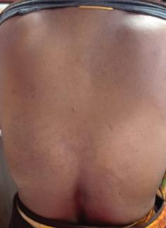
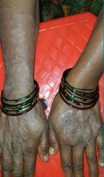
A 45-year-old female patient presented to the clinic with a suspected case of tinea corporis, a common dermatophyte infection of the skin. Upon examination, the patient exhibited raised, ring-shaped scaly patches distributed across her back, arms, and the dorsal aspects of her hands. These characteristic lesions strongly indicated tinea corporis. To confirm the diagnosis, a clinical examination was conducted, which corroborated the initial suspicion. Given the confirmed diagnosis of tinea corporis, a decision was made to initiate treatment with itraconazole.
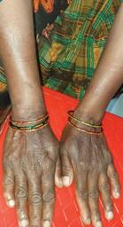
Itraconazole was selected due to its potent systemic antifungal properties, which are particularly effective against dermatophytes— the fungi responsible for tinea corporis. The patient was prescribed a three-week course of itraconazole, and she adhered to this treatment regimen as directed. Following the initiation of itraconazole therapy, the patient showed significant clinical improvement. The lesions that were initially observed demonstrated a notable reduction in both size and scaling. This positive response to itraconazole was evident as the treatment progressed, indicating the medication's efficacy in treating the infection. By the end of the three-week course, the patient experienced a substantial resolution of symptoms, with a marked improvement in the appearance of the affected skin areas. The successful treatment of tinea corporis in this patient highlights the effectiveness of systemic antifungal therapy with itraconazole. The resolution of the lesions and restoration of skin integrity underscore the importance of appropriate antifungal treatment in managing dermatophyte infections.
A Case Study of Tinea Corporis in A 45 Year Old Female Patient
This case reinforces the clinical utility of itraconazole in achieving favourable outcomes for patients with tinea corporis.
Before treatment
After treatment
Before treatment
After treatment
Figure 1: Raised, ring-shaped scaly patches on back
Figure 2: Raised, ring-shaped scaly patches on the dorsal aspects of hands
A Case Study of Tinea Corporis
Before treatment
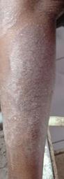
3: Raised, scaly patches on leg
Diagnosis
Laboratory investigations are crucial for diagnosing dermatophytosis, particularly in tinea corporis cases. Collecting skin scrapings from the lesion's active margin and examining them with 10–20% potassium hydroxide under direct microscopy allows rapid visualization of dermatophytic elements. Fluorescent staining enhances sensitivity by binding to fungal cell wall chitin. Culture on Sabouraud dextrose agar (SDA) or modified SDA with antibiotics incubates for 7–14 days, with dermatophyte test medium (DTM) indicating fungal growth through phenol red color change.3
Antifungal susceptibility testing (AST) is crucial in diagnosing and managing tinea corporis, especially in recurrent or treatment-resistant cases. Two main methods for AST are the microdilution method and Minimum Fungicidal Concentration (MFC) determination. The microdilution method assesses minimum inhibitory concentrations (MICs) of antifungal agents by diluting them in a microplate with culture medium inoculated with fungal isolates. MIC values indicate the lowest drug concentration inhibiting visible fungal growth after incubation, providing quantitative susceptibility data. MFC determination complements MIC testing by determining the lowest concentration needed to kill rather than inhibit fungi. After MIC determination,
subculturing on drug-free media checks for fungal growth inhibition. MFC is the minimum concentration preventing visible growth upon subculture, indicating fungicidal activity crucial for effective tinea corporis management.3
Histopathology is essential in diagnosing tinea corporis, revealing characteristic changes like epidermal hyperkeratosis, parakeratosis, and septate hyphae within the stratum corneum and hair follicles. Inflammatory cells such as neutrophils and lymphocytes indicate the immune response against the fungus, with additional features like spongiosis, vesiculation, or dermal inflammation helping to assess infection severity. Dermoscopy aids in distinguishing tinea corporis by noting structureless yellowish scales, an erythematous halo, central clearing, peripheral scaling, and marginal hyperpigmentation, guiding effective treatment strategies for fungal skin infections.3
Diagnosis of tinea corporis using Polymerase Chain Reaction (PCR) and nucleic acid sequencebased amplification (NASBA) involves molecular techniques that detect
Figure
After treatment
A Case Study of Tinea Corporis in A 45 Year Old Female Patient
fungal DNA or RNA directly from skin samples. PCR targets specific genetic sequences of dermatophytes, amplifying them to detectable levels for identification. NASBA, on the other hand, amplifies RNA molecules, offering a complementary method to PCR for detecting fungal RNA. These molecular methods provide rapid and sensitive detection of dermatophytes, especially in cases where conventional diagnostic methods like microscopy or culture may be inconclusive or challenging.3
Matrix-assisted laser desorption ionizationtime of flight mass spectrometry (MALDI-TOF MS) is a novel molecular method revolutionizing the evaluation of tinea corporis. This technique allows rapid identification of fungal species by analyzing their protein profiles. By comparing the mass spectra of unknown fungal isolates to a reference database, MALDI-TOF MS accurately identifies dermatophytes causing tinea corporis with high specificity and sensitivity.3 Reflectance confocal microscopy (RCM) is revolutionizing the evaluation of tinea corporis by offering non-
invasive, real-time imaging of skin lesions at a cellular level. Dermatologists can visualize fungal structures like hyphae and spores directly on the skin surface, leveraging RCM's ability to detect epidermal changes such as hyperkeratosis and parakeratosis. This method also reveals the inflammatory response around fungal elements, aiding in diagnosis by providing detailed assessments of lesion morphology, including scales, erythema, and the characteristic annular pattern of tinea corporis.3
The effective diagnosis of tinea corporis relies on a thorough clinical assessment supplemented by appropriate diagnostic tests when needed. Early and accurate identification of this superficial fungal infection allows for prompt initiation of targeted antifungal therapy, thereby facilitating rapid resolution of symptoms and reducing the risk of complications.
Treatment
Treatment of dermatophyte infections, particularly localized tinea corporis, relies on the strategic application of topical or oral antifungal treatments. The primary goal of initial management is to provide
a focused approach that directly targets the superficial fungal infection, aiming to eradicate the causative dermatophytes and alleviate accompanying symptoms.
Topical antifungal therapy
Azoles, such as econazole, ketoconazole, miconazole, clotrimazole, oxiconazole, sulconazole, sertaconazole, eberconazole, and luliconazole, exert their antifungal effects by inhibiting lanosterol 14α-demethylase, a key enzyme involved in ergosterol biosynthesis. Ergosterol plays a crucial role in preserving the structural integrity of fungal cell membranes. By blocking this enzyme, azoles disrupt ergosterol synthesis, leading to the accumulation of toxic methylated sterols and causing structural and functional damage to the fungal cell membrane. This disruption ultimately results in impaired fungal growth and survival.4 Allylamines, including naftifine and terbinafine, exert their antifungal effects by inhibiting squalene epoxidase, a crucial enzyme in the ergosterol biosynthesis pathway of fungi. By blocking this enzyme, allylamines disrupt
A Case Study of Tinea Corporis in
the formation of fungal cell membranes, leading to the accumulation of squalene and depletion of ergosterol. This disruption compromises the structural integrity and function of the fungal cell membrane, ultimately resulting in fungal cell death. Terbinafine, in particular, is widely used topically and is welltolerated, making it a preferred treatment option for localized dermatophyte infections.4 Butenafine, a benzylamine derivative, exerts its antifungal action by inhibiting squalene epoxidase, an enzyme crucial for ergosterol biosynthesis in fungi. This ultimately leads to fungal cell death.4 Ciclopirox, chelates with polyvalent cations, particularly iron (Fe^3+), which are essential for various fungal cellular processes. By depriving fungi of these essential ions, ciclopirox disrupts the integrity and function of their cell membranes.4 Tolnaftate acts primarily by inhibiting squalene monooxygenase, an enzyme crucial in the biosynthesis of ergosterol, an essential component of fungal cell membranes. By blocking this enzyme, tolnaftate disrupts the synthesis of ergosterol, leading to structural and functional abnormalities in
the fungal cell membrane.4 Amphotericin B functions by binding to ergosterol, an essential component of fungal cell membranes. This binding disrupts membrane integrity, leading to leakage of cellular contents and ultimately fungal cell death. This mechanism of action is broad-spectrum, effective against a wide range of fungal pathogens, including those resistant to other antifungal agents.3 Amorolfine alters the membrane structure, increasing permeability and ultimately leading to fungal cell death. It exhibits broad-spectrum antifungal activity and is well-tolerated, characterized by minimal systemic absorption and fewer side effects compared to systemic antifungal therapies.3
Oral antifungal therapy
Oral itraconazole is utilized in the treatment of tinea corporis, a superficial dermatophyte infection. As an azole antifungal, it inhibits ergosterol synthesis in fungal cell membranes, disrupting membrane integrity and causing fungal cell death. It is administered once daily in capsule or tablet form for a specified treatment duration. Itraconazole demonstrates efficacy against a range
of dermatophytes, including resistant strains not responsive to topical therapies. This systemic approach is particularly beneficial for managing extensive or persistent infections inadequately controlled by topical treatments.4
Oral terbinafine is prescribed for severe or resistant cases due to its rapid onset of action and favourable safety profile when used correctly. Fluconazole, a triazole antifungal agent, disrupts fungal membrane integrity, leading to cell death and resolving tinea corporis. Administered orally once daily, it effectively treats a range of dermatophytes causing the infection, particularly suitable for widespread or recurrent cases. Griseofulvin disrupts fungal cell division and mitosis in the keratinized epidermal layers, targeting dermatophytes responsible for tinea corporis. Administered orally over several weeks, it clears infections, including those resistant to topical treatments, by preventing fungal growth and spread.4
Adjunctive therapies
Adjunctive therapies play a crucial role in the comprehensive ............. management of tinea
corporis, a superficial fungal infection of the skin caused by dermatophytes. Topical steroids are often used to alleviate inflammation and itching, which are common symptoms accompanying the infection. They help reduce discomfort and promote patient comfort during treatment. In cases where there is a secondary bacterial infection complicating tinea corporis, antibiotics may be prescribed to address bacterial overgrowth and prevent further complications. Emollients and moisturizers are beneficial adjuncts that soothe and hydrate the skin, promoting healing and restoring skin barrier function compromised by fungal infection and inflammation. These adjunctive therapies complement antifungal treatments by managing symptoms and supporting skin recovery, contributing to comprehensive care and enhancing patient outcomes in the management of tinea corporis.3
Effective treatment of tinea corporis involves a comprehensive approach, combining topical and systemic antifungal therapies as necessary, along with adjunctive
A
Case Study
of Tinea Corporis in A 45 Year Old Female Patient
measures to alleviate symptoms and aid skin recovery. Customized treatment plans and patient education on preventive measures are crucial for successful outcomes and minimizing recurrence in this prevalent skin condition.
Discussion
Tinea corporis, the most common dermatophytosis globally, exhibits a higher prevalence in tropical regions. The lifetime risk of acquiring tinea corporis ranges from 10% to 20%. It primarily affects postpubertal children and young adults, although rare cases have been reported in newborns. There is no predilection based on gender. Humans contract the infection through close contact with infected individuals, animals (especially domestic dogs or cats), contaminated objects, or soil. Spread can also occur from other sites of dermatophyte infection (e.g., tinea capitis, tinea pedis, onychomycosis). Household transmission among family members, particularly children from infected household members, is the most common route. Autoinfection from dermatophytes elsewhere on the body is possible. Factors that predispose
individuals to tinea corporis include personal history of dermatophytosis (e.g., tinea capitis, tinea pedis, tinea cruris, tinea unguium), presence of affected family members, pets in the home, crowded living conditions, participation in sports involving skin-toskin contact (e.g., wrestling, martial arts), hyperhidrosis, low β-defensin 4 levels, immunodeficiency, ........... diabetes mellitus, genetic predisposition (particularly tinea imbricata), xerosis, and ichthyosis. Transmission is facilitated by moist, warm environments, sharing of towels and clothing, and wearing occlusive clothing. Tinea corporis, predominantly caused by dermatophytes, is commonly attributed to Trichophyton rubrum, T. tonsurans, and Microsporum canis T. rubrum is the most prevalent worldwide and the leading cause of tinea corporis in North America. Secondary infections from tinea capitis are typically due to T. tonsurans, whereas contact with dogs or cats often leads to infections caused by M. canis. Other notable pathogens include T. interdigitale (formerly T. mentagrophytes), T. verrucosum, T. violaceum, T. concentricum,
A Case Study of Tinea Corporis in
Epidermophyton floccosum, M. audouinii, and M. gypseum. 5
The differential diagnosis of tinea corporis includes pityriasis rosea, presenting with a non-itchy herald patch followed by a 'Christmas tree' pattern rash. Tinea versicolor manifests as scaly macules in fair-skinned individuals and hypopigmented patches in dark-skinned individuals. Nummular eczema shows coinshaped, pruritic lesions, while plaque psoriasis features erythematous plaques with silvery scales. Atopic dermatitis presents with flexural involvement and intense itching. Contact dermatitis is localized due to irritants or allergens, and seborrheic dermatitis shows greasy scales.4 Other conditions include granuloma annulare, fixed drug eruptions, lupus erythematosus types, urticaria, pityriasis lichenoides chronica, lichen planus, erythema migrans, erythema multiforme, erythema dyschromicum perstans, erythema marginatum, erythema annulare centrifugum, impetigo contagiosa, erythema gyratum repens, and secondary syphilis with macules or patches
on the trunk and limbs. Tinea corporis is highly contagious, leading to significant psychological, social, and occupational impacts. Scratching and skin abrasions can predispose to secondary bacterial infections. Postinflammatory changes such as hypopigmentation and hyperpigmentation are common sequelae.3 Dermatophytid reaction, also known as autoeczematization, may occur in response to a fungal infection or antifungal treatment, presenting as widespread, intensely itchy, erythematous papules, vesicles, or pustules. This reaction is thought to involve a delayed-type hypersensitivity response to fungal antigens. Additionally, rare instances of psoriatic flares triggered by tinea corporis have been reported.4
Preventing tinea corporis hinges on minimizing conditions favourable to fungal growth. Patients should be counselled to wear breathable, loosefitting clothing to reduce moisture and warmth, which promote fungal proliferation. It's essential to maintain meticulous personal hygiene practices, including frequent handwashing
and ensuring skin remains clean and dry. Avoiding direct contact with items or clothing used by individuals with tinea corporis is crucial to prevent transmission of dermatophytes. These measures are fundamental in controlling the spread of this fungal infection within community and household environments. The prognosis for tinea corporis is generally favourable with adherence to proper treatment and patient compliance. Education plays a crucial role in prevention, emphasizing measures such as wearing light and loose-fitting clothing and maintaining clean, dry skin to minimize recurrence. When initiating topical antifungal therapy, patient compliance is essential, although symptomatic relief may not be immediate. Patients should be informed that resolution of symptoms may take several weeks with consistent treatment. Encouraging patient understanding and compliance can lead to successful management of tinea corporis and improve overall outcomes.4, 5
Conclusion
Managing tinea corporis effectively demands a holistic approach that
A Case Study of Tinea Corporis in A 45
Year Old Female Patient
addresses its chronic nature and potential for resistance. Utilizing advanced therapies such as oral antifungals, combination treatments, and adjunctive medications like corticosteroids and immunomodulators is pivotal in achieving successful outcomes. The availability of newer antifungal agents further broadens treatment options, especially for cases resistant to conventional therapies. Vigilant monitoring of treatment response and resistance is critical to adapting therapies as needed. Patient education on hygiene practices is equally indispensable to prevent recurrence and transmission. By integrating these advanced treatments with thorough patient care and education, healthcare providers can significantly enhance treatment effectiveness, minimize relapse rates, and elevate the overall management of tinea corporis, ultimately improving patient outcomes and quality of life.
References
1. Yee G, Al Aboud AM. Tinea Corporis. In: StatPearls. Treasure Island (FL): StatPearls Publishing; August 8, 2023.
2. Leung AK, Lam JM, Leong KF, Hon KL. Tinea corporis: an updated review. Drugs Context.
2020; 9:2020-5-6. Published 2020 Jul 20. doi:10.7573/ dic.2020-5-6.
3. Sahoo AK, Mahajan R. Management of tinea corporis, tinea cruris, and tinea pedis: A comprehensive review. Indian Dermatol Online J. 2016;7(2):7786. doi:10.4103/22295178.178099.
4. Jartarkar SR, Patil A, Goldust Y, et al. Pathogenesis, Immunology and Management of Dermatophytosis. J Fungi (Basel). 2021; 8(1):39. Published 2021 Dec 31. Doi: 10.3390/ jof8010039.
5. Khurana A, Sharath S, Sardana K, Chowdhary A, Panesar S. Therapeutic Updates on the Management of Tinea Corporis or Cruris in the Era of Trichophyton Indotineae: Separating Evidence from Hype-A Narrative Review. Indian J Dermatol. 2023;68(5):525-540. doi:10.4103/ijd.ijd_832_23.